Embed Size (px)
Citation preview
Woo
dhea
d Pu
blis
hing
Lim
ited
Woodhead Publishing Limited; proof copy not for publication
123456789
10111213141516171819202122232425262728293031323334353637383940414243
© Woodhead Publishing Limited, 2012
224
10Mesoscale mobile robots for gastrointestinal
minimally invasive surgery (MIS)
J. L. GorLewicz, r. J. webster ii i and P. VaLdastri, Vanderbilt University, Usa
Abstract: wireless capsule endoscopes, swallowable devices, such as commercial pill cameras designed for minimally invasive surgery of the gastrointestinal tract are reviewed and robotic capsules are described in terms of the basic modules they can contain and the latest research results are given for each module. a perspective is presented on the future prospects for clinical implementation of wireless robotic capsule-based surgical systems.
Key words: wireless capsule endoscopy, robotic capsules, swallowable devices, surgical endoscopy, minimally invasive surgery.
10.1 Introduction
as discussed in previous chapters of this book, robotic systems have been applied to a wide range of surgical procedures including abdominal surgery, neurosurgery, cardiac surgery, otolaryngology, and urology, among many others. in the future, one can expect to see many more customized robotic systems, purposely designed for specific clinical needs in specific surgical procedures. From a technical perspective, future robots will become smaller and increasingly less invasive in their approach to the surgical site. one way to achieve this using endoscope-like tools is to move toward natural orifice surgery, as reviewed in chapter 9. an alternate paradigm is to ‘cut the cord’, making the robot completely untethered and mobile within the body. this enables the robot to enter the body though either a natural orifice or a very small incision, and then maneuver itself to a surgical site that need not be in close proximity to the point of entry. such mobile medical robot systems may be created on a variety of scales including the micro- and nanoscales, as discussed in chapters 8, 9 and 11. Unlike micro- and nanoscale robots which are useful in areas such as the eye (ergeneman et al. 2008) or the capillaries (Martel et al. 2009) where larger robots are precluded by anatomical constraints, mesoscale robots are ideal for areas like the gastrointestinal (Gi) tract and abdomen where their larger payload capacity is advantageous. The mesoscale (broadly defined as
MedicalRobotics-Gomes-10.indd 224 8/1/12 5:05:30 PM
225Mesoscale mobile robots for gastrointestinal MIS
Woo
dhea
d Pu
blis
hing
Lim
ited
Woodhead Publishing Limited; proof copy not for publication
123456789
10111213141516171819202122232425262728293031323334353637383940414243
© Woodhead Publishing Limited, 2012
1–100 mm) includes robots that are small, but easily visible and manipulable by unaided human eyes and hands. although there do exist some robots in this category designed to be inserted through incisions (Lehman et al. 2009, Patronik et al. 2009, Platt et al. 2009, rentschler et al. 2006, simi et al. 2012), many more robots in this category are designed to be swallowable. Because it provides a natural means of robot locomotion and is difficult to access any other way, the Gi tract has been the proving ground for the vast majority of mesoscale mobile robots developed to date. For this reason, and because the basic modules in mesoscale mobile robots are similar regardless of method of entry into the body, in this chapter we will focus on swallowable capsule robots intended for the Gi tract. as an emerging technology, many of these robots are still in the research and development phase and have not yet been clinically implemented, although an increasing number have been applied in animal studies. combined with the commercial and clinical success enjoyed by cameral pills, the future of mesoscale mobile robots as platforms for surgical interventions is bright.
10.2 Commercial gastrointestinal wireless capsule endoscopes
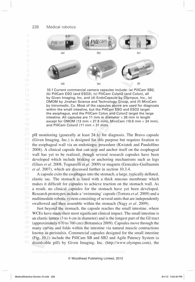
The first wireless capsule endoscopes (WCEs) were pills containing cameras, which captured images deep within the GI tract. The first commercial WCE, the Pillcam (Given imaging, inc, http://www.givenimaging.com) and other commercial capsules (Fig. 10.1) have been used to diagnose diseases such as obscure gastrointestinal bleeding (oGib), cancer, celiac disease, and crohn’s disease, all of which occur in the small intestine (Moglia et al. 2009). in addition to becoming the preferred method of diagnosis within the small intestine, commercial wces have recently been developed for other parts of the Gi tract. the Gi tract consists of the esophagus, stomach, small intestine, and large intestine (colon), which vary in size and shape, creating many different challenges for wces. the esophagus, a hollow muscular tube approximately 25 cm long and 1.5–2 cm in diameter (http://www.britannica.com) is the first area a capsule reaches after being swallowed. because a capsule passes through the esophagus in approximately 10 s, images must be rapidly acquired. For this reason, commercial esophageal capsules (Pillcam eso and eso2 by Given imaging, inc.) have dual cameras and capture images at higher frame rates than the capsules used in the small and large intestine (Moglia et al. 2007). studies with these esophageal capsules show that they can achieve very high accuracies in diagnosing barrett esophagus and portal hypertension, but are not sensitive enough to diagnose neoplastic lesions (Heresbach et al. 2010). Gastro esophageal reflux disease (GERD) requires long-term
MedicalRobotics-Gomes-10.indd 225 8/1/12 5:05:30 PM
226 Medical robotics
Woo
dhea
d Pu
blis
hing
Lim
ited
Woodhead Publishing Limited; proof copy not for publication
123456789
10111213141516171819202122232425262728293031323334353637383940414243
© Woodhead Publishing Limited, 2012
pH monitoring (generally at least 24 h) for diagnosis. the bravo capsule (Given Imaging, Inc.) is designed for this purpose but requires fixation to the esophageal wall via an endoscopic procedure (Kwiatek and Pandolfino 2008). a clinical capsule that can stop and anchor itself on the esophageal wall has yet to be realized, though several research capsules have been developed which include braking or anchoring mechanisms such as legs (Glass et al. 2008, tognarelli et al. 2009) or magnets (Gonzalez-Guillaumin et al. 2007), which are discussed further in section 10.3.4. A capsule exits the esophagus into the stomach, a large, typically deflated, elastic sac. the stomach is lined with a thick mucous membrane which makes it difficult for capsules to achieve traction on the stomach wall. As a result, no clinical capsules for the stomach have yet been developed. research prototypes include a ‘swimming’ capsule (tortora et al. 2009) and a multimodule robotic system consisting of several units that are independently swallowed and then assemble within the stomach (Nagy et al. 2009). Just beyond the stomach, the capsule reaches the small intestine, where WCEs have made their most significant clinical impact. The small intestine is an elastic lumen (3 to 4 cm in diameter) and is the longest part of the Gi tract (approximately 670 to 760 cm) (britannica 2009). capsules move through the many curves and folds within the intestine via natural muscle contractions known as peristalsis. commercial capsules designed for the small intestine (Fig. 10.1) include the Pillcam sb and sb2 and agile Patency system (a dissolvable pill) by Given imaging, inc. (http://www.olympus.com), the
(a) (b) (c)
(d) (e) (f)
10.1 Current commercial camera capsules include: (a) PillCam SB2, (b) PillCam ESO (and ESO2), (c) PillCam Colon2 (and Colon), all by Given Imaging, Inc. and (d) EndoCapsule by Olympus, Inc., (e) OMOM by Jinshan Science and Technology Group, and (f) MiroCam by Intromedic, Co. Most of the capsules above are used for diagnosis within the small intestine, but the PillCam ESO and ESO2 target the esophagus, and the PillCam Colon and Colon2 target the large intestine. All capsules are 11 mm in diameter ¥ 26 mm in length except for OMOM (13 mm ¥ 27.9 mm), MiroCam (10.8 mm ¥ 24 mm), and PillCam Colon2 (11 mm ¥ 31 mm).
MedicalRobotics-Gomes-10.indd 226 8/1/12 5:05:30 PM
227Mesoscale mobile robots for gastrointestinal MIS
Woo
dhea
d Pu
blis
hing
Lim
ited
Woodhead Publishing Limited; proof copy not for publication
123456789
10111213141516171819202122232425262728293031323334353637383940414243
© Woodhead Publishing Limited, 2012
endocapsule by olympus, inc., the oMoM capsule by the Jinshan science and technology Group co., Ltd., and the Mirocam capsule by intromedic co. the agile capsule, an accessory to the Pillcam, can be used before the ingestion of the Pillcam sb to verify adequate capsule passage through the Gi tract in patients with known or suspected strictures. this capsule assists in detecting the problem of capsule retention, which occurs in 0.75% of oGib cases, but may occur at a rate of up to 6.7% in patients with crohn’s disease (Moglia et al. 2007). these commercial capsules designed for use in the small intestine vary slightly in size, optical sensors such as complementary metal oxide semiconductor (cMos) or change coupled device (ccd), and frame rate (cavallotti et al. 2009). a capsule generally takes approximately 8–10 h to pass through the entire Gi tract, with the majority of the time spent in the small and large intestines (drossman et al. 2005). the large intestine or colon, which is about 150 cm in length and 6 cm in diameter, is the last region a capsule encounters before exiting the body. because of the larger lumen diameter, it is almost impossible for capsules to view the entire surface area of the internal intestinal wall. Furthermore, because the capsules are much smaller in size than the lumen itself, they tend to tumble within the lumen, making visualization even more difficult. To address this, the PillCam Colon and colon2, the only commercially available capsules for the colon, are longer in length than the other commercial capsules and contain two on-board cameras. in a multicenter performance study, the Pillcam colon2 achieved a sensitivity of 89% and a specificity of 76% in detecting colonic polyps ≥ 6 mm, suggesting that it is a safe and effective method for visualization in the colon and for detecting lesions (eliakim et al. 2009). it is not yet clear what clinical impact these colon capsules have as neither the colon or colon2 have received Food and drug administration (Fda) approval yet.
10.3 Robotic capsule modules
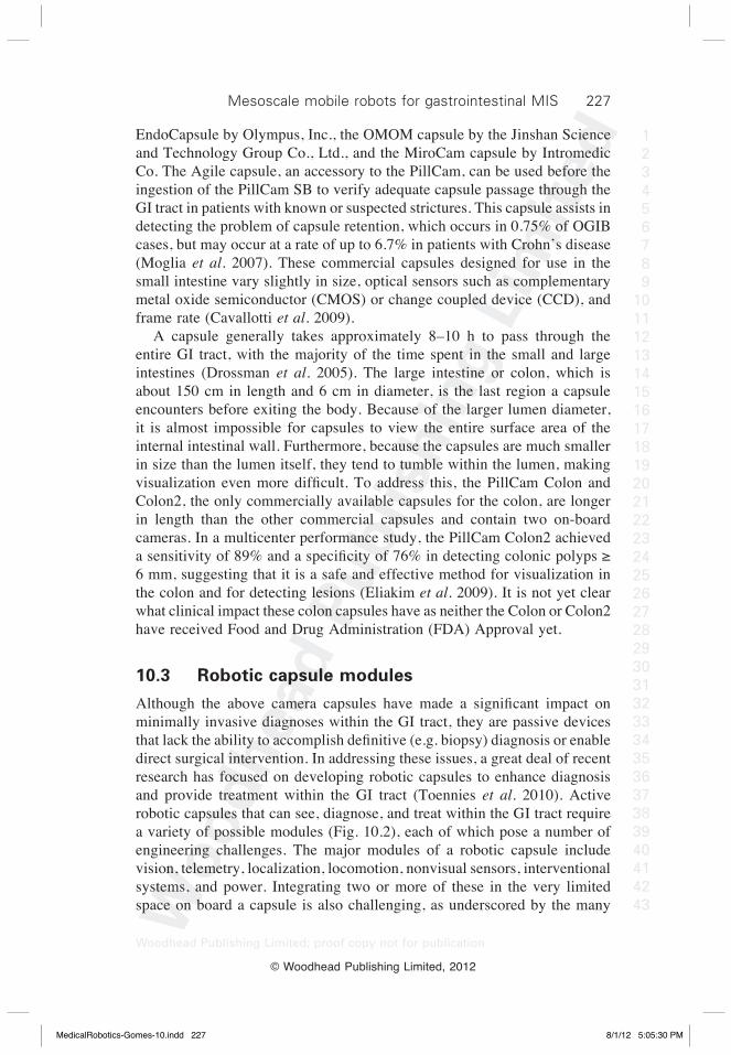
Although the above camera capsules have made a significant impact on minimally invasive diagnoses within the Gi tract, they are passive devices that lack the ability to accomplish definitive (e.g. biopsy) diagnosis or enable direct surgical intervention. in addressing these issues, a great deal of recent research has focused on developing robotic capsules to enhance diagnosis and provide treatment within the Gi tract (toennies et al. 2010). active robotic capsules that can see, diagnose, and treat within the Gi tract require a variety of possible modules (Fig. 10.2), each of which pose a number of engineering challenges. the major modules of a robotic capsule include vision, telemetry, localization, locomotion, nonvisual sensors, interventional systems, and power. integrating two or more of these in the very limited space on board a capsule is also challenging, as underscored by the many
MedicalRobotics-Gomes-10.indd 227 8/1/12 5:05:30 PM
228 Medical robotics
Woo
dhea
d Pu
blis
hing
Lim
ited
Woodhead Publishing Limited; proof copy not for publication
123456789
10111213141516171819202122232425262728293031323334353637383940414243
© Woodhead Publishing Limited, 2012
prototype capsules built to date that only contain a subset of these modules. whether all of these modules can be integrated into one capsule that can perform all tasks or whether a co-operative multicapsule approach will be required is not yet known, though a large number of robotic modules will be needed in either case. the current technical progress of each of these modules is discussed in detail in the following sections.
10.3.1 Vision
the very existence of wces can be attributed to the development of miniature camera(s) integrated within swallowable pills (swain 2008). typical images obtained from a camera pill are shown in Fig. 10.3. the primary goal of all vision systems is to provide clear, high resolution internal images of the Gi tract. in order to do this, a number of hardware components are required on board a capsule including a vision sensor, a lens, an illumination source, and a compression chip. the primary image acquisition chip used by most capsules, including the Pillcam (Given imaging, inc.), oMoM (Jinshan science and technology Group, Ltd., co.), and Mirocam (bang et al. 2009), is based on cMos technology, though the olympus endocapsule (olympus, inc.) relies on a charged coupled device (ccd). although both imaging chips provide similar diagnostic performance, recent studies have shown that the endocapsule generally scores higher in image quality and resolution than the Pillcam sb (cave et al. 2007, Metzger et al. 2009). a detailed review of the vision capabilities associated with each technology is available (Moglia et al. 2009).
Camera and LEDs
Human fingertip
(for scale)
Vision
Telemetry
Locomotion
Power
Tissue
interaction
Additional
diagnostics
10.2 Potential modules required for a wireless robotic capsule include vision, telemetry, localization, locomotion, sensing, intervention, and power.
MedicalRobotics-Gomes-10.indd 228 8/1/12 5:05:30 PM
229Mesoscale mobile robots for gastrointestinal MIS
Woo
dhea
d Pu
blis
hing
Lim
ited
Woodhead Publishing Limited; proof copy not for publication
123456789
10111213141516171819202122232425262728293031323334353637383940414243
© Woodhead Publishing Limited, 2012
current commercial imaging chips must compromise between image quality and power consumption (Vatteroni et al. 2010). several researchers have been working to develop smaller, more efficient image acquisition chips. For example, cMos technology has been used to develop a monolithic 320 ¥ 240 active-pixel red, green, blue (rGb) or gray level camera-on-a-chip sensor to improve imaging sensitivity while decreasing the power consumption of the chip (Vatteroni et al. 2010). this imaging chip has been tested in an ex vivo setting, and preliminary results suggest low noise and good image uniformity (Vatteroni et al. 2010). Furthermore, in developing a high-dynamic-range vision system, a linear–logarithmic cMos pixel with a programmable dynamic range extendable beyond 110 db was developed and integrated in a 100 ¥ 100 array within a monochrome imager (Vatteroni et al. 2011). an integrated 3d sensor which uses a multispectral cMos sensor and a pulsed pattern projector for 3d reconstruction of images has been demonstrated on a large-scale by (Kolar et al. 2009). there has also been some work in combining technologies from both cMos and ccd to develop an improved hybrid camera (swain 2008). the type of optics, including both the lens and illumination source, is also important in obtaining quality images from a capsule platform. to date, all available WCEs have a fixed focal length lens (Cavallotti et al. 2009). although this is acceptable for passive capsules, because the lumen is almost always collapsed around the capsule keeping the distance from the camera to the lumen wall constant, it would not be ideal for capsules that can actively locomote or distend tissue. to this end, cavallotti et al. (2009) developed a prototype system that uses a liquid lens actuated by a pulse width modulated signal to adjust the focal length from 15 to 100 mm. Good illumination is
(a) (b)
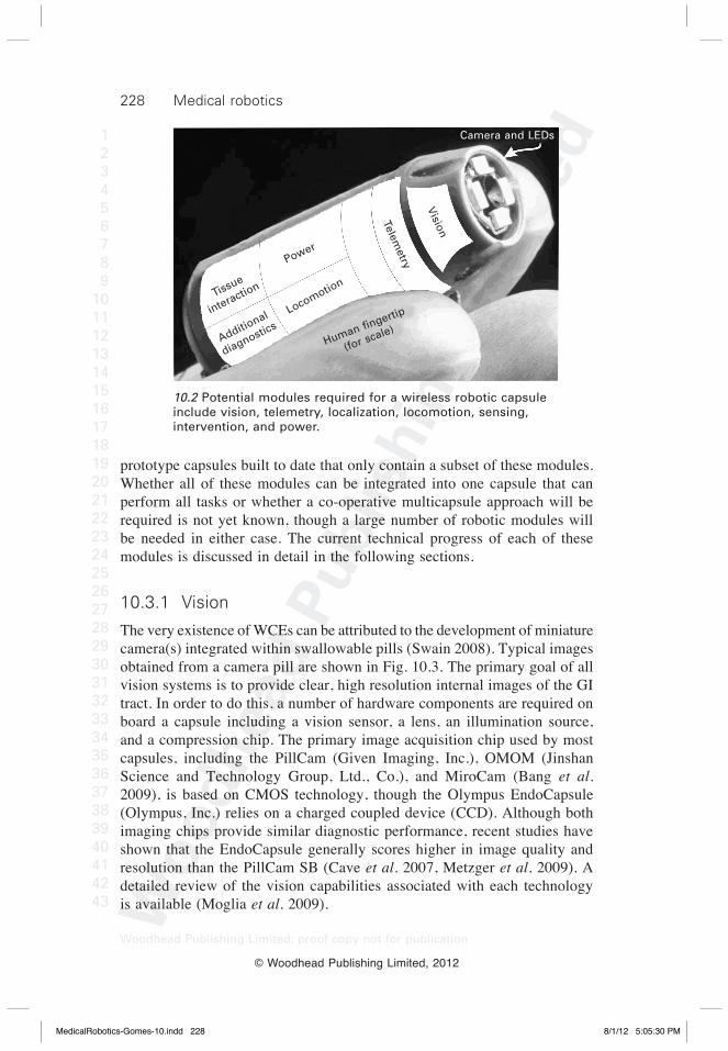
10.3 (a) A typical image showing a bleeding lesion obtained from a PillCamSB, (Image courtesy of Rajesh Kumar, Johns Hopkins University). (b) An image of the gastric cavity with the pylorus seen distally from an Olympus capsule endoscope (© 2010 Georg Thieme Verlag KG, reprinted with permission, Rey et al. 2010).
MedicalRobotics-Gomes-10.indd 229 8/1/12 5:05:30 PM
230 Medical robotics
Woo
dhea
d Pu
blis
hing
Lim
ited
Woodhead Publishing Limited; proof copy not for publication
123456789
10111213141516171819202122232425262728293031323334353637383940414243
© Woodhead Publishing Limited, 2012
also necessary for obtaining high-quality images from a capsule. current commercial capsules use anywhere from 4–6 light-emitting diodes (Leds). beyond this, new image-enhanced technologies have been developed that allow physicians to view the Gi tract in much greater detail than ever before, even on a cellular level (Hasan and wallace 2009). a few of these techniques include chromoendoscopy and autofluoresence (light working in conjunction with dyes applied to the Gi mucosa or with chemicals in the tissue that fluoresce, respectively), confocal laser endomicroscopy (for real-time histology), and spectroscopy (which has been used to detect dysplasia) (Hasan and wallace 2009). these technologies may be integrated into future robotic capsules, enabling optical diagnoses deep within the Gi tract. a second, yet equally important, goal of a capsule vision system is the transmission of acquired images to an offline receiver. Because thousands of images are captured by the capsule, each containing a large amount of information, these images must be compressed in order to transmit them over the relatively low data rate telemetry links currently available (section 10.3.2). Image compression is challenging, as it requires retaining sufficient image information while keeping power consumption and transmission time low. the optimal level of compression is dependent upon the medical objective of the capsule. currently available capsules for the small intestine, which have frame rates ranging from 2–3 Hz (cavallotti et al. 2009), produce diagnostically useful results, though there is little doubt that higher frame rates would be desirable. when higher frame rates are achieved, such as in the Pillcam eso and eso2, which acquire images at a rate of 14–18 frames per second (fps), the battery life of the capsule is significantly reduced. Fortunately, because esophageal capsules only need to function for a few minutes at most, battery life is not as much of a concern for these capsules (Moglia et al. 2007). For active robotic capsules that may be teleoperated by a surgeon, it is expected that frame rates of 20 fps or more will be required (turgis and Puers 2005). Several researchers have worked on developing efficient compression algorithms and low-power compression chips (Lin et al. 2006, turcza et al. 2008, turgis and Puers 2005). toward addressing power consumption, chen et al. (2009) developed a system that can reduce average power consumption by 45% using a power efficient image compression module, a power management unit, and a novel wireless wake-up subsystem with zero stand-by current. A versatile field-programmable gate array (FPGA)-based system has been developed by (cavallotti et al. 2011) which enables testing of different configurations of the submodules that compose the entire vision system, including the camera, illumination, image compression, and telemetry. The optimal configuration that was found for endoscopic capsules attained an average frame rate of 19 fps over a transmission channel of 1.5 Mbit s–1 (cavallotti et al. 2011).
MedicalRobotics-Gomes-10.indd 230 8/1/12 5:05:30 PM
231Mesoscale mobile robots for gastrointestinal MIS
Woo
dhea
d Pu
blis
hing
Lim
ited
Woodhead Publishing Limited; proof copy not for publication
123456789
10111213141516171819202122232425262728293031323334353637383940414243
© Woodhead Publishing Limited, 2012
once the images have been compressed and transmitted, they must be analyzed either manually by a physician or automatically by computer algorithms. this external post-processing of images is very useful, as a typical small intestine capsule returns approximately 50 000 images from a single use (bejakovic et al. 2009), but only a few contain useful diagnostic information. to alleviate physicians from the task of manually sorting through these images, algorithms are being developed to automatically guide the physician to the most important images (bejakovic et al. 2009). although the above techniques are for off-board analysis of images, on-board analysis would enable closed-loop control of camera orientation (zabulis et al. 2008) and the ability to detect and predict upcoming video images that may be of concern to a physician (wang and Meng 2009). such in situ video analysis could also be used to help control capsule movement, by increasing or decreasing speed, based on the images collected (wang and Meng 2009). although current vision systems in capsules are good enough for diagnostic tasks, there is little doubt that speed and resolution improvements would be desirable to enhance diagnosis and are necessary to enable future teleoperated robotic capsules.
10.3.2 Telemetry
as mentioned in section 10.3.1, images obtained from a capsule must be transmitted over a wireless communication link to an external receiver. the objective is to transmit information as quickly as possible using the least amount of power. currently available commercial capsules are only capable of unidirectional communication (sending images from the capsule). the Pillcams accomplish this using telemetry chips produced by zarlink, inc. other custom unidirectional chips (shen et al. 2005, thone et al. 2009)) are also available which have slightly lower power consumption, but also slower data rates than the zarlink chip. because robotic capsules must be able to send and receive commands, bidirectional communication are probably essential. to this end, susilo et al. (2009) have developed an architecture that incorporates zigbee (which is based on the ieee 802.15.4 standard) and a commercial microcontroller to achieve bidirectional communication of control signals. Another promising method of information transfer is electric-field propagation, which is used in the Mirocam capsule by intromedic co. it is expected that this technique may have higher data rates and lower power consumption than existing radio frequency (rF) technologies, because the human body is used as the conductive medium for data transmission.
10.3.3 Localization
because the Gi tract is a long, tubular environment lacking obvious landmarks, it is challenging to localize capsule position with respect to the intestine, and
MedicalRobotics-Gomes-10.indd 231 8/1/12 5:05:30 PM
232 Medical robotics
Woo
dhea
d Pu
blis
hing
Lim
ited
Woodhead Publishing Limited; proof copy not for publication
123456789
10111213141516171819202122232425262728293031323334353637383940414243
© Woodhead Publishing Limited, 2012
thereby determine the location of lesions or other structures of interest seen in capsule images. to provide a means of capsule localization, a number of systems have been explored. one approach, used in the Pillcam sb, involves rF triangulation. this technique uses an external sensor array to measure the signal strength of capsule transmissions at multiple points and then estimates the distance the capsule has traveled based on these measurements (Fischer et al. 2004). Unfortunately, this method often returns noisy measurements, resulting in distance estimates having errors of up to 37.7 mm in one study (wang et al. 2006). a potentially more accurate approach is to use magnetic tracking of a permanent magnet embedded in the capsule and detected by a skin-mounted magnetoresistive sensor array (wang et al. 2006). one 3d magnetic system called 3d-MaGMa (innovent technology, Germany) uses 27 sensors to determine the location of a magnetic pill and can achieve accuracies of 5 mm in position and 2° in orientation (Hocke et al. 2008). Using biplane fluoroscopy for visualization, Laulicht et al. (2011) achieved real-time monitoring and control of a magnetic capsule in the small intestines of rats. such a system has potential applications for localizing drug delivery capsules (see section 10.3.6). a similar approach was taken by carpi et al. (2011)who used fluoroscopic imaging in conjunction with a robotic navigation system (Niobe, stereotaxis, inc.) originally developed for cardiovascular clinical procedures for 3d localization and steering (see section 21.3.3) of a magnetic capsule (carpi et al. 2011). they achieved 3d localization of the capsule with an error of 1 mm. a simpler localization approach achieved 6° accuracy in estimating capsule orientation using inertial sensing (ciuti et al. 2010a). electromagnetic localization using eight small magnetized coils was used to localize an inch-worm robot (see sections 10.3.4 and 10.3.7) and was able to achieve positional errors <10 mm and orientation errors <2° (Li et al. 2008). the intelisite and enterion drug delivery capsules (discussed in more detail in section 10.3.6) use gamma scintigraphy for localization. this method requires a radioactive agent to be placed inside the capsule, which can then be tracked using gamma cameras (wilding et al. 2000). several noteworthy approaches using computer vision as a means of localizing the capsule have also been developed. one approach achieves 95% accuracy in classifying images as belonging to the upper or lower (that is, the colon) Gi tract (bulat et al. 2007), whereas another classifies different digestive organs based on their unique muscular contraction patterns with 76% accuracy (Lee et al. 2007). Yet another possible localization approach, which has not yet been tested in the Gi tract, uses ultrasonic pulses emitted outside of the body and echoed back by the capsule to determine the capsule location (arshak and adepoju 2006). another idea drawn from a review of patent literature (Moglia et al.
MedicalRobotics-Gomes-10.indd 232 8/1/12 5:05:31 PM
233Mesoscale mobile robots for gastrointestinal MIS
Woo
dhea
d Pu
blis
hing
Lim
ited
Woodhead Publishing Limited; proof copy not for publication
123456789
10111213141516171819202122232425262728293031323334353637383940414243
© Woodhead Publishing Limited, 2012
2008) is the possibility of combining magnetics and x-ray imaging for localization (Kuth et al. 2007). at the time of writing this chapter, it remains unclear which localization system is likely to be most useful and easily implementable, while providing sufficiently accurate information. However, it is clear that a localization system that can acquire not only the capsule’s 3d position in space, but also the distance it has traveled and its position relative to the Gi tract would be extremely valuable in both diagnostic and therapeutic applications of wces.
10.3.4 Locomotion
current passive commercial capsules move freely through the Gi tract using peristalsis, making their motion erratic and unpredictable, and leading to incomplete evaluations approximately 20% of the time (westerhof et al. 2009). active locomotion has the potential to improve capsule imaging consistency and evaluation by enabling the capsule to move forward, backward, or even stop at places of interest (Valdastri et al. 2009). to achieve this, a number of prototype capsules have been developed implementing internal or external locomotion strategies. internal locomotion involves micromechanisms integrated on board the capsule, while external locomotion utilizes propulsive forces transmitted by an external system, typically a magnetic field.
Internal locomotion
despite the challenges of designing, manufacturing, and integrating micromechanisms in the limited space available on board a capsule, a number of different internal locomotion systems have been developed over the past few years. one design that is mechanically very simple utilizes vibratory actuation, where a micromotor contains an asymmetric mass on the rotor to create vibrations around its central axis (ciuti et al. 2011). this vibration aids the forward progression of a capsule by reducing friction with the intestinal wall, but can make orientation control and the acquisition of good images difficult. Another capsule uses two directional friction spirals surrounding the outside ends of the capsule body, combined with two magnets (one that rotates and one that translates), to propel itself forward or backward (Yim and Jeona 2009). a few capsule robots that mimic earthworm-like motion have also been developed. two prototypes developed by Kim and coworkers use cyclic compression/extension of shape memory alloy (sMa) spring actuators to propel the capsule forward relying on directional spines to alternately anchor itself in between cycles (Kim et al. 2005b, 2005c, Kwon et al. 2007). these prototypes were slightly larger in size than the commercial Pillcam colon, but
MedicalRobotics-Gomes-10.indd 233 8/1/12 5:05:31 PM
234 Medical robotics
Woo
dhea
d Pu
blis
hing
Lim
ited
Woodhead Publishing Limited; proof copy not for publication
123456789
10111213141516171819202122232425262728293031323334353637383940414243
© Woodhead Publishing Limited, 2012
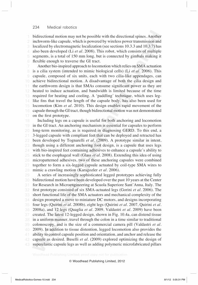
bidirectional motion may not be possible with the directional spines. another inchworm-like capsule, which is powered by wireless power transmission and localized by electromagnetic localization (see sections 10.3.3 and 10.3.7) has also been developed (Li et al. 2008). this robot, which consists of multiple segments, is a total of 150 mm long, but is connected by gimbals making it flexible enough to traverse the GI tract. another bio-inspired approach to locomotion which relies on sMa actuation is a cilia system (intended to mimic biological cells) (Li et al. 2006). this capsule, composed of six units, each with two cilia-like appendages, can achieve bidirectional motion. a disadvantage of both the cilia design and the earthworm design is that SMAs consume significant power as they are heated to induce actuation, and bandwidth is limited because of the time required for heating and cooling. a ‘paddling’ technique, which uses leg-like fins that travel the length of the capsule body, has also been used for locomotion (Kim et al. 2010). this design enables rapid movement of the capsule through the Gi tract, though bidirectional motion was not demonstrated on the first prototype. including legs on a capsule is useful for both anchoring and locomotion in the Gi tract. an anchoring mechanism is essential for capsules to perform long-term monitoring, as is required in diagnosing Gerd. to this end, a 3-legged capsule with compliant feet that can be deployed and retracted has been developed by tognarelli et al. (2009). a prototype similar in intent, though using a different anchoring foot design, is a capsule that uses legs with bio-inspired feet containing adhesives to enhance a capsule’s ability to stick to the esophageal wall (Glass et al. 2008). extending this idea of using micropatterned adhesives, two of these anchoring capsules were combined together to form a six-legged capsule actuated by coil-type sMa wires to mimic a crawling motion (Karagozler et al. 2006). a series of increasingly sophisticated legged prototypes achieving fully bidirectional motion have been developed over the past 10 years at the center for research in Microengineering at scuola superiore sant’anna, italy. the first prototype consisted of six SMA-actuated legs (Gorini et al. 2006). the short functional life of the sMa actuators and mechanical complexity of the design prompted a move to miniature dc motors, and designs incorporating four legs (Quirini et al. 2008b), eight legs (Quirini et al. 2007, Quirini et al. 2008a), and 12 legs (Quaglia et al. 2009, Valdastri et al. 2009) have been created. the latest 12-legged design, shown in Fig. 10.4a, can distend tissue in a uniform manner, travel through the colon in a time similar to traditional colonoscopy, and is the size of a commercial camera pill (Valdastri et al. 2009). in addition to tissue distention, legged locomotion also provides the ability to control capsule position and orientation, and anchor and release the capsule as desired. buselli et al. (2009) explored optimizing the design of superelastic capsule legs as well as adding polymeric microfabricated pillars
MedicalRobotics-Gomes-10.indd 234 8/1/12 5:05:31 PM
235Mesoscale mobile robots for gastrointestinal MIS
Woo
dhea
d Pu
blis
hing
Lim
ited
Woodhead Publishing Limited; proof copy not for publication
123456789
10111213141516171819202122232425262728293031323334353637383940414243
© Woodhead Publishing Limited, 2012
(c)
(b)
(a)
Robotic system
Wireless link
CR2B controller
Remote PC
RV-JSB Rotobic arm6 DOF Control
peripheral
Display – vision feedback
Permanent magnet
TCP/IP link
Magnetic link
Endoscopic device HMI
• Internalpermanent magnets (IPM)
•Wiredvisionmodule
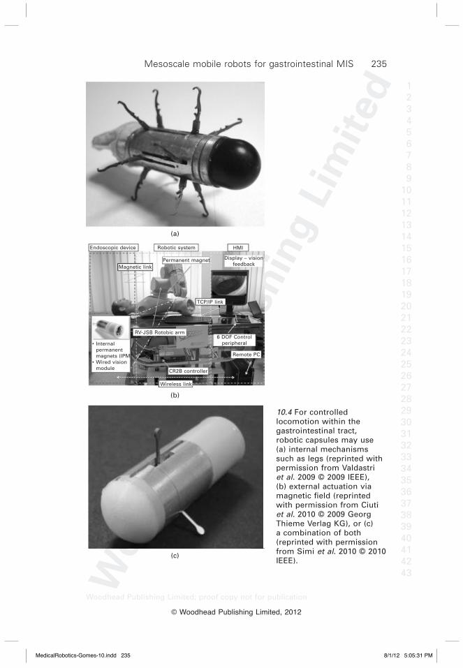
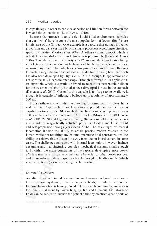
10.4 For controlled locomotion within the gastrointestinal tract, robotic capsules may use (a) internal mechanisms such as legs (reprinted with permission from Valdastri et al. 2009 © 2009 IEEE), (b) external actuation via magnetic field (reprinted with permission from Ciuti et al. 2010 © 2009 Georg Thieme Verlag KG), or (c) a combination of both (reprinted with permission from Simi et al. 2010 © 2010 IEEE).
MedicalRobotics-Gomes-10.indd 235 8/1/12 5:05:31 PM
236 Medical robotics
Woo
dhea
d Pu
blis
hing
Lim
ited
Woodhead Publishing Limited; proof copy not for publication
123456789
10111213141516171819202122232425262728293031323334353637383940414243
© Woodhead Publishing Limited, 2012
to capsule legs in order to enhance adhesion and friction forces between the legs and the colon tissue (buselli et al. 2010). Because the stomach is an elastic, liquid-filled environment, capsules that can ‘swim’ have become the most popular form of locomotion for use in this area of the Gi tract. one example is a capsule that utilizes propeller propulsion and can steer itself by actuating its propellers according to direction, speed, and rotation (tortora et al. 2009). another swimming robot, which is actuated by animal-derived muscle tissue, was proposed by (Herr and dennis 2004). though their current prototype is 12 cm long, the idea of using living muscle tissue for actuation may be beneficial for future capsule endoscopes. a swimming microrobot which uses two pairs of external Helmholtz coils to create a magnetic field that causes a fin-like tail to swing back and forth has also been developed by (byun et al. 2011), though its applications are not specific to GI capsule endoscopy. Though different in its application, an ingestible wireless capsule designed to release an intragastric balloon for the treatment of obesity has also been developed for use in the stomach (Kencana et al. 2010). currently, this capsule is too large to be swallowed, though it is capable of inflating a balloon up to a volume of approximately 200 mL. From earthworm-like motion to crawling to swimming, it is clear that a wide variety of approaches have been taken to provide internal locomotion capabilities to capsules. other methods that have also been explored (swain 2008) include electrostimulation of Gi muscles (Mosse et al. 2001, woo et al. 2006, 2009) and flagellar swimming (Kosa et al. 2008); some patents also allude to magnetically actuated propellers (iddan and Gilad 2006) and self-propulsion through jets (iddan 2006). the advantages of internal locomotion include the ability to obtain precise motion relative to the lumen, while not requiring any external magnetic field generators, and the ability to achieve tissue distention away from the on-board camera in some cases. the challenges associated with internal locomotion, however, include designing and manufacturing complex mechanical systems small enough to fit within the space constraints of the capsule, developing more power efficient mechanisms to run on miniature batteries or other power sources, and to manufacture these capsules cheaply enough to be disposable (which may be preferred) or robust enough to be sterilized.
External locomotion
an alternative to internal locomotion mechanisms on board capsules is to use external systems (primarily magnetic fields) to induce locomotion. external locomotion is being pursued in the research community, and also in the commercial arena by Given imaging, inc. and olympus, inc. Magnetic fields can be generated outside the patient either by electromagnetic coils or
MedicalRobotics-Gomes-10.indd 236 8/1/12 5:05:31 PM
237Mesoscale mobile robots for gastrointestinal MIS
Woo
dhea
d Pu
blis
hing
Lim
ited
Woodhead Publishing Limited; proof copy not for publication
123456789
10111213141516171819202122232425262728293031323334353637383940414243
© Woodhead Publishing Limited, 2012
by permanent magnets. Coils can provide electronic control of field strength and direction, whereas permanent magnets typically generate higher fields in smaller form factors. One of the earliest groups to utilize magnetic fields for capsule locomotion was the Norika Project team, who developed a capsule controlled by external coils embedded in a jacket worn by the patient (Moglia et al. 2007, Uehara and Hoshina 2003). olympus has also been developing a bidirectional magnetically actuated capsule that has an internal magnet which interacts with a varying, controllable, rotating field created by three pairs of external magnets (Moglia et al. 2007). Using an olympus magnetic capsule endoscope and a siemens magnetic guidance system, rey et al. (2010) showed that magnetically navigated capsules can accomplish gastric examinations. Given Imaging, Inc. has taken a slightly different approach to using magnetic fields, using an external hand-held plate magnet to manipulate a magnetic capsule under gastroscopic visualization (Keller et al. 2011, swain et al. 2010). Feasibility studies using the hand-held magnetic system suggest that remote manipulation of the capsule is feasible in both the esophagus and stomach (Keller et al. 2011, swain et al. 2010). a custom-designed magnetic capsule navigation system using a pair of electromagnets has been developed and tested in both simulation and in a small-scale physical setup by (Lam and Mintchev 2009). incorporating diamagnetic materials in the capsule casing to provide finer control and stabilization of on-board magnets is also being explored (Lam and Mintchev 2009). Yet another approach uses a rotating external magnetic field to move a variable diameter capsule that contains an internal magnet and a spiral fin around its body (zhang et al. 2010). Controlling a magnetic field by fixing a permanent magnet to a cartesian robot was proposed by Kim et al. (2005a). this idea was studied in more depth by ciuti et al. (2010), who exploited the precise control of a six degree-of-freedom (doF) robotic manipulator with a permanent magnet fixed to its end effector to control capsule locomotion Fig. 10.4b. Using this system, robotic control for magnetic steering of capsule endoscopes was demonstrated to be more precise and reliable than manual operation (ciuti et al. 2010a). an innovative approach utilizing two commercially available technologies, the Niobe system by stereotaxis, inc. and the Pillcam sb by Given imaging, inc. was proposed by carpi et al. (2006, 2011 and carpi and Pappone 2009). In this system, a magnetic sleeve was created to fit around the PillCam SB, whose orientation was then controlled using the Niobe system. omnidirectional steering accuracy of 1° was achieved. Using magnetic fields to directly steer the camera of a capsule has also been proposed by Valdastri et al. (2010). in this work, a remote controlled capsule, known as the magnetic internal mechanism (MIM) capsule, uses two internal permanent magnets fixed to a toothed gear, which rotates when a motor is activated. when placed in an
MedicalRobotics-Gomes-10.indd 237 8/1/12 5:05:31 PM
238 Medical robotics
Woo
dhea
d Pu
blis
hing
Lim
ited
Woodhead Publishing Limited; proof copy not for publication
123456789
10111213141516171819202122232425262728293031323334353637383940414243
© Woodhead Publishing Limited, 2012
external magnetic field, the internal magnets remain aligned with the external magnetic field, causing the entire capsule to pivot about a single point, enabling fine adjustment of the camera orientation (Valdastri et al. 2010). the primary advantage of external locomotion, in comparison with internal locomotion, is a reduction in components on board the capsule, enabling a larger diagnostic or therapeutic payload. the drawback is the requirement of a large and complex system outside the patient to generate the magnetic field. Methods to determine the necessary field characteristics for a given desired capsule motion or force application are also open research areas (abbott et al. 2007, ciuti et al. 2010b). one other challenge in using magnetic locomotion is that the deflated intestine significantly impedes their motion (this is why many of the existing prototypes are tested in conjunction with endoscopy, which can control insufflation during the test). Recently, wireless insufflation has been proposed to overcome this obstacle (Toennies et al. 2010, toennies and webster 2009). it is also possible to combine internal and external locomotion in a hybrid capsule, (Fig. 10.4c), which is primarily magnetically actuated but uses legs to assist with locomotion and tissue distention as needed (simi et al. 2010). another hybrid approach uses a spiral ridge which is rotated by a micromotor as its primary source of locomotion and uses external magnetic fields to provide assisting force and increase speed as needed (wang et al. 2010).
10.3.5 Diagnostic systems
Many different sensing systems have been developed for wireless capsules. an example is a swallowable pH-sensing capsule that uses voltage differences to measure pH and half-ring electrodes for impedance monitoring, developed for diagnosis of Gerd (Gonzalez-Guillaumin et al. 2007). this capsule is held to the esophageal wall by an external magnet near the throat. several capsules have been developed that can measure temperature in the Gi tract. examples include the Vitalsense ingestible telemetric physiological monitoring system, which uses thermistor-based sensing, and the cortemp pill system, which measures crystal oscillations for temperature sensing, (McKenzie and osgood 2004). other capsules that can measure both temperature and pressure include the Philips intellicap (www.philips.com) (also discussed in section 10.3.6) (Forgione 2009) and a design by van der schaar and coworkers that can expel a small amount of liquid (Moglia et al. 2008). two other sensing capsules reviewed (twomey and Marchesi 2009) include the Lab-on-a-Pill capsule (Johannessen et al. 2004) which can sense pH, conductivity, and oxygen levels, and the standford endocapsule (allison et al. 2006), which has optical, chemical, temperature, and pH measuring capabilities. Moglia et al. (2008) also review other recent patents on capsule-based biosensors.
MedicalRobotics-Gomes-10.indd 238 8/1/12 5:05:31 PM
239Mesoscale mobile robots for gastrointestinal MIS
Woo
dhea
d Pu
blis
hing
Lim
ited
Woodhead Publishing Limited; proof copy not for publication
123456789
10111213141516171819202122232425262728293031323334353637383940414243
© Woodhead Publishing Limited, 2012
optical biopsy may also be a useful diagnostic tool for capsule endoscopes, because traditional biopsies often require multiple samples to be taken from the same site and analyzed within 1–2 h of extraction. Unlike traditional biopsies, optical biopsies rely on the properties of light for diagnosis (sauk and itzkowitz 2011). one example of a capsule system that detects lesions or internal bleeding within the Gi tract was developed by (Liu et al. 2010) and another was described in a patent by (schostek et al. 2007). other promising techniques include fluorescence endoscopy, optical coherence tomography, confocal microendoscopy, light-scattering spectroscopy, raman spectroscopy, and molecular imaging, all of which are discussed in detail by wang and Van dam (2004).
10.3.6 Intervention
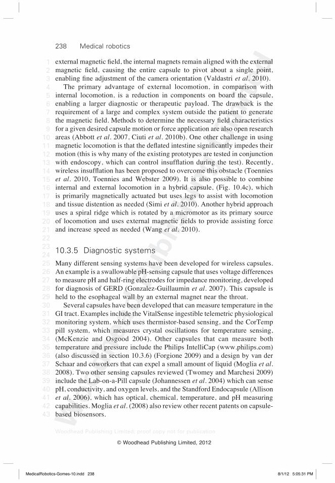
Perhaps the most compelling aspect of robotic capsules is their potential to both diagnose and treat lesions they encounter in situ. although current commercial capsules have significantly improved physicians’ ability to see areas deep within the Gi tract, they are still passive devices and subsequent surgery is often required in order to perform an intervention. compared with many of the other robotic modules discussed in this chapter, interventional capabilities of capsules are still in their infancy. despite this, a few innovative designs have been developed which include a clip deployment capsule, drug delivery capsules, and biopsy sampling. The first therapeutic capsule of its kind, recently developed by Valdastri et al. (2008), can deploy a single preloaded sMa clip at the site of a lesion, based on a wirelessly transmitted command. this capsule (Fig. 10.5), also
(a) (b)
10.5 This interventional capsule (a) uses magnetic locomotion to position itself near a lesion and then (b) deploys a clip to stop the bleeding (reprinted with permission from Valdastri et al. 2008, © 2009 Georg Thieme Verlag KG).
MedicalRobotics-Gomes-10.indd 239 8/1/12 5:05:31 PM
240 Medical robotics
Woo
dhea
d Pu
blis
hing
Lim
ited
Woodhead Publishing Limited; proof copy not for publication
123456789
10111213141516171819202122232425262728293031323334353637383940414243
© Woodhead Publishing Limited, 2012
has permanent magnets embedded inside of it, enabling active locomotion via an external magnetic field so that the capsule can be properly aligned with the site of interest. other therapeutic capsules have also been designed to deliver topical drugs to an area of interest. it is hoped that delivering drugs directly at the site of interest will lower the dosage levels required for treatment and lower the time required for healing to begin. the intelicap, under development at Philips, inc., can measure and transmit temperature and pH levels (as mentioned in section 10.3.5) in addition to delivering a treatment agent on command. as mentioned in section 10.3.3, Laulicht et al. (2011) are developing a magnetic localization platform that allows a user to adjust a capsule’s location within the Gi tract in order to optimize the release of drugs in the area of greatest absorption or therapeutic action. Yet another drug delivery capsule uses a novel micromachined thruster that converts chemical energy into propulsion energy which produces sufficient pressure to empty a drug reservoir on board the capsule (Pi et al. 2010). other drug delivery capsules include the intelisite capsule (innovative devices) which uses sMa wires to line perforated inner and outer sleeves to disperse a drug through holes and the enterion capsule (Phaeton research and Pharmaceutical Profiles), which uses a piston/spring system to deliver a treatment agent (wilding et al. 2000). both (twomey and Marchesi 2009) and (wilding et al. 2000) provide further in-depth discussions of drug delivery capsules. as mentioned in section 10.3.5, conventional biopsy procedures often require taking multiple samples from one location and then having these samples analyzed within 1–2 h of extraction. although optical biopsy from a capsule platform has been proposed, a biopsy device which uses a rotational razor for cutting tissue has been implemented on board a capsule and was successful in extracting a tissue sample in an ex vivo animal intestine (Kong et al. 2005). it is unknown yet whether this type of capsule is able to take multiple samples as required in traditional biopsy for accurate histological analysis. a few patent disclosures (Moglia et al. 2008) also suggest other ideas for therapeutic and interventional capsules including capsules housing ultrasonic transducers (Miyake 2006, taniguchi 2005) and a dual-capsule camera biopsy system (swain 2007). interventional capsules are one of the most compelling aspects of endoscopic capsule research from the perspectives of reducing invasiveness and costs of treatment. endowing capsules with interventional capabilities, however, is challenging owing to the intelligence and miniaturized mechanisms that are required for both diagnosis and treatment. such capabilities, however, will probably propel capsule endoscopes into becoming truly robotic devices.
MedicalRobotics-Gomes-10.indd 240 8/1/12 5:05:31 PM
241Mesoscale mobile robots for gastrointestinal MIS
Woo
dhea
d Pu
blis
hing
Lim
ited
Woodhead Publishing Limited; proof copy not for publication
123456789
10111213141516171819202122232425262728293031323334353637383940414243
© Woodhead Publishing Limited, 2012
10.3.7 Power
the need for an energy-dense power source is the greatest challenge facing researchers developing mobile robotic surgery platforms today. For vision alone, commercial Gi capsules rely on two silver oxide coin cell batteries which take up almost half of the space available onboard (Moglia et al. 2009). in general, batteries do not scale down well to the mesoscale, primarily because the batteries that are small enough to fit within a robotic capsule are not energy-dense enough to power robotic mechanisms for very long. Lithium ion polymer batteries (LiPo) are the best source of electrical power available today because they can source peak currents up to 20 times their nominal current. Furthermore, they can be shaped or slightly bent for optimal placement within a capsule. Valdastri et al. (2009) elaborate on the power requirements of their actively locomoting 12-legged capsule, which they estimated would require 430 mw, plus an additional 180 mw for real-time vision. they determined that the smallest (10 mm in diameter ¥ 30 mm in length) battery available to power their 12-legged capsule was a 100 ma h battery. this battery is almost the same size as those used in commercial capsule endoscopes. thus, future advancements in battery technology are needed to enable completely wireless robots with complex mechanisms. one alternative is wireless power transfer via inductive coupling, which uses internal coils on board a capsule to derive power from a magnetic field established by an external solenoid coil. this approach was used in the intracorporal video probe (Lenaerts and Puers 2007) and was also used in powering a propeller-driven capsule, shown in Fig. 10.6 (carta et al. 2009). Further, the Norika project team proposed using inductive coupling to power a capsule by having a patient wear a vest that had coils embedded in it for both power transmission and direction control (Uehara and Hoshina 2003). wireless power transmission was also used to power an inchworm-like robot (Li et al. 2008), where the maximum applied power was 35 w, and the average received power was 490 mw. by using electromagnetic localization and synthesis of the magnetic field vector, a wireless power system that can automatically tune the power-transmitting coils and can regulate the output power based on the capsule’s need was developed (Li et al. 2010). One potential alternative to electrical energy is fluid power. Fluid power involves the phase transition of a liquid fuel to a gas, which generates pneumatic pressure. this concept was initially demonstrated in a prosthetic arm by Goldfarb et al. (2003), where experiments indicated that it could improve an energetic figure of merit (a metric that encompasses energy density, conversion efficiency, and actuator power density) by an order of magnitude. Based on this, the idea of utilizing fluid power for capsule robots has also been proposed (toennies et al. 2010, toennies and webster 2009), where initial experiments for wireless insufflation were performed as
MedicalRobotics-Gomes-10.indd 241 8/1/12 5:05:31 PM
242 Medical robotics
Woo
dhea
d Pu
blis
hing
Lim
ited
Woodhead Publishing Limited; proof copy not for publication
123456789
10111213141516171819202122232425262728293031323334353637383940414243
© Woodhead Publishing Limited, 2012
a first step toward using fluid power to actuate more complex mechanisms. these feasibility studies suggest that it is possible for a capsule to carry a sufficient volume of fluid to generate sufficient gas to significantly enhance visualization. at this point, it is unclear which form of power generation is most efficient for robotic capsules. It is clear, however, that capsule robots require enhanced batteries or alternative sources of power such as inductive coupling, fluid power, or other novel technologies to be developed at the scales, power levels, and current sourcing capacities required to achieve their full potential.
10.4 Future trends in mobile surgical devices
Future innovations in several aspects of robotic capsules will open the door to many new surgical applications. the most important need is a highly energy dense power supply. also extremely useful would be miniature wireless cameras capable of high-definition imaging and high frame rates. This will be enabled by advancements in flexible electronics and 3D packaging of integrated electronics and optics components. design of miniature, high-performance mechanisms is the key to enabling biopsy and interventional capabilities.
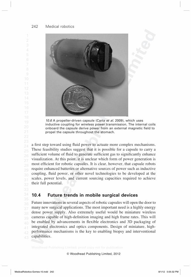
10.6 A propeller-driven capsule (Carta et al. 2009), which uses inductive coupling for wireless power transmission. The internal coils onboard the capsule derive power from an external magnetic field to propel the capsule throughout the stomach.
MedicalRobotics-Gomes-10.indd 242 8/1/12 5:05:32 PM
243Mesoscale mobile robots for gastrointestinal MIS
Woo
dhea
d Pu
blis
hing
Lim
ited
Woodhead Publishing Limited; proof copy not for publication
123456789
10111213141516171819202122232425262728293031323334353637383940414243
© Woodhead Publishing Limited, 2012
as the technologies described in each of the preceding sections evolve, mobile surgical robots are poised to become increasingly useful diagnostic and interventional devices in the Gi tract and in abdominal surgery. as the robotic technologies described in previous sections are combined, miniaturized, and made more power-efficient, mobile surgical robots will take on increasingly complex tasks in many different kinds of surgery. Future systems can be expected to evolve along several different parallel trajectories. First, mobile robots containing cameras (the first few examples of which have already appeared in academic laboratories, as discussed earlier in this chapter) will provide physicians with many new view angles on existing surgical sites. this will enable image-processing techniques to provide even more information to the physician, including 3d reconstructions of anatomical information, and augmented reality interfaces. other sensors can also be integrated onto mobile platforms to enable in situ diagnosis of disease (e.g. by spectroscopy or chemical sensors). to overcome the power issue, early generations of mobile surgical platforms will be ‘softly tethered’, meaning that they incorporate very small and lightweight tethers to provide power and other capabilities. Magnets can be used in conjunction with soft tethers to enable several robots to work co-operatively to perform laparoscopic surgery. the natural progression as advances in each aspect of mobile surgical robots are made will be to cut the cord and remove these tethers. wireless interventional capsules have already been demonstrated in the Gi tract, although much work is needed before they become clinically available, and many new designs will be needed to address different pathologies. Microscale mobile robots are also an active area of research as reviewed in chapters 8, 9 and 11. they are particularly useful as the target environment becomes smaller (e.g. eye, blood vessels). in terms of manufacturing, microscale robots typically require micromanufacturing techniques and/or integration of biological components. in contrast, mesoscale robots can often be fabricated using high-precision, but fundamentally standard manufacturing processes. Looking to the future, rather than converging to one single device that can do everything, it is likely that specific robot systems will be designed for specific surgical indications. Also, teams of mobile robots will probably collaborate to meet surgical objectives. these devices or teams of devices will make surgery less invasive and more effective, enabling physicians to see, diagnose, and apply proper therapy, simultaneously.
10.5 Conclusion
in this chapter, we have reviewed the state-of-the art in mesoscale mobile robots. we have focused on robotic capsules for endoscopy within the Gi
MedicalRobotics-Gomes-10.indd 243 8/1/12 5:05:32 PM
244 Medical robotics
Woo
dhea
d Pu
blis
hing
Lim
ited
Woodhead Publishing Limited; proof copy not for publication
123456789
10111213141516171819202122232425262728293031323334353637383940414243
© Woodhead Publishing Limited, 2012
tract, because this is the most accessible location in the human body to capsule robots, and has thus served as the proving ground for many current devices. although passive imaging capsules have rapidly become the gold standard for diagnosis within the small intestine, robotic capsules have yet to be implemented clinically owing to the many challenges associated with miniaturizing internal mechanisms and the lack of an energy-dense power source. despite this, many robotic capsules are currently being developed and enabling technologies are being explored to overcome the current challenges in applying capsule robots in surgery. in the future, it is likely that these technologies will propel capsules into clinical implementation and enable them to see, diagnose, and treat diseases within the human body from a mobile, minimally invasive platform.
10.6 Referencesabbott, J. J., ergeneman, o., Kummer, M. P., Hirt, a. and Nelson, b. (2007), ‘Modeling
magnetic torque and force for controlled manipulation of soft-magnetic bodies’, IEEE Transactions on Robotics, 23, 1247–1252.
allison, e., Kiraly, z., springer, G. and dam, J. (2006), ‘endocapsule’. Patent Number wo/2006/045011.
arshak, K. and adepoju, F. (2006), ‘capsule tracking in the Gi tract: a novel microcontroller based solution’, IEEE Sensors Applications Symposium, Houston, TX, USA, pp. 186–191.
bang, s., Park, J., Jeong, s., Kim, Y., shim, H., Kim, t., Lee, d. and song, s. (2009), ‘First clinical trial of the ‘Miro’ capsule endoscope by using a novel transmission technology: electric-field propagation’, Clinical Endoscopy, 69, 253–259.
bejakovic, s., Kumar, r., dassopoulos, t., Mullin, G. and Hager, G. (2009), ‘analysis of crohn’s disease lesions in capsule endoscopy images’, IEEE International Conference on Robotics and Automation, Kobe, Japan.
bulat, J., duda, K., duplaga, M., Fraczek, r., skalski, a., socha, M., turcza, P. and zielinski, t. (2007), ‘data processing tasks in wireless Gi endoscopy: image-based capsule localization and navigation with video compression’, Proceedings of IEEE/EMBS, Lyon, France, pp. 2815–2818.
buselli, e., Pensabene, V., castrataro, P., Valdastri, P., Menciassi, a., and dario, P. (2010), ‘evaluation of friction enhancement through soft polymer micro-patterns in active capsule endoscopy’, Measurement Science and Technology, 21, 105802 (7pp).
buselli, e., Valdastri, P., Quirini, M., Menciassi, a. and dario, P. (2009), ‘superelastic leg design optimization for an endoscopic capsule with active locomotion’, Smart Materials and Structures, Pisa, italy 18, 015001 (8 pp).
byun, d., choi, J., cha, K., Park, J. and Park, s. (2011), ‘swimming microrobot actuated by two pairs of Helmholtz coils system’, Mechatronics, 21, 357–364.
carpi, F., Galbiati, s. and carpi, a. (2006), ‘Magnetic shells for gastrointestinal endoscopic capsules as a means to control their motion’, Biomedicine and Pharmacotherapy, 60, 370–374.
carpi, F., Kastelein, N., talcott, M. and Pappone, c. (2011), ‘Magnetically controllable gastrointestinal steering of video capsules’, IEEE Transactions on Biomedical Engineering, 58, 231–234.
MedicalRobotics-Gomes-10.indd 244 8/1/12 5:05:32 PM
245Mesoscale mobile robots for gastrointestinal MIS
Woo
dhea
d Pu
blis
hing
Lim
ited
Woodhead Publishing Limited; proof copy not for publication
123456789
10111213141516171819202122232425262728293031323334353637383940414243
© Woodhead Publishing Limited, 2012
carpi, F. and Pappone, c. (2009), ‘Magnetic maneuvering of endoscopic capsules by means of a robotic navigation system’, IEEE Transactions on Biomedical Engineering, 56, 1482–1490.
carta, r., tortora, G., Lenaerts, b., thone, J., Valdastri, P., Menciassi, a., Puers, r. and dario, P. (2009), ‘wireless powering for a self-propelled and steerable endoscopic capsule for stomach inspection’, Biosensors and Bioelectronics, 25, 845–851.
cavallotti, c., Merlino, P., Vatteroni, M., Valdastri, P., abramo, a., Menciassi, a. and dario, P. (2011), ‘an FPGa-based versatile development system for endoscopic capsule design optimization’, Sensors and Actuators A, 172, 301–307.
cavallotti, c., Piccigallo, M., susilo, e., Valdastri, P., Menciassi, a. and dario, P. (2009), ‘an integrated vision system with autofocus for wireless capsular endoscopy’, Sensors and Actuators A, 156, 72–78.
cave, d., Fleischer, d., Gostout, c., Faigel, d., Leighton, J., Heigh, r., sharma, V., Mergener, K., bhattacharya, K., rajan, e., Foley, a., Lee, M., Knipschield, M. and Hibberd, P. (2007), ‘a multi-center randomized comparison of the endocapsule: olympus inc and the Pillcam sb: Given imaging in patients with obscure Gi bleeding [abstract]’, Gastrointestinal Endoscopy 65, ab125.
chen, X., zhang, X., zhang, L., Li, X., Qi, N., Jiang, H. and wang, z. (2009), ‘a wireless capsule endoscope system with low-power controlling and processing asic’, IEEE Transactions on Biomedical Circuits and Systems, 3, 11–21.
ciuti, G., donlin, r., Valdastri, P., arezzo, a., Menciassi, a., Morino, M. and dario, P. (2010a), ‘robotic versus manual control in magnetic steering of an endoscopic capsule’, Endoscopy, 42, 148–152.
ciuti, G., Pateromichelakis, N., sfakiotakis, M., Valdastri, P., Menciassi, a., tsakiris, d. P., and dario, P. (2011,), ‘a wireless module for vibratory motor control and inertial sensing in capsule endoscopy’, Sensors and Actuators A: Physical, doi: 10.1016/j.sna.2011.12.024.
ciuti, G., Valdastri, P., Menciassi, a. and dario, P. (2010b), ‘robotic magnetic steering and locomotion of microsystems for diagnostic and surgical endoluminal procedures’, Robotica, 28, 199–207.
drossman, d. a., Grimm, i. and shaheen, N. (2005), Handbook of gastroenterologic procedures – fourth edition, Lippincott williams and wilkins, Philadelphia, Pa.
eliakim, r., Yassin, K., Niv, Y., Metzger, Y., Lachter, J., Gal, e., spaznikov, b., Konikoff, F., Leichtmann, G., Fireman, z., Kopelman, Y. and adler, s. N. (2009), ‘Prospective multicenter performance evaluation of the second-generation colon capsule compared with colonoscopy’, Endoscopy, 41, 1026–1031.
ergeneman, o., dogangil, G., Kummer, M. P., abbott, J. J., Nazeeruddin, M. K. and Nelson, b. J. (2008), ‘a magnetically controlled wireless optical oxygen sensor for intraocular measurements’, IEEE Sensors Journal of the American Chemical Society, 8, 29–37.
Fischer, d., schreiber, r., Levi, d. and eliakim, r. (2004), ‘capsule endoscopy: the localization system’, Gastrointestinal Endoscopy Clinics of North America, 14, 25–31.
Forgione, A. (2009), ‘In vivo microrobots for natural orifice transluminal surgery. current status and future perspectives’, Surgical Oncology, 18, 121–129.
Glass, P., cheung, e. and sitti, M. (2008), ‘a legged anchoring mechanism for capsule endoscopes using micropatterned adhesives’, IEEE Transactions on Biomedical Engineering, 55, 2759–2767.
Goldfarb, M., barth, e., Gongola, M. and wehrmeyer, J. (2003), ‘design and energetic
MedicalRobotics-Gomes-10.indd 245 8/1/12 5:05:32 PM
246 Medical robotics
Woo
dhea
d Pu
blis
hing
Lim
ited
Woodhead Publishing Limited; proof copy not for publication
123456789
10111213141516171819202122232425262728293031323334353637383940414243
© Woodhead Publishing Limited, 2012
characterization of a liquid-propellant-powered actuator for self-powered robots’, IEEE/ASME Transactions on Mechatronics, 8, 254–262.
Gonzalez-Guillaumin, J., sadowski, d., Kaler, K. and Mintchev, M. (2007), ‘ingestible capsule for impedance and pH monitoring in the esophagus’, IEEE Transactions on Biomedical Engineering, 54, 2231–2236.
Gorini, s., Quirini, M., Menciassi, a., Pernorio, G., stefanini, c. and dario, P. (2006), ‘a novel sMa-based actuator for a legged endoscopic capsule’, IEEE/RAS-EMBS International Conference on Biomedical Robotics and Biomechatronics, Pisa, italy pp. 443–449.
Hasan, M. and wallace, M. (2009), ‘image-enhanced endoscopy’, American Society for Gastrointestinal Endoscopy, 16, 1–5.
Heresbach, d., Leray, e., d’Halluin, P. N., cholet, F., Lapalus, M. G., Gaudric, M., ben soussan, e., Gaudin, J. L., Vahedi, K., Quentin, V., Filoche, b., saurin, J. c., chaussade, s. and Ponchon, t. (2010), ‘diagnostic accuracy of esophageal capsule endoscopy versus conventional upper digestive endoscopy for suspected esophageal squamous cell carcinoma’, Endoscopy, 42, 93–97.
Herr, H. and dennis, r. (2004), ‘a swimming robot actuated by living muscle tissue’, Journal of NeuroEngineering and Rehabilitation, 1, 6.
Hocke, M., schone, U., richert, H., Gornert, P., Keller, J., Layer, P. and stallmach, a. (2008), ‘every slow-wave impulse is associated with motor activity of the human stomach’, American Journal of Physiology Gastrointestinal and Liver Physiology, 296, 709–716.
iddan, G. (2006), ‘self propelled device’. Us Patent Number 2006/0030754 a1.iddan, G. and Gilad, z. (2006), ‘Motor for an in-vivo device’. Patent Number wo
2006/070350 a2.Jinshan science and technology Group, Ltd., co. http://www.omom.us/main.
php?sLaN=en.Johannessen, e. a., wang, L., cui, L., tang, t. b., ahmadian, M., astaras, a., reid,
s. w. J., Yam, P. s., Murray, a. F., Flynn, b. w., beaumont, s. P., cumming, d. r. s. and cooper, J. M. (2004), ‘implementation of multichannel sensors for remote biomedical measurements in a microsystems format’, IEEE Transactions on Biomedical Engineering, 51, 525–535.
Karagozler, M., cheung, e., Kwon, J. and sitti, M. (2006), ‘Miniature endoscopic capsule robot using biomimetic micro-patterned adhesives’, IEEE/RAS-EMBS International Conference on Biomedical Robotics and Biomechatronics, Pisa, italy pp. 105–111.
Keller, J., Fibbe, c., Volke, F., Gerber, J., Mosse, a. c., reimann-zawadzki, M., rabinovitz, e., Layer, P., schmitt, d., andresen, V., rosien, U. and swain, P. (2011), ‘inspection of the human stomach using remote-controlled capsule endoscopy: a feasibility study in healthy volunteers (with videos)’, Gastrointestinal Endoscopy, 73, 22–28.
Kencana, a. P., rasouli, M., Huynh, V. a., ting, e. K., Lai, J. c. Y., Huy, Q. d. Q., tan, s. L., wong, K. J. and Phee, s. J. (2010), ‘an ingestible wireless capsule for treatment of obesity’, 32nd Annual International Conference of the IEEE EMBS Conference, pp. 963–966.
Kim, b. K., Park, J. o. and Hong, Y. s. (2005a), ‘capsule type endoscope control system’, Korean institute of science and technology. Patent Number wo/2005/122866.
Kim, b., Lee, s., Park, J. H. and Park, J. o. (2005b), ‘design and fabrication of a locomotive mechanism for capsule-type endoscopes using shape memory alloys (sMas)’, IEEE/ASME Transactions on Mechatronics, 10, 77–86.
Kim, b., Park, s., Yoon, s. J. and Jee, c. Y. (2005c), ‘an earthworm-like locomotive
MedicalRobotics-Gomes-10.indd 246 8/1/12 5:05:32 PM
247Mesoscale mobile robots for gastrointestinal MIS
Woo
dhea
d Pu
blis
hing
Lim
ited
Woodhead Publishing Limited; proof copy not for publication
123456789
10111213141516171819202122232425262728293031323334353637383940414243
© Woodhead Publishing Limited, 2012
mechanism for capsule endoscopes’, IEEE/RSJ International Conference on Intelligent Robots and Systems, edmonton, alberta, canada pp. 2997–3002.
Kim, H. M., Yang, s., Kim, J., Park, s., cho, J. c., Park, J. Y., Kim, t. s., Yoon, e., song, s. Y. and bang, s. (2010), ‘active locomotion of a paddling-based capsule endoscope in an in vitro and in vivo experiment (with videos)’, Gastrointestinal Endoscopy, 72, 381–387.
Kolar, a., Pinna, a., romain, o., Viateur, s., ea, t., belhaire, e., Graba, t. and Granado, b. (2009), ‘a multishutter time sensor for multispectral imaging in a 3-d reconstruction integrated sensor’, IEEE Sensors Journal, 9, 478–484.
Kong, K., cha, J., Jeon, d. and cho, d. (2005), ‘a rotational micro biopsy device for the capsule endoscope’, IEEE/RSJ International Conference on Intelligent Robots and Systems, edmonton, alberta, canada pp. 1839–1843.
Kosa, G., Jakb, P., Hata, N., Jolesz, F., Neubach, z., shoham, M., zaaroor, M. and szekely, G. (2008), ‘Flagellar swimming for medical micro robots: theory, experiments, and application’, IEEE/RAS-EMBS International Conference on Biomedical Robotics and Biomechatronics, scottsdale, az Usa pp. 258–263.
Kuth, r., reinschke, J. and roeckelein, r. (2007), ‘Method for determining the position and orientation of an endoscopy capsule guided through an examination object by using a navigating magnetic field generated by means of a navigation device’. German patent, de102005032370.
Kwiatek, M. A. and Pandolfino, J. (2008), ‘The Bravo(TM) pH capsule system’, Digestive and Liver Disease, 40, 156–160.
Kwon, J., Park, s., Park, J. and Kim, b. (2007), ‘evaluation of the critical stroke of an earthworm-like robot for capsule endoscopes’, Proceedings of the IMechE Part H: Journal of Engineering in Medicine, 221, 397–405.
Lam, M. and Mintchev, M. (2009), ‘diamagnetically stabilized levitation control of an intraluminal magnetic capsule’, Physiological Measurement, 30, 763–777.
Laulicht, b., Gidmark, N. J., tripathi, a. and Mathiowitz, e. (2011), ‘Localization of magnetic pills’, Proceedings of the National Academy of Science, 108, 2252–2257.
Lee, J., Oh, J., Shah, S., Yuan, X. and Tang, S. (2007), ‘Automatic classification of digestive organs in wireless endoscopy videos’, Proceedings of the ACM Symposium on Applied Computing, seoul, Korea, pp. 1041–1045.
Lehman, a., dumpert, J., wood, N., redden, L., Visty, a., Farritor, s., Varnell, b. and Oleynikov, D. (2009), ‘Natural orifice cholecystectomy using a miniature robot’, Surgical Endoscopy, 23, 260–266.
Lenaerts, b. and Puers, r. (2007), ‘an inductive power link for a wireless endoscope’, Biosensors and Bioelectronics, 22, 1390–1395.
Li, H., Yan, G. and Gao, P. (2010), ‘a method for improving the wireless power transmission efficiency of an endoscopic capsule based on electromagnetic localization and synthesis of magnetic field vector’, Proceedings of the IMechE Part C: Journal of Mechanical Engineering Science, 224, 1463–1471.
Li, H., Yan, G. and Ma, G. (2008), ‘an active endoscopic robot based on wireless power transmission and electromagnetic localization’, International Journal of Medical Robotics and Computer Assisted Surgery, 4, 355–367.
Li, w., Guo, w., Li, M. and zhu, Y. (2006), ‘a novel locomotion principle for endoscopic robot’, IEEE International Conference on Mechatronics and Automation, Luoyang, china pp. 1658–1662.
Lin, M. c., dung, L. r. and weng, P. K. (2006), ‘an ultra-low-power image compressor for capsule endoscope’, BioMedical Engineering OnLine, 5, 14.
MedicalRobotics-Gomes-10.indd 247 8/1/12 5:05:32 PM
248 Medical robotics
Woo
dhea
d Pu
blis
hing
Lim
ited
Woodhead Publishing Limited; proof copy not for publication
123456789
10111213141516171819202122232425262728293031323334353637383940414243
© Woodhead Publishing Limited, 2012
Liu, H., wang, G., wei, K., Pi, X., zhu, L., zheng, X. and wen, z. (2010), ‘an intelligent electronic capsule system for automated detection of gastrointestinal bleeding’, Journal of Zheijiang University SCIENCE B, 11, 937–943.
Martel, s., Felfoul, o., Mathieu, J.b., chanu, a., tamaz, s., Mohammadi, M., Mankiewicz, M. and tabatabaei, N. (2009), ‘Mri-based medical nanorobotic platform for the control of magnetic nanoparticles and flagellated bacteria for target interventions in human capillaries’, International Journal of Robotics Research, 28, 1169–1182.
McKenzie, J. and osgood, d. (2004), ‘Validation of a new telemetric core temperature monitor’, Journal of Thermal Biology, 29, 605–611.
Metzger, Y., adler, s., shitrit, a., Koslowsky, b. and bjarnason, i. (2009), ‘comparison of a new Pillcam sb2 video capsule versus the standard Pillcam sb for detection of small bowel disease’, Medical Imaging, 2, 7–11.
Miyake, K. (2006), ‘capsule ultrasonic endoscope and capsule ultrasonic endoscope system’. Japanose Patent JP325874.
Moglia, a., Menciassi, a. and dario, P. (2008), ‘recent patents on wireless capsule endoscopy’, Recent Patents on Biomedical Engineering, 1, 24–33.
Moglia, a., Menciassi, a., dario, P. and cuschieri, a. (2009), ‘capsule endoscopy: progress update and challenges ahead’, Natural Reviews in Gastroenterology and Hepatology, 6, 353–362.
Moglia, a., Menciassis, a., schurr, M. and dario, P. (2007), ‘wireless capsule endoscopy: from diagnostic devices to multipurpose robotic systems’, Biomed Microdevices, 9, 235–243.
Mosse, a., Mills, t., appleyard, M., Kadirkamanathan, s. and swain, P. (2001), ‘electrical stimulation for propelling endoscopes’, Gastrointestinal Endoscopy, 54, 79–83.
Nagy, z., Harada, K., Fluckiger, M., susilo, e., Kaliakatsos, i. K., Menciassi, a., Hawkes, E., Abbott, J. J., Dario, P. and Nelson, B. J. (2009), ‘Assembling reconfigurable endoluminal surgical systems: opportunities and challenges’, International Journal of Biomechatronics and Biomedical Robotics, 1, 3–16.
Patronik, N., ota, t., zenati, M. and riviere, c. (2009), ‘a miniature mobile robot for navigation and positioning on the beating heart’, IEEE Transactions on Robotics, 25, 1109–1124.
Pi, X., Lin, Y., wei, K., Liu, H., wang, G., zheng, X., wen, z. and Li, d. (2010), ‘a novel micro-fabricated thruster for drug release in remote controlled capsule’, Sensors and Actuators A, 159, 227–232.
Platt, s., Hawks, J. and rentschler, M. (2009), ‘Vision and task assistance using modular wireless in vivo surgical robots’, IEEE Transactions an Biomedical Engineering, 56, 1700–1710.
Quaglia, c., buselli, e., webster iii, r. J., Valdastri, P., Menciassi, a. and dario, P. (2009), ‘an endoscopic capsule robot: a meso-scale engineering case study’, Journal of Micromechanics and Microengineering, 19, 105007, 11p.
Quirini, M., Menciassi, a., scapellato, s., dario, P., rieber, F., Ho, c. N., schostek, s. and schurr, M. (2008a), ‘Feasibility proof of a legged locomotion capsule for the Gi tract’, Gastrointestinal Endoscopy, 67, 1153–1158.
Quirini, M., Menciassi, a., scapellato, s., stefanini, c. and dario, P. (2008b), ‘design and fabrication of a motor legged capsule for the active exploration of the gastrointestinal tract’, IEEE/ASME Transactions on Mechatronics, 13, 169–179.
Quirini, M., scapellato, s., Valdastri, P., Menciassi, a. and dario, P. (2007), ‘an approach to capsular endoscopy with active motion’, IEEE/EMBS, Lyon, France, pp. 2827–2830.
MedicalRobotics-Gomes-10.indd 248 8/1/12 5:05:32 PM
249Mesoscale mobile robots for gastrointestinal MIS
Woo
dhea
d Pu
blis
hing
Lim
ited
Woodhead Publishing Limited; proof copy not for publication
123456789
10111213141516171819202122232425262728293031323334353637383940414243
© Woodhead Publishing Limited, 2012
rentschler, M., dumper, J., Platt, s., iagnemma, K., oleynikov, d. and Farritor, s. (2006), ‘Modeling, analysis, and experimental study of in vivo wheeled robotic mobility’, IEEE Transactions on Robotics, 22, 308–321.
rey, J. F., ogata, H., Hosoe, N., ohtsuka, K., ogata, N., ikeda, K., aihara, H., Pangtay, i., Hibi, t., Kudo, s. and tajiri, H. (2010), ‘Feasibility of stomach exploration with a guided capsule endoscope’, Endoscopy, 42, 541–545.
sauk, J. and itzkowitz, s. (2011), ‘optical enhancements in diagnosis and surveillance of colorectal neoplasia’, Current Colorectal Cancer Reports, 7, 24–32.
schostek, s., rieber, F., Ho, c. and schurr, M. (2007), ‘device for hemorrhage detection’. Us Patent Number 11/772603.
shen, M. w., Lee, c. Y. and bor, J. c. (2005), ‘a 4.0-mw 2-Mbps programmable bFsK transmitter for capsule endoscope applications’, IEEE Asian Solid-State Circuits Conference, Hsinchu, taiwan.
simi, M., Valdastri, P., Quaglia, c., Menciassi, a. and dario, P. (2010), ‘design, fabrication, and testing of a capsule with hybrid locomotion for gastrointestinal tract exploration’, IEEE/ASME Transactions on Mechatronics, 15, 170–180.
simi, M., tolou, N., Valdastri, P., Herder, J. L., Menciassi, a. and dario, P. (2012), ‘Modeling of a compliant joint in a magnetic levitation system for an endocscopic camera’, Mechanical Sciences, 3, 5–14.
susilo, e., Valdastri, P., Menciassi, a. and dario, P. (2009), ‘a miniaturized wireless control platform for robotic capsular endoscopy using advanced pseudokernel approach’, Sensors and Actuators A: Physical, 156, 49–58.
swain, c. P. (2007), ‘Method, system and device for in-vivo biopsy’. Patent Number eP1757099.
swain, P. (2008), ‘the future of wireless capsule endoscopy’, World Journal of Gastroenterology, 14, 4142–4145.
swain, P., toor, a., Volke, F., Keller, J., Gerber, J., rabinovitz, e. and rothstein, r. i. (2010), ‘remote magnetic manipulation of a wireless capsule endoscope in the esophagus and stomach of humans (with videos)’, Gastrointestinal Endoscopy, 71, 1290–1293.
taniguchi, Y. (2005), ‘intracoelomic mobile body, and capsule-type ultrasonic endoscope’. Patent Number Us20050119577.
thone, J., radiom, s., turgis, d., carta, r., Gielen, G. and Puers, r. (2009), ‘design of a 2 Mbps FSK near-field transmitter for wireless capsule endoscopy’, Sensors and Actuators A, 156, 43–48.
toennies, J. L., ciuti, G., smith, b. F., a., M., Valdastri, P. and webster iii, r. J. (2010), ‘Toward tetherless insufflation of the gi tract’, Proceedings of the IEEE Engineering in Medicine and Biology Conference (EMBC), pp. 1946–1949.
toennies, J. L., tortora, G., simi, M., Valdastri, P. and webster iii, r. J. (2010), ‘swallowable medical devices for diagnosis and surgery: the state of the art’, Proceedings of the IMechE, Part C: Journal of Mechanical Engineering Science, 224, 1397–1414.
Toennies, J. L. and Webster III, R. J. (2009), ‘A wireless insufflation system for capsular endoscopes’, ASME Journal of Medical Devices, 3(2), 27514.
tognarelli, s., Quaglia, c., Valdastri, P., susilo, e., Menciassi, a. and dario, P. (2009), ‘innovative stopping mechanism for esophageal wireless capsular endoscopy’, Procedia Chemistry, 1, 485–488.
tortora, G., Valdastri, P., susilo, e., Menciassi, a., dario, P., rieber, F. and schurr, M. (2009), ‘Propeller-based wireless device for active capsular endoscopy in the gastric district’, Minimally Invasive Therapy and Allied Technologies, 18, 280–290.
MedicalRobotics-Gomes-10.indd 249 8/1/12 5:05:32 PM
250 Medical robotics
Woo
dhea
d Pu
blis
hing
Lim
ited
Woodhead Publishing Limited; proof copy not for publication
123456789
10111213141516171819202122232425262728293031323334353637383940414243
© Woodhead Publishing Limited, 2012
turcza, P., zielinski, t. and duplaga, M. (2008), ‘Hardware implementation aspects of new low complexity image coding algorithm for wireless capsule endoscopy’, International Conference on Computational Science, 5101, 476–485.
turgis, d. and Puers, r. (2005), ‘image compression in video radio transmission for capsule endoscopy’, Sensors and Actuators A, 123–124, 129–136.
twomey, K. and Marchesi, J. r. (2009), ‘swallowable capsule technology: current perspectives and future directions’, Endoscopy, 41, 357–362.
Uehara, a. and Hoshina, K. (2003), ‘capsule endoscope NoriKa system’, Minimally Invasive Therapy and Allied Technologies, 1, 227–234.
Valdastri, P., Quaglia, c., buselli, e., arezzo, a., di Lorenzo, N., Morino, M., Menciassi, a. and dario, P. (2010), ‘a magnetic internal mechanism for precise orientation of the camera in wireless endoluminal applications’, Endoscopy, 42, 481–486.
Valdastri, P., Quaglia, c., susilo, e., Menciassi, a., dario, P., Ho, c. N., anhoeck, G. and schurr, M. o. (2008), ‘wireless therapeutic endoscopic capsule: in vivo experiment’, Endoscopy, 40, 979–982.
Valdastri, P., webster, iii, r. J., Quaglia, c., Quirini, M., Menciassi, a. and dario, P. (2009), ‘a new mechanism for mesoscale legged locomotion in compliant tubular environments’, IEEE Transactions on Robotics, 25, 1047–1057.
Vatteroni, M., covi, d., cavallotti, c., clementel, L., Valdastri, P., Menciassi, a., dario, P. and sartori, a. (2010), ‘smart optical cMos sensor for endoluminal applications’, Sensors and Actuators A, 162, 297–303.
Vatteroni, M., Valdastri, P., sartori, a., Menciassi, a. and dario, P. (2011), ‘Linear–logarithmic cMos pixel with tunable dynamic range’, IEEE Transactions on Electron Devices, 58, 1108–1115.
wang, t. d. and Van dam, J. (2004), ‘optical biopsy: a new frontier in endoscopic detection and diagnosis’, Clinical Gastroenterology and Hepatology, 2, 744–753.
wang, X. and Meng, M. (2009), ‘in situ analysis of capsule endoscopy images and preliminary results’, IEEE/RSJ International Conference on Intelligent Robots and Systems, pp. 5737–5742.
wang, X., Meng, M. and Hu, c. (2006), ‘a localization method using 3-axis magnetoresistive sensors for tracking of capsule endoscope’, Proceedings of IEEE/EMBS, New York, NY Usa pp. 2522–2525.
wang, X., Meng, M. Q. H. and chen, X. (2010), ‘a locomotion mechanism with external magnetic guidance for active capsule endoscope’, 32nd annual international conference of the ieee eMbs conference pp. 4375–4378.
westerhof, J., weersman, r. and Koornstra, J. (2009), ‘risk factors for incomplete small-bowel capsule endoscopy’, Gastrointestinal Endoscopy, 69, 74–80.
wilding, i., Hirst, P. and connor, a. (2000), ‘development of a new engineering-based capsule for human drug absorption studies’, Pharmaceutical Science and Technology Today, 3, 385–392.
woo, s. H., Jang, J. Y., Jung, e. s., Lee, J. H., Moon, Y. K., Kim, t. w., won, c. H., choi, H. c. and cho, J. H. (2006), ‘electrical stimuli capsule for control moving direction at the small intestine’, Proceedings of IASTED International Conference on Biomedical Engineering, innsbruck, austria.
woo, s. H., Kim, t. w., Lee, J. H., Kim, P. U., won, c. H. and cho, J. H. (2009), ‘implemented edge shape of an electrical stimulus capsule’, International Journal of Medical Robotics and Computer Assisted Surgery, 5, 59–65.
Yim, s. and Jeona, d. (2009), ‘capsular microrobot using directional friction spiral’, IEEE International Conference on Robotics and Automation, pp. 4444–4449.
MedicalRobotics-Gomes-10.indd 250 8/1/12 5:05:32 PM
251Mesoscale mobile robots for gastrointestinal MIS
Woo
dhea
d Pu
blis
hing
Lim
ited
Woodhead Publishing Limited; proof copy not for publication
123456789
10111213141516171819202122232425262728293031323334353637383940414243
© Woodhead Publishing Limited, 2012
zabulis, X., argyros, a. and tsakiris, d. (2008), ‘Lumen detection for capsule endoscopy’, IEEE/RSJ International Conference on Intelligent Robots and Systems, Nice, France pp. 3921–3926.
zhang, Y. s., Jiang, s. Y., zhang, X. w., Yu, H. H., wang, d. L. and Guo, d. M. (2010), ‘dynamic characteristics of an intestine capsule robot with variable diameter’, Chinese Science Bulletin, 55, 1813–1821.
MedicalRobotics-Gomes-10.indd 251 8/1/12 5:05:32 PM


![my.vanderbilt.edu Mischer ketone Fragmentation Intramolecular Aldo] Dibromocarbene addition Luciduline Evans Diels Alder with ketene equivalent Oxy-Cope rearrangement](https://img.pdfslide.us/doc/110x75/5b378a3c7f8b9a40428c7b99/my-mischer-ketone-fragmentation-intramolecular-aldo-dibromocarbene-addition-luciduline.jpg)