Embed Size (px)
Citation preview
LITERATURE AND PUBLITIONSOF GK MECHANICAL HEART VALVE
LITERATURE AND PUBLITIONSOF GK MECHANICAL HEART VALVE
GK Mechanical Heart ValveGK Mechanical Heart Valve

LITERATURE AND PUBLITIONSOF GK MECHANICAL HEART VALVE

6. 248 Clinical Cases of G-K Mechanical Heart Valve Liao Chongxian, Li Zengqi, Chen Daozhong, Chen Jianping, Lai Tianjie, Weng Qingyong
9. Animal experiments of GK bileaflet prosthetic heart valve ZHONG Jiug, TA N G Yue, MENG Liaug, et al (Department of Cardio-thoracic Surgery, General Hospital of Air Force, Beijing 100036, China)
16. Comparison of Clinical Effects between Native GK-double –leaflet Mechanical Heart Valve and Edward
Kang Kai, Jiang Shu-lin, Xie Bao-dong, et al (the second affiliated Hospital of Harbin Medical University, Harbin 150086, China)
21. Primary clinical application of new (GK) bileaflet mechanical heart valve ZHONG Jing, WAN Shi-jie, WANG Wei-xin, et al (Department of Caridovascular Surgery, General
Hospital of Air Force, Beijing 10036, China)
27. Clinical Application of New (GK) Bileaflet Mechanical Heart Valve Zhong Jing, Yi Ding-hua, Jiang Shulin, Li Tong, Han Zhen, Wan Shi-jie (1. Department of Cardiovascular
Surgery, General Hospital of Air Force, Beijing 10036, China; 2. Department of Cardiovascular Surgery, Xijing Hospital, the Fourth Military Medical University, Xi'an 710032, China; 3. Department of Cardiovascular Surgery, the second Clinical College, Harbin Medical University, Harbin 150086, China)
33. The preliminary clinical report about the domestic GK bileaflet mechanical heart valves ZHONG Jing Cardiovascular Surgery of the Xijing Hospital of The Fourth Military Medical University
Cardiovascular Surgery of the Second Affiliated Hospital of Harbin Medical University
38. The preliminary clinical report about the domestic GK bileaflet mechanical heart valves ZHONG Jing Cardiovascular Surgery of the Xijing Hospital of The Fourth Military Medical University
Cardiovascular Surgery of the Second Affiliated Hospital of Harbin Medical University
44. Visualization Of Cavitation On Mechanical Heart Valve HE Zhao-ming, XI Bao-shu: ZHU Ke-qin: ZU Pei-zhen (Institute of Biomechanics, Department of
Engineering Mechanics Tsinghua University Beijing 100084)
Chinese Journal of Thoracic and Cardiovascular Surgery February 1997, Vol. 13, No.1
248 Clinical Cases of G-K Mechanical Heart Valve
Liao Chongxian, Li Zengqi, Chen Daozhong, Chen Jianping, Lai Tianjie, Weng
Qingyong
From Jan 1988 to Jan 1994, we have
performed 248 valve replacement
procedures with the use of 268
Hook-port (G-K) tilting disk valves. The
report of these cases is as follows.
Clinical data
There were 248 patients in this group,
including 112 males and 136 females.
The average age was 35.4±12.6 years of
age, with a range from 7 to 66 years of
age. These cases included 2 cases of
congenital aortic valve insufficiency (AI)
with ventricular septal defect (VSD), 2
cases of mitral incompetence (MI) , 2
cases of ruptured aneurysm of Valsalva
sinus with AI, 2 cases of Marfan
syndrome, 1case of incompetence
caused by traumatic rupture chordate
tendineae mitral valve, 2 cases of
incompetence caused by myxoid
transformation of mitral valve, 23 cases
of rheumatic mitral stenosis (MS), 15
cases of MI, 93 cases of MS + MI, 14
cases of AI, 13 cases of AI + aortic valve
stenosis (AS), 23 cases of MS + MI+ AI,
31 cases of MS + MI + AI + AS, 13
cases of postoperative MS + MI after
closed mitral commisurotomy, 4 cases of
bioprosthetic valve failure and 8 cases of
infective endocarditis. The average
disease course was 11.2±5.8 years, with
a range from 4 months to 35 years. 14
patients were of grade II preoperative
heart function (NYHA), 153 patients
were of grade III and 81 patients were of
grade IV. The average cardiothoracic
ratio was 0.65±0.05, with a range from
0.5 to 0.9. The cardiothoracic ratio was
more than 0.7 in 44 cases (18.1%). 116
patients (63.7%) experienced the
complication of atrial fibrillation. 116
patients underwent mitral valve
replacement (MVR); 44 patients
underwent aortic valve replacement
( AVR) and 38 patients underwent MVR
+ AVR (18 using imported bileaflet
valves in AVR and 20 using G-K valves
in AVR). 12 aortic valves were of Model
25, 30 of Model 23 and 22 of Model 21
of all the aortic valves. 19 mitral valves
were of Model 25, 123 of Model 27 and
62 of Model 29.
Operation method
We performed cardioplegia perfusion
with 4 ℃ cold cardioplegia solution
containing potassium at 10 ml/kg
through the aortic root or left or right
coronary artery after performing
cardiopulmonary bypass under a
medium to low temperature
(22~28℃).Half amount of cardioplegia
perfusion was repeated every 30 minutes.
Pericardial cavity was surrounded with
flake ice or placed in an icebag. Mitral
valve replacement was performed using
the interatrial groove path more
frequently. However, if the patient
needed to undergo tricuspid
valvuloplasty or replacement reoperation,
we would choose the right atrium -
1994 2010 China Academic Journal Electronic Publishing House. All Rights reserved. http://www.cnki.net
GK Mechanical Heart Valve
6
Literature and Publitions
7
Chinese Journal of Thoracic and Cardiovascular Surgery February 1997, Vol. 13, No.1
interatrial septum path. During aortic
valve replacement, the impaired valve
was resected through oblique incision in
the ascending aortic root. The
subvalvular structures under the mitral
valve would be partially or totally
reserved in patients with a larger left
ventricular chamber (all of the 3 cases in
this group using this method had a good
outcome after the operation). After the
measurement of the actual size of mitral
annulus, we chose to use a one size
smaller artificial valve. We used everting
mattress suture with a pad in all valve
fixation.
Result
(1) There were 13 cases (5.24%) of early
deaths (within 30 days after the
operation) in this group. Causes of
deaths included seriously low cardiac
output in 4 cases, serious cardiac
dysrhythmia in 2 cases, respiratory
failure in 2 cases, renal failure in 2 cases
and mycotic endocarditis in 1 case and
advanced pericardial tamponade in 2
cases. Of all these 13 early deaths, 9
were of grade IV preoperative cardiac
function and 4 grade III. None of early
deaths was directly related to artificial
valves. (2) There were 3 cases (1.22%)
of late deaths (later than 30 days after
the operation). Of these 3 cases, 2 cases
with mycotic endocarditis died in month
2 and 6 postoperation, respectively, 1
with excessive anticoagulation died of
cerebral hemorrhage in year 1
postoperation. (3) Postoperative cardiac
function recovery: 218 cases were
followed for 6 months to 6 years, with
an average of 29.6±13.1 months. 68
cases of grade IV preoperative cardiac
function changed to grade I
postoperative in 18 cases, grade II in 36
cases and grade III in 14 cases. 140
cases of grade III preoperative cardiac
function changed to grade 0 in 22 cases,
grade I in 86 cases and grade II in 30
cases, and no change in 2 cases. 10 cases
of grade II preoperative cardiac function
changed to grade 0 in 6 cases and grade
I in 3 cases and no change in 1 case.
Discussion
G-K valves are hook-port disk valves.
After 6 years of clinical application and
following-up observation, we concluded
that G-K valves are advantageous
because of: (1) Excellent hemodynamics.
In this case group, postoperative cardiac
function was significantly improved.
Echocardiogram showed the disks can
normally open or close with a larger
valve orifice and lesser gradient pressure,
which is in accordance with domestic
reports [1,2]. (2) G-K valves with one
lesser column in comparison with
similar importing tilting disk valves as
Medtronic valves may consequently
reduce the chance to destroy formed
elements in blood and lesser chance of
thrombogenesis or serious haematolysis
with larger effective orifice area. In our
experience, no artificial valvular
thrombogenesis was observed so far, but
the long-term still needed to be followed
up. (3) G-K valves can rotate around
suture ring. At surgery, the disk opening
position can rotate until satisfying
opening and closing is observed. (4)
G-K valves have lower valve frame,
which can less frequently cause left
ventricle rupture. In this group, no case
of left ventricle rupture was observed. (5)
Reliable quality and excellent
abradability. (6) The price is suitable and
1994 2010 China Academic Journal Electronic Publishing House. All Rights reserved. http://www.cnki.net

GK Mechanical Heart Valve
8
Chinese Journal of Thoracic and Cardiovascular Surgery February 1997, Vol. 13, No.1
it may be easily accepted by patients in
the extensive impoverished area.
G-K valves have the following
restrictions: (1) Some patients were not
easily adapted to the undesirable sound
when the disk opened or closed. (2)
Doctors might feel a sense of
nonfluency inserting the needle into the
aortic valvular suture ring. (3) Some
sature rings might become deformed
after ligature , while deformation of the
sature ring may cause the knot plugging
into the orifice, which should be noted
in surgery.
References
1 Dongqing Wang, Gongsong Li,
Langbiao Zhu, et al. Report of 100
clinical cases with G-K heart valves.
Chinese Journal of Thoracic and
Cardiovascular Surgery, 1990, 6: 16.
2 Yongzan Gang, Chongxian Liao,
Daozhong Chen, et al. Evaluation of
postoperative valvular function after
mitral disk valve replacement with
Doppler ultrasound. Chinese Journal of
Physical Medicine, 1990, 4: 203.
Received date: 1995-03-13. Accepted
date: 1996-10-03
Author affiliation: Heart surgical
department, The Union Hospital
Affiliated to Fujian Medical University,
350001
1994 2010 China Academic Journal Electronic Publishing House. All Rights reserved. http://www.cnki.net
Literature and Publitions
9
No.4, Vol. 21, 2005 Journal of General Hospital of Air Force
Animal experiments of GK bileaflet prosthetic heart valve
ZHONG Jiug �TA N G Yue �MENG Liaug�et al
(Department of Cardio-thoracic Surgery�General Hospital of Air Force�Beijing 100036�China)
Abstract: Objective To observe and evaluate the long-term existence result of a
new type of bileaflet mechanical prosthetic heart valve (GK bileaflet valve) after its
implantation to the animal bodies. Methods Seven sheep were operated upon
with either mural valve or pulmonary valve under the extracorperal circulation ,totally
7 GK bileaflet valves were implanted. Through animal general appearance
observation , circulation and the breath system monitor, the blood biochemical and
bacteriologies examination ,pathologic histology check, long-term existence time and
existence quantity�we analyzed and evaluated the general function of GK bileaflet
valves and their effect on the each main organs. Results One animal died during
the surgical operations; and 6 animals had long-term existence (over 30 d) ,the longest
existence time was 378 days. Over a long period of time existent animals ,did not
appear any infection and the valve-related complications. According to the plan S
animals were sent to autopsy in 1 ,3 and 6 months respectively ,the proshetic valvular
had smooth surface ,opened and closed freely. The embolisms and abnormalities
were not found in the main organs including the lung, kidney, liver ,spleen ,and
myocardium during general and histological examinations. Conclusion GK
bileaflet prosthetic heart valve exhibits good biological consistency, satisfied
durability ,and excellent hemodynamic properties. Implanting into the animal body
can acquire the satisfied long-term existence result.
Key words :Heart valve prosthesis;Animals;Survival rate
GK bileaflet prosthetic heart valve is a
new type of valve developed jointly by
General Hospital of Air Force and
Beijing Beijing Star Medical Devices
Co., Ltd. After in vitro performance and
fatigue test, it was applied to animal
long-term survival testing from March
2001 to March 2003 in the current study,
and a desired result was achieved.
1 Materials and Methods
1.1 Preoperative preparation Seven
Small-tailed Han sheep (1 to 2 years,
male, weighted 37~67 (47.9 ±7.3kg), in
accordance with the experimental
animal requirements after 1 week
quarantine). A 24-hour fasting was
undertaken preoperatively. The blood
was collected from the same specie of
sheep for intraoperative use.
1.2 Anesthesia and position: Ketamine
(20~30mg/kg) and diazepam
(2~2.5mg/kg) intramuscular injection
and systemic induction of anesthesia.
After Secoverine (1mg/kg)
intramuscular injection, artificial
ventilation was established by
endotracheal tube (ET), and ketamine (8
~ 10mg/kg) and diazepam (0.8~1mg/kg),
or only fentanyl (2~4μg/kg) for
1
GK Mechanical Heart Valve
10
No.4, Vol. 21, 2005 Journal of General Hospital of Air Force
maintenance of anesthesia through
intermittent intravenous injection. Either
left- or right-lateral position was
acceptable. The arterial pressure and
central venous pressure were monitored
with Limb lead ECG, transcutaneous
oxygen saturation continuous
monitoring, and internal carotid artery
and internal jugular vein cannulation. At
the same time, nasopharyngeal
temperature, hemoglobin, hematocrit,
serum electrolytes, blood gas analysis,
blood activated clotting time (ACT),
urine output and other indicators were
recorded.
1.3 Extracorporeal Circulation (EC)
Methods: Sans 7000 artificial
heart-lung machine, Xijing Bubble
Oxygenator, whole blood as priming
fluid, 706 plasma substitute and crystal
fluid were used to establish EC. Arterial
perfusion tube was inserted at the root of
ascending aorta; superior and inferior
vena cava tube or atrioventricular tube
was implanted through right atrium.
Heparin (dosage: 3mg/kg) was used to
maintain an ACT above 400s. The whole
operation was conducted under room
temperature and without heart arrest.
The nasopharyngeal temperature was
kept at between 360C ~ 370C during EC.
Protamine was used to neutralize
heparin after it was completed.
1.4 Surgical Methods: The left
fourth intercostal thoracotomy approach
was applied to three sheep, and right
approach to other sheep. Systemic
heparinization after opening of
pericardium. Double purse-string was
sutured at the ascending aorta, and aortic
perfusion tube inserted. The purse-string
suture was applied at right atrium, in
which superior and inferior vena cava
tube or 1 atrioventricular tube was
implanted. Afterwards, EC was started
under mildly low-temperature. During
the replacement of mitral valve, after
sectioning along the long axis of the left
atrial appendage, the mitral valve was
exposed and removed. Afterwards,
continuous 2-point 4.0 Prolene suturing
was carried out and prosthetic mitral
valve was implanted. The prosthetic
mitral valve was sutured on the
completely-reserved mitral valve, in
only one case. In the pulmonary valve
replacement, transverse incision was
performed on the main pulmonary artery,
and three 4.0 Prolene suture was used to
continuously sew up the prosthetic valve,
with partial pulmonary valve leaflets as
pad. After the replacement completed,
the opening and closing of valve leaflets
were examined. The incisions were
sewed up on the left and right atrium
and pulmonary artery. Gradually in
experimental animals the extracorporeal
circulation was reduced, piping was
removed and pericardial incision was
left open. Intrathoracic drain tube was
placed at the lower intercostal position,
sutured incisions and closed thoracic
cavity in sequence.
1.5 Postoperative Care After closure of
thoracic cavity, breathing machine was
used for mechanical ventilation. After
the removal of endotracheal intubation,
the animals changed into standing
position. The arterial and venous
pressure and ECG were monitored
continuously for 24~48h to maintain a
stable circulation. The vasoactive drugs
were utilized if necessary. Blood would
be transfused in accordance with the
volume of drainage fluid and the
2
Literature and Publitions
11
No.4, Vol. 21, 2005 Journal of General Hospital of Air Force
concentration of hemoglobin. The
homeostasis should be maintained and
blood pH and electrolytes be kept in the
normal range.
1.7 Observed Indicators: For
long-term survived animals, the quality
of their life (including eating, drinking,
characteristics of stool and urine, metal
state, and daily activities, etc.),
hemorrhage, embolism, infection and
other abnormalities, should be closely
observed and strictly recorded. The
autopsy and pathological examinations
should be carried out in the animals that
died postoperatively and were executed
regularly on schedule, to gain a
comprehensive understanding of the
abnormal changes on their lung, kidney,
liver, spleen, heart, brain and other main
organs.
1.6 Antibiotics and anticoagulant
therapy Postoperatively
intravenous administration of antibiotic
was continued for 5~7d. Cefazolin
sodium was applied to No. 12 and 13
animals while Penicillin to others.
Continuous venous injecction of heparin
was started with micro pump to maintain
ACT at about 200s. After the removal of
endotracheal intubation and moving to
the recovery room, triple anticoagulation
was continuously utilized on animals.
While the sheep left the recovery room,
the increased dosage of warfarin was
administrated but heparin and aspirin
were discontinued. INR should be kept
at 3.0 � 4.0 in the first week. For
long-term survived sheep, INR was
examined once each one or two weeks
and adjusted to 2. 5�3.0. Three months
later, the anticoagulant was discontinued
on schedule.
1.8 Statistical analysis: SPSS
10.0 software was used for data analysis.
All data was expressed as
mean±standard deviation (x±s). t test
was applied to group comparison, and P
<0. 05 are statistically significant.
Kaplan-Meier Regression method was
for survival and event correlation
analysis while Log-Rank test for the
comparison of overall difference.
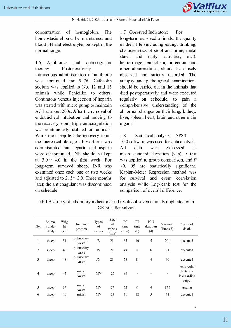
Tab 1 A variety of laboratory indicators a nd results of seven animals implanted with
GK bileaflet valves
No.
Animal
s under
Study
Weig
ht
(kg)
Implant
position
Types
of
valves
Size
of
valves
(mm)
EC
time
(min)
ET
time
(h)
ICU
duration
(d)
Survival
Time (d)
Cause of
death
1 sheep 51 pulmonary
valve AV 21 65 10 5 201 executed
2 sheep 46 pulmonary
valve AV 21 49 8 6 91 executed
3 sheep 48 pulmonary
valve AV 21 58 11 4 40 executed
4 sheep 43 mitral
valve MV 25 80 - - -
ventricular
dilatation,
low cardiac
output
5 sheep 67 mitral
valve MV 27 72 9 4 378 trauma
6 sheep 40 mitral MV 25 51 12 5 41 executed
3
GK Mechanical Heart Valve
12
No.4, Vol. 21, 2005 Journal of General Hospital of Air Force
valve
7 sheep 57 mitral
valve MV 27 83 7 5 31 executed
tota
l -
50.6±
6.8 - - -
65.4±
11.2
9.5±
5.1 4.8±1.3
130.2±7
1.5 -
MV: mitral valve prosthesis; AV: aortic valve prosthesis
2 Results
Seven GK bileaflet valves were
implanted in seven animals (See Table
1). The operation was successfully
performed in six with normal size,
filling level, and opening and closing of
valves in atrium and ventricle, however,
the hyperdistension of heart or errhysis
of incisions made one survive only on
bypass machine. Six survived for over 1
month, in which three over 3 months,
and one even successfully for 378d. The
mean survival time was 130. 2 ±71.5 d
respectively. In the six long-term
survived animals, five were executed at
the end of 1st , 3rd , and 6th month in line
with study plan, respectively, and the
other one died of trauma. Six animals,
successively getting rid of bypass
machine, kept stable hemodynamic with
their blood pressure at 90/60~130/70
mmHg. A small amount of dopamine
(2~5μg•mi n -1•kg -1) was administrated
to two sheep during perioperative period
with their heart rate at 140~150/min in
sinus rhythm. Sinus tachycardia
appeared in No. 1 animal
postoperatively with their heart rate at
140~150/min, and returned to normal
after symptomatic treatment. Blood
routine test, plasma free hemoglobin,
blood biochemistry and blood culture
were regularly examined and all kept in
normal range, except for a low
hemoglobin concentration observed in
No. 1 sheep. The regular blood culture
and prosthetic valve bacterial culture
after execution were negative.
Triple anticoagulant was applied in the
study to maintain an ACT at 200s and
INR at 3.6±0.9 (2.5�6.1), respectively,
when the animals stayed in ICU. After
they moved out, INR was measured
once one or two weeks and kept at
3.1±0.6 (2.3�5.1) with the daily dosage
of warfarin at 9 ~ 21 mg and its mean at
13.9 ± 4.5 mg. No thrombosis,
embolism and bleeding were observed.
The autopsy and pathological
examinations were undertaken in all
long-term survived animals, executed on
schedule or died unexpectedly. The
smooth surface of prosthetic valves was
observed without thrombosis. No
embolism and other abnormal change
were observed in the lung, kidney, liver,
spleen, heart, brain and other organs.
3 Discussion
Chronic animal survival test should be
conducted for the new prosthetic heart
valve before clinical application,
however, there were few studies
documented on the survival test for its
considerable difficulties, long
observation period, various factors
involved and high expenses [1,2].
China-made bileaflet mechanical valve,
though having been developed for many
years, failed to be utilized clinically,
mainly due to the strict national
standards formulated in accordance with
4
Literature and Publitions
13
No.4, Vol. 21, 2005 Journal of General Hospital of Air Force
international standards, in which animal
long-term survival test was a key
rate-limiting step [3]. In the recent two
years, we conducted such an animal test
on GK bileaflet valve, and achieved an
increasing success rate and satisfactory
results via continuous improvement of
test strategies and techniques,
3.1 Experimental standard of prosthetic
mechanical valve: In line with
National Standards in 1990, implantable
prosthetic heart valves include
mechanical and bioprosthetic valve.
Before clinical application, heart valves
should be implanted into at least three
animals, and their survival time over 30
days. Furthermore, according to the
latest standards of FDA (ISO 5840296),
it is required that there are at least three
experimental animals surviving more
than three months, in order to gain a
comprehensive understanding of the
relative characteristics of implanted
prosthetic heart valves [3]. In the present
study, the prosthetic valve replacement
had been conducted in seven animals, in
which six survived over one month,
three over three months, and one even
over one year. In this sense, the survival
time in the study was far longer than that
required by national standard.
3.2 The choice of experimental
animals: Animal model has been
applied to the clinical evaluation of
mechanical heart valve for many years,
but a variety of animal models have their
own their advantages and disadvantages.
Dog is light-weighted and difficult to
manage. Pig is not suitable for long-term
animal model due to the difficulties in
endotracheal intubation and
postoperative management. The rapid
growth of calf will lead to relative
stenosis of the implanted valve several
months after operation, so it is not an
ideal animal model, either. Sheep, with
the similar characteristics of
hemodynamic and laboratory indicators
with man, is often selected as animal
model in valve replacement. General
anesthesia and extracorporeal circulation
are enough for operation, and its
postoperative treatment is relatively
simple, too. Besides, other factors, such
as docile temperament, easy
management, little postoperative
infection, easy long-term feeding and
high long-term survival rate, make sheep
an ideal choice for chronic test of heart
valve replacement.
3.3 Choice of implant position of
prosthetic valve Tricuspid valve was
mostly chosen in the previous long-term
survival animal test with valve
replacement for its easy exposure. There
are rare reports on pulmonary valve
replacement. However, as an implant
position, pulmonary valve possesses
many advantages that makes it an ideal
position worth recommending to
evaluate the long-term survival of
mechanical valves, if the survival of
experimental animal is taken into
account, such as enlarged visual
operative field, easy operation, the
whole replacement under room
temperature and without heart arrest, no
need for cold delivery tube inserted at
the aortic root or occlusion clamp,
simplified surgery steps, shorten
operation time and reduced various
surgical complications. However, as a
pre-clinical experiment of new type
prosthetic valve, the experimental
position should be that most commonly
5
GK Mechanical Heart Valve
14
No.4, Vol. 21, 2005 Journal of General Hospital of Air Force
used in valve replacement. In this sense,
the mitral valve replacement was chosen
in the latest four animals, and No. 25 or
27 mitral valve was selected to match
the weight of animal. All operations
were successfully completed. It
indicated that mitral valve could be
chosen as position for replacement in
accordance with clinical practice.
3.4 Experimental techniques of
prosthetic valve implantation under
extracorporeal circulation: To date, the
animal experiment abroad of valve
replacement were mostly completed via
left atrial appendage or right atrium
under heart arrest [4], in which the
leaflet was removed first, the suture was
separated at the level of valve ring and
there prosthetic valve was implanted.
However, mitral implant in the current
study is completed via left atrial
appendage without occlusion of
ascending aorta and heart arrest, under
the support of extracorporeal circulation.
The strength of this operation type is
that the operation at aortic root could be
left out and the possibility of myocardial
ischemia is avoided. Furthermore, the
opening and closing of valves could be
observed under direct vision after
implantation, to detect whether there is
abnormal paravalvular blood flow,
which could be found as two returning
blood flow jetting out of the bileaflet
valve axis. In the study we also kept
whole mitral valve untouched, sutured
the prosthetic valve above the mitral
valve ring with the needle sewing into
from atrial wall and out from valve ring.
The above suture technique ensured the
successful implantation of the prosthetic
valves with suitable size or type.
3.5 The significance of long-term
surviva l In six long-term survived
animals, five were executed on schedule,
and autopsy indicated the good
properties of opening and closing of
heart valves without paravalvular
leakage, hrombosis, and embolism or
bleeding on main organs. The other died
of trauma 378 days after operation, in
which no complications related to valve
implantation were observed via autopsy.
It is reasonable to consider that GK
bileaflet valve can achieve an ideal
effect of anticoagulation through early,
active postoperative treatment after
implantation in animals with its good
blood compatibility and anti-thrombotic
properties.
Seven GK bileaflet prosthetic valves
was implanted in seven animals in the
present study, in which six survived for
over one month in good conditions,
normal mental state, feeding and
drinking, and activities. No structural
abnormalities and mechanical failure
were observed on all seven GK bileaflet
valves. Anatomical and histological
examination indicated that the prosthetic
valves had smooth surface and no
thrombosis, embolism and other
abnormalities in major organs. In
conclusion, the present study indicated
that China-made GK bileaflet valve had
achieved a satisfactory long-term
survival effect for its stable application
safety, ideal hydrodynamic properties,
good biocompatibility and reliable
durability.
References:
[1] Wang Dongqing, Zhu Langbiao, Liu
Minghui, et al,. The animal study of
6
Literature and Publitions
15
No.4, Vol. 21, 2005 Journal of General Hospital of Air Force
China-made ZDM bileaflet prosthetic
heart valves [J]. Academic Journal of
PLA Postgraduate Medical School.
1996 ,17 (3): 159-161
[4] Irwin E, Lang G, Clack K,et al.
Long-term evaluation of prosthetic
mural valves in sheep [J]. J Invest Surg�993 ,6 (2):133-141.
[2] Dong Aiqiang, Chen Rukun, Zhang
Changming, et al. The animal study of
China-made pyrolytic carbon bileaflet
valve. [J]. Journal of Zhejiang
University (Medical Sciences) ,2000, 29
( 4):162-164.
[5] Ali MI, Kumar SP, Bjornstad K,et al.
The sheep as an animal model for heart
valve research [J〕.Cadiovasc Surg�I 996,
4(4) :543-549.
[6] Salerno CT,Droel J ,Bianco RW.
Current state of in vivo preclinical heart
valve evaluation[J]. J Heart Valve Dis�1998 ,7(2):158-162.
[3] Johnson DM, Sapirstein W. FDA’s
requirements for in-vivo performance
data for prosthetic heart valves[J〕 .J
Heart Valve Dis ,1994 ,3 (4) ,350-355.
7
GK Mechanical Heart Valve
16
No.2, Vol. 29, Feb 2005, Heilongjiang Medical Journal
Comparison of Clinical Effects between Native GK-double –leaflet
Mechanical Heart Valve and Edward
Kang Kai, Jiang Shu-lin, Xie Bao-dong, et al
(the second affiliated Hospital of Harbin Medical University, Harbin 150086, China)
Abstract: Objective To evaluate the clinical effects of native GK-bileaflet
mechanical heart valve. Methods From April 2003 to Augest 2003, 20 patients with
heart valve disease were perfomed on heart valve replacement with native GK
bileaflet mechanical heart valve (group 1). Their Postoperative outcome was
compared with another 20 patients who underwent heart valve replacement with
Edward bileaflet valve (group II). Results a suicide was committed and 2 cases of
leaking around the valve occurred in group 1 7d, 2d, 23d respectively after the
operation; 1 case of leaking around the valve happened 2 months after the operation in
group II. All the three patients came back to health through the second repair surgery,
and the operative courses of the other patients were fee and smoothly. There was no
significant difference between both groups in their ICU stay, hospital stay after
operation and assistant time of hypertension drug (P>0.05 respectively), while the
cost in group I was much lower than that in group II (P< 0.005). There were no
mistakes prosthesis-related occurred in both groups during follow-up period and no
statistically significant difference was observed in the ratio of postoperative relief of
cardiac function in the corresponding period. Conclusion The homemade GK
bileaflet prosthesis could be taken as an ideal alternative for native heart valve for its
safety, reliability, high qual ity and low price in our undeveloped area at present.
Key words: Cardiovascular surgery; heart valve replacement; Mechnical heart valve’
comparison
Beijing Star Medical Devices Co., Ltd is
responsible for the researching,
development and manufacturing of GK
– double – leaflet prosthesis which is the
first generation of native double – leaflet
valves. Our institute was submitted to
perform valve replacement on 20
patients in comparison with 20 patients
in the control group who were
performed valve replacement with
importing double – leaflet prosthesis
(Edward valves) from April, 2003 to
August, 2003 in order to further evaluate
the clinical quality and efficacy of GK –
double – leaflet prosthesis. The trial
report is as follows.
1 Clinical Data
Patients were divided into two groups of
20 patients. Clinically all of the patients
experienced palpitation, chest distress
and short breath after exercises. Patients
with infectious endocarditis were
experiencing or had experienced
long-term febrile diseases. Before
1
Literature and Publitions
17
No.2, Vol. 29, Feb 2005, Heilongjiang Medical Journal
inclusion, we examined all patients for
their medical history, and all of these
patients underwent physical examination,
electrocardiographic examination, chest
X- ray and echocardiogram diagnosis.
1.1 Native valve group (group I): There
were 20 patients in this group, including
8 males and 12 females. The average
age was 47.1±9.94 a with a range from
21 to 60 a. The average weight was
60.3±10.6 kg with a range from 44.5 to
94.0 kg. There was 1 case of grade II
preoperative cardiac function, 17 of
grade III and 2 of grade IV. Preoperative
diagnosis: 15 cases of rheumatic heart
disease including 2 cases of combined
valvular heart disease, 9 cases of left
atrioventricular valve disease, 2 cases of
infectious endocarditis, 2 cases of
degenerative aortic valve disease and 1
case of patent ductus arteriosus and left
atrioventricular valve insufficiency. We
performed double valve replacement on
6 patients ( 2 patients using GK – double
– leaflet valves both and 4 patients using
GK – double – leaflet valves and GK –
single – leaflet valves simultaneously).
12 patients were performed on left
atrioventricular valve replacement and 2
patients underwent aortic valve
replacement. Intraoperative procedures
concluded 2 cases of left atrial
thrombectomy, 1 case of ligation of
patent arterial duct and 5 cases of right
atrioventricular Devega plasty.
1.2 Importing valve group (group II):
There were 20 patients in this group,
including 11 males and 9 females. The
average age was 46.0±12.73 a with a
range from 13 to 62 a. The average
weight was 64.58±13.49 kg with a range
from 43 to 98 kg. There were 18 cases
of grade III preoperative cardiac
function and 2 of grade IV. Preoperative
diagnosis: 13 cases of rheumatic heart
disease including 6 cases of combined
valvular heart disease, 7 cases of left
atrioventricular valve disease, 4 cases of
degenerative aortic valve disease and 3
cases of infectious endocarditis
including 1 case of combined patent
ductus arteriosus. We performed double
valve replacement on 6 patients, left
atrioventricular valve replacement on 10
patients and aortic valve replacement on
4 patients. Importing Edward – double –
leaflet valves were applied on all of the
patients in this group. Intraoperative
procedures included 2 cases of left atrial
thrombectomy, 1 case of patent arterial
duct , 3 cases of right atrioventricular
Devega plasty and 1 case of left atrial
volume reduction surgery.
2 Operation procedures
Both group underwent conventional
cardiopulmonary bypass cannulating
under general anesthesia, and
anterograde perfusion with cold blood or
modified Thomas cardioplegia ( once for
single valve replacement and twice for
double valve replacement). Continuous
suture was applied in both left
atrioventricular and aortic replacement.
Mural thrombectomy, right
atrioventricular valve Devega plasty
ligation of patent arterial duct or left
atrial volume reduction surgery were
performed on part of the patients
simultaneously. The average blocking
time was (36.4±9.08) min and
(3.83±12.40) min for the two groups
respectively. The average connecting
time was (65.45±10.46) min and
(67.65±15.64) min for the two groups
2
GK Mechanical Heart Valve
18
No.2, Vol. 29, Feb 2005, Heilongjiang Medical Journal
respectively.
3 Statistical methods
All values were designated as ±s. T
test and 2 te swt er uese di n
comparison of measurement data and
enumeration data respectively.
4 Results
No intraoperative death occurred in both
group. 1 case of suicide was committed
in group I after oral application of
valium. 2 cases of leaking around the
valve occurred in group I 2d and 23d
after the operation, respectively. 1 case
of leaking around the valve occurred in
group II in an car accident two months
after the operation. These three patients
recovered from leaking around the valve
after reoperation and were then
discharged.
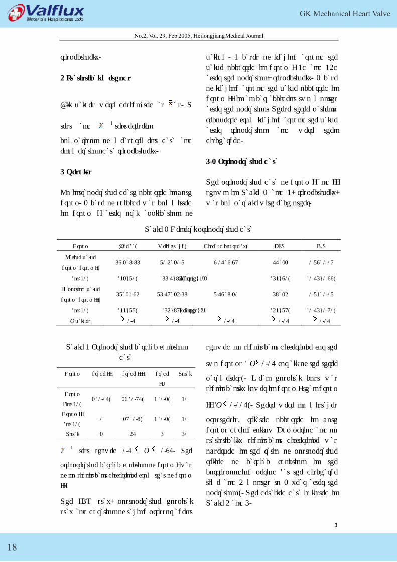
4.1 Preoperative data
The preoperative data of group I and II
shown in Table 1 and 2, respectively,
was comparable with each other.
Table 1 General preoperative data
Group Age (a) Weight (kg) Disease course (y) EF% C/T
Native valve
group (group I) 47.1±9.94 60.3±10.6 7.05±7.78 55±11 0.67±0.08
(n=20) (21~60) (44.5~94) ymeaornst)h ~2(11 (42~70) (0.54~0.77)
Importing valve
group (group II) 46±12.73 64.58±13.49 6.57±9.10 49±13 0.62±0.06
(n=20) (22~66) (43~98) myeoanrtsh) s~3(20 (32~68) (0.54~0.80)
P values 0.5 0.5 0.05 0.05 0.05
Table 2 Preoperative cardiac function
data
Group grade II grade III grade
IV
Total
Group
I(n=20) 1 (0.05) 17 (0.85) 2 (0.1) 20
Group II
(n=20) 0 18 (0.9) 2 (0.1) 20
Total 1 35 4 40
2 test showed 0.5 P 0.75. The
preoprerative cardiac function of group I was
of no significant difference from that of group
II.
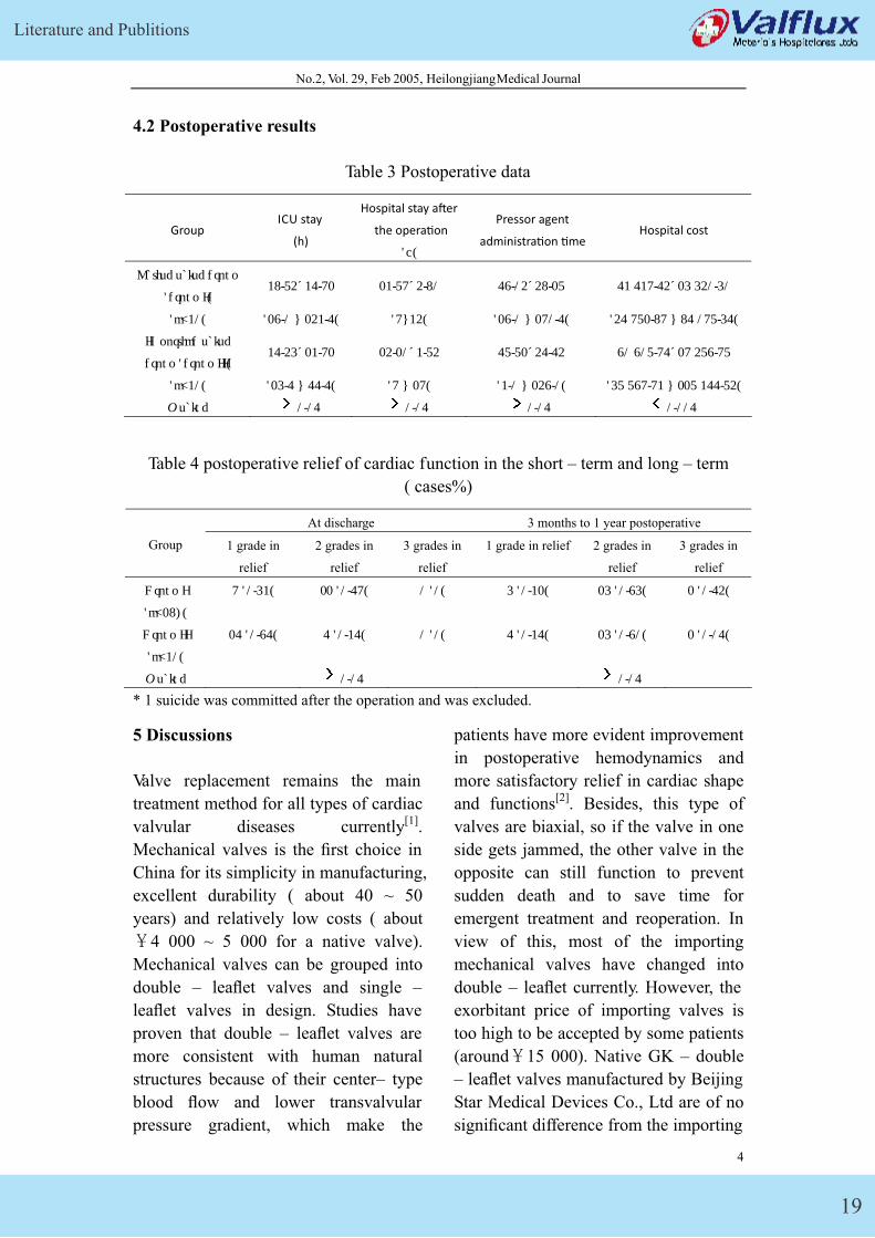
The ICU stay, postoperative hospital
stay and duration of taking pressor agent
showed no significant difference for the
two groups ( P 0.05 for all of the three
parameters). Mean hospital cost was
significantly lower in group I than group
II (P 0.005). There were no mistakes
prosthesis- related occurred in both
groups during follow – up period and no
statistically significant difference was
observed in the ratio of postoperative
relief of cardiac function in the
corresponding period (at the discharge
time and 3 months to 1 year after the
operation ). The detailed data is listed in
Table 3 and 4.
3
Literature and Publitions
19
No.2, Vol. 29, Feb 2005, Heilongjiang Medical Journal
4.2 Postoperative results
Table 3 Postoperative data
Group ICU stay
(h)
Hospital stay a�er
the opera�on
(d)
Pressor agent
administra�on �me Hospital cost
Native valve group
(group I) 29.63±25.81 12.68±3.90 57.03±39.16 52 528.53±14 430.40
(n=20) (17.0 ~ 132.5) (8~23) (17.0 ~ 180.5) (35 861.98 ~ 95 086.45)
Importing valve
group (group II) 25.34±12.81 13.10±2.63 56.61±35.53 70 706.85±18 367.86
(n=20) (14.5 ~ 55.5) (8 ~ 18) (2.0 ~ 137.0) (46 678.82 ~ 116 255.63)
P value 0.05 0.05 0.05 0.005
Table 4 postoperative relief of cardiac function in the short – term and long – term
( cases%)
At discharge 3 months to 1 year postoperative
Group 1 grade in
relief
2 grades in
relief
3 grades in
relief
1 grade in relief 2 grades in
relief
3 grades in
relief
Group I
(n=19*)
8 (0.42) 11 (0.58) 0 (0) 4 (0.21) 14 (0.74) 1 (0.53)
Group II
(n=20)
15 (0.75) 5 (0.25) 0 (0) 5 (0.25) 14 (0.70) 1 (0.05)
P value 0.05 0.05
* 1 suicide was committed after the operation and was excluded.
5 Discussions
Valve replacement remains the main
treatment method for all types of cardiac
valvular diseases currently[1].
Mechanical valves is the first choice in
China for its simplicity in manufacturing,
excellent durability ( about 40 ~ 50
years) and relatively low costs ( about
�4 000 ~ 5 000 for a native valve).
Mechanical valves can be grouped into
double – leaflet valves and single –
leaflet valves in design. Studies have
proven that double – leaflet valves are
more consistent with human natural
structures because of their center– type
blood flow and lower transvalvular
pressure gradient, which make the
patients have more evident improvement
in postoperative hemodynamics and
more satisfactory relief in cardiac shape
and functions[2]. Besides, this type of
valves are biaxial, so if the valve in one
side gets jammed, the other valve in the
opposite can still function to prevent
sudden death and to save time for
emergent treatment and reoperation. In
view of this, most of the importing
mechanical valves have changed into
double – leaflet currently. However, the
exorbitant price of importing valves is
too high to be accepted by some patients
(around�15 000). Native GK – double
– leaflet valves manufactured by Beijing
Star Medical Devices Co., Ltd are of no
significant difference from the importing
4
GK Mechanical Heart Valve
20
No.2, Vol. 29, Feb 2005, Heilongjiang Medical Journal
valves in failure rate, short – term and
long – term therapeutic effect, while the
hospital cost is significantly lower in
native valves. Given the domestic
economic conditions at present, the
homemade GK – double – leaflet
prosthesis could be taken as an ideal
alternative for its safety, reliability, high
quality and low price.
References:
[1] Guoqi Qi, Xiaodong Zhu, Shengshou
Hu, et al. Long-term follow up of
Chinese with mechanical prosthetic
heart valve replacement and the status
quo of anti-coagulation treatment [J].
Chinese Journal of Thoracic and
Cardiovascular Surgery, 2004, 6,
20(3):145~147.
[2] Baoren Zhang, Jialin Zhu. Artificial
cardiac valves and valve replacement
[M]. Second edition. Beijing: People's
Medical Publishing House, 1999.
321~324.
(Editor: Xuezhen Liu)
(Accepted date: 2004 – 12 – 07)
5
Literature and Publitions
21
Acad emic Journal of General Hospital of Air Force, No.2, Vol.21, 2005
Primary clinical application of
new (GK) bileaflet mechanical heart valve
ZHONG Jing, WAN Shi-jie, WANG Wei-xin, et al
(Department of Caridovascular Surgery, General Hospital of Air Force, Beijing 10036, China)
Abstract: Objective to introduce a new type of bileaflet mechnical prosthetic heart valve( GK
bileaflet valve) and evaluate clinically the early hemodynamic effect and shor term follow up after
its replacement. Methods 20 patients were operated upon with a mean age of 44.5±10.74 years.
85 percents(17/20) had NYHA class III and IV hear function. The mitral valve replacement was
performed in 14 patients, aortic valve replacement in 4 patients and double valve replacement in 2
patients. Follow-up is 100% and extended 1 to 2.5 years. Result There was no any early or late
mortality. Without valve-related complications all patients have lived for more than 1 to 2.5 years.
Conclusion Early clinical results and short-term followup demonstrate that GK bileaflet
prosthetic heart valve exhibits excellent hemodynamic properties, satisfied blood consistency and
a low incidence of valve-related complications.
Key words: heart valve prosthesis; heart valve diseases/surgery
GK bileaflet mechanical heart valve
(GK bileaf valve in abbreviation below)
is a new bileaflet mechanical heart valve,
which is developed by Air Force
General Hospital and Beijing Star
Medical Devices Co., Ltd together. It
was applied clinically from Sep 2002
after the completion of eatracorporeal
tests and animal experiments, and 20
bileaflet valves were applied in 22 heart
valve replacement surgeries during one
and a half years. The conditions of
clinical application and recent follow-up
results are summarized as followings.
1 Materials and methods
1.1 Mechanical heart valves GK
bileaflet valve is a kind of mechanical
valves which are low in valve support
with three-channel central flow and
bileaflet. Graphite acts as the base of
valve support and valve (leaf), and
surface is covered by pyrolytic carbon.
Opening angle of leaflet (in
one-direction) is 850, a sella peak
structure which owns a pair of processes
at inflow side is adopted in appearance,
ball-socket design is applied into
joint-twisting structure in valve hub, and
sewing cuff is filament fabric.
1.2 Patient data Among 20
patients, 9 were male and 11 female.
Age ranged from 31 to 60 years old
(44.5±10.74 years old in average),
weight from 43 to 83kg �57±12.4 in
average�, and history from 0.6 to 36
years, 8.78 years in average. Symptoms
of palpitation, brachypnea, dyspnea, and
so on were clinically available after
activity, and coronary diseases were
excluded by pre-operative examination,
ECG, chest film, echocardiogrpahy, and
alike. Pre-operative diagnosis: 50 cases
for rheumatic heart disease, among
1
GK Mechanical Heart Valve
22
Acad emic Journal of General Hospital of Air Force, No.2, Vol.21, 2005
which 18 cases for joint valvular disease,
2 for mitral disease, 13 for restriction
after closed mitral commissurotomy, 1
for degenerative disease of aortic valve,
and 1 for infective endocarditis. 7 cases
were accompanied by chronic atrial
fibrillation, with 4 cases for left atrium
thrombosis. 3 cases for cardiac function
of level II, 13 cases for cardiac
function of level III and 4 cases for that
of level IV.
1.3 Surgical methods: Surgeries
were all conducted on patients under
moderate hypothermic general
anesthesia and extracorporeal circulation,
with application of antegrade cold blood
or cold crystal cardioplegia for sole
valve replacement and that of both
antergrade and retrograde cold blood
cardiogplegia for bileaflet replacement.
Continuous suture along right
atrium-interventricular septum routine
was adopted for mitral valve
replacement; Interrupted mattress suture
was mostly placed by 2-0 Ethicon
stitches with shims for aortic valve
replacement, and 3-stitch Prolene
continuous suture was applied in few
patients. Partial patients were also
conducted on by left atrium wall
mechanical thrombectomy, tricuspid De
Vega plastic surgery or left atrium
volume-reduction. Warfarin was initially
administrated orally 48hs after operation,
to regulate prothrombin time (PT) to 1.5
times of contrast value, and international
standardized value�INR�was 1.5-2.5
(when international sensitive index was
1.2).
1.4 Hemodynamic and
hemocompatibility observing parameters
Such index was observed dynamically as
routine blood test, routine urine test,
liver function and changing process of
free serum hemoglobulin. Those
parameters were examined, like
cardiothorax rate (chest film),
measurement of all atriums and
ventricles, effective area of mechanical
valve opening, flow velocity at valve
opening, transvalvular pressure gradient
(echocardiography) and so on during
follow-up.
1.5 Follow-up Patients were
followed half a year and one year later
respectively, and follow-up ways were
inclusive of letter, call, outpatient visit
and so on. Cardiac function grading,
valve-related complications and alike
were judged according to activity,
administration, examination results and
so on, referred to American
Cardiothorax Association Standard
which was set up in 1996 by Edmunds
and others [2].
1.6 Statistic analysis SPSS 10.0
software was applied, data was
expressed in χ ±S, t-test was conducted
on intergroup contrast, and it is
significant statistically when P<0.05.
Kaplan-Meier regression was applied
into correlation analysis of survival rate
and incident, with Log-Rank test to
compare population difference.
2 Results
2.1 Early results Among 20 cases in
this group, 14 cases receiving mitral
valve replacement, 4 receiving valve
replacement, 2 receiving both mitral and
aortic valve replacement, 4 cases also
receiving left atrium thrombectomy and
2
Literature and Publitions
23
Acad emic Journal of General Hospital of Air Force, No.2, Vol.21, 2005
5 also receiving tricuspidoplasty.
Blocking time for sole valve
replacement was 29-112� 64.75±27.5�minutes with CPB time of 46-163
� 96.35±38.52 � minutes. That for
bivalve replacement was
67-146(78.66±22.36�minutes with CPB
time of 85-176(106.16±23.29�minutes.
Total number of GK bileaflet implanted
was 22, among which there were 16
mitral valves (25M 5、27M 10、29M 1),
and 6 aortic valves (21M 2、23M 4 ). No
death was existent in through group
(within 30 days). Tubes intubated in
patients were all pulled out within 24
hours after operation, and their
consciousness was clear with stability of
hemodynamics and absence of severe
arrhythmia. Mild low cardiac output
syndrome occurred on 2 cases, and
another open heart surgery was
conducted on one case due to poststernal
blood oozing at 6 hour after the
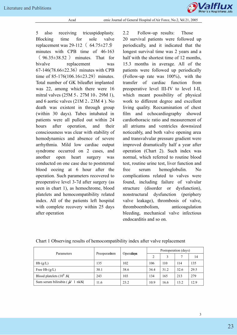
operation. Such parameters recovered to
preoperative level 3-7d after surgery (as
seen in chart 1), as hemochrome, blood
platelets and hemocompatibility related
index. All of the patients left hospital
with complete recovery within 25 days
after operation
2.2 Follow-up results: Those
20 survival patients were followed up
periodically, and it indicated that the
longest survival time was 2 years and a
half with the shortest time of 12 months,
15.3 months in average. All of the
patients were followed up periodically
(Follow-up rate was 100%)�with the
transfer of cardiac function from
preoperative level III-IV to level I-II,
which meant possibility of physical
work to different degree and excellent
living quality. Reexamination of chest
film and echocardiography showed
cardiothoracic ratio and measurement of
all atriums and ventricles downsized
noticeably, and both valve opening area
and transvalvular pressure gradient were
improved dramatically half a year after
operation (Chart 2). Such index was
normal, which referred to routine blood
test, routine urine test, liver function and
free serum hemoglobulin. No
complications related to valves were
found, including failure of valvular
structure (disorder or dysfunction),
nonstructural dysfunction (periphery
valve leakage), thrombosis of valve,
thromboembolism, anticoagulation
bleeding, mechanical valve infectious
endocarditis and so on.
Chart 1 Observing results of hemocompatibility index after valve replacement
Postoperation (days) Parameters Preoperation Operadtiaoyns
2 3 7 14
Hb (g/L) 135 102 106 110 114 135
Free Hb (g/L) 30.1 38.6 34.4 31.2 32.6 29.5
Blood platelets (109 /L) 243 103 134 165 213 279
Sum serum bilirubin ( μ mol/L) 11.6 23.2 10.9 16.6 13.2 12.9
3
GK Mechanical Heart Valve
24
Acad emic Journal of General Hospital of Air Force, No.2, Vol.21, 2005
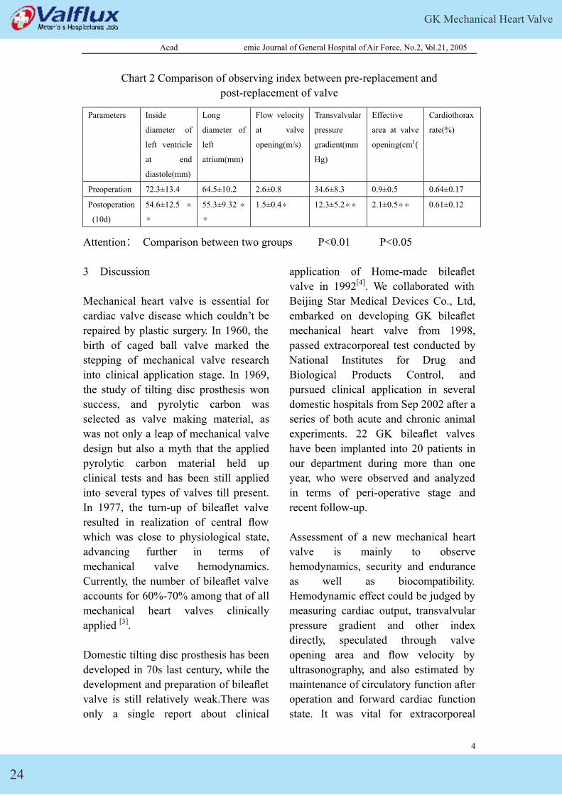
Chart 2 Comparison of observing index between pre-replacement and
post-replacement of valve
Parameters Inside
diameter of
left ventricle
at end
diastole(mm)
Long
diameter of
left
atrium(mm)
Flow velocity
at valve
opening(m/s)
Transvalvular
pressure
gradient(mm
Hg)
Effective
area at valve
opening(cm2)
Cardiothorax
rate(%)
Preoperation 72.3±13.4 64.5±10.2 2.6±0.8 34.6±8.3 0.9±0.5 0.64±0.17
Postoperation
(10d)
54.6±12.5 �
�
55.3±9.32 �
�
1.5±0.4� 12.3±5.2�� 2.1±0.5�� 0.61±0.12
Attention� Comparison between two groups P<0.01 P<0.05
3 Discussion
Mechanical heart valve is essential for
cardiac valve disease which couldn’t be
repaired by plastic surgery. In 1960, the
birth of caged ball valve marked the
stepping of mechanical valve research
into clinical application stage. In 1969,
the study of tilting disc prosthesis won
success, and pyrolytic carbon was
selected as valve making material, as
was not only a leap of mechanical valve
design but also a myth that the applied
pyrolytic carbon material held up
clinical tests and has been still applied
into several types of valves till present.
In 1977, the turn-up of bileaflet valve
resulted in realization of central flow
which was close to physiological state,
advancing further in terms of
mechanical valve hemodynamics.
Currently, the number of bileaflet valve
accounts for 60%-70% among that of all
mechanical heart valves clinically
applied [3].
Domestic tilting disc prosthesis has been
developed in 70s last century, while the
development and preparation of bileaflet
valve is still relatively weak.There was
only a single report about clinical
application of Home-made bileaflet
valve in 1992[4]. We collaborated with
Beijing Star Medical Devices Co., Ltd,
embarked on developing GK bileaflet
mechanical heart valve from 1998,
passed extracorporeal test conducted by
National Institutes for Drug and
Biological Products Control, and
pursued clinical application in several
domestic hospitals from Sep 2002 after a
series of both acute and chronic animal
experiments. 22 GK bileaflet valves
have been implanted into 20 patients in
our department during more than one
year, who were observed and analyzed
in terms of peri-operative stage and
recent follow-up.
Assessment of a new mechanical heart
valve is mainly to observe
hemodynamics, security and endurance
as well as biocompatibility.
Hemodynamic effect could be judged by
measuring cardiac output, transvalvular
pressure gradient and other index
directly, speculated through valve
opening area and flow velocity by
ultrasonography, and also estimated by
maintenance of circulatory function after
operation and forward cardiac function
state. It was vital for extracorporeal
4
Literature and Publitions
25
Acad emic Journal of General Hospital of Air Force, No.2, Vol.21, 2005
accelerated fatigue test to reckon
endurance. Clinical observation of
valve-related incidence not only
indicated whether it was endurable or
not but also reflected its safety generally.
Such index was rather significant for
assessment of mechanical valve function,
as evaluation of biocompatibility after
valve implantation and clinical
observation of thrombosis, embolism,
hemolysis, as well as hemolytic anemia.
Three aspects above were observed
systematically and analyzed generally
during primary clinical application. It
was observed during surgery that surface
of valve was smooth, both opening and
closing of valve were flexible, twisting
was free, conforming to basic
requirements of clinical application.
Patients in this group broke away from
extracorporeal circulation successfully,
and circulatory system was stable after
operation without severe low cardiac
output syndrome�passing peri-operative
stage safely. Cardiac function was all
improved dramatically, as shown in the
follow-up of 1-2.5 years later after
operation, indicating excellent
hemodynamic effect was present for 89
bileaflet valves in patients. All of such
index backed to normal range within one
week, which referred to hemochrome,
blood platelets, bilirubin, free serum
hemoglobulin. No complications were
available, like thrombosis and embolism,
even under anticoagulation with low
intensity, as demonstrated that
short-term hemocompatibilty was good
and forward effect remained to be seen.
No complications correlated to valves
were present after relatively close
follow-up. Although it was still years
away for assessment of valve endurance,
observation of infection and periphery
valve leakage lasted for over one year
for the whole group without noticeable
hemolysis, thrombosis and embolism,
and complications concerning
anticoagulation. Reexamination of
ultracardiography indicated that such
parameters were all among normal range,
as maximum flow velocity at valve
opening, transvalvular pressure gradient,
effective area of valve opening and so
on, showing there was no significant
difference between this kind of valves
and other imported bileaflet valves
which were usually applied clinically.
4 Summa ry
This type of heart valve was excellent in
function, stable in hemodynamics, easy
in suture, low in valve noise, reliable in
function, through clinical application of
22 GK bileaflet valves into 20 cases and
short-term follow-up observation after
operation. No death was existent in this
group both recently and forwards
without valve-related complications
during follow-up, inclusive of valve
structure failure, valve thrombosis,
embolism, bleeding, mechanical valve
infectious endocarditis and so on. Given
that the number of surgery cases was not
large and follow-up time was not long,
further follow-up and observation were
still essential for judgment of forward
effect.
References:
[1] Yuo Tang, Shengshou Hu, Liang
Meng, and so on. Chronic experimental
study on Home-made bileaflet mitral
valve implanted into sheep�J�.Chinese
5
GK Mechanical Heart Valve
26
Acad emic Journal of General Hospital of Air Force, No.2, Vol.21, 2005
Circulation Journa , 2003,18(4):303
-305
[2] Edmunds LH, Clark RE, Cohn LH ,
et al. Guidelines for morbidity and
mortality after cardiac valvular
operation [J]. J Thorac Cardiovasc Surg,
1996, 112(3): 708-711.
[3] Weiyong Liu, Status quo and
development trend of
cardiomeningology � J � Chinese
Journal of Clinical Thoraxic Surgery,
1999� 6 �2��65-66
[4�Zipu Tian, Chuanxing Luo, Xuzhong
Huang, and others. Development of
bileaflet mechnical valve and primary
report of its clinical application [J].
Chinese Journal of Clinical Thoraxic
and Cardiovascular Surgery �1992,
8(1):1
[5] Vitale N, Cappabianca G, Visicchio G,
et al. Midtem evaluation of the Sorin
Bicarbon heart valve prosthesis:
single-center experience[J]. Ann Thorac
Surg, 2004, 77(2):527-531.
[6]Wu Y, Gregorio R, Renzulli A, et al.
Mechanical heart valves: are two leaflets
better than one?[J]. J Thorac Cardiovasc
Surg, 2004, 127(4):1171-1179.
6
Literature and Publitions
27
China J Clinical Thorac Cardiovascular Surgery, No.5, Vol.12, October 2005 1
Clinical Application of New (GK) Bileaflet Mechanical Heart Valve
Zhong Jing, Yi Ding-hua, Jiang Shulin, Li Tong, Han Zhen, Wan Shi-jie
(1. Department of Cardiovascular Surgery, General Hospital of Air Force, Beijing 10036, China; 2.
Department of Cardiovascular Surgery, Xijing Hospital, the Fourth Military Medical University,
Xi’an 710032, China; 3. Department of Cardiovascular Surgery, the second Clinical College,
Harbin Medical University, Harbin 150086, China)
Abstract: Objective To introduce a new type of bileaflet mechnical prosthetic
heart valve (GK bileaflet valve) and evaluate clinically the early hemodynamic effect
and short term follow-up after its replacement. Methods Sixty-one patients with heart
valve diseases were operated upon. The mitral valve replacement was performed in 34
patients, aortic valve replacement in 16 patients and double valve replacement in 11
patients. A total of 72 GK bileaflet mechanical valves were implanted, 45 in mitral
position, and 27 in aortic positions. Blood consistency and hemodynamics were
monitored. Follow-up was carried out routinely to check whether there were some
valve-related complications. Results There was no early mortality(<30d). Only
one patient died of trauma 2 months after the operation. Follow-up was 100% and
extended 1 to 2.5 years. Without valve-related complications all patients have lived
for more than 1 to 2.5 years. In 98% (60/61) of survivors heart functional performance
have improved to New York Hear t Association class I or II. Conclusion Early
clinical results and short-term follow-up demonstrate that GK bileaflet prosthetic
heart valve exhibits excellent hemodynamic properties, satisfied blood consistency
and a low incidence of valve-related complications. Midterm and long-term results
should be observed further.
Key words: Heart valve diseases; Prosthetic hear valve; Implantation of heart valve
prosthesis
GK bileaflet mechanical heart valve
(GK bileaf valve in abbreviation below)
is a new bileaflet mechanical heart valve,
which is developed by Air Force
General Hospital and Beijing Star
Medical Devices Co., Ltd together. It
was applied clinically from Sep 2002
after the completion of eatracorporeal
tests and animal experiments, and 61
bileaflet valves were applied in 72 heart
valve replacement surgeries during one
and a half years. The conditions of
clinical application and recent follow-up
results are summarized as followings.
1 Materials and methods
GK Mechanical Heart Valve
28
China J Clinical Thorac Cardiovascular Surgery, No.5, Vol.12, October 2005 2
1.1 Mechanical heart valves
GK bileaflet valve is a kind of
mechanical valves which are low in
valve support with three-channel central
flow and bileaflet. Graphite acts as the
base of valve support and valve (leaf),
and surface is covered by pyrolytic
carbon. Opening angle of leaflet (in
one-direction) is 850, a sella peak
structure which owns a pair of processes
at inflow side is adopted in appearance,
ball-socket design is applied into
joint-twisting structure in valve hub, and
sewing cuff is filament fabric.
1.2 Clinical data
61 cases of valve replacement were
completed by three hospitals (21 by
Second Clinical Institute of Harbin
Medical University, 20 by Xijing
Hospital affiliated to The Fourth
Military Medical University, 20 by Air
Force General Hospital), among which
29 were male and 32 female. Age ranged
from 18 to 60 years old (41.5±10.74
years old on average), weight from
38.5 to 90.4 kg � 54.2± 12.4 on
average�, and history from 0.6 to 36.0
years, 8.8 years on average. Symptoms
of palpitation, brachypnea, dyspnea, and
so on were clinically available for all
cases after activity, whose diagnosis was
proved by physical examination, ECG,
chest x-ray, echocardiography and alike
prior to operation. Coronary
angiography served cases over 50 years
old to exclude coronary disease.
Pre-operative diagnosis: rheumatic heart
disease in 47, among which joint
valvular disease was in 11, mitral
disease in 34, restriction after closed
mitral commissurotomy in 1, bio-valve
degeneration in 1, degenerative disease
of aortic valve in 5, and infective
endocarditis in 6. 23 cases were
accompanied by chronic atrial
fibrillation, with left atrium thrombosis
in 8, cardiac function of level II in 9,
cardiac function of level III in 40 and
cardiac function of level IV in 12.
1.3 Surgical methods
Surgeries were all conducted on patients
under moderate hypothermic general
anesthesia and extracorporeal circulation,
with application of antegrade cold blood
or cold crystal cardioplegia for sole
valve replacement and that of both
antergrade and retrograde cold blood
cardiogplegia for bileaflet replacement.
Continuous suture along right
atrium-interventricular septum routine
was adopted for mitral valve
replacement; Interrupted mattress suture
was mostly placed by 2-0 Ethicon
stitches with shims for aortic valve
replacement, and 3-stitch Prolene
continuous suture was applied in few
patients. Partial patients were also
conducted on by left atrium wall
mechanical thrombectomy, tricuspid De
Vega plastic surgery or left atrium
volume-reduction. Warfarin was initially
administrated orally 48hs after operation,
to regulate prothrombin time (PT) to 1.5
times of contrast value with international
standardized value� INR�of 1.5-2.5
(when international sensitive index was
1.2).
1.4 Hemodynamic and
hemocompatibility observing parameters
Such index was observed dynamically as
routine blood test, routine urine test,
Literature and Publitions
29
China J Clinical Thorac Cardiovascular Surgery, No.5, Vol.12, October 2005 3
liver function and changing process of
free serum hemoglobulin within 2 weeks
after operation.. Those parameters were
examined during follow-up, like
cardiothorax rate by chest X-ray,
measurement of all atriums and
ventricles by echocardiography,
effective area of mechanical valve
opening, flow velocity at valve opening,
transvalvular pressure gradient
(echocardiography) and so on.
1.5 Follow-up
Patients were followed half a year and
one year later respectively, and
follow-up ways were inclusive of letter,
call, outpatient visit and so on. Cardiac
function grading, valve-related
complications and alike were judged
according to activity, administration,
examination results and so on, referred
to American Cardiothorax Association
Standard which was set up in 1996 by
Edmunds and others [2].
1.6 Statistic analysis
Data was expressed in χ ±S, t-test was
conducted on intergroup contrast, and it
was significant statistically when P<0.05.
Kaplan-Meier regression was applied
into correlation analysis of survival rate
and incident, with Log-Rank test to
compare population difference. SPSS
10.0 software was applied for statistics.
2 Results
2.1 Early results
Among 61 cases in this group, 34 cases
receiving mitral valve replacement, 16
receiving valve replacement, 11
receiving both mitral and aortic valve
replacement, 4 cases also receiving left
atrium thrombectomy and 5 also
receiving tricuspidoplasty. Blocking
time for sole valve replacement was
29-112� 64.75±27.5) min with CPB
time of 46-163�96.35±38.52� minutes.
That for bivalve replacement was
67-146(78.66±22.36� min with CPB
time of 85-176(106.16±23.29� min.
Total number of GK bileaflet implanted
was 72, among which there were 45
mitral valves (25M 8, 27M 24, 29M 3),
and 27 aortic valves (21M 11, 23M
5 ,25A 1 ). No death was existent in
through group (within 30 days). Tubes
intubated in patients were all pulled out
within 24 hours after operation, and their
consciousness was clear with stability of
hemodynamics and absence of severe
arrhythmia. Mild low cardiac output
syndrome occurred on 5 cases, and 2
cases received surgeries again on 2nd
and 21st day after operation respectively,
due to periphery valve leakage. Another
open heart surgery was conducted on
one case due to poststernal blood oozing
at 6th hour after operation. All of the
patients left hospital with complete
recovery within 25 days after operation.
Hemocompatibility related index of 61
cases backed to normal range during 3-7
days after operation.
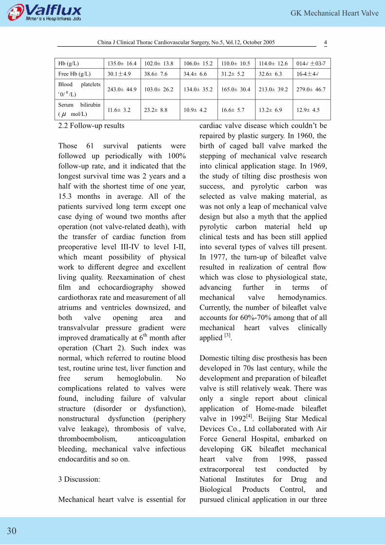
Chart 1 Observing results of hemocompatibility index of 61 cases after valve
replacement Postoperation (days) Parameters Preoperation Operation
days 2d 3d 7d 14d
GK Mechanical Heart Valve
30
China J Clinical Thorac Cardiovascular Surgery, No.5, Vol.12, October 2005 4
Hb (g/L) 135.0± 16.4 102.0± 13.8 106.0± 15.2 110.0± 10.5 114.0± 12.6 125.0±14.8
Free Hb (g/L) 30.1±4.9 38.6± 7.6 34.4± 6.6 31.2± 5.2 32.6± 6.3 27.5±5.0
Blood platelets
(109 /L) 243.0± 44.9 103.0± 26.2 134.0± 35.2 165.0± 30.4 213.0± 39.2 279.0± 46.7
Serum bilirubin
( μ mol/L) 11.6± 3.2 23.2± 8.8 10.9± 4.2 16.6± 5.7 13.2± 6.9 12.9± 4.5
2.2 Follow-up results
Those 61 survival patients were
followed up periodically with 100%
follow-up rate, and it indicated that the
longest survival time was 2 years and a
half with the shortest time of one year,
15.3 months in average. All of the
patients survived long term except one
case dying of wound two months after
operation (not valve-related death), with
the transfer of cardiac function from
preoperative level III-IV to level I-II,
which meant possibility of physical
work to different degree and excellent
living quality. Reexamination of chest
film and echocardiography showed
cardiothorax rate and measurement of all
atriums and ventricles downsized, and
both valve opening area and
transvalvular pressure gradient were
improved dramatically at 6th month after
operation (Chart 2). Such index was
normal, which referred to routine blood
test, routine urine test, liver function and
free serum hemoglobulin. No
complications related to valves were
found, including failure of valvular
structure (disorder or dysfunction),
nonstructural dysfunction (periphery
valve leakage), thrombosis of valve,
thromboembolism, anticoagulation
bleeding, mechanical valve infectious
endocarditis and so on.
3 Discussion:
Mechanical heart valve is essential for
cardiac valve disease which couldn’t be
repaired by plastic surgery. In 1960, the
birth of caged ball valve marked the
stepping of mechanical valve research
into clinical application stage. In 1969,
the study of tilting disc prosthesis won
success, and pyrolytic carbon was
selected as valve making material, as
was not only a leap of mechanical valve
design but also a myth that the applied
pyrolytic carbon material held up
clinical tests and has been still applied
into several types of valves till present.
In 1977, the turn-up of bileaflet valve
resulted in realization of central flow
which was close to physiological state,
advancing further in terms of
mechanical valve hemodynamics.
Currently, the number of bileaflet valve
accounts for 60%-70% among that of all
mechanical heart valves clinically
applied [3].
Domestic tilting disc prosthesis has been
developed in 70s last century, while the
development and preparation of bileaflet
valve is still relatively weak. There was
only a single report about clinical
application of Home-made bileaflet
valve in 1992[4]. Beijing Star Medical
Devices Co., Ltd collaborated with Air
Force General Hospital, embarked on
developing GK bileaflet mechanical
heart valve from 1998, passed
extracorporeal test conducted by
National Institutes for Drug and
Biological Products Control, and
pursued clinical application in our three
Literature and Publitions
31
China J Clinical Thorac Cardiovascular Surgery, No.5, Vol.12, October 2005 5
hospitals from Sep 2002 after a series of
both acute and chronic animal
experiments. 72 GK bileaflet valves
have been implanted into 61 patients in
our department during more than one
year, who were observed and analyzed
in terms of peri-operative stage and
recent follow-up.
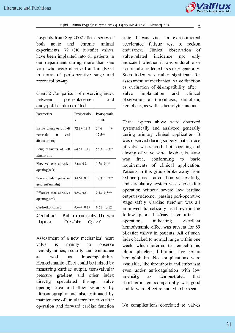
Chart 2 Comparison of observing index
between pre-replacement and
post-replacement of valve
Parameters Preoperatio
n
Postoperatio
n 10d
Inside diameter of left
ventricle at end
diastole(mm)
72.3± 13.4 54.6 ±
12.5**
Long diameter of left
atrium(mm)
64.5± 10.2 55.3± 9.3**
Flow velocity at valve
opening(m/s)
2.6± 0.8 1.5± 0.4*
Transvalvular pressure
gradient(mmHg)
34.6± 8.3 12.3± 5.2**
Effective area at valve
opening(cm2)
0.9± 0.5 2.1± 0.5**
Cardiothorax rate 0.64± 0.17 0.61± 0.12
Attention� Comparison between two
groups P<0.05, P<0.01
Assessment of a new mechanical heart
valve is mainly to observe
hemodynamics, security and endurance
as well as biocompatibility.
Hemodynamic effect could be judged by
measuring cardiac output, transvalvular
pressure gradient and other index
directly, speculated through valve
opening area and flow velocity by
ultrasonography, and also estimated by
maintenance of circulatory function after
operation and forward cardiac function
state. It was vital for extracorporeal
accelerated fatigue test to reckon
endurance. Clinical observation of
valve-related incidence not only
indicated whether it was endurable or
not but also reflected its safety generally.
Such index was rather significant for
assessment of mechanical valve function,
as evaluation of biocompatibility after
valve implantation and clinical
observation of thrombosis, embolism,
hemolysis, as well as hemolytic anemia.
Three aspects above were observed
systematically and analyzed generally
during primary clinical application. It
was observed during surgery that surface
of valve was smooth, both opening and
closing of valve were flexible, twisting
was free, conforming to basic
requirements of clinical application.
Patients in this group broke away from
extracorporeal circulation successfully,
and circulatory system was stable after
operation without severe low cardiac
output syndrome�passing peri-operative
stage safely. Cardiac function was all
improved dramatically, as shown in the
follow-up of 1-2.5 years later after
operation, indicating excellent
hemodynamic effect was present for 89
bileaflet valves in patients. All of such
index backed to normal range within one
week, which referred to hemochrome,
blood platelets, bilirubin, free serum
hemoglobulin. No complications were
available, like thrombosis and embolism,
even under anticoagulation with low
intensity, as demonstrated that
short-term hemocompatibilty was good
and forward effect remained to be seen.
No complications correlated to valves
GK Mechanical Heart Valve
32
China J Clinical Thorac Cardiovascular Surgery, No.5, Vol.12, October 2005 6
were present after relatively close
follow-up. Although it was still years
away for assessment of valve endurance,
observation of infection and periphery
valve leakage lasted for over one year
for the whole group without noticeable
hemolysis, thrombosis and embolism,
and complications concerning
anticoagulation. Reexamination of
ultracardiography indicated that such
parameters were all among normal range,
as maximum flow velocity at valve
opening, transvalvular pressure gradient,
effective area of valve opening and so
on, showing there was no significant
difference between this kind of valves
and other imported bileaflet valves
which were usually applied clinically.
This type of heart valve was excellent in
function, stable in hemodynamics, easy
in suture, low in valve noise, reliable in
function, through clinical application of
72 GK bileaflet valves into 61 cases and
short-term follow-up observation after
operation. No death was existent in this
group for short term but one due to
wound forwards, without valve-related
complications during follow-up,
inclusive of valve structure failure, valve
thrombosis, embolism, bleeding,
mechanical valve infectious endocarditis
and so on. Given that the number of
surgery cases was not large and
follow-up time was not long, further
follow-up and observation were still
essential for judgment of forward effect.
References:
[1] Yao Tang, Shengshou Hu, Liang
Meng, and so on. Chronic experimental
study on Home-made bileaflet mitral
valve implanted into sheep�J�.Chinese
Circulation Journa , 2003,18(4):303
-305
[3] Weiyong Liu, Status quo and
development trend of
cardiomeningology � J � Chinese
Journal of Clinical Thoraxic Surgery,
1999� 6 �2��65-66
[4�Zipu Tian, Chuanxing Luo, Xuzhong
Huang, and others. Development of
bileaflet mechnical valve and primary
report of its clinical application [J].
Chinese Journal of Clinical Thoraxic
and Cardiovascular Surgery �1992,
8(1):1
Literature and Publitions
33
Conference Proceeding of the national Cardiovascular heart valve surgery congress April, 2005
806. The preliminary clinical report about the domestic GK bileaflet
mechanical heart valves
ZHONG Jing Cardiovascular Surgery of the Xijing Hospital of The Fourth Military Medical
University Cardiovascular Surgery of the Second Affiliated Hospital of Harbin Medical
University
Abstract Objectives To observe the clinical therapeutic effect and long term follow-up result of
GK type artificial bileaflet heart valves(GK BHV). Methods We followed up 61 cases of
artificial heart valve replacement surgery regularly and checked for related complications, 34 of
which were mitral valve replacement surgery(MVR), 16 of which were aortic valve replacement
surgery(AVR), and 11 of which were double valve replacement surgery(DVR). Results No
operative mortality was observed (within 30d from surgery). And there was only one case of
trauma death in the long term. From the follow-up lasting 1 year to 2.5 year, the condition of the
rest patients survived have all improved, with elevated cardiac functions from III~IV to I~II,
better hemodynamic performances, good biocompatibility, and improved quality of life.
Conclusions The preliminary application of GK BHV has obtained a satisfactory therapeutic
effect and the recent follow-up result didn’t show any complications related to the valves.
GK type artificial mechanical bileaflet heart valve is a new domestic artificial bileaflet heart
valve developed and produced by the Beijing Star Medical Devices Co. Ltd. To further evaluate
its therapeutic effect, we applied GK BHV on 61 cases of heart valve replacement surgery. And
the results of the clinical application and long term follow-up are as follows.
1. Materials and methods
1.1 Patients: The 61 patients, 29 of which were male and 32 of which were female, aging 18 to
60 years old (41.2±9.8 in average), and weighting 28.5 to 90.4 kg (54.2±10.8 kg in average), all
presented a symptom of heart palpitations, shortness of breath and chest distress after physical
activities. Before the surgery, we confirmed the diagnosis through examination,
electrocardiograph, chest film and echocardiogram, and ruled out the possibility of coronary
artery disease by running coronary arteriography on elder patients over 50 years old. The
preoperative diagnosis were as follows: 47 cases of rheumatic heart disease, among which 11
cases were combined valvular disease, 34 cases were mitral lesions, 1 case was mitral stenosis
after closed mitral commisurotomy,
1
GK Mechanical Heart Valve
34
Conference Proceeding of the na�onal Cardiovascular heart valve surgery congress April, 2005
5 cases were degenerative aortic valve disease; and 6 cases were infective endocarditis. 23 cases
were complicated by chronic atrial fibrillation, and 8 cases were complicated by left atrial
thrombus. 9 out of the 63 patients reached the cardiac function grades of II, 40 out of 61 were III,
and 12 out of 61 were IV.
1.2 Operation methods: The surgery was performed with a cardiopulmonary bypass at moderate
hypothermia using general anesthesia. We chose cold blood cardioplegia cis irrigation during
univalve replacement surgery, and combined cold blood cardioplegia cis irrigation with trans
irrigation during double valve replacement surgery. To replace the mitral valve, we adopted
continuous suture through the right-sided atrial septum pathway. To replace the aortic valve, we
adopted interrupted mattress suture with a gasket using the 1—0 Ethicon on most patients,
continuous suture using 1 or 3 needles. We also performed thrombectomy of mural thrombus in
the left atrium on 8 patients and De Vega’s tricuspid valve angioplasty or left atrium volume
reduction surgery on 5 patients at the same time.
1.3 Anticoagulant therapy: when taking Warfarin per oral at 48 hours after surgery, the
prothrombin time (PT) increased 1.5 times comparing to the control value, while the
international normalized ratio(INR) reached 1.5~2.5(when international sensitive index is 1.2), in
which range the INR of aortic valve replacement can be relatively low while the mitral valve
replacement and double valves replacement can be relatively high.
1.4 Outcome measures of hemodynamics and blood compatibility: The size of atriums and
ventricles, the area of valves and the gradient of transvascular pressure were measured
respectively at preoperative period, early postoperative period(when discharged) and
follow-up(after half year and one year). And the fluctuation of the result of routine blood
examination, routine urine examination, liver function and free-haemoglobin in plasma were also
followed closely.
1.5 Postoperative follow-up: Further consultations were demanded at 3 months, half year and 1
year postoperatively to acquire the information of medical record, physical examination,
echocardiography, electrocardiography and some laboratory examinations. The important task
was to get to know the quality of life, recovery of cardiac function and whether there emerged
the valve-related complication.
Statistical methods: All of the numerical value should be expressed using the form of mean ±
standard deviation. The comparison of measurement data should use t test, while the comparison
of enumeration data should use x2 test.
2. Results
2.1 Early result
2
Literature and Publitions
35
Conference Proceeding of the na�onal Cardiovascular heart valve surgery congress April, 2005
Among the 61 cases, 34 cases were mitral valve replacement, 16 cases were aortic valve
replacement and 11 cases were double valve replacement. The clamping time of univalve
replacement and double valves replacement were 29~132 (43.62±21.31) min, and 67~146
(72.66±22.31) min respectively, and the bypass time were 46~163(74.23±32.51) min and
85~176(98.14±28.41) min respectively. The GK BHV implanted totaled 72, among which 45
were mitral valves (18 25M, 24 27M, 3 29M), 27 were aortic valve (11 21A, 15 23A, 1 25A).
No operative mortality occurred(within 30d from surgery). Tracheal incubations were all
withdrawn in 24 hours after surgery, with patients being conscious, stable in hemodynamics. No
serious arrhythmias occurred, while mild low cardiac output syndrome appeared on 8 cases,
reoperation due to paravalvular leak at 2d and 21d postoperatively occurred on 2cases, and
re-thoracotomy due to retrosternal bleeding at 6h postoperatively occurred on 1case. There was
no case of mechanical valve failure or malfunction observed. The related outcome measurement
index such as haematochrome, blood platelet and blood compatibility index restored the
preoperative level only 3~7d after surgery(see Fig.1), and all patients recovered and left the
hospital within 30d after surgery.
2.2 Follow-up results
We did regular follow-up of the 61 patients, lasting from 1 year to 2.5 years: except for 1 patient
died from trauma (not valve-related death) after 2 months from surgery, the other patients all
enjoyed a long-term survival, the longest of which lasted 2 years and 6 months and the average
of which lasted 1.83 years. And the cardiac function of these patients had changed from III~ IV
to I~ II. Hence, they became able to undertake physical labor of varying degrees, and enjoyed a
satisfying quality of life. The chest film and echocardiograph obtained from the reexamination
half year from the surgery showed that the cardiothoracic ratio and the size of atrium and
ventricle significantly decreased, and the area of the valve and the gradient of the
transmemberane pressure improved remarkably (Fig 3,4). We didn’t discover any valve-related
complications in the follow-up, including valve structure damage (mechanical failure or valve
malfunction), non-structural dysfunction, valve thrombosis, embolism, drug withdrawal or
hospitalization due to bleeding from anticoagulant drug, and artificial valve infective
endocarditis, etc.
From the reexamination results in the follow-up, including routine blood examination, routine
urine examination, liver function and free plasma haematoglobin, only one patient was low in the
red blood cell counting, hematocrit, and haematoglobin level and normal in the other tests.
3. Discussion
Artificial heart valve replacement surgery is often performed on patients whose heart valve
diseases can’t repaired using general surgery. The invention of caged-ball valve in 1960 marked
the research of artificial heart valve’s heading into clinical application stage. Then in 1969, the
creation of single tilting disc valve using pyrolytic carbon as material was very successful, which
led to the wide use of this material. With the invention of bileaflet heart valve, the hemodynamic
3
GK Mechanical Heart Valve
36
Conference Proceeding of the national Cardiovascular heart valve surgery congress April, 2005
character of artificial heart valve was further improved to a level more analogous with the
physiological situation. At present, the bileaflet heart valve takes up 60~70% of the total artificial
heart valve applied in the clinics. In China, the manufacture of single tilting disc valve has begun
since 1970s, but the invention and manufacture of bileaflet heart valve is not fully developed and
there was only one case-report of clinical application in 1992. The GK type artificial mechanical
bileaflet heart valve produced by the Beijing Star Medical Devices Co. Ltd has passed all the
tests of the National Institute for Food and Drug, a series of animal experiment, stepped into
clinical application in Oct. 2002, during which 72 GK BHV were implanted into 61 patients, and
obtained a satisfying recent therapeutic effect.
When evaluating a new type of artificial heart valve, people often consider these three aspects:
hemodynamics, safety and durability, and biocompatibility. The effect in hemodynamics can be
judged through measuring the heart output and transmemberane pressure gradient directly, or
deducing from the valve area and blood velocity using ultrasound indirectly, or assessing the
preservation of circulation at and after surgery and also the long-term cardiac function
postoperatively. As for the safety and durability, in -vitro accelerated fatigue test can reflect the
durability indirectly, while the clinical observation and valve-related complications can only tell
part of the durability but reflect the safety comprehensively. To evaluate the biocompatibility
which is especially significant for the evaluation of mechanical character of valve, what we
usually do in the clinics is observing the occurrence of thrombosis, embolism, haemolysis and
hemolytic anemia, which not complete but very principal.
During the clinical validation, we did systematic observation and comprehensive analysis of all
of the three aspects. What we observed in the surgery was that the surface of the valve was
smooth, the open and shut of the valve was flexible, and the suture of the valve ring was
convenient. After the surgery, 61 patients all got off the bypass successfully, had stable
circulation systems postoperatively with no serious low cardiac output syndrome, and pulled
through the perioperative period. The postoperative follow-up in 1~2.5 years from surgery
showed that their cardiac function were all significantly elevated, which suggested that the
hemodynamic effect of the GK BHV in vivo was very good.
We monitored the related measurements of the patients after surgery including haematochrome,
platelet, bilirubin, and free heamatoglubin in plasma. The result indicated that the index all
restored the normal level in one week. And no thrombosis or embolism appeared, which
suggested a good blood compatibility in short term, but the effect in long term remained to be
discovered.
Through the intensive follow-up, we didn’t find out any valve-related complications. Though the
the durability still remained to be evaluated, the observation of infection and paravalvular leak
already lasted over 1 year, which showed that, same as the other bileaflet valves applied in the
clinics, there was no thrombosis and embolism, or any anticoagulation-related complication.
4. Conclusions
4

Literature and Publitions
37
Conference Proceeding of the national Cardiovascular heart valve surgery congress April, 2005
From the implantation of 72 GK BHV into 61 patients and short-term postoperative follow-up,
we concluded that this heart valve has excellent in performance, stable in hemodynamics, easy to
be sutured, low in valve noise, and reliable in safety and quality. We observed no operative
mortality in the short time, no valve-related death in the long time, no valve-related complication
in the follow-up, including valve structure failure(mechanical failure or valve malfunction),
valve thrombosis, embolism, bleeding from anticoagulant drug, and artificial valve infective
endocarditis, etc. Considering the number of the cases and the time of the follow-up is still
limited, we suggest a further follow-up and observation to determine its therapeutic effect in the
long term.
5
GK Mechanical Heart Valve
38
No.1, Volume 21, 2005, Academic Journal of General Hospital of Air Force
GK-2 prosthetic heart valve: twenty-year experience
ZHONG Jing, WAN Shi-jie, ZHANG Hong-chao, et al
(Department of Cardiovascular Surgery,
General Hospital of Air Force, Beijing 10036, China)
Abstract: Objective The GK-2 mechanical heart valve was introduced in 1985. We
firstly implanted it in 1986. The purpose of this study was to analyze our 20-year
clinical experience. Methods Between March 1985 and June 2004, 402 valves were
implanted in 301 patients, 113 men and 188 women with a mean age of 37.59± 10.23
years. Ninety-one percents had NYHA class III or IV heart function. The single valve
replacement was performed in 92 patients. Follow-up is 89% and extended 1 to 20
years with cumulative 2252.3 patient-years. Results The early mortality was
3.32%( 10/30). The main cause of death included low cardiac output syndrome
(LCOS) in 5 patients, operated vascular endocardics, bleeding and arrhythmia. There
were 17 late deaths (1.39% per patient-year). The congestive heart failure, arrhythmia
and bleeding were the major reasons. In 92.3% of survivor’s heart functional
performance had improved to NYHA class II or class I. Conclusion Early and
long-term results demonstrated that the GK-2 prosthetic heart valve exhibits us
excellent hemodynamic properties, mechanical durability and a low incidence of
valve-related complication.
Key words: Heart valve prosthesis; Heart valve surgery; follow-up studies.
GK-2 Tilting Disc Prosthesis with a
number of 402 was applied in valve
replacement with cases of 301 during 20
years, from early 1985 to Jun 2004. Now
the results are reported as followings.
1 Data and Methods
1.1 Clinical data 113 cases
were male, and 188 female. Age ranged
from 1.6 to 68 years old(�37.59±10.23�
years old. 270 cases�89.7%� were of
rheumatic valvular heart disease, 18
�5.98%� of congenital heart disease
with valve involvement, 7 �2.33%� of
valve damage due to infectious
endocarditis, 6 � 1.99% � of valve
degeneration, aneurysm, or heart tumor,
among which 10 were of restriction and
(or) insufficiency after closed mitral
dilatation, 4 of biological deterioration,
and one of aortic regurgitation after
aortic aneurysm rupture diorthosis.
History differed from 3 months to 42
years, 12.8±4.6 years on average. 21
cases were of cardiac function level II,
293 of level III, and 87 of level IV.
Cardiothorax rate was 0.45-0.98
�0.62±0.08�, and 133 cases �44.19%�underwent atrial fibrillation.
Left atrial end diastolic diameter was
41-111�71.2±11.8�mm, indicated by
echocardiography.
1
Literature and Publitions
39
No.1, Volume 21, 2005, Academic Journal of General Hospital of Air Force
1.2 Surgical methods Operations
were all performed under moderate
hypothermia and extracorporeal
circulation, with incision at medium of
sternum (Two cases were of right
anterolateral incision). Apart from three
cases of valve replacement without heart
arrest, 53 cases were infused by 4 ℃
potassium cold cardioplegia, 10ml/kg
with repeat infusion of half amount
every 30 min, through aortic root or left
and right coronary artery directly, and
flake ice was added into pericardium to
protect myocardium. 148 cases received
infusion of myocardium-protective
liquid with the approach of lasting cold
blood antegrade infusion or retrograde
infusion through coronary sinus, and
received terminal warm blood in fusion
at fifth minute prior to opening.
The main surgical routine of mitral
valve replacement was right
atrium-interatrial septum route, with 27
cases (27/301) of interatrial groove path.
Excision of abnormal valve by oblique
incision along the root of ascending
aorta served aortic valve replacement.
Since 1993, subvalvular structure of
mitral valve was partially or thoroughly
retained for cases with large cavity in
left ventricle, with maintenance of
posterior valve and chordae tendinae in
43 and reservation of utter mitral
structure in 2. Continuous suture was
performed for all cases except those
with severe valve ring of calcification or
with node and rigidity, for which
interrupted suture was vital [1]. A
continuous circle of suture should start
from 6th point for mitral valve, while one
Prolene thread was demanded to stitch
continuously and terminated at
commissure for aortic valve. Surgical
procedure included mitral valve
replacement �MVR�in 183�60.8%�,
mitral valve with aortic valve
replacement�DVR�in 92�30.56%�,
aortic valve replacement �AVR�in 21
� 6.98% � , and tricuspid valve
replacement�TVR�in 5�1.66%�, with
thrombectomy on left atrial wall in 34,
tricuspid valve plastic surgery in 43, left
atrioplication in 7, malformation
correcting for all sorts of congenital
diseases in 15, transection of abnormal
bundles in pre-excitation syndrome in 2,
excision of left atrial aneurysm in 1,
CPB in 1 and ascending aorta
replacement in 2. Blocking time for sole
valve replacement was 21-114
�51.90±19.32�min with CPB time of
52-147�91.16±27.05�min; That for
bivalve replacement was 61-125
� 95.03±19.81 � with CPB time of
91-201�139.32±30.19�min. Number of
cases with automatic heart self-recovery
was 187�62.13%�, and assist circulatory
time was 1/3-1/2 times of aortic
blocking time after heart self-recovery.
After operation, warfarin or other new
anticoagulation tablets were
administrated after thoracic catheter was
pulled out. Prothrombin time was
maintained at 1.5-2 times of normal
prothrombin time range�PTR�. In the
group, 290 GK-2 mitral valves were
implanted with aortic valves in 112
(including 51 wave-suture aortic valves
of self-design).
2 Results
2.1 Early results Early death
(within 30 days after operation) reached
10 with mortality rate of 3.32%�10/301�,
among which MVR death was in 8
�5.26%� , with DVR death in 1
2
GK Mechanical Heart Valve
40
No.1, Volume 21, 2005, Academic Journal of General Hospital of Air Force
�1.67%�, TVR death in 1�25%�, and
absence of AVR death. Death reasons
included low cardiac output syndrome in
5, and intraoperative hemorrhage,
infectious endocarditis, obstinate
ventricular arrhythmia, respiratory
failure and deep coma in 1, respectively.
Other main complications were
inclusive of blocking for a second or
third time or transfer in 6 (difficult
resuscitation in 1, resewing of aortic
incision, left atrial hemorrhage,
hemorrhage of pulmonary artery in 1,
respectively), acute and chronic
pericardial tamponade in 1, respectively,
severe ventricular arrhythmia in 4
(multiple defibrillation in 2), severe low
cardiac output syndrome
[dopamine>10ug (min·kg), lasting more
than 6h] in 11, respiratory failure
(ventilator assist over 72h), coma in 4,
hemiplegia in 3 (transient occurrence in
2), severe psychotic symptoms in 2,
renal failure in 1, and multiple organ
dysfunction in 1. Periphery valve
leakage and mechanical valve disorder
and other valve-related death were
absent early after operation. Mean
transvalvular pressure difference of
mechanical valve was 21.23mm by
echocardiography examination after
operation, with that of aortic valve of
12.3, 11.2mm Hg; 25, 27, 29mm and
mitral valve of 7.8, 5.2, 4.8mm Hg.
2.2 Late results The period of
survived 291 cases ranged from 31 days
to 20 years with an average of
10.48±4.35 years. Death number of
interviewees was 17, and the chief
reasons of late death included heart
failure in 9, arrhythmia in 2, cerebral
hemorrhage in 1, cerebral infarct in 1,
pulmonary infection and infectious
endocarditis in 4 and so on, with no
record of mechanical valve disorder.
Severe complications were inclusive of
muscular hemorrhage of lower limbs
due to excessive anticoagulation in 2,
peritoneal hematoma in 1, venous
thromboembolism of lower limbs due to
insufficient anticoagulation in 1, and
cerebral infarct and hemiplegia in 1. All
of above cases administrated
anticoagulant orally without detection of
prothrombin time according to
requirements with other main
complications of transfusion related
hepatitis in 6. Cardiac function was
improved to different extent after
operation, with survived cases of 232
within current follow-up, whose cardiac
function recovered to level I, II mostly,
with merely 16 cases of level III, IV.
Cardiothorax rate and all atrial and
ventricular sizes downsized dramatically
after operation, compared with those
prior to surgery. K-P survival curve was
not cited to describe year-mortality rate
of patients in this paper, by virtue of the
effect of follow-up rate and basic
medical conditions of our nation, while
the real follow-up data was listed in
chart 1.
3 Discussion:
3.1 Technical traits of GK Tilting Disc
Prosthesis
This kind of valves was found to be
advantageous in many aspects, and
reliable in clinical application after
20-year clinical application and
postoperative follow-up observation.
Short-term mortality rate of the group
was only 3.32% with long-term
mortality rate of interviewee of barely
3
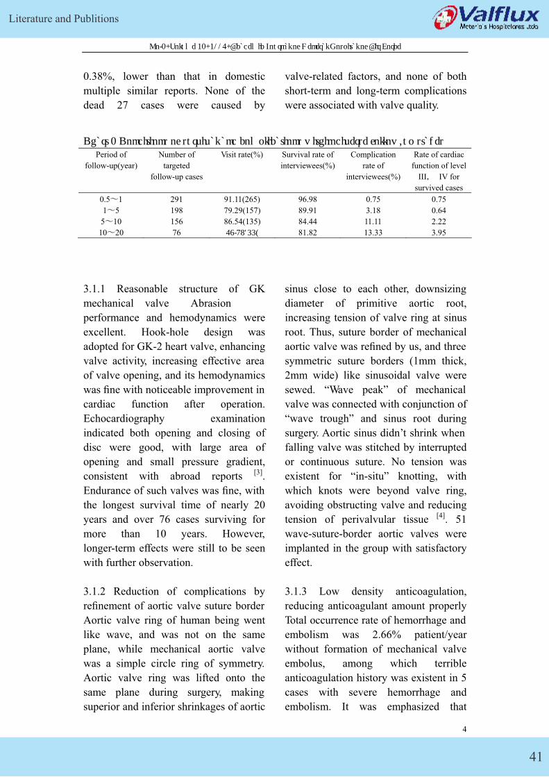
Literature and Publitions
41
No.1, Volume 21, 2005, Academic Journal of General Hospital of Air Force
0.38%, lower than that in domestic
multiple similar reports. None of the
dead 27 cases were caused by
valve-related factors, and none of both
short-term and long-term complications
were associated with valve quality.
Chart 1 Conditions of survival and complications within diverse follow-up stages Period of
follow-up(year)
Number of
targeted
follow-up cases
Visit rate(%) Survival rate of
interviewees(%)
Complication
rate of
interviewees(%)
Rate of cardiac
function of level
III� IV for
survived cases
0.5�1 291 91.11(265) 96.98 0.75 0.75
1�5 198 79.29(157) 89.91 3.18 0.64
5�10 156 86.54(135) 84.44 11.11 2.22
10�20 76 57.89(44) 81.82 13.33 3.95
3.1.1 Reasonable structure of GK
mechanical valve Abrasion
performance and hemodynamics were
excellent. Hook-hole design was
adopted for GK-2 heart valve, enhancing
valve activity, increasing effective area
of valve opening, and its hemodynamics
was fine with noticeable improvement in
cardiac function after operation.
Echocardiography examination
indicated both opening and closing of
disc were good, with large area of
opening and small pressure gradient,
consistent with abroad reports [3].
Endurance of such valves was fine, with
the longest survival time of nearly 20
years and over 76 cases surviving for
more than 10 years. However,
longer-term effects were still to be seen
with further observation.
3.1.2 Reduction of complications by
refinement of aortic valve suture border
Aortic valve ring of human being went
like wave, and was not on the same
plane, while mechanical aortic valve
was a simple circle ring of symmetry.
Aortic valve ring was lifted onto the
same plane during surgery, making
superior and inferior shrinkages of aortic
sinus close to each other, downsizing
diameter of primitive aortic root,
increasing tension of valve ring at sinus
root. Thus, suture border of mechanical
aortic valve was refined by us, and three
symmetric suture borders (1mm thick,
2mm wide) like sinusoidal valve were
sewed. “Wave peak” of mechanical
valve was connected with conjunction of
“wave trough” and sinus root during
surgery. Aortic sinus didn’t shrink when
falling valve was stitched by interrupted
or continuous suture. No tension was
existent for “in-situ” knotting, with
which knots were beyond valve ring,
avoiding obstructing valve and reducing
tension of perivalvular tissue [4]. 51
wave-suture-border aortic valves were
implanted in the group with satisfactory
effect.
3.1.3 Low density anticoagulation,
reducing anticoagulant amount properly
Total occurrence rate of hemorrhage and
embolism was 2.66% patient/year
without formation of mechanical valve
embolus, among which terrible
anticoagulation history was existent in 5
cases with severe hemorrhage and
embolism. It was emphasized that
4
GK Mechanical Heart Valve
42
No.1, Volume 21, 2005, Academic Journal of General Hospital of Air Force
prothrombin time should be maintained
at 2 times of normal value years before,
and hemorrhage incidence was much
higher than that of embolism.
Postoperative anticoagulation could not
only prevent embolus formation
effectively but also avoid complication
of hemorrhage. From dimension of
valve manufactureing, reduction of its
activation to blood system was the key
for elevation of hemocompatibility and
antiembolism ability, which was
dependent on treatment on surface
coating and smoothness of mechanical
valve. Identical carbon deposition
technology was adopted for GK
mechanical valve with higher level of
smoothness, whose hook-hole design
reduced destruction of formed elements
in blood and adhesion of fibrin, lessened
embolus formation and occurrence of
severe hemolysis. In the recent five
years, previous method of large amount
to reach anticoagulation standard was
transferred to that of smaller amount to
achieve anticoagulation standard
gradually, and prothrombin time was
maintained at a level a little higher than
1.5 times of normal value. Postoperative
hemorrhage rate declined, while
embolus occurrence rate didn’t increase
noticeably. Low intensity
anticoagulation was also adopted by
some abroad scholars [5], and their
conclusion was similar to that of the
group.
3.2 Consequence of long-term follow-up
Beside higher requirement of
mechanical valve, enhancement of
intraoperative care, logical surgery mode
and selection of surgery time, and
assertion of postoperative cardiac
function and anticoagulation were also
equally significant for effect of valve
replacement [6]. Heart was arrested under
oxygen by the way of lasting
oxygenated cold blood infusion and
warm blood infusion prior to opening of
aorta for patients in the group with large
hearts, severe disease, double-valve
replacement or valve replacement once
again, beneficial for automatic heart
self-recovery. Retaining of posterior
valve and subvalvular structure as well
as detection and treatment of tricuspid
valve (De Vega or Kay plastic surgery)
were performed during operation with
satisfactory effect.
K-P survival curve was not utilized to
describe year-mortality rate of patients
in the group, because larger difference
between interviewed group and missed
group would influence precision of
survival curve greatly, due to patient
resource in the group from multiple
social classes and the difference between
current medical level and personal
health consciousness in our country [7].
According to the real follow-up data of
the group, follow-up rate lessened
gradually and also did survival rate of
interviewees with the prolonging of
follow-up period. It was difficult to
explain effect coefficient of mechanical
valve as a result of longer follow-up
period of the group and the presence of
other death factors beside hearts of
patients as well as heart failure and
ventricular remodeling in cases with
rheumatic heart disease. The number of
cases with cardiac function of level III�
IV didn’t increase dramatically during
follow-up, for survived patients over 10
years in the group, as indicated
indirectly that the main related factors of
cardiac function during late follow-up of
5
Literature and Publitions
43
No.1, Volume 21, 2005, Academic Journal of General Hospital of Air Force
patients were preoperative conditions of
cardiac function, and reliability and
endurance of mechanical valve in
long-term application. 7 cases went with
severe anticoagulation related
complications, and 5 with terrible
anticoagulation history�7.15%� , as
demonstrated that logical publicity and
education as well as supervision, and
long-term anticoagulation were rather
significant.
3.3 Future of single-leaf disc valve
Bileaflet mechanical valve was
clinically widespread in recent years, but
it was found that thrombosis occurrence
rate and mechanical disorder rate of
single-leaf disc valve were lower than
those of bileaflet valve according to
comparison between single-leaf and
bileaflet conducted by some research
centers, and some other researchers
studied and deemed single-leaf valve
could elevate coronary blood flow
[2]Chunjiu Gu, Demin Yan, Demian
Xiao and others. 510 cases of heart
valve replacement [J]. Chinese Journal
of Thoracic and Cardiovascular Surgery,
1999, 15(5):290
【 】8 .
Advantage of single-leaf valve could
also be seen in terms of mechanical
security and anticoagulation from cases
in the group. Traditional study held that
single-leaf valve was small in opening
area, louder in noise, and eccentric in
blood flow, compared with bileaflet.
Except eccentric blood flow which
couldn’t be altered, actual effective area
of single-leaf valve was enough to cater
to demands of ventricular filling and
ejection, although its effective opening
area was relatively smaller. It could be
seen that left atrium and ventricle of
most patients with valve replacement
downsized, based on research of the
group. The tested transvalvular pressure
difference was also ideal, and the
problem of valve noise was believed to
be improved gradually with the
development of material science and
manufacturing technology.
Reference
[3]Chongxian Liao, Zengqi Li,
Daozhong Chen and others, Clinical
application of 248 GK mechanical
valves [J]. Chinese Journal of Thoracic
and Cardiovascular Surgery, 1997,
13(1):45
[4] Shisheng Jie, Jing Zhong, Shijie Wan
and others. Development and clinical
application of wave-suture border
mechanical aortic valves [J]. Chinese
Medical Journal, 1995, 75(5):306-307
[5]Ping Zhu, Guocui Long, 70 cases of
valve replacement by Carbo Medics
mechanical valve [J]. Chinese Journal of
Thoracic and Cardiovascular
Surgery.1998, 14(1): 29-30
6
GK Mechanical Heart Valve
44
Vol.21, No 4, August 2002Chinese Journal of Biomedical Engineering
Visualization Of Cavitation On Mechanical Heart Valve
HE Zhao-ming, XI Bao-shu: ZHU Ke-qin: ZU Pei-zhen
(Institute of Biomechanics, Department of Engineering Mechanics Tsinghua University Beijing 100084)
Abstract: Cavitation has been documented for GK mechanical heart valve in an in
vitro flow system simulating the closing event in mural position. From picture taken
with digital camera: cavitation clouds moved outwards along the seat stop on the
occlude surface after closure. C'avitati on was investigated and explained by
squeezing flow from the impact between occlude and seat stops at the closure of
mechanical heart valve. Its mechanism is vortex cavitation from wall jet. Velocity of
squeezing flow was estimated to be up to 9 m/s causing local low pressure
contributing much to cavitation. Closing velocity of occlude was an important factor
for cavitalion potential of UK valve. C'avitation duration varied with the ventricular
loading rate and was up to 440fcs at loading rate of 2750mmH}/s. The loading rate
threshold of cavitation was 420mmH}/s. This value was lower than that。[normal
physiological condition of 750mmH}/s in animal test. It was concluded that there was
possibility of cavitation for UK mechanical heart valve.
Key words: Mechanical heart valve; Cuvita tion visualization; Squeezing flow;
Wall jet
0 Introduction
Cavitation erosion pits were observed on pyrolytic carbon occluder of mechanical
mitral valve removed by valve replacements, therefore, it was suspected that there
was cavitation on the in vivo mechanical mitral valve. Furthermore, that could lead to
potential risk of thrombosis and mechanical valve damage[1]. In recent years a
number of studies on mechanical heart valve cavitation were undertaken, most of
them indicated that the squeezing flow between mechanical valve occluder and seat
stops or valve ring was the main underlying mechanism�2-4�, however, the causes of
cavitation and its specific process still remain unclear. To date, in China, no study has
been carried out on the cavitation of China-made mechanical valve. To fill gaps in this
field and gain certain understanding of the characteristics or trends of China-made
mechanical valves, the cavitation visualization of GK valve was conducted in the
present study. The foreign and domestic products have quite similar constructures,
however, their influence on cavitation was still under study at home and abroad.
1
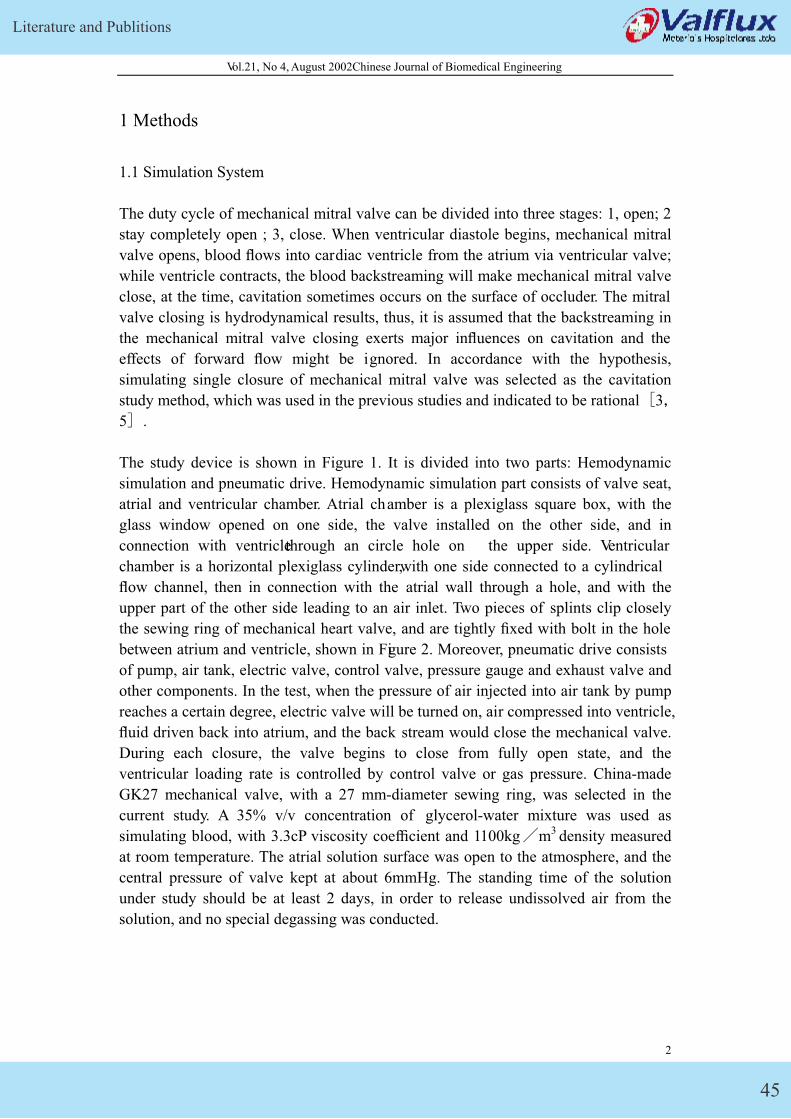
Literature and Publitions
45
Vol.21, No 4, August 2002Chinese Journal of Biomedical Engineering
1 Methods
1.1 Simulation System
The duty cycle of mechanical mitral valve can be divided into three stages: 1, open; 2
stay completely open ; 3, close. When ventricular diastole begins, mechanical mitral
valve opens, blood flows into cardiac ventricle from the atrium via ventricular valve;
while ventricle contracts, the blood backstreaming will make mechanical mitral valve
close, at the time, cavitation sometimes occurs on the surface of occluder. The mitral
valve closing is hydrodynamical results, thus, it is assumed that the backstreaming in
the mechanical mitral valve closing exerts major influences on cavitation and the
effects of forward flow might be ignored. In accordance with the hypothesis,
simulating single closure of mechanical mitral valve was selected as the cavitation
study method, which was used in the previous studies and indicated to be rational�3�5�.
The study device is shown in Figure 1. It is divided into two parts: Hemodynamic
simulation and pneumatic drive. Hemodynamic simulation part consists of valve seat,
atrial and ventricular chamber. Atrial chamber is a plexiglass square box, with the
glass window opened on one side, the valve installed on the other side, and in
connection with ventricle through an circle hole on the upper side. Ventricular
chamber is a horizontal plexiglass cylinder, with one side connected to a cylindrical
flow channel, then in connection with the atrial wall through a hole, and with the
upper part of the other side leading to an air inlet. Two pieces of splints clip closely
the sewing ring of mechanical heart valve, and are tightly fixed with bolt in the hole
between atrium and ventricle, shown in Figure 2. Moreover, pneumatic drive consists
of pump, air tank, electric valve, control valve, pressure gauge and exhaust valve and
other components. In the test, when the pressure of air injected into air tank by pump
reaches a certain degree, electric valve will be turned on, air compressed into ventricle,
fluid driven back into atrium, and the back stream would close the mechanical valve.
During each closure, the valve begins to close from fully open state, and the
ventricular loading rate is controlled by control valve or gas pressure. China-made
GK27 mechanical valve, with a 27 mm-diameter sewing ring, was selected in the
current study. A 35% v/v concentration of glycerol-water mixture was used as
simulating blood, with 3.3cP viscosity coefficient and 1100kg�m3 density measured
at room temperature. The atrial solution surface was open to the atmosphere, and the
central pressure of valve kept at about 6mmHg. The standing time of the solution
under study should be at least 2 days, in order to release undissolved air from the
solution, and no special degassing was conducted.
2
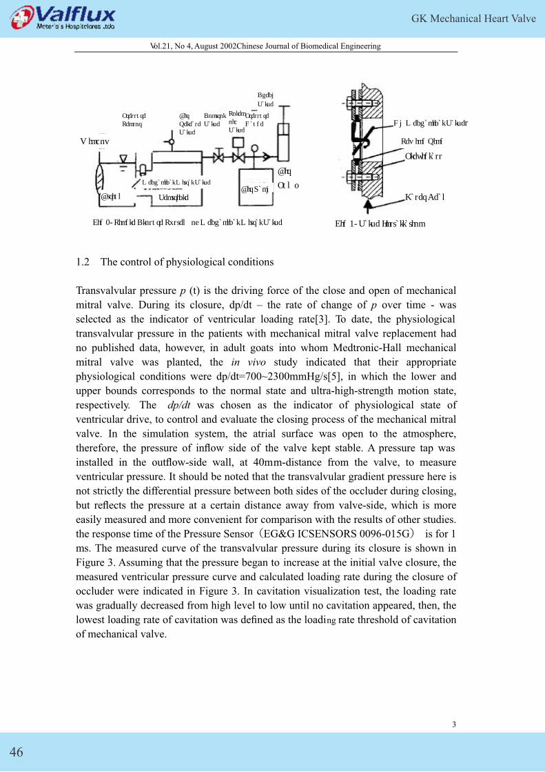
GK Mechanical Heart Valve
46
Vol.21, No 4, August 2002Chinese Journal of Biomedical Engineering
Air Release Valve
Pressure Gauge
Solenoid Valve
Check
Valve
Control Valve
Pressure Sensor Gk Mechanical Valves
Window
Plexiglass
Sewing Ring
Air
PumpMechanical Mitral Valve Air Tank
Laser BeamAtrium Ventricle
Fig 1. Single Closure System of Mechanical Mitral Valve Fig 2. Valve Installation
1.2 The control of physiological conditions
Transvalvular pressure p (t) is the driving force of the close and open of mechanical
mitral valve. During its closure, dp/dt – the rate of change of p over time - was
selected as the indicator of ventricular loading rate[3]. To date, the physiological
transvalvular pressure in the patients with mechanical mitral valve replacement had
no published data, however, in adult goats into whom Medtronic-Hall mechanical
mitral valve was planted, the in vivo study indicated that their appropriate
physiological conditions were dp/dt=700~2300mmHg/s[5], in which the lower and
upper bounds corresponds to the normal state and ultra-high-strength motion state,
respectively. The dp/dt was chosen as the indicator of physiological state of
ventricular drive, to control and evaluate the closing process of the mechanical mitral
valve. In the simulation system, the atrial surface was open to the atmosphere,
therefore, the pressure of inflow side of the valve kept stable. A pressure tap was
installed in the outflow-side wall, at 40mm-distance from the valve, to measure
ventricular pressure. It should be noted that the transvalvular gradient pressure here is
not strictly the differential pressure between both sides of the occluder during closing,
but reflects the pressure at a certain distance away from valve-side, which is more
easily measured and more convenient for comparison with the results of other studies.
the response time of the Pressure Sensor�EG&G ICSENSORS 0096-015G� is for 1
ms. The measured curve of the transvalvular pressure during its closure is shown in
Figure 3. Assuming that the pressure began to increase at the initial valve closure, the
measured ventricular pressure curve and calculated loading rate during the closure of
occluder were indicated in Figure 3. In cavitation visualization test, the loading rate
was gradually decreased from high level to low until no cavitation appeared, then, the
lowest loading rate of cavitation was defined as the loading rate threshold of cavitation
of mechanical valve.
3
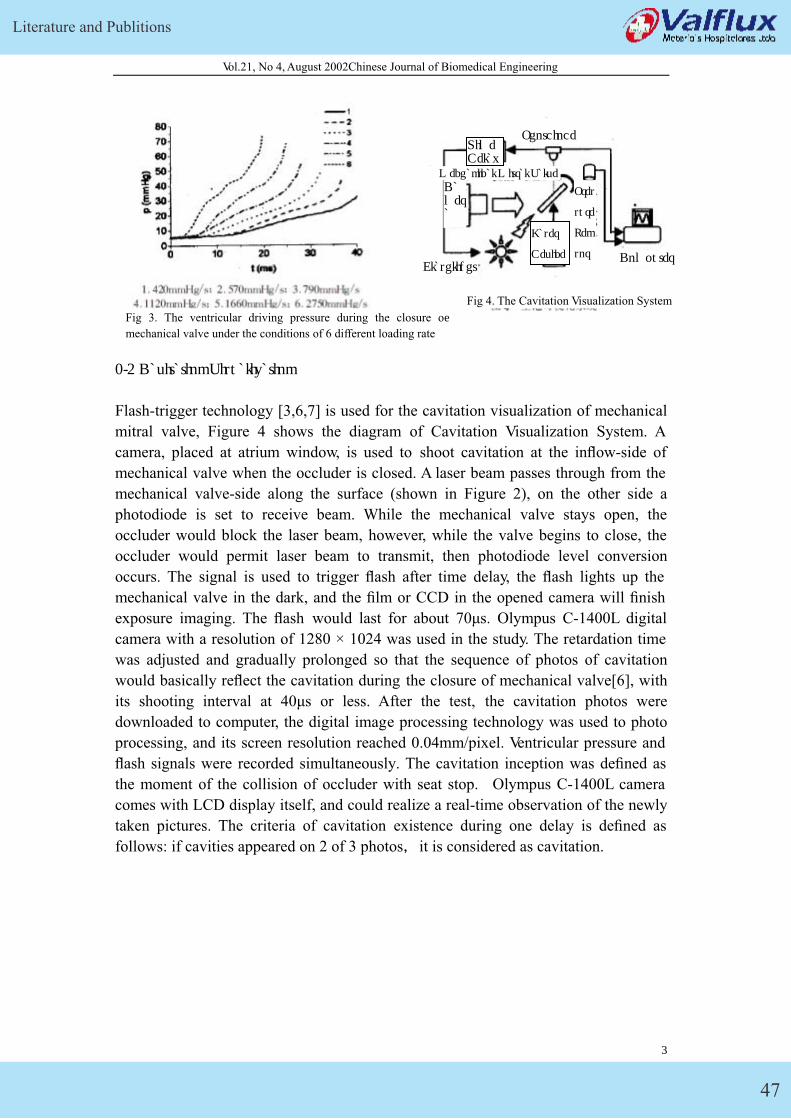
Literature and Publitions
47
Vol.21, No 4, August 2002Chinese Journal of Biomedical Engineering
Photdiode
Mechanical Mitral Valve
Time Delay
Camera
Pres
sure
Sen
sor
Laser
Device ComputerFlashlight
Fig 3. The ventricular driving pressure during the closure of
mechanical valve under the conditions of 6 different loading rate
1.3 Cavitation Visualization
Flash-trigger technology [3,6,7] is used for the cavitation visualization of mechanical
mitral valve, Figure 4 shows the diagram of Cavitation Visualization System. A
camera, placed at atrium window, is used to shoot cavitation at the inflow-side of
mechanical valve when the occluder is closed. A laser beam passes through from the
mechanical valve-side along the surface (shown in Figure 2), on the other side a
photodiode is set to receive beam. While the mechanical valve stays open, the
occluder would block the laser beam, however, while the valve begins to close, the
occluder would permit laser beam to transmit, then photodiode level conversion
occurs. The signal is used to trigger flash after time delay, the flash lights up the
mechanical valve in the dark, and the film or CCD in the opened camera will finish
exposure imaging. The flash would last for about 70μs. Olympus C-1400L digital
camera with a resolution of 1280 × 1024 was used in the study. The retardation time
was adjusted and gradually prolonged so that the sequence of photos of cavitation
would basically reflect the cavitation during the closure of mechanical valve[6], with
its shooting interval at 40μs or less. After the test, the cavitation photos were
downloaded to computer, the digital image processing technology was used to photo
processing, and its screen resolution reached 0.04mm/pixel. Ventricular pressure and
flash signals were recorded simultaneously. The cavitation inception was defined as
the moment of the collision of occluder with seat stop. Olympus C-1400L camera
comes with LCD display itself, and could realize a real-time observation of the newly
taken pictures. The criteria of cavitation existence during one delay is defined as
follows: if cavities appeared on 2 of 3 photos�it is considered as cavitation.
4
Fig 4. The Cavitation Visualization System
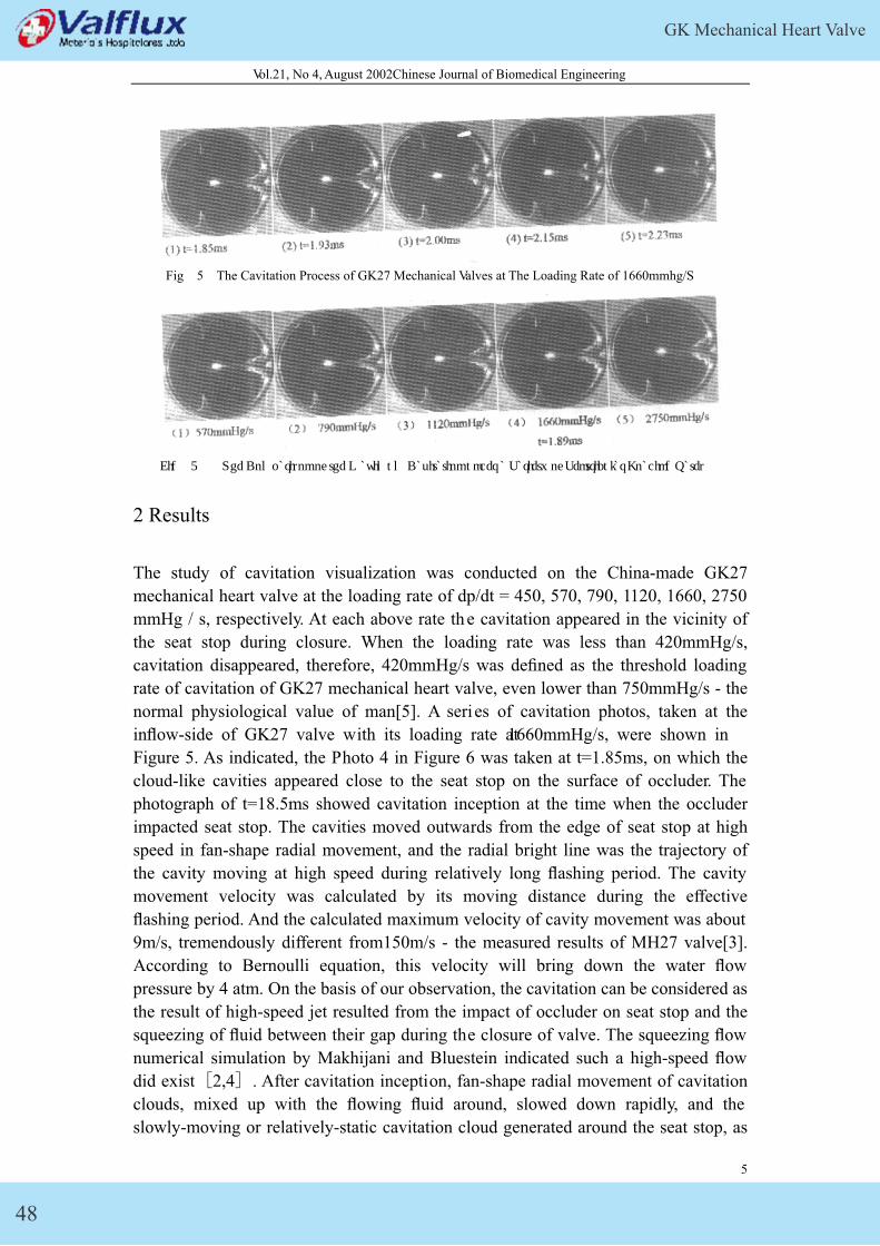
GK Mechanical Heart Valve
48
Vol.21, No 4, August 2002Chinese Journal of Biomedical Engineering
Fig 5 The Cavitation Process of GK27 Mechanical Valves at The Loading Rate of 1660mmhg/S
Fig 6 The Comparison of the Maximum Cavitation under a Variety of Ventricular Loading Rates
2 Results
The study of cavitation visualization was conducted on the China-made GK27
mechanical heart valve at the loading rate of dp/dt = 450, 570, 790, 1120, 1660, 2750
mmHg / s, respectively. At each above rate th e cavitation appeared in the vicinity of
the seat stop during closure. When the loading rate was less than 420mmHg/s,
cavitation disappeared, therefore, 420mmHg/s was defined as the threshold loading
rate of cavitation of GK27 mechanical heart valve, even lower than 750mmHg/s - the
normal physiological value of man[5]. A series of cavitation photos, taken at the
inflow-side of GK27 valve with its loading rate at 1660mmHg/s, were shown in
Figure 5. As indicated, the Photo 4 in Figure 6 was taken at t=1.85ms, on which the
cloud-like cavities appeared close to the seat stop on the surface of occluder. The
photograph of t=18.5ms showed cavitation inception at the time when the occluder
impacted seat stop. The cavities moved outwards from the edge of seat stop at high
speed in fan-shape radial movement, and the radial bright line was the trajectory of
the cavity moving at high speed during relatively long flashing period. The cavity
movement velocity was calculated by its moving distance during the effective
flashing period. And the calculated maximum velocity of cavity movement was about
9m/s, tremendously different from150m/s - the measured results of MH27 valve[3].
According to Bernoulli equation, this velocity will bring down the water flow
pressure by 4 atm. On the basis of our observation, the cavitation can be considered as
the result of high-speed jet resulted from the impact of occluder on seat stop and the
squeezing of fluid between their gap during the closure of valve. The squeezing flow
numerical simulation by Makhijani and Bluestein indicated such a high-speed flow
did exist�2,4�. After cavitation inception, fan-shape radial movement of cavitation
clouds, mixed up with the flowing fluid around, slowed down rapidly, and the
slowly-moving or relatively-static cavitation cloud generated around the seat stop, as
5
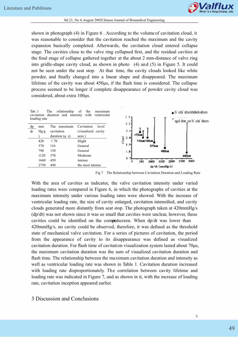
Literature and Publitions
49
Vol.21, No 4, August 2002Chinese Journal of Biomedical Engineering
shown in photograph (4) in Figure 6 . According to the volume of cavitation cloud, it
was reasonable to consider that the cavitation reached the maximum and the cavity
expansion basically completed. Afterwards, the cavitation cloud entered collapse
stage. The cavities close to the valve ring collapsed first, and the residual cavities at
the final stage of collapse gathered together at the about 2 mm-distance of valve ring
into gridle-shape cavity cloud, as shown in photo (4) and (5) in Figure 5. It could
not be seen under the seat stop. At that time, the cavity clouds looked like white
powder, and finally changed into a linear shape and disappeared. The maximum
lifetime of the cavity was about 450μs, if the flash time is considered. The collapse
process seemed to be longer if complete disappearance of powder cavity cloud was
considered, about extra 100μs.
With the area of cavities as indicator, the valve cavitation intensity under varied
loading rates were compared in Figure 6, in which the photographs of cavities at the
maximum intensity under various loading rates were showed. With the increase of
ventricular loading rate, the size of cavity enlarged, cavitation intensified, and cavity
clouds generated more distantly from seat stop. The photograph taken at 420mmHg/s
(dp/dt) was not shown since it was so small that cavities were unclear, however, these
cavities could be identified on the computer screen. When dp/dt was lower than
420mmHg/s, no cavity could be observed, therefore, it was defined as the threshold
state of mechanical valve cavitation. For a series of pictures of cavitation, the period
from the appearance of cavity to its disappearance was defined as visualized
cavitation duration. For flash time of cavitation visualization system lasted about 70μs,
the maximum cavitation duration was the sum of visualized cavitation duration and
flash time. The relationship between the maximum cavitation duration and intensity as
well as ventricular loading rate was shown in Table 1. Cavitation duration increased
with loading rate disproportionately. The correlation between cavity lifetime and
loading rate was indicated in Figure 7, and as shown in it, with the increase of loading
rate, cavitation inception appeared earlier.
Tab. 1 The relationship of the maximum cavitation duration and intensity with ventricular loading rate
dp
dt
mm
Hg/g
)
The maximum
cavitation
duration (μ s)
Cavitation level
(visualized cavity
area )
420 �70 Slight
570 110 General
790 150 General
1120 370 Moderate
1660 450 intense
2750 440 the most intense
the end of cavitation
cavitation inception
Fig 7 The Relationship between Cavitation Duration and Loading Rate
3 Discussion and Conclusions
6
GK Mechanical Heart Valve
50
Vol.21, No 4, August 2002Chinese Journal of Biomedical Engineering
When valve is closed, the end of occluder rotates at the highest linear velocity and
collides with seat stop, during which local squeezing flow generates. The primary
location shape of cavitation inception is consistent with squeezing contact surface
during the impact of occluder on seat stop, thus, it is known that, for GK mechanical
valve, the jet, generated in the squeezing between occluder and seat stop during valve
closure, is the main cause of cavitation. On the basis of the mechanism, some one
considered it as Venturi cavitation, for the cavity movement speed is 9m/s, calculated
by its movement trajectory, which could cause a drop of 4 atm. If the potential energy
of the flow provided by the squeezing is not enough to make up such a drop, the
pressure in the jet would be less than cavitation threshold pressure. In addition, the
severe vortex of high-speed jet can intensify the conversion of the heat energy to
kinetic energy, the local pressure would lower, and the gas nucleus in the jet will
expand in the high-speed movement. When the jet with growing-up cavities rushes
out and squeezes the gap between valve and seat stop, the cavities can be observed to
move outwards on the taken pictures. However, the author proposed vortex cavitation
mechanism, that is, this cavitation is a vortex cavitation caused by jet. This squeezing
jet is a kind of wall jet according to jet environment. The surface of discontinuity,
formed at the free mixing zone of the jet exit, might lose its stability and develop into
a vortex. The fluid surrounding the vortex will be entrained and absorbed into the jet,
at the same time, turbulence will come into being after continuous moving,
deformating, and separating. The turbulence generated great pulse pressure, and made
the core pressure of vortex decrease less than cavitation threshold pressure in a flash.
The gas nucleus enlarged in high-speed movement, and cavities can be observed to
move outwards at the jet free mixing area outside of the squeezing gap exit. This is
the mechanism of vortex cavitation caused by wall jet. The drop of the fluid pressure
resulted from dilatation wave generated during the closure of valve, generally
speaking, is not enough to lead to cavitation [3], for the cavities generated by
dilatation wave are only separate ball-shape cavities [6] with unstable position.
Furthermore, according to the shape and distribution characteristics of the cavities, the
cavitation observed in the study is not produced by such a dilatation wave, however,
the pressure drop here could strengthen the trends of jet cavitation, and delay cavity
collapse, and prolong cavitation lifetime [8]. The enlargement of cavitation cloud
volume in the squeezing jet is the interaction results of cavity development and
continuous low-pressure. With the slow-down of mixing up of jet with surrounding
fluid, as well as the reflection of dilatation wave, the pressure began to recover and
the cavities developed into collapse stage. The collapse duration of cavity cloud
seems to be longer than its development period, which can be explained by cavity
kinetic. For the collapse location of cavity clouds, the last cavities disappeared at
2-mm distance from the valve ring. Further studies are required on the damage degree
of this cavity collapse. To date, there are no clinical reports related to the evidence of
cavitation at the above location of mechanical valve. Some cavities collapse at the
near end of occluder might be the underlying reason of the cavitation at the edge of
valve and occluder. Lee had indicated that, for MH mechanical valve and other similar
products, the threshold loading rate of cavitation was only half the normal loading rate
7
Vol.21, No 4, August 2002Chinese Journal of Biomedical Engineering
of man. GK mechanical valve might have the same characteristics as MH for their
similar structures. The result provides support for the explanation of squeezing jet as
the underlying reason of mechanical valve cavitation, however, for the caviation
mechanism, Lee proposed that this was a kind of vortex cavitation caused by wall jet.
According to Figure 7, cavitation duration prolonged with, but out of proportion to,
the increase of loading rate. It might be caused by the experimental system error. The
cavitation duration might be a little shorter at 1120 and 1660mmHg/s, and at the same
time, the collapse time of cavities is disperse. The collapse stage seemed to be a little
longer. At the end stage of collapse, no obvious change could be observed of the
volume of cavities in powder state, therefore, it is difficult for the naked eye to
accurately determine the time of complete cavity collapse. However, it is relatively
easy to determine the starting time of cavitation, for the sudden growth of cavity at the
cavitation inception would make the change of its volume obvious. With the increase
of the loading rate, the time of cavitation inception will occur earlier. The position of
occluder, used for the measurement of starting time in delayed timing, is immovable,
and the cavitation duration is the period from the time when the occluder at initial
reference location to its impact on seat stop, on the basis of which the closing rate of
valve could be evaluated. With the increase of loading rate, the time of the impact of
occluder on seat stop will be earlier, furthermore, the impact velocity will rise, too. It
shows that the impact velocity of occluder has a greater effect on the cavitation, which
is a support that the squeezing jet is the underlying reason of cavitation.
According to this study, the following conclusions could be drawn:
� The loading rate under normal physiological state is about 750mmHg/s. The
present study indicated that the threshold loading rate of cavitation of GK27
mechanical heart valve was 420mmHg/s, similar to that of Medtronic Hall mechanical
valve, but far less than that under normal physiological state.
� Cloud-like cavities appeared in the vicinity of seat stop, moved radially outwards.
Therefore, the cavitation is caused by the local squeezing jet generated during the
impact of occluder on seat stop during the GK mechanical valve closure. As for
cavitation mechanism, the author think it is a vortex cavitation caused by wall jet,.
� With the rise of loading rate, the closing velocity of valve is increased, and the
GK mechanical valve cavitation is intensified, too. Therefore, the closing velocity of
valve is a key indicator to evaluate the cavitation trend GK mechanical valve.
� Modify the design of valve, for instance, to reduce appropriately the squeezing
contact area of seat stop, or to decrease the velocity of occluder at the final stage of
closing, etc, so as to decrease squeezing jet velocity and improve the cavitation trend
of mechanical valve.
8
Literature and Publitions
51
Vol.21, No 4, August 2002Chinese Journal of Biomedical Engineering
4 Reference
[1] Zhao-ming HE, XI Pao-shu, ZHU Ke-qin. Advances in the study of mechanical
valve cavitation [J]. Journal of Biomedical Engineering, 1999�16�2��243-248.
[2] Bluestein D, Einav S, Hwang NHC. A squeeze flow ph enomenon at the closing of
a Bileaflet mechanical heart valve prosthesis [J]. Journal of Biomechanics, 1994,
27(11):1369-1378.
[3] Lee CS, Chandran KB, Chen LD. Cavitation dynamics of Medtronic – Hall
mechanical heart valve prosthesis fluid squeezing effect [J]. Journal of Biomechanical
Engineering, 1996, 118(1): 97-105.
[4] Makhijani VB, Siegel JM, Hwang NHC. Numerical study of squeeze-flow in
tilting dise mechanical heart valves [J]. Journal of Heart Valve Disease, 1996, 3:
97-103
[5] Chandran KB, Dexter EU, Richenbacher WE. Negative pressure transients with
mechanical heart-valve closure: correlation between in vitro and in vivo results [J].
Annals of Biomedical Engineering, 1998, 26(4): 546-556.
[6] Zhao-ming HE, XI Pao-shu, ZHU Ke-qin., etc. Cavitation visualization study on
the surface of piston in decelerated motion [J]. Journal of Experimental Mechanics.
1999�14(3):309-315.
[7] Lamson TC, Stinebring DR, Deutsch S, et al. Real-time in vitro observation of
cavitation in a prosthetic heart valve [C]. ASAIO transactions, 1991,37: 351-353.
[8] Zhao-ming HE, XI Pao-shu, ZHU Ke-qin, etc. The calculation of surface
pressure of piston in the decelerated motion and the prediction of cavitation inception
�J�. Tsinghua University (Natural Science) .1999,39(11):54-57.
[9] Chandran KB, Aluri S. Mechanical valve closing dynamics: relationship between
velocity of closing pressure transients, and cavitation initiation[J]. Annals of
Biomedical Engineering 1997 25( 6 ): 926-938.
9
GK Mechanical Heart Valve
52
Lepu Medical Technology (Beijing) Co., Ltd.Address: 3rd Floor, Beikong Building, No.10 Baifuquan Road, Chagping, Beijing, China 102200Tel: (86) 10-80120666Fax: (86) 10-80120600Marketng and Sales Center20F, Tower 2,, Xihuan Plaza, No.1 Xizhimeenwai St., Xicheng District, Beijing, China 100044Tel: (86) 10-58305000Fax: (86) 10-58305100WWW.LEPUMEDICAL.COM