Embed Size (px)
Citation preview
Gilead -Topics in Human Pathophysiology
Fall 2010 Drug Safety and Public Health
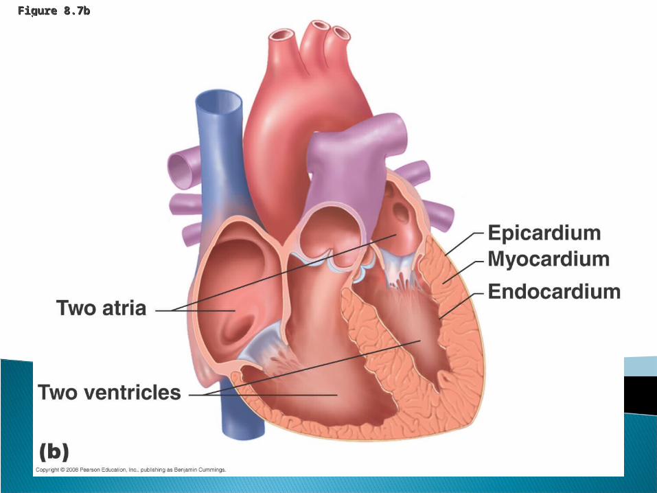
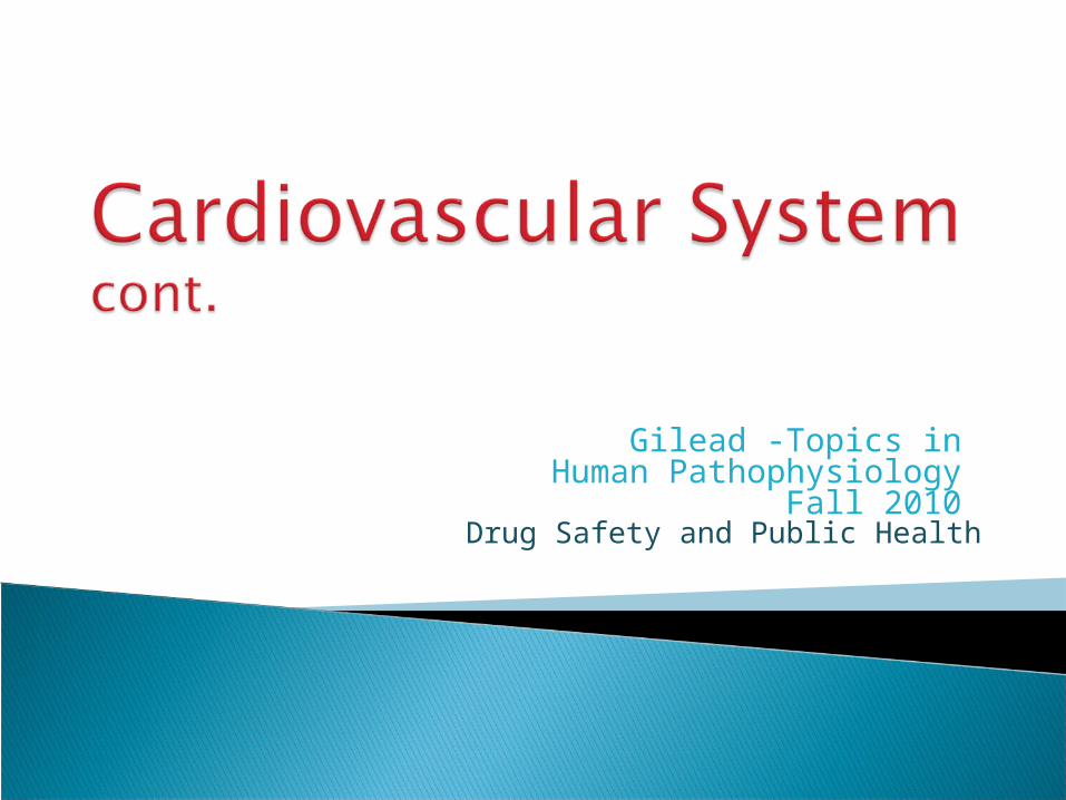
Figure 8.7bFigure 8.7b
Systole – ventricles contract, semilunar valves open to allow blood to large arteries; AV valves close to prevent backflow to the atria
Diastole – ventricles relax, semilunar valves close to prevent backflow into the ventricles, AV valves open to allow ventricles to fill
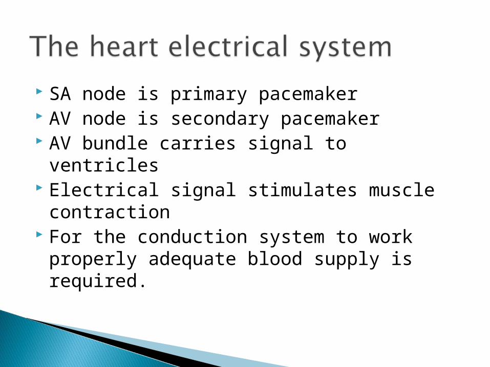
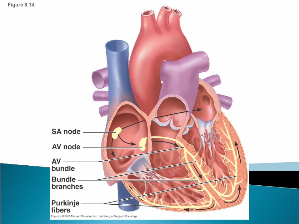
SA node is primary pacemaker AV node is secondary pacemaker AV bundle carries signal to ventricles Electrical signal stimulates muscle
contraction For the conduction system to work properly
adequate blood supply is required.
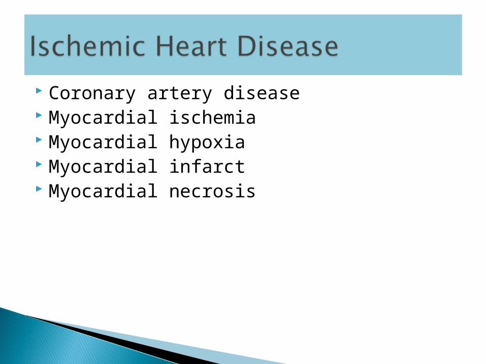
Coronary artery disease Myocardial ischemia Myocardial hypoxia Myocardial infarct Myocardial necrosis
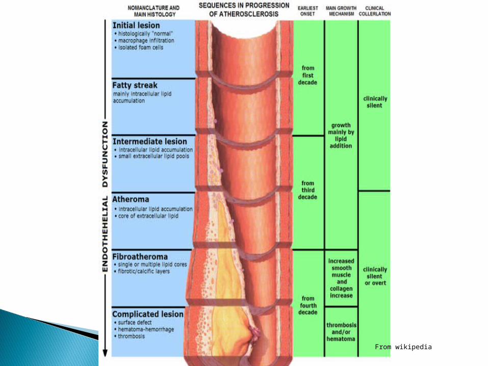
From wikipedia
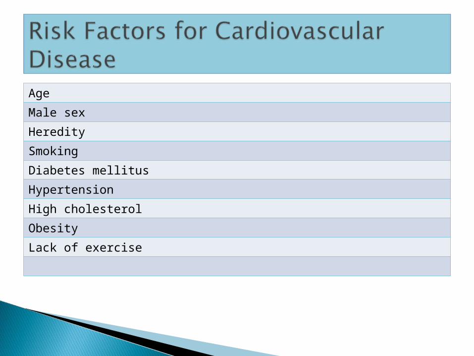
Age
Male sex
Heredity
Smoking
Diabetes mellitus
Hypertension
High cholesterol
Obesity
Lack of exercise

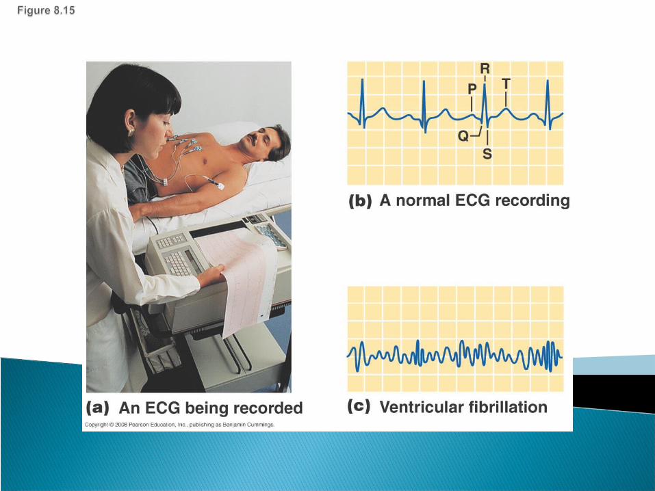
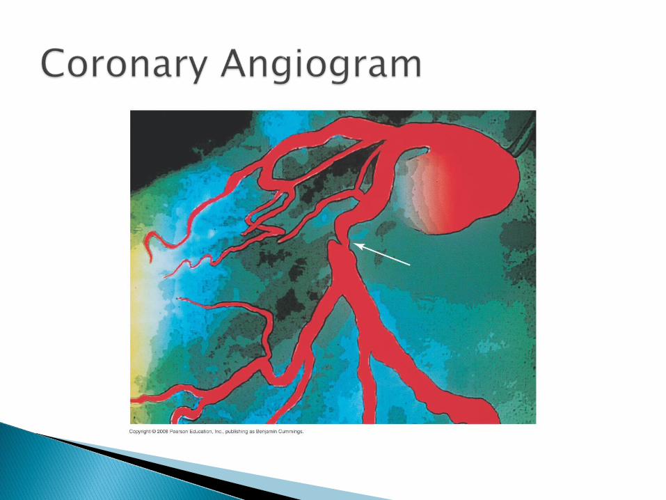
Diagnosis◦ BP monitoring◦ Symptoms◦ ECG◦ Angiogram◦ Stress Test◦ Nuclear myocardial
perfusion tests
For myocardial perfusion imaging (MPI) A2A adenosine receptor agonist Vasodilates coronary arteries as if exercising Injected into blood stream prior to gamma
camera scan Can give a good indication of myocardial
perfusion
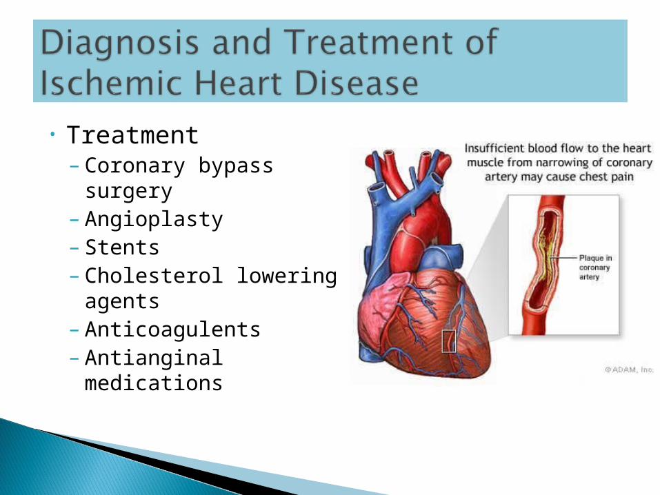
• Treatment– Coronary bypass
surgery– Angioplasty– Stents– Cholesterol lowering
agents– Anticoagulents– Antianginal
medications
For angina Thought to inhibit a sodium ion
channel in the cardiac muscle cells Contraction of those cells might
normally cause compression of cardiac blood vessels during diastole.
Can be taken with other anti-anginal meds
Very effective
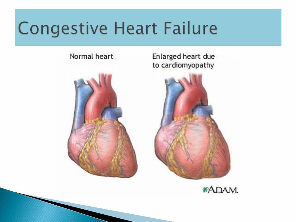
Congestive Heart Failure
• Heart becomes weak• Blood backs up in veins and capillaries• Fluid excess in tissues• Symptoms include shortness of breath,
edema, difficulty breathing (especially when lying down,) difficulty exercising
Congestive Heart Failure
• Causes: – cardiomyopathy– hypertension– lung disease– coronary artery disease– previous MI– valve disease
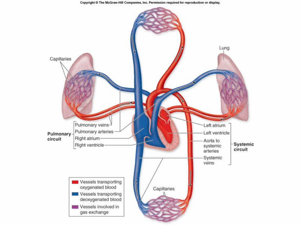
Blood Vessels and Pressure
• Artery structure and function• Control over smooth muscle• Vascular Disease
– Atherosclerosis– Pulmonary hypertension

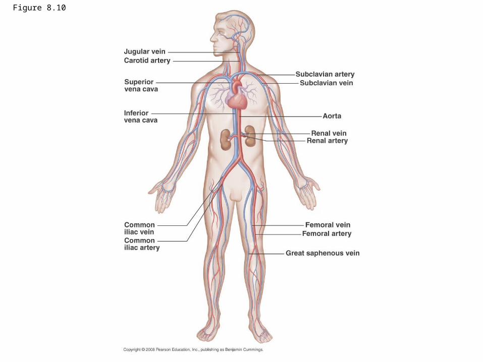
Figure 8.10
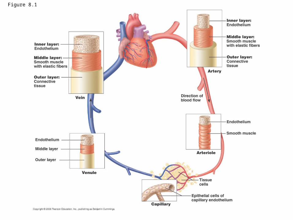
Figure 8.1
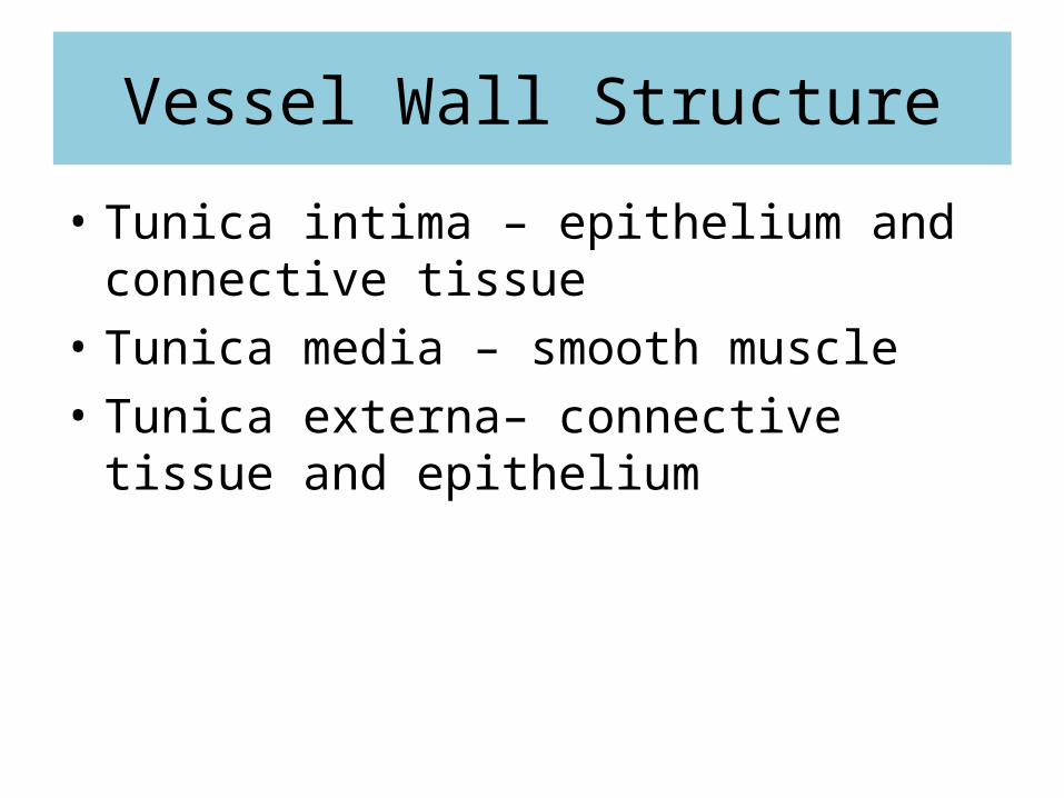
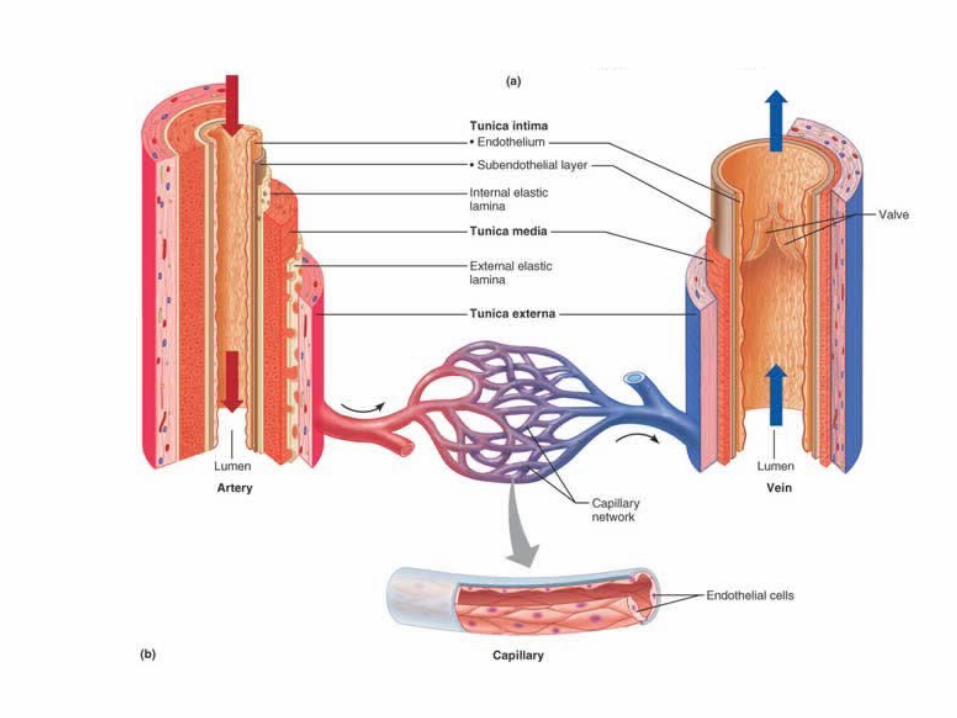
Vessel Wall Structure
• Tunica intima – epithelium and connective tissue
• Tunica media – smooth muscle• Tunica externa– connective tissue and
epithelium
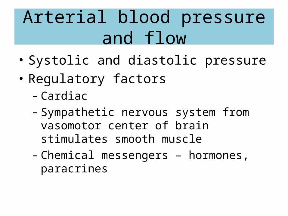
Arterial blood pressure and flow
• Systolic and diastolic pressure• Regulatory factors
– Cardiac – Sympathetic nervous system from vasomotor
center of brain stimulates smooth muscle– Chemical messengers – hormones, paracrines
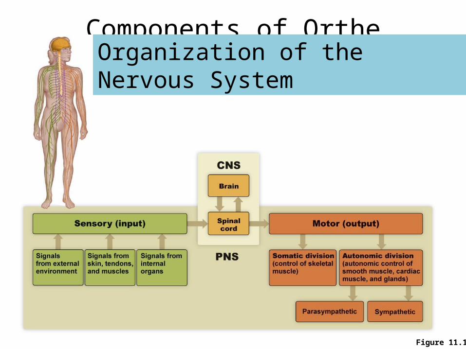
Components of Orthe Nervous System
Figure 11.1
Organization of the Nervous System
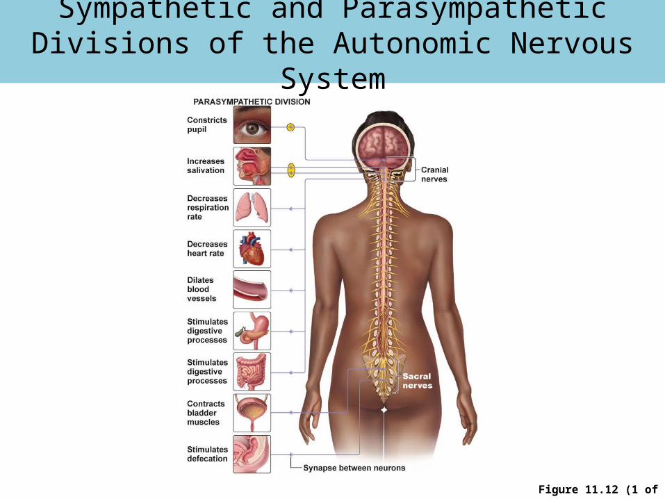
Figure 11.12 (1 of 2)
Sympathetic and Parasympathetic Divisions of the Autonomic Nervous System
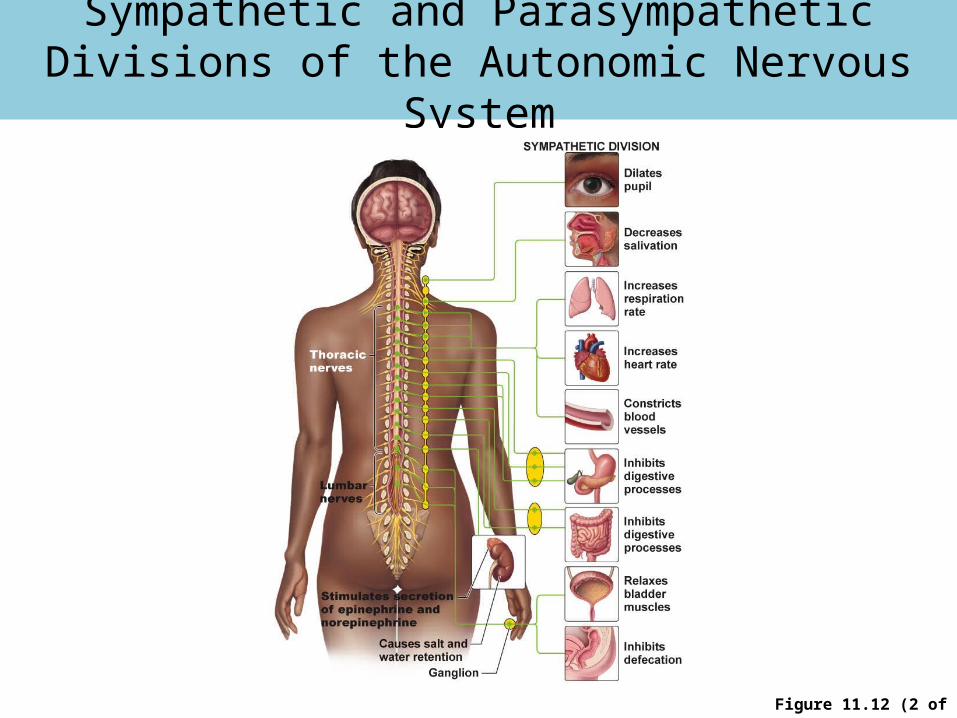
Figure 11.12 (2 of 2)
Sympathetic and Parasympathetic Divisions of the Autonomic Nervous System
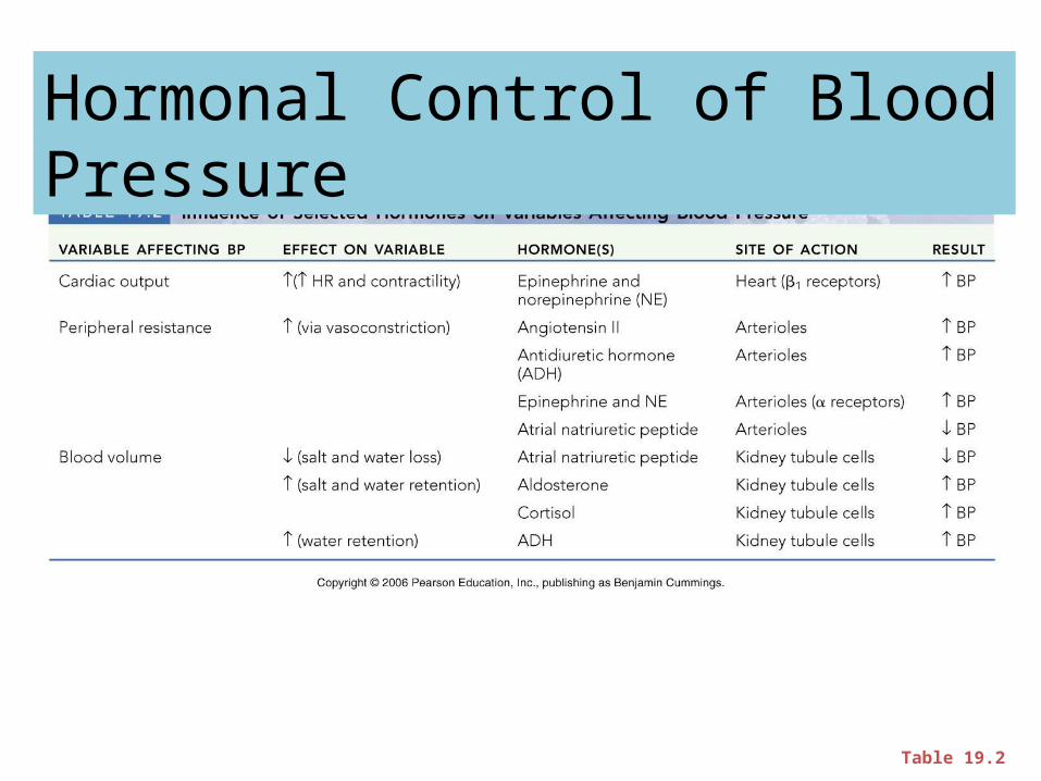
Table 19.2
Hormonal Control of Blood Pressure
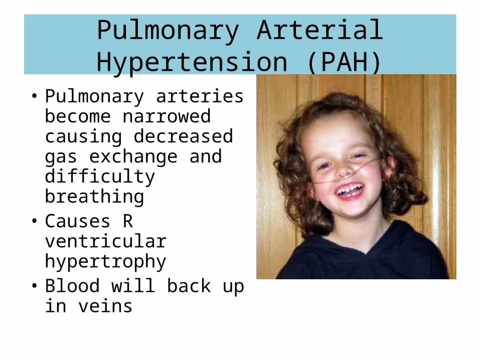
Pulmonary Arterial Hypertension (PAH)
• Pulmonary arteries become narrowed causing decreased gas exchange and difficulty breathing
• Causes R ventricular hypertrophy
• Blood will back up in veins
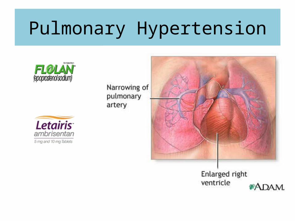
Pulmonary Hypertension
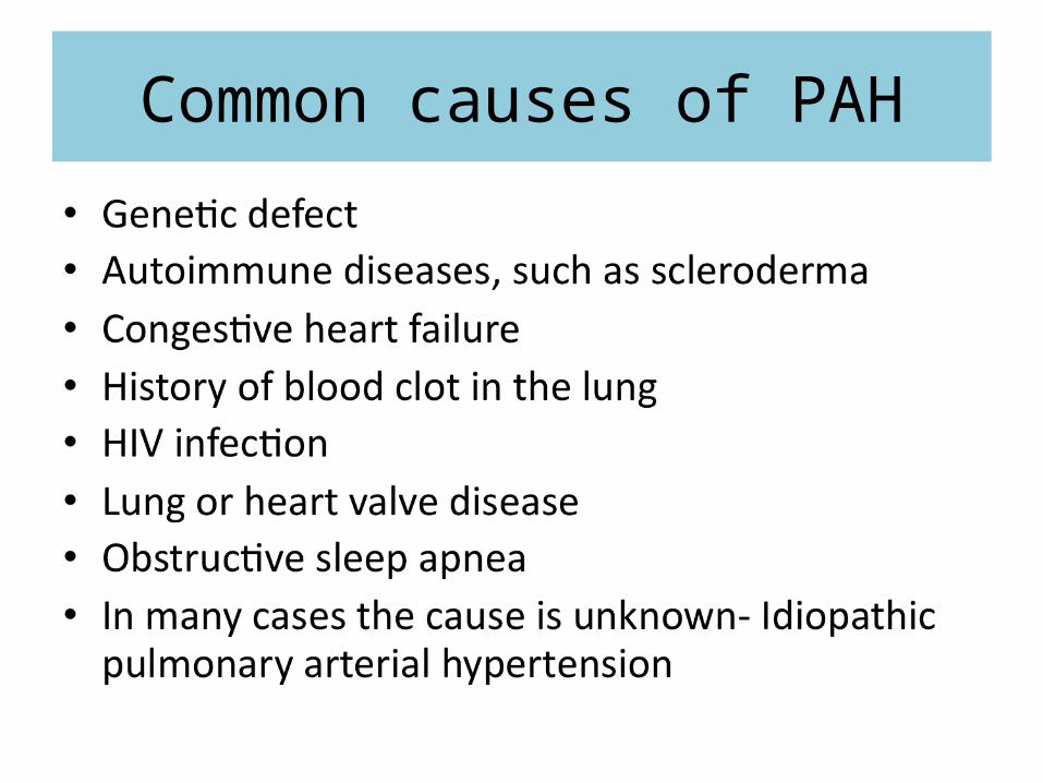
Common causes of PAH

Symptoms of PAH
• Chest pain, usually in the front of the chest• Dizziness• Fainting• Fatigue• Leg edema• Light-headedness during exercise• Shortness of breath during activity• Weakness
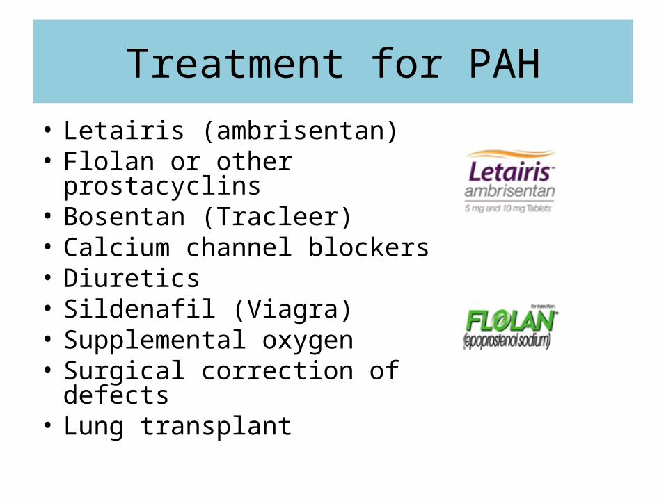
Treatment for PAH• Letairis (ambrisentan)• Flolan or other prostacyclins• Bosentan (Tracleer)• Calcium channel blockers• Diuretics• Sildenafil (Viagra)• Supplemental oxygen• Surgical correction of defects• Lung transplant