Embed Size (px)
Citation preview
lable at ScienceDirect
Food and Chemical Toxicology 97 (2016) 232e242
Contents lists avai
Food and Chemical Toxicology
journal homepage: www.elsevier .com/locate/ foodchemtox
Genotoxicity assessment of the flavouring agent, perillaldehyde
Cheryl A. Hobbs a, *, Sean V. Taylor b, Carol Beevers c, Melvyn Lloyd c, Rachael Bowen c,Lucinda Lillford c, Robert Maronpot d, Shim-mo Hayashi e
a Toxicology Program, Integrated Laboratory Systems, Inc., PO Box 13501, Research Triangle Park, NC 27709, USAb International Organization of the Flavor Industry, 1101 17th Street NW, Suite 700, Washington, DC 20036, USAc Covance Laboratories Ltd, Otley Road, Harrogate, North Yorkshire, HG3 1PY, England, UKd Maronpot Consulting LLC, 1612 Medfield Road, Raleigh, NC 27607, USAe Japan Flavor and Fragrance Materials Association, Sankei Nihonbashi Bldg 6F, 4-7-1 Nihonbashihon-cho, Chuo-ku, Tokyo, 103-0023, Japan
a r t i c l e i n f o
Article history:Received 6 July 2016Received in revised form25 August 2016Accepted 27 August 2016Available online 1 September 2016
Keywords:GenotoxicityFlavouring agentPerillaldehydep-mentha-1,8-dien-7-alPerilla aldehydeDNA damage
Abbreviations: EFSA, European Food Safety AuthoriGLP, Good Laboratory Practice; HPRT, hypoxanthine-Pharmaceuticals for Human Use; JaCVAM, Japanese Celog; MN, micronucleus or micronuclei; MN-PCE, mipolychromatic erythrocytes; (Q)SAR, quantitative sOrganization.* Corresponding author. Integrated Laboratory Syst
E-mail address: [email protected] (C.A. Hobbs).
http://dx.doi.org/10.1016/j.fct.2016.08.0290278-6915/© 2016 The Authors. Published by Elsevier
a b s t r a c t
Perillaldehyde, a natural monocyclic terpenoid found most abundantly in the herb perilla, has a longhistory of use as a flavouring ingredient to add spiciness and citrus taste to foods. Previously, it wasjudged to be safe by several international expert panels. To confirm the safety of flavourings placed onthe European Union list of flavourings, perillaldehyde was selected by the European Food Safety Au-thority as a representative of a subgroup of alicyclic aldehyde flavouring substances to be evaluated forgenotoxic potential. Perillaldehyde was tested in a bacterial reverse mutation assay, an in vitro micro-nucleus assay in human lymphocytes, an HPRT assay in mouse lymphoma cells, and a micronucleus/comet assay in Han Wistar rats. In contrast to previously published results, perillaldehyde inducedmutation in Salmonella typhimurium strain TA98 in the absence of metabolic activation. The comet assaywas negative for duodenum and weakly positive for liver but only at a hepatotoxic dose of perillaldehyde.All other genotoxicity assays were negative. These data do not provide an indication of any genotoxicpotential for perillaldehyde, and they provide the primary basis for recent scientific opinions regardingperillaldehyde genotoxicity announced by several international organizations responsible for safetyassessment of food additives and flavourings.© 2016 The Authors. Published by Elsevier Ltd. This is an open access article under the CC BY-NC-ND
license (http://creativecommons.org/licenses/by-nc-nd/4.0/).
1. Introduction
Perillaldehyde (also known as l-perillaldehyde, perilla aldehyde,l-perilla aldehyde, and p-mentha-1,8-dien-7-al) is a natural com-pound found abundantly in the annual herb perilla and the peel ofcitrus fruits. Perillaldehyde and volatile oils from perilla rich inperillaldehyde are used as flavouring agents to add spiciness and awoody, citrus taste to foods such as baked goods, puddings, meatproducts, salad dressing, sauces, pickled vegetables, and beverages.It is also used for its mint-like cinnamon odor in the perfume in-dustry and is being investigated for potential hypolipidemic, anti-
ty; FAO, Food and Agriculture Orgaguanine phosphoribosyl transferasnter for the Validation of Alternaticronucleated polychromatic erythrtructure activity relationship; UK
ems, Inc, P.O. Box #13501, Researc
Ltd. This is an open access article u
inflammatory, neuroprotective, antidepressant-like, and anti-fungal effects (Ji et al., 2014; Omari-Siaw et al., 2016; Tian et al.,2015; Xu et al., 2014). Perillaldehyde is considered as “generallyrecognized as safe” (GRAS) by the Expert Panel of the U.S. Flavorand Extract Manufacturers Association (FEMA) (Oser and Ford,1978), was judged to be safe by the Food and Agriculture Organi-zation of the United Nations (FAO)/World Health Organization(WHO) Joint Expert Committee on Food Additives (JECFA) (JECFA,2003), and has been designated unlikely to harm human healthby the Japanese Ministry of Health, Labour and Welfare under theFood Sanitation Act (http://www.mhlw.go.jp/english/topics/
nization of the United Nations; FEMA, Flavor and Extract Manufacturers Association;e; ICH, International Conference on Harmonisation of Technical Requirements forve Methods; JECFA, FAO/WHO Joint Expert Committee on Food Additives; ln, naturalocytes(s); OECD, Organization for Economic Cooperation and Development; PCE,EMS, United Kingdom Environmental Mutagen Society; WHO, World Health
h Triangle Park, NC 27709, USA.
nder the CC BY-NC-ND license (http://creativecommons.org/licenses/by-nc-nd/4.0/).
C.A. Hobbs et al. / Food and Chemical Toxicology 97 (2016) 232e242 233
foodsafety/foodadditives/index.html) since 1948.Perillaldehyde is a monocyclic terpenoid containing an a,b-un-
saturated aldehyde functional group (Fig. 1). It is quickly metabo-lized, largely by oxidation of the side chain to a carboxylic acid,which is excreted unchanged and as conjugates (JECFA, 2003). In asafety assessment program for confirming the safety of flavouringslisted on the European Union (EU) list, the European Food SafetyAuthority (EFSA) Panel on Food Contact Materials, Enzymes, Fla-vourings and Processing Aids requested additional data related tothe possible genotoxic potential of flavouring substances contain-ing a,b-unsaturated aldehyde and ketone structures, or their po-tential precursors [Flavouring Group Evaluation 19 (FGE.19)]. Theserequests were driven by the structural alerts for potential geno-toxicity associated with a,b-unsaturated carbonyl compounds,which are believed to react with nucleophilic sites in DNA througha 1,4-nucleophilic addition (Michael reaction). To facilitate the datacollection, the FGE.19 list of compounds was divided into 19 sub-groups and representative substances were selected for each sub-group on the basis of chain length, branching, lipophilicity, possibleadditional functional groups, and consideration of the possible in-fluence of substituents on the Michael reaction and (Q)SAR pre-dictions (EFSA, 2008b). Perillaldehyde was selected as a chemicalfor which genotoxicity data could be considered representative ofthe other substances in subgroup 2.2, which included p-menth-1,8-dien-7-ol, myrtenol, myrtenal, 2,6,6-trimethyl-1-cyclohexen-1-carboxaldehyde, myrtenyl formate, p-mentha-1,8-dien-7-yl ace-tate, myrtenyl acetate, myrtenyl 2-methylbutyrate, and myrtenyl 3-methylbutyrate. Relevant industries were requested to submitgenotoxicity study data for substances considered representative ofa subgroup, including perillaldehyde.
In response to the request of EFSA for genotoxicity data for usein risk assessment, perillaldehyde was evaluated in a Good Labo-ratory Practice (GLP) test battery compliant with EFSA and OECDguidance on genotoxicity testing (EFSA, 2008a, 2012; OECD, 1997a,b, c; OECD 2008). Specifically, perillaldehyde was evaluated in abacterial reverse mutation assay (OECD 471) using Salmonella andE. coli tester strains, an in vitro MN assay using human peripheralblood lymphocytes (OECD 487), and an in vitro HPRT mutationassay in mouse L5178Y lymphoma cells (OECD 476). In addition, acombined MN (OECD 474) and comet assay was conducted in maleHan Wistar rats. Although an OECD test guideline for the in vivocomet assay did not exist at the time this study was conducted, theassay was performed in accordance with recommendations of EFSAand expert working groups (Burlinson et al., 2007; EFSA, 2012; Ticeet al., 2000). The results of this comprehensive genotoxicity testingof perillaldehyde are reported.
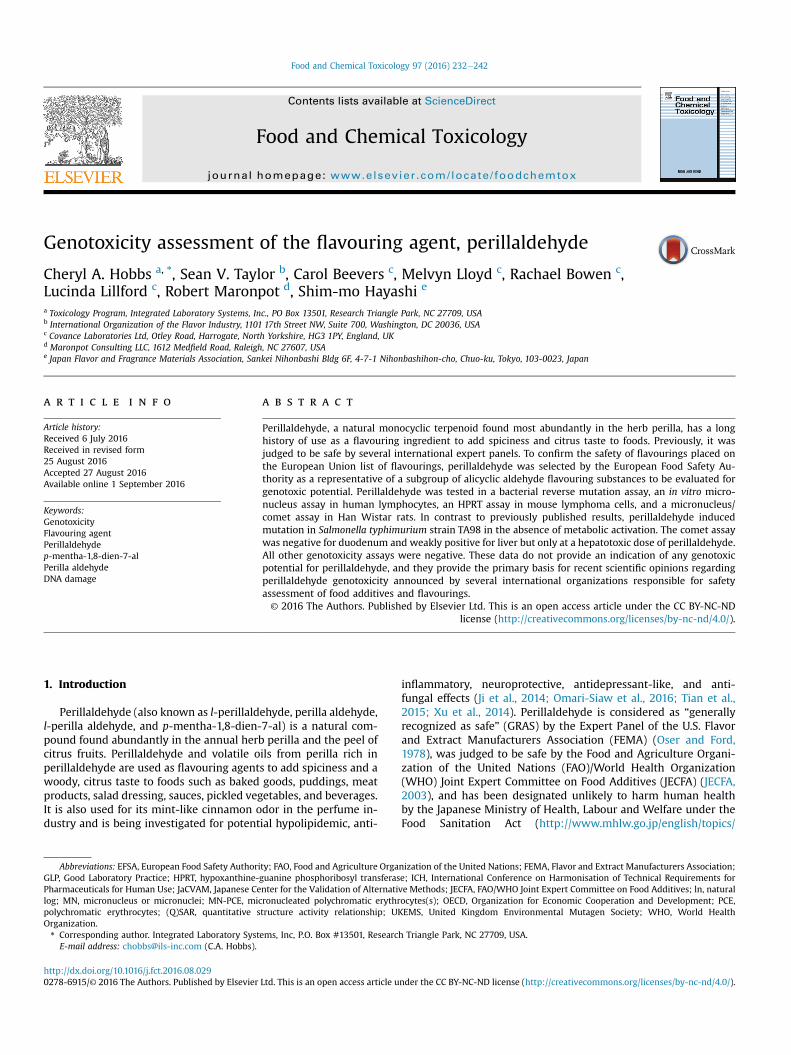
Fig. 1. Chemical structure of perillaldehyde. Perillaldehyde is a naturally occurringmonocyclic terpenoid with an a,b-unsaturated aldehyde structure.
2. Material and methods
2.1. Chemicals
All genotoxicity assays were conducted according to OECD testguidelines or EFSA guidance (comet assay) and were GLP-compliant with the exception that dose formulations were notanalyzed for achieved concentration. Perillaldehyde (91.9e94.2%pure; CAS No. 2111-75-3; Nippon Terpene Chemicals, Inc., Kobe,Japan) solutions were prepared by dissolving in anhydrousanalytical grade dimethyl sulfoxide (DMSO) or suspending in cornoil under subdued light conditions with continual stirring beforeand during dosing. The test article solutions were protected fromlight and used within 2 (MN/comet and HPRTmutation assays) or 5(bacterial mutagenicity and in vitro MN assays) hours of initialformulation. Sodium azide, mitomycin C, vinblastine, and ethylmethanesulfonate were formulated in water; 2-nitrofluorene, 9-aminoacridine, benzo[a]pyrene, 2-aminoanthracene, and cyclo-phosphamidewere formulated in DMSO. Aliquots of stock solutionswere either prepared in advance and stored refrigerated (benzo[a]pyrene and mitomycin C for the bacterial reverse mutation test) orfrozen at �80 �C (2-nitrofluorene, 9-aminoacridine, sodium azide,and cyclophosphamide) in the dark, or they were preparedimmediately prior to use (ethyl methanesulfonate, vinblastine, andmitomycin C for the in vitro MN assay). All chemicals and reagentswere obtained from Sigma-Aldrich Chemical Co. (Poole, UK) orequivalent suppliers unless specifically stated otherwise.
2.2. Bacterial reverse mutation assay
The procedures used in this study were in accordance withOECD Guideline 471 (OECD, 1997a). Mutagenicity assays of peril-laldehyde, with and without metabolic activation, were conductedusing five strains of Salmonella typhimurium bacteria (TA98, TA100,TA1535, TA1537 and TA102). All the tester strains, with theexception of strain TA102, were originally obtained from the UKNCTC. Strain TA102 was derived from a culture obtained fromGlaxoGroup Research Limited. All strains were checked previously for themaintenance of genetic markers. Metabolic activationwas providedby 10% liver post-mitochondrial fraction (S9) prepared from maleSprague Dawley rats induced with Aroclor 1254 (Moltox, Boone,NC). The composition of the S9 mix was: 10% S9, 8 mM MgCl2,33 mM KCl, 1.5 mg/mL glucose-6-phosphate, 3.2 mg/mL b-nico-tinamide adenine dinucleotide phosphate (NADP), 0.1 M phosphatebuffer, 40 mg/mL L-histidine HCl (in 250 mM MgCl2), and 49 mg/mLd-biotin. Strain specific positive controls tested without metabolicactivation were 2-nitrofluorene (TA98; 5 mg/plate), sodium azide(TA100 and TA1535; 2 mg/plate), 9-aminoacridine (TA1537; 50 mg/plate), and mitomycin C (TA102; 0.2 mg/plate). Benzo[a]pyrene(10 mg/plate) and 2-aminoanthracene (TA100, TA1535, TA1537 at5 mg/plate; TA102 at 20 mg/plate) were used as the positive controlsfor TA98, and all other strains, respectively, tested with metabolicactivation. Bacteria were cultured at 37 �C for 10 h in nutrient brothcontaining ampicillin (TA98, TA100) or ampicillin and tetracycline(TA102) as appropriate. Incubation was carried out with shaking inan anhydric incubator. All treatments were completed within 6 h ofthe end of the incubation period. For plate incorporation assays,bacteria, control or test article formulation, and 10% S9 mix orbuffer were added to molten agar at 46 ± 1 �C, mixed rapidly, andpoured onto Vogel-Bonner E plates. For the pre-incubation assay,perillaldehyde or control formulation, bacteria, and 10% S9 mixwere mixed and incubated at 37 ± 1 �C for 1 h prior to the additionof molten agar and plating. Once set, triplicate plates per concen-tration (five plates for vehicle control) were incubated at 37 �C inthe dark for 3 days. Colonies were counted using the Sorcerer
C.A. Hobbs et al. / Food and Chemical Toxicology 97 (2016) 232e242234
Colony Counter (Perceptive Instruments, Ltd., Suffolk, UK) ormanually when confounding factors such as precipitation affectedthe accuracy of the automated counter. The background lawn wasinspected for signs of toxicity. Data were confirmed to meet thefollowing acceptability criteria: 1) the mean vehicle control countsfell within the laboratory's historical 99% confidence intervals forgroup means and/or 2) each vehicle control plate count fell withinthe historical 99% reference ranges, and 3) the positive control platecounts were comparable with the historical 99% reference ranges.
2.3. In vitro micronucleus assay
The methodology used in this study was based on draft OECDTest Guideline 487 (OECD, 2008). An appropriate volume of wholeblood from two healthy, non-smoking male volunteers was drawnfrom the peripheral circulation into heparinized tubes within twodays of culture initiation. Blood was stored refrigerated and pooledusing equal volumes from each donor prior to use. Whole bloodcultures were established in sterile disposable centrifuge tubes byplacing 0.4 mL of pooled heparinized blood into 9 mL of HEPES-buffered RPMI medium containing 10% (v/v) heat inactivated fetalcalf serum and 50 mg/mL gentamycin. Phytohaemagglutinin (PHA,reagent grade, Gibco, Loughborough, UK) was included in the cul-ture medium at a final concentration of approximately 2% tostimulate the lymphocytes to divide. Replicate cultures (four forvehicle controls, two for positive controls and each test articleconcentration) were established for each test condition. Cultureswere incubated at 37 �C for 48 h and rocked continuously. Imme-diately prior to treatment, 0.1 mL of culture medium was removedfrom the 24 h continuous cultures to achieve a final pre-treatmentvolume of 9.3 mL. The S9 mix used for metabolic activation wasprepared by mixing glucose-6-phosphate (180 mg/mL), NADP(25 mg/mL), KCl (150 mM) and rat liver S9 in the ratio 1:1:1:2. Thefinal concentration of the liver homogenate in the test system was2%; an equivalent volume of 150 mM KCl was added to culturestreated in the absence of S9. S9 mix or KCl (0.5 mL per culture) wasadded and the cultureswere treatedwith perillaldehyde or controls(0.1 mL per culture). Cyclophosphamide (CPA; 12.5 mg/mL), mito-mycin C (MMC; 0.08 mg/mL), and vinblastine (VIN; 0.02 mg/mL)were used as indirect and direct-acting positive controls. Cyto-chalasin B in DMSO (0.1 mL) was added to the 24 h continuouscultures at the time of treatment. Cultures were incubated at 37 �Cfor 3 (±S9) or 24 (-S9) hours. Following exposure, the 3 h (±S9)cultures were centrifuged at 300 g for 10 min, washed twice withsterile saline (pre-warmed to 37 �C), and resuspended in fresh pre-warmed medium containing fetal calf serum, gentamycin andCytochalasin B at 6 mg/mL. These cultures were allowed to recoverat 37 �C for 21 h. At the defined sampling time, cultures werecentrifuged at 300 g for 10 min. Pelleted cells were resuspended in4mL of 0.075MKCl at 37 �C for 4min to allow cell swelling to occur.Cells were then fixed by dropping the KCl suspension into fresh,cold methanol/glacial acetic acid (3:1, v/v). The fixative waschanged by centrifugation (300 g, 10 min) and resuspension andthe process repeated as necessary (1250 g, 2e3 min) until the cellpellets were clean.
Lymphocytes were maintained refrigerated in fixative until atleast the following day to ensure adequate fixation. Cells werecentrifuged and resuspended in a minimal amount of fresh fixativeas required to produce a milky suspension. Several drops of cellsuspension were gently spread onto multiple clean microscopeslides and allowed to dry. The slides were stained for 5 min infiltered 4% (v/v) Giemsa in Gurr's phosphate buffer, pH 6.8, rinsed,dried, and mounted with coverslips. Slides were examined forproportions of mono-, bi-, and multinucleate cells (500 cells/cul-ture). Slides from the concentration at which approximately 55%
(typically 50%e60%) reduction in replication index (RI; [numberbinucleate cells þ 2(number multinucleate cells)]/total number ofcells in treated cultures) had occurred and at least two lowerconcentrations were selected for microscopic analysis, such that arange of cytotoxicity from maximum (not excessive) to little wascovered. Coded slides were analyzed by scoring 1000 binucleatecells from each culture (2000 per concentration) for micronuclei.
2.4. In vitro HPRT mutation assay in L5178Y mouse lymphoma cells
The methodology and fluctuation protocol used in this studycomplies with OECD Test Guideline 476 (OECD, 1997b). The stock ofL5178Y tkþ/� (3.7.2C) mouse lymphoma cells was originally pro-vided by Dr. Donald Clive (Burroughs Wellcome Co.). Cells werecultured in RPMI medium containing 100 U/mL penicillin andstreptomycin, 2.5 mg/mL amphotericin B, 0.5 mg/mL Pluronic, and 5or 10% heat inactivated horse serum. For 3 h exposures, at least107 cells in a volume of 18.8 mL of RPMI medium (5% serum) wereseeded in sterile 50mL centrifuge tubes. For 24 h exposures, at least4 � 106 cells in a volume of 19.8 mL RPMI medium (10% serum)were seeded in 75 cm2 tissue culture flasks. For all exposure con-ditions, 0.2 mL of vehicle, perillaldehyde, or positive control solu-tions was added to the culture. The S9 mix used for metabolicactivation was prepared by mixing glucose-6-phosphate (180 mg/mL), NADP (25mg/mL), KCl (150mM) and Aroclor 1254-induced ratliver S9 (Moltox, Boone, NC) in the ratio 1:1:1:2. The final con-centration of the liver homogenate in the test system was 2%; anequivalent volume of 150 mM KCl was added to cultures treated inthe absence of S9. Duplicate (single for positive controls) exposures(±S9) were performed in a final volume of 20 mL. After incubationfor 3 h at 37 �C with gentle agitation or stationary incubation at37 �C for 24 h, cultures were centrifuged at 200 g for 5 min, washed,and resuspended in 20 mL RPMI medium (10% serum). Cell den-sities were adjusted to 2 � 105 cells/mL and cells were transferredto flasks for growth throughout the expression period or werediluted and plated for survival assessment. For survival measure-ments, 0.2 mL of cells diluted to 8 cells/mL was placed into eachwell of two 96-well microtiter plates. The plates were incubated at37 �C in a humidified incubator with 5% v/v CO2 in air for 7 daysuntil scoreable. Wells containing viable clones were visually iden-tified using background illumination and counted. For mutationfrequency assessment, cultures weremaintained in flasks for 7 daysto allow for expression of Hprt- mutations. Sub-culturing was per-formed as necessary to avoid exceeding 1 � 106 cells/mL, retainingat least 6 � 106 cells/flask if possible.
Cultures to be plated for viability and 6-thioguanine resistancewere selected on the basis of observations of recovery and growthof the cultures during the expression period. Following the 7 dayexpression period, cell densities in the selected cultures wereadjusted to 1� 105 cells/mL. For viability assessment, samples fromthese were further diluted to 8 cells/mL, plated, and scored after7e8 days as described above. For mutation frequency assessment,6-thioguanine was added to the cultures to give a final concen-tration of 15 mg/mL and 0.2 mL of each suspension was transferredto each well of four 96-well microtiter plates. Plates were incubateduntil scoreable (12e14 days), and wells containing clones wereidentified and counted.
From the zero term of the Poisson distribution, the probablenumber of clones/well (P) on microtiter plates is given by the for-mula: P ¼ -ln(empty wells/total wells). Plating efficiency (PE) ofeach culture was calculated as PE ¼ P/number of cells plated perwell. Percentage relative survival (% RS) in each test culture wasdetermined by comparing plating efficiencies in test and controlcultures: % RS ¼ [PE(test)/PE(control)] � 100. Any loss of cells duringthe 3 h exposure period was accounted for by adjusting the % RS
C.A. Hobbs et al. / Food and Chemical Toxicology 97 (2016) 232e242 235
values for each test article concentration as follows: adjusted %RS ¼ % RS � (post-treatment cell concentration for test articletreatment/post-treatment cell concentration for vehicle control).Mutant frequency (MF) is usually expressed as “mutants/106 viablecells”: MF ¼ [PE(mutant)/PE(viable)] � 106. Since 2 � 104 cells wereplated/well for mutation to 6-thioguanine resistance and anaverage of 1.6 cells were plated/well on viability plates,MF¼ [P(mutant)/2� 104]/[P(viable)/1.6]� 106¼ (-ln[empty wells/totalwells(mutant)]/-ln[empty wells/total wells(viable)]) � 80.
2.5. In vivo micronucleus/comet assay
2.5.1. Animal husbandryMale Han Wistar rats (Charles River (UK) Ltd., Margate, UK)
were 8e10 weeks of age at the time of treatment. Animals werehoused in wire topped, solid bottomed cages (three animals percage) with European softwood bedding (Datesand Ltd., Manchester,UK), in a temperature and humidity controlled AAALAC accreditedfacility with a 12 h light/12 h dark cycle. SQC Rat and MouseMaintenance Diet No 1, Expanded (Special Diets Services Ltd.,Witham, UK) and water were provided ad libitum. Animals wereprovided wooden Aspen chew blocks and rodent retreats forenvironmental enrichment and were acclimatized for at least 5days prior to initiation of dosing.
2.5.2. Experimental designA combined micronucleus and comet assay was conducted in
accordance with OECD test guideline 474 (OECD, 1997c) for themicronucleus assay and current recommendations for the cometassay at the time the study was conducted (Burlinson et al., 2007;EFSA, 2012; Tice et al., 2000). A dose range-finding study wasconducted to estimate the maximum tolerated dose (MTD) ofperillaldehyde in male and female rats. Since no substantial genderdifferences in toxicity were revealed in the range-finder experi-ment, male rats were selected for the MN/comet assay. Five groupsof male rats (6 rats/group) were administered perillaldehyde atdose levels of 175, 350, and 700 mg/kg/day (estimated to be 25%MTD, 50% MTD, and MTD, respectively), vehicle (corn oil), or thepositive control compound, ethyl methanesulfonate in purifiedwater at 150 mg/kg/day, by oral gavage daily for three days (0, 24and 45 h). Three hours after the final dose the animals wereeuthanized and femurs were collected for analysis of micronuclei inthe bone marrow, liver and duodenum tissues were collected forthe comet assay and histopathology, and blood samples werecollected for clinical chemistry as described below.
2.5.3. Erythrocyte micronucleus assayFemurs were cleaned of adherent tissue and the ends removed
from the shanks. Using a syringe and needle, bone marrow wasflushed from each femur with 2 mL of fetal bovine serum (FBS). Thesamples were filtered through cellulose columns containing anequal mix of type 50 and a-cellulose at 50 mg/mL. Once the ma-jority of the FBS had passed through the column an additional 4 mLof FBS were added to the sample tubes and loaded onto the col-umns. The filtered bone marrow cells were centrifuged at 200 g for5 min at 15e25 �C and the cell pellets were gently resuspended in3 mL of FBS and pelleted again; the supernatant was aspirated toleave one or two drops used to resuspend the cell pellet using aPasteur pipette. One drop of cell suspension was placed on the endof each slide and smeared by drawing the end of a clean slide alongthe sample slide. Slides were air-dried prior to fixing for 10 min inabsolute methanol, rinsed several times in distilled water, and air-dried. One slide per animal was immediately stained for 5 min in12.5 mg/mL acridine orange in 0.1 M phosphate buffer pH 7.4.Stained slides were rinsed in phosphate buffer, then dried and
stored protected from light at room temperature. At least 500erythrocytes were analyzed for the relative proportions of poly-chromatic erythrocytes (PCE), visible as bright orange enucleatedcells, and normochromatic erythrocytes (NCE), visible as smallerdark green enucleated cells. At least 2000 PCE/animal wereexamined for the presence of micronuclei. All slides were coded toeliminate scorer bias.
2.5.4. Comet assayLiver samples were washed thoroughly in Merchants solution
(0.5 mM NaEDTA, 10% DMSO in phosphate buffered saline, pH 7.4),cut into small pieces in fresh Merchants solution, and pushedthrough bolting cloth (pore size of 150 mm) with approximately4 mL of Merchants solution to produce single cell suspensions.Duodenum samples were washed thoroughly in Merchants solu-tion and vortexed in fresh Merchants solution for approximately15 s. The tissue was removed from the Merchants solution and theinner surface gently scraped (released material discarded) usingthe back of a scalpel blade. The tissue was vortexed in Merchantssolution for a further 15 s prior to gently scraping the inside of theduodenum with the back of a scalpel blade. All cell suspensionswere maintained on ice prior to slide preparation. Four slides wereprepared per tissue for each animal. Slides were dipped in moltennormal melting point agarose, the underside of the slides waswiped clean, and the slides were allowed to dry. Each single cellsuspension (40 mL) was added to 400 mL of 0.7% low melting pointagarose at approximately 37 �C and 100 mL of cell suspension/agarose mix were placed on each slide. The slides were then cov-erslipped and placed on ice. Once gelled, coverslips were removedand the slides placed in lysis buffer (2.5 M NaCl, 100 mM EDTA,10 mM Tris, pH 10 [adjusted with NaOH], 1% Triton X-100, 10%DMSO) at 2e8 �C protected from light. One slide for each animalwas incubated in lysis buffer for 1 h (“halo”); remaining slides(comet assay) were lysed overnight. Following lysis, the slides forthe comet assay were washed in purified water for 5 min andtransferred to electrophoresis buffer (300 mM NaOH, 1 mM EDTA,pH > 13) at 2e8 �C to allow the DNA to unwind for 20 min (duo-denum) or 30 min (liver). The slides were electrophoresed in thesame buffer at 0.7 V/cm for 20 min (duodenum) or 40 min (liver). Ablock design was employed for the unwinding and electrophoreticsteps in order to avoid excessive variation across the groups foreach electrophoretic run (i.e., the same number of slides was pro-cessed at a time for each animal). Following electrophoresis, slideswere neutralized in 0.4M Tris, pH 7.0 (3� 5minwashes), dried, andstored at room temperature. Following lysis, the “halo” slides wereplaced in 0.4 M Tris, pH 7.0 for approximately 3 � 5 min and thendried and stored as described above.
Immediately prior to scoring, coded slides were stained with100 mL of 2 mg/mL ethidium bromide and coverslipped. Scoring wascarried out using fluorescence microscopy at an appropriatemagnification and with suitable filters. Comet scoring was per-formed using “Comet Assay IV” image analysis system (PerceptiveInstruments, Suffolk, U.K.). Measurements of tail intensity (% DNAin tail) and Olive tail moment were generally obtained from at least150 cells/animal scored on 2e3 slides. The number of “hedgehog”cells [a morphology indicative of highly damaged cells which canbe associated with severe cytotoxicity, necrosis or apoptosis(Burlinson et al., 2007; Lorenzo et al., 2013; Tice et al., 2000); alsoknown as “clouds” and “ghosts”] observed during comet scoringwas tabulated for each animal but these cells were not scored ascomets. Halo slides, used to evaluate cytotoxicity, were scored byassessing 100 cells/slide and recording the number with a clearhalo of DNA around the nucleus. Acceptable cytotoxicity criteriawere considered to be �30% hedgehogs and/or �30% cells withhalos in the vehicle control.
C.A. Hobbs et al. / Food and Chemical Toxicology 97 (2016) 232e242236
2.5.5. Clinical chemistry and histopathologyBlood obtained from the abdominal aorta under deep anesthesia
was collected into lithium heparin anticoagulant, mixed for twominutes, cooled, centrifuged at 2300 g for 10 min at 4 �C, andplasma was analyzed for clinical chemistry parameters (see Table 7for list of parameters). Samples of liver were preserved in neutralbuffered formalin, embedded in paraffin wax, sectioned at 5 mm,and stained with haematoxylin and eosin. Slides were examined bythe study pathologist and digitized whole slide scans underwentpeer-review evaluation.
2.6. Statistical analyses
For the bacterial mutagenicity study, Dunnett's test was used tocompare the counts at each concentration with the vehicle controland the presence of a concentration-dependent response wasassessed. The test was considered positive for mutagenicity if areproducible concentration related significant response (p � 0.01)was induced. For the in vitro micronucleus test, the proportions ofbinucleate cells with micronuclei in the replicate samples wereused to establish acceptable heterogeneity between replicates bymeans of a binomial dispersion test. The proportion of binucleatecells with micronuclei for each treatment condition was comparedwith the proportion in negative controls by using Fisher's exact test.
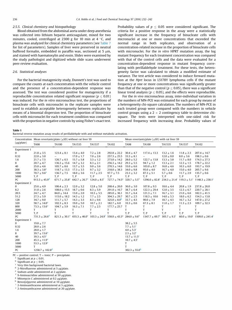
Table 1Bacterial reverse mutation assay results of perillaldehyde with and without metabolic ac
Concentration(mg/plate)
Mean revertants/plate (±SD) without rat liver S9
TA98 TA100 TA1535 TA1537 TA102
Experiment 10 21.8 ± 3.5 123.4 ± 8.1 13.4 ± 4.0 7.2 ± 2.8 292.8 ±0.32 22.0 ± 3.0 e 17.0 ± 1.7 7.0 ± 2.6 257.3 ±1.6 21.7 ± 7.5 126.7 ± 6.5 15.7 ± 5.8 3.3 ± 1.2 273.0 ±8 20.7 ± 4.7 136.3 ± 15.6 14.7 ± 3.2 8.3 ± 2.1 256.3 ±40 25.0 ± 4.6 105.7 ± 8.6 13.7 ± 3.5 9.0 ± 3.6 270.3 ±200 38.3 ± 3.8a 118.7 ± 13.3 17.3 ± 3.5 9.7 ± 3.2 218.0 ±1000 70.7 ± 9.6a 136.7 ± 7.5 18.0 ± 3.6 11.7 ± 2.5 97.7 ± 75000 T, P T, P T, P T, P T, PPC 913.3 ± 45.9d 671.7 ± 25.8e 642.7 ± 26.7e 124.0 ± 8.5f 727.7 ±Experiment 20 25.6 ± 4.9 106.4 ± 2.3 12.0 ± 3.2 12.8 ± 5.0 268.4 ±8.2 31.0 ± 2.6 108.0 ± 15.5 18.7 ± 0.6 8.3 ± 5.9 291.0 ±20.5 24.7 ± 4.7 84.0 ± 14.4 13.0 ± 2.0 10.3 ± 5.5 285.0 ±51.2 27.3 ± 5.9 115.0 ± 18.2 14.3 ± 1.2 7.7 ± 2.5 304.3 ±128 34.7 ± 9.0 111.7 ± 5.7 14.3 ± 3.5 8.3 ± 0.6 325.0 ±320 58.7 ± 14.0a 102.3 ± 8.3 19.0 ± 5.0 10.7 ± 2.1 182.7 ±800 73.3 ± 13.6a 104.7 ± 5.9 16.3 ± 7.1 7.7 ± 2.5 177.7 ±2000 T T T T T5000 T, P T T T TPC 731.3 ± 26.6d 821.3 ± 30.1e 655.3 ± 46.0e 103.3 ± 24.9f 558.0 ±Experiment 30 19.8 ± 7.70.32 28.0 ± 2.61.6 28.0 ± 1.78 39.7 ± 13.6a
40 39.3 ± 4.5a
200 45.3 ± 11.9a
1000 53.3 ± 12.9a
5000 TPC 1258.7 ± 102.8d
PC ¼ positive control; T ¼ toxic; P ¼ precipitate.a Significant at p � 0.01.b Significant at p � 0.05.c Very thin background bacterial lawn.d 2-Nitrofluorene administered at 5 mg/plate.e Sodium azide administered at 2 mg/plate.f 9-Aminoacridine administered at 50 mg/plate.g Mitomycin C administered at 0.2 mg/plate.h Benzo[a]pyrene administered at 10 mg/plate.i 2-Aminoanthracene administered at 5 mg/plate.j 2-Aminoanthracene administered at 20 mg/plate.
Probability values of p � 0.05 were considered significant. Thecriteria for a positive response in the assay were a statisticallysignificant increase in the frequency of binucleate cells withmicronuclei at one or more concentrations that exceeded thenormal range in both replicates, and observation of aconcentration-related increase in the proportion of binucleate cellswith micronuclei. For the in vitro HPRT mutation assay, the logmutant frequency for each treatment concentration was comparedwith that of the control cells and the data were evaluated for aconcentration-dependent response in mutant frequency corre-lating with perillaldehyde treatment. For these tests, the hetero-geneity factor was calculated to obtain a modified estimate ofvariance. The test article was considered to induce forward muta-tion at the Hprt locus in L5178Y lymphoma cells if the mutantfrequency at one or more concentrations was significantly greaterthan that of the negative control (p � 0.05), there was a significantlinear trend analysis (p � 0.05), and the effects were reproducible.
For the in vivo micronucleus assay, inter-individual variation inthe numbers of MN-PCE was estimated for each group by means ofa heterogeneity chi-square calculation. The numbers of MN-PCE ineach treated group were compared with the numbers in vehiclecontrol groups using a 2 � 2 contingency table to determine chi-square. The tests were interpreted with one-sided risk forincreased frequency with increasing dose. Probability values of
tivation.
Mean revertants/plate (±SD) with rat liver S9
TA98 TA100 TA1535 TA1537 TA102
22.2 30.4 ± 4.7 117.4 ± 13.3 13.2 ± 1.6 11.8 ± 2.3 207.4 ± 14.725.5 33.3 ± 8.1 e 12.0 ± 0.0 8.0 ± 3.6 198.3 ± 0.614.2 28.0 ± 5.2 122.7 ± 13.8 13.3 ± 3.8 11.7 ± 8.0 174.3 ± 27.314.2 29.3 ± 5.5 94.7 ± 1.2 13.3 ± 2.1 12.3 ± 1.5 178.7 ± 22.214.6 33.0 ± 6.6 103.0 ± 8.7 16.0 ± 4.6 10.3 ± 6.0 193.7 ± 19.938.3 34.0 ± 9.8 95.0 ± 4.0 16.7 ± 4.0 15.0 ± 4.0 205.7 ± 9.6.5 23.3 ± 3.2 87.3 ± 5.1 5.7 ± 0.6 11.7 ± 2.9 135.7 ± 0.6
T, P T, P T, P T, P T, P74.5g 320.7 ± 5.5h 1296.0 ± 82.8i 234.3 ± 21.4i 119.3 ± 5.1i 1148.3 ± 238.1j
26.0 36.0 ± 3.0 107.8 ± 9.3 18.6 ± 4.4 20.8 ± 1.9 237.8 ± 26.016.7 38.7 ± 6.4 122.3 ± 29.4 12.0 ± 3.5 12.3 ± 6.7 220.7 ± 28.136.1 35.7 ± 6.4 121.3 ± 7.1 16.7 ± 3.1 21.0 ± 6.6 182.3 ± 41.529.7 30.7 ± 2.5 118.3 ± 10.6 14.0 ± 5.3 18.0 ± 6.2 154.3 ± 13.88.0b 33.7 ± 4.5 90.0 ± 7.9 18.7 ± 4.5 16.7 ± 3.2 147.0 ± 27.26.0 33.3 ± 0.6 67.3 ± 8.1 11.0 ± 1.7 11.3 ± 2.3 109.7 ± 32.325.7 T T T T T
T T T T TT, P T, P T, P T, P T, P
43.3g 284.0 ± 9.6h 1347.7 ± 69.7i 305.7 ± 9.3i 60.0 ± 19.0i 1509.0 ± 241.4j
38.2 ± 5.57.7 ± 5.1c
4.0 ± 5.3c
2.3 ± 3.2c
13.7 ± 11.5c
10.7 ± 4.5c
TT302.3 ± 55.0h
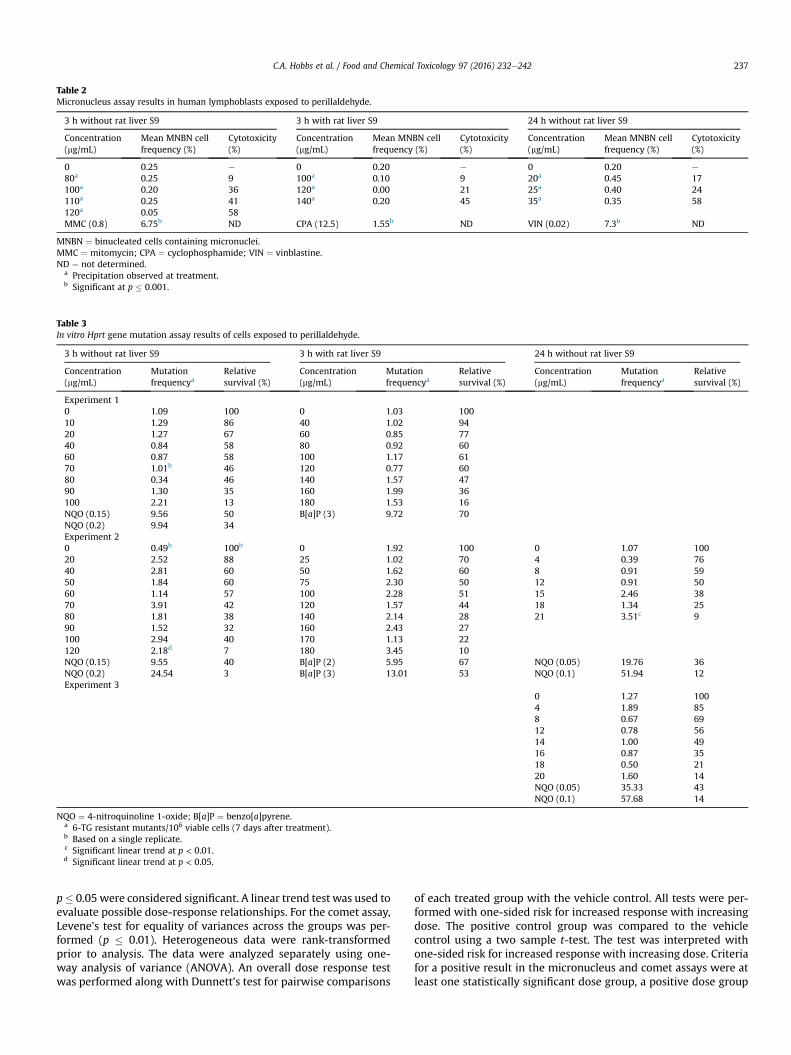
Table 2Micronucleus assay results in human lymphoblasts exposed to perillaldehyde.
3 h without rat liver S9 3 h with rat liver S9 24 h without rat liver S9
Concentration(mg/mL)
Mean MNBN cellfrequency (%)
Cytotoxicity(%)
Concentration(mg/mL)
Mean MNBN cellfrequency (%)
Cytotoxicity(%)
Concentration(mg/mL)
Mean MNBN cellfrequency (%)
Cytotoxicity(%)
0 0.25 e 0 0.20 e 0 0.20 e
80a 0.25 9 100a 0.10 9 20a 0.45 17100a 0.20 36 120a 0.00 21 25a 0.40 24110a 0.25 41 140a 0.20 45 35a 0.35 58120a 0.05 58MMC (0.8) 6.75b ND CPA (12.5) 1.55b ND VIN (0.02) 7.3b ND
MNBN ¼ binucleated cells containing micronuclei.MMC ¼ mitomycin; CPA ¼ cyclophosphamide; VIN ¼ vinblastine.ND ¼ not determined.
a Precipitation observed at treatment.b Significant at p � 0.001.
Table 3In vitro Hprt gene mutation assay results of cells exposed to perillaldehyde.
3 h without rat liver S9 3 h with rat liver S9 24 h without rat liver S9
Concentration(mg/mL)
Mutationfrequencya
Relativesurvival (%)
Concentration(mg/mL)
Mutationfrequencya
Relativesurvival (%)
Concentration(mg/mL)
Mutationfrequencya
Relativesurvival (%)
Experiment 10 1.09 100 0 1.03 10010 1.29 86 40 1.02 9420 1.27 67 60 0.85 7740 0.84 58 80 0.92 6060 0.87 58 100 1.17 6170 1.01b 46 120 0.77 6080 0.34 46 140 1.57 4790 1.30 35 160 1.99 36100 2.21 13 180 1.53 16NQO (0.15) 9.56 50 B[a]P (3) 9.72 70NQO (0.2) 9.94 34Experiment 20 0.49b 100b 0 1.92 100 0 1.07 10020 2.52 88 25 1.02 70 4 0.39 7640 2.81 60 50 1.62 60 8 0.91 5950 1.84 60 75 2.30 50 12 0.91 5060 1.14 57 100 2.28 51 15 2.46 3870 3.91 42 120 1.57 44 18 1.34 2580 1.81 38 140 2.14 28 21 3.51c 990 1.52 32 160 2.43 27100 2.94 40 170 1.13 22120 2.18d 7 180 3.45 10NQO (0.15) 9.55 40 B[a]P (2) 5.95 67 NQO (0.05) 19.76 36NQO (0.2) 24.54 3 B[a]P (3) 13.01 53 NQO (0.1) 51.94 12Experiment 3
0 1.27 1004 1.89 858 0.67 6912 0.78 5614 1.00 4916 0.87 3518 0.50 2120 1.60 14NQO (0.05) 35.33 43NQO (0.1) 57.68 14
NQO ¼ 4-nitroquinoline 1-oxide; B[a]P ¼ benzo[a]pyrene.a 6-TG resistant mutants/106 viable cells (7 days after treatment).b Based on a single replicate.c Significant linear trend at p < 0.01.d Significant linear trend at p < 0.05.
C.A. Hobbs et al. / Food and Chemical Toxicology 97 (2016) 232e242 237
p� 0.05 were considered significant. A linear trend test was used toevaluate possible dose-response relationships. For the comet assay,Levene's test for equality of variances across the groups was per-formed (p � 0.01). Heterogeneous data were rank-transformedprior to analysis. The data were analyzed separately using one-way analysis of variance (ANOVA). An overall dose response testwas performed along with Dunnett's test for pairwise comparisons
of each treated group with the vehicle control. All tests were per-formed with one-sided risk for increased response with increasingdose. The positive control group was compared to the vehiclecontrol using a two sample t-test. The test was interpreted withone-sided risk for increased response with increasing dose. Criteriafor a positive result in the micronucleus and comet assays were atleast one statistically significant dose group, a positive dose group
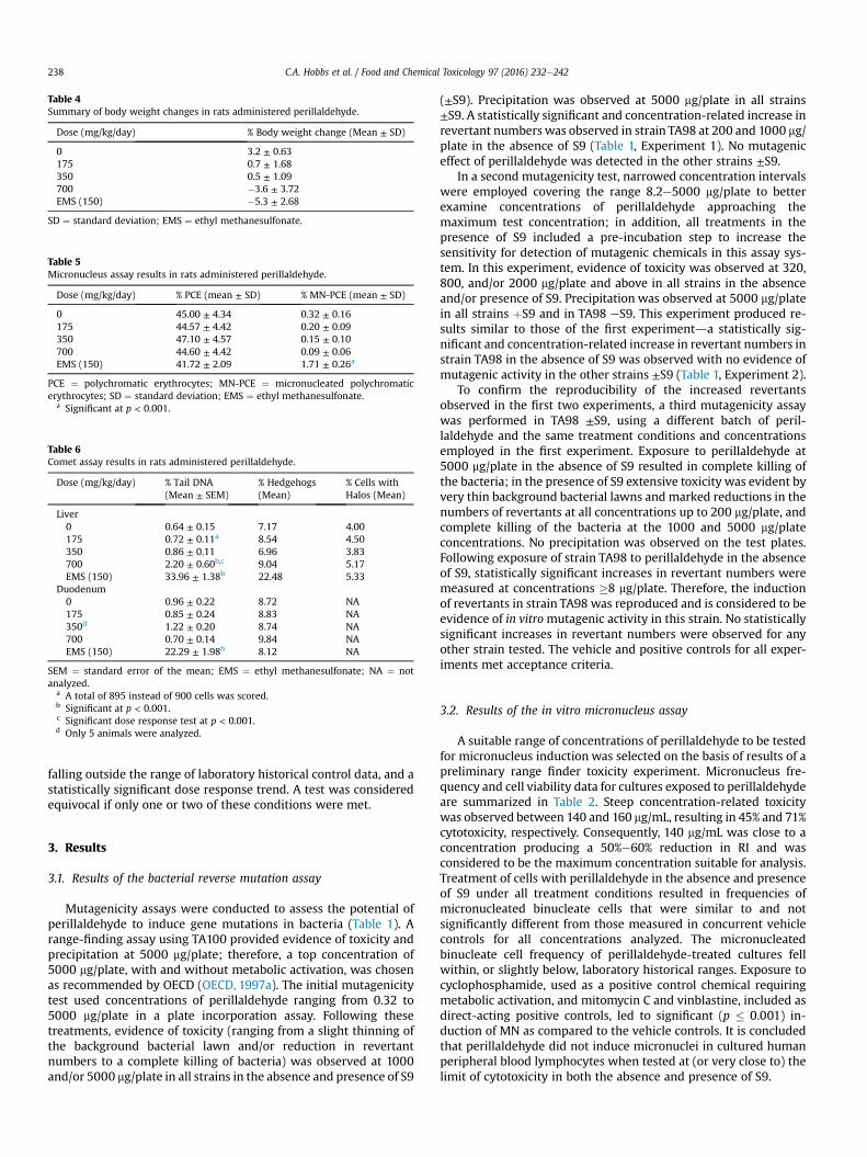
Table 4Summary of body weight changes in rats administered perillaldehyde.
Dose (mg/kg/day) % Body weight change (Mean ± SD)
0 3.2 ± 0.63175 0.7 ± 1.68350 0.5 ± 1.09700 �3.6 ± 3.72EMS (150) �5.3 ± 2.68
SD ¼ standard deviation; EMS ¼ ethyl methanesulfonate.
Table 5Micronucleus assay results in rats administered perillaldehyde.
Dose (mg/kg/day) % PCE (mean ± SD) % MN-PCE (mean ± SD)
0 45.00 ± 4.34 0.32 ± 0.16175 44.57 ± 4.42 0.20 ± 0.09350 47.10 ± 4.57 0.15 ± 0.10700 44.60 ± 4.42 0.09 ± 0.06EMS (150) 41.72 ± 2.09 1.71 ± 0.26a
PCE ¼ polychromatic erythrocytes; MN-PCE ¼ micronucleated polychromaticerythrocytes; SD ¼ standard deviation; EMS ¼ ethyl methanesulfonate.
a Significant at p < 0.001.
Table 6Comet assay results in rats administered perillaldehyde.
Dose (mg/kg/day) % Tail DNA(Mean ± SEM)
% Hedgehogs(Mean)
% Cells withHalos (Mean)
Liver0 0.64 ± 0.15 7.17 4.00175 0.72 ± 0.11a 8.54 4.50350 0.86 ± 0.11 6.96 3.83700 2.20 ± 0.60b,c 9.04 5.17EMS (150) 33.96 ± 1.38b 22.48 5.33
Duodenum0 0.96 ± 0.22 8.72 NA175 0.85 ± 0.24 8.83 NA350d 1.22 ± 0.20 8.74 NA700 0.70 ± 0.14 9.84 NAEMS (150) 22.29 ± 1.98b 8.12 NA
SEM ¼ standard error of the mean; EMS ¼ ethyl methanesulfonate; NA ¼ notanalyzed.
a A total of 895 instead of 900 cells was scored.b Significant at p < 0.001.c Significant dose response test at p < 0.001.d Only 5 animals were analyzed.
C.A. Hobbs et al. / Food and Chemical Toxicology 97 (2016) 232e242238
falling outside the range of laboratory historical control data, and astatistically significant dose response trend. A test was consideredequivocal if only one or two of these conditions were met.
3. Results
3.1. Results of the bacterial reverse mutation assay
Mutagenicity assays were conducted to assess the potential ofperillaldehyde to induce gene mutations in bacteria (Table 1). Arange-finding assay using TA100 provided evidence of toxicity andprecipitation at 5000 mg/plate; therefore, a top concentration of5000 mg/plate, with and without metabolic activation, was chosenas recommended by OECD (OECD, 1997a). The initial mutagenicitytest used concentrations of perillaldehyde ranging from 0.32 to5000 mg/plate in a plate incorporation assay. Following thesetreatments, evidence of toxicity (ranging from a slight thinning ofthe background bacterial lawn and/or reduction in revertantnumbers to a complete killing of bacteria) was observed at 1000and/or 5000 mg/plate in all strains in the absence and presence of S9
(±S9). Precipitation was observed at 5000 mg/plate in all strains±S9. A statistically significant and concentration-related increase inrevertant numbers was observed in strain TA98 at 200 and 1000 mg/plate in the absence of S9 (Table 1, Experiment 1). No mutageniceffect of perillaldehyde was detected in the other strains ±S9.
In a second mutagenicity test, narrowed concentration intervalswere employed covering the range 8.2e5000 mg/plate to betterexamine concentrations of perillaldehyde approaching themaximum test concentration; in addition, all treatments in thepresence of S9 included a pre-incubation step to increase thesensitivity for detection of mutagenic chemicals in this assay sys-tem. In this experiment, evidence of toxicity was observed at 320,800, and/or 2000 mg/plate and above in all strains in the absenceand/or presence of S9. Precipitation was observed at 5000 mg/platein all strains þS9 and in TA98 eS9. This experiment produced re-sults similar to those of the first experimentda statistically sig-nificant and concentration-related increase in revertant numbers instrain TA98 in the absence of S9 was observed with no evidence ofmutagenic activity in the other strains ±S9 (Table 1, Experiment 2).
To confirm the reproducibility of the increased revertantsobserved in the first two experiments, a third mutagenicity assaywas performed in TA98 ±S9, using a different batch of peril-laldehyde and the same treatment conditions and concentrationsemployed in the first experiment. Exposure to perillaldehyde at5000 mg/plate in the absence of S9 resulted in complete killing ofthe bacteria; in the presence of S9 extensive toxicity was evident byvery thin background bacterial lawns and marked reductions in thenumbers of revertants at all concentrations up to 200 mg/plate, andcomplete killing of the bacteria at the 1000 and 5000 mg/plateconcentrations. No precipitation was observed on the test plates.Following exposure of strain TA98 to perillaldehyde in the absenceof S9, statistically significant increases in revertant numbers weremeasured at concentrations �8 mg/plate. Therefore, the inductionof revertants in strain TA98 was reproduced and is considered to beevidence of in vitromutagenic activity in this strain. No statisticallysignificant increases in revertant numbers were observed for anyother strain tested. The vehicle and positive controls for all exper-iments met acceptance criteria.
3.2. Results of the in vitro micronucleus assay
A suitable range of concentrations of perillaldehyde to be testedfor micronucleus induction was selected on the basis of results of apreliminary range finder toxicity experiment. Micronucleus fre-quency and cell viability data for cultures exposed to perillaldehydeare summarized in Table 2. Steep concentration-related toxicitywas observed between 140 and 160 mg/mL, resulting in 45% and 71%cytotoxicity, respectively. Consequently, 140 mg/mL was close to aconcentration producing a 50%e60% reduction in RI and wasconsidered to be the maximum concentration suitable for analysis.Treatment of cells with perillaldehyde in the absence and presenceof S9 under all treatment conditions resulted in frequencies ofmicronucleated binucleate cells that were similar to and notsignificantly different from those measured in concurrent vehiclecontrols for all concentrations analyzed. The micronucleatedbinucleate cell frequency of perillaldehyde-treated cultures fellwithin, or slightly below, laboratory historical ranges. Exposure tocyclophosphamide, used as a positive control chemical requiringmetabolic activation, and mitomycin C and vinblastine, included asdirect-acting positive controls, led to significant (p � 0.001) in-duction of MN as compared to the vehicle controls. It is concludedthat perillaldehyde did not induce micronuclei in cultured humanperipheral blood lymphocytes when tested at (or very close to) thelimit of cytotoxicity in both the absence and presence of S9.
Table 7Summary of clinical chemistry parameters in plasma of rats administered perillaldehyde.a
Clinical chemistry Dose (mg/kg/day)
0 175 350 700
Aspartate aminotransferase (IU/L) 78 ± 8.4 (5)b 77 ± 10.4 (5) 79 ± 7.9 (5) 110 ± 44.9 (6)Alanine aminotransferase (IU/L) 72 ± 15.1 (6) 53 ± 11.5 (5) 64 ± 12.0 (5) 89 ± 27.7 (6)Alkaline phosphatase (IU/L) 220 ± 44.6 (6) 182 ± 12.7 (6) 216 ± 32.7 (6) 216 ± 45.5 (6)Total cholesterol (mmol/L) 2.0 ± 0.24 (6) 1.7 ± 0.14 (6) 1.8 ± 0.24 (6) 1.5 ± 0.27c (6)Bilirubin (mmol/L)d <1.7 (1) <1.7 ± 0.00 (3) <1.7 ± 0.00 (2) <1.8 ± 0.15 (4)Total protein (g/L) 56 ± 1.5 (6) 58 ± 2.8 (6) 58 ± 1.8 (6) 59 ± 2.8 (6)Albumin (g/L) 39 ± 1.4 (6) 41 ± 2.7 (6) 41 ± 1.8 (6) 43 ± 2.3e (6)Globulin (g/L) 17 ± 1.6 (6) 17 ± 1.5 (6) 17 ± 0.8 (6) 16 ± 1.4 (6)Albumin/globulin ratio 2.4 ± 0.29 (6) 2.5 ± 0.29 (6) 2.4 ± 0.16 (6) 2.7 ± 0.28 (6)Sodium (mmole/L) 138 ± 1.6 (6) 139 ± 1.4 (6) 139 ± 1.4 (6) 135 ± 1.6c (6)Potassium (mmole/L) 4.5 ± 0.41 (6) 4.3 ± 0.24 (6) 4.3 ± 0.15 (6) 3.2 ± 0.44f (6)Chloride (mmole/L) 100 ± 1.6 (6) 99 ± 1.3 (6) 99 ± 1.4 (6) 94 ± 2.2f (6)Calcium (mmol/L) 2.56 ± 0.072 (6) 2.61 ± 0.054 (6) 2.65 ± 0.061 (6) 2.51 ± 0.032g (6)Inorganic phosphorus (mmol/L) 2.2 ± 0.16 (6) 2.2 ± 0.21(6) 2.4 ± 0.10 (6) 2.9 ± 0.31f (6)Creatinine (mmol/L) 21 ± 2.1 (6) 25 ± 2.7 (6) 23 ± 2.2 (6) 23 ± 3.1 (6)Urea (mmol/L) 3.6 ± 0.22 (6) 4.3 ± 1.01 (6) 3.9 ± 0.42 (6) 6.8 ± 1.77f (6)Glucose (mmol/L) 11.8 ± 1.45 (6) 11.9 ± 1.03 (6) 9.7 ± 1.14 (6) 5.4 ± 2.52f,g (6)
a Mean ± standard deviation.b Number of animals is provided in parentheses.c Significant at p < 0.005.d Not analyzed for statistical significance.e Significant at p < 0.05.f Significant at p < 0.0001.g Significant trend at p < 0.05.
C.A. Hobbs et al. / Food and Chemical Toxicology 97 (2016) 232e242 239
3.3. Results of the in vitro HPRT mutation assay
Following cytotoxicity range-finder experiments to determineconcentrations of perillaldehyde providing >10% relative survivalunder different test conditions, perillaldehyde was assayed for theability to induce mutation at the hypoxanthine-guanine phos-phoribosyl transferase (Hprt) locus [6-thioguanine resistance] inmouse L5178Y lymphoma cells. Results of the HPRT mutation assayare summarized in Table 3. An initial experiment was conducted for3 h in the absence and presence of metabolic activation. The highestconcentrations analyzed to determine viability and 6-thioguanineresistance were 100 mg/mL in the absence of S9 and 180 mg/mL inthe presence of S9, which resulted in 13% and 16% relative survival,respectively. A second experiment was conducted for 3 and 24 h inthe absence of S9 and 3 h in the presence of S9. None of the testedconcentrations for the 3 and 24 h (-S9) conditions produced10e20% relative survival; therefore, the highest concentrationselected to determine viability and 6-thioguanine resistance for the3 and 24 h (-S9) conditions were 120 mg/mL (which resulted in 7%relative survival) and 21 mg/mL (which resulted in 9% relative sur-vival), respectively. The highest concentration analyzed for the 3 h(þS9) condition was 180 mg/mL, which resulted in 10% relativesurvival. Subsequently, a third experiment was conducted for 24 hin the absence of S9 to clarify the equivocal results for this treat-ment condition obtained in the previous experiment. In the thirdexperiment, the highest concentration analyzed to determineviability and 6-thioguanine resistance was 20 mg/mL, which resul-ted in 14% relative survival. In all three experiments, no statisticallysignificant increases in mutant frequency were detected followingexposure to perillaldehyde at any concentration tested in theabsence and presence of metabolic activation. Statistically signifi-cant linear trendsweremeasured following 3 and 24 h exposures inthe absence of S9 in the second experiment; however, there wereno statistically significant increases in mutant frequency at anyconcentration evaluated under these conditions. The small, spo-radic increases in the magnitude of mutant frequency and thepositive linear trend observed in the second experiment were notreproduced in the third experiment. In all three experiments,
mutant frequencies in vehicle control cultures fell within accept-able ranges based on laboratory historical data and significant in-creases inmutationwere induced by the positive control chemicals,4-nitroquinoline 1-oxide (-S9) and benzo[a]pyrene (þS9). Sincenone of the concentrations tested in three independent experi-ments induced mutations, and the positive trends measured for 3and 24 h exposures in the absence of S9 were not reproduced overtwo experiments, it is concluded that perillaldehyde did not inducemutation at the Hprt locus of L5178Y mouse lymphoma cells underthe conditions tested in this study.
3.4. Results of the in vivo MN/comet assays
Based on the results of a preliminary dose setting study, acombined MN/comet assay was conducted in which male HanWistar rats were administered perillaldehyde at 175, 350, and700 mg/kg/day for 3 consecutive days. Clinical signs of toxicitywere limited to animals dosed at 700mg/kg/day, for which reducedlevels of activity were observed in 5/6 animals; in addition, oneanimal displayed symptoms of ataxia and a second animal exhibi-ted piloerection. Dose related decreases in body weight gain orweight loss were observed at all dose levels (Table 4).
Results of analysis of MN-PCE and PCE frequencies are sum-marized in Table 5. Rats treated with perillaldehyde at all dosesexhibited group mean %PCE that were similar to the concurrentvehicle control group, indicating a lack of bone marrow toxicity.There were no statistically significant increases in MN-PCE for anyof the groups treated with perillaldehyde compared to the con-current vehicle control, the micronucleus frequencies wereconsidered consistent with the laboratory's historical vehicle con-trol data, and the positive control chemical, ethyl methanesulfo-nate, elicited a statistically significant response. Thus, under theconditions of this study, perillaldehyde did not induce an increasein MN-PCE of the bone marrow of male rats following oral gavageadministration of doses up to 700 mg/kg/day.
The results of the assessment of DNA damage in two potentialtarget organs of rats administered perillaldehyde, as measured bythe comet assay, are provided in Table 6 and Supplemental Data
C.A. Hobbs et al. / Food and Chemical Toxicology 97 (2016) 232e242240
Table I. There was no evidence of DNA damage induced by peril-laldehyde in the duodenum of exposed rats. Group mean % tailintensity values for the liver of animals in the 175 and 350 mg/kg/day dose groups were not significantly different from the concur-rent vehicle control. At the highest dose (700 mg/kg/day), a smallbut statistically significant increase in liver % tail intensity wasobserved. A statistically significant linear trend was also measured.Damage due to chemical-related cytotoxicity or to excessive me-chanical disruption during cell isolation can confound interpreta-tion of comet assay results. In this study, there was no dose-relatedincrease in % hedgehogs or % cells with halos in liver cells ofexposed animals. Five out of the six animals in the 700 mg/kg/daydose group had % tail intensity values that exceeded those of theconcurrent vehicle control animals. However, all six animals fellwell within the laboratory's historical vehicle control 95thpercentile range (0.02e11.39; n ¼ 165) and only two of the sixanimals had % tail intensity values that exceeded the mean % tailintensity of the historical control data (2.22%). Administration ofthe positive control chemical, ethyl methanesulfonate, induced astatistically significant increase in DNA damage in both liver andduodenum falling well outside of the confidence range of historicalvehicle control data.
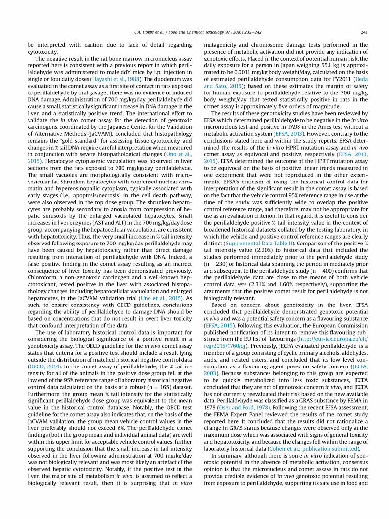
There were no macroscopic findings considered to be related toadministration of perillaldehyde. On microscopic examination,hepatocytes in the liver of animals administered perillaldehyde at700mg/kg/day were enlarged due to the presence of multiple smallcytoplasmic vacuoles consistent with microvesicular fat depositswith attendant compression of sinusoidal channels (Fig. 2). Thisvacuolation was predominantly periportal to mid-lobular in
Fig. 2. Photomicrographs of liver from vehicle control rat and rat exposed to thehighest dose (700 mg/kg/day) of perillaldehyde. (A) Vehicle control: clear areas incytoplasm of normal hepatocytes represent glycogen. (B) High dose rat: hepatocytesare enlarged with collapse of sinusoidal spaces and cytoplasmic vacuolation consistentwith microvesicular fatty change. Individual degenerating hepatocytes are shrunkenwith hyper-eosinophilic cytoplasm and condensed nuclear chromatin (denoted witharrows).
localization, but sometimes affected the centrilobular hepatocytesand was present in all six rats in the perillaldehyde dose group.Centrilobular hepatocyte degeneration characterized by loss ofcytoplasmic and nuclear detail was occasionally evident in rats inthe 700 mg/kg/day dose group, typically in areas in which cyto-plasmic vacuolation was less severe. Individual as well as smallclusters of shrunken hepatocytes with condensed nuclear chro-matin and hypereosinophilic cytoplasm were present to varyingdegrees only in this dose group. During clinical chemistry assess-ment of blood samples it was noted that a high number of sampleswere lipemic; this may be attributed to the corn oil used as thevehicle for test article formulation, which was administered justthree hours prior to blood sampling. As a consequence, somesamples were deemed unsuitable for statistical analysis of certainparameters. Measurements were obtained for at least five animals/group for aspartate aminotransferase (AST) and alanine amino-transferase (ALT); although mean serum values for these enzymeswere not statistically significant, three of the six rats dosed with700 mg/kg/day perillaldehyde exhibited high activity of both ALTand AST, indicative of hepatic toxicity. Statistically significantfindings supportive of general toxicity that were present only in therats in the 700 mg/kg/day dose group included increased serumurea and decreases in serum cholesterol, glucose, sodium, chloride,and potassium (Table 7).
4. Discussion
These studies were conducted in response to a request by EFSAfor genotoxicity data for flavouring substances considered repre-sentative of groups of compounds containing a,b-unsaturatedaldehyde and ketone structures, or their potential precursors.Specifically, the genotoxicity of perillaldehyde was evaluated as arepresentative substance of FGE.19 subgroup 2.2. The ability ofperillaldehyde to induce mutations, chromosomal abnormalities,and DNA damage was evaluated in accordance with the test strat-egy outlined by EFSA for evaluation of FGE.19 subgroup substances(EFSA, 2008a). The results of these tests are summarized in Table 8.
In a bacterial reverse mutation assay, perillaldehyde inducedmutation in histidine-requiring Salmonella typhimurium strainTA98 in the absence of S9. This result differs from the previouslypublished negative results for a study conducted in a variety ofbacterial strains (Ishidate et al., 1984). However, it is unclear if thetreatment conditions for these studies are comparable. Moreover,the published results were reported as “e or þ”, so observation of aweak response (<2� the concurrent control) cannot be ruled out. Inan in vitro micronucleus assay testing perillaldehyde in humanlymphocytes, no induction of micronuclei formation was detectedin a short (3 h) exposure in the presence and absence of metabolicactivation or in a long (24 h) exposure in the absence of metabolicactivation. These results do not support published positive resultsof a chromosome aberration study (Ishidate et al., 1984) that should
Table 8Summary of genotoxicity assessments of perillaldehyde.
Assay Result
Bacterial mutagenicity Positive (TA98 eS9)In vitro micronucleus NegativeIn vitro Hprt mutation Negativea
In vivo micronucleus NegativeIn vivo CometLiver Negativeb
Duodenum Negative
a Positive trend tests not reproduced.b Statistically positive only at a hepatotoxic dose; all data within his-
torical negative control 95% confidence range.
C.A. Hobbs et al. / Food and Chemical Toxicology 97 (2016) 232e242 241
be interpreted with caution due to lack of detail regardingcytotoxicity.
The negative result in the rat bone marrow micronucleus assayreported here is consistent with a previous report in which peril-laldehyde was administered to male ddY mice by i.p. injection insingle or four daily doses (Hayashi et al., 1988). The duodenumwasevaluated in the comet assay as a first site of contact in rats exposedto perillaldehyde by oral gavage; there was no evidence of inducedDNA damage. Administration of 700 mg/kg/day perillaldehyde didcause a small, statistically significant increase in DNA damage in theliver. and a statistically positive trend. The international effort tovalidate the in vivo comet assay for the detection of genotoxiccarcinogens, coordinated by the Japanese Center for the Validationof Alternative Methods (JaCVAM), concluded that histopathologyremains the “gold standard” for assessing tissue cytotoxicity, andchanges in % tail DNA require careful interpretationwhenmeasuredin conjunction with severe histopathological changes (Uno et al.,2015). Hepatocyte cytoplasmic vacuolation was observed in liversections from the rats exposed to 700 mg/kg/day perillaldehyde.The small vacuoles are morphologically consistent with micro-vesicular fat. Shrunken hepatocytes with condensed nuclear chro-matin and hypereosinophilic cytoplasm, typically associated withearly stages (i.e., apoptosis/necrosis) in the cell death pathway,were also observed in the top dose group. The shrunken hepato-cytes are probably secondary to anoxia from compression of he-patic sinusoids by the enlarged vacuolated hepatocytes. Smallincreases in liver enzymes (ASTand ALT) in the 700mg/kg/day dosegroup, accompanying the hepatocellular vacuolation, are consistentwith hepatotoxicity. Thus, the very small increase in % tail intensityobserved following exposure to 700 mg/kg/day perillaldehyde mayhave been caused by hepatotoxicity rather than direct damageresulting from interaction of perillaldehyde with DNA. Indeed, afalse positive finding in the comet assay resulting as an indirectconsequence of liver toxicity has been demonstrated previously.Chloroform, a non-genotoxic carcinogen and a well-known hep-atotoxicant, tested positive in the liver with associated histopa-thology changes, including hepatocellular vacuolation and enlargedhepatocytes, in the JaCVAM validation trial (Uno et al., 2015). Assuch, to ensure consistency with OECD guidelines, conclusionsregarding the ability of perillaldehyde to damage DNA should bebased on concentrations that do not result in overt liver toxicitythat confound interpretation of the data.
The use of laboratory historical control data is important forconsidering the biological significance of a positive result in agenotoxicity assay. The OECD guideline for the in vivo comet assaystates that criteria for a positive test should include a result lyingoutside the distribution of matched historical negative control data(OECD, 2014). In the comet assay of perillaldehyde, the % tail in-tensity for all of the animals in the positive dose group fell at thelow end of the 95% reference range of laboratory historical negativecontrol data calculated on the basis of a robust (n ¼ 165) dataset.Furthermore, the group mean % tail intensity for the statisticallysignificant perillaldehyde dose group was equivalent to the meanvalue in the historical control database. Notably, the OECD testguideline for the comet assay also indicates that, on the basis of theJaCVAM validation, the group mean vehicle control values in theliver preferably should not exceed 6%. The perillaldehyde cometfindings (both the group mean and individual animal data) are wellwithin this upper limit for acceptable vehicle control values, furthersupporting the conclusion that the small increase in tail intensityobserved in the liver following administration at 700 mg/kg/daywas not biologically relevant and was most likely an artefact of theobserved hepatic cytotoxicity. Notably, if the positive test in theliver, the major site of metabolism in vivo, is assumed to reflect abiologically relevant result, then it is surprising that in vitro
mutagenicity and chromosome damage tests performed in thepresence of metabolic activation did not provide any indication ofgenotoxic effects. Placed in the context of potential human risk, thedaily exposure for a person in Japan weighing 55.1 kg is approxi-mated to be 0.0011 mg/kg body weight/day, calculated on the basisof estimated perillaldehyde consumption data for FY2011 (Uedaand Sato, 2015); based on these estimates the margin of safetyfor human exposure to perillaldehyde relative to the 700 mg/kgbody weight/day that tested statistically positive in rats in thecomet assay is approximately five orders of magnitude.
The results of these genotoxicity studies have been reviewed byEFSAwhich determined perillaldehyde to be negative in the in vitromicronucleus test and positive in TA98 in the Ames test without ametabolic activation system (EFSA, 2013). However, contrary to theconclusions stated here and within the study reports, EFSA deter-mined the results of the in vitro HPRT mutation assay and in vivocomet assay as equivocal and positive, respectively (EFSA, 2013,2015). EFSA determined the outcome of the HPRT mutation assayto be equivocal on the basis of positive linear trends measured inone experiment that were not reproduced in the other experi-ments. EFSA's criticism of using the historical control data forinterpretation of the significant result in the comet assay is basedon the fact that the vehicle control 95% reference range in use at thetime of the study was sufficiently wide to overlap the positivecontrol reference range, and therefore, may not be appropriate foruse as an evaluation criterion. In that regard, it is useful to considerthe perillaldehyde positive % tail intensity value in the context ofbroadened historical datasets collated by the testing laboratory, inwhich the vehicle and positive control reference ranges are clearlydistinct (Supplemental Data Table II). Comparison of the positive %tail intensity value (2.20%) to historical data that included thestudies performed immediately prior to the perillaldehyde study(n ¼ 230) or historical data spanning the period immediately priorand subsequent to the perillaldehyde study (n¼ 400) confirms thatthe perillaldehyde data are close to the means of both vehiclecontrol data sets (2.31% and 1.60% respectively), supporting thearguments that the positive comet result for perillaldehyde is notbiologically relevant.
Based on concern about genotoxicity in the liver, EFSAconcluded that perillaldehyde demonstrated genotoxic potentialin vivo and was a potential safety concern as a flavouring substance(EFSA, 2015). Following this evaluation, the European Commissionpublished notification of its intent to remove this flavouring sub-stance from the EU list of flavourings (http://eur-lex.europa.eu/eli/reg/2015/1760/oj). Previously, JECFA evaluated perillaldehyde as amember of a group consisting of cyclic primary alcohols, aldehydes,acids, and related esters, and concluded that its low level con-sumption as a flavouring agent poses no safety concern (JECFA,2003). Because substances belonging to this group are expectedto be quickly metabolized into less toxic substances, JECFAconcluded that they are not of genotoxic concern in vivo, and JECFAhas not currently reevaluated their risk based on the new availabledata. Perillaldehyde was classified as a GRAS substance by FEMA in1978 (Oser and Ford, 1978). Following the recent EFSA assessment,the FEMA Expert Panel reviewed the results of the comet studyreported here. It concluded that the results did not rationalize achange in GRAS status because changes were observed only at themaximum dose which was associated with signs of general toxicityand hepatotoxicity, and because the changes fell within the range oflaboratory historical data (Cohen et al.; publication submitted).
In summary, although there is some in vitro indication of gen-otoxic potential in the absence of metabolic activation, consensusopinion is that the micronucleus and comet assays in rats do notprovide credible evidence of in vivo genotoxic potential resultingfrom exposure to perillaldehyde, supporting its safe use in food and

C.A. Hobbs et al. / Food and Chemical Toxicology 97 (2016) 232e242242
beverages. These results reinforce the original conclusions of JECFAand the more recent reevaluation by the FEMA Expert Panel thatperillaldehyde poses no safety concern for humans when used as aflavouring agent and consumed at estimated dietary exposures.Furthermore, by extrapolation on the basis of structural similarities(EFSA, 2008b), it seems unlikely that the other a,b-unsaturatedalicyclic aldehyde flavourings categorized as FGE.19 subgroup 2.2would pose a genotoxic risk to humans.
Acknowledgements
These studies were conducted at Covance Laboratories Ltd. andfunded by the International Organization of the Flavor Industry(IOFI). ILS, Inc. was contracted to write the manuscript. MaronpotConsulting LLC, a consultant for the Japan Flavor and FragranceMaterials Association, provided pathology peer-review assessmentof liver histopathology.
Appendix A. Supplementary data
Supplementary data related to this article can be found at http://dx.doi.org/10.1016/j.fct.2016.08.029.
Transparency document
Transparency document related to this article can be foundonline at http://dx.doi.org/10.1016/j.fct.2016.08.029.
References
Burlinson, B., Tice, R.R., Speit, G., Agurell, E., Brendler-Schwaab, S.Y., Collins, A.R.,Escobar, P., Honma, M., Kumaravel, T.S., Nakajima, M., Sasaki, Y.F., Thybaud, V.,Uno, Y., Vasquez, M., Hartmann, A., 2007. Fourth International Workgroup onGenotoxicity testing: results of the in vivo Comet assay workgroup. Mutat. Res.627, 31e35.
Cohen S.M., Fukushima S., Gooderham N.J., Guengerich F.P., Hecht S.S., RietjensI.M.C.M., Smith R.L., Bastaki M., Harman C.L., McGowen M.M., Taylor S.V., FEMAexpert panel review of p-mentha-1,8-dien-7-al genotoxicity testing results,(submitted).
EFSA, 2008a. Statement of the Panel on food contact materials, enzymes, flavour-ings and processing Aids (CEF) on genotoxicity test strategy for substancesbelonging to subgroups of FGE.19. EFSA J. 854, 1e5.
EFSA, 2008b. Statement of the Panel on food contact materials, enzymes, flavour-ings and processing Aids (CEF) on list of alpha, beta-unsaturated aldehydes andketones representative of FGE.19 substances for genotoxicity testing. EFSA J.910, 1e7.
EFSA, 2012. Minimum criteria for the acceptance of in vivo alkaline comet assayreports. EFSA J. 10, 2977.
EFSA, 2013. EFSA Panel on food contact materials, enzymes, flavourings and pro-cessing Aids scientific opinion on flavouring group evaluation 208 (FGE.208):consideration of genotoxicity data on representatives for 10 alicyclic aldehydeswith the a,b-unsaturation in ring/side-chain and precursors from chemical
subgroup 2.2 of FGE.19. EFSA J. 11, 3151.EFSA, 2015. EFSA Panel on food contact materials, enzymes, flavourings and pro-
cessing Aids scientific opinion on flavouring group evaluation 208(FGE.208Rev1): consideration of genotoxicity data on representatives for 10alicyclic aldehydes with the a,b-unsaturation in ring/side-chain and precursorsfrom chemical subgroup 2.2 of FGE.19. EFSA J. 13, 4173.
Hayashi, M., Kishi, M., Sofuni, T., Ishidate Jr., M., 1988. Micronucleus tests in mice on39 food additives and eight miscellaneous chemicals. Food Chem. Toxicol. 26,487e500.
Ishidate Jr., M., Sofuni, T., Yoshikawa, K., Hayashi, M., Nohmi, T., Sawada, M.,Matsuoka, A., 1984. Primary mutagenicity screening of food additives currentlyused in Japan. Food Chem. Toxicol. 22, 623e636.
JECFA, 2003. Safety evaluation of certain food additives. In: Fifty-ninth Meeting ofthe Joint FAO/WHO Expert Committee on Food Additives. WHO Food AdditiveSeries, no. 50. http://www.inchem.org/documents/jecfa/jecmono/v50je01.htm.
Ji, W.W., Wang, S.Y., Ma, Z.Q., Li, R.P., Li, S.S., Xue, J.S., Li, W., Niu, X.X., Yan, L.,Zhang, X., Fu, Q., Qu, R., Ma, S.P., 2014. Effects of perillaldehyde on alternationsin serum cytokines and depressive-like behavior in mice after lipopolysaccha-ride administration. Pharmacol. Biochem. Behav. 116, 1e8.
Lorenzo, Y., Costa, S., Collins, A.R., Azqueta, A., 2013. The comet assay, DNA damage,DNA repair and cytotoxicity: hedgehogs are not always dead. Mutagenesis 28,427e432.
OECD, 1997a. OECD Guideline for the Testing of Chemicals: Bacterial Reverse Mu-tation Test, p. 471.
OECD, 1997b. OECD Guideline for the Testing of Chemicals: in Vitro Mammalian CellGene Mutation Test, p. 476.
OECD, 1997c. OECD Guideline for the Testing of Chemicals: Mammalian ErythrocyteMicronucleus Test, p. 474.
OECD, 2008. OECD Guideline for the Testing of Chemicals: in Vitro Mammalian CellMicronucleus Test, p. 487.
OECD, 2014. OECD Guideline for the Testing of Chemicals: in Vivo MammalianAlkaline Comet Assay, p. 489.
Omari-Siaw, E., Zhu, Y., Wang, H., Peng, W., Firempong, C.K., Wang, Y.W., Cao, X.,Deng, W., Yu, J., Xu, X., 2016. Hypolipidemic potential of perillaldehyde-loadedself-nanoemulsifying delivery system in high-fat diet induced hyperlipidemicmice: formulation, in vitro and in vivo evaluation. Eur. J. Pharm. Sci. 85,112e122.
Oser, B.L., Ford, R.A., 1978. Recent progress in the consideration of flavoring in-gredients under the Food Additives Amendment.11. GRAS substances. FoodTechnol. 32, 60e70.
Tian, J., Wang, Y., Zeng, H., Li, Z., Zhang, P., Tessema, A., Peng, X., 2015. Efficacy andpossible mechanisms of perillaldehyde in control of Aspergillus Niger causinggrape decay. Int. J. Food Microbiol. 202, 27e34.
Tice, R.R., Agurell, E., Anderson, D., Burlinson, B., Hartmann, A., Kobayashi, H.,Miyamae, Y., Rojas, E., Ryu, J.C., Sasaki, Y.F., 2000. Single cell gel/comet assay:guidelines for in vitro and in vivo genetic toxicology testing. Environ. Mol.Mutagen. 35, 206e221.
Ueda, H., Sato, H., 2015. Research on daily exposure estimate based on poundagedata of food additives. JAFAN 35, 1e38.
Uno, Y., Kojima, H., Omori, T., Corvi, R., Honma, M., Schechtman, L.M., Tice, R.R.,Beevers, C., De Boeck, M., Burlinson, B., Hobbs, C.A., Kitamoto, S., Kraynak, A.R.,McNamee, J., Nakagawa, Y., Pant, K., Plappert-Helbig, U., Priestley, C.,Takasawa, H., Wada, K., Wirnitzer, U., Asano, N., Escobar, P.A., Lovell, D.,Morita, T., Nakajima, M., Ohno, Y., Hayashi, M., 2015. JaCVAM-organized inter-national validation study of the in vivo rodent alkaline comet assay for detec-tion of genotoxic carcinogens: II. Summary of definitive validation study results.Mutat. Res. Genet. Toxicol. Environ. Mutagen. 786e788, 45e76.
Xu, L., Li, Y., Fu, Q., Ma, S., 2014. Perillaldehyde attenuates cerebral ischemia-reperfusion injury-triggered overexpression of inflammatory cytokines viamodulating Akt/JNK pathway in the rat brain cortex. Biochem. Biophys. Res.Commun. 454, 65e70.