Embed Size (px)
Citation preview

RESEARCH ARTICLE
Genotoxic potential of diesel exhaust particlesfrom the combustion of first- and second-generation biodieselfuels—the FuelHealth project
Magdalena Kowalska1 & Aneta Wegierek-Ciuk1& Kamil Brzoska2 &
Maria Wojewodzka2 & Sylwia Meczynska-Wielgosz2 & Joanna Gromadzka-Ostrowska3 &
Remigiusz Mruk4& Johan Øvrevik5
& Marcin Kruszewski2,6,7 & Anna Lankoff1,2
Received: 3 April 2017 /Accepted: 22 August 2017 /Published online: 9 September 2017# The Author(s) 2017. This article is an open access publication
Abstract Epidemiological data indicate that exposure to dieselexhaust particles (DEPs) from traffic emissions is associatedwith higher risk of morbidity and mortality related to cardio-vascular and pulmonary diseases, accelerated progression ofatherosclerotic plaques, and possible lung cancer. While theimpact of DEPs from combustion of fossil diesel fuel on humanhealth has been extensively studied, current knowledge ofDEPs from combustion of biofuels provides limited and incon-sistent information about its mutagenicity and genotoxicity, aswell as possible adverse health risks. The objective of the pres-ent work was to compare the genotoxicity of DEPs from com-bustion of two first-generation fuels, 7% fatty acidmethyl esters(FAME) (B7) and 20% FAME (B20), and a second-generation20% FAME/hydrotreated vegetable oil (SHB: synthetic hydro-carbon biofuel) fuel. Our results revealed that particulate engineemissions from each type of biodiesel fuel induced genotoxiceffects in BEAS-2B and A549 cells, manifested as the in-
creased levels of single-strand breaks, the increased frequenciesof micronuclei, or the deregulated expression of genes involvedin DNA damage signaling pathways. We also found that noneof the tested DEPs showed the induction of oxidative DNAdamage and the gamma-H2AX-detectable double-strandbreaks. The most pronounced differences concerning the testedparticles were observed for the induction of single-strandbreaks, with the greatest genotoxicity being associated withthe B7-derived DEPs. The differences in other effects betweenDEPs from the different biodiesel blend percentage and biodie-sel feedstock were also observed, but the magnitude of thesevariations was limited.
Keywords Diesel exhaust particles . First- andsecond-generation biodiesel fuels . Single- and double-strandbreaks . Oxidative DNA damage . Chromosomal damage
Responsible editor: Philippe Garrigues
Electronic supplementary material The online version of this article(https://doi.org/10.1007/s11356-017-9995-0) contains supplementarymaterial, which is available to authorized users.
* Anna [email protected]
1 Department of Radiobiology and Immunology, Institute of Biology,Jan Kochanowski University, 15 Swietokrzyska Str,25-406 Kielce, Poland
2 Center for Radiobiology and Biological Dosimetry, Institute ofNuclear Chemistry and Technology, 16 Dorodna Str,03-195 Warsaw, Poland
3 Faculty of Human Nutrition and Consumer Science, WarsawUniversity of Life Sciences, 166 Nowoursynowska Str,02-787 Warsaw, Poland
4 Faculty of Production Engineering, Warsaw University of LifeSciences, 166 Nowoursynowska Str, 02-787 Warsaw, Poland
5 Domain of Infection Control and Environmental Health, NorwegianInstitute of Public Health, P.O. Box 4404, Nydalen,0403 Oslo, Norway
6 Department of Molecular Biology and Translational Research,Institute of Rural Health, Jaczewskiego 2, 20-090 Lublin, Poland
7 Faculty of Medicine, University of Information Technology andManagement in Rzeszow, Sucharskiego 2, 35-225 Rzeszow, Poland
Environ Sci Pollut Res (2017) 24:24223–24234DOI 10.1007/s11356-017-9995-0

Introduction
The overall impact of engine emissions on human health hasbeen studied for a long time, mostly due to the presence ofpolycyclic aromatic hydrocarbons (PAHs) and their deriva-tives (nitro-PAHs) in diesel exhaust particles (DEPs).Epidemiological data indicate that exposure to DEPs fromtraffic emissions is associated with a higher risk of morbidityand mortality related to cardiovascular and pulmonary dis-eases (Vieira et al. 2017; Steiner et al. 2016). There is alsocompelling evidence from animal experimental models andhumans that exposure to DEPs and ambient air particles isassociated with accelerated progression of atheroscleroticplaques (Cao et al. 2016). Recently, evidence has also beenobtained about their possible carcinogenicity (Hesterberg et al.2012). While the impact of DEPs from combustion of fossildiesel fuel on human health has been extensively studied,current knowledge of DEPs from combustion of biodieselsprovides limited and inconsistent information about their mu-tagenicity and genotoxicity, as well as possible adverse healthrisks. The vast majority of studies comparing DEPs from com-bustion of fossil diesels and biodiesels used the bacterial re-verse mutation assay (so-called Ames test). Early research onmutagenicity of DEPs in Salmonella typhimurium testerstrains TA98 (frame-shift mutation) and TA100 (base-pairsubstitution) has shown that biodiesel emissions induced sub-tly lower mutagenic potency when compared to the exhaust offossil fuels (Bünger et al. 1998; Westerholm et al. 2001).Other studies revealed either no differences or significant in-crease in mutagenic activity of biodiesel emission extracts(Westphal et al. 2012; Krahl et al. 2008). The more recentstudies revealed a clear reduction of mutagenic effects, show-ing less differences between the biodiesels compared to refer-ence fuels (Bünger et al. 2012). It is believed that the maincontributors to the mutagenicity of biodiesel DEPs are thePAH and nitro-PAH compounds adsorbed onto the particlesurface. Besides the mutagenicity determined in a prokaryotemodel, genotoxic properties of biodiesel-derived DEPs havealso been evaluated in eukaryote models. Two studies dealingwith the formation of bulky DNA adducts have shown con-tradictory results. Ross et al. (2015) reported the formation ofmultiple DNA adducts by the in vitro metabolic activation oforganic extracts of DEPs from combustion of fossil diesel andsoy biodiesel. However, no formation of DNA adducts waspresented in vitro by André et al. (2015), even if cells werefully able to metabolize nitroaromatics and PAHs. Otherin vitro studies revealed the induction of DNA strand breaks,but either no differences or significant differences were ob-served between DEPs from combustion of fossil diesel andbiodiesel (Jalava et al. 2010; Hemmingsen et al. 2011; Jalavaet al. 2012). Similarly, inconsistent results were observed forthe induction of micronuclei by DEPs from combustion offossil diesel and biodiesel, showing either no difference or
significant increase of chromosomal DNA damage bybiodiesel-derived DEPs (Leme et al. 2012; Cervena et al.2016). In summary, these contradictory trends are hard to in-terpret and may be caused by chance in light of relativelylimited data.Moreover, a comparison of the results of differenttoxicological studies for exhaust particles produced by biodie-sel combustion is difficult because of differences in the exper-imental approach, including age and type of diesel engine,drive cycle, feedstock blend, and its percentage in the blendedfuel. The objective of the present work was to compare thegenotoxicity of different DEPs from combustion of first- andsecond-generation biodiesel fuels in relation to their physico-chemical properties. DEPs were produced by the 1.3 JTDengine (Euro V stage), fueled with three biodiesel fuels ofcommercial interest: the first-generation B7 biodiesel fuel(7% fatty acid methyl esters (FAME)), which is currently usedin the EU; the first-generation B20 biodiesel fuel (20%FAME); and the second-generation (synthetic hydrocarbonbiofuel (SHB)) biodiesel fuel (7% FAME and 13% synthetichydrotreated vegetable oil (HVO)). These biodiesel fuels werecombusted under identical engine operation conditions, andemissions were evaluated during a certified test cycle.Detailed physicochemical characterizations of DEPs were per-formed to investigate how the composition of three types ofDEPs affects their biological effects in vitro, measured as in-duction of single- and double-strand breaks, oxidative DNAdamage, and chromosomal damage. In addition, the expressionof genes involved in DNA damage signaling was also evalu-ated to screen for possible molecular mechanisms of toxicity.
Materials and methods
Collection of DEPs
A Fiat Panda with compression ignition engine 1.3 JTD (com-mon rail third-generation injection system, engine capacity1248 cm3, max power 75 bhp, max torque 190 Nm, productionyear 2014), fulfilling the requirements of the Euro V stage, wasused as a DEP source. The engine was tested under controlledconditions on a chassis dynamometer (SchenckKomeg EMDY48) at constant engine speed and load of 340 rpm and 45.7%,respectively, corresponding to a constant vehicle speed of43.75 km/h. The engine temperature was kept at 94 °C duringthe test cycle. The enginewas fueled by three different mixturesof diesel oil and biocomponents: (1) first-generation biodieselfuel BB7,^ containing 7% vol. FAME in diesel oil; (2) first-generation biodiesel fuel BB20,^ containing 20% vol. FAME indiesel oil; and (3) second-generation biodiesel fuel BSHB,^containing 13% vol. synthetic HVO (NExBTL) and 7% vol.FAME in diesel oil. According to the newest EU Directive2015/1513 that encourages the use of second-generationbiofuels (such as HVO), instead of first-generation biofuels
24224 Environ Sci Pollut Res (2017) 24:24223–24234

(such as FAME and Rapeseed oil methyl ester (RME)), wechosen these two types of biofuels: HVO which is NesteRenewable biodiesel (formerly NEXBTL), produced in a pat-ented vegetable oil refining process and commercialized by theFinnish oil and refining company Neste (Rantanen et al. 2005)and FAME, which is currently used in EU and produced in theprocess of transesterification of plant oils with methanol byORLEN Poland. The DEPs used in the present study werecollected from the main diesel exhaust without a diesel particlefilter on PTFE-coated glass fiber filters (Pallflex, Emfab filters,TX40HI20WW, 70 mm). For the quantitative analysis, theblank sampling filters were marked and weighed. The filtersfrom individual fuels were separately re-weighed after sam-pling, and the mass differences before and after the samplingwere compared and analyzed to determine the total particulatematter collected. Driving cycles and particulate collectionswere repeated with the same fuel several times to new filtersets to ensure that a sufficiently large sample mass would beobtained in order to complete the toxicological and chemicalanalyses. Filters were stored in the freezer at − 20 °C for 1 weekbefore analysis. Particles for in vitro experiments were scrapedfrom the filters using a clean stainless steel blade.
Preparation of diesel exhaust particles
DEP-stock solutions were prepared by dispersion of 2 mg ofparticles in 1 mL of LHC-9 serum-free medium (for experi-ments with BEAS-2B cells) or in 1 mL of F12 Ham mediumsupplemented with 10% FCS (for experiments with A549cells). DEP-dispersions were then sonicated on ice using theSonic Vibra Cell ultrasonic liquid processor (USA).Ultrasonic energy (3 kJ) was provided in pulses (30 s on,10 s off) at 60% amplitude. Stock solutions were dispensed(100 μL) into sterile 1-mL cryogenic vials and stored at– 20 °C. The samples were thawed before each set of exper-iments at 37 °C for 60 s, dispersed in the corresponding me-dium at a ratio of 1:10, and mixed prior to use (workingsolution).
Physicochemical characterization of diesel exhaustparticles
Physicochemical characterization of diesel exhaust particleswas described in detail by Lankoff et al. (2017). Briefly, sam-ple size distribution was measured by the NanoparticleTracking Analysis (NTA) with a NanoSight LM20(NanoSight, Amesbury, UK), equipped with a sample cham-ber with a 640-nm laser. Zeta potential and polydispersityindex were determined by DLS method at 25 °C in a foldedcapillary cell at 150 V and M3-PALS detection using non-invasive backscatter at 173° with an Avalanche photodiode,Q.E. > 50% at 633 nm (Malvern, Malvern Hills, UK). Theshape of DEPs was analyzed by transmission electron
microscopy (TEM) (JEOL 1200 EXII, JEOL, JAPAN) oper-ating at an acceleration voltage of 120 kV. Elemental analysisof DEPs was performed by digital scanning electron micros-copy (SEM) type DSM 942 (Zeiss, Germany) in the second-ary electron (SE) mode using the energy dispersive X-rayspectrometry (EDS) with Quantax 400 (Bruker, Germany)system. Separation and analysis of PAHs from particulate ex-tracts were described in detail by Czarnocka andOdziemkowska (2016). The content of 17 PAHswas measuredby the Agilent 7890A GC System chromatograph coupled witha mass spectrometer MS 5975C using a low-polarity Rtx-5mscapillary column (30 m × 0.25 mm × 0.25 μm) (Restek,Bellefonte, PA, USA).
Cell cultures
The human type-II-like alveolar epithelial cell line A549 andthe human bronchial epithelial cell line BEAS-2B were pur-chased from the American Type Tissue Culture Collection(ATCC, Rockville, MD) and maintained according to ATCCprotocols. Briefly, A549 were cultured in F12 Ham mediumsupplemented with 10% FCS and 2 mM L-glutamine, where-as BEAS-2B were cultured in LHC-9 serum-free bronchialepithelial growth medium on non-coated plates. Both celllines were maintained in an incubator at 37 °C with 5%CO2. The exponentially growing BEAS-2B and A549 cellswere incubated with 1, 10, 25, and 50 μg/mL of DEPs for 6,24, or 48 h (depending on the assay procedure). The doses andtreatment times were chosen based on the cytotoxicity results(e.g., induction of apoptosis and necrosis, inhibition of proteinsynthesis, generation of free radicals), published previously byour group (Lankoff et al. 2017).
Determination of single-strand DNA breaks and oxidativeDNA damage by the comet assay
The exponentially growing BEAS-2B and A549 cells wereincubated with 1, 10, 25, and 50 μg/mL of DEPs for 24 h.Positive control cells were irradiated with a dose of 2 Gy of X-rays at a dose rate of 1.14 Gy/min (Xylon International Smart200-E irradiator, Xylon, San Jose, CA). The comet assay (sin-gle cell gel electrophoresis) was performed as previously de-scribed (Wojewódzka et al. 1998). Briefly, an aliquot of cellsuspension was mixed with an equal volume of 2% low melt-ing point agarose (type VII, Sigma), put on a microscope slidepre-coated with 0.5% regular agarose (type I-A, Sigma), andleft on ice. After agarose solidification, the slides were im-mersed in a cold lysing solution (2.5 M NaCl, 100 mMNa2EDTA, 10 mM Tris, and 1% Triton X-100, pH 10) or leftfor 30 min in culture media at 37 °C to allow damage repair.After 40 min lysis, the slides were placed on a horizontal gelelectrophoresis unit filled with fresh electrophoretic buffer(1 mM Na2EDTA (sodium ethylenediamine tetraacetate)
Environ Sci Pollut Res (2017) 24:24223–24234 24225

and 300 mM NaOH) and allowed to stay in this buffer for40 min for DNA unwinding. Next, electrophoresis was per-formed (1.2 V/cm, 30 min, 10 °C). After electrophoresis, theslides were washed with 0.4 M Tris, pH 7.5 (3 × 5 min), andstained with DAPI (4′,6-diamidino-2-fenylindole), 50 μL(1 μg/mL). Basically, the same test was applied for the mea-surement of DNA base damage. Incubation of irradiatedcells with the formamidopyrimidine glycosylase (FPG),BioLabs, was carried out as previously described(Kruszewski et al. 1998). Briefly, after lysis, the slideswere washed 3 × 5 min with the buffer (40 mM Hepes(4-(2-hydroxyethyl)-1-piperazineethanesulfonic acid),0.1 M KCl, 0.5 mM EDTA, 0.2 mg/mL bovine serumalbumin, pH 8) at 4 °C. Further, 50 μL of FPG solution(4.8 × 10⁻² U) in the buffer was placed on each slide, coveredwith cover glass, and incubated for 30 min in a light-protectedbox at 37 °C. Slides were stained with DAPI (1 μg/mL) andanalyzed as described above. Image analysis of data was per-formed by the Comet Assay IV image analysis system(Perceptive Instruments, UK). Data for 75 randomly selectedcomets per point were analyzed. Percent of DNA in comet’stail was chosen as a measure of DNA damage.
Analysis of double-strand DNA breaks by the γ-H2AXassay
BEAS-2B and A549 cells at exponential growth were incubat-ed with 1, 10, 25, 50, and 100 μg/mL of the different DEPs for24 h. Positive control cells were irradiated with a dose of 2 Gyof X-rays at a dose rate of 1.14 Gy/min (Xylon InternationalSmart 200-E irradiator, Xylon, San Jose, CA). γ-H2AX fociwere detected with theγ-H2AX (H2A.X PHOS) DetectionKit(Upstate Biotechnology, USA). Briefly, after incubation, thecells were washed, fixed, and resuspended in a perme-abilization solution (0.5% saponin, 10 mM HEPES, 140 mMNaCl, 2.5 mMCaCl2). Unspecific binding was blocked for 1 hin blocking buffer BSA-T-PBS (1% BSA, 0.1% Triton X-100in PBS). Thereafter, the cells were suspended in BSA-T-PBScontaining 2 μg of the fluorescein-conjugated γ-H2AX anti-body (monoclonal anti-phosphohistone H2AX antibody,Upstate Biotechnology) for 20 min. Cells were analyzed witha FACScan (Becton Dickinson, San Jose, CA, USA). Twentythousand cells per point were analyzed for γ-H2AX intensity.
Determination of chromosomal DNA damageby the micronucleus assay
The exponentially growing BEAS-2B and A549 cells wereincubated with 1, 10, 25, and 50 μg/mL of DEPs for 24 and48 h. Positive control cells were irradiated with a dose of 2 Gyof X-rays at a dose rate of 1.14 Gy/min (Xylon InternationalSmart 200-E irradiator, Xylon, San Jose, CA). After 1 h oftreatment, cytochalasin-B (final concentration of 10 μg/mL)
was added into the cell culture medium in order to blockcytokinesis and obtain binucleated cells. Cytochalasin-B isan inhibitor of microfilament ring assembly required for thecompletion of cytokinesis. This inhibition allows to generateonce-divided binucleated cells, which are the cells that canexpress micronuclei. Restricting scoring of micronuclei in bi-nucleated cells prevents confounding effects caused by sub-optimal or altered cell division kinetics, which is a major var-iable in the micronucleus assay protocol that does not distin-guish between non-dividing cells that cannot expressmicronuclei and dividing cells that can (Fenech 2007). Afterthe exposure, the cells were harvested by centrifugation andsubjected to cold mild hypotonic treatment (0.075 M KCl) for8 min, fixed twice with methanol/acetic acid/ringer solution(13:12:3), and then dropped on clean, dry slides. The cellswere mounted and stained in the Vectashield MountingMedium containing DAPI (4′,6-diamidyno-2-fenyloindol).The slides were coded, and the frequency of micronuclei(MN) in 3000 binucleated cells (BNC) per dose (1000 cells/replicate) was analyzed with the fully automated image acqui-sition and analysis system Metafer (Metasystems, Germany)by one skilled scorer. The frequency of MN in untreated con-trols and positive controls (2 Gy) was also scored in 3000BNC (1000 cells/replicate), according to the criteria proposedby Fenech (2007).
RNA isolation, reverse transcription, and real-time PCR
Total RNA was extracted from cell pellets using the RNeasyMini Kit (Qiagen) according to manufacturer’s protocol. Toassess the concentration and purity of RNA, the portion ofevery RNA sample was diluted in TE buffer (pH 8.0) andthe absorbance at 230, 260, and 280 nm was measured usingCary 50 UV-Vis spectrophotometer (Varian). All RNA sam-ples used in subsequent analyses had a concentration≥ 100 ng/μL, as well as A260/A280 and A260/A230 ratios≥ 2.0. RNA integrity was tested by agarose gel electrophore-sis. For PCR array analysis, 1 μg of total RNAwas convertedto complementary DNA (cDNA) in a 20-μL reaction volumeusing RT2 First Strand Kit (Qiagen). The cDNA was dilutedwith 91 μl distilled water and used for the expression profilingusing the human DNA damage signaling pathway PCR array(Qiagen, cat. no. PAHS-029Z) according to manufacturer’sinstructions. Briefly, a total volume of 25 μL of PCRreaction mixture, which included 12.5 μL of RT2 SYBRGreen/ROX qPCR Master Mix from Qiagen (containingHotStart DNA Taq polymerase, SYBR Green dye and theROX reference dye), 11.5 μL of double-distilled H2O, and1 μL of diluted template cDNA, was used for each primerset in each well of the PCR array. One technical replicatewas performed for each sample. PCR amplificationwas carriedout using 7500 Real-Time PCR System (ThermoFisherScientific) with an initial 10-min step at 95 °C followed by
24226 Environ Sci Pollut Res (2017) 24:24223–24234

40 cycles of 95 °C for 15 s and 60 °C for 1 min. Relative geneexpression was calculated using the ΔΔCt method withACTB, B2M, GAPDH, HPRT1, and RPLP0 as reference con-trols. Calculations were done using the Relative QuantificationSoftware version 3.2.1-PRC-build1 (Thermo Fisher Cloud).Statistical differences were examined by Student’s t test withp < 0.05 considered to be statistically significant.
Statistical evaluation
Statistical analysis of the obtained data was performed usingthe Statistica 7.1 software (Stat Soft. Inc., Tulsa, USA). Thedata were expressed as mean ± standard deviation (SD) of atleast three independent experiments. Data were evaluated byKruskal-Wallis one way analysis of variance on ranks(ANOVA) followed by the post hoc Fisher’s test. Correlationcoefficients between the obtained data were evaluated by thePearson product-moment. Differences were considered statis-tically significant when the p value was less than < 0.05.
Results
Physicochemical characterization of DEPs
A detailed physicochemical characterization of the DEPsused in this study has previously been published (Lankoffet al. 2017. As shown in Table 1, the average hydrodynam-ic diameters of all three types of DEPs were comparable, ifthe same culture medium was used. However, the B7 bio-fuel generated the lowest number of particles with diameterin the range 1–90 nm (~ 55%), as compared with the B20biofuel (~ 70%) and the SHB biofuel (~ 85%). The poly-dispersity index values for all DEPs were less than 0.5,indicating high homogeneity of the suspension. The zetapotentials were negative and comparable for all testedDEPs, indicating stability of the colloidal system. TEManalysis revealed that all three types of DEPs were nearlyspherical, and the particles were present as single particles,clusters, or chain-like aggregates. All DEPs were composedprimarily of carbon (~ 85%). The next most abundant ele-ments were oxygen and nitrogen. Zinc was present in allDEPs at low concentration. Concentrations of sulfur, cop-per, and chlorine were below 0.5% in all samples, with theexception of chlorine in SHB-DEP, which was slightlyabove 1%. Silver and iron could only be detected inSHB-DEP samples. The content of PAHs in DEP-derivedorganic extracts was the highest in the B7-DEP sample(165.78 ng/mg) and the lowest in the SHB-DEPs sample(69.93 ng/mg). The data showed that pyrene, fluoranthene,phenanthrene, and chrysene were the most abundant PAHsin all samples.
DEPs induce single-strand breaks but not oxidative DNAdamage in BEAS-2B and A549 cells
BEAS-2B andA549 cells were cultured in the presence of threetypes of DEPs (1, 10, 25, and 50 μg/mL) for 24 h. DEPsinduced DNA damage in BEAS-2B cells versus correspondingcontrol cultures as shown in Fig. 1. The increased level ofsingle-strand DNA breaks (SSBs) was observed in cells incu-bated with 10, 25, and 50 μg/mL of B7-DEP (p = 0.0003,
Table 1 Physicochemical characteristics of diesel exhaust particlesfrom the combustion of B7, B20, and SHB biodiesel fuels. Elementalcomposition of DEPs (wt%), PAHs (ng/mg) in the organic extractsfrom DEPs, particle size (nm), polydispersity index, and zeta potential(mV) of B7-DEPs, B20-DEPs, and SHB-DEPs in cell culture mediumF12+FBS or LHC-9. All data presented as mean of triplicates ± SD
Chemical constituents ofthe particulate samples
Diesel exhaust particles
B7-DEP B20-DEP SHB-DEP
Elemental composition (wt%)CarbonOxygenNitrogenZincSulfurCopperChlorineSilverIron
85.53 ± 0.937.51 ± 1.025.12 ± 0.971.40 ± 0.440.22 ± 0.040.18 ± 0.030.04 ± 0.01–
–
86.76 ± 1.096.31 ± 1.215.15 ± 0.871.24 ± 0.050.22 ± 0.040.19 ± 0.080.10 ± 0.02–
–
87.51 ± 0.862.24 ± 0.855.11 ± 0.852.25 ± 0.770.26 ± 0.070.45 ± 0.211.33 ± 0.360.72 ± 0.120.21 ± 0.03
Organic components (ng/mg)NaphthaleneAcenaphthyleneAcenaphthaleneFluorinePhenanthreneAnthraceneFluoranthenePyreneBenzo(a)anthraceneChryseneBenzo(b)fluorantheneBenzo(k)fluorantheneBenzo(a)pyreneBenzo(a)fluorantheneIndeno(1,2,3-c,d)pyreneDibenzo(a,h)anthraceneDibenzo(g,h,i)perylene
Total PAHs
3.860.260.110.5612.331.1336.0490.332.0710.264.302.590.380.650.360.070.47165.78
7.740.920.191.4120.831.736.0643.231.143.981.651.580.831.570.440.240.3393.54
3.130.140.070.235.130.566.3640.531.766.922.801.380.220.270.190.050.2069.93
Hydrodynamicdiameter (nm)
78 ± 55a
126 ± 64b80 ± 43a
107 ± 49b68 ± 37a
113 ± 48b
Polydispersity index 0.185 ± 0.02a
0.190 ± 0.01b0.455 ± 0.05a
0.470 ± 0.08b0.334 ± 0.06a
0.383 ± 0.02b
Zeta potential (mV) − 22.4 ± 3.22a
− 21.5 ± 2.31b− 20.1 ± 1.98a
− 19.93 ± 3.98b− 23.5 ± 3.03a
− 22.1 ± 2.98b
a F12+FBSb LHC-9
Environ Sci Pollut Res (2017) 24:24223–24234 24227

p = 0.0001, and p = 0.0001, respectively) (Fig. 1a), in cellsincubated with 50 μg/mL of B20-DEP (p = 0.0036) (Fig. 1b),and in cells incubated with 25 and 50 μg/mL of SHB-DEP(p = 0.0002, p = 0.0001, respectively). None of the testedDEPs showed significant induction of oxidative DNA damagein BEAS-2B. The effect of DEPs on induction of DNA damagein A549 cells is shown in Fig. 2. The increased level of SSBswas observed in cells incubated with 10, 25, and 50 μg/mL ofB7-DEP (p = 0.0018, p = 0.0001, and p = 0.0001, respectively)(Fig. 2a); 25 and 50 μg/mL of B20-DEP (p = 0.0383 andp = 0.0001, respectively) (Fig. 2b); and in cells incubated with10, 25, and 50 μg/mL of SHB-DEP (p = 0.0301, p = 0.0010and p = 0.0001, respectively) (Fig. 2c). None of the tested DEPs
showed significant induction of oxidative DNA damage inA549 cells. B7-DEP were the most effective in inducingSSBs in BEAS-2B (p = 0.0001 for B7-DEP vs B20-DEP,p = 0.0001 for B7-DEP vs SHB-DEP, p = 0.2256 for B20-DEP vs SHB-DEP) and A549 cells (p = 0.0019 for B7-DEPvs B20-DEP, p = 0.0232 for B7-DEP vs SHB-DEP, p = 0.3697for B20-DEP vs SHB-DEP).
DEPs do not induce double-strand breaks in BEAS-2Band A549 cells
The gamma-H2AX-detectable double-strand breaks were an-alyzed in BEAS-2B and A549 cells cultured in the presence of
Fig. 1 Effect of DEPs on induction of single-strand breaks (SSBs) andoxidative DNA damage (FPG) in BEAS-2B cells. DNA damage wasdetermined by the comet assay. a B7-derived DEPs, b B20-derivedDEPs, and c SHB-derived DEPs. Data are expressed as means ± S.D.from three independent experiments. p < 0.05. Asterisk denotes statisti-cally significant difference versus corresponding control group
Fig. 2 Effect of DEPs on induction of single-strand breaks (SSBs) andoxidative DNA damage (FPG) in A549 cells. DNA damage was deter-mined by the comet assay. a B7-derived DEPs, b B20-derived DEPs, andc SHB-derived DEPs. Data are expressed as means ± S.D. from threeindependent experiments. p < 0.05. Asterisk denotes statistically signifi-cant difference versus corresponding control group
24228 Environ Sci Pollut Res (2017) 24:24223–24234

three types of DEPs (10, 25, 50, and 100 μg/mL) for 24 h. Aspresented in Fig. 3, none of the tested DEPs showed signifi-cant induction of the gamma-H2AX foci fluorescence inBEAS-2B (Fig. 3a) and A549 (Fig. 3b) cells. The positiveassay control (2 Gy of ionizing radiation) significantly in-creased the gamma-H2AX foci fluorescence in both cell lines.
DEPs induce chromosomal damage in BEAS-2Band A549 cells
BEAS-2B and A549 cells were cultured in the presence ofthree types of DEPs (1, 10, and 50 μg/mL) for 24 and 48 h.The effect of DEPs on the frequency ofMN in BEAS-2B cellsversus corresponding control cultures is shown in Fig. 4. Theincreased frequency of MN was observed in cultures incubat-ed with 10 and 50 μg/mL of B7-DEP for 24 h (p = 0.0177 andp = 0.0001, respectively) and for 48 h (p = 0.0004 andp = 0.0001, respectively) (Fig. 4a). As presented in Fig. 4b,the increased frequency of MN was observed in cultures in-cubated with 50 μg/mL of B20-DEP for 24 h (p = 0.0001) andfor 48 h (p = 0.0001). As shown in Fig. 4c, the increasedfrequency of MN was observed in cultures incubated with50 μg/mL of SHB-DEP for 24 h (p = 0.0001) and with 10
and 50 μg/mL for 48 h (p = 0.0032, p = 0.0016, respectively).The effect of DEPs on the frequency of MN in A549 cellsversus corresponding control cultures is shown in Fig. 5. Theincreased frequency of MN was observed in cultures incubat-ed with 50 μg/mL of B7-DEP for 24 h (p = 0.0016) and with10 and 50 μg/mL for 48 h (p = 0.0326 and p = 0.0002, re-spectively) (Fig. 5a). As presented in Fig. 5b, the increasedfrequency ofMNwas observed also in cultures incubatedwith50 μg/mL of B20-DEP for 48 h (p = 0.0032). As shown inFig. 5c, the increased frequency of MN was observed in cul-tures incubated with 10 and 50 μg/mL of SHB-DEP for 48 h
Fig. 4 Effect of DEPs on induction of micronuclei in BEAS-2B cells.The frequency of micronuclei (MN) was determined by the micronucleusassay. a B7-derived DEPs, b B20-derived DEPs, and c SHB-derivedDEPs. Data are expressed as means ± S.D. from three independent ex-periments. p < 0.05. Asterisk denotes statistically significant differenceversus corresponding control group, BNC—binucleated cell
Fig. 3 Effect of DEPs (B7, B20, SHB) on induction of double-strandbreaks in BEAS-2B (a) and A549 cells (b). Double-strand breaks weredetermined by the gamma-H2AX assay. Data are expressed as means ±S.D. from three independent experiments. p < 0.05. Asterisk denotesstatistically significant difference versus corresponding control group
Environ Sci Pollut Res (2017) 24:24223–24234 24229
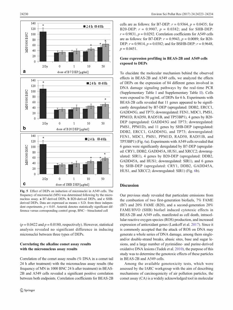
(p = 0.0422 and p = 0.0180, respectively). However, statisticalanalysis revealed no significant difference in inducingmicronuclei between three types of DEPs.
Correlating the alkaline comet assay resultswith the micronucleus assay results
Correlation of the comet assay results (% DNA in a comet tail24 h after treatment) with the micronucleus assay results (thefrequency of MN in 1000 BNC 24 h after treatment) in BEAS-2B and A549 cells revealed a significant positive correlationbetween both endpoints. Correlation coefficients for BEAS-2B
cells are as follows: for B7-DEP: r = 0.9364, p = 0.0435; forB20-DEP: r = 0.9907, p = 0.0382; and for SHB-DEP:r = 0.9831, p = 0.0292. Correlation coefficients for A549 cellsare as follows: for B7-DEP: r = 0.9943, p = 0.0089; for B20-DEP: r = 0.9814, p = 0.0382; and for BSHB-DEP: r = 0.9648,p = 0.0451.
Gene expression profiling in BEAS-2B and A549 cellsexposed to DEPs
To elucidate the molecular mechanism behind the observedeffects in BEAS-2B and A549 cells, we analyzed the effectsof DEPs on the expression of 84 different genes involved inDNA damage signaling pathways by the real-time PCR(Supplementary Table I and Supplementary Table II). Cellswere exposed to 50 μg/mL of DEPs for 6 h. Experiments withBEAS-2B cells revealed that 11 genes appeared to be signifi-cantly deregulated by B7-DEP (upregulated: DDB2, ERCC1,GADD45G, and TP73; downregulated: FEN1, MDC1, PMS1,PPM1D, RAD50, RAD51B, and TP53BP1), 4 genes by B20-DEP (upregulated: GADD45G and TP73; downregulated:PMS1, PPM1D), and 11 genes by SHB-DEP (upregulated:DDB2, ERCC1, GADD45G, and TP73; downregulated:FEN1, MDC1, PMS1, PPM1D, RAD50, RAD51B, andTP53BP1) (Fig. 6a). Experiments with A549 cells revealed that6 genes were significantly deregulated by B7-DEP (upregulat-ed: CRY1, DDB2, GADD45A, HUS1, and XRCC2; downreg-ulated: SIR1), 4 genes by B20-DEP (upregulated: DDB2,GADD45A, and HUS1; downregulated: SIR1), and 6 genesby SHB-DEP (upregulated: CRY1, DDB2, GADD45A,HUS1, and XRCC2; downregulated: SIR1) (Fig. 6b).
Discussion
Our previous study revealed that particulate emissions fromthe combustion of two first-generation biofuels, 7% FAME(B7) and 20% FAME (B20), and a second-generation 20%FAME/HVO (SHB) biofuel induced cytotoxic effects inBEAS-2B and A549 cells, manifested as cell death, intracel-lular reactive oxygen species (ROS) production, and increasedexpression of antioxidant genes (Lankoff et al. 2017). Since itis commonly accepted that the attack of ROS on DNA maygenerate a whole series of DNA damage, among them single-and/or double-strand breaks, abasic sites, base and sugar le-sions, and a large number of pyrimidine- and purine-derivedoxidative DNA lesions (Tudek et al. 2010), the purpose of thisstudy was to determine the genotoxic effects of these particlesin BEAS-2B and A549 cells.
Among the available genotoxicity tests, which wereassessed by the IARC workgroup with the aim of describingmechanisms of carcinogenicity of air pollution particles, thecomet assay (CA) is a widely acknowledged tool in molecular
Fig. 5 Effect of DEPs on induction of micronuclei in A549 cells. Thefrequency of micronuclei (MN) was determined following by the micro-nucleus assay. a B7-derived DEPs, b B20-derived DEPs, and c SHB-derived DEPs. Data are expressed as means ± S.D. from three indepen-dent experiments. p < 0.05. Asterisk denotes statistically significant dif-ference versus corresponding control group, BNC—binucleated cell
24230 Environ Sci Pollut Res (2017) 24:24223–24234

epidemiology and genetic toxicology (Araldi et al. 2015). Weapplied the alkaline comet assay, which is typically referred toas measuring BDNA SSBs^ or BDNA damage.^ Our resultsrevealed a dose-dependent increase in the level of SSBs incells treated with all types of DEPs. Among the studiedDEPs, B7-DEP exposure caused maximum DNA damage,while B20-DEP and SHB-DEP induced SSBs at the similarlevel. This is most likely due to differences in the physico-chemical properties of the three types of DEP tested. A num-ber of characteristic parameters of particles affect their toxic-ity, including their size, shape, surface reactivity, surfacecharge, surface coating, and elemental composition (Øvreviket al. 2015).While the size, morphology, and surface charge ofthe three types of DEPs tested in this study were similar, thepresence of organic compounds including PAH and/or nitro-PAH compounds adsorbed onto the particle surface could beexpected to have an impact. B7-derived DEP contained thehighest concentration of PAHs. The increase in bioadditiveratio caused a decrease in the PAH concentration of sufficientmagnitude to diminish the observed effects, as demonstratedfor B20-DEPs and SHB-DEPs. The role of PAHs adsorbed ondiesel soot emissions has been extensively reviewed, showingthat genotoxic effects of organic extracts from combustion-
generated particles are mainly connected with PAHs and theirderivative (Topinka et al. 2012). Nevertheless, studies relatedto the associations between biodiesel-derived DEP exposuresand the CA endpoints are almost completely missing. Amongthese reports available, Jalava et al. (2010) used the CA todetermine DNA damage in the 264.7 macrophages followingexposure to DEPs from the combustion of 100% diesel oil andtwo biodiesels (100%HVO and 100%RME). They found thatall DEP samples induced SSBs at the same level, exceptweaker response for the RME sample with a catalyst. Twoyears later, the same group published the results showing thatDEPs from the combustion of five fuels (100% diesel, 100%HVO, 30% HVO, 100% RME, 30% RME) induced a dose-dependent fragmentation of chromosomal DNA. Emissionparticles from the engine powered by 100% diesel and 30%HVO were the most potent inducers of SSBs (Jalava et al.2012).
One of the commonest sources of SSBs is oxidativeattack by endogenous ROS. SSBs can arise directly viadisintegration of the oxidized sugar or indirectly duringthe DNA base excision repair (BER) of oxidized basesand abasic sites, as well as during the nucleotide excisionrepair (NER) of damaged or altered nucleotides. Morerecently, it has emerged that SSBs can also arise as aresult of erroneous or abortive activity of cellular enzymessuch as DNA topoisomerase 1 (Top1) or erroneous incor-poration of ribonucleotides into DNA (Caldecott 2014).The alkaline CA does not distinguish between SSBs dueto direct oxidative DNA damage and indirect SSBs(Collins et al. 2017). Thus, it has been of particular inter-est to elucidate the origin of SSBs induced by DEPs testedin our study. We therefore used the enzyme-modified CA,which enable the detection of oxidized bases by combin-ing the assay with the use of a formamidopyrimidine-DNA glycosylase (FPG) converting altered purines intoDNA breaks (Collins 2014). We found that none of the testedDEPs showed significant induction of oxidative DNA basedamage in BEAS-2B and A549 cells, suggesting the DNArepair-related origin of SSBs rather than oxidative stress-relat-ed. On the contrary to our findings, Hemmingsen et al. (2011)found that SSBs and FPG sensitive sites showed concentration-dependent increases in A549 cells exposed to particles from thecombustion of RME-derived biofuel (B20) and pure diesel,with small differences between the B20 and B0 particles.However, along with our study, Gualtieri et al. (2011) reportedthat urban PM2.5weremore potent compared to PM10 for SSBgeneration in BEAS-2B cells, whereas there was no effect onFPG total sites. On the contrary, Jantzen et al. (2012) reportedthat the reference DEPs (SRM2975 and SRM1650b) generatedSSBs and oxidatively damaged DNA, measured using theenzyme-modified CA on formamidopyrimidine-DNAglycosylase or oxoguanine DNA glycosylase (hOGG1)-sensi-tive sites in A549 and THP-1a cells, as well as in co-cultures of
Fig. 6 Changes in gene expression in BEAS-2B cells (a) and A549 cells(b) after treatment with 50 μg/mL of three types of DEPs for 6 h. Meanfold change values from three independent experiments are presented.Error bars represent minimum and maximum values in sample. Foldchanges statistically significant in Student’s t test are highlighted(asterisk)
Environ Sci Pollut Res (2017) 24:24223–24234 24231

A549 and THP-1a cells. Recently, Vattanasit et al. (2014) ob-served the dose-dependent oxidative DNA damage in the lym-phoblasts and lung cells exposed to DEPs.
Single-strand breaks can have an impact on cell fate, ifthey are not repaired rapidly and appropriately. The mostlikely consequence of the increased level of SSBs in non-proliferating cells is cell death by stalling of RNA poly-merases during transcription. However, in proliferatingcells, the most likely consequence is the blockage or col-lapse of DNA replication forks during the S phase of thecell cycle, leading to the formation of double-strand breaks(DSBs), chromosome rearrangements, and genomic insta-bility (Woodbine et al. 2011). To elucidate whether the in-creased level of SSBs observed in the comet assay results ininduction of DSBs, we treated BEAS-2B and A549 cells inthe same manner as for the analysis of SSBs and used the γ-H2AX assay, which is considered attributable to the directformation of DSBs (Rogakou et al. 1998). Our results re-vealed that none of the tested DEPs showed induction of thegamma-H2AX-detectable double-strand breaks. To the best ofour knowledge, a study investigating the effects of biodiesel-derived DEPs or their organic extracts on induction of DSBshas not been published yet, so comparable data are not avail-able. However, Toyooka et al. (2012) determined the forma-tion of DSBs in A549, MCF7, HaCaT, and A549 cells ex-posed to 9,10-phenanthrenequinone (9,10-PQ), a major qui-none in DEPs, and reported small amounts of γ-H2AX,shown as fluorescence foci.
To further elucidate whether the increased level of SSBsobserved in the comet assay may be related to DNA damage atthe chromosome level, we applied the micronucleus assay, amulti-target genotoxic endpoint, assessing not onlyclastogenic and aneugenic events but also some epigeneticeffects (Kirsch-Volders et al. 2011). Our results revealed thatall types of DEPs increased the frequency of micronuclei in adose- and time-dependent manner in BEAS-2B and A549cells when compared to controls. However, statistical analysisrevealed no significant difference in inducing micronuclei be-tween the three types of DEPs. Our results are in line withthe findings of Cervena et al. (2016), who observed sig-nificantly increased frequency of MN in BEAS-2B cellsexposed to extractable organic matter from particle emis-sions from combustion of various biodiesel fuels and purediesels (B0, B30, B100). The genotoxicity of these ex-tracts was comparable. On the contrary, Leme et al.(2012) studied the genotoxic mode of action of pure soy-bean biodiesel water extract (B100) and its blends in die-sel oil (B5, B20, B50) using the flow cytometry-basedMN assay. These authors reported a clear increase in theMN frequency after exposure of CHO-K1 to the B100sample and suggested a clastogenic mode of action.Apart from the B100 sample, no other test extract showedsignificant MN induction in this test system.
Comparative analysis of genotoxicity of the three typesof DEPs in BEAS-2B and A549 cells by the alkaline CA,the enzyme-modified CA, the gamma-H2AX assay, andthe MN assay showed significant positive correlations be-tween endpoints evaluated by the alkaline CA and theMN assay. The overall results suggest that the increasedlevel of SSBs is likely the indicator of DNA damage in-duction and repair due to the presence of breaks in thelesion repair via BER or NER. To confirm our assump-tion, we analyzed the expression of 84 different genesinvolved in DNA damage signaling pathways by thereal-time PCR. Supplementary Table I summarizes thedifferential expression of the genes tested in BEAS-2Bcells. Among these genes, commonly upregulated geneswere as follows: DDB2, ERCC1, TP73, and GADD45G.Elevated expression of DDB2 (damaged DNA bindingprotein 2) may be interpreted as response to DNA damageand repair, since this gene encodes a smaller subunit ofthe damaged DNA binding protein DDB, which recog-nizes DNA damage and is required for efficient NER, aDNA repair pathway involved in the removal of bulkyDNA adducts (Forestier et al. 2015). ERCC1 (DNA exci-sion repair protein 1) forms the ERCC1-XPF enzymecomplex that participates in NER and repair of inter-strand crosslinks (Formica et al. 2017). Upregulated ex-pression of TP73 (p73) is an indicator of DNA damage asTP73 promotes a growth arrest and/or apoptosis similar top53 (Candi et al. 2014). GADD45G (the growth arrestand DNA damage-inducible 45) protein plays an impor-tant role in cellular genotoxic and non-genotoxic stressresponses including NER and cell cycle control(Hildesheim et al. 2002). In addition, downregulation ofFEN1, MDC1, PMS1, PPM1D, RAD50, and TP53BP1may suggest disturbances of DNA damage recognitionand repair since FEN1 (flap endonuclease 1) removes 5′ove r hang i ng Bf l a p s^ o f s i ng l e - s t r a nd ed DNA(Balakrishnan and Bambara 2013); MDC1 (mediator ofDNA damage checkpoint protein 1) is part of the DNAdamage response pathway, the mechanism through whichcells respond to damaged DNA (Coster and Goldberg2010); PPP1R15A (protein phosphatase 1 regulatory sub-unit 15A) responds to treatment with DNA-damagingagents; PMS1 (PMS1 protein homolog 1) is involved inDNA mismatch repair (Goellner et al. 2015); PPM1D(Protein phosphatase 1D) is a negative regulator of cellstress response pathways (Zhu and Bulavin 2012);RAD50 (DNA repair protein RAD50) is involved inDSBs repair; and TP53BP1 (tumor suppressor p53-binding protein 1) plays a key role in response to DNAdamage (Panier and Boulton 2014). We also identifiedseveral genes in A549 cells that were differentiallye x p r e s s e d a s c om p a r e d t o B EAS - 2 B c e l l s(Supplementary Table II) . Among these genes,
24232 Environ Sci Pollut Res (2017) 24:24223–24234

commonly upregulated genes were as follows: HUS1and XRCC2. Elevated expression of HUS1 (checkpoint pro-tein HUS1) may suggest response to DNA damage since theprotein encoded by this gene is a component of genotoxin-activated checkpoint complex that is involved in the cell cyclearrest in response to DNA damage (Weiss et al. 2000).XRCC2 (DNA repair protein XRCC2) is involved in the re-pair of DNA double-strand breaks by homologous recombi-nation (Thacker and Zdzienicka 2004). However, SIR1(sirtuin 1) is an intracellular regulatory protein withmono-ADP-ribosyltransferase activity (Abdellatif 2012).Taken together, these data clearly indicate that all typesof DEPs deregulated expression of genes, which encodeproteins playing an important role in response to DNAdamage and repair in BEAS-2B and A549 cells. Theseproteins are mainly involved in recognition of DNA dam-age, cell cycle arrest in response to DNA damage, and itsrepair by NER, a DNA repair pathway involved in theremoval of bulky DNA adducts. Therefore, the gene ex-pression results support our assumption that the increasedlevel of SSBs is likely the indicator of DNA repair due tothe presence of breaks in the lesion repair via NER.
Conclusions
To conclude, our findings indicate that particulate engineemissions from each type of biodiesel fuel inducedgenotoxic effects in BEAS-2B and A549 cells, manifestedeither as the increased levels of single-strand breaks, theincreased frequencies of micronuclei, or deregulated ex-pression of genes involved in DNA damage signalingpathways. Our results revealed also that none of the testedDEPs caused the induction of oxidative DNA damage andthe gamma-H2AX-detectable double-strand breaks. Themost pronounced differences concerning the tested parti-cles were observed for the induction of single-strandbreaks, with the greatest genotoxicity being associatedwith the B7-derived DEPs. Differences in other effectsbetween DEP from the different biodiesel blend percent-age and biodiesel feedstock were also observed, but themagnitude of these differences were rather marginal.Overall, this suggests that increasing the concentrationof FAME in biodiesel from the current 7 to 20% orsubstituting FAME with HVO affects the toxicity fromDEP emissions, but the biological significance of thismay be moderate. However, these results should be takenwith some caution, since they were obtained in in vitrosystems. A combination of these results with the resultsfrom in vivo genotoxicity studies, performed as part of theFuelHealth project (unpublished results), should help tobetter understand the toxicity induced by DEPs from thecombustion of various biodiesel fuels.
Funding This work was supported by Polish-Norwegian ResearchCooperation Programme, Project FuelHealth: Green fuels and humanhealth—toxicity of engine emissions from 1st and 2nd generation biodie-sel fuels [Pol-Nor/201040/72/2013].
Open Access This article is distributed under the terms of the CreativeCommons At t r ibut ion 4 .0 In te rna t ional License (h t tp : / /creativecommons.org/licenses/by/4.0/), which permits unrestricted use,distribution, and reproduction in any medium, provided you giveappropriate credit to the original author(s) and the source, provide a linkto the Creative Commons license, and indicate if changes were made.
References
Abdellatif M (2012) Sirtuins and pyridine nucleotides. Circ Res111:642–656
André V, Barraud C, Capron D, Preterre D, Keravec V, Vendeville C,Cazier F, Pottier D, Morin JP, Sichel F (2015) Comparative muta-genicity and genotoxicity of particles and aerosols emitted by thecombustion of standard vs. rapeseed methyl ester supplemented bio-diesel fuels. Mut Res Gen Toxicol Environ Mut 777:33–42
Araldi RP, de Melo TC, Mendes TB, de Sá Júnior PL, Nozima BH, ItoET, de Carvalho RF, de Souza EB, de Cassia Stocco R (2015) Usingthe comet and micronucleus assays for genotoxicity studies: a re-view. Biomed Pharmacother 72:74–82
Balakrishnan L, Bambara RA (2013) Flap endonuclease 1. Annu RevBiochem 82:119–138
Bünger J, Krahl J, Franke H-U, Munack A, Hallier E (1998) Mutagenicand cytotoxic effects of exhaust particulate matter of biodiesel com-pared to fossil diesel fuel. Mut Res Gen Tox EnvironMut 415:13–23
Bünger J, Krahl J, Schröder O, Schmidt L, Westphal GA (2012) Potentialhazards associated with combustion of bio-derived versuspetroleum-derived diesel fuel. Crit Rev Toxicol 42:732–750
Caldecott KW (2014) DNA single-strand break rep air. Exp. Cell. Res.32:2–8
Candi E, Agostini M, Melino G, Bernassola F (2014) How the TP53family proteins TP63 and TP73 contribute to tumorigenesis: regula-tors and effectors. Hum Mutat 35:702–714
Cao Y, Long J, Ji Y, Chen G, Shen Y, Gong Y, Li J (2016) Foam cellformation by particulate matter (PM) exposure: a review. InhalToxicol 28:583–590
Cervena T, Rossnerova A, Sikorova J, Beranek V, Vojtisek-Lom M,Ciganek M, Topinka J, Rossner P Jr (2016) DNA damage potentialof engine emissions measured in vitro by micronucleus test in hu-man bronchial epithelial cells. Basic Clin Pharmacol Toxicol.https://doi.org/10.1111/bcpt.12693
Collins AR (2014) Measuring oxidative damage to DNA and its repairwith the comet assay. Biochim Biophys Acta 1840:794–800
Collins A, El Yamani N, Dusinska M (2017) Sensitive detection of DNAoxidation damage induced by nanomaterials. Free Rad Biol Med107:69–76
Coster G, Goldberg M (2010) The cellular response to DNA damage: afocus on MDC1 and its interacting proteins. Nucleus 1:166–178
Czarnocka J, OdziemkowskaM (2016) Characterization of the polycyclicaromatic hydrocarbons emitted from a compression ignition enginepowered with biofuels of the 1st and 2nd generation. CHEMIC 8:419–425
Fenech M (2007) Cytokinesis-block micronucleus cytome assay. NatProtoc 2:1084–1104
Forestier A, Douki T, De Rosa V, Béal D, RachidiW (2015) Combinationof Aβ secretion and oxidative stress in an Alzheimer-like cell lineleads to the over-expression of the nucleotide excision repair pro-teins DDB2 and XPC. Int J Mol Sci 16:17422–17444
Environ Sci Pollut Res (2017) 24:24223–24234 24233

Formica V, Doldo E, Antonetti FR, Nardecchia A, Ferroni P, Riondino S,Morelli C, Arkenau HT, Guadagni F, Orlandi A, Roselli M (2017)Biological and predictive role of ERCC1 polymorphisms in cancer.Crit Rev Oncol Hematol 111:133–114
Goellner EM, Putnam CD, Kolodner RD (2015) Exonuclease 1-dependent and independent mismatch repair. DNA Repair (Amst)32:24–32
Gualtieri M, Ovrevik J, Mollerup S, Asare N, Longhin E, Dahlman HJ,Camatini M, Holme JA (2011) Airborne urban particles (Milan win-ter-PM2.5) cause mitotic arrest and cell death: effects on DNA,mitochondria, AhR binding and spindle organization. Mutat Res713:18–31
Hemmingsen JG, Møller P, Nøjgaard JK, Roursgaard M, Loft S (2011)Oxidative stress, genotoxicity, and vascular cell adhesion moleculeexpression in cells exposed to particulate matter from combustion ofconventional diesel and methyl ester biodiesel blends. Environ SciTechnol 45:8545–8551
Hesterberg TW, Long CM, Bunn WB, Lapin CA, McClellan RO, ValbergPA (2012) Health effects research and regulation of diesel exhaust: anhistorical overview focused on lung cancer risk. Inhal Toxicol 1:1–45
Hildesheim J, Bulavin DV, Anver MR, Alvord WG, Hollander MC,Vardanian L (2002) Gadd45a protects against UV irradiation-induced skin tumors, and promotes apoptosis and stress signalingvia MAPK and p53. Cancer Res 62:7305–7315
Jalava PI, Tapanainen M, Kuuspalo K, Markkanen A, Hakulinen P,Happo MS, Pennanen AS, Ihalainen M, Yli-Pirilä P, Makkonen U,Teinilä K, Mäki-Paakkanen J, Salonen RO, Jokiniemi J, HirvonenMR (2010) Toxicological effects of emission particles from fossil-and biodiesel-fueled diesel engine with and without DOC/POC cat-alytic converter. Inhal Toxicol 2:48–58
Jalava PI, Aakko-Saksa P, Murtonen T, Happo MS, Markkanen A, Yli-Pirilä P, Hakulinen P, Hillamo R, Mäki-Paakkanen J, Salonen RO,Jokiniemi J, Hirvonen MR (2012) Toxicological properties of emis-sion particles from heavy duty engines powered by conventionaland bio-based diesel fuels and compressed natural gas. Part FibreToxicol 9:37
Jantzen K, Roursgaard M, Desler C, Loft S, Rasmussen LJ, Møller P(2012) Oxidative damage to DNA by diesel exhaust particle expo-sure in co-cultures of human lung epithelial cells and macrophages.Mutagenesis 27:693–701
Kirsch-Volders M, Plas G, Elhajouji A, Lukamowicz M, Gonzalez L,Vande Loock K, Decordier I (2011) The in vitro MN assay in 2011:origin and fate, biological significance, protocols, high throughputmethodologies and toxicological relevance. Arch Toxicol 85:873–899
Krahl J, Munack A, Ruschel Y, Schröder O, Bünger J (2008) Exhaust gasemissions and mutagenic effects of diesel fuel, biodiesel and biodie-sel blends. SAE Technical Paper. 2008-01-2508
Kruszewski M, Wojewódzka M, Iwaneńko T, Collins AR, Szumiel I(1998) Application of the comet assay for monitoring DNA damagein workers exposed to chronic low-dose irradiation. II Base damageMutation Res 416:37–57
Lankoff A, Brzoska K, Czarnocka J, Kowalska M, Lisowska H, Mruk R,Øvrevik J, Wegierek-Ciuk A, Zuberek M, Kruszewski M (2017) Acomparative analysis of in vitro toxicity of diesel exhaust particlesfrom combustion of 1st- and 2nd-generation biodiesel fuels in rela-tion to their physicochemical properties-the FuelHealth project.Environ Sci Pollut Res Int. https://doi.org/10.1007/s11356-017-9561-9
Leme DM, Grummt T, Heinze R, Sehr A, Renz S, Reinel S, de OliveiraDP, Ferraz ER, de Marchi MR, Machado MC, Zocolo GJ, Marin-Morales MA (2012) An overview of biodiesel soil pollution: databased on cytotoxicity and genotoxicity assessments. J Hazard Mater199-200:343–349
Øvrevik J, Refsnes M, Låg M, Holme JA, Schwarze PE (2015)Activation of proinflammatory responses in cells of the airway
mucosa by particulate matter: oxidant- and non-oxidant-mediatedtriggering mechanisms. Biomol Ther 5:1399–1440
Panier S, Boulton SJ (2014) Double-strand break repair: 53BP1 comesinto focus. Nat Rev Mol Cell Biol 15:7–18
Rantanen L, Linnaila R, Aakko P, Harju T (2005) NExBTL - BiodieselFuel of the SecondGeneration. SAETechnical Paper 2005-01-3771.https://doi.org/10.4271/2005-01-3771
Rogakou EP, Pilch DR, Orr AH, Ivanova VS, Bonner WM (1998) DNADouble-stranded Breaks Induce Histone H2AX Phosphorylation onSerine 139. J Biol Chem 273:5858–5868
Ross JA, Nelson GB, Mutlu E, Warren SH, Gilmour MI, DeMarini DM(2015) DNA adducts induced by activation of extracts of diesel andbiodiesel exhaust particles. Inhal Toxicol 27:576–584
Steiner S, Bisig C, Petri-Fink A, Rothen-Rutishauser B (2016) Dieselexhaust: current knowledge of adverse effects and underlying cellu-lar mechanisms. Arch Toxicol 90:1541–1553
Thacker J, Zdzienicka MZ (2004) The XRCC genes: expandingroles in DNA double-strand break repair. DNA Repair (Amst)8-9:1081–1090
Topinka J, Milcova A, Schmuczerova J, Mazac M, Pechout M, Vojtisek-LomM (2012) Genotoxic potential of organic extracts from particleemissions of diesel and rapeseed oil powered engines. Toxicol Lett1:11–17
Toyooka T, Shinmen T, Aarts JM, Ibuki Y (2012) Dual effects of N-acetyl-L-cysteine dependent on NQO1 activity: suppressive or pro-motive of 9,10-phenanthrenequinone-induced toxicity. ToxApplPharmacol 3:404–412
Tudek B,Winczura A, Janik J, Siomek A, Foksinski M, Oliński R (2010)Involvement of oxidatively damaged DNA and repair in cancerdevelopment and aging. Am J Transl Res 3:254–284
Vattanasit U, Navasumrit P, Khadka MB, Kanitwithayanun J,Promvijit J, Autrup H, Ruchirawat M (2014) Oxidative DNAdamage and inflammatory responses in cultured human cells andin humans exposed to traffic-related particles. Int J Hyg EnvironHealth 217:23–33
Vieira JL, Macedo FY, Benjo AM, Guimarães GV, Contreras JP, BocchiEA (2017) Systemic effects of controlled exposure to diesel exhaust:a meta-analysis from randomized controlled trials. Ann Med 49:165–175.
Weiss RS, Leder P, Enoch T (2000) A conserved role for the Hus1 check-point protein in eukaryotic genome maintenance. Cold Spring HarbSymp Quant Biol 65:457–466
Westerholm R, Christensen A, Törnqvist M, Ehrenberg L, Rannug U,Sjögren M, Rafter J, Soontjens C, Almén J, Grägg K (2001)Comparison of exhaust emissions from Swedish environmentalclassified diesel fuel (MK1) and European Program on Emissions,Fuels and Engine Technologies (EPEFE) reference fuel: a chemicaland biological characterization, with viewpoints on cancer risk.Environ. Sci. Technol. 9:1748–1754
Westphal GA, Krahl J, Munack A, Ruschel Y, Schröder O, Hallier E,Brüning T, Bünger J (2012) Mutagenicity of diesel engine exhaustis eliminated in the gas phase by an oxidation catalyst but onlyslightly reduced in the particle phase. Environ. Sci. Technol. 11:6417–6424
Wojewódzka M, Kruszewski M, Iwaneńko T, Collins AR, Szumiel I(1998) Application of the comet assay for monitoring DNA damagein workers exposed to chronic low-dose irradiation. I Strand break-age Mutat Res 416:21–35
Woodbine L, Brunton H, Goodarzi AA, Shibata A, Jeggo PA (2011)Endogenously induced DNA double strand breaks arise in hetero-chromatic DNA regions and require ataxia telangiectasia mutatedand Artemis for their repair. Nucleic Acids Res 39:6986–6997
Zhu YH, Bulavin DV (2012) Wip1-dependent signaling pathways inhealth and diseases. Prog Mol Biol Transl Sci Prog Mol BiolTransl Sci 106:307–325
24234 Environ Sci Pollut Res (2017) 24:24223–24234