Embed Size (px)
Citation preview

New Phytol. (1998), 139, 341–352
Genetic variability in intergenic spacers of
ribosomal DNA in Pisolithus isolates
associated with pine, eucalyptus and
Afzelia in lowland Kenyan forests
B FRANCIS MARTIN "*, CHRISTINE DELARUELLE "
MIKE IVORY #
"Equipe de Microbiologie Forestie[ re, Institut National de la Recherche Agronomique,
Centre de Recherches de Nancy, F-54280 Champenoux, France
#Queensland Forestry Research, Institute, 80 Meier’s Road, Indooroopilly,
Queensland 4068, Australia
(Received May 1997; accepted 26 February 1998)
Basidiocarps of Pisolithus associated with indigenous (Afzelia quanzensis Welw.) and introduced (Pinus caribaea
Mor. and Eucalyptus camaldulensis Dehnh.) hosts in the lowland forests of the Coast Province of Kenya are
morphologically distinct. Genetic variability among 52 Pisolithus basidiocarps, collected beneath the various host
plants, was examined based on sequence polymorphism within the internal transcribed spacer (ITS) and
intergenic spacer (IGS1) of ribosomal DNA genes. Variability in ITS and IGS1 sequences indicated that the three
host-associated morphotypes were genetically different. Consensus trees generated by bootstrap analysis of
sequence data of Pisolithus isolates from Australia and Kenya are polyphyletic and strongly suggest that the three
different morphotypes}genotypes present in Kenya represent separate biological species. In addition, our data
indicate that little genetic exchange occurs in silva between these species.
Key words: Afzelia, ectomycorrhizal fungus, eucalypt, internal transcribed spacer, intergenic spacer, pine,
Pisolithus, ribosomal DNA.
Although considerable variation exists in terms of
basidiocarp, spore and isolated mycelium morpho-
logy, taxa within the genus Pisolithus are widely
regarded as conspecific (Watling et al., 1995).
However, distinct Pisolithus species, including P.
aurantioscabrosus Watling et al., P. kisslingi E. Fisch,
P. microcarpus (Cke. & Mass.) G. Cunn, P. pusillum
Pat. and P. tinctorius (Pers.) Coker & Couch [Syn.: P.
arhizus (Scop. per Pers.) Rauschert] have been
described based on distinctive basiocarp and ba-
sidiospore morphology (Cunningham, 1942;
Watling et al., 1995).
Large variations in enzyme activity (Ho, 1987),
protein patterns (Burgess, Malajczuk & Dell, 1995),
rDNA sequence (Anderson, Chambers & Cairney,
1998), mycorrhizal ability (Tonkin, Malajczuk &
* To whom correspondence should be addressed.
E-mail : fmartin!nancy.inra.fr
McComb, 1989; Lamhamedi et al., 1990) and the
morphology of basidiocarps and basidiospores (Bur-
gess, Dell & Malajczuk, 1994) of P. tinctorius isolates
have been reported, suggesting that several biological
species can be recognized within the group currently
described as P. tinctorius (Bronchart, Calogne &
Demoulin, 1975; Calonge & Demoulin, 1975; Bur-
gess et al., 1995; Watling et al., 1995). The
occurrence of a tetrapolar mating system is seen as
further evidence that several biological species exist
within the current P. tinctorius grouping (Kope &
Fortin, 1990). Previous studies have stressed that
host and geographical origin of isolates may play
significant roles in this variability (Marx, 1980;
Malajczuk, Lapeyrie & Garbaye, 1990; Burgess et
al., 1994).
P. tinctorius has a world-wide distribution and has
been found in a range of habitats including roadside
areas, orchard and forest sites, as well as eroded and
mine-site soils (Marx, 1977). It is an early colonizer
342 F. Martin, C. Delaruelle, M. Ivory
(a )
(b)
(c )
Figure 1. Mature basidiocarps of Pisolithus associated to
(a) Afzelia quanzensis, (b) Eucalyptus camaldulensis, and (c)Pinus caribaea collected in the Coast Province of Kenya
demonstrating the large variation in form. Magnification:
(a), ¬0±4; (b), ¬0±6; (c), ¬0±33.
and has been demonstrated to be a very successful
symbiotic partner of a wide range of gymnosperms
and angiosperms (Marx, 1977; Cairney & Chambers
1997). This fungus has potential for practical
application in afforestation programmes with growth
increases following inoculation reported for pines
and eucalypts (Marx, Bryan & Cordell, 1977;
Garbaye, Delwaulle & Diangana, 1988; Malajczuk et
al., 1994). Growth responses of Eucalyptus and Pinus
spp. to inoculation with P. tinctorius are, however,
variable and strongly influenced by fungal genotype
(Tonkin et al., 1989; Burgess et al., 1994; Cairney &
Chambers 1997). A thorough examination of phy-
logeny with P. tinctorius and related biological
species will probably reveal a genetic basis for
differential host specificity and growth response of
inoculated plants.
In the Coast Province of Kenya, woodlands of
Miombo-like vegetation comprise mainly legume
trees of the Caesalpinioideae (e.g. Afzelia quanzensis
Welw., Brachystegia spiciformis Benth., Julbernardia
magnistipulata (Harms.) Troupin). Large plantations
of Pinus caribaea Mor. and Eucalyptus camaldulensis
Dehnh. have also been established in Kenya since
1900, mainly in upland areas. Pisolithus appears to
associate with pines, eucalypts, and a native leg-
uminous tree (A. quanzensis). The basidiocarps
exhibit considerable morphological variation and are
separable into three distinct morphotypes (Fig. 1),
each associated with only one of the above hosts
(Ivory et al., 1996).
The present study aimed to provide comparative
information on the genetic variability and phylo-
genetic relationships of the three distinct Pisolithus
morphotypes found in Kenya. RFLP and sequencing
of the intergenic spacers (ITS, IGS1) of the rDNA
gene complex of Pisolithus collected beneath A.
quanzensis, P. caribaea and E. camaldulensis were
undertaken because these regions are known to be
variable at the inter- and intraspecific level (Gardes
et al., 1991; Henrion, Le Tacon & Martin, 1992;
Appel & Gordon, 1996; Kretzer et al., 1996). In
addition, ITS region sequences can diagnose phylo-
genetic relationships at many phylogenetic levels
(Hershkovitz & Lewis, 1996). Our sampling identi-
fied three distinct RFLP haplotypes, each specific to
a single host. Pairwise comparison of ITS and IGS1
nucleotide sequences between these haplotypes
indicates that divergence among them is extensive
and suggest that different Pisolithus species are
present in the study area.
Sites and source of collections
Basidiocarps of Pisolithus tinctorius were collected in
1993, 1994 and 1995 in Kenya from all its known
habitats in the indigenous ectomycorrhizal forests in
the lowland coastal region at Arabuko–Sokoke Forest
Reserve, in P. caribaea and E. camaldulensis
plantations at Gede Forest Station and the pine

Genetic variability in Pisolithus 343
Kwale FS
Mombasa
Malindi
Gede FS
Arabuko pine trial
ArabukoSokoke ForestReserve
Galana River
INDIAN OCEAN
4°
3°
Figure 2. Map of the study area in the Coast Province of
Kenya with the sampling sites (E) where Pisolithusbasidiocarps were collected. F.S., Forest Station.
mycorrhiza trial sites at Arabuko, Kwale Forest
Station (Fig. 2). As far as possible, different areas in
each locality and 2–12 basidiocarps in each area were
sampled. Samples can thus be ranked from sub-
Table 1. Pisolithus collections used in this study with their host plant and rDNA spacer haplotypes
Sample
no.
Year
collected Locality Host Haplotype
720 1993 Gede F.S.a, office yard Afzelia quanzensis Ab
721 1993 Gede F.S., eucalypt plantation E. camaldulensis E
732 1993 Gede F.S., office yard A. quanzensis A
737 1993 Arabuko-Sokoke, mixed Afzeliawoodland, site 4
A. quanzensis A
758 1993 Gede F.S., plot 4 (planted 1982) A. quanzensis A
824 (1–12)c 1994 Gede F.S., plot 1 (planted 1974) A. quanzensis A
826 1994 Gede F.S., plot 3 (planted 1982) A. quanzensis A
827 (1–2)c 1994 Gede F.S., (12 m from plot 3) A. quanzensis A
828 (1–2)c 1994 Gede F.S., plot 4 (planted 1982) A. quanzensis A
862 1994 Kwale F.S., mycorrhiza trial P. caribaea P
943 1994 Gede F.S., office yard A. quanzensis A
944 1994 Arabuko-Sokoke, mixed Afzeliawoodland, site 4
A. quanzensis A
5072 (1–5)c 1995 Gede F.S., office yard A. quanzensis A
5103 (1–5)c 1995 Gede F.S., office yard A. quanzensis A
5105 (1–3)c 1995 Gede F.S., Afzelia plot 2 A. quanzensis A
5106 (1–4)c 1995 Gede F.S., Afzelia plot 3 A. quanzensis A
5107 (1–3)c 1995 Gede F.S., Afzelia plot 4 A. quanzensis A
5110 (1–10)d 1995 Gede F.S., roadside plantation E. camaldulensis E
5111 (1–10)e 1995 Arabuko, mycorrhiza trial P. caribaea P
5148 (1–3)e 1995 Kwale F.S., mycorrhiza trial P. caribaea P
For easy reference in the text, localities or areas sampled are listed. Haplotypes A, E and P stand for Afzelia-,
Eucalyptus, and Pinus- RFLP types (see Figs 3, 6). The numbers in brackets refer to individual basidiocarps collected
in the mentioned area.a F.S., Forest Station.b Haplotypes: A, Afzelia ; E, Eucalyptus ; P, Pinus.c Sample comprises (¬) different basidiocarps collected within 10 m of the host tree.d Sample comprises 10 different basidiocarps collected along a roadside bordering the eucalypt plantation.e Samples comprises (¬) different basidiocarps, each collected beneath a different host tree within the plantation.
regional (C100 km) to area (C100 m) and tree scale
(C10 m). Collection numbers and sampling
localities are given in Table 1. The lowland forests
occur on infertile, well-drained, deep, sandy soils
which are mostly acidic. However, some soils at
Gede Forest Station are neutral or slightly alkaline
when derived from coral limestone. Mean annual
rainfall is c. 1000 mm and temperature remains fairly
constant around 28–32 °C.
Basidiocarp morphological characteristics includ-
ing size, shape, and colour were recorded for selected
isolates and a basidiocarp of each representative
morphotype was retained for producing mycelial
cultures and then preserved by drying for taxonomic
studies using procedures described previously
(Ivory, 1987). Immediately after collection, samples
(c. 100 mg) for DNA analysis were excised from the
central part of basidiocarp stipes (to avoid con-
tamination by other micro-organisms or spore DNA)
and fixed in 1 ml of glycerol}ethanol}water
(30}30}40).
DNA extraction
DNA was routinely extracted by the cetyltrimethyl-
ammonium bromide (CTAB) protocol according to
Henrion, Chevalier & Martin (1994). For recalcitrant

344 F. Martin, C. Delaruelle, M. Ivory
samples giving no amplification or low amplification
yield, DNA was extracted using a guanidinium
buffer followed by DNA purification using
GeneClean2 glassbeads (Grube et al., 1995; Martin
et al., 1997).
PCR amplification and RFLP
The ITS and IGS1 regions of the rDNA were
amplified in duplicate by PCR using the primers
ITS1}ITS4 and CNL12}5SA (Henrion et al., 1992)
respectively, Taq DNA polymerase, and the buffer
provided by the manufacturer (Applige' ne-Oncor,
Illkirch, France) according to Henrion et al. (1994).
Amplifications were carried out on a GeneAmp2PCR System 9600 (Perkin Elmer). The thermal
cycling parameters were an initial denaturation at
94 °C for 3 min, followed by 30 cycles of
denaturation at 94 °C for 30 s, annealing at 50 °C for
30 s, and extension at 72 °C for 2 min with a final
extension at 72 °C for 10 min. Controls with no
DNA were included in every series of amplification
to test for the presence of contamination of reagents
and reaction buffers. For RFLP analysis, one tenth
of the amplified DNA was digested for between 1 h
and overnight with 5–10 units of various restriction
enzymes (Alu I, Hinf I, Mbo I, Rsa I) (Promega,
BioLabs) according to the manufacturers’ instruc-
tions. The amplification products were size-
fractionated using a 1±5% regular agarose gel,
whereas restriction fragments were separated using
2% composite agarose (1±5% wide-range agarose
and 0±5% regular agarose) or 10% polyacrylamide
gels (Sambrook, Fritsch & Maniatis 1989). Gels
were stained with ethidium bromide, and photo-
graphed under u.v. light. Size standards were:
φX174 DNA, digested with Hae III, and BRL 100-
bp ladder.
DNA sequencing and phylogenetic analyses
ITS and IGS1 were amplified from two basidiocarps
(collection nos 5105, 5110 and 5111) representative
of each of the three haplotypes as described above.
Double-stranded products were purified with
QuickSpin2 columns (Qiagen, Dusseldorf,
Germany) and sequenced for both strands. The
sequencing reactions were performed using the
PRISM2 Ready Reaction Dye Primer Cycle
Sequencing kit (Applied Biosystems, Foster City,
CA, USA), Taq FS polymerase, and ITS1}ITS4
primers for ITS and CNL12}5SA for IGS1. The
sequencing reaction products were analysed using a
ABI model 373S DNA sequencer (Perkin Elmer
Applied Biosystems) at the Sequencing Facilities of
Laval University (Que!bec City, Que!bec, Canada).
Raw sequence data were edited using Sequencher2(Gene Codes Corporation, Ann Arbor, MI) for
Macintosh2 and were deposited in the National
Center for Biotechnology Information (NCBI) data-
bases under the following GenBank accession nos:
no. 5105 ITS: AF003915, no. 5110 ITS: AF003914,
no. 5111 ITS: AF003916, no. 5106 IGS1:
AF061182, no. 5110 IGS1: AF061183, and no. 5111
IGS1: AF061184.
Search for sequence homology in the NCBI
GenBank DNA database was carried out by Gapped
BLAST (NCBI) (Altschul et al., 1997) WWW
network services. Multiple sequence alignments
were initially constructed with the aid of the
MultAlin program (Corpet, 1988) on the WWW
ProDom server (INRA, Toulouse, France): for the
IGS1 sequences, and for the ITS sequences of the
Kenyan Pisolithus (present study), P. tinctorius 441
collected under Eucalyptus citriodora in Brazil
(GenBank accession no. U62666) (Carnero Diaz,
Tagu & Martin, 1997) and six Pisolithus isolates
from New South Wales (Australia) [CS01 (GenBank
accession no. AF004732), LJ07 (AF004733), R01
(AF004735), W15 (AF004736) W16 (AF004737)
and WM01 (AF004734)] (Anderson et al., 1998).
Owing to short hypervariable areas in the ITS
sequences, sequence alignments were hand edited to
improve alignments using the SeqPup (D. Gilbert,
National Institutes of Health, Bethesda, MD, USA)
sequence editor. The PAUP 3\1\1 program
(Swofford, 1993) was used to construct parsimony-
based trees. Both heuristic searches, using the
nearest-neighbour interchange method, and branch
and bound method searches were performed with
similar results. All alignment gaps were treated as
missing data. Validities of the clades were tested
using bootstrap analysis (1000 iterations). Trees
were drawn using PAUP 3\1\1 or using TreeView
(version1\3,writtenbyRodericD. M. Page,http}:}}taxonomy.zoology.gla.ac.uk}rod}treeview.html).
Basidiocarp collections
Fifty-two basidiocarps of Pisolithus were collected at
various times between 1993 and 1995 from areas
within the localities listed in Table 1. Other localities
where likely hosts grow were also searched for the
fungus, without success. The three morphotypes
therefore appear to have very restricted distributions
in the lowland coast region in Kenya. The basi-
diocarps collected were all readily separated into the
three morphotypes associated with particular host
trees in Kenya on the basis of macroscopic characters
(Fig. 1). The pine-associated taxon has large basi-
diocarps with a massive stipe, brown context, yellow
external hyphae, large peridioles which extend into
the stipe, and a thin dark, fragile exoperidium. The
eucalypt-associated taxon has smaller basidiocarps
with a less-massive stipe, bright orange}yellow stipe
context, brown external hyphae, large peridioles not

Genetic variability in Pisolithus 345
1000
600
300
200
100
M 1 2 3 4
(a )
(b)
(c )
1000600
300
200
100
1000
600
300
200
100
Figure 3. The amplified ITS and RFLP patterns of
Pisolithus isolates associated with (a) Afzelia (¯haplotype
A), (b) Eucalyptus (¯haplotype E), or (c) Pinus (¯hap-
lotype P) in the lowland forests of the Coast Province
of Kenya. Lane 1, uncut ITS; lane 2, 3, and 4: ITS cut
with Mbo I, Alu I and Hinf I, respectively. 50-bp bands
lanes (a) 2 and 3 are primers-dimers. Uncut ITS remains
in lane 3. M, fragment size markers in bp.
Table 2. Size of the restriction fragments (in base pairs) of the rDNA ITS
and IGS1 obtained using Alu I, Hinf I and Mbo I
Endonuclease Afzelia-type Eucalyptus-type Pinus-type
ITS
Alu I 85, 120, 480 85, 500 80, 85, 95, 360
Hinf I 95, 110, 140, 190 110, 210, 240 85, 120, 160, 220
Mbo I 120, 140, 170, 290 120, 170, 280 260, 300
IGS1
Alu I 160, 225 135, 340 160, 215
Hinf I 115, 280 105, 115, 125, 150 110, 145, 160
Mbo I 225, 255 105, 155, 215 220, 250
extending into the stipe, and a thin, pale, fragile
exoperidium. The Afzelia-associated taxon has small
basidiocarps with a slender stipe, brown context,
brown external hyphae, small peridioles not
extending into the stipe, and a thick, fragmenting
exoperidium. The latter morphotype will be de-
scribed elsewhere as a separate species (M.
Honrubia, University of Murcia, Spain, pers.
comm.).
RFLP and sequence of ITS
The rDNA ITS region of 52 basidiocarps of the
three different morphotypes of Pisolithus were ampli-
fied with the conserved fungal primers ITS1 and
ITS4. All basidiocarps of the Pinus, Eucalyptus and
Afzelia morphotypes produced a single ITS band of
c. 600 bp (Fig. 3). All basidiocarps presented re-
striction sites within the ITS region for each
endonuclease used (Table 2), except for Rsa I which
was excluded from further analysis. After digestion
using endonucleases Mbo I, Alu I or Hinf I, each of
the 52 basidiocarps produced one of three distinct
rDNA haplotypes, referred to as A, E or P, with two
to four restricted fragments per pattern (Fig. 3a, b, c).
These groups corresponded to collections sampled
under Afzelia, eucalypt or pine, respectively. No
restricted fragments were shared between these
groups. Within each group, RFLP patterns of
basidiocarps collected under the same tree (e.g.
collection no. 5107 including basidiocarps 1, 2 and 3)
or geographically-separated basidiocarps (up to
120 km for pine-associated isolates) (e.g. nos 5111}1-
10 and 5148) were identical. RFLP patterns of
basidiocarps coming from the same localities but
collected in different years (Table 1) showed
identical patterns.
Restriction digests of the amplified ITS revealed a
variability indicative of striking sequence differences
among the three distinct Pisolithus groups. Both
strands of the amplified fragments, containing ITS1,
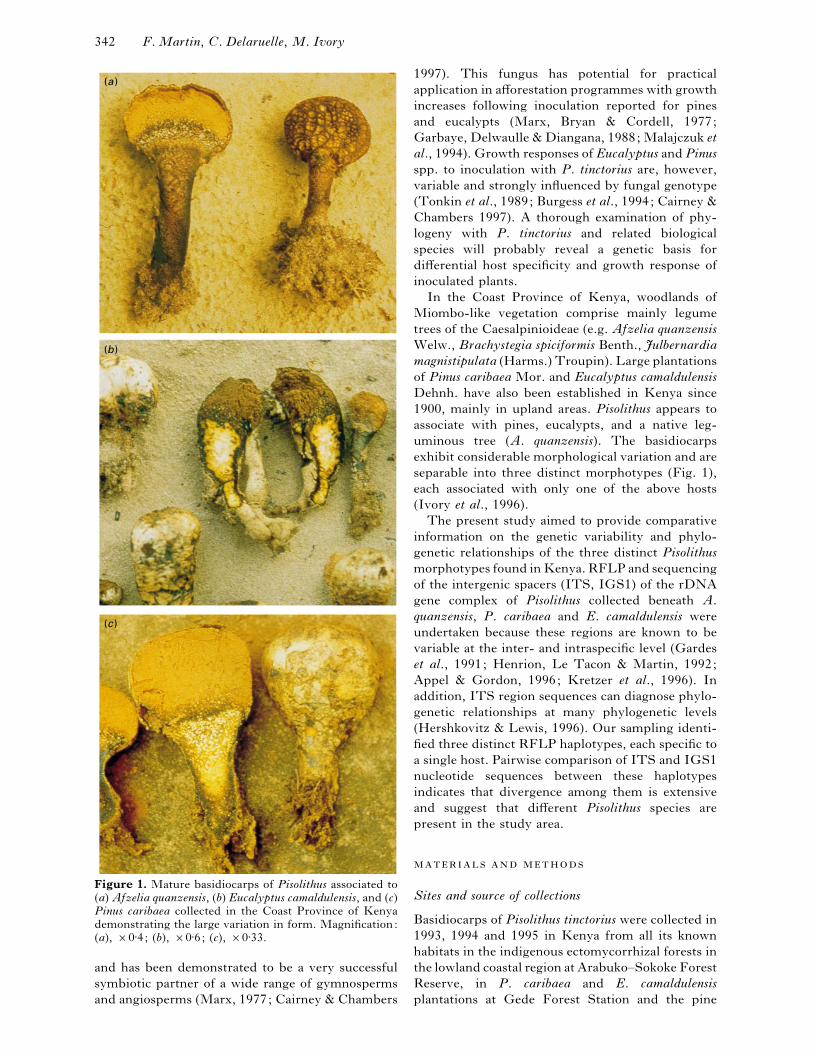
5\8S, and ITS2, from the three morphotypes}haplotypes were sequenced (GenBank accession no.
AF003914, AF003915 and AF003916) and aligned
(Fig. 4) with known Pisolithus ITS sequences
346 F. Martin, C. Delaruelle, M. Ivory
Figure 4. For legend see opposite.

Genetic variability in Pisolithus 347
Figure 4. Multiple sequence alignment of the ITS region of basidiocarps of Pisolithus associated to Eucalyptuscamaldulensis, Pinus caribaea, or Afzelia quanzensis in Kenya, together with Pisolithus ITS sequences available
in the NCBI database. no. 5105 (GenBank accession no. AF003915), no. 5110 (accession no. AF003914), no.
5111 (accession no. AF003916), basidiocarps associated with A. quanzensis, E. camaldulensis, and P. caribaea,
respectively; 441, vegetative mycelium of P. tinctorius 441 associated with Eucalyptus citriodora in Brazil ; CS01
(GenBank accession no. AF004732), LJ07 (AF004733), R01 (AF004735), W15 (AF004736), W16 (AF004737)
and WM01 (AF004734), isolates of Pisolithus collected in New South Wales (Australia) (Anderson et al., 1998;
with kind permission). –, gap resulting from insertion}deletion.
(Anderson et al., 1998, Carnero-Diaz et al., 1997).
The Pisolithus ITS sequences were all clearly related
to each other. The actual length of the corresponding
fragment was 589–627 bp, also containing a part of
the 3«-end of the 18S rDNA and the 5«-end of the
28S rDNA. Typically, a high degree of similarity

348 F. Martin, C. Delaruelle, M. Ivory
100
100
79
10
WM01 no. 5110
LJ07
no. 5105
no. 5111
W16
W15
CS01
R01
441
Figure 5. Radial parsimony tree of ITS sequences of
Pisolithus isolates. The tree was constructed from the
sequence alignment data set shown in Figure 4. The
illustration is one of three equally parsimonious trees of
length 329 which were found using PAUP 3\1\1 to
perform a heuristic search with simple addition. Numerical
values, shown along the stem of each supported clade, are
those deriving from 1000 replicates of heuristic parsimony
bootstrap analysis. The scale bar on the bottom right
indicates the length of 10 steps in the tree. Outlines and
shading indicate particular groupings of ITS referred to in
the text.
was present within the 194 bp-5\8S rDNA region in
all the Pisolithus collections (!95%) (Fig. 4) and the
Pisolithus 5\8S showed a high similarity with
basidiomycetous ITS sequences deposited in the
NCBI DNA database (data not shown).
The ITS1 and ITS2 sequences of the three
distinct Kenyan Pisolithus morphotypes differed by
several insertions}deletions and base substitutions,
but conservation patterns with phylogenetic diag-
nostic value (Hershkovitz & Zimmer, 1996), could
be easily identified. The multiple sequence align-
ment (Fig. 4) indicated that isolates no. 5110
(eucalypt-associated morphotype), and LJ07 and
WM01 from NSW (Australia) had 98% sequence
homology with each other, but only 85% homology
with the other isolates. Their sequence are charac-
terized by the presence of a CT-rich insertion in the
3«-end of the ITS2 region. Similarly, isolates CS01,
1000
500
300
200
100
M 1 2 3 4
(a )
(b)
(c )
1000
500
300
200
100
1000
500
300
200
100
Figure 6. The amplified IGS1 and RFLP patterns of
Pisolithus isolates associated with (a) Afzelia (¯haplotype
A), (b) Eucalyptus (¯haplotype E), or (c) Pinus (¯hap-
lotype P) in the lowland forests of the Coast Province
of Kenya. Lane 1, uncut IGS1; lane 2, 3, and 4: ITS cut
with Mbo I, Alu I and Hinf I, respectively. M, fragment
size markers in bp. Partial digestion products are visible
above 300 bp in (c) 4.
R01, W15, W16 from NSW and P. tinctorius 441
from Brazil showed a high degree of sequence
homology, being 95–97% homologous. The ITS
sequences of Afzelia- (no. 5105) and Pine- (no. 5111)
associated isolates were divergent with each other
and strikingly divergent from other investigated
Pisolithus isolates.
Equally weighted, unordered parsimony analysis
of the data set of all ITS sequences resulted in three
equally parsimonious trees (differences between the
trees were due to branch swapping of haplotypes).
The strict consensus radial tree (Fig. 5), deriving
from 1000 iterations of heuristic parsimony boot-
strap analysis, shows the relationships among the 10
ITS sequences. It supports the clear differentiation
of the three major groups revealed by sequence
pairwise comparison. The various Pisolithus groups
were supported by very strong branches (79–100%
of 1000 bootstrap iterations).

Genetic variability in Pisolithus 349
Figure 7. Multiple sequence alignment of IGS1 of basidiocarps of Pisolithus associated to Eucalyptuscamaldulensis (5110; accession no. AF0061183), Pinus caribaea (5111, accession no. AF0061184) and Afzeliaquanzensis (5106, GenBank accession no. AF0061182). –, gap resulting from insertion}deletion.
RFLP and sequence of IGS
The basidiocarps were further examined by PCR}RFLP of the rDNA IGS, a rDNA region known as
highly variable in fungi (Henrion et al., 1992, 1994;
Apel & Gordon, 1996; Selosse et al., 1996). All
basidiocarps of the Pinus-, Eucalyptus- and Afzelia-
morphotypes produced a single IGS band of 450 bp
(Fig. 6). Heteroduplex formation resulting from
rDNA heterozygosity (Selosse et al., 1996) was not
observed in the amplification of Pisolithus rDNA
IGS. Three distinct restriction patterns, with two to
four restricted fragments per pattern, were detected
using Alu I, Hinf I, and Mbo I (Fig. 6; Table 2). The
clustering of basidiocarps was identical to the one
achieved by PCR}RFLP of the rDNA ITS. In both
analyses, the 52 basidiocarps formed three clusters
which correspond to the three morphotypes of
Pisolithus. By contrast, RFLPs of IGS1 of isolates,
collected over a wide geographical scale (100–
120 km), were identical within each morphotype}haplotype.
Both strands of the amplified IGS1 were
sequenced (GenBank accession no. AF001182,
AF0061183 and AF0061184) and aligned (Fig. 7).
These sequences were clearly related to each other.
The actual length of the corresponding fragment was
446–449 bp, also containing a part of the 3«-end of
the 25S rDNA and the 5«-end of the 5S rDNA. The
aligned sequences indicated that isolates nos 5110

350 F. Martin, C. Delaruelle, M. Ivory
(eucalypt-associated), and 5111 (eucalypt-associ-
ated) and 5105 (Afzelia-associated) had 21% se-
quence divergence with each other.
Because collections within the Pisolithus taxon
exhibit considerable variability in basidiocarp and
basidiospore morphology, it is widely thought that
Pisolithus is a complex of evolutionarily independent
biological species (Bronchart et al., 1975; Kope &
Fortin 1990; Burgess et al., 1995; Watling et al.,
1995). Various methods based on the PCR have been
proposed to characterize ectomycorrhizal fungi
(Gardes et al., 1991; Henrion et al., 1992) and to
examine phylogenetic relationships in the various
taxa (Bruns, White & Taylor, 1991). Direct
sequencing of genes coding for nuclear and mito-
chondrial rRNA genes and intergenic spacers
amplified by PCR (Bruns et al., 1989; Bruns &
Szaro, 1992; Kretzer et al., 1996; Selosse et al.,
1996) and RFLP analysis of these PCR-amplified
sequences (Gardes et al., 1991; Henrion et al., 1992,
1994) have been used to characterize ectomycorrhizal
fungi at the intraspecific and interspecific phylo-
genetic levels. In the present investigation, we have
used this approach to investigate genetic and phylo-
genetic relationships between the morphotypes of
Pisolithus found in plantations and woodlands of the
Coast Province of Kenya.
In the area studied, host association appeared to be
a key feature in the clustering of Pisolithus isolates
since all of the basidiocarps, isolated under a specific
host plant (A. quanzensis, P. caribaea, or E.
camaldulensis), but ranking from subregional to local
area scale, produced identical rDNA patterns and
sequences. They showed no obvious correlation with
geographical origin. This lack of genetic variability
within highly polymorphic DNA regions, such as
the IGS1, has been confirmed by using micro-
satellite-primed PCR (data not shown) and suggests
that little breeding occurs between Pisolithus
associated with pine and eucalypt plantations and the
indigenous morphotype associated with a native tree
(Afzelia) in forest reserves even when basidiocarps
of different morphology}haplotype occur in close
proximity (C10–100 m). The variation in
basidiocarp morphology and the degree of host
specificity (Ivory et al., 1996), together with the
present genetic analysis suggest that eucalypt-com-
patible and pine-compatible strains of Pisolithus
have been introduced to Kenya and now persist in a
few pine and eucalypt plantations.
The sequence analysis of the rDNA ITS and
IGS1 have confirmed that the Pisolithus types in the
study area are genetically highly divergent. The
three groups formed by analysis of sequence poly-
morphism of rDNA spacers correlated very well
with basidiocarp morphotypes and host range (Ivory
et al., 1996). The estimated pairwise distance among
Pisolithus ITS sequences (up to 20%) is extensive
when compared to that found in some other fungal
species (1–10%) (Gardes et al., 1991; Hibbett et al.,
1995; Bruns et al., 1991; Hseu et al., 1996).
Comparable levels of sequence divergence between
distinct species and species groups within the
basidiomycetes (e.g. Lentinus ; Hibbett et al. (1995))
and ascomycetes (e.g. Cenococcum geophilum ;
Lobuglio, Rogers & Wang (1991)) have been
interpreted as indicating significant evolutionary
divergence within these species.
Similarly, the high degree of rDNA ITS di-
vergence among Pisolithus from Kenya (present
study) and Australia (Anderson et al., 1998) suggests
that we are dealing with a complex of evolutionarily
independent biological species. Although highly
variable at the interspecific level, the ITS region
contains conserved patterns and could be used to
diagnose phylogenetic relationships (Hershkovitz &
Lewis, 1996; Hershkovitz & Zimmer, 1996). A
parsimony analysis of the data set of all ITS
sequences of Pisolithus available in DNA databases
was therefore performed. The phylogenetic analysis
clearly separated a group of isolates including the
eucalypt-associated no. 5110 (Kenya), LJ07 and
WM01 (Australia) from the cluster containing other
Australian isolates (W16, W15, CS01, R01) and no.
441 from Brazil. These findings confirm and extend
a study, based on RAPD and ITS sequence analyses,
of NSW Pisolithus (Anderson et al., 1998). Sep-
aration of the eucalypt-associated isolates on two
strongly supported branches (100% bootstrap values
from 1000 iterations) indicates that these two groups
might represent two different species. Basidiocarp
and basidiospore morphology of the Australian
isolates CS01, R01, W15 and W16 conformed to
Cunningham’s description (Cunningham, 1942) of
P. tinctorius (Anderson et al., 1998). This suggests
that the group, including isolates CS01, R01, W15,
W16 and no. 441, probably represents P. tinctorius,
whereas the other cluster (no. 5110, LJ07 and
WM01) represents another, probably undescribed
Pisolithus species. Isolates of Pisolithus associated
to Afzelia (no. 5105) or pine (no. 5111) were related,
but widely separated from other clades. They might
also represent additional species, but more isolates
need to be included in future studies to fully resolve
the genetic heterogeneity within these two Pisolithus
types. Interestingly, ITS sequence of Pinus-associ-
ated basidiocarps (no. 5111) found in Kenya was
identical to the sequence of isolates of Pisolithus
collected beneath pine species on different continents
suggesting a common origin for this morphotype
(Dell et al., unpublished).
It appears that distinct evolutionary lineages occur
amongst the P. tinctorius populations world-wide
(Bronchart et al., 1975; Calogne & Demoulin, 1975;
Burgess et al., 1995; Watling et al., 1995; Anderson

Genetic variability in Pisolithus 351
et al., 1998; present study). An analysis of rDNA
ITS sequence variation amongst these world-wide
populations would be useful for exploring the
phylogenetic relationships existing between these
lineages.
The above study forms part of a joint research programme
conducted by INRA-Nancy, Oxford Forestry Institute,
Kenya Forestry Research Institute, and Universidad de
Murcia, which is entirely funded by the EU project TS3-
CT92-0124. We would like to thank Ce! line Di Battista
(INRA-Nancy) for her assistance in PCR typing during
the initial phase of this project, Bernie Dell (Murdoch
University) for critical reading of an early version of the
manuscript and his timely suggestions, and John Cairney
(University of Western Sydney) for providing ITS
sequences of Australian Pisolithus before publication.
Discussions with Linus Mwangi (KEFRI) have been very
helpful and appreciated.
Altschul SF, Madden TL, Schaffer AA, Zhang J, Zhang Z,Miller W, Lipman DJ. 1997. Gapped BLAST and PSI-
BLAST: a new generation of protein database search programs.
Nucleic Acids Research 25 : 3389–3402.
Anderson IC, Chambers SM, Cairney JWG. 1998. Molecular
determination of genetic variation in Pisolithus isolates from a
defined region in New South Wales, Australia. New Phytologist
138 : 151–162.
Appel DJ, Gordon TR. 1996. Relationships among pathogenic
and nonpathogenic isolates of Fusarium oxysporum based on the
partial sequence of the intergenic spacer region of the ribosomal
DNA. Molecular Plant–Microbe Interactions 9 : 125–138.
Bronchart R, Calogne FD, Demoulin V. 1975. Nouvelle
contribution a' l’e! tude de l’ultrastructure de la paroi sporale des
Gaste! romyce' tes. Bulletin Trimestriel de la SocieU teU Mycologique
de France 91 : 232–246.
Bruns TD, Szaro TM. 1992. Rate and mode differences between
nuclear and mitochondrial small-subunit rRNA genes in
mushrooms. Molecular Biology and Evolution 9 : 836–855.
Bruns TD, Fogel R, White TJ, Palmer JD. 1989. Accelerated
evolution of a false-truffle from a mushroom ancestor. Nature
339 : 140–142.
Bruns TD, White TJ, Taylor JW. 1991. Fungal molecular
systematics. Annual Review of Ecology and Systematics 22 :
525–564.
Burgess T, Dell B, Malajczuk N. 1994. Variation in mycorrhizal
development and growth stimulation by 20 Pisolithus isolates
inoculated onto Eucalyptus grandis W. Hill ex Maiden. New
Phytologist 127 : 731–739.
Burgess T, Malajczuk N, Dell B. 1995. Variation in Pisolithus
based on basidiome and basidiospore morphology, culture
characteristics and analysis of polypeptides using 1D–SDS–
PAGE. Mycological Research 99 : 1–13.
Cairney JWG, Chambers SM. 1997. Interactions between
Pisolithus tinctorius and its hosts : a review of current knowledge.
Mycorrhiza 7 : 117–131.
Calogne FD, Demoulin V. 1975. Les Gaste! romyce' tesd’Espagne. Bulletin Trimestriel de la SocieU teU Mycologique de
France 91 : 247–292.
Carnero Diaz ME, Tagu D, Martin F. 1997. Ribosomal DNA
internal transcribed spacers to estimate the proportion of
Pisolithus tinctorius and Eucalyptus globulus RNAs in ecto-
mycorrhiza. Applied and Environmental Microbiology 63 :
840–843.
Corpet F. 1988. Multiple sequence alignment with hierarchical
clustering. Nucleic Acids Research 16 : 10881–10890.
Cunningham GH. 1942. The Gasteromycetes of Australia and
New Zealand. Dunedin, New Zealand: John McIndoe.
Garbaye J, Delwaulle JC, Diangana D. 1988. Growth response
of eucalypts in the Congo to ectomycorrhizal inoculation. Forest
Ecology and Management 24 : 151–157.
Gardes M, White TJ, Fortin JA, Bruns TD, Taylor JW. 1991.Identification of indigenous and introduced symbiotic fungi in
ectomycorrhizae by amplification of nuclear and mitochondrial
ribosomal DNA. Canadian Journal of Botany 69 : 189–190.
Grube M, Depriest PT, Gargas A, Hafellner J. 1995. DNA
isolation from lichen ascomata. Mycological Research 99 :
1321–1324.
Henrion B, Le Tacon F, Martin F. 1992. Rapid identification of
genetic variation of ectomycorrhizal fungi by amplification of
ribosomal RNA genes. New Phytologist 122 : 289–298.
Henrion B, Chevalier G, Martin F. 1994. Typing truffle species
by PCR amplification of the ribosomal DNA spacers.
Mycological Research 98 : 37–43.
Hershkovitz MA, Lewis LA. 1996. Deep-level diagnostic value
of the rDNA-ITS region. Molecular Biology and Evolution 13 :
1276–1295
Hershkovitz MA, Zimmer EA. 1996. Conservation patterns in
angiosperm rDNA ITS2 sequences. Nucleic Acids Research 24 :
2857–2867.
Hibbett DS, Fukusama-Nakai Y, Tsuneda A, Donoghue MJ.1995. Phylogenetic diversity in shiitake inferred from nuclear
ribosomal DNA sequences. Mycologia 87 : 618–638.
Ho I. 1987. Comparison of eight Pisolithus tinctorius isolates for
growth rate, enzyme activity, and phytohormone production.
Canadian Journal of Forestry Research 17 : 31–35.
Hseu RS, Wang HH, Wang HF, Moncalvo JM. 1996.Differentiation and grouping of isolates of the Ganoderma
lucidum complex by random amplified polymorphic DNA–PCR
compared with grouping on the basis of internal transcribed
spacer sequences. Applied and Environmental Microbiology 62 :
1354–1363.
Ivory MH. 1987. Diseases and disorders of pines in the tropics.
Overseas Research Publication 31, Overseas Development
Administration, London}Oxford Forestry Institute, Oxford.
Ivory MH, Honrubia M, Mburu BK, Mwangi LM. 1996.Putative ectomycorrhizal fungi from native and exotic forests in
Kenya. In: Azcon-Aguilar C, Barea JM, eds. Mycorrhizas in
Integrated Systems from Genes to Plant Development. Brussels,
Luxembourg: Office for official publications of the European
Communities, COST Report, 125–127.
Kope HH, Fortin JA. 1990. Germination and comparative
morphology of basidiospores of Pisolithus arhizus. Mycologia
82 : 350–357.
Kretzer A, Li Y, Szaro T, Bruns TD. 1996. Internal transcribed
spacer sequences from 38 recognized species of Suillus sensu
lato : phylogenetic and taxonomic implications. Mycologia 88 :
776–785.
Lamhamedi MS, Fortin JA, Kope HH, Kropp BR. 1990.Genetic variation in ectomycorrhiza formation by Pisolithus
arhizus on Pinus pinaster and Pinus banksiana. New Phytologist
115 : 689–697.
Lobuglio KF, Rogers SO, Wang CJK. 1991. Variation in
ribosomal DNA among isolates of the mycorrhizal fungus
Cenococcum geophilum. Canadian Journal of Botany 69 :
2331–2343.
Malajczuk N, Grove TS, Bougher TS, Dell B, Gong Minquin.1994. Ectomycorrhizas and nutrients – their importance to
eucalypts in China. Brown AG, eds. Australian Tree Species
Research in China. Canberra, Australia : Australian Centre for
International Agricultural Research, 132–139.
Malajczuk N, Lapeyrie F, Garbaye J. 1990. Infectivity of pine
and eucalypt isolates of Pisolithus tinctorius on roots of
Eucalyptus urophylla in vitro 1. Mycorrhiza formation in model
systems. New Phytologist 114 : 627–631.
Martin F, Costa G, Delaruelle C, Diez J. 1997. Genomic
fingerprinting of ectomycorrhizal fungi by microsatellite-
primed PCR. In: Varma A, Hock B, eds. Mycorrhiza Manual.
Springer Lab Manual Berlin: Springer-Verlag, 463–474.
Marx DH. 1977. Tree host range and world distribution of the
ectomycorrhizal fungus Pisolithus tinctorius. Canadian Journal
of Microbiology 23 : 217–223.
Marx DH. 1980. Variability in ectomycorrhizal development and
growth among isolates of Pisolithus tinctorius as affected by
source, age, and re-isolation. Canadian Journal of Forest
Research 11 : 168–174.
Marx DH, Bryan WC, Cordell CE. 1977. Survival and growth of

352 F. Martin, C. Delaruelle, M. Ivory
pine seedlings with Pisolithus ectomycorrhizae after two years
on reforestation sites in North Carolina and Florida. Forest
Science 16 : 363–373.
Mehmann B, Egli S, Braus GH, Brunner I. 1995. Coincidence
between molecularly or morphologically classified ecto-
mycorrhizal morphotypes and fruitbodies in a spruce forest. In:
Stocchi V, Bonfante P, Nuti M, eds. Biotechnology of Ecto-
mycorrhizae. Molecular Approaches. New York: Plenum Press,
41–52.
Sambrook J, Fritsch EF, Maniatis T. 1989. Molecular Cloning.
A Laboratory Manual, 2nd edn. New York: Cold Springer
Harbor Laboratory Press.
Selosse M-A, Costa G, Di Battista C, Le Tacon F, Martin F.
1996. Meiotic segregation and recombination of the intergenic
spacer of the ribosomal DNA in the ectomycorrhizal basi-
diomycete Laccaria bicolor. Current Genetics 30 : 332–337.
Swofford DL. 1993. PAUP: Phylogenetic analysis using par-
simony, version 3.1.1. Computer program distributed by the
Illinois Natural History Survey, Champaign, IL, USA.
Tonkin CM, Malajczuk N, McComb JA. 1989. Ecto-
mycorrhizal formation by micropropagated clones of Eucalyptus
marginata inoculated with isolates of Pisolithus tinctorius. New
Phytologist 111 : 209–24.
Watling R, Taylor A, Lee SS, Sims K, Alexander IJ. 1995. A
rainforest Pisolithus : its taxonomy and ecology. Nova Hedwigia
61 : 417–429.