Embed Size (px)
Citation preview
REVIEW
GENETIC TOOLBOX
Genetic and Genomic Toolbox of the ChordateCiona intestinalisAlberto Stolfi and Lionel Christiaen
Center for Developmental Genetics, Department of Biology, New York University, New York, New York 10003
ABSTRACT The experimental malleability and unique phylogenetic position of the sea squirt Ciona intestinalis as part of the sistergroup to the vertebrates have helped establish these marine chordates as model organisms for the study of developmental geneticsand evolution. Here we summarize the tools, techniques, and resources available to the Ciona geneticist, citing examples of studiesthat employed such strategies in the elucidation of gene function in Ciona. Genetic screens, germline transgenesis, electroporation ofplasmid DNA, and microinjection of morpholinos are all routinely employed, and in the near future we expect these to be comple-mented by targeted mutagenesis, homologous recombination, and RNAi. The genomic resources available will continue to support thedesign and interpretation of genetic experiments and allow for increasingly sophisticated approaches on a high-throughput, whole-genome scale.
THE sea squirt Ciona intestinalis (Figure 1) has recentlyemerged as a powerful model organism for biological
research in the postgenomic era. Ever since their days asa favorite of early developmental biologists like LaurentChabry (Chabry 1887), Ed Conklin (Conklin 1905), andeven famed geneticist Thomas Hunt Morgan (Morgan1923), sea squirts, or ascidians, have been recognized aschoice organisms for experimental embryology due to theirsimple embryos, rapid development, and ease of manipula-tion. These advantages, coupled to their amenability to ge-nomic and systems approaches (Satoh et al. 2003), todaydrive the sure but steady acceptance of ascidians into themainstream of developmental biology.
Ascidians are a paraphyletic group within the subphylumTunicata in the chordate phylum. For a long time it wasbelieved that cephalochordata as the sister group to thevertebrates. However, improved phylogenetic methods andsequencing of tunicate (also known as “urochordate”) andcephalochordate genomes clearly confirmed that the tuni-cates, although highly divergent in body plan and genomicarchitecture, are our closest living invertebrate relatives(Delsuc et al. 2006; Putnam et al. 2008). This phylogenetic
position has been an important factor for the adoption ofthe sea squirt as a “model organisms” for the biomedicalsciences.
We will not provide an in-depth discussion of all theadvantages or disadvantages of using ascidians, morespecifically Ciona spp., in biological research. Nor do weintend this review to be a primer on ascidian biology. Thesehave been covered elsewhere (Christiaen et al. 2009c;Lemaire 2009, 2011). Instead, we will focus on the genetictools available to the biologist studying C. intestinalis andrelated species. These include not only the traditional ap-proaches of forward and reverse genetic screens, but alsomolecular genetic techniques, especially those based ontransient transfection of whole embryos with plasmid DNA.The two major experimental species, C. intestinalis andC. savignyi, are weakly cross-fertile (Byrd and Lambert2000) and are treated here almost interchangeably, referredto as simply Ciona. In fact, evidence points to the groupingof more than one cryptic species within C .intestinalis(Suzuki et al. 2005; Iannelli et al. 2007), so it would bemore appropriate to talk about a Ciona species complex.Embryogenesis is virtually identical in all Ciona spp., despiteneutral genomic sequence variation 10 times greater thanthat observed between rat and mouse (Hill et al. 2008).
Other ascidian species utilized as experimental species inthe laboratory include Halocynthia roretzi, Phallusia mam-milata, Molgula spp., Ascidiella aspersa, Ecteinascidia turbi-nata, and Botryllus schlosseri. Extreme conservation of
Copyright © 2012 by the Genetics Society of Americadoi: 10.1534/genetics.112.140590Manuscript received March 18, 2012; accepted for publication April 30, 2012Available freely online through the author-supported open access option.Corresponding authors: Department of Biology, New York University, 100 WashingtonSquare E., New York, NY 10003. E-mail: [email protected] and [email protected]
Genetics, Vol. 192, 55–66 September 2012 55
embryonic development among these distantly related spe-cies is a general rule (Lemaire 2009), in spite of profounddifferences in genomic sequence (Oda-Ishii et al. 2005) or inchoice of cell signaling pathway used for induction of a givencell fate (Tokuoka et al. 2007; Hudson and Yasuo 2008).This dichotomy of phenotypic constancy and molecular di-vergence is of great interest to those studying the evolutionof development. Overall, the remarkable conservation ofascidian embryogenesis also provides some hope that mosttechniques described here might be readily applied to othersea squirt species.
Forward Genetics
The latest techniques and strategies for genetic screens inCiona have recently been reviewed and outlined in excep-tional detail (Veeman et al. 2011). Here we attempt to sum-marize basic aspects of doing forward genetics in Ciona. Oneparticular advantage that has motivated the development offorward genetics in Ciona, especially for developmentalstudies, is the minimal overlap in gene functions. Due totunicates having branched off from vertebrates before thelatter underwent two whole-genome duplication events(Dehal and Boore 2005), several paralogous gene familiesin vertebrates are each represented by a single ortholog inCiona. This means the requirement for these genes can bereadily tested without the need for double or triple mutantsto circumvent such overlap in gene function.
The generation time of Ciona is 1–3 mo. Under certainculturing conditions, sperm can be obtained in 1 mo andeggs after 2 mo (Sasakura et al. 2003). Most solitary asci-dians are hermaphroditic broadcast spawners. This presentsadvantages as well as disadvantages to the geneticist study-ing them. One advantage is that Ciona adults show a modestability to self-fertilize. This allows one to screen for recessivemutations in the progeny of selfed F0 animals. Second, their
broadcast spawning strategy is tied to their large effectivepopulation sizes, which has resulted in extreme polymor-phism rates (Small et al. 2007b). Genome-wide average sin-gle nucleotide polymorphism (SNP) heterozygosity is at1.2% in C. intestinalis and reaches 4.5% in C. savignyi (Dehalet al. 2002; Kim et al. 2007; Small et al. 2007b). In otherwords, any two Ciona individuals from the same populationare as different from one another, at the sequence level, ashumans are from chimpanzees. This means that there is noshortage of SNPs to use as genetic markers, averaging a SNPor indel per 80 bp of genome. Indeed, the latest positionalmapping strategies in Ciona use direct sequencing of SNPs(Veeman et al. 2011).
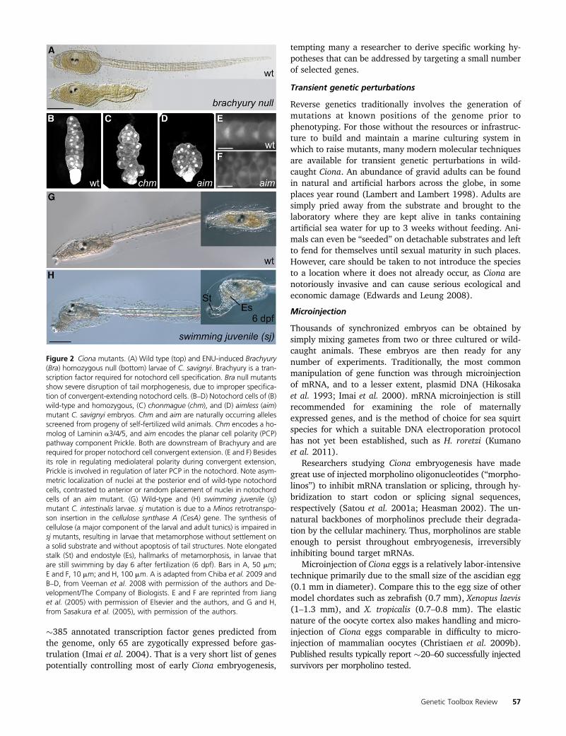
Unfortunately, the elevated levels of polymorphism makeidentifying the particular mutation underlying a phenotypedifficult to pinpoint. Nevertheless, successful identificationof genes underlying mutant phenotypes has been carriedout. Screening for short-tailed mutants revealed essentialroles for prickle and laminin a3/4/5 in notochord cell po-larity and convergence (Figure 2, B–F) (Jiang et al. 2005;Veeman et al. 2008). These mutants were identified byscreening progeny from self-fertilized “wild-caught” Ciona.This demonstrates how the extreme natural variation in wildCiona populations also provides a wealth of naturally occur-ring recessive mutations. Additionally, N-ethyl-N-nitrosourea(ENU) has been successfully used to induce mutations (Fig-ure 2A) (Chiba et al. 2009). Mutant strains are propagatedfor distribution by the Ascidian Stock Center at the Universityof California, Santa Barbara (http://www.ascidiancenter.ucsb.edu/). Currently, there are no isogenic inbred lines availablefor Ciona.
Ciona also present unique challenges relating primarily totheir captivity and husbandry. As marine filter-feeders, theyrequire circulating sea water. Both open circulation systemsutilizing a natural source of sea water and food as well asclosed circulation systems utilizing cultured microplanktonas a food source have been developed to rear Ciona (Hen-drickson et al. 2004; Joly et al. 2007). Open systems areeasier to maintain, but may be difficult to set up, dependinggreatly on local availability of microplankton-laden sea wa-ter. Closed systems can be set up far inland, but may en-counter problems such as inadequate food supply, waterfouling, pests, etc. Nonetheless, the breeding and rearingof Ciona in the laboratory is feasible and stands to be furtherdeveloped as more groups begin to incorporate geneticscreens and genetic engineering in their studies (see below).
Reverse Genetics
The sequencing of the C. intestinalis (Dehal et al. 2002) andC. savignyi (Vinson et al. 2005; Small et al. 2007a) genomesin the early to mid 2000s immediately made reverse geneticsin Ciona quite alluring. This is especially the case for studiesin developmental biology where a limited number of candi-date genes identified from genomic and EST sequences arepredicted to effect early embryogenesis. For instance, of
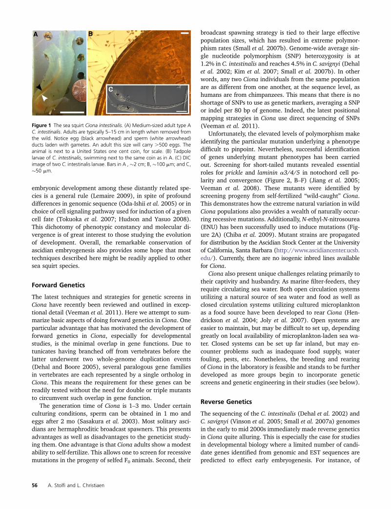
Figure 1 The sea squirt Ciona intestinalis. (A) Medium-sized adult type AC. intestinalis. Adults are typically 5–15 cm in length when removed fromthe wild. Notice egg (black arrowhead) and sperm (white arrowhead)ducts laden with gametes. An adult this size will carry .500 eggs. Theanimal is next to a United States one cent coin, for scale. (B) Tadpolelarvae of C. intestinalis, swimming next to the same coin as in A. (C) DICimage of two C. intestinalis larvae. Bars in A , �2 cm; B,�100 mm; and C,�50 mm.
56 A. Stolfi and L. Christiaen
�385 annotated transcription factor genes predicted fromthe genome, only 65 are zygotically expressed before gas-trulation (Imai et al. 2004). That is a very short list of genespotentially controlling most of early Ciona embryogenesis,
tempting many a researcher to derive specific working hy-potheses that can be addressed by targeting a small numberof selected genes.
Transient genetic perturbations
Reverse genetics traditionally involves the generation ofmutations at known positions of the genome prior tophenotyping. For those without the resources or infrastruc-ture to build and maintain a marine culturing system inwhich to raise mutants, many modern molecular techniquesare available for transient genetic perturbations in wild-caught Ciona. An abundance of gravid adults can be foundin natural and artificial harbors across the globe, in someplaces year round (Lambert and Lambert 1998). Adults aresimply pried away from the substrate and brought to thelaboratory where they are kept alive in tanks containingartificial sea water for up to 3 weeks without feeding. Ani-mals can even be “seeded” on detachable substrates and leftto fend for themselves until sexual maturity in such places.However, care should be taken to not introduce the speciesto a location where it does not already occur, as Ciona arenotoriously invasive and can cause serious ecological andeconomic damage (Edwards and Leung 2008).
Microinjection
Thousands of synchronized embryos can be obtained bysimply mixing gametes from two or three cultured or wild-caught animals. These embryos are then ready for anynumber of experiments. Traditionally, the most commonmanipulation of gene function was through microinjectionof mRNA, and to a lesser extent, plasmid DNA (Hikosakaet al. 1993; Imai et al. 2000). mRNA microinjection is stillrecommended for examining the role of maternallyexpressed genes, and is the method of choice for sea squirtspecies for which a suitable DNA electroporation protocolhas not yet been established, such as H. roretzi (Kumanoet al. 2011).
Researchers studying Ciona embryogenesis have madegreat use of injected morpholino oligonucleotides (“morpho-linos”) to inhibit mRNA translation or splicing, through hy-bridization to start codon or splicing signal sequences,respectively (Satou et al. 2001a; Heasman 2002). The un-natural backbones of morpholinos preclude their degrada-tion by the cellular machinery. Thus, morpholinos are stableenough to persist throughout embryogenesis, irreversiblyinhibiting bound target mRNAs.
Microinjection of Ciona eggs is a relatively labor-intensivetechnique primarily due to the small size of the ascidian egg(0.1 mm in diameter). Compare this to the egg size of othermodel chordates such as zebrafish (0.7 mm), Xenopus laevis(1–1.3 mm), and X. tropicalis (0.7–0.8 mm). The elasticnature of the oocyte cortex also makes handling and micro-injection of Ciona eggs comparable in difficulty to micro-injection of mammalian oocytes (Christiaen et al. 2009b).Published results typically report �20–60 successfully injectedsurvivors per morpholino tested.
Figure 2 Ciona mutants. (A) Wild type (top) and ENU-induced Brachyury(Bra) homozygous null (bottom) larvae of C. savignyi. Brachyury is a tran-scription factor required for notochord cell specification. Bra null mutantsshow severe disruption of tail morphogenesis, due to improper specifica-tion of convergent-extending notochord cells. (B–D) Notochord cells of (B)wild-type and homozygous, (C) chonmague (chm), and (D) aimless (aim)mutant C. savignyi embryos. Chm and aim are naturally occurring allelesscreened from progeny of self-fertilized wild animals. Chm encodes a ho-molog of Laminin a3/4/5, and aim encodes the planar cell polarity (PCP)pathway component Prickle. Both are downstream of Brachyury and arerequired for proper notochord cell convergent extension. (E and F) Besidesits role in regulating mediolateral polarity during convergent extension,Prickle is involved in regulation of later PCP in the notochord. Note asym-metric localization of nuclei at the posterior end of wild-type notochordcells, contrasted to anterior or random placement of nuclei in notochordcells of an aim mutant. (G) Wild-type and (H) swimming juvenile (sj)mutant C. intestinalis larvae. sj mutation is due to a Minos retrotranspo-son insertion in the cellulose synthase A (CesA) gene. The synthesis ofcellulose (a major component of the larval and adult tunics) is impaired insj mutants, resulting in larvae that metamorphose without settlement ona solid substrate and without apoptosis of tail structures. Note elongatedstalk (St) and endostyle (Es), hallmarks of metamorphosis, in larvae thatare still swimming by day 6 after fertilization (6 dpf). Bars in A, 50 mm;E and F, 10 mm; and H, 100 mm. A is adapted from Chiba et al. 2009 andB–D, from Veeman et al. 2008 with permission of the authors and De-velopment/The Company of Biologists. E and F are reprinted from Jianget al. (2005) with permission of Elsevier and the authors, and G and H,from Sasakura et al. (2005), with permission of the authors.
Genetic Toolbox Review 57
Despite the relative difficulty of morpholino injections,commitment and hard work can make it pay off in a big way.It was using morpholinos that the provisional gene regula-tory “blueprint” of the Ciona embryo was generated. Imaiand colleagues assayed the expression of 73 developmentalregulators at different embryonic stages, in 25 different mor-phant backgrounds generated by morpholino injection. Thisrevealed over 3000 gene regulatory connections operatingin the early Ciona embryo (Imai et al. 2006). Thus, micro-injection skills are, for now, indispensable for a rigorous andthorough investigation of gene function in Ciona.
Electroporation
Due to the constraints of the microinjection technique,a protocol for electroporating plasmid DNA into fertilized,single-cell zygotes was developed (Corbo et al. 1997). Al-though electroporation has been used to transfect cells intissue culture or in avian embryos, as far as we know Cionaare the only animals for which electroporation is used tointroduce DNA to whole embryos, en masse. For this tech-nique, plasmid DNA is simply mixed with embryos at theone-cell stage. This mixture is placed in an electroporationcuvette, which is subjected to an electric current, much akinto electroporation of cultured mammalian cells (Chu et al.1987). It is no exaggeration to say this revolutionized thestudy of Ciona embryogenesis as thousands of synchronized,transgenic embryos could be generated at the push of a but-ton. Today, electroporation remains the main molecular ge-netic technique used in most Ciona laboratories worldwide(Zeller 2004).
After cloning and preparation of plasmid DNA, the entireprocedure from adult dissection to fertilization, dechoriona-tion, and electroporation of embryos, takes �1 hr. (Christiaenet al. 2009a). Several plasmids can be tested in hundreds ofembryos each, daily. The sheer numbers of transfected em-bryos generated in short time by the electroporation tech-nique allow for robust, large-scale yet rapid and low-costphenotyping not possible in other chordate model organ-isms. In one such instance, Brown and colleagues were ableto assay 85,506 transgenic embryos resulting from 1237electroporations (Brown et al. 2007).
The typical plasmid electroporated into Ciona embryosconsists of a driver and a transgene. We refer to the combi-nation of distal cis-regulatory (“enhancers”) and basal pro-moter elements as the “driver.” The compact genomes ofCiona species allow for easy cloning and testing of intergenicand/or intronic sequences for enhancer activity. This hasbeen facilitated by a shared library of plasmids that allowsfor the Gateway method of restriction-free, modular assem-bly of driver/transgene combinations (Roure et al. 2007).Several lineage-, tissue- and cell-type–specific drivers (Fig-ure 3) have been described and are constantly being discov-ered and added to the researcher’s toolkit. Transgenes canbe reporter genes (“reporters”) and/or perturbation genes.We refer to perturbation genes as simply any sequenceexpected to have a biological function in Ciona. We will later
discuss which types of perturbation genes are frequentlyused in Ciona and which are still but promising possibilities.
General strategies, caveats, and surprisesof the electroporation technique
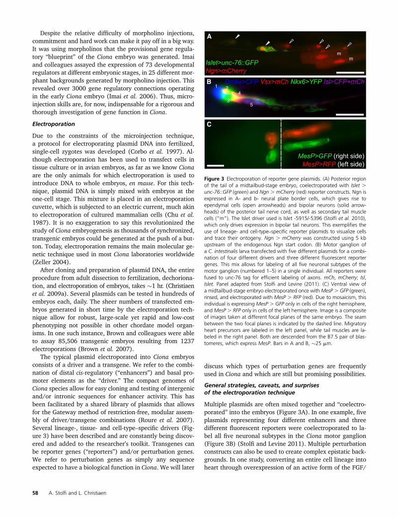
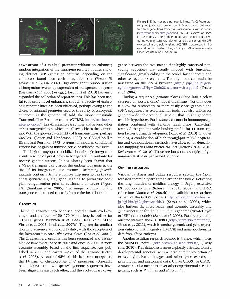
Multiple plasmids are often mixed together and “coelectro-porated” into the embryos (Figure 3A). In one example, fiveplasmids representing four different enhancers and threedifferent fluorescent reporters were coelectroporated to la-bel all five neuronal subtypes in the Ciona motor ganglion(Figure 3B) (Stolfi and Levine 2011). Multiple perturbationconstructs can also be used to create complex epistatic back-grounds. In one study, converting an entire cell lineage intoheart through overexpression of an active form of the FGF/
Figure 3 Electroporation of reporter gene plasmids. (A) Posterior regionof the tail of a midtailbud-stage embryo, coelectroporated with Islet .unc-76::GFP (green) and Ngn . mCherry (red) reporter constructs. Ngn isexpressed in A- and b- neural plate border cells, which gives rise toependymal cells (open arrowheads) and bipolar neurons (solid arrow-heads) of the posterior tail nerve cord, as well as secondary tail musclecells (“m”). The Islet driver used is Islet -5915/-5396 (Stolfi et al. 2010),which only drives expression in bipolar tail neurons. This exemplifies theuse of lineage- and cell-type–specific reporter plasmids to visualize cellsand trace their ontogeny. Ngn . mCherry was constructed using 5 kbupstream of the endogenous Ngn start codon. (B) Motor ganglion ofa C. intestinalis larva transfected with five different plasmids for a combi-nation of four different drivers and three different fluorescent reportergenes. This mix allows for labeling of all five neuronal subtypes of themotor ganglion (numbered 1–5) in a single individual. All reporters werefused to unc-76 tag for efficient labeling of axons. mCh, mCherry; Isl,Islet. Panel adapted from Stolfi and Levine (2011). (C) Ventral view ofa midtailbud-stage embryo electroporated once withMesP. GFP (green),rinsed, and electroporated with MesP . RFP (red). Due to mosaicism, thisindividual is expressing MesP . GFP only in cells of the right hemisphere,andMesP. RFP only in cells of the left hemisphere. Image is a compositeof images taken at different focal planes of the same embryo. The seambetween the two focal planes is indicated by the dashed line. Migratoryheart precursors are labeled in the left panel, while tail muscles are la-beled in the right panel. Both are descended from the B7.5 pair of blas-tomeres, which express MesP. Bars in A and B, �25 mm.
58 A. Stolfi and L. Christiaen
MAPK-signaling effector Ets1/2 while simultaneously block-ing cell migration by expressing a repressor form of thetranscription factor FoxF, resulted in the uncoupling of spec-ification and directed migration of cardiac precursor cells(Beh et al. 2007).
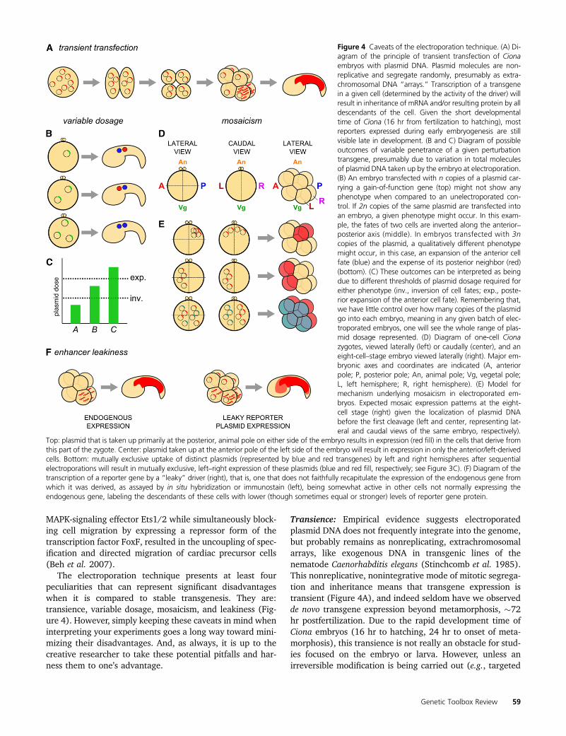
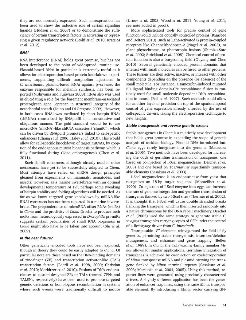
The electroporation technique presents at least fourpeculiarities that can represent significant disadvantageswhen it is compared to stable transgenesis. They are:transience, variable dosage, mosaicism, and leakiness (Fig-ure 4). However, simply keeping these caveats in mind wheninterpreting your experiments goes a long way toward mini-mizing their disadvantages. And, as always, it is up to thecreative researcher to take these potential pitfalls and har-ness them to one’s advantage.
Transience: Empirical evidence suggests electroporatedplasmid DNA does not frequently integrate into the genome,but probably remains as nonreplicating, extrachromosomalarrays, like exogenous DNA in transgenic lines of thenematode Caenorhabditis elegans (Stinchcomb et al. 1985).This nonreplicative, nonintegrative mode of mitotic segrega-tion and inheritance means that transgene expression istransient (Figure 4A), and indeed seldom have we observedde novo transgene expression beyond metamorphosis, �72hr postfertilization. Due to the rapid development time ofCiona embryos (16 hr to hatching, 24 hr to onset of meta-morphosis), this transience is not really an obstacle for stud-ies focused on the embryo or larva. However, unless anirreversible modification is being carried out (e.g., targeted
Figure 4 Caveats of the electroporation technique. (A) Di-agram of the principle of transient transfection of Cionaembryos with plasmid DNA. Plasmid molecules are non-replicative and segregate randomly, presumably as extra-chromosomal DNA “arrays.” Transcription of a transgenein a given cell (determined by the activity of the driver) willresult in inheritance of mRNA and/or resulting protein by alldescendants of the cell. Given the short developmentaltime of Ciona (16 hr from fertilization to hatching), mostreporters expressed during early embryogenesis are stillvisible late in development. (B and C) Diagram of possibleoutcomes of variable penetrance of a given perturbationtransgene, presumably due to variation in total moleculesof plasmid DNA taken up by the embryo at electroporation.(B) An embryo transfected with n copies of a plasmid car-rying a gain-of-function gene (top) might not show anyphenotype when compared to an unelectroporated con-trol. If 2n copies of the same plasmid are transfected intoan embryo, a given phenotype might occur. In this exam-ple, the fates of two cells are inverted along the anterior–posterior axis (middle). In embryos transfected with 3ncopies of the plasmid, a qualitatively different phenotypemight occur, in this case, an expansion of the anterior cellfate (blue) and the expense of its posterior neighbor (red)(bottom). (C) These outcomes can be interpreted as beingdue to different thresholds of plasmid dosage required foreither phenotype (inv., inversion of cell fates; exp., poste-rior expansion of the anterior cell fate). Remembering that,we have little control over how many copies of the plasmidgo into each embryo, meaning in any given batch of elec-troporated embryos, one will see the whole range of plas-mid dosage represented. (D) Diagram of one-cell Cionazygotes, viewed laterally (left) or caudally (center), and aneight-cell–stage embryo viewed laterally (right). Major em-bryonic axes and coordinates are indicated (A, anteriorpole; P, posterior pole; An, animal pole; Vg, vegetal pole;L, left hemisphere; R, right hemisphere). (E) Model formechanism underlying mosaicism in electroporated em-bryos. Expected mosaic expression patterns at the eight-cell stage (right) given the localization of plasmid DNAbefore the first cleavage (left and center, representing lat-eral and caudal views of the same embryo, respectively).
Top: plasmid that is taken up primarily at the posterior, animal pole on either side of the embryo results in expression (red fill) in the cells that derive fromthis part of the zygote. Center: plasmid taken up at the anterior pole of the left side of the embryo will result in expression in only the anterior/left-derivedcells. Bottom: mutually exclusive uptake of distinct plasmids (represented by blue and red transgenes) by left and right hemispheres after sequentialelectroporations will result in mutually exclusive, left–right expression of these plasmids (blue and red fill, respectively; see Figure 3C). (F) Diagram of thetranscription of a reporter gene by a “leaky” driver (right), that is, one that does not faithfully recapitulate the expression of the endogenous gene fromwhich it was derived, as assayed by in situ hybridization or immunostain (left), being somewhat active in other cells not normally expressing theendogenous gene, labeling the descendants of these cells with lower (though sometimes equal or stronger) levels of reporter gene protein.
Genetic Toolbox Review 59
mutagenesis by overexpression of zinc-finger nucleases, asproposed below), plasmid electroporation is not a suitablemethod for genetic perturbations in juveniles or adults. Thiscan actually be an advantage, because certain driver . per-turbation combinations that might be adult lethal or sterilecan be easily assayed during embryogenesis.
Variable dosage: The uptake of DNA by electroporated cellsis not well understood (Escoffre et al. 2009). What we doknow is that we have an imprecise control over exactly howmany copies of the plasmid go into each embryo. This meansthat, in any batch of electroporated embryos, one sees a gra-dient of transgene dosage, though there is no simple way toaccurately quantify this. This is why the large numbers oftransfected embryos obtained by electroporating them enmasse is so useful, as it allows one to average phenotypicquantification data over large numbers of individuals.
The one advantage of this variability is the possibility ofuncovering the effects of gene dosage in a particular process.In this way, the gradient of plasmid dosage seen in a batch ofelectroporated embryos is akin to an allelic series. Whereasdifferent transgenic strains must be generated to uncoverthe phenotypes associated with variable gene copy numberin organisms such as flies, in Ciona one can see the entirerange of phenotypes on a single slide. In some cases, qual-itatively different phenotypes can arise from different plas-mid doses, if, when, or where certain regulatory thresholdsare met (Figure 4, B and C).
Mosaicism: This term relates to the spatial variation inexpression of exogenous DNA, another issue inherent to theelectroporation technique. One hypothesis is that mosaicismmight occur by random, unequal distribution of plasmidDNA molecules into one of two daughter cells resulting froma mitotic division. Another possibility is random loss orsilencing of plasmid DNA. Regardless of the underlyingcause, transgene expression is frequently seen only in cellsdescended from one of the major “quadrants” or hemispheresof the early embryo (Figure 4, D and E). Mosaicism withina lineage is also known to occur (Zeller et al. 2006). However,with the appropriate optimization of electroporation rigs andparameters, one can increase or decrease the incidence ofmosaicism at will, suggesting the underlying cause of mosa-icism is closely linked to the initial distribution of plasmidDNA within the unicellular zygote (Zeller et al. 2006).
Mosaicism can be exploited to deliberately drive expres-sion of different transgenes in equivalent cells of differentlineages or on either side of the bilaterally symmetricembryo (Stolfi and Levine 2011). In embryos electroporatedmore than once, each time with a distinct plasmid, someproportion of embryos will randomly incorporate plasmidin one hemisphere and another plasmid in the oppositehemisphere (Figure 3C). This provides an internal controlin an experiment in which cells on a genetically manipulatedside of the embryo can be compared to cells on the opposite,unmanipulated side.
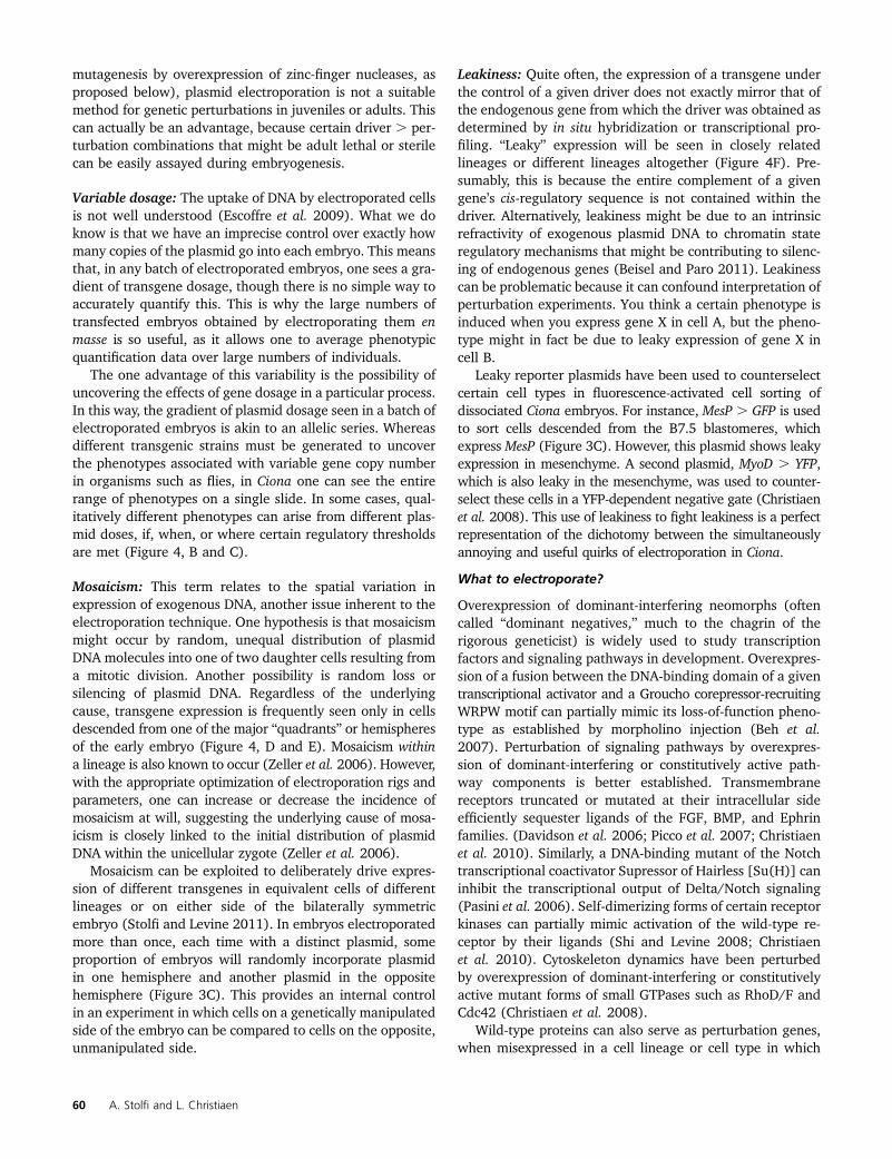
Leakiness: Quite often, the expression of a transgene underthe control of a given driver does not exactly mirror that ofthe endogenous gene from which the driver was obtained asdetermined by in situ hybridization or transcriptional pro-filing. “Leaky” expression will be seen in closely relatedlineages or different lineages altogether (Figure 4F). Pre-sumably, this is because the entire complement of a givengene’s cis-regulatory sequence is not contained within thedriver. Alternatively, leakiness might be due to an intrinsicrefractivity of exogenous plasmid DNA to chromatin stateregulatory mechanisms that might be contributing to silenc-ing of endogenous genes (Beisel and Paro 2011). Leakinesscan be problematic because it can confound interpretation ofperturbation experiments. You think a certain phenotype isinduced when you express gene X in cell A, but the pheno-type might in fact be due to leaky expression of gene X incell B.
Leaky reporter plasmids have been used to counterselectcertain cell types in fluorescence-activated cell sorting ofdissociated Ciona embryos. For instance, MesP . GFP is usedto sort cells descended from the B7.5 blastomeres, whichexpress MesP (Figure 3C). However, this plasmid shows leakyexpression in mesenchyme. A second plasmid, MyoD . YFP,which is also leaky in the mesenchyme, was used to counter-select these cells in a YFP-dependent negative gate (Christiaenet al. 2008). This use of leakiness to fight leakiness is a perfectrepresentation of the dichotomy between the simultaneouslyannoying and useful quirks of electroporation in Ciona.
What to electroporate?
Overexpression of dominant-interfering neomorphs (oftencalled “dominant negatives,” much to the chagrin of therigorous geneticist) is widely used to study transcriptionfactors and signaling pathways in development. Overexpres-sion of a fusion between the DNA-binding domain of a giventranscriptional activator and a Groucho corepressor-recruitingWRPW motif can partially mimic its loss-of-function pheno-type as established by morpholino injection (Beh et al.2007). Perturbation of signaling pathways by overexpres-sion of dominant-interfering or constitutively active path-way components is better established. Transmembranereceptors truncated or mutated at their intracellular sideefficiently sequester ligands of the FGF, BMP, and Ephrinfamilies. (Davidson et al. 2006; Picco et al. 2007; Christiaenet al. 2010). Similarly, a DNA-binding mutant of the Notchtranscriptional coactivator Supressor of Hairless [Su(H)] caninhibit the transcriptional output of Delta/Notch signaling(Pasini et al. 2006). Self-dimerizing forms of certain receptorkinases can partially mimic activation of the wild-type re-ceptor by their ligands (Shi and Levine 2008; Christiaenet al. 2010). Cytoskeleton dynamics have been perturbedby overexpression of dominant-interfering or constitutivelyactive mutant forms of small GTPases such as RhoD/F andCdc42 (Christiaen et al. 2008).
Wild-type proteins can also serve as perturbation genes,when misexpressed in a cell lineage or cell type in which
60 A. Stolfi and L. Christiaen
they are not normally expressed. Such misexpression hasbeen used to show the inductive role of certain signalingligands (Hudson et al. 2007) or to demonstrate the suffi-ciency of certain transcription factors in activating or repres-sing a given regulatory network (Stolfi et al. 2010; Kratsioset al. 2012).
RNAi
RNA interference (RNAi) holds great promise, but has notbeen developed to the point of widespread, routine use.Plasmid-based RNAi in particular is extremely useful as itallows for electroporation-based protein knockdown experi-ments, supplanting difficult morpholino injections. InC. intestinalis, plasmid-based RNAi against tyrosinase, theenzyme responsible for melanin synthesis, has been re-ported (Nishiyama and Fujiwara 2008). RNAi also was usedin elucidating a role for the basement membrane-associatedproteoglycan gene Leprecan in structural integrity of thenotochordal sheath (Dunn and Di Gregorio 2009). However,in both cases RNAi was mediated by short hairpin RNAs(shRNAs) transcribed by RNApolIII in a constitutive andubiquitous manner. The gold standard would be to usemicroRNA (miRNA)-like shRNA cassettes (“shmiR”), whichcan be driven by RNApolII promoters linked to cell-specificenhancers (Chang et al. 2006; Haley et al. 2010). This wouldallow for cell-specific knockdown of target mRNAs, by coop-tion of the endogenous miRNA biogenesis pathway, which isfully functional during Ciona embryogenesis (Chen et al.2011).
Such shmiR constructs, although already used in othermetazoans, have yet to be successfully adapted to Ciona.Most attempts have relied on shRNA design principlesgleaned from experiments on mammals, nematodes, andinsects. However, as a marine invertebrate with an optimaldevelopmental temperature of 19�, perhaps some tweakingof hairpin stability and folding algorithms will be needed. Asfar as we know, targeted gene knockdown by miRNA-likeRNAi constructs has not been reported in a marine inverte-brate. The preponderance of microRNA-offset RNAs (moRs)in Ciona and the proclivity of Ciona Drosha to produce suchmoRs from heterologously expressed in Drosophila pri-miRssuggests certain peculiarities of small RNA biogenesis inCiona might also have to be taken into account (Shi et al.2009).
In the near future?
Other genetically encoded tools have not been explored,though in theory they could be easily adapted to Ciona. Ofparticular note are those based on the DNA-binding domainsof zinc-finger (ZF) and transcription activator-like (TAL)transcription factors (Beerli et al. 1998, 2000; Christianet al. 2010; Morbitzer et al. 2010). Fusions of DNA endonu-cleases to custom-designed ZFs or TALs (termed ZFNs andTALENs, respectively) have been used to promote targetedgenetic deletions or homologous recombination in systemswhere such events were traditionally difficult to induce
(Urnov et al. 2005; Wood et al. 2011; Young et al. 2011;see note added in proof).
More sophisticated tools for precise control of genefunction would include optically controlled proteins (Riggsbeeand Deiters 2010), such as light-activated G protein-coupledreceptors like Channelrhodopsin-2 (Nagel et al. 2003), orplant phytochrome, or phototropin fusions (Shimizu-Satoet al. 2002; Strickland et al. 2008). Chemical control of pro-tein function is also a burgeoning field (Ouyang and Chen2010). Several genetically encoded protein domains thatinteract with small molecules can be fused to other proteins.These fusions are then active, inactive, or interact with othercomponents depending on the presence (or absence) of thesmall molecule. For instance, a tamoxifen-induced mutatedER ligand binding domain-Cre recombinase fusion is rou-tinely used for small molecule-dependent DNA recombina-tion in mouse (Feil et al. 1997). Such methods could allowfor another layer of precision on top of the spatiotemporalcontrol of gene expression already afforded by the use ofcell-specific drivers, taking the electroporation technique tonew heights.
Stable transgenesis and reverse genetic screens
Stable transgenesis in Ciona is a relatively new developmentthat holds great promise in expanding the scope of geneticanalysis of ascidian biology. Plasmid DNA introduced intoCiona eggs rarely integrates into the genome (Matsuokaet al. 2005). Two methods have been developed for increas-ing the odds of germline transmission of transgenes, onebased on co-injection of I-SceI meganuclease (Deschet et al.2003) and one based on Tc1/mariner superfamily transpos-able elements (Sasakura et al. 2003).
I-SceI meganuclease is an endonuclease from yeast thatrecognizes an 18-bp target sequence (Monteilhet et al.1990). Co-injection of I-SceI enzyme into eggs can increasethe rate of genome integration and germline transmission oftransgenes flanked by two I-SceI sites (Thermes et al. 2002).It is thought that I-SceI will cause double stranded breaksflanking the transgene, which is then inserted randomly intoa native chromosome by the DNA repair machinery. Deschetet al. (2003) used the same strategy to generate stable C.savignyi transgenics carrying copies of GFP under the controlof a Brachyury driver from C. intestinalis.
Transposable “P” elements reinvigorated the field of flygenetics, permitting stable transgenesis, insertion/deletionmutagenesis, and enhancer and gene trapping (Bellenet al. 1989). In Ciona, the Tc1/mariner-family member Mi-nos allows for similar applications. Germline integration oftransgenes is achieved by co-injection or coelectroporationof Minos transposase mRNA and plasmid carrying the trans-gene flanked by Minos terminal repeats (Sasakura et al.2003; Matsuoka et al. 2004, 2005). Using this method, re-porter lines were generated using previously characterizeddrivers. A slightly different application has been the gener-ation of enhancer trap lines, using the same Minos transpos-able element. By introducing a Minos vector carrying GFP
Genetic Toolbox Review 61
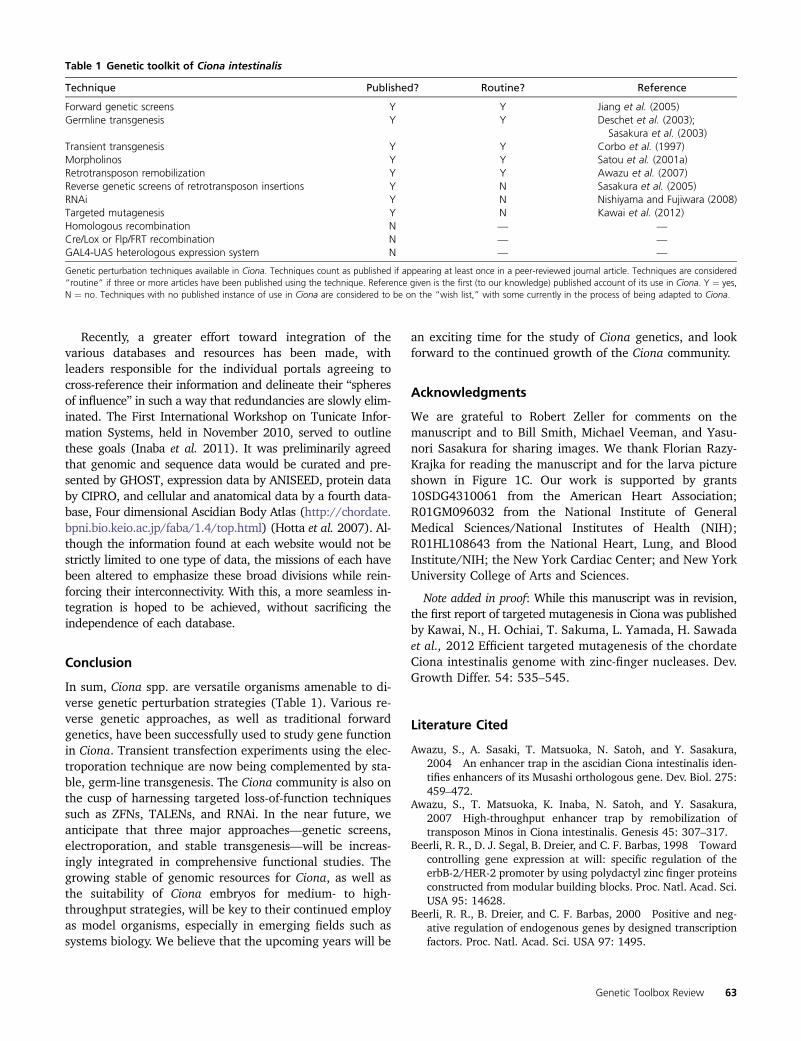
downstream of a minimal promoter without an enhancer,random integration of the transgene resulted in lines show-ing distinct GFP expression patterns, depending on theenhancers found near each integration site (Figure 5)(Awazu et al. 2004, 2007). High-throughput remobilizationof integration events by expression of transposase in sperm(Sasakura et al. 2008) or egg (Hozumi et al. 2010) has sinceexpanded the collection of reporter lines. This has been use-ful to identify novel enhancers, though a paucity of embry-onic reporter lines has been observed, perhaps owing to thechoice of minimal promoter used or the rarity of embryonicenhancers in the genome. All told, the Ciona intestinalisTransgenic Line Resource center (CITRES, http://marinebio.nbrp.jp/ciona/) has 41 enhancer trap lines and several otherMinos transgenic lines, which are all available to the commu-nity. With the growing availability of transgenic lines, perhapsCre/Lox- (Sauer and Henderson 1988) or GAL4/UAS-like(Brand and Perrimon 1993) systems for modular, conditionalgenetic loss or gain of function could be adapted to Ciona.
The high-throughput remobilization of single integrationevents also holds great promise for generating mutants forreverse genetic screens. It has already been shown thata Minos transgene can disrupt the endogenous gene at thesite of its integration. For instance, swimming juvenilemutants contain a Minos enhancer trap insertion in the cel-lulose synthase A (CesA) gene, leading to premature bodyplan reorganization prior to settlement of larvae (Figure2G) (Sasakura et al. 2005). The unique sequence of thetransgene can be used to easily locate the insertion site.
Genomics
The Ciona genomes have been sequenced at draft-level cov-erage, and are both �150–170 Mb in length, coding for�16,000 genes. (Simmen et al. 1998; Dehal et al. 2002;Vinson et al. 2005; Small et al. 2007a). They are the smallestchordate genomes sequenced to date, with the exception ofthe larvacean tunicate Oikopleura dioica (Seo et al. 2001).The C. intestinalis genome has been sequenced and assem-bled de novo twice, once in 2002 and once in 2005. A moreaccurate assembly, based on the first sequence, was pub-lished in 2008 and covers �75% of the genome (Satouet al. 2008). A total of 65% of this has been mapped tothe 14 pairs of chromosomes of C. intestinalis (Shoguchiet al. 2006). The two species’ genome sequences havebeen aligned against each other, and the evolutionary diver-
gence between the two means that highly conserved non-coding sequences are usually imbued with functionalsignificance, greatly aiding in the search for enhancers andother cis-regulatory elements. The alignment can easily benavigated on the VISTA browser (http://pipeline.lbl.gov/cgi-bin/gateway2?bg=Cioin2&selector=vistapoint) (Frazeret al. 2004).
Having a sequenced genome places Ciona into a selectcategory of “postgenomic” model organisms. Not only doesit allow for researchers to more easily clone genomic andcDNA sequences as experimental tools, but also allows forgenome-wide observational studies that might generatetestable hypotheses. For instance, chromatin immunoprecip-itation combined with genome tiling chips (ChIP-chip)revealed the genome-wide binding profile for 11 transcrip-tion factors during development (Kubo et al. 2010). In otherstudies, a combination of high-throughput mRNA sequenc-ing and computational methods have allowed for detectionand mapping of Ciona microRNA loci (Hendrix et al. 2010;Keshavan et al. 2010). These are but some examples of ge-nome-scale studies performed in Ciona.
On-line resources
Various databases and online resources serving the Cionaresearch community are spread around the world. Reflectingthe long tradition of ascidian biology in Japan, extensiveEST sequencing data (Satou et al. 2001b, 2002a) and cDNAcollections (Satou et al. 2002b) are available to researchersas part of the GHOST portal (http://ghost.zool.kyoto-u.ac.jp/cgi-bin/gb2/gbrowse/kh/) (Satou et al. 2005), whichalso harbors the most recent and accurate assembly andgene annotation for the C. intestinalis genome (“KyotoHoya”or “KH” gene models) (Satou et al. 2008). For more protein-oriented research, there is CIPRO (http://cipro.ibio.jp/current/)(Endo et al. 2011), which is another genomic and gene expres-sion database that integrates 2D-PAGE and mass-spectrometrydata from Ciona embryos.
Another ascidian research hotspot is France, which hoststhe ANISEED portal (http://www.aniseed.cnrs.fr/) (Tassyet al. 2010). This database is more explicitly oriented towarddevelopmental genetics, with a large curated collection ofin situ hybridization images and other gene expression,gene model, and anatomical data. Unlike GHOST or CIPRO,ANISEED is also meant to cover other experimental ascidiangenera, such as Phallusia and Halocynthia.
Figure 5 Enhancer trap transgenic lines. (A–C) Postmeta-morphic juveniles from different Minos-based enhancertrap transgenic lines from the Bioresource Project in Japan(http://marinebio.nbrp.jp/ciona/). (A) GFP expression seenin the endostyle, retropharyngeal band, esophagus, cen-tral nervous system, oral siphon, and atrial siphon. (B) GFPexpressed in the pyloric gland. (C) GFP is expressed in thecentral nervous system. Bar, �100 mm. All images unpub-lished, courtesy of Y. Sasakura.
62 A. Stolfi and L. Christiaen
Recently, a greater effort toward integration of thevarious databases and resources has been made, withleaders responsible for the individual portals agreeing tocross-reference their information and delineate their “spheresof influence” in such a way that redundancies are slowly elim-inated. The First International Workshop on Tunicate Infor-mation Systems, held in November 2010, served to outlinethese goals (Inaba et al. 2011). It was preliminarily agreedthat genomic and sequence data would be curated and pre-sented by GHOST, expression data by ANISEED, protein databy CIPRO, and cellular and anatomical data by a fourth data-base, Four dimensional Ascidian Body Atlas (http://chordate.bpni.bio.keio.ac.jp/faba/1.4/top.html) (Hotta et al. 2007). Al-though the information found at each website would not bestrictly limited to one type of data, the missions of each havebeen altered to emphasize these broad divisions while rein-forcing their interconnectivity. With this, a more seamless in-tegration is hoped to be achieved, without sacrificing theindependence of each database.
Conclusion
In sum, Ciona spp. are versatile organisms amenable to di-verse genetic perturbation strategies (Table 1). Various re-verse genetic approaches, as well as traditional forwardgenetics, have been successfully used to study gene functionin Ciona. Transient transfection experiments using the elec-troporation technique are now being complemented by sta-ble, germ-line transgenesis. The Ciona community is also onthe cusp of harnessing targeted loss-of-function techniquessuch as ZFNs, TALENs, and RNAi. In the near future, weanticipate that three major approaches—genetic screens,electroporation, and stable transgenesis—will be increas-ingly integrated in comprehensive functional studies. Thegrowing stable of genomic resources for Ciona, as well asthe suitability of Ciona embryos for medium- to high-throughput strategies, will be key to their continued employas model organisms, especially in emerging fields such assystems biology. We believe that the upcoming years will be
an exciting time for the study of Ciona genetics, and lookforward to the continued growth of the Ciona community.
Acknowledgments
We are grateful to Robert Zeller for comments on themanuscript and to Bill Smith, Michael Veeman, and Yasu-nori Sasakura for sharing images. We thank Florian Razy-Krajka for reading the manuscript and for the larva pictureshown in Figure 1C. Our work is supported by grants10SDG4310061 from the American Heart Association;R01GM096032 from the National Institute of GeneralMedical Sciences/National Institutes of Health (NIH);R01HL108643 from the National Heart, Lung, and BloodInstitute/NIH; the New York Cardiac Center; and New YorkUniversity College of Arts and Sciences.
Note added in proof: While this manuscript was in revision,the first report of targeted mutagenesis in Ciona was publishedby Kawai, N., H. Ochiai, T. Sakuma, L. Yamada, H. Sawadaet al., 2012 Efficient targeted mutagenesis of the chordateCiona intestinalis genome with zinc-finger nucleases. Dev.Growth Differ. 54: 535–545.
Literature Cited
Awazu, S., A. Sasaki, T. Matsuoka, N. Satoh, and Y. Sasakura,2004 An enhancer trap in the ascidian Ciona intestinalis iden-tifies enhancers of its Musashi orthologous gene. Dev. Biol. 275:459–472.
Awazu, S., T. Matsuoka, K. Inaba, N. Satoh, and Y. Sasakura,2007 High-throughput enhancer trap by remobilization oftransposon Minos in Ciona intestinalis. Genesis 45: 307–317.
Beerli, R. R., D. J. Segal, B. Dreier, and C. F. Barbas, 1998 Towardcontrolling gene expression at will: specific regulation of theerbB-2/HER-2 promoter by using polydactyl zinc finger proteinsconstructed from modular building blocks. Proc. Natl. Acad. Sci.USA 95: 14628.
Beerli, R. R., B. Dreier, and C. F. Barbas, 2000 Positive and neg-ative regulation of endogenous genes by designed transcriptionfactors. Proc. Natl. Acad. Sci. USA 97: 1495.
Table 1 Genetic toolkit of Ciona intestinalis
Technique Published? Routine? Reference
Forward genetic screens Y Y Jiang et al. (2005)Germline transgenesis Y Y Deschet et al. (2003);
Sasakura et al. (2003)Transient transgenesis Y Y Corbo et al. (1997)Morpholinos Y Y Satou et al. (2001a)Retrotransposon remobilization Y Y Awazu et al. (2007)Reverse genetic screens of retrotransposon insertions Y N Sasakura et al. (2005)RNAi Y N Nishiyama and Fujiwara (2008)Targeted mutagenesis Y N Kawai et al. (2012)Homologous recombination N — —
Cre/Lox or Flp/FRT recombination N — —
GAL4-UAS heterologous expression system N — —
Genetic perturbation techniques available in Ciona. Techniques count as published if appearing at least once in a peer-reviewed journal article. Techniques are considered“routine” if three or more articles have been published using the technique. Reference given is the first (to our knowledge) published account of its use in Ciona. Y ¼ yes,N ¼ no. Techniques with no published instance of use in Ciona are considered to be on the “wish list,” with some currently in the process of being adapted to Ciona.
Genetic Toolbox Review 63
Beh, J., W. Shi, M. Levine, B. Davidson, and L. Christiaen,2007 FoxF is essential for FGF-induced migration of heart pro-genitor cells in the ascidian Ciona intestinalis. Development134: 3297–3305.
Beisel, C., and R. Paro, 2011 Silencing chromatin: comparingmodes and mechanisms. Nat. Rev. Genet. 12: 123–135.
Bellen, H. J., C. J. O’Kane, C. Wilson, U. Grossniklaus, R. K. Pearsonet al., 1989 P-element-mediated enhancer detection: a versa-tile method to study development in Drosophila. Genes Dev. 3:1288.
Brand, A. H., and N. Perrimon, 1993 Targeted gene expression asa means of altering cell fates and generating dominant pheno-types. Development 118: 401–415.
Brown, C. D., D. S. Johnson, and A. Sidow, 2007 Functional ar-chitecture and evolution of transcriptional elements that drivegene coexpression. Science 317: 1557.
Byrd, J., and C. C. Lambert, 2000 Mechanism of the block tohybridization and selfing between the sympatric ascidians Cionaintestinalis and Ciona savignyi. Mol. Reprod. Dev. 55: 109–116.
Chabry, L., 1887 Contribution to the normal and teratologicalembryology of solitary ascidians. F. Alcan (translated fromFrench).
Chang, K., S. J. Elledge, and G. J. Hannon, 2006 Lessons fromnature: microRNA-based shRNA libraries. Nat. Methods 3: 707–714.
Chen, J. S., M. San Pedro, and R. W. Zeller, 2011 miR-124 func-tion during Ciona intestinalis neuronal development includesextensive interaction with the Notch signaling pathway. Devel-opment 138: 4943–4953.
Chiba, S., D. Jiang, N. Satoh, and W. C. Smith, 2009 Brachyurynull mutant-induced defects in juvenile ascidian endodermalorgans. Development 136: 35–39.
Christiaen, L., B. Davidson, T. Kawashima, W. Powell, H. Nollaet al., 2008 The transcription/migration interface in heart pre-cursors of Ciona intestinalis. Science 320: 1349.
Christiaen, L., E. Wagner,W. Shi, andM. Levine, 2009a Electroporationof transgenic DNAs in the sea squirt Ciona. Cold Spring Harbor Pro-tocols, doi: 10.1101/pdf.prot5345.
Christiaen, L., E. Wagner, W. Shi, andM. Levine, 2009b Microinjectionof morpholino oligos and RNAs in sea squirt (Ciona) embryos. ColdSpring Harbor Protocols, doi: 10.1101/pdb. prot5347.
Christiaen, L., E. Wagner, W. Shi, and M. Levine, 2009c The seasquirt Ciona intestinalis. Cold Spring Harbor Protocols, doi:10.1101/pdb. emo138.
Christiaen, L., A. Stolfi, and M. Levine, 2010 BMP signaling coor-dinates gene expression and cell migration during precardiacmesoderm development. Dev. Biol. 340: 179–187.
Christian, M., T. Cermak, E. L. Doyle, C. Schmidt, F. Zhang et al.,2010 Targeting DNA double-strand breaks with TAL effectornucleases. Genetics 186: 757.
Chu, G., H. Hayakawa, and P. Berg, 1987 Electroporation for theefficient transfection of mammalian cells with DNA. NucleicAcids Res. 15: 1311.
Conklin, E. G., 1905 Organ-forming substances in the eggs ofascidians. Biol. Bull. 8: 205.
Corbo, J. C., M. Levine, and R. W. Zeller, 1997 Characterization ofa notochord-specific enhancer from the Brachyury promoter re-gion of the ascidian, Ciona intestinalis. Development 124: 589–602.
Davidson, B., W. Shi, J. Beh, L. Christiaen, and M. Levine,2006 FGF signaling delineates the cardiac progenitor field inthe simple chordate, Ciona intestinalis. Genes Dev. 20: 2728.
Dehal, P., and J. L. Boore, 2005 Two rounds of whole genomeduplication in the ancestral vertebrate. PLoS Biol. 3: e314.
Dehal, P., Y. Satou, R. K. Campbell, J. Chapman, B. Degnan et al.,2002 The draft genome of Ciona intestinalis: insights intochordate and vertebrate origins. Science 298: 2157.
Delsuc, F., H. Brinkmann, D. Chourrout, and H. Philippe,2006 Tunicates and not cephalochordates are the closest liv-ing relatives of vertebrates. Nature 439: 965–968.
Deschet, K., Y. Nakatani, and W. C. Smith, 2003 Generation of Ci-Brachyury-GFP stable transgenic lines in the ascidian Ciona sa-vignyi. Genesis 35: 248–259.
Dunn, M. P., and A. Di Gregorio, 2009 The evolutionarily con-served leprecan gene: its regulation by Brachyury and its role inthe developing Ciona notochord. Dev. Biol. 328: 561–574.
Edwards, P. K., and B. Leung, 2008 Re-evaluating eradication ofnuisance species: invasion of the tunicate, Ciona intestinalis.Front. Ecol. Environ 7: 326–332.
Endo, T., K. Ueno, K. Yonezawa, K. Mineta, K. Hotta et al.,2011 CIPRO 2.5: Ciona intestinalis protein database, a uniqueintegrated repository of large-scale omics data, bioinformaticanalyses and curated annotation, with user rating and reviewingfunctionality. Nucleic Acids Res. 39: D807.
Escoffre, J. M., T. Portet, L. Wasungu, J. Teissié, D. Dean et al.,2009 What is (still not) known of the mechanism by whichelectroporation mediates gene transfer and expression in cellsand tissues. Mol. Biotechnol. 41: 286–295.
Feil, R., J. Wagner, D. Metzger, and P. Chambon, 1997 Regulationof Cre recombinase activity by mutated estrogen receptor li-gand-binding domains. Biochem. Biophys. Res. Commun. 237:752–757.
Frazer, K. A., L. Pachter, A. Poliakov, E. M. Rubin, and I. Dubchak,2004 VISTA: computational tools for comparative genomics.Nucleic Acids Res. 32: W273–W279.
Haley, B., B. Foys, and M. Levine, 2010 Vectors and parametersthat enhance the efficacy of RNAi-mediated gene disruption intransgenic Drosophila. Proc. Natl. Acad. Sci. USA 107: 11435.
Heasman, J., 2002 Morpholino oligos: Making sense of antisense?Dev. Biol. 243: 209–214.
Hendrickson, C., L. Christiaen, K. Deschet, D. Jiang, J. S. Joly et al.,2004 Culture of adult ascidians and ascidian genetics. Meth-ods Cell Biol. 74: 143–170.
Hendrix, D., M. Levine, andW. Shi, 2010 MethodmiRTRAP, a com-putational method for the systematic identification of miRNAsfrom high throughput sequencing data. Genome Biol. 11: R39.
Hikosaka, A., N. Satoh, and K. W. Makabe, 1993 Regulated spa-tial expression of fusion gene constructs with the 59 upstreamregion of Halocynthia roretzi muscle actin gene in Ciona sa-vignyi embryos. Dev. Genes Evol. 203: 104–112.
Hill, M. M., K. W. Broman, E. Stupka, W. C. Smith, D. Jiang et al.,2008 The C. savignyi genetic map and its integration with thereference sequence facilitates insights into chordate genomeevolution. Genome Res. 18: 1369–1379.
Hotta, K., K. Mitsuhara, H. Takahashi, K. Inaba, K. Oka et al.,2007 A web-based interactive developmental table for the as-cidian Ciona intestinalis, including 3D real-image embryo recon-structions: I. From fertilized egg to hatching larva. Dev. Dyn.236: 1790–1805.
Hozumi, A., N. Kawai, R. Yoshida, Y. Ogura, N. Ohta et al.,2010 Efficient transposition of a single Minos transposon copyin the genome of the ascidian Ciona intestinalis with a transgenicline expressing transposase in eggs. Dev. Dyn. 239: 1076–1088.
Hudson, C., and H. Yasuo, 2008 Similarity and diversity in mech-anisms of muscle fate induction between ascidian species. Biol.Cell 100: 265–277.
Hudson, C., S. Lotito, and H. Yasuo, 2007 Sequential and combi-natorial inputs from Nodal, Delta2/Notch and FGF/MEK/ERKsignalling pathways establish a grid-like organisation of distinctcell identities in the ascidian neural plate. Development 134:3527–3537.
Iannelli, F., G. Pesole, P. Sordino, and C. Gissi, 2007 Mitogenomicsreveals two cryptic species in Ciona intestinalis. Trends Genet. 23:419–422.
64 A. Stolfi and L. Christiaen
Imai, K., N. Takada, N. Satoh, and Y. Satou, 2000 (beta)-cateninmediates the specification of endoderm cells in ascidian em-bryos. Development 127: 3009–3020.
Imai, K. S., K. Hino, K. Yagi, N. Satoh, and Y. Satou, 2004 Geneexpression profiles of transcription factors and signaling mole-cules in the ascidian embryo: towards a comprehensive under-standing of gene networks. Development 131: 4047–4058.
Imai, K. S., M. Levine, N. Satoh, and Y. Satou, 2006 Regulatoryblueprint for a chordate embryo. Science 312: 1183.
Inaba, K., T. Endo, Y. Satou, K. Hotta, H. Nishida et al.,2011 Tunicate databases: toward a comprehensive informa-tive basis at molecular and cellular level for tunicate community.6th International Tunicate Meeting, McGill University, Mon-treal, Canada.
Jiang, D., E. M. Munro, and W. C. Smith, 2005 Ascidian prickleregulates both mediolateral and anterior-posterior cell polarityof notochord cells. Curr. Biol. 15: 79–85.
Joly, J. S., S. Kano, T. Matsuoka, H. Auger, K. Hirayama et al.,2007 Culture of Ciona intestinalis in closed systems. Dev.Dyn. 236: 1832–1840.
Kawai, N., H. Ochiai, T. Sakuma, L. Yamada, H. Sawada et al.,2012 Efficient targeted mutagenesis of the chordate Ciona in-testinalis genome with zinc-finger nucleases. Dev. Growth Differ.54: 535–545.
Keshavan, R., M. Virata, A. Keshavan, and R. W. Zeller,2010 Computational identification of Ciona intestinalis Micro-RNAs. Zoolog. Sci. 27: 162–170.
Kim, J. H., M. S. Waterman, and L. M. Li, 2007 Diploid genomereconstruction of Ciona intestinalis and comparative analysiswith Ciona savignyi. Genome Res. 17: 1101–1110.
Kratsios, P., A. Stolfi, M. Levine, and O. Hobert, 2012 Coordinatedregulation of cholinergic motor neuron traits through a conservedterminal selector gene. Nat. Neurosci. 15: 205–214.
Kubo, A., N. Suzuki, X. Yuan, K. Nakai, N. Satoh et al.,2010 Genomic cis-regulatory networks in the early Ciona in-testinalis embryo. Development 137: 1613–1623.
Kumano, G., N. Takatori, T. Negishi, T. Takada, and H. Nishida,2011 A maternal factor unique to ascidians silences the germ-line via binding to P-TEFb and RNAP II regulation. Curr. Biol.21: 1308–1313.
Lambert, C. C., and G. Lambert, 1998 Non-indigenous ascidiansin southern California harbors and marinas. Mar. Biol. 130:675–688.
Lemaire, P., 2009 Unfolding a chordate developmental program,one cell at a time: invariant cell lineages, short-range inductionsand evolutionary plasticity in ascidians. Dev. Biol. 332: 48–60.
Lemaire, P., 2011 Evolutionary crossroads in developmental bi-ology: the tunicates. Development 138: 2143.
Matsuoka, T., S. Awazu, N. Satoh, and Y. Sasakura, 2004 Minostransposon causes germline transgenesis of the ascidian Cionasavignyi. Dev. Growth Differ. 46: 249–255.
Matsuoka, T., S. Awazu, E. Shoguchi, N. Satoh, and Y. Sasakura,2005 Germline transgenesis of the ascidian Ciona intestinalisby electroporation. Genesis 41: 67–72.
Monteilhet, C., A. Perrin, A. Thierry, L. Colleaux, and B. Dujon,1990 Purification and characterization of the in vitro activityof I-Sce I, a novel and highly specific endonuclease encoded bya group I intron. Nucleic Acids Res. 18: 1407–1413.
Morbitzer, R., P. Römer, J. Boch, and T. Lahaye, 2010 Regulationof selected genome loci using de novo-engineered transcriptionactivator-like effector (TALE)-type transcription factors. Proc.Natl. Acad. Sci. USA 107: 21617.
Morgan, T., 1923 Removal of the block to self-fertilization in theascidian Ciona. Proc. Natl. Acad. Sci. USA 9: 170.
Nagel, G., T. Szellas, W. Huhn, S. Kateriya, N. Adeishvili et al.,2003 Channelrhodopsin-2, a directly light-gated cation-selec-tive membrane channel. Proc. Natl. Acad. Sci. USA 100: 13940.
Nishiyama, A., and S. Fujiwara, 2008 RNA interference by ex-pressing short hairpin RNA in the Ciona intestinalis embryo.Dev. Growth Differ. 50: 521–529.
Oda-Ishii, I., V. Bertrand, I. Matsuo, P. Lemaire, and H. Saiga,2005 Making very similar embryos with divergent genomes:conservation of regulatory mechanisms of Otx between the as-cidians Halocynthia roretzi and Ciona intestinalis. Development132: 1663–1674.
Ouyang, X., and J. K. Chen, 2010 Synthetic strategies for studyingembryonic development. Chem. Biol. 17: 590–606.
Pasini, A., A. Amiel, U. Rothbächer, A. Roure, P. Lemaire et al.,2006 Formation of the ascidian epidermal sensory neurons:insights into the origin of the chordate peripheral nervous sys-tem. PLoS Biol. 4: e225.
Picco, V., C. Hudson, and H. Yasuo, 2007 Ephrin-Eph signallingdrives the asymmetric division of notochord/neural precursorsin Ciona embryos. Development 134: 1491–1497.
Putnam, N. H., T. Butts, D. E. K. Ferrier, R. F. Furlong, U. Hellstenet al., 2008 The amphioxus genome and the evolution of thechordate karyotype. Nature 453: 1064–1071.
Riggsbee, C. W., and A. Deiters, 2010 Recent advances in thephotochemical control of protein function. Trends Biotechnol.28: 468–475.
Roure, A., U. Rothbächer, F. Robin, E. Kalmar, G. Ferone et al.,2007 A multicassette Gateway vector set for high throughputand comparative analyses in Ciona and vertebrate embryos.PLoS ONE 2: e916.
Sasakura, Y., S. Awazu, S. Chiba, and N. Satoh, 2003 Germ-linetransgenesis of the Tc1/mariner superfamily transposon Minosin Ciona intestinalis. Proc. Natl. Acad. Sci. USA 100: 7726.
Sasakura, Y., K. Nakashima, S. Awazu, T. Matsuoka, A. Nakayamaet al., 2005 Transposon-mediated insertional mutagenesis re-vealed the functions of animal cellulose synthase in the ascidianCiona intestinalis. Proc. Natl. Acad. Sci. USA 102: 15134.
Sasakura, Y., A. Konno, K. Mizuno, N. Satoh, and K. Inaba,2008 Enhancer detection in the ascidian Ciona intestinaliswith transposase-expressing lines of Minos. Dev. Dyn. 237:39–50.
Satoh, N., Y. Satou, B. Davidson, and M. Levine, 2003 Ciona in-testinalis: an emerging model for whole-genome analyses.Trends Genet. 19: 376–381.
Satou, Y., K. S. Imai, and N. Satoh, 2001a Action of morpholinosin Ciona embryos. Genesis 30: 103–106.
Satou, Y., N. Takatori, L. Yamada, Y. Mochizuki, M. Hamaguchiet al., 2001b Gene expression profiles in Ciona intestinalistailbud embryos. Development 128: 2893–2904.
Satou, Y., N. Takatori, S. Fujiwara, T. Nishikata, H. Saiga et al.,2002a Ciona intestinalis cDNA projects: expressed sequencetag analyses and gene expression profiles during embryogenesis.Gene 287: 83–96.
Satou, Y., L. Yamada, Y. Mochizuki, N. Takatori, T. Kawashimaet al., 2002b A cDNA resource from the basal chordate Cionaintestinalis. Genesis 33: 153–154.
Satou, Y., T. Kawashima, E. Shoguchi, A. Nakayama, and N. Satoh,2005 An integrated database of the ascidian, Ciona intestina-lis: towards functional genomics. Zoolog. Sci. 22: 837–843.
Satou, Y., K. Mineta, M. Ogasawara, Y. Sasakura, E. Shoguchiet al., 2008 Improved genome assembly and evidence-basedglobal gene model set for the chordate Ciona intestinalis: newinsight into intron and operon populations. Genome Biol. 9:R152.
Sauer, B., and N. Henderson, 1988 Site-specific DNA recombina-tion in mammalian cells by the Cre recombinase of bacterio-phage P1. Proc. Natl. Acad. Sci. USA 85: 5166.
Seo, H. C., M. Kube, R. B. Edvardsen, M. F. Jensen, A. Beck et al.,2001 Miniature genome in the marine chordate Oikopleuradioica. Science 294: 2506.
Genetic Toolbox Review 65
Shi, W., and M. Levine, 2008 Ephrin signaling establishes asym-metric cell fates in an endomesoderm lineage of the Ciona em-bryo. Development 135: 931–940.
Shi, W., D. Hendrix, M. Levine, and B. Haley, 2009 A distinct classof small RNAs arises from pre-miRNA–proximal regions in a sim-ple chordate. Nat. Struct. Mol. Biol. 16: 183–189.
Shimizu-Sato, S., E. Huq, J. M. Tepperman, and P. H. Quail,2002 A light-switchable gene promoter system. Nat. Biotech-nol. 20: 1041–1044.
Shoguchi, E., T. Kawashima, Y. Satou, M. Hamaguchi, T. Sin et al.,2006 Chromosomal mapping of 170 BAC clones in the ascid-ian Ciona intestinalis. Genome Res. 16: 297–303.
Simmen, M. W., S. Leitgeb, V. H. Clark, S. J. M. Jones, and A. Bird,1998 Gene number in an invertebrate chordate, Ciona intesti-nalis. Proc. Natl. Acad. Sci. USA 95: 4437.
Small, K., M. Brudno, M. Hill, and A. Sidow, 2007a A haplomealignment and reference sequence of the highly polymorphicCiona savignyi genome. Genome Biol. 8: R41.
Small, K. S., M. Brudno, M. M. Hill, and A. Sidow, 2007b Extremegenomic variation in a natural population. Proc. Natl. Acad. Sci.USA 104: 5698.
Stinchcomb, D., J. Shaw, S. H. Carr, and D. Hirsh, 1985 Extra-chromosomal DNA transformation of Caenorhabditis elegans. Mol.Cell. Biol. 5: 3484.
Stolfi, A., and M. Levine, 2011 Neuronal subtype specification inthe spinal cord of a protovertebrate. Development 138: 995.
Stolfi, A., T. B. Gainous, J. J. Young, A. Mori, M. Levine et al.,2010 Early chordate origins of the vertebrate second heartfield. Science 329: 565.
Strickland, D., K. Moffat, and T. R. Sosnick, 2008 Light-activatedDNA binding in a designed allosteric protein. Proc. Natl. Acad.Sci. USA 105: 10709.
Suzuki, M. M., T. Nishikawa, and A. Bird, 2005 Genomic ap-proaches reveal unexpected genetic divergence within Cionaintestinalis. J. Mol. Evol. 61: 627–635.
Tassy, O., D. Dauga, F. Daian, D. Sobral, F. Robin et al., 2010 TheANISEED database: digital representation, formalization, and
elucidation of a chordate developmental program. GenomeRes. 20: 1459–1468.
Thermes, V., C. Grabher, F. Ristoratore, F. Bourrat, A. Choulikaet al., 2002 I-SceI meganuclease mediates highly efficienttransgenesis in fish. Mech. Dev. 118: 91–98.
Tokuoka, M., G. Kumano, and H. Nishida, 2007 FGF9/16/20 andWnt-5a signals are involved in specification of secondary musclefate in embryos of the ascidian, Halocynthia roretzi. Dev. GenesEvol. 217: 515–527.
Urnov, F. D., J. C. Miller, Y. L. Lee, C. M. Beausejour, J. M. Rocket al., 2005 Highly efficient endogenous human gene cor-rection using designed zinc-finger nucleases. Nature 435:646–651.
Veeman, M. T., Y. Nakatani, C. Hendrickson, V. Ericson, C. Linet al., 2008 Chongmague reveals an essential role for lami-nin-mediated boundary formation in chordate convergenceand extension movements. Development 135: 33–41.
Veeman, M. T., S. Chiba, and W. C. Smith, 2011 Ciona genetics.Methods Mol. Biol. 770: 401.
Vinson, J. P., D. B. Jaffe, K. O’Neill, E. K. Karlsson, N. Stange-Thomann et al., 2005 Assembly of polymorphic genomes:algorithms and application to Ciona savignyi. Genome Res.15: 1127–1135.
Wood, A. J., T. W. Lo, B. Zeitler, C. S. Pickle, E. J. Ralston et al.,2011 Targeted genome editing across species using ZFNs andTALENs. Science 333: 307.
Young, J. J., J. M. Cherone, Y. Doyon, I. Ankoudinova, F. M. Farajiet al., 2011 Efficient targeted gene disruption in the soma andgerm line of the frog Xenopus tropicalis using engineered zinc-finger nucleases. Proc. Natl. Acad. Sci. USA 108: 7052.
Zeller, R. W., 2004 Generation and use of transgenic ascidianembryos. Methods Cell Biol. 74: 713–730.
Zeller, R. W., M. J. Virata, and A. C. Cone, 2006 Predictable mo-saic transgene expression in ascidian embryos produced witha simple electroporation device. Dev. Dyn. 235: 1921–1932.
Communicating editor: O. Hobert
66 A. Stolfi and L. Christiaen