Embed Size (px)
Citation preview
Adrenocortical hypofunction . Polyglandular autoimmune
syndromes
Tímea Baló, MDSemmelweis University 3rd Department of Internal Medi cine
3rd Year, Faculty of Medicine2018/2019 Academic Year, 2nd Semester
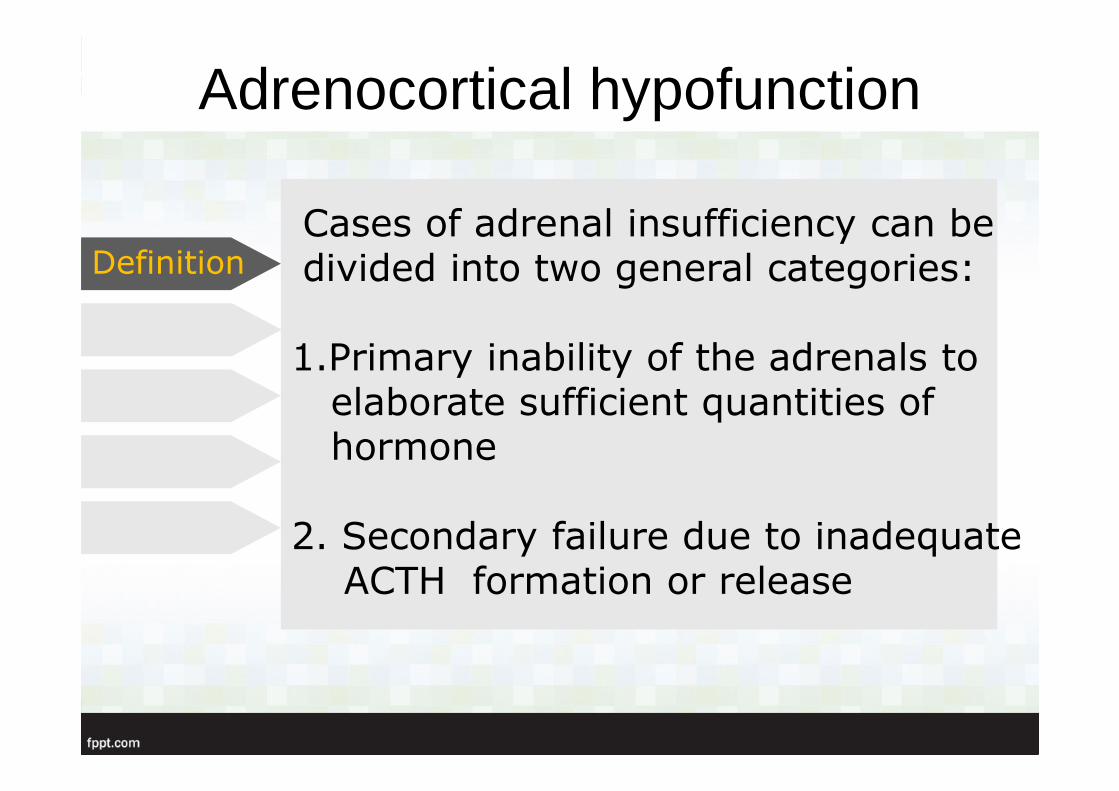
Cases of adrenal insufficiency can be divided into two general categories:
1.Primary inability of the adrenals toelaborate sufficient quantities of hormone
2. Secondary failure due to inadequateACTH formation or release
Adrenocortical hypofunction
Definition
• Primary
• Secondary
Adrenal insufficiency
Classification
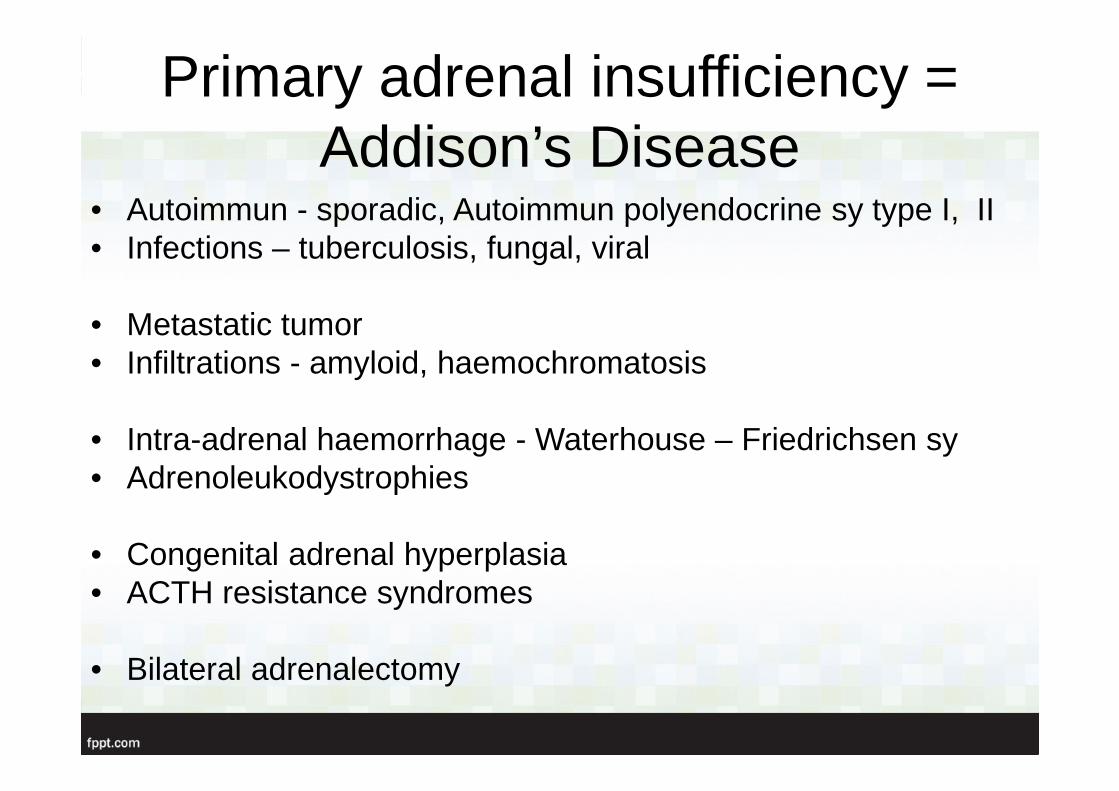
Primary adrenal insufficiency = Addison’s Disease
• Autoimmun - sporadic, Autoimmun polyendocrine sy type I, II• Infections – tuberculosis, fungal, viral
• Metastatic tumor• Infiltrations - amyloid, haemochromatosis
• Intra-adrenal haemorrhage - Waterhouse – Friedrichsen sy• Adrenoleukodystrophies
• Congenital adrenal hyperplasia• ACTH resistance syndromes
• Bilateral adrenalectomy
Adrenocortical hypofunctionAddison’s disease
„ general languor and debility, feebleness of the heart’s action, irritability of the stomach, and a peculiar change of the color of the skin”
The original description of Addison’s disease:
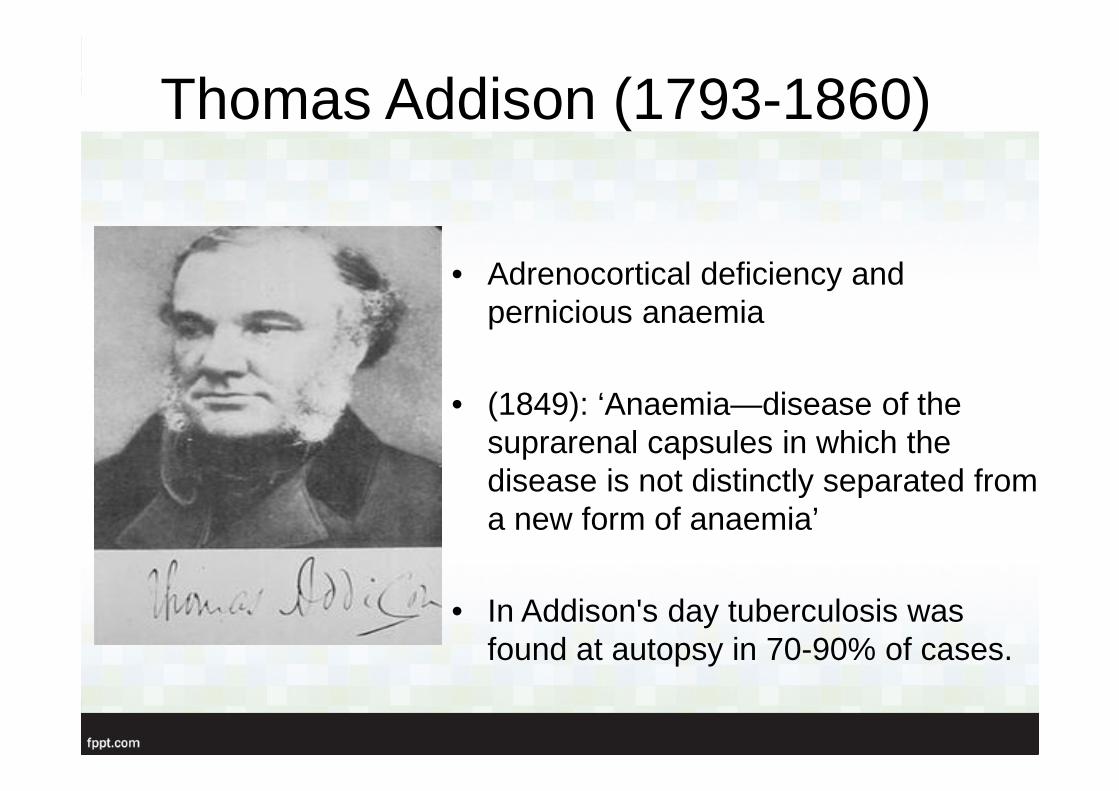
Thomas Addison (1793-1860)
• Adrenocortical deficiency and pernicious anaemia
• (1849): ‘Anaemia—disease of the suprarenal capsules in which the disease is not distinctly separated from a new form of anaemia’
• In Addison's day tuberculosis was found at autopsy in 70-90% of cases.
Autoimmune adrenalitis
• 70% of all cases• Atrophic glands, but medulla is intact• Autoantibodies• 50% of patients have an
associated autoimmun disease (APS)
Infections• A very common cause as well
• Tuberculosis: extraadrenal disease is also evident, both the cortex and medulla are affected
• Fungal infections (cryptococcosis, histoplasmosis), cytomegalovirus
• AIDS – 10 % of patients show subnormal response to Synacthen test
• Some drugs for treatment may precipitate the adrenal insufficiency ( ketokonazol: inhibits cortisol synthesis, rifampicin: increases cortizol metabolism)
Acquired primary adrenalinsufficiency
• Adrenal metastases: breast, lung-rarely cause adrenal insufficiency
• Necrosis of the glands – should be considered in any severely sick patient (infection, trauma, coagulopathy)
• Waterhouse – Friedrichsen sy associated with meningococci
Waterhouse- Friedrichsensyndrome
• bleeding into the gland• Severe infection with
meningococcus bacteria
• It can be caused by procoagulants
• Other causes: low platelet count, primary antiphospholipid syndrome, renal vein thrombosis, steroid use
Inherited primary adrenalinsufficiency
• Adrenal hypoplasia congenita – X-linked disorder (combined with primary and central hypogonadotropic hypogonadism)
• Adrenoleukodystrophy• Familial glucocorticoid deficiency – AR cause, it
ususally manifests in childhood• Triple A syndrome (adrenal insuff, achalasia,
alacrima)
Adrenoleukodystrophy
• X-linked inherited disorder • Prevalence 1:20000• Disease of the very-long chain fatty acid
metabolism• Progressive neurological symptoms of
demyelinisation
Secondary causes• Glucocorticoid therapy • Hypopituitarism• Selective removal of ACTH secreting pit. adenoma• Pituitary tumors, and pituitary surgery,
craniopharyngeomas, pituitary apoplexy, pituitary irradiation
• Granulomatous disease• Postpartum pituitary infarction (Sheehan’s sy)• Secondary tumor deposits• Isolated ACTH deficiency, Multiple pituitary hormon
deficiencies
Hypadrenalism during critical illness
• Even in individuals with previously intact HPA axis• Functional adrenal insufficiency• Hypadrenia is transient, no structural lesion• Uncertain etiology• Inability to mount an adequate and appropriate cortisol
response to stress on intensive care units• Increases the risk of death during acute illness• Treatment with relatively high doses of hydrocortizon,
or with methylprednisolon in septic shock, and early phase of acute respiratory distress is recommended
Clinical features
Addison’ s disease - epidemiology
• Prevalence: 93-140/1 million
• Incidence: 4.7 – 6.2/ 1 million/year
• Young adults
• Woman are affected (more frequent)
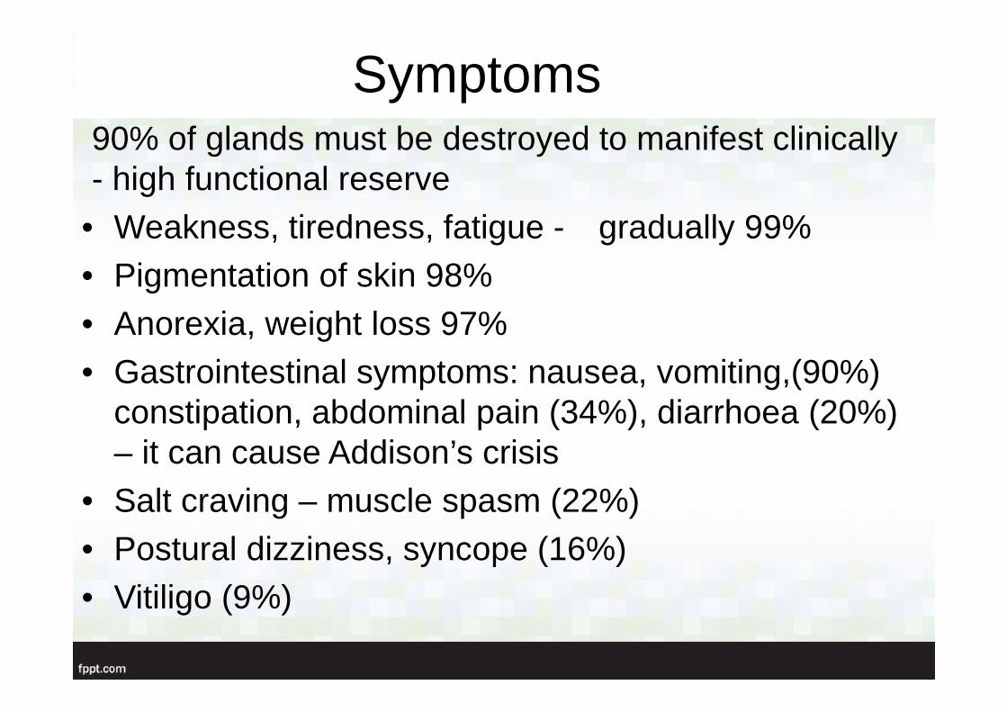
Symptoms90% of glands must be destroyed to manifest clinically- high functional reserve
• Weakness, tiredness, fatigue - gradually 99%• Pigmentation of skin 98%• Anorexia, weight loss 97%• Gastrointestinal symptoms: nausea, vomiting,(90%)
constipation, abdominal pain (34%), diarrhoea (20%) – it can cause Addison’s crisis
• Salt craving – muscle spasm (22%)• Postural dizziness, syncope (16%)• Vitiligo (9%)
Signs and laboratory findings
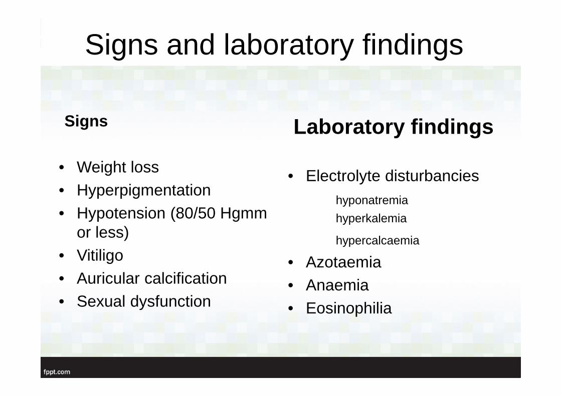
Signs
• Weight loss• Hyperpigmentation• Hypotension (80/50 Hgmm
or less)• Vitiligo• Auricular calcification• Sexual dysfunction
Laboratory findings
• Electrolyte disturbancieshyponatremia
hyperkalemia
hypercalcaemia
• Azotaemia• Anaemia• Eosinophilia
Addison’s disease
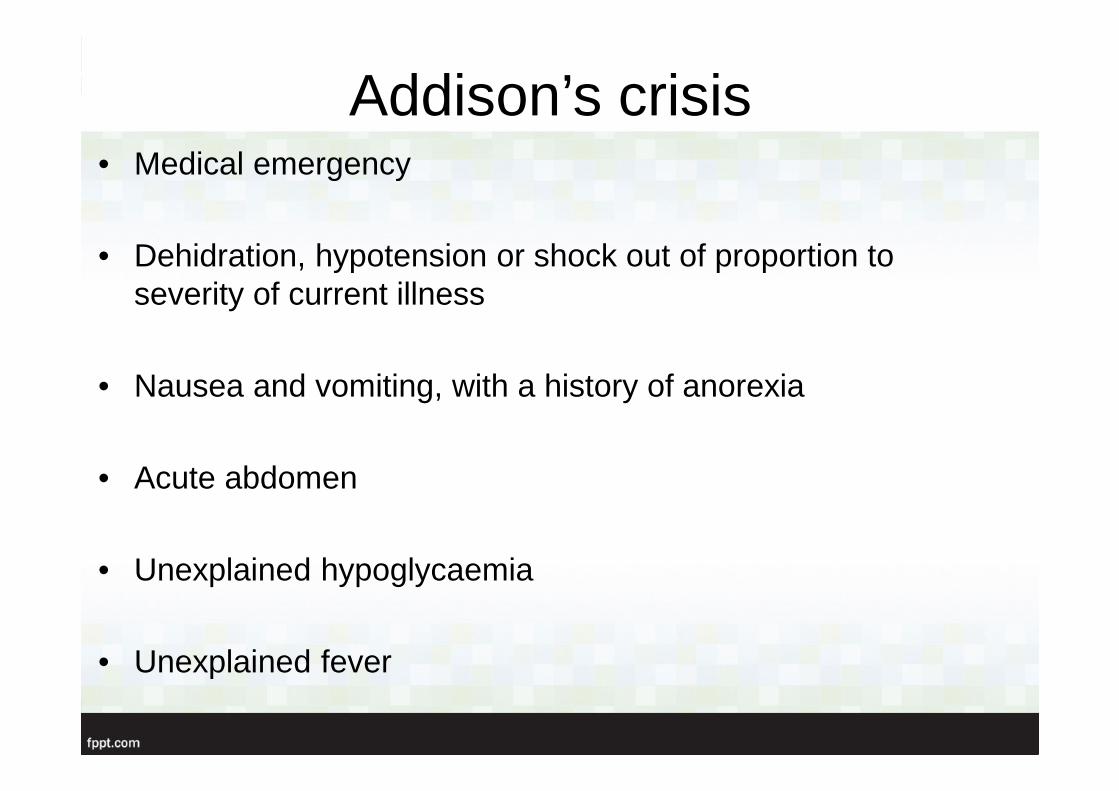
Addison’s crisis• Medical emergency
• Dehidration, hypotension or shock out of proportion to severity of current illness
• Nausea and vomiting, with a history of anorexia
• Acute abdomen
• Unexplained hypoglycaemia
• Unexplained fever
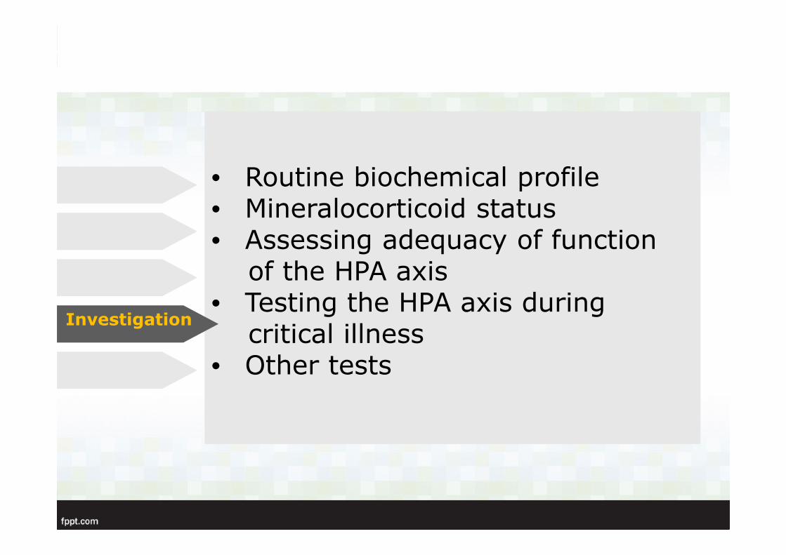
• Routine biochemical profile• Mineralocorticoid status• Assessing adequacy of function
of the HPA axis• Testing the HPA axis during
critical illness• Other tests
Investigation

Assessing adequacy of function of the HPA axis
• Basal plasma cortisol and UFC levels are often in the low-normal range
• A basal cortisol value greater than 14.5 ug/dl (400 nmol/l) indicates an intact HPA axis
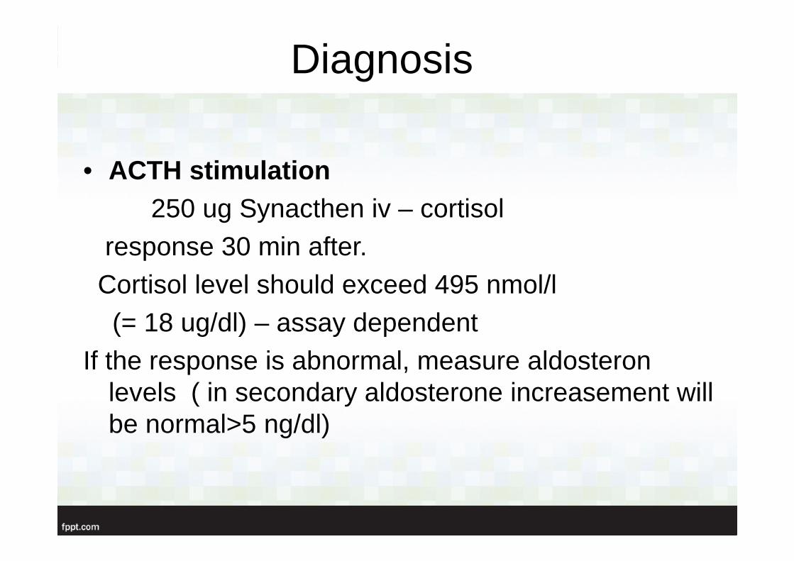
Diagnosis
• ACTH stimulation 250 ug Synacthen iv – cortisol
response 30 min after.Cortisol level should exceed 495 nmol/l (= 18 ug/dl) – assay dependent
If the response is abnormal, measure aldosteron levels ( in secondary aldosterone increasement will be normal>5 ng/dl)
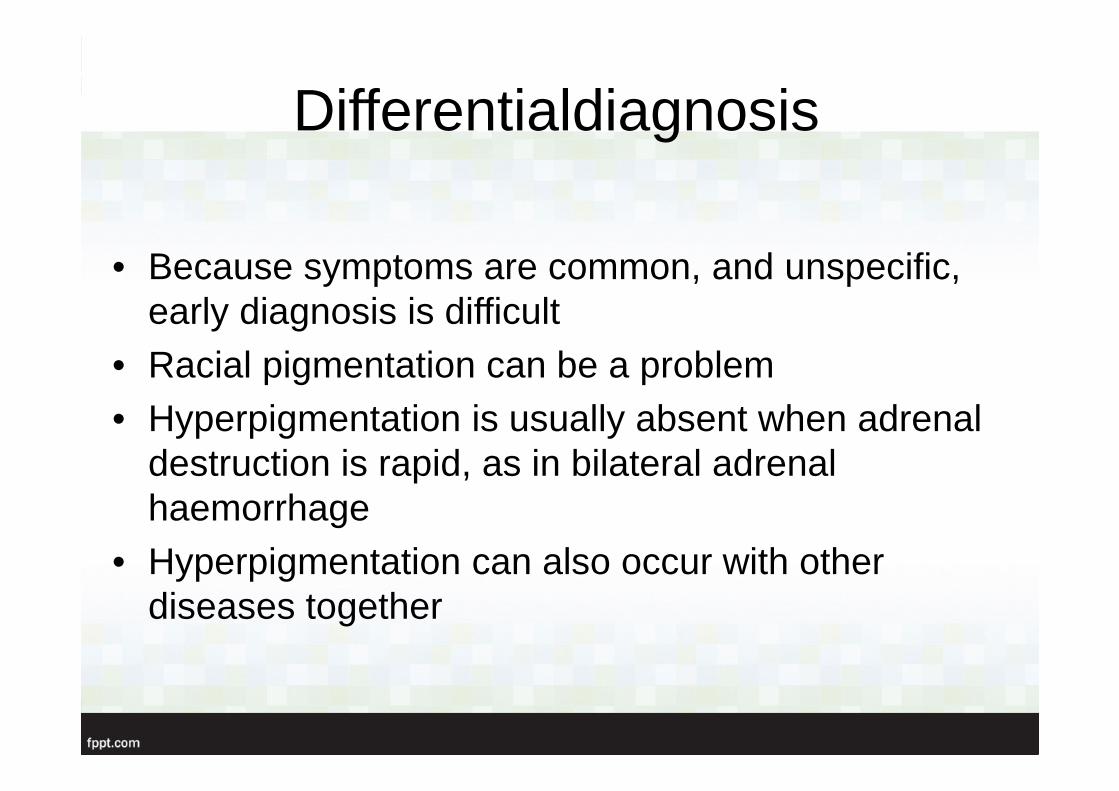
Differentialdiagnosis
• Because symptoms are common, and unspecific, early diagnosis is difficult
• Racial pigmentation can be a problem• Hyperpigmentation is usually absent when adrenal
destruction is rapid, as in bilateral adrenal haemorrhage
• Hyperpigmentation can also occur with other diseases together
• Treatment of acute adrenalinsufficiency
• Long-term replacementtherapy
Treatment
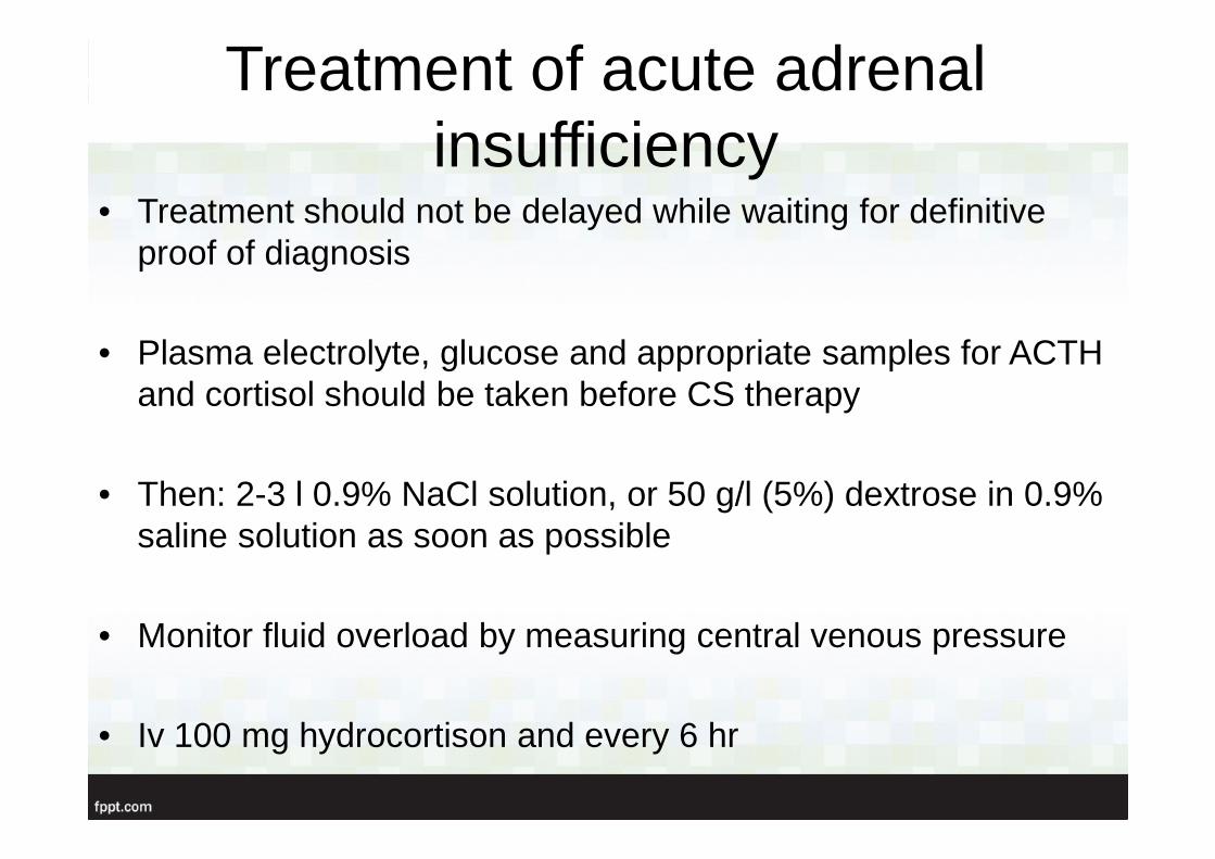
Treatment of acute adrenalinsufficiency
• Treatment should not be delayed while waiting for definitiveproof of diagnosis
• Plasma electrolyte, glucose and appropriate samples for ACTH and cortisol should be taken before CS therapy
• Then: 2-3 l 0.9% NaCl solution, or 50 g/l (5%) dextrose in 0.9% saline solution as soon as possible
• Monitor fluid overload by measuring central venous pressure
• Iv 100 mg hydrocortison and every 6 hr
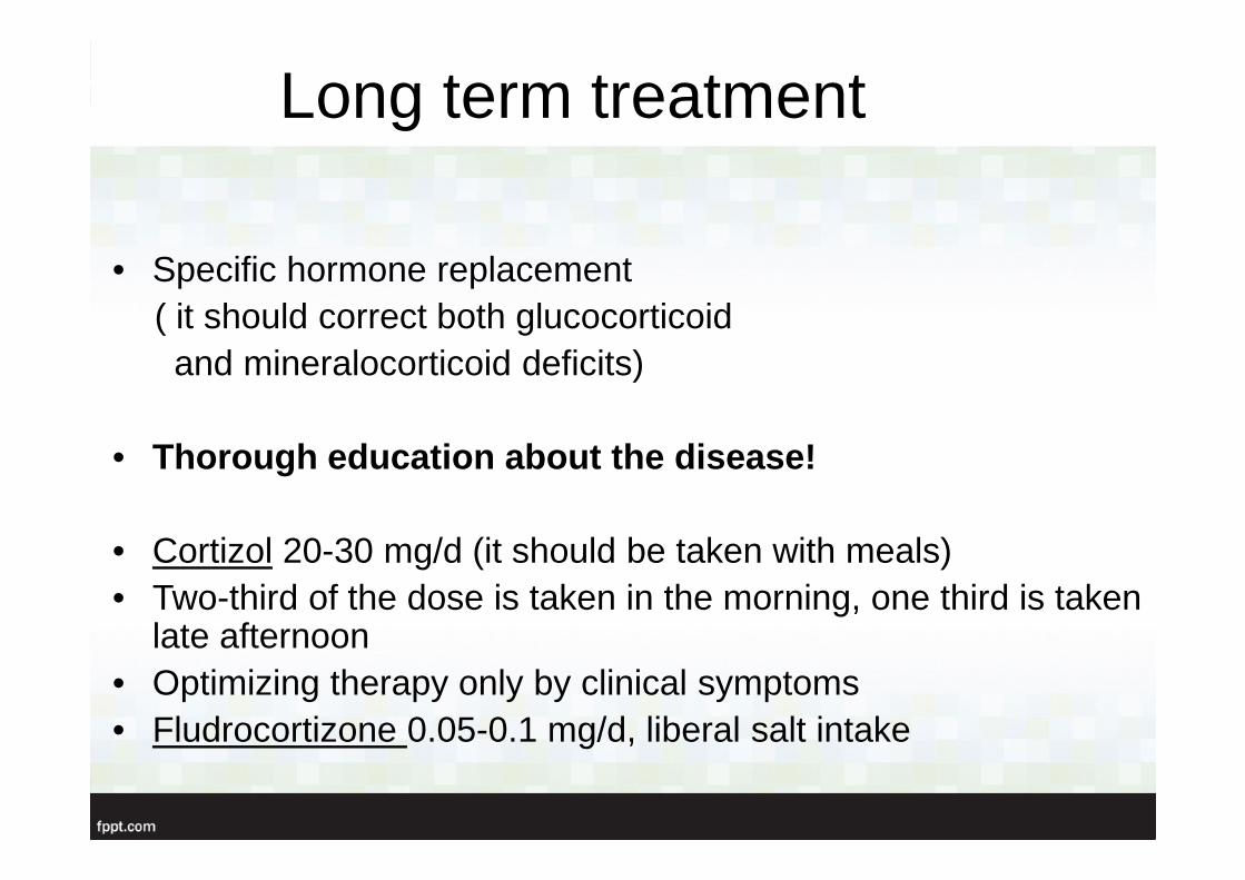
Long term treatment
• Specific hormone replacement( it should correct both glucocorticoidand mineralocorticoid deficits)
• Thorough education about the disease!
• Cortizol 20-30 mg/d (it should be taken with meals)• Two-third of the dose is taken in the morning, one third is taken
late afternoon• Optimizing therapy only by clinical symptoms• Fludrocortizone 0.05-0.1 mg/d, liberal salt intake
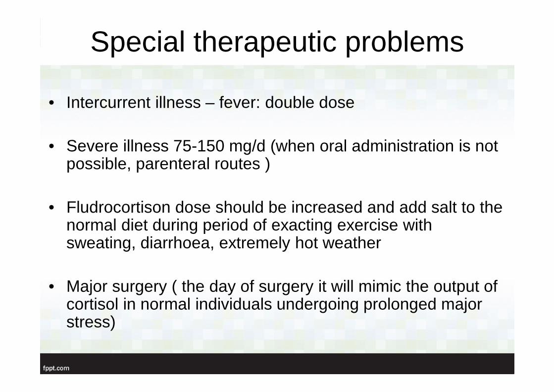
Special therapeutic problems
• Intercurrent illness – fever: double dose
• Severe illness 75-150 mg/d (when oral administration is not possible, parenteral routes )
• Fludrocortison dose should be increased and add salt to the normal diet during period of exacting exercise with sweating, diarrhoea, extremely hot weather
• Major surgery ( the day of surgery it will mimic the output of cortisol in normal individuals undergoing prolonged major stress)
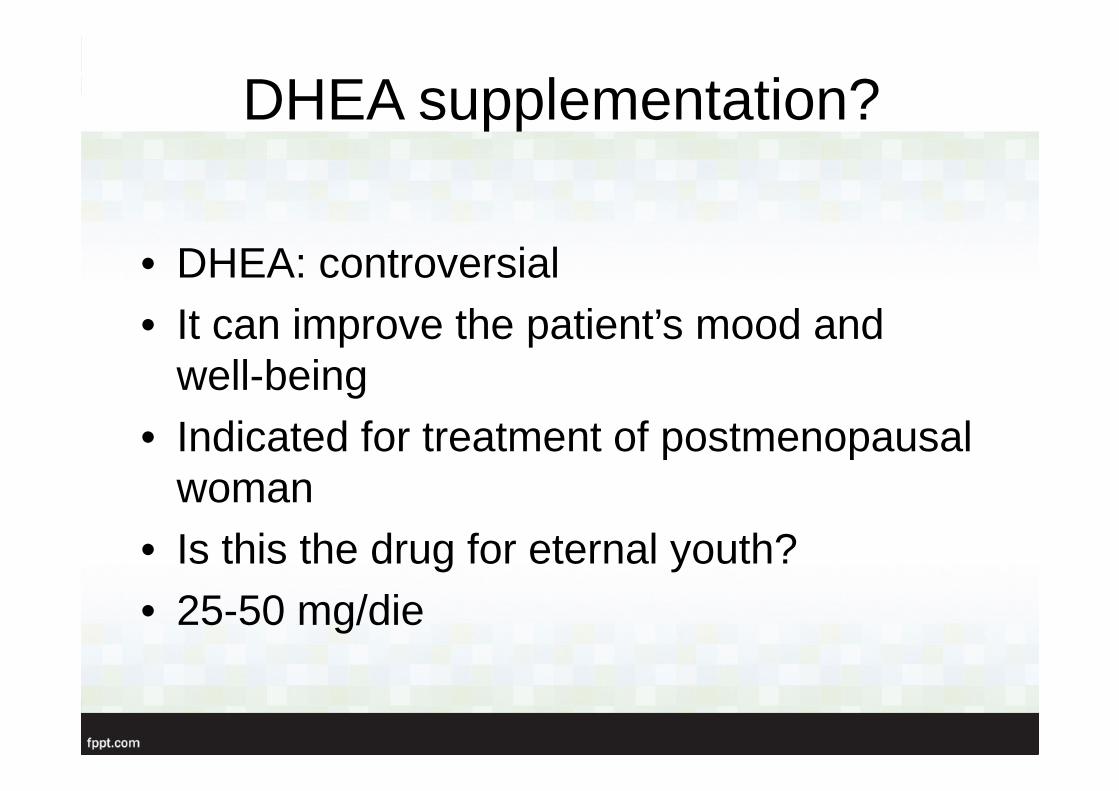
DHEA supplementation?
• DHEA: controversial• It can improve the patient’s mood and
well-being• Indicated for treatment of postmenopausal
woman• Is this the drug for eternal youth? • 25-50 mg/die
Secondary adrenal insufficiency
• HPA axis failure- deficiency of glucocorticoids and adrenal androgens- mineralocorticoids are unaffected
• 1 cause = chronic exogenous glucocorticoid- suppresses diurnal CRH/ACTH release- both time and dose related (short course, and daily dose of prednisolone 5 mg or less)
- reversible (recovery may take up to a year)
Summary
„ Unexplained hyponatremia and hyperkalemia in the setting of hypotension unresponsive for catecholamin and fluid administration…. …..should receive 100 mg hyrocortisone intravenously”
Case presentation
Sz. V. 29 years old female
• History: appendectomy, nasal plastical surgery• No drugs taken• Smoking: 2-3 cigarettes/day• Family:sister hypothyreosis, psoriasis• Menses regular
• Complaints: since 1 year progressive tiredness, weaknessmainly in evenings, brownish skin color

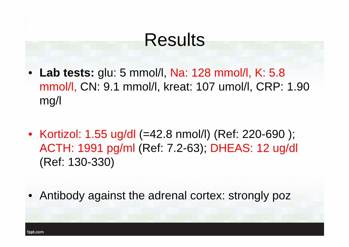
Results
• Lab tests: glu: 5 mmol/l, Na: 128 mmol/l, K: 5.8 mmol/l, CN: 9.1 mmol/l, kreat: 107 umol/l, CRP: 1.90 mg/l
• Kortizol: 1.55 ug/dl (=42.8 nmol/l) (Ref: 220-690 ); ACTH: 1991 pg/ml (Ref: 7.2-63); DHEAS: 12 ug/dl (Ref: 130-330)
• Antibody against the adrenal cortex: strongly poz
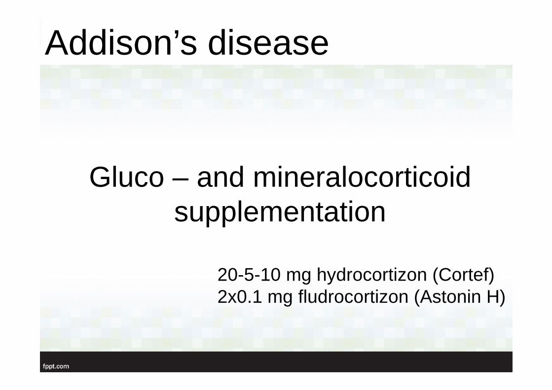
Gluco – and mineralocorticoid supplementation
20-5-10 mg hydrocortizon (Cortef)2x0.1 mg fludrocortizon (Astonin H)
Addison’s disease
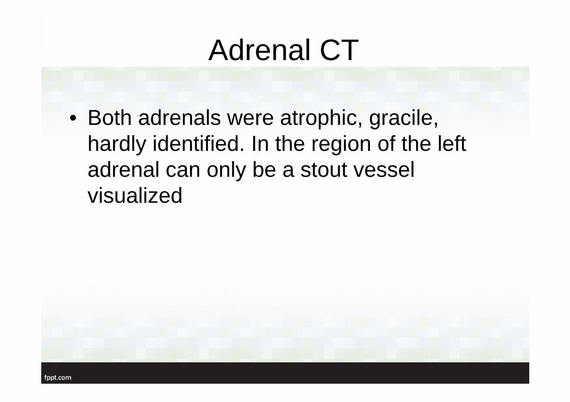
Adrenal CT
• Both adrenals were atrophic, gracile, hardly identified. In the region of the left adrenal can only be a stout vessel visualized
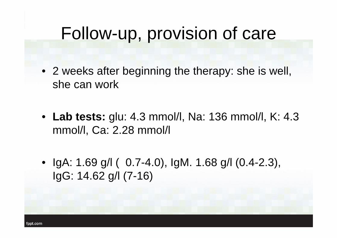
Follow-up, provision of care
• 2 weeks after beginning the therapy: she is well, she can work
• Lab tests: glu: 4.3 mmol/l, Na: 136 mmol/l, K: 4.3 mmol/l, Ca: 2.28 mmol/l
• IgA: 1.69 g/l ( 0.7-4.0), IgM. 1.68 g/l (0.4-2.3), IgG: 14.62 g/l (7-16)
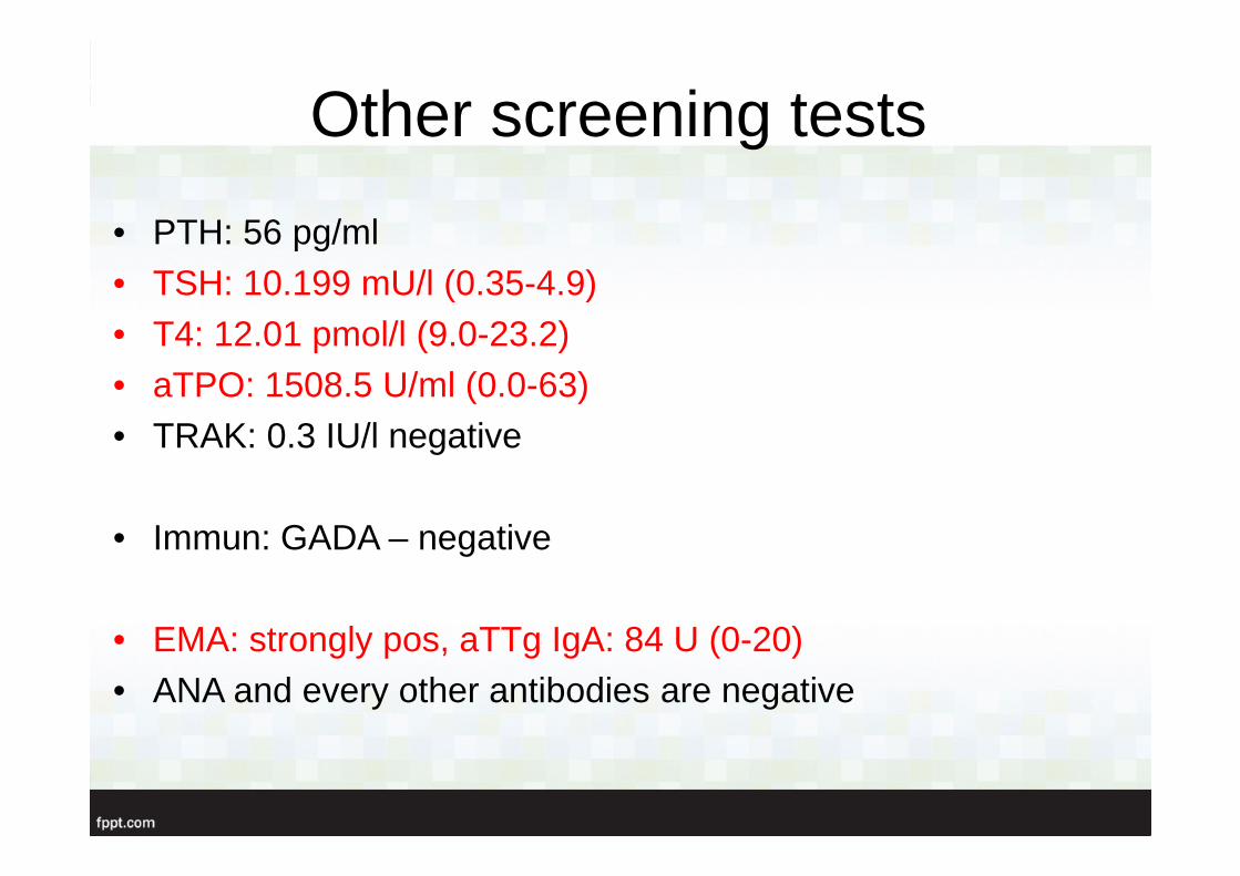
Other screening tests
• PTH: 56 pg/ml• TSH: 10.199 mU/l (0.35-4.9)• T4: 12.01 pmol/l (9.0-23.2)• aTPO: 1508.5 U/ml (0.0-63)• TRAK: 0.3 IU/l negative
• Immun: GADA – negative
• EMA: strongly pos, aTTg IgA: 84 U (0-20)• ANA and every other antibodies are negative
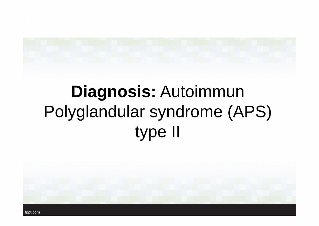
Diagnosis : Autoimmun Polyglandular syndrome (APS)
type II
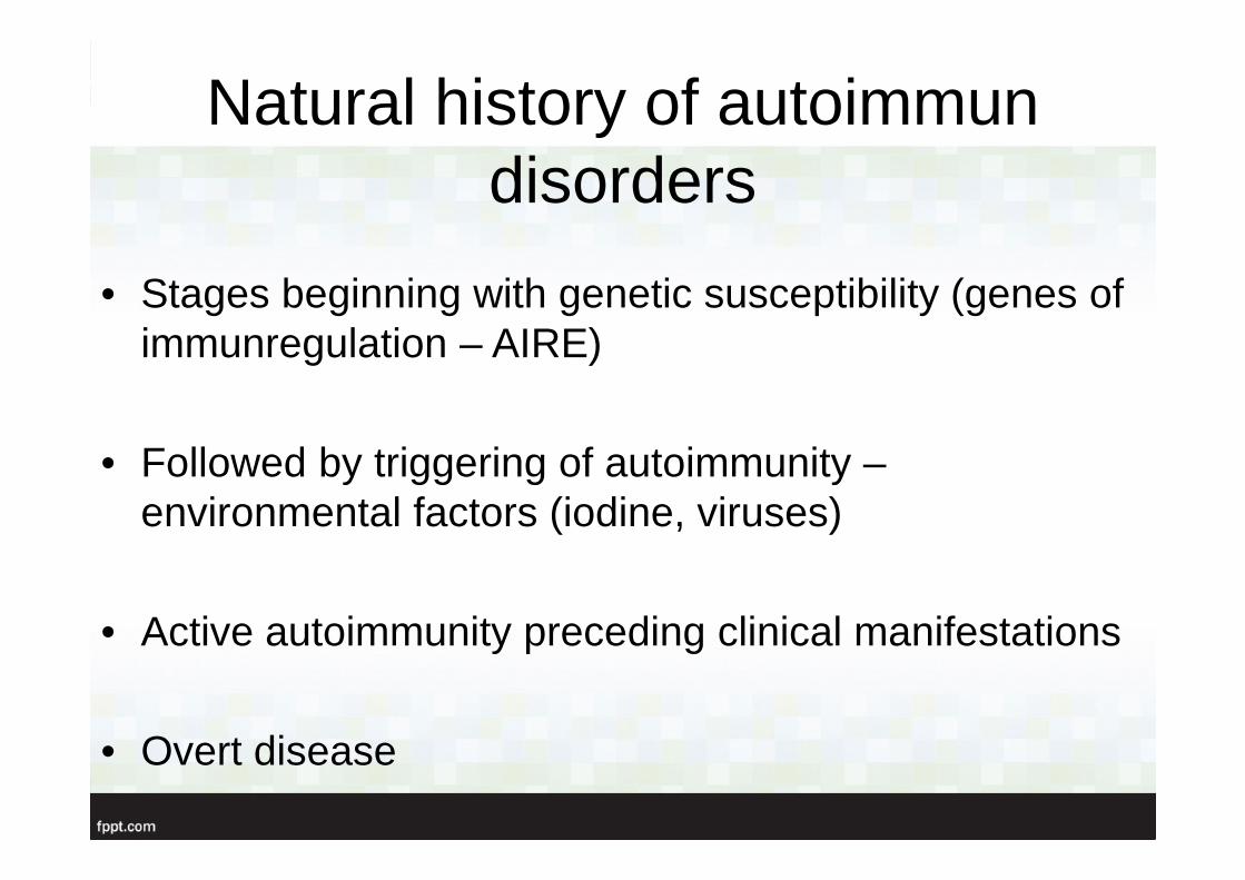
Natural history of autoimmun disorders
• Stages beginning with genetic susceptibility (genes of immunregulation – AIRE)
• Followed by triggering of autoimmunity –environmental factors (iodine, viruses)
• Active autoimmunity preceding clinical manifestations
• Overt disease
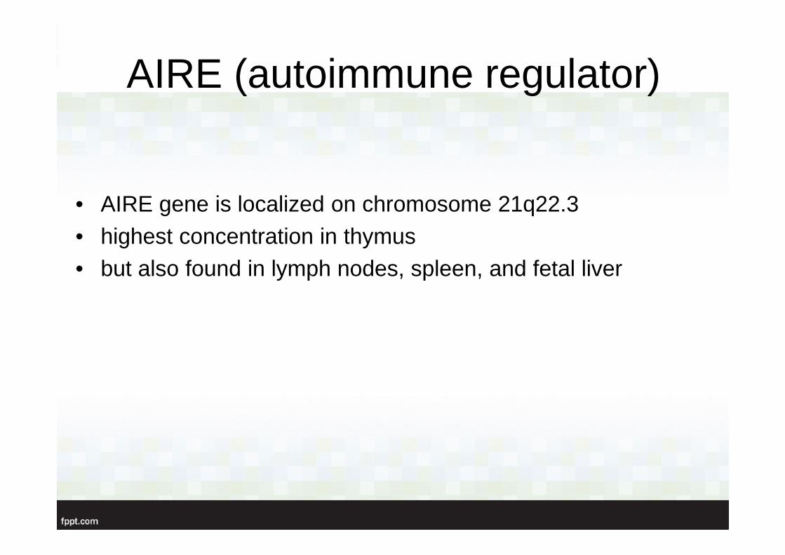
AIRE (autoimmune regulator)
• AIRE gene is localized on chromosome 21q22.3• highest concentration in thymus • but also found in lymph nodes, spleen, and fetal liver
Model of the pathogenesis of autoimmunity in polyendocrine
disorders
Introduction
• The term “polyendocrine” itself is a misname• not all patients have multiple endocrine
disorders• many have nonendocrine autoimmune
diseases
N Engl J Med 2004;350:2068-79
APS – I.
• Epidemiology 1:9000 – 1:200000• Female:male = 1 : 2.4• Imunregulatory gene defect (AIRE gen – 21 q)• Candidiasis: because of a T-cell defect• Genetic: AR, monogenic, inherited disease, doesn’t
accompanied to HLA antigens• Mutations of AIRE gene
APS – I.
• = APECED (Autoimmune polyendocrinopathy, Candidiasis, Ectodermal Dystrophy) = Whitaker-sy
• It begins in childhood• Minor symptoms can present prior to the main
symptomps in the early 20th years(hepatitis, keratoconjunctivitis, periodic rashes with
fever, chronic diarrhea, celiac disease, severe
obstipation, alopecia, or vitiligo)
Introduction
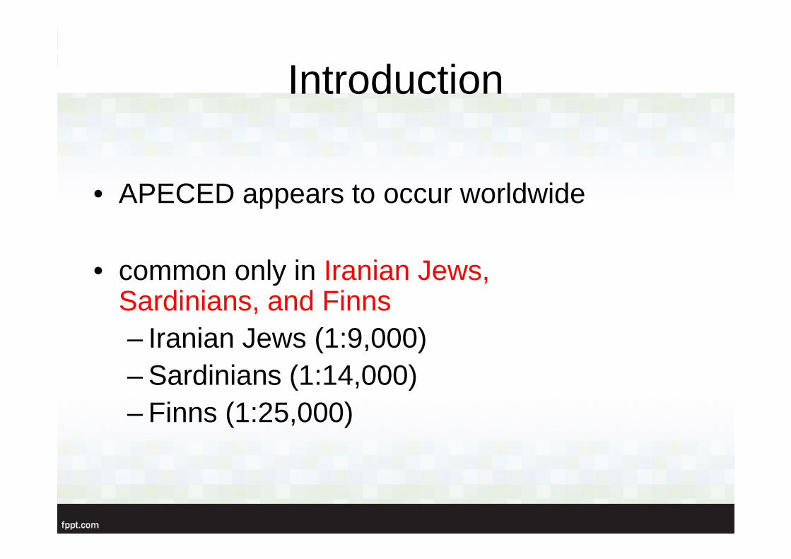
• APECED appears to occur worldwide
• common only in Iranian Jews, Sardinians, and Finns– Iranian Jews (1:9,000)– Sardinians (1:14,000)– Finns (1:25,000)
Clinical manifestation
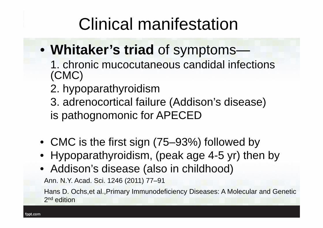
• Whitaker’s triad of symptoms—1. chronic mucocutaneous candidal infections(CMC)2. hypoparathyroidism3. adrenocortical failure (Addison’s disease)is pathognomonic for APECED
• CMC is the first sign (75–93%) followed by• Hypoparathyroidism, (peak age 4-5 yr) then by • Addison’s disease (also in childhood)
Hans D. Ochs,et al.,Primary Immunodeficiency Diseases: A Molecular and Genetic 2nd edition
Ann. N.Y. Acad. Sci. 1246 (2011) 77–91
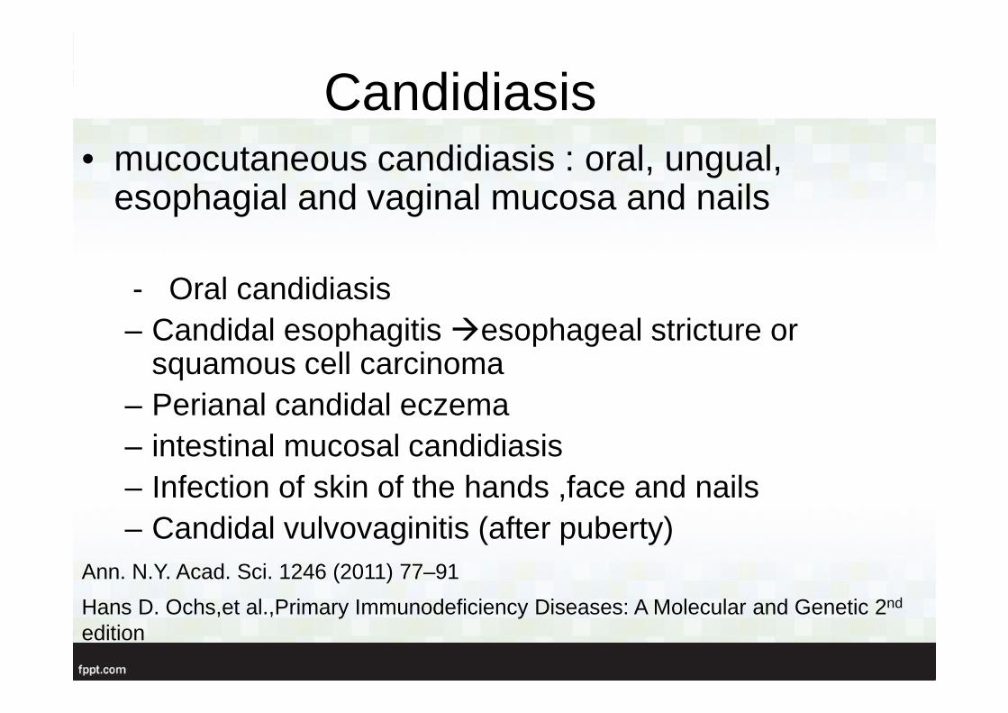
Candidiasis• mucocutaneous candidiasis : oral, ungual,
esophagial and vaginal mucosa and nails
- Oral candidiasis– Candidal esophagitis �esophageal stricture or
squamous cell carcinoma– Perianal candidal eczema – intestinal mucosal candidiasis– Infection of skin of the hands ,face and nails– Candidal vulvovaginitis (after puberty)
Ann. N.Y. Acad. Sci. 1246 (2011) 77–91
Hans D. Ochs,et al.,Primary Immunodeficiency Diseases: A Molecular and Genetic 2nd
edition

Endocrine manifestation
• Apart from hypoparathyroidism and Addison’s disease,– hypergonadotropic hypogonadism– type 1 diabetes– autoimmune thyroid diseases– pituitary defects– gastric parietal cell atrophy
� autoimmune originoften associated with a specific set of organ-specific
autoantibodies
Ann. N.Y. Acad. Sci. 1246 (2011) 77–91
Management
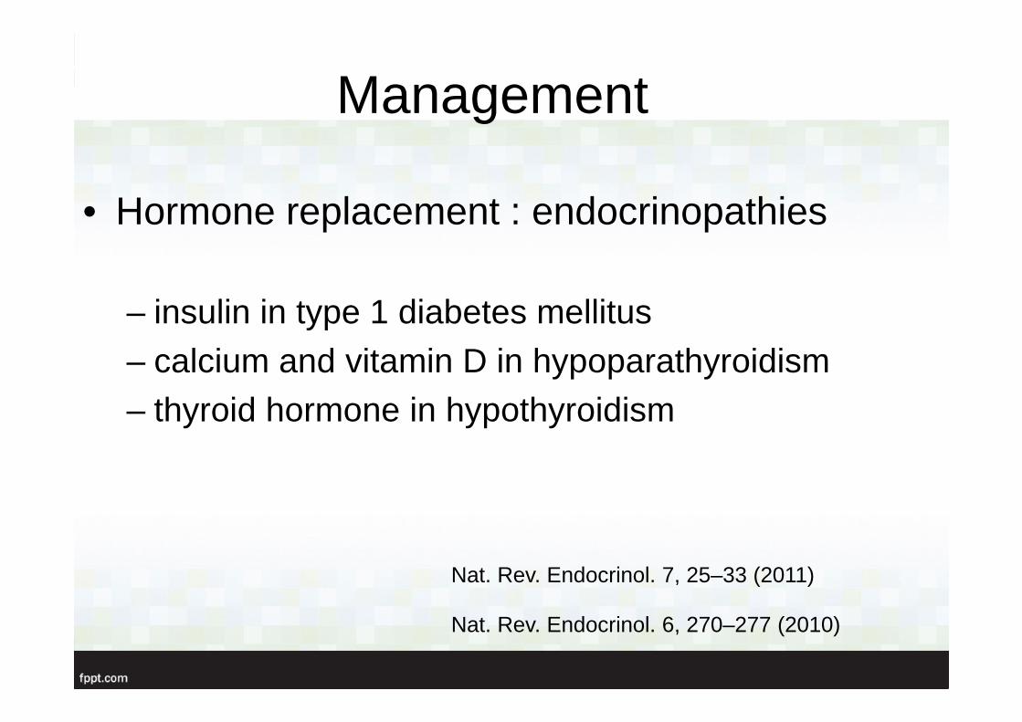
• Hormone replacement : endocrinopathies
– insulin in type 1 diabetes mellitus– calcium and vitamin D in hypoparathyroidism– thyroid hormone in hypothyroidism
Nat. Rev. Endocrinol. 6, 270–277 (2010)
Nat. Rev. Endocrinol. 7, 25–33 (2011)
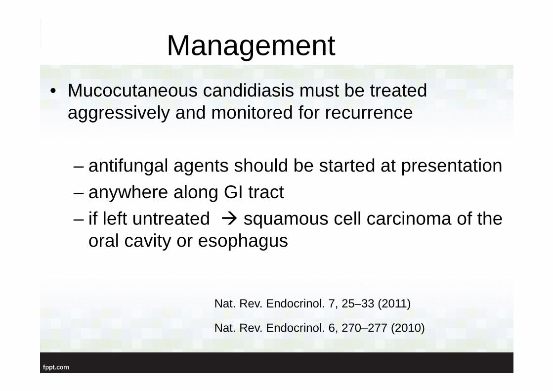
Management• Mucocutaneous candidiasis must be treated
aggressively and monitored for recurrence
– antifungal agents should be started at presentation– anywhere along GI tract– if left untreated � squamous cell carcinoma of the
oral cavity or esophagus
Nat. Rev. Endocrinol. 6, 270–277 (2010)
Nat. Rev. Endocrinol. 7, 25–33 (2011)
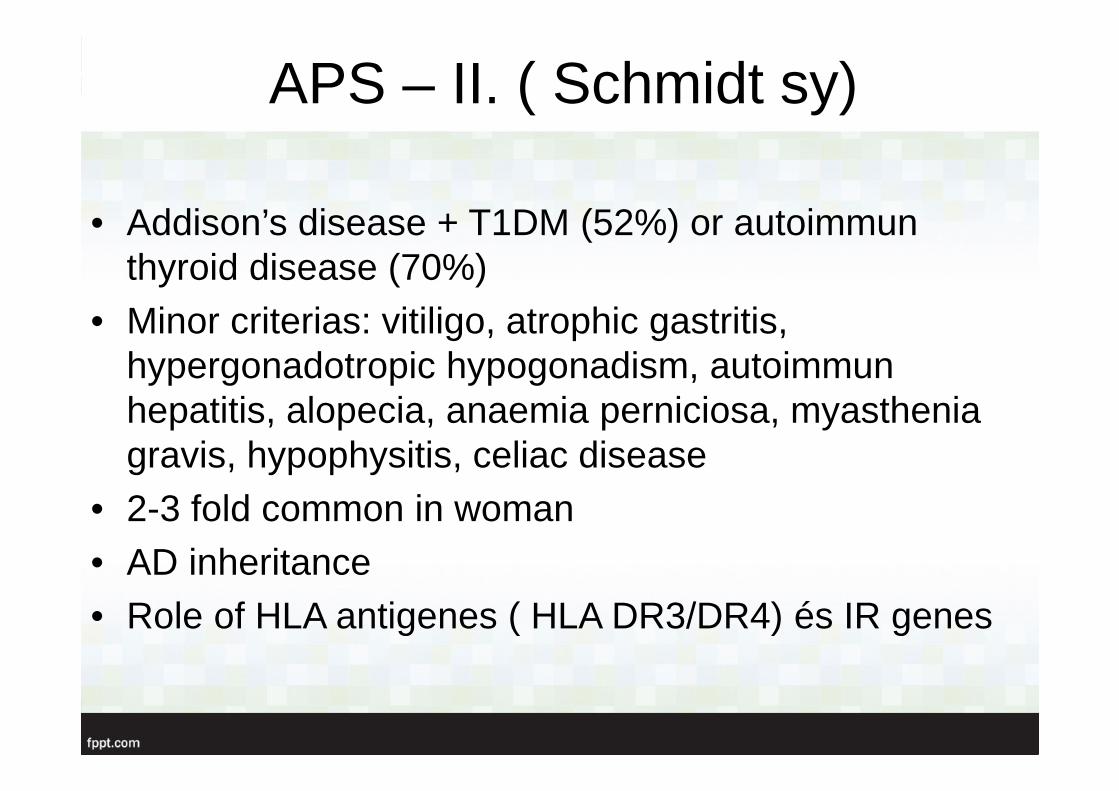
APS – II. ( Schmidt sy)
• Addison’s disease + T1DM (52%) or autoimmun thyroid disease (70%)
• Minor criterias: vitiligo, atrophic gastritis, hypergonadotropic hypogonadism, autoimmun hepatitis, alopecia, anaemia perniciosa, myastheniagravis, hypophysitis, celiac disease
• 2-3 fold common in woman• AD inheritance• Role of HLA antigenes ( HLA DR3/DR4) és IR genes
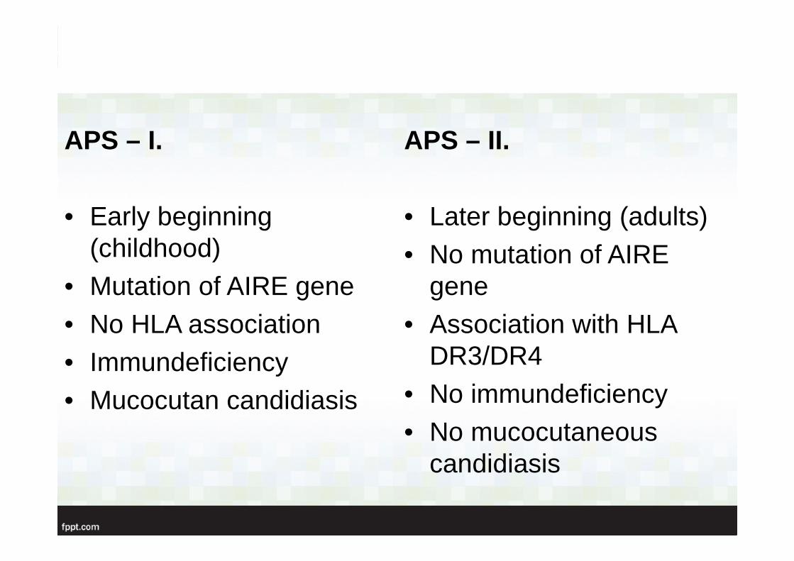
APS – I.
• Early beginning(childhood)
• Mutation of AIRE gene• No HLA association• Immundeficiency• Mucocutan candidiasis
APS – II.
• Later beginning (adults) • No mutation of AIRE
gene• Association with HLA
DR3/DR4• No immundeficiency• No mucocutaneous
candidiasis

APS – III.
• Autoimmun thyroid disease( TAD = thyreoidassociated disease: Hashimoto, Basedow, endokrin orbitopathy)
+ 28% other autoimmun disease: Sjögren, coeliakia, SLE, myasthenia
• Many autoimmun disorders are mild, subclinical form
• 7-8% in the population
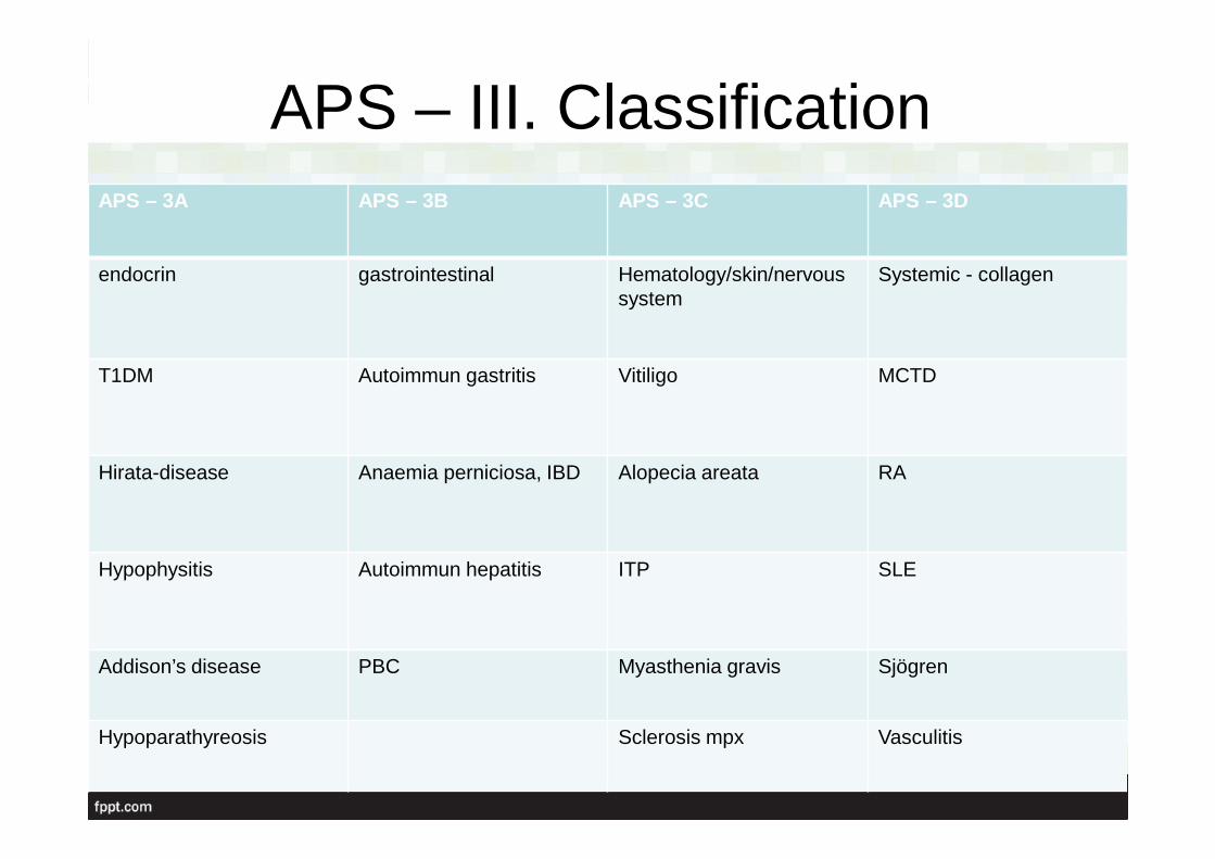
APS – III. ClassificationAPS – 3A APS – 3B APS – 3C APS – 3D
endocrin gastrointestinal Hematology/skin/nervoussystem
Systemic - collagen
T1DM Autoimmun gastritis Vitiligo MCTD
Hirata-disease Anaemia perniciosa, IBD Alopecia areata RA
Hypophysitis Autoimmun hepatitis ITP SLE
Addison’s disease PBC Myasthenia gravis Sjögren
Hypoparathyreosis Sclerosis mpx Vasculitis
Differential diagnosis
• Turner-sy ( autoimmun thyreoiditis 30% and otherendocrinopathies)
• Kearns - Syre sy : main complaint is myopathy, buthypoparathyreosis, primary hypogonadism, T1DM, hypopituitarism can also occur
• Wolfram sy – begins in childhood( DM, DI, atrophy of the nervus opticus, deafness)
• With thorough treatment patients can usually cope with the disease and their life expectancy is only slightly decreased
• oral squamous cell carcinoma or a sudden onset of the disease by hypocalcemic or Addisonian crisis or acute hepatitis can sometimes be of a fulminant nature

Thank you for your attention!


![The Hypothesis of NMDA Receptor Hypofunction for …cent hypothesis of schizophrenia as a “glutamate disorder” [12], the glutamatergic hypofunction hypothesis is not in confl](https://img.pdfslide.us/doc/110x75/5fd7f5f77ba0784ee13d01f1/the-hypothesis-of-nmda-receptor-hypofunction-for-cent-hypothesis-of-schizophrenia.jpg)