Embed Size (px)
Citation preview
Abstract Rodents have a toothless diastema region be-tween the incisor and molar teeth which may contain ru-dimentary tooth germs. We found in upper diastema re-gion of the mouse (Mus musculus) three small toothgerms which developed into early bud stage before theirapoptotic removal, while the sibling vole (Microtus ros-siaemeridionalis) had only a single but larger tooth germin this region, and this developed into late bud stage be-fore regressing apoptotically. To analyze the geneticmechanisms of the developmental arrest of the rudimen-tary tooth germs we compared the expression patterns ofseveral developmental regulatory genes (Bmp2, Bmp4,Fgf4, Fgf8, Lef1, Msx1, Msx2, p21, Pitx2, Pax9 and Shh)between molars and diastema buds of mice and voles. Indiastema tooth buds the expression of all the genes dif-fered from that of molars. The gene expression patternssuggest that the odontogenic program consists of partial-ly independent signaling cascades which define the ex-act location of the tooth germ, initiate epithelial budding,and transfer the odontogenic potential from the epitheli-um to the underlying mesenchyma. Although the diaste-ma regions of the two species differed, in both speciesthe earliest difference that we found was weaker expres-sion of mesenchymal Pax9 in the diastema region than inmolar and incisor regions at the dental lamina stage.However, based on earlier tissue recombination experi-ments it is conceivable that the developmental arrest isdetermined by the early oral epithelium.
Key words Developmental genes · Mouse · Microtus rossiaemeridionalis · Rudimentary · Tooth
Introduction
Mouse (Mus musculus) and sibling vole (Microtus rossi-aemeridionalis) are muroid rodents which, according toimmunological data, diverged from a common ancestorabout 20 million years ago (Nikoletopoulos et al. 1992;Robison et al. 1997). Both species have one incisor andthree molars in each jaw quadrant, and between the inci-sor and the first molar is a toothless gap, a diastema. Thediastema has existed in rodents since at least the Eoceneepoch, or for over 50 million years (Luckett 1985; Menget al. 1994), but many rodents, including the mouse,have rudimentary tooth germs in the diastema region(Luckett 1985; Peterková et al. 1995). Rudimentary dia-stema tooth germs are believed to be remnants of theprimitive Eutherian dental formula of three incisors, acanine, four premolars, and three molars in each jawquadrant (Luckett 1985; Peterková et al. 1995), and stud-ies on the genetic mechanisms of their developmental ar-rest may hence shed light on the mechanisms of the evo-lution of rodent dentition. Furthermore, the developmen-tal arrest of diastema teeth represents a natural “experi-ment” which can be compared to the loss of teeth in geneknockout experiments.
All teeth, regardless of shape or identity, pass throughthe same developmental stages, and consist of the sametissues (Butler 1995; Stock et al. 1997). The oral epithe-lium invaginates into underlying mesenchyma and formsthe tooth bud. The mesenchyma condenses, and anenamel knot is induced into the tip of the bud at late budstage. The cervical loops are formed lateral to the enamelknot, and these grow to surround the tooth crown baseduring the cap stage. Crown morphogenesis and cytodif-ferentiation occur during the bell stage. After the toothcrown is mineralized, it begins to erupt. In rudimentarytooth germs the development may be arrested at any ofthese stages, and the tooth germ may then regress (Luck-ett 1985; Moss-Salentijn 1978). Because the basic devel-opment is similar in all teeth, they are considered to beserially homologous (Stock et al. 1997). Hence the ge-netic mechanisms involved in basic morphogenesis and
Edited by R. Balling
S.V.E. Keränen (✉) · P. Kettunen · T. Åberg · I. ThesleffJ. JernvallDevelopmental Biology Program, Institute of Biotechnology, Biocenter 1, University of Helsinki, P.O. Box 56, Helsinki, FIN-00014, Finlande-mail: [email protected].: +358-9-70859393; Fax: +358-9-70859560
Dev Genes Evol (1999) 209:495–506 © Springer-Verlag 1999
O R I G I N A L A RT I C L E
Soile V.E. Keränen · Päivi Kettunen · Thomas ÅbergIrma Thesleff · Jukka Jernvall
Gene expression patterns associated with suppression of odontogenesis in mouse and vole diastema regions
Received: 1 February 1999 / Accepted: 30 March 1999
cytodifferentiation should be conserved between teeth ofdifferent identity, as they are between different species,for example, mouse and vole (Keränen et al. 1998).
Tooth development is regulated by a complex seriesof epithelial mesenchymal interactions. The odontogenicpotential, which originally resides in the oral epithelium,is transferred into the underlying neural crest derivedmesenchyma, and mesenchymal signals are necessaryfor further epithelial morphogenesis and cytodifferentia-tion. Extensive studies on mouse odontogenesis have re-vealed several genes which regulate the tooth morpho-genesis and differentiation. To analyze the genetic basisof the rudimentary diastema tooth germ development wecompared the expression patterns of genes for several se-creted signaling molecules (Bmp2, Bmp4, Fgf4, Fgf8 andShh) and their intracellular targets (Lef1, Msx1, Msx2,Pitx2, Pax9 and p21) between the diastema tooth germsand molars. It has been shown that these genes functionin four major, interacting signaling pathways, namelybone morphogenetic protein BMP), fibroblast growthfactor (FGF), Sonic Hedgehog (Shh), and Wingless(Wnt) signaling pathways (for reviews, see e.g., Maasand Bei 1997; Stock et al. 1997; Thesleff and Pispa1998; Thesleff and Sharpe 1997).
All of these genes are known or suggested to have im-portant functions in regulation of tooth development(Hardcastle et al. 1998; Maas and Bei 1997; Neubüser etal. 1997; Thesleff and Jernvall 1997). For example,Bmp4 and Fgf8 are expressed in the early dental epitheli-um, and BMP4 induces the mesenchymal expression ofMsx1, Msx2, and Lef1, while FGF8 induces mesenchy-mal Pax9 and Msx1 (Jernvall et al. 1998; Kettunen et al.1998; Neubüser et al. 1997; Vainio et al. 1993). Msx1and Pax9 regulate mesenchymal Bmp4, which acts onthe epithelium, regulating, for example, p21 expression,and is essential for the transition from bud to cap stage(Chen et al. 1996; Jernvall et al. 1998; Peters et al.1998). Epithelial Lef1 is also essential for transitionfrom bud to cap stage (Kratochwil et al. 1996), and mis-expressed Lef1 can induce ectopic tooth germs (Zhou etal. 1995). Lef1 is induced in the epithelium by BMP2and BMP4 (our unpublished results), but Lef1 can alsomediate Wnt-signaling (Kengaku et al. 1998). In micelacking the function of Lef1, Msx1 or Pax9 tooth devel-opment is arrested at late bud stage; while when bothMsx1 and Msx2 genes are nonfunctional, odontogenesisis arrested already at dental lamina stage (Maas and Bei1997). On the other hand, Shh can induce ectopic epithe-lial invaginations in oral epithelium (Hardcastle et al.1998), and it can also induce Pitx2 expression in thechicken node (St Amand et al. 1998). Haploinsufficiencyof the transcription factor Pitx2/RIEG causes missingteeth in humans (Semina et al. 1996). Shh is coexpressedin molars with Bmp2, Fgf4, Lef1, and p21. The coexpres-sion of these genes is typical to primary and secondaryenamel knots, which apparently are epithelial signalingcenters at cap and early bell stage (Jernvall et al. 1998).The expression of these genes in an anterior lingualswelling in early bud stage molars suggests that this
swelling is an early epithelial signaling center, analogousto the enamel knots and necessary for normal morpho-genesis (Keränen et al. 1998). Hence, the comparison ofspatial and temporal expression patterns of the studiedgenes between diastema buds and molars may pinpointthe parts of the signaling networks, which are lacking inrudimentary tooth germs.
We found that although the final dental formulas ofmouse and sibling vole are the same, mouse upper dia-stema region contains three small rudimentary toothgerms which develop into early bud stage before theirapoptotic removal, while the single larger vole diastematooth germ develops into late bud stage before its degen-eration. The initiation of tooth buds in the diastema re-gion indicates that the odontogenic program is partiallyactivated. We found significant differences in the down-regulation or upregulation of various developmental reg-ulatory genes in mouse and vole diastema buds as com-pared to the molars. However, the changes were not sim-ilar in different genes, which suggests that the basic od-ontogenic program consists of partially separate subpro-grams involving semi-independent signaling cascades,some of which appeared to be primarily affected. Be-cause none of the developmental genes is likely to havebeen lost, the evolution of the stage of developmental ar-rest and the numbers of the rudimentary tooth germsprobably results from spatiotemporal changes in the ear-ly expression patterns of some but not all epithelialgenes. Our results support the model by which tooth de-velopment requires a strong induction of Pax9 expres-sion by FGF8, but they also indicate that the lack ofstrong Pax9 expression is not the only factor causing theevolution of tooth loss in murine diastema region.
Materials and methods
The mouse (Mus musculus) tissues were obtained from CBAT6T6X NMRI matings, (vaginal plug=E0). The sibling vole (Microtusrossiaemeridionalis or M. epiroticus) tissues were obtained from acolony kept at the Department of Animal Physiology (Universityof Helsinki), and the animals were allowed to become accustomedto each other in separate cages for a day before being mated over-night; the following day was counted as E0.
The tissues for radioactive in situ hybridization and terminaldeoxynucleotidyl transferase-mediated dUTP nick end labeling(TUNEL) staining were fixed overnight in 4% paraformaldehyde(PFA), dehydrated, embedded in paraffin and serially sectioned at7 or 10 µm for histology. The tissues for whole-mount in situ hy-bridization were fixed overnight in 4% PFA, dehydrated, andstored in 100% methanol or 70% ethanol.
The radioactive in situ hybridization with [35S]UTP-labeled(Amersham) single-stranded RNA was carried out as described byKettunen and Thesleff (1998) and the TUNEL staining as de-scribed by Vaahtokari et al. (1996). Whole-mount in situ hybrid-ization was performed as described by Jernvall et al. (1998), ex-cept that the prehybridized tissues were usually stored in –20°Cfor at least overnight before hybridization with 0.5–1.0 µg of dig-oxygenin labeled single strand RNA probe in 1 ml of hybridiza-tion buffer overnight at +55°C. The antibody blocking with alka-line phosphatase antidigoxygenin FAB fragment (concentration1/2000) was carried out at +4°C overnight and the color reactionwith NBT and BCIP for 30’ to 3 h in RT.
496
The probes used for radioactive in situ hybridization were mu-rine Bmp2 (240-bp cDNA fragment), murine Bmp4 (285-bp cDNAfragment), murine Fgf4 (620-bp cDNA fragment), murine Fgf8(full-length cDNA), murine Lef1 (660-bp 3’-truncation of the GL1clone at NdeI site), murine Msx1 (600-bp cDNA fragment), mu-rine Msx2 (800-bp cDNA fragment), murine Pitx2 (1.8-kb cDNAfragment), murine Pax9 (1370-bp fragment) murine p21CIP1/WAF1
(740-bp cDNA fragment), and rat Shh (2.6-kb cDNA fragment).For whole-mount in situ hybridization we used the same Shh and alonger Bmp2 probe (1.2-kb cDNA fragment).
The bright-field and dark-field images of radioactive in situ hy-bridization results were digitized using a Macintosh PPC computerwith Cohu 4912–5000 CCD (Cohu, Calif., USA) camera and ScionLG-3 Frame Grabber card (Scion, MD, USA). The TUNEL apop-tosis stainings were digitized as bright field images. Digitizing wascarried out using the public domain NIH Image 1.61 program(United States National Institutes of Health, available from the In-ternet by anonymous FTP from zippy.nimh.nih.gov). For Figs. 3and 4 the grains from dark field pictures were selected, coloredblack, and added to the bright field pictures in Photoshop 4.
The expression patterns for both species are also available inhttp://honeybee.helsinki.fi/toothexp, which is our database of geneexpression patterns in teeth.
Results
Morphological development of mouse and vole diastema tooth germs
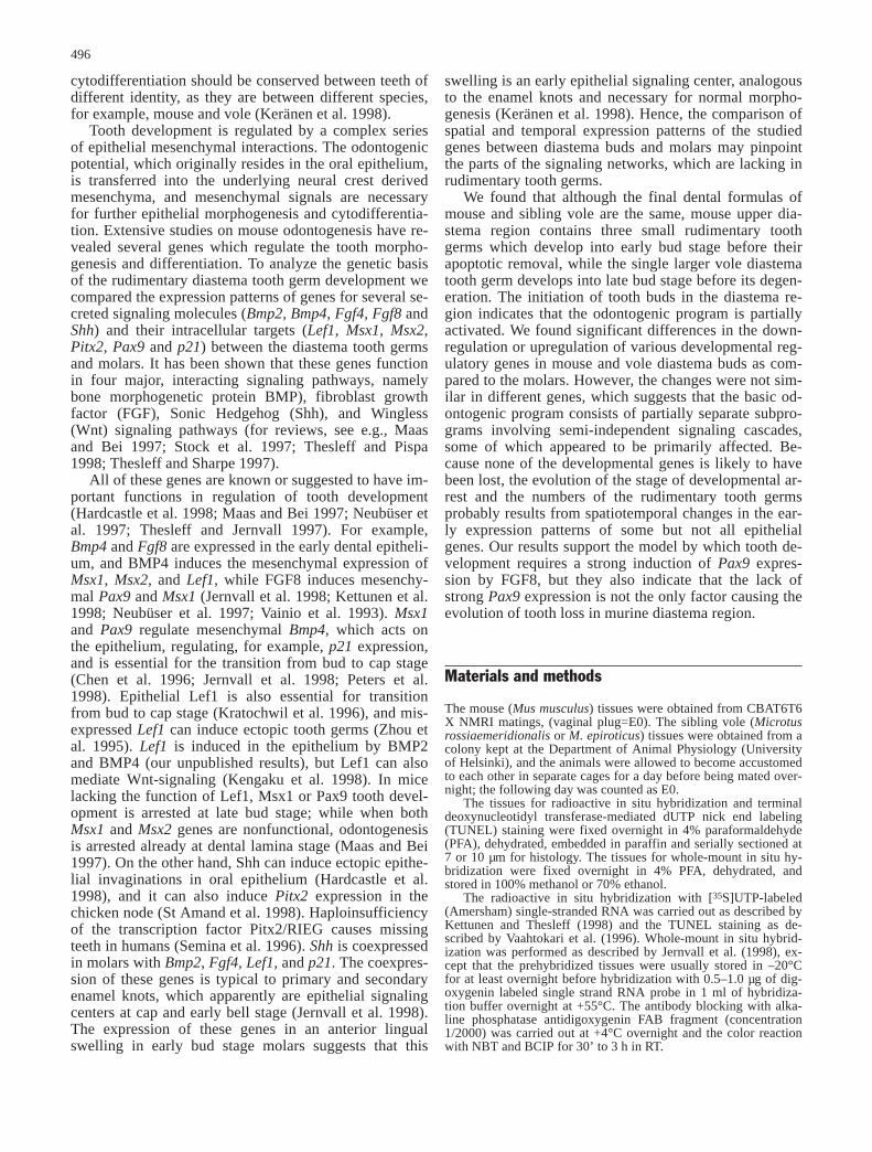
In careful analysis of frontal serial sections we found inmouse three upper diastema region rudimentary toothgerms (but see Peterková et al. 1998), while only a sin-gle rudimentary tooth germ was detected in the vole up-per diastema (Fig. 1). We did not find rudimentary tooth
germs in the lower jaws of either species (not shown).Thus, despite having the same final number of teeth,mouse and vole have different embryological dental for-mulas.
The first mouse diastema tooth germ (D1) developedby E12 near the incisors (Fig. 1K), but it was located inthe maxillary instead of the frontonasal process. The sec-ond (D2; Fig. 1M) and third (D3; Fig. 1N) diastematooth germs developed in the dental lamina posterior toD1 but clearly anterior to the first upper molar and theywere connected to the palatal rugae (Fig. 1L–N). Thevole diastema tooth germ was located in the maxillaryprocess next to the primary choana in a similar locationas D1 in the mouse, and hence it appears to be the vole
497
Fig. 1 Histological sections of developing first lower vole molarsfrom E12 to E16 (A–E), vole upper jaw diastema tooth germsfrom E12 to E16 (F–J) and mouse upper jaw diastema toothgerms D1 at E12 (K), D2/D3 at E12 (L), D2 at E13 (M), and D3at E13 (N). db Vole diastema bud; d1 mouse first diastema bud;d2/d3 presumptive mouse second and third diastema buds; d2mouse second diastema bud; d3 mouse third diastema bud; m mo-lar; pc primary choana; pr palatal ruga. Bar 150 µm
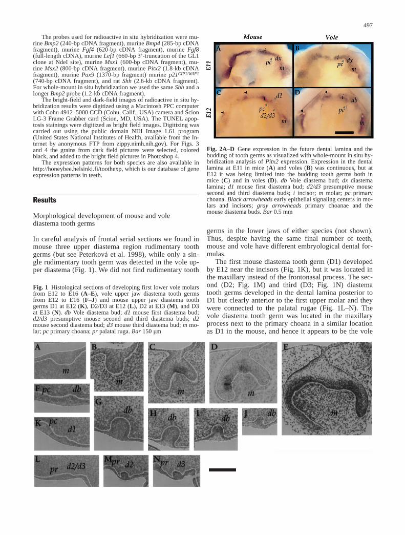
Fig. 2A–D Gene expression in the future dental lamina and thebudding of tooth germs as visualized with whole-mount in situ hy-bridization analysis of Pitx2 expression. Expression in the dentallamina at E11 in mice (A) and voles (B) was continuous, but atE12 it was being limited into the budding tooth germs both inmice (C) and in voles (D). db Vole diastema bud; dx diastemalamina; d1 mouse first diastema bud; d2/d3 presumptive mousesecond and third diastema buds; i incisor; m molar; pc primarychoana. Black arrowheads early epithelial signaling centers in mo-lars and incisors; gray arrowheads primary choanae and themouse diastema buds. Bar 0.5 mm

counterpart of mouse D1 (Fig. 1F). All diastema toothgerms originated from the dental lamina, which wasclearly illustrated in the oral view of whole-mount in situhybridizations (Fig. 2A, B). They did not, however, growin mesiodistal direction as molars (Fig. 2C, D).
All mouse rudimentary tooth germs remained as smallepithelial swellings, and they were quickly removed apo-ptotically at early bud stage. In D1 some apoptosis wasdetected at E12, and the germ disappeared at E13 (Fig.3E). The D2 and D3 became clearly discernible frompalatal rugae only after E12 (Fig. 1L), and we could notdistinguish between D2 and D3 at E12. D2 and D3 weremaximally developed at E13, but their apoptosis beganat E12.5 (Fig. 3G, H), and they disappeared at E14 (notshown). (For more detailed descriptions of the develop-ment of mouse diastema tooth germs, see Peterková etal. 1995, Turecková et al. 1996.)
The epithelial thickening of the vole diastema toothgerm was seen at E11.5, at about the same time as in in-cisors but later than in molars. The bud became visible atE12, and the development was maximal between E13and E14 (Fig. 1F–G). Instead of proceeding into the capstage after the late bud stage, the bud degenerated apo-ptotically. Apoptosis began later in the vole bud, and itsurvived longer than the mouse diastema buds. First apo-ptotic cells were detected in the neck of late E13 diaste-ma bud epithelium, and the bud was removed by E16 toE17 (Fig. 3A–D, Fig. 1H–J).
Although there was some epithelial cell rearrange-ment in the tip of vole E13 diastema bud (not shown),we did not find morphologically distinct enamel knots.The mesenchyma around the diastema buds did not con-dense properly as in molars (Fig. 1F–N). We did not de-tect mesenchymal apoptosis, although the rudimentarycondensation disappeared slowly.
Gene expression patterns in the diastema tooth germs
The expression patterns of several developmental regula-tory genes (Bmp2, Bmp4, Fgf4, Fgf8, Lef1, Msx1, Msx2,p21, Pax9, Pitx2, and Shh) were compared between thediastema buds and the normal tooth germs of the twospecies. We analyzed frontal serial sections of stagesfrom E11 to E13 in mice and from E11 to E15 in voles
and whole mounts of stages from E10 to E13 in bothspecies.
We detected expression of all these genes in both mo-lars and incisors (not shown). As most of the gene ex-pression patterns have already been reported for molars(Keränen et al. 1998), we compared the gene activitiesprimarily between diastema buds and molars.
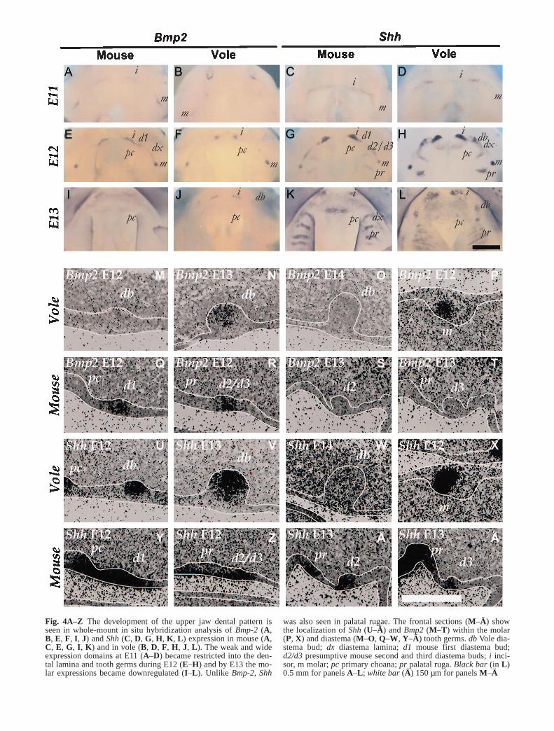
Shh was present in the odontogenic epithelia of bothspecies from E11 onwards. It was first expressedthroughout the dental lamina (Fig. 4C, D). Then it be-came limited to the forming palatal rugae and to the bud-ding tooth germs where expression was upregulated inthe early epithelial signaling centers (Fig. 4G, H, K, L,U–Ä). It was lost from late bud stage molars at E13, butbecame again upregulated in the enamel knots by the capstage (Keränen et al. 1998). In mouse the whole diaste-ma buds expressed Shh, and the expression was continu-ous with either palatal rugae or primary choanae (Fig.4Y–Ä). In voles the Shh expression, which was original-ly connected to the primary choanae, became limited in-to the tip of the E13 diastema bud (Fig. 4U–W). The tipof the E13 vole diastema bud also expressed Bmp2, Lef1,and p21, which are known to be coexpressed with Shh inearly epithelial signaling centers (Keränen et al. 1998).Because the morphological development of the vole dia-stema buds was delayed 1 day compared to molars, itseems that the tip of the vole diastema bud correspondsto the E12 molar early epithelial signaling center.
As with Shh, Bmp2 was expressed in odontogenic epi-thelium, in the forming tooth germs and the early epithe-lial signaling centers, but unlike Shh, Bmp2 was not seenin the palatal rugae (Fig. 4A, B, E, F, I, J, M–T). Bmp2was upregulated later than Shh, especially in voles, andone of the differences between mouse and vole diastemabuds was that mouse diastema buds expressed Bmp2 ear-ly while in vole diastema buds it was seen only at E13.Based on Bmp2 expression we detected D2/D3 at E12,before it could be distinguished morphologically (Fig.4R).
As with Shh, Pitx2 expression was first continuous inthe dental lamina in both species and became limited in-to the budding tooth germs, including the diastema toothgerms in mouse and vole. Pitx2 became downregulatedin the early epithelial signaling centers of both incisorsand molars in both species (Fig. 2C, D) while other re-
498
Fig. 3 Apoptosis in vole andmouse diastema buds as detect-ed by TUNEL staining: E12(A), E13 (B), E14 (C), and E15(D) vole diastema buds; E12(E) mouse D1, E12 (F) mouseD2/D3, E13 (G) mouse D2 andE13 (H) mouse D3 buds. Bar150 µm
499
Fig. 4A–Z The development of the upper jaw dental pattern isseen in whole-mount in situ hybridization analysis of Bmp-2 (A,B, E, F, I, J) and Shh (C, D, G, H, K, L) expression in mouse (A,C, E, G, I, K) and in vole (B, D, F, H, J, L). The weak and wideexpression domains at E11 (A–D) became restricted into the den-tal lamina and tooth germs during E12 (E–H) and by E13 the mo-lar expressions became downregulated (I–L). Unlike Bmp-2, Shh
was also seen in palatal rugae. The frontal sections (M–Ä) showthe localization of Shh (U–Ä) and Bmp2 (M–T) within the molar(P, X) and diastema (M–O, Q–W, Y–Ä) tooth germs. db Vole dia-stema bud; dx diastema lamina; d1 mouse first diastema bud;d2/d3 presumptive mouse second and third diastema buds; i inci-sor, m molar; pc primary choana; pr palatal ruga. Black bar (in L)0.5 mm for panels A–L; white bar (Ä) 150 µm for panels M–Ä

gions of dental epithelium continued to express it in-tensely.
Epithelial Lef1 expression resembled that of Shh. Lef1was expressed in the early epithelium (not shown) Aswell as in the forming tooth germs and palatal rugae andin the early epithelial signaling centers (Fig. 5A–H). Invole diastema buds Lef1 was downregulated at E14 (Fig.5C), while its expression continued in the tips of molarbuds and in enamel knots (not shown, Keränen et al.1998). The mesenchymal Lef1 expression was downreg-ulated in the diastema region (Fig. 5A–H) but main-
tained and upregulated in molar and incisor tooth germs(not shown).
As reported earlier by Turescová et al. (1995), Msx1expression was intense in the mouse diastema region upto E13 (Fig. 5M–P). In voles the initially high expressionof Msx1 at E11 became quickly downregulated in the di-astema mesenchyma as compared to molars (Fig. 5I–L).We found that unlike in molars but as in incisors, the dia-stema bud epithelia in both species expressed Msx1 (Fig.5I–S).
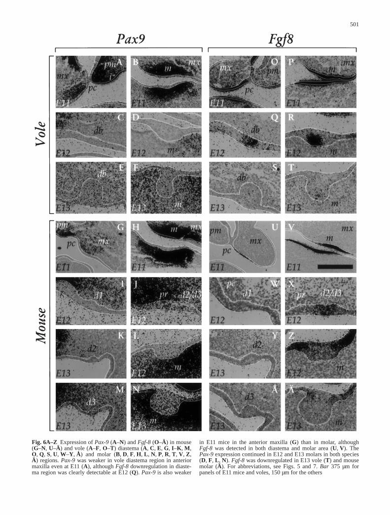
The clearest early difference that we detected betweenthe diastema tooth germs and the molars was the weakermesenchymal Pax9 expression in E11 diastema regions ofboth species (Fig. 6A, B, G, H). The Pax9 expression wasweaker in the E11 anterior maxilla or future diastema re-gion of the serial sections than in future molar or incisorregions and was not upregulated there even later althoughits expression continued in molar (and incisor) mesenchy-
500
Fig. 5A–S Lef-1 (A–H) and Msx-1 (I–S) expression in frontalsections of vole (A–D, I–L, Q) and mouse (E–H, M–P, R, S). db Vole diastema bud; d1 mouse first diastema bud; d2/d3 pre-sumptive mouse second and third diastema buds; d2 mouse seconddiastema bud; d3 mouse third diastema bud; i incisor; m molar; pc primary choana; pr palatal ruga. Bar 150 µm
501
Fig. 6A–Z Expression of Pax-9 (A–N) and Fgf-8 (O–Ä) in mouse(G–N, U–Ä) and vole (A–F, O–T) diastema (A, C, E, G, I–K, M,O, Q, S, U, W–Y, Å) and molar (B, D, F, H, L, N, P, R, T, V, Z,Ä) regions. Pax-9 was weaker in vole diastema region in anteriormaxilla even at E11 (A), although Fgf-8 downregulation in diaste-ma region was clearly detectable at E12 (Q). Pax-9 is also weaker
in E11 mice in the anterior maxilla (G) than in molar, althoughFgf-8 was detected in both diastema and molar area (U, V). ThePax-9 expression continued in E12 and E13 molars in both species(D, F, L, N). Fgf-8 was downregulated in E13 vole (T) and mousemolar (Ä). For abbreviations, see Figs. 5 and 7. Bar 375 µm forpanels of E11 mice and voles, 150 µm for the others
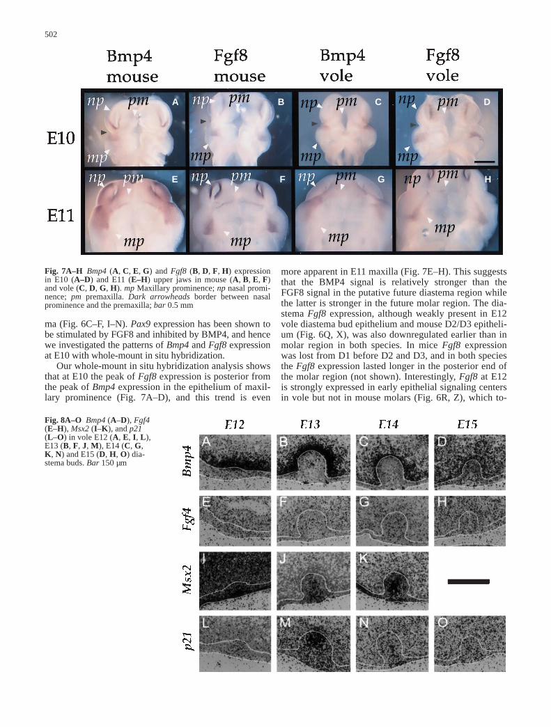
ma (Fig. 6C–F, I–N). Pax9 expression has been shown tobe stimulated by FGF8 and inhibited by BMP4, and hencewe investigated the patterns of Bmp4 and Fgf8 expressionat E10 with whole-mount in situ hybridization.
Our whole-mount in situ hybridization analysis showsthat at E10 the peak of Fgf8 expression is posterior fromthe peak of Bmp4 expression in the epithelium of maxil-lary prominence (Fig. 7A–D), and this trend is even
502
A B C D
E F G H
Fig. 7A–H Bmp4 (A, C, E, G) and Fgf8 (B, D, F, H) expressionin E10 (A–D) and E11 (E–H) upper jaws in mouse (A, B, E, F)and vole (C, D, G, H). mp Maxillary prominence; np nasal promi-nence; pm premaxilla. Dark arrowheads border between nasalprominence and the premaxilla; bar 0.5 mm
Fig. 8A–O Bmp4 (A–D), Fgf4(E–H), Msx2 (I–K), and p21(L–O) in vole E12 (A, E, I, L),E13 (B, F, J, M), E14 (C, G,K, N) and E15 (D, H, O) dia-stema buds. Bar 150 µm
more apparent in E11 maxilla (Fig. 7E–H). This suggeststhat the BMP4 signal is relatively stronger than theFGF8 signal in the putative future diastema region whilethe latter is stronger in the future molar region. The dia-stema Fgf8 expression, although weakly present in E12vole diastema bud epithelium and mouse D2/D3 epitheli-um (Fig. 6Q, X), was also downregulated earlier than inmolar region in both species. In mice Fgf8 expressionwas lost from D1 before D2 and D3, and in both speciesthe Fgf8 expression lasted longer in the posterior end ofthe molar region (not shown). Interestingly, Fgf8 at E12is strongly expressed in early epithelial signaling centersin vole but not in mouse molars (Fig. 6R, Z), which to-
gether with Fgf4 expression in E12 vole but not inmouse (Keränen et al. 1998), suggests that Fgf signalingis upregulated more strongly in early vole than in earlymouse molars, although its developmental significanceremains to be studied.
In voles Bmp4 was downregulated in the diastemaepithelium by E12, earlier than in the molars (Figs.8A–D, 9) while in E12 mouse diastema buds Bmp4 waslocated in central and superficial cells (not shown), as re-ported by Turecková et al. (1995). How this expressioncompares with the late vole epithelial Bmp4 expressionin single cells (Fig. 8B, C), and whether it is involved inthe apoptotic removal of the diastema buds (Peterková etal. 1998) remains to be studied. Bmp4 was also initiallypresent in the subepithelial mesenchyma of both species,but was weaker than in molar areas even at E12 (not
shown), and in voles it colocalized with the disappearingrudimentary mesenchymal condensation (Fig. 8A–D).
Unlike in molars, where both Bmp4 and Msx2 are bi-ased to the buccal side of the tooth germ, in vole diaste-ma buds their expression domains were similar in bothbuccal and lingual sides (Fig. 8A–D, I–K). These geneexpression differences may be associated with the find-ing that the early epithelial signaling center in the volediastema buds was located at the tip of the bud, not in itslingual aspect as in molars.
The early epithelial signaling center of the vole dia-stema bud coexpressed Shh, Bmp2, p21, and Lef1 (Figs.4M–O, U–W, 5A–C, 8L–O). However, other epithelialgenes such as Bmp4 and Fgf8, which are present in mo-lar signaling centers, were downregulated (Fig. 9) andMsx2 and Fgf4 were not upregulated (Fig. 8E–K), indi-
503
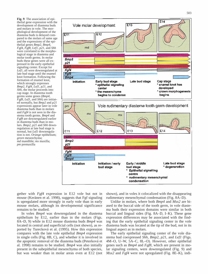
Fig. 9 The association of epi-thelial gene expression with thedevelopment of diastema budsand molars in vole. The mor-phological development of thediastema buds is delayed com-pared to the molars of same ageand the expressions of the epi-thelial genes Bmp2, Bmp4,Fgf4, Fgf8, Lef1, p21, and Shhwere correlated to the morpho-logical stage in diastema andmolar tooth germs. In molarbuds these genes were all ex-pressed in the early epithelialsignaling center. Except forLef1, all were downregulated atlate bud stage until the enamelknot formation. Following theformation of enamel knot,which strongly expressesBmp2, Fgf4, Lef1, p21, andShh, the molar proceeds intocap stage. In diastema toothgerms some genes (Bmp4,Fgf8, Lef1, and Shh) are initiat-ed normally, but Bmp2 and p21expressions appear later in volediastema buds than in molarsand Fgf4 is not seen in the dia-stema tooth germs. Bmp4 andFgf8 are downregulated earlierin diastema buds than in mo-lars. Bmp2, p21 and Shh down-regulation at late bud stage isnormal, but Lef1 downregula-tion is not. Orange epithelium;green mesenchyma; md mandible; mx maxilla; pm premaxilla
cating that it is different from molar early epithelial sig-naling center. The lack of a morphologically distinguish-able enamel knot with strong Fgf4 expression in the volediastema bud indicates that its development stops at thestage that corresponds to E13 bud stage molars beforethe transition to cap stage (Fig. 9).
Discussion
Although mouse and vole have the same number of mo-lars and incisors, and the histological development oftheir teeth and the correlation of gene expression pat-terns to the developing histology (except for early Fgfsignaling) are similar in the two species (Keränen et al.1998), the diastema regions of the two species differ.Mouse has three small diastema buds which develop intoearly bud stage, whereas vole has one large diastema budwhich develops into late bud stage. Mouse diastemabuds do not contain separate signaling centers, but aclear early epithelial signaling center develops within thevole diastema buds. This center, however, is located atthe tip of the bud unlike in molars, where it is locatedlingually, and in the tips of the diastema buds the timingof gene expression differs from that of the molar earlyepithelial signaling center. Subsequently, the vole diaste-ma buds neither develop enamel knots nor proceed to thecap stage. All diastema buds in both species degenerateapoptotically. To investigate the genetic basis of the de-velopmental arrest in the two species we compared theexpression patterns of the developmental regulatorygenes Bmp2, Bmp4, Fgf4, Fgf8, Lef1, Msx1, Msx2, p21,Pax9, Pitx2, and Shh between the rudimentary diastematooth germs and the normal teeth.
Early odontogenic epithelium and rudimentary diastematooth germs
Teeth are serially homologous organs (Stock et al. 1997).Hence the basic genetic mechanisms governing odonto-genesis ought to be conserved between teeth of differentidentity as well as between the teeth in different species(Keränen et at 1998). Therefore the developmental dif-ferences between tooth germs should be reflected in lo-cal modifications in the expression patterns of shared de-velopmental regulatory genes. The species-specific loca-tions (and numbers) of tooth germs presumably dependon where the basic genetic program for tooth germ initia-tion is activated (Tucker et al. 1998). Classic recombina-tion experiments have shown that the identity (and hencethe morphological development) of an individual toothgerm depends on the early epithelium, which containsthe necessary information for odontogenesis (Kollar andMina 1991; Lumsden 1988; Mina and Kollar 1989), andrecently Bmp4 has been implicated in the determinationof identity and tooth specific morphogenesis (Tucker etal. 1998).
All tooth germs, including the diastema buds, arosefrom the dental lamina in both species. The oral epitheli-
um forming the future dental lamina could be visualizedwith the expression of Pitx2 and Shh genes (Fig. 2A–D,4C–D). These genes were first expressed as a continuousband through maxillary and premaxillary epithelium, andthey became subsequently limited into budding toothgerms, including the diastema buds, where they were up-regulated. The tooth germ epithelia thereafter becamecompartmentalized as the early epithelial signaling cen-ters expressing, for example, Shh, Lef1, Bmp2, p21, wereformed in molars and incisors of both species and in volediastema buds. The restriction of expressions into indi-vidual tooth germs occurred at E12, i.e., after the odon-togenic potential had been transferred into the mesen-chyma. However, we did not note similar localized pat-terning in any of the mesenchymal genes that we studied,and the exact locations of the individual tooth germsmay be determined within the epithelium, although apermissive signal from the mesenchyma is possible.
Although the developing tooth germs could be visual-ized with the expression of epithelial genes such as Shhand Pitx2, the developmental arrest of the diastema toothgerms was reflected by spatiotemporal differences ingene expressions as compared with molars. We detectedall analyzed genes except Fgf4 in the vole diastema toothgerms, and these were downregulated or upregulated atdifferent times than with the molars (Fig. 9). Some of thegenes (epithelial Fgf8 and Bmp4, all the mesenchymalgenes) were downregulated earlier the molars, somewere not induced in the early signaling centers (Msx2,Fgf4) while others were expressed normally until latebud stage (Shh, Lef1), and the upregulation of yet othergenes (Bmp2, p21) was delayed (Fig. 9). Since thesegenes may be involved in the early events of the odonto-genesis, the differences between diastema buds and mo-lars suggest both that the signaling cascades governingvarious components of early odontogenesis (e.g., the de-termination of the tooth location, epithelial invagination,determination of tooth identity, and transfer of odonto-genic potential from epithelium to mesenchyma) are atleast partially separate, and that only some of these areoriginally affected in the diastema buds.
Although the early odontogenesis depends on the oralepithelium, the earliest clear genetic difference that wefound was weaker Pax9 expression in diastema than inmolar (or incisor) mesenchymas of both species at dentallamina stage (E11). Pax9 is induced by epithelial FGF8,but at E10 BMP4 can inhibit Pax9 expression (Fig. 9;Neubüser et al. 1997; Peters et al. 1998). The peak ofFgf8 expression seemed to be posterior from the peak ofBmp4 expression in E10 mouse and vole maxillaryprominences (Fig. 7A–D), suggesting that the downregu-lation of Pax9 in the anterior maxilla, or future diastemaregion may be associated with stronger expression ofBmp4 than Fgf8.
Fgf8 and Bmp4 have been suggested to participate inthe transfer of odontogenic potential from epithelium tothe mesenchyma by inducing mesenchymal Pax9 andMsx1 expression (Neubüser et al. 1997; Tucker et al.1998; Vainio et al. 1993), both of which are necessaryfor the mesenchymal Bmp4 expression. BMP4 is essen-
504
tial for tooth morphogenesis (Chen et al. 1996). It can in-duce epithelial p21 and Lef1, and it has been speculatedthat mesenchymal BMP signals are necessary to induceenamel knot formation in tooth germs before they canproceed into cap stage (Kratochwil et al. 1996; Jernvallet al. 1998). In vole diastema buds both mesenchymalBmp4 and other mesenchymal genes became downregu-lated after Pax9. Together with the early downregulationof epithelial Bmp4 and Fgf8, this suggests that the Pax9mediated transfer of odontogenic potential is disturbedspecifically in vole diastema buds. The vole diastematooth germs resemble morphologically the tooth germsof Pax9–/– mutant mice (Peters et al. 1998), and it is pos-sible that early disturbances in the Pax9-dependent sig-naling pathway cause the late bud stage developmentalarrest in the vole diastema tooth germs, even when otherdevelopmental processes are initially normal.
Indeed, the expression patterns of Shh and Lef1 werequite similar in vole diastema buds and molars until latebud stage. Both Shh and Lef1 have been implicated inepithelial invagination and early dental patterning (Hard-castle et al. 1998; Zhou et al. 1995). The species-specificdifferences in their expression patterns and their late dis-turbance in vole diastema buds suggest that these genesare involved in the early determination and/or budding oftooth germs. Moreover, the restriction of epithelial geneexpression (Bmp2, Lef1, Pitx2, Shh) into forming toothgerms occurs after the early induction of Pax9. This re-striction and budding occurs also in the areas which donot express Pax9, suggesting that the determination ofexact locations of the tooth germs and their early devel-opment occurs parallel to and at least partly indepen-dently of the early induction of odontogenic mesenchy-ma by BMP4.
In addition to the differences between diastema toothgerms and molars (or incisors), there also were differ-ences between the diastema tooth germs of the two spe-cies. Unlike the vole diastema buds, the mouse diastemabuds degenerate at the early bud stage. Interestingly, wefound that Bmp2 expression was both upregulated anddownregulated earlier in the mouse than in the vole dia-stema buds (Fig. 4E, F, M–T). Another difference be-tween the two species was the continuing Msx1 expres-sion in the mouse but not in the vole diastema mesenchy-ma (Fig. 5I–P). Neither of these differences can explainthe differences between the diastema regions of the twospecies, but it is reasonable to hypothesize that some ear-ly epithelial odontogenic signaling pathway may be dif-ferently affected in mouse than in vole, although the ear-ly expressions of Bmp4, Fgf8, and Pax9 in the two spe-cies are similar.
Evolution of rudimentary diastema tooth germs
The most primitive rodent dental formula, also found inthe earliest known rodent from the Eocene (Meng et al.1994), consists of one incisor, two premolars, and threemolars. Because mice have three diastema buds, at leastone of them may have persisted for over 50 millionyears.
The persistence of rudimentary organs was been com-mented upon as early as Darwin (1856). Unlike the pseu-dogenes, which degenerate relatively soon under the ran-dom mutation load (e.g., Li and Graur 1991), the devel-opmental programs of rudimentary organs seem to bemore resistant to modifications. The reason for this isprobably the modular use of the individual signalingpathways for various developmental processes. If evenone gene within such signaling cascade were lost, the ef-fects could be lethal or deleterious in multiple organs, in-cluding other teeth (Behrens et al. 1997; Feldman et al.1995; van Genderen et al. 1994; Hogan 1996; Peters et al.1998; Roessler et al. 1996; Satokata and Maas 1994; Se-mina et al. 1996). On the other hand, although the devel-opmental programs themselves are conserved, the mor-phological evolution can be fast (Brunet-Lecomte andChaline 1991), which suggests that the spatiotemporal ap-plication of the conserved programs can evolve flexibly.
Our data support the view that the early epithelialgene expression patterns are important in the patterningof the mesenchyma, depending on the identity of thetooth germs (Tucker et al. 1998). Because there are nodiastema buds in the mandibles of either species, and be-cause the reduction in tooth numbers in rodents tends tobe more pronounced in the lower than the upper jaws(Luckett 1985), the loss of tooth germs is probably lessfavored in the upper than the lower jaw diastema re-gions. We found that both vole diastema buds and mouseD1 are associated with the primary choanae, where themaxillary processes fuse with the frontonasal process,while the mouse D2 and D3 were connected to the pala-tal rugae. The morphological associations were seen alsoas continuous expression areas of some genes such asLef1 and Shh. The morphological connections betweentooth germs, nasal epithelium, and palatal rugae and thesimilarities in their differentiation support the notion thatthese structures develop during ontogeny from a com-mon epithelial precursor (Peterková 1985). It is thereforepossible that the more complex morphological develop-ment of the upper than the lower jaw also slows the evo-lutionary loss of upper diastema tooth germs by support-ing the early epithelial patterning necessary for the spa-tial determination and initiation of the tooth germs.
Acknowledgements We thank M. Holopainen, K. Kettunen, M.Mäkinen, P. Pekkarinen, R. Santalahti, and A. Tuomi for their ex-pert technical help. We also thank Drs. C. Basilico, T. Edlund, R.Grosschedl, C. MacArthur, T. Mitsiadis, A. Neubüser, P. Sharpe,B. Vogelstein, E. Wang, and J. Wozney for probe constructs. Thiswork was funded by the Finnish Academy, the Sigrid JuseliusFoundation, and University of Helsinki Graduate School for Bio-technology.
References
Behrens M, Langecker TG, Wilkens H, Schmale H (1997) Com-parative analysis of Pax-6 sequence and expression in the eyedevelopment of the blind cave fish Astyanax fasciatus and itsepigean conspecific. Mol Biol Evol 14:299–308
Brunet-Lecomte P, Chaline J (1991) Morphological evolution andphylogenetic relationships of the European ground voles (Ar-vicolidae, Rodentia). Lethaia 24:45–54
505
Butler PM (1995) Ontogenetic aspects of dental evolution. Int JDev Biol 39:25–34
Chen Y, Bei M, Woo I, Satokata I, Maas R (1996) Msx1 controlsinductive signaling in mammalian tooth morphogenesis. De-velopment 122:3035–3044
Darwin C (1856) The origin of species: by means of natural selec-tion or the preservation of favoured races in the struggle forlife, chap XIV. Murray, London
Feldman B, Poueymirou W, Papaioannou VE, DeChiara TM,Goldfarb M (1995) Requirement of FGF-4 for postimplanta-tion mouse development. Science 267:246–249
Genderen C van, Okamura RM, Farinas I, Quo RG, Parslow TG,Bruhn L, Grosschedl R (1994) Development of several organsthat require inductive epithelial-mesenchymal interactions isimpaired in LEF-1-deficient mice. Genes Dev 8:2691–2703
Hardcastle Z, Mo R, Hui C-C, Sharpe PT (1998) The Shh signal-ling pathway in tooth development: defects in Gli2 and Gli3mutants. Development 125:2803–2811
Hogan BL (1996) Bone morphogenetic proteins in development.Curr Opin Genes Dev 6:432–438
Jernvall J, Åberg T, Kettunen P, Keränen S, Thesleff I (1998) Thelife history of an embryonic signaling center: BMP-4 inducesp21 and is associated with apoptosis in the mouse tooth enam-el knot. Development 125:161–169
Kengaku M, Capdevila J, Rodriguez-Esteban C, De La Pena J,Johnson RL, Belmonte JCI, Tabin CJ (1998) Distinct WNTpathways regulating AER formation and dorsoventral polarityin the chick limb bud. Science 280:1274–1277
Keränen SVE, Åberg T, Kettunen P, Thesleff I, Jernvall J (1998)Association of developmental regulatory genes with the devel-opment of different molar tooth shapes in two species of ro-dents. Dev Genes Evol 208:477–486
Kettunen P, Thesleff I (1998) Expression and function of FGFs-4,-8, and -9 suggest functional redundancy and repetitive use asepithelial signals during tooth morphogenesis. Dev Dyn211:256–268
Kollar EJ, Mina M (1991) Role of the early epithelium in the pat-terning of the teeth and Meckel’s cartilage. J Craniofac GenetDev Biol 11:223–228
Kratochwil K, Dull M, Farinas I, Galceran J, Grosschedl R (1996)Lef1 expression is activated by BMP-4 and regulates inductivetissue interactions in tooth and hair development. Genes Dev10:1382–1394
Li W-H, Graur D (1991) Fundamentals of molecular evolution.Sinauer, Sunderland
Luckett WP (1985) Superordinal and intraordinal affinities of ro-dents: developmental evidence from the dentition and placen-tation. In: Luckett WP, Hartenberger J-L (eds) Evolutionaryrelationships among rodents. NATO ASI Series, Ser. A, vol92. Plenum, New York, pp 227–276
Lumsden AGS (1988) Spatial organization of the epithelium andthe role of neural crest cells in the initation of the mammaliantooth germ. Development 103 [Suppl]:155–169
Maas R, Bei M (1997) The genetic control of early tooth develop-ment. Crit Rev Oral Biol Med 8:4–39
Meng J, Wyss AR, Dawson MR, Zhai R (1994) Primitive fossilrodent from Inner Mongolia and its implications for mammali-an phylogeny. Nature 370:134–136
Mina M, Kollar EJ (1987) The induction of odontogenesis in non-dental mesenchyme combined with early murine mandibulararch epithelium. Arch Oral Biol 32:123–127
Moss-Salentijn L (1978) Vestigial teeth in the rabbit, rat andmouse; their relationship to the problem of lacteal dentitions.In: Butler PM, Joysey KA (eds) Development, function andevolution of teeth. Academic, London, pp 13–29
Neubüser A, Peters H, Balling R, Martin GR (1997) Antagonisticinteractions between FGF and BMP signaling pathways: amechanism for positioning the sites of tooth formation. Cell90:247–255
Nikoletopoulos NP, Chondropoulos BP, Fraguedakis-Tsolis SE(1992) Albumin evolution and phylogenetic relationshipsamong Greek rodents of the families Arvicolidae and Muridae.J Zool 228:445–453
Peterková R (1985) The common developmental origin and phylo-genetic aspects of teeth, rugae palatinae, and fornix vestibulioris in the mouse. J Craniofac Genet Dev Biol 5:89–104
Peterková R, Peterka M, Vonesch JL, Ruch JV (1995) Contribu-tion of 3-D computer-assisted reconstructions to the study ofthe initial steps of mouse odontogenesis. Int J Dev Biol39:239–247
Peterková R, Peterka M, Vonesch JL, Turecková J, Viriot L RuchJV, Lesot H (1998) Correlation between apoptosis distributionand BMP-2 and BMP-4 expression in vestigial tooth primordiain mice. Eur J Oral Sci 106:667–670
Peters H, Neubüser A, Kratochwil K, Balling R (1998) Pax9-defi-cient mice lack pharyngeal pouch derivatives and teeth and ex-hibit craniofacial and limb abnormalities. Genes Dev 12:2735–2747
Robinson M, Catzeflis F, Briolay J, Mouchiroud D (1997) Mole-cular phylogeny of rodents, with special emphasis on murids:evidence from nuclear gene LCAT. Mol Phylogenet Evol 8:423–434
Roessler E, Belloni E, Gaudenz K, Jay P, Berta P, Scherer SW,Tsui LC, Muenke M (1996) Mutations in the human SonicHedgehog gene cause holoprosencephaly. Nat Genet 14:357–360
Satokata I, Maas R (1994) Msx1 deficient mice exhibit cleft palateand abnormalities of craniofacial and tooth development. NatGenet 6:348–356
Semina EV, Reiter R, Leysens NJ, Alward WL, Small KW, DatsonNA, Siegel-Bartelt J, Bierke-Nelson D, Bitoun P, Zabel BU,Carey JC, Murray JC (1996) Cloning and characterization of anovel bicoid-related homeobox transcription factor gene,RIEG, involved in Rieger syndrome. Nat Genet 14:392–399
St Amand TR, Ra J, Zhang Y, Hu Y, Baber SI, Qiu M, Chen Y(1998) Cloning and expression pattern of chicken Pitx2: a newcomponent in the SHH signaling pathway controlling embry-onic heart looping. Biochem Biophys Res Commun 247:100–105
Stock DW, Weiss KM, Zhao Z (1997) Patterning of the mammali-an dentition in development and evolution. Bioessays 19:481–490
Thesleff I, Jernvall J (1997) The enamel knot – a putative signal-ing center regulating tooth development. In: Symposium 62:pattern formation during development. Cold Spring HarborLaboratory, Cold Spring Harbor, pp 257–267
Thesleff I, Pispa J (1998) The teeth as models for studies on themolecular basis of the development and evolution of organs.In: Chuong C-M (ed) Molecular basis of epithelial appendagemorphogenesis. Landes, Georgetown, pp 157–179
Thesleff I, Sharpe P (1997) Signalling networks regulating dentaldevelopment. Mech Dev 67:111–123
Tucker AS, Matthews KL, Sharpe PT (1998) Transformation oftooth type induced by inhibition of BMP signaling. Science282:1136–1138
Turecková J, Sahlberg C, Åberg T, Ruch JV, Thesleff I, PeterkováR (1995) Comparison of expression of the msx-1, msx-2,BMP-2 and BMP-4 genes in the mouse upper diastemal andmolar tooth primordia. Int J Dev Biol 39:459–468
Turescová J, Lesot H, Vonesch J-L, Peterka M, Peterková R, RuchJV (1996) Apoptosis is involved in the disappearance of thediastema dental primordia in mouse embryo. Int J Dev Biol40:483–489
Vaahtokari A, Åberg T, Thesleff I (1996) Apoptosis in the devel-oping tooth: association with an embryonic signaling centerand suppression by EGF and FGF-4. Development 122:121–129
Vainio S, Karavanova I, Jowett A, Thesleff I (1993) Identificationof BMP-4 as a signal mediating secondary induction betweenepithelial and mesenchymal tissues during early tooth devel-opment. Cell 75:45–58
Zhou P, Byrne C, Jacobs J, Fuchs E (1995) Lymphoid enhancerfactor 1 directs hair follicle patterning and epithelial cell fate.Genes Dev 9:700–713
506