Embed Size (px)
Citation preview
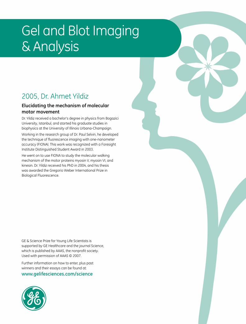
Further information on how to enter, plus pastwinners and their essays can be found at:
www.gelifesciences.com/science
GE & Science Prize for Young Life Scientists issupported by GE Healthcare and the journal Science,which is published by AAAS, the nonprofit society. Used with permission of AAAS © 2007.
Gel and Blot Imaging& Analysis
2005, Dr. Ahmet Yildiz Elucidating the mechanism of molecular motor movementDr. Yildiz received a bachelor's degree in physics from Bogazici University, Istanbul, and started his graduate studies in biophysics at the University of Illinois Urbana-Champaign.
Working in the research group of Dr. Paul Selvin, he developed the technique of fluorescence imaging with one-nanometer accuracy (FIONA). This work was recognized with a Foresight Institute Distinguished Student Award in 2003.
He went on to use FIONA to study the molecular walking mechanism of the motor proteins myosin V, myosin VI, and kinesin. Dr. Yildiz received his PhD in 2004, and his thesiswas awarded the Gregorio Weber International Prize in Biological Fluorescence.
C
M
Y
CM
MY
CY
CMY
K
PAGE 372 GEH Ch11ToC FINAL.pdf 09/06/2008 12:54:29


Gel
and
Blo
t Im
agin
g &
Ana
lysi
s
10
PA
GE
: 374 - C
UR
RE
NC
Y: U
SD
PA
GE
: 374
- CU
RR
EN
CY
: US
DP
AG
E: 3
74 - C
UR
RE
NC
Y: U
SD
PA
GE
: 37
4 - CU
RR
EN
CY
: US
D
Imaging Systems, Software, and Accessories
GE HealthcareFor more information, visit www.gelifesciences.com374
SELECTION GUIDE – Imaging Systems
Typhoon imagers are our high-end variable mode imagers encompassing phosphor and fl uorescence applications. Typhoon imagers deliver outstanding linearity, quantitative accuracy, and extremely low limits of detection. Automated multicolor scanning permits detection of multiple samples in the same experiment. This ensures accuracy of analysis, increases throughput, and saves time.
Storm Gel and Blot Imaging Systems provide proven quality andhigh-performance for fi lmless autoradiography and specifi c fl uorapplications. The systems have wide exposure range and accurate signal quantitation that yield publication quality images on the fi rst exposure. Storm phosphorimagers use storage phosphor screens instead of fi lm to deliver high-resolution phosphorimages and accurate quantitation of various sources of ionizing radiation.
Ettan DIGE Imager is a high-quality imager for a wide range of fl uorescence gel and blot applications. In particular it is validated for use with Cy2-, Cy3- and Cy5-labeled 2-D DIGE gels and Amersham ECL Plex quantitative Western blotting applications.
ImageScanner III is a versatile densitometer with high linearity that is designed for accurate and sensitive densitometric quantitation of SDS-PAGE gels, blots, membranes, and slides.
ImageQuant CCD-based imaging systems cover the full range of gel documentation, chemiluminescence, and fl uorescence applications. CCD imaging creates digital data for archiving, publication, and, with some models, quantitative analysis. For some users, a CCD imager is an appropriate partner or replacement to fi lm–based chemiluminescence applications.
ch10.fm Page 374 Wednesday, June 18, 2008 4:26 PM

Gel and Blot Im
aging & Analysis
10
PA
GE
: 37
5 -
CU
RR
EN
CY
: US
DP
AG
E: 3
75 -
CU
RR
EN
CY
: U
SD
PA
GE
: 3
75 -
CU
RR
EN
CY
: U
SD
PA
GE
: 37
5 -
CU
RR
EN
CY
: US
D
Imaging Systems, Software, and Accessories
GE Healthcare For more information, visit www.gelifesciences.com 375
SELECTION GUIDE – Typhoon, Storm, and ImageQuant Imager PerformanceTyphoon Typhoon Typhoon Typhoon Typhoon Typhoon Storm Storm Storm ImageQuant ImageQuant
Trio+ Trio 9410 9400 9210 9200 860 840 820 RT ECL 350
Storage Phosphor32P, 125I, 14C, 33P, 35S, 3H ++++ ++++ ++++ ++++ ++++ ++++ ++++ ++++ ++++ - -
Macroarray (radiolabeled) ++++ ++++ ++++ ++++ ++++ ++++ ++++ ++++ ++++ - -
Fluorescence—ProteomicsCyDye DIGE Fluors Cy2 ++++ ++++ ++++ ++++ + + ++ ++ - - -
Cy3 ++++ ++++ ++++ ++++ ++++ ++++ - - - - - Cy5 ++++ ++++ ++++ ++++ ++++ ++++ +++ - - -
ECL Plex Fluors Cy2 +++ +++ +++ +++ + + ++ ++ - - - Cy3 ++++ ++++ ++++ ++++ ++++ ++++ - - - - -
Cy5 ++++ ++++ ++++ ++++ ++++ ++++ +++ - - - -
Protein Stains Deep Purple Total Protein Stain ++++ ++++ ++++ ++++ ++++ ++++ + + - +++ ++
SYPRO Ruby ++++ ++++ ++++ ++++ +++ +++ ++ ++ - +++ ++
NanoOrange (solutions) +++ +++ +++ +++ ++ ++ + + - - - Pro-Q Diamond (phosphorylated proteins) ++++ ++++ ++++ ++++ ++++ ++++ - - - - -
Pro-Q Sapphire 532 (Histidine-tagged proteins)
++++ ++++ ++++ ++++ ++++ ++++ - - - - -
Fluorescence—GenomicsMicroarray Cy3 and Cy5 +++ ++ +++ ++ +++ ++ - - - - - Alexa Fluor 532 and Alexa Fluor 633 +++ ++ +++ ++ +++ ++ - - - - -
Nucleic Acid Stains Ethidium Bromide (post stain) ++++ ++++ ++++ ++++ ++++ ++++ - - - ++ ++ Vistra Green, SYBR Gold, SYBR Green I & II ++++ ++++ ++++ ++++ +++ +++ ++ ++ - +++ +++
PicoGreen, RiboGreen +++ +++ +++ +++ ++ ++ ++ ++ - ++ ++
Chemifluorescence (enzyme catalyzed)ECL Plus Western Blotting +++ +++ ++++ ++++ - - ++ ++ - +++ ++ECF, AlkPhos Direct ECF ++++ ++++ ++++ ++++ +++ +++ +++ +++ - + +
DDAO Phosphate ++++ ++++ ++++ ++++ ++++ ++++ +++ - -
Multipurpose FluorescenceCy2 ++++ ++++ ++++ ++++ + + ++ ++ - - -Cy3 ++++ ++++ ++++ ++++ ++++ ++++ - - - - -
Cy5 ++++ ++++ ++++ ++++ ++++ ++++ +++ - - - -
Fluorescein, FAM, FITC, Alexa Fluor 488 ++++ ++++ ++++ ++++ +++ +++ + + - +++ +++TET, HEX, ROX, TAMRA ++++ ++++ ++++ ++++ ++++ ++++ - - -
Other ApplicationsGFP +++ +++ +++ +++ ++ ++ + + - +++ +++
Chemiluminescence + + + + + + - - -ECL ++ ++
ECL Plus ++++ +++
ECL Advance ++++ +++
++++ = superior performance+++ = high performance++ = good performance+ = acceptable performance– = not compatibleRatings are based on overall system performance including model-specific features, versatility, and sensitivity (limit of detection)
ch10.fm Page 375 Wednesday, June 18, 2008 4:26 PM

Gel
and
Blo
t Im
agin
g &
Ana
lysi
s
10
PA
GE
: 376 - C
UR
RE
NC
Y: U
SD
PA
GE
: 376
- CU
RR
EN
CY
: US
DP
AG
E: 3
76 - C
UR
RE
NC
Y: U
SD
PA
GE
: 37
6 - CU
RR
EN
CY
: US
D
Imaging Systems, Software, and Accessories
GE HealthcareFor more information, visit www.gelifesciences.com376
Typhoon Variable Mode Imager
Typhoon 9410 high performance gel and blot imager that can also image microarrays.
J Versatile system platform, handles gel sandwiches, agarose and polyacrylamide gels, membranes, microplates, and even microarrays.J Powerful excitation sources and innovative high-quality
confocal optics allow for the sensitive detection of low-abundance targets.J Red-, green-, and blue-excitation wavelengths and a wide
choice of emission filters enable imaging of an extensive variety of fluorophores.J Automated four-color fluorescence scanning allows
multiplexing of multiple targets in the same sample ensuring accuracy of analysis, increasing throughput, and saving time.J Storage phosphor technology delivers high-resolution
imaging and accurate quantitation of 3H, 14C, 125I, 32P, 33P, 35S, and other sources of ionizing radiation.J Highly sensitive optics enable direct chemiluminescent
imaging without intermediate exposure to films or screens.
Four-color competitive gel shift assay to determine the relative affinities of the Salmonella spp. bacteriophage P22 repressor Mnt (1). In the assay the repressor is allowed to bind four different oligonucleotides labeled with FAM, HEX, TAMRA, and ROX respectively at the same time. The intensities for each fluorophore in a lane are related directly to the relative affinity of the repressor protein for the oligonucleotide used.
ORDERING INFORMATIONProduct Quantity Code Number
Typhoon 9200 1 63-0055-74Includes: ImageQuant TL†
Typhoon 9210 1 63-0055-76Includes: ImageQuant TL†
Typhoon Trio 1 63-0055-87Includes: ImageQuant TL†
Typhoon Trio+ 1 63-0055-89Includes: ImageQuant TL†
Typhoon 9400 1 63-0055-78Includes: ImageQuant TL†
Typhoon 9410 1 63-0055-80Includes: ImageQuant TL†
Workstation scanner with monitor 1 28-4086-78AccessoriesLow-fluorescence Glass Plate, Large, 3 mm thick,
33 u 42 cm1 63-0028-90
Low-fluorescence Glass Plate, Small, 3 mm thick, 20 u 36 cm
1 63-0028-92
Low-fluorescence Glass Plates, 27 u 21 cm, for Ettan DALT (including spacers)
1 80-6475-58
Low-fluorescence Glass Plates for SE600, 18 u 16 cm 2 80-6442-14Spacers for SE 600/SE 400 Vertical Gel Units2 cm u 16 cm u 1.00 mm (W u L u T)
2 80-6180-70
Spacers for SE 600/SE 400 Vertical Gel Units1 cm u 16 cm u 1.00 mm (W u L u T)
2 80-6179-94
Kapton Tape Roll 3 mm u 33 m 1 63-0028-94Wonder Wedge plate separation tool 1 80-6127-88Microarray Slide Holder* 1 63-0039-99Image Eraser 1 63-0007-83Ettan DIGE Gel Alignment Guides for Ettan DALT 1 80-6496-10Ettan DIGE Gel Alignment Guides for SE 600 1 80-6496-29Typhoon Multislide Tray 1 63-0054-34
For pricing information, visit www.gelifesciences.com/orderonline* For Typhoon 9210 and Typhoon 9410 models only† Requires approved computer
Related Products Refer To
Storage Phosphor Screens and Cassettes page 381
Image Eraser page 382
ImageQuant TL page 388
Amersham ECL Plex Western Blotting Combination Packs page 354
Amersham ECL Plex CyDye-conjugated Antibodies page 354
CyDye DIGE Fluors page 264
Ettan DIGE Imager page 383
DeCyder 2-D Differential Analysis Software v6.5 page 265
DeCyder Extended Data Analysis (EDA) Software v1.0 page 266
ImageMaster 2D Platinum v6.0 page 268
Ettan DALTtwelve Large Vertical System page 277
Ettan DALTsix Large Vertical System page 274
Multiphor II Electrophoresis System page 291
Ettan IPGphor 3 IEF System page 272
Typhoon 9410 Variable Mode Imager unites proven storage phosphor autoradiography technology with four-color, nonradioactive fluorescent labeling techniques. For DNA, RNA, and protein samples, choose from:
J Storage phosphor autoradiographyJ Direct blue-excited fluorescence (457, 488 nm)J Direct green-excited fluorescence (532 nm)J Direct red-excited fluorescence (633 nm)J Chemiluminescence
ch10.fm Page 376 Wednesday, June 18, 2008 4:26 PM
Gel and Blot Im
aging & Analysis
10
PA
GE
: 37
7 -
CU
RR
EN
CY
: US
DP
AG
E: 3
77 -
CU
RR
EN
CY
: U
SD
PA
GE
: 3
77 -
CU
RR
EN
CY
: U
SD
PA
GE
: 37
7 -
CU
RR
EN
CY
: US
D
Imaging Systems, Software, and Accessories
GE Healthcare For more information, visit www.gelifesciences.com 377
Typhoon Variable Mode Imager (continued)
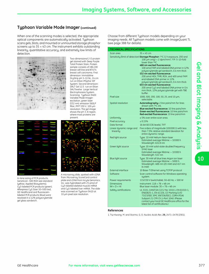
When one of the scanning modes is selected, the appropriate optical components are automatically activated. Typhoon scans gels, blots, and mounted or unmounted storage phosphor screens up to 35 u 43 cm. The instrument exhibits outstanding linearity, quantitative accuracy, and extremely low limits of detection.
Two-dimensional 2-D protein gel stained with Deep Purple Total Protein Stain. Protein sample consists of HBL100 breast cell line and BT474 breast cell carcinoma. First dimension: Immobiline DryStrip pH 3–10 NL, 24 cm run on Ettan IPGphor IEF System. Second dimension: DALT Gel 12.5 run on Ettan DALTtwelve Large Vertical Electrophoresis System. Scanning: Typhoon 9400 Variable Mode Imager: excitation, green laser (532 nm); emission 560LP filter, PMT 530 V, 100 µm resolution. The gel image shows the pH 3–8 region, where most proteins are present.
In-lane sizing of PCR products. GeneScan -500 ROX size standard (yellow, Applied Biosystems), Cy3-labeled PCR products (green), Alfexpress Cy5 Sizer 50–500 (red, GE Healthcare) and fluorescein-labeled PCR products (blue) wereresolved in a 12% polyacrylamidegel glass sandwich.
A microarray slide, spotted with cDNA from Microarray ScoreCard control plate and cDNA from Incyte Genomics, Inc. was hybridized with 25 pmol of Cy3-labeled skeletal muscle mRNA and Cy5-labeled liver mRNA. The slide was scanned on Typhoon 9410 at 10 µm pixel size resolution.
Choose from different Typhoon models depending on your imaging needs. All Typhoon models come with ImageQuant TL (see page 388 for details).
TECHNICAL SPECIFICATIONSScan area 35 u 43 cm
Sensitivity (limit of detection) Storage Phosphor: 14C (1 h exposure, 200 and 100 µm only): v 2 dpm/mm2; 32P: 5–10-fold lower than 14C
488 nm excited fluorescence: 100 amol FAM end-labeled DNA primer in 12% polyacrylamide gel sandwich, 0.4 mm thick
532 nm excited fluorescence: 200 amol HEX, TMR, ROX, and 400 amol FAM end-labeled DNA primer in 12% polyacrylamide gel sandwich, 0.4 mm thick
633 nm excited fluorescence: 200 amol Cy5 end-labeled DNA primer in 0.4 mm thick, 12% polyacrylamide gel with TBE buffer.
Pixel size 1000, 500, 200, 100, 50, 25, and 10 µm, selectable.
Spatial resolution Autoradiography: 2 line pairs/mm for lines drawn with 14C ink.
Blue-excited fluorescence: 10 line pairs/mmGreen-excited fluorescence: 10 line pairs/mmRed-excited fluorescence: 10 line pairs/mm
Uniformity ± 5% over entire scan area.
Pixel accuracy ± 0.15%
Data format 16-bit (65 536 levels), TIFFLinear dynamic range and
linearityFive orders of magnitude (100 000:1) with less
than 7.5% relative standard deviation for entire dynamic range.
Red light source Type: 10 mW Helium-Neon laser Estimated average lifetime: z 10 000 h Wavelength: 632.8 nm
Green light source Type: 20 mW solid-state doubled frequency SYAG laser Estimated average lifetime: z 10 000 h Wavelength: 532 nm
Blue light source Type: 30 mW all blue lines Argon ion laser Estimated average lifetime: z 5000 h Wavelength: 488 nm (20 mW) and 457 nm (4 mW)
External interface 10 Base-T Ethernet using TCP/IP protocol
Software Scan control software for Windows operating system.
Power requirements 115/230 V (switchable), 50–60 Hz, v 500 W
Dimensions(W u D u H)
Instrument: 118 u 78 u 48 cmBlue laser module: 30 u 78 u 48 cm
Safety certifications UL 3101, CAN/CSA C22.2 No 1010.1, EN 61010-1, EN60825-1, EN 61326, CE Marking (LVD 73/23/EEC EMC 89/336/EEC), CDRH (FDA) Subpart 21 CFR Ch.1-Part 1040. (Please contact your local GE Healthcare office for the latest list of certifications.)
References1. Tsz-Kwong, M. and Stormo, G. D. Nucleic Acids Res. 29, 2471–2478 (2001).
ch10.fm Page 377 Wednesday, June 18, 2008 4:26 PM

Gel
and
Blo
t Im
agin
g &
Ana
lysi
s
10
PA
GE
: 378 - C
UR
RE
NC
Y: U
SD
PA
GE
: 378
- CU
RR
EN
CY
: US
DP
AG
E: 3
78 - C
UR
RE
NC
Y: U
SD
PA
GE
: 37
8 - CU
RR
EN
CY
: US
D
Imaging Systems, Software, and Accessories
GE HealthcareFor more information, visit www.gelifesciences.com378
Typhoon Variable Mode Imager (continued)
Typhoon Trio imager is enabled for 2-D DIGE provides multicolor scanning and optimized detection of CyDye DIGE Fluor dye-labeled proteins.
J Provides optimized detection of Cy2, Cy3, and Cy5 CyDye DIGE Fluor minimal and saturation dyes, enabling visualization of up to three differently labeled samples on a single gel.J Exceptional signal-to-noise increases the number of
statistically significant differences detected.J Consistent point-light illumination produces high-quality
uniform images and reduces photo-bleaching.J Gels are scanned between glass plates, preventing drying
and shrinkage and allowing further running and rescanning if required.
Typhoon Trio imager unites the ability to detect an extensive variety of fluors with proven storage phosphor autoradiography technology and direct imaging of chemiluminescence. Powerful excitation sources and innovative high quality confocal optics allow sensitive detection of low-abundance targets.
Typhoon Trio imager delivers outstanding linearity, quantitative accuracy, and extremely low limits of detection. Automated multicolor scanning permits detection of multiple samples in the same experiment, which ensures accuracy of analysis, increasing throughput and saving time.
Typhoon Trio and Ettan DIGE Imager have been designed for the Ettan DIGE System and allow simple scanning of large-format gels. Typhoon can scan up to two DALT 12.5 and four SE600 CyDye DIGE Fluor-labeled gels, and Ettan DIGE Imager can scan one DALT 12.5 gel. Correct alignment of gels is achieved using the appropriate Gel Alignment Guides or gel cassettes. These greatly simplify the process of loading and unloading gels.
SELECTION GUIDE – Typhoon Model Capabilities
Typhoon modelStorage phosphorautoradiography Chemiluminescence
Direct red-excitedfluorescence
Direct green-excitedfluorescence
Direct blue-excited fluorescence Microarray imagingcapability457 nm 488 nm
9200 t t t t - - -
9210 t t t t - - t
Trio t t t t - t -
Trio+t t t t - t t
9400 t t t t t t -
9410 t t t t t t t
Typhoon Multislide Tray
J Typhoon Multislide Tray allows the user to scan up to 30 slides at one time on Typhoon Variable Mode Imager.J The Typhoon Multislide Tray positions the slides at the proper
height above the glass platen of the Typhoon so that the instrument can correctly collect data.J Instrument control software provides simple, yet flexible
interface for the user to set up scanning parameters.J Compatible with Code 39, Code 128, Code I2of5, and Codabar
barcodes.J Individual files are created for each slide, with the option of
adding barcode information to the file name. J Slides are held securely in place by compression pads located
on the lid while being scanned.
Typhoon Multislide tray is an accessory to the Typhoon Instrument allowing the user to expand the microarray slide scanning from 1 to 30 slides, as their application grows. The tray has been designed to position slides in optimal position to collect quantitative data.
TECHNICAL SPECIFICATIONSSlide Dimensions
Width 24.89–25.65 mm (0.98–1.01 in)
Length 74.50–76.45 mm (2.93–3.01 in)Thickness 0.94–1.07 mm (0.037–0.042 in)
Slides that do not meet the specifications above might not fit securely in the slide slots of Typhoon Multislide Tray
ORDERING INFORMATIONProduct Quantity Code Number
Typhoon Multislide Tray 1 63-0054-34
For pricing information, visit www.gelifesciences.com/orderonline
Related Products Code Number Refer To
Typhoon 9210 63-0055-76 page 376
Typhoon 9210 with PC 63-0055-77 page 376
Typhoon 9410 63-0055-80 page 376
Typhoon 9410 with PC 63-0055-81 page 376
Typhoon Multislide Tray holds up to 30 slides for microarray analysis.
ch10.fm Page 378 Wednesday, June 18, 2008 4:26 PM

Gel and Blot Im
aging & Analysis
10
PA
GE
: 37
9 -
CU
RR
EN
CY
: US
DP
AG
E: 3
79 -
CU
RR
EN
CY
: U
SD
PA
GE
: 3
79 -
CU
RR
EN
CY
: U
SD
PA
GE
: 37
9 -
CU
RR
EN
CY
: US
D
Imaging Systems, Software, and Accessories
GE Healthcare For more information, visit www.gelifesciences.com 379
Storm
Storm 860 Gel and Blot Imaging System offers filmless autoradiography, fluorescence and chemifluorescence imaging.
J Large format imaging systems for filmless autoradiography, fluorescence, and chemifluorescence imaging.J Filmless autoradiography based on proven PhosphorImager
technology.J Direct red- and blue-excited fluorescence enable rapid and
accurate quantitation using fluorescence- and chemifluorescence-based methods.J Five orders of linear dynamic range ensure accurate,
publication-quality results on the first exposure.
Storm Gel and Blot Imaging System delivers proven PhosphorImager capability for autoradiography, direct fluorescence for nucleic acid and protein gel analysis, and chemifluorescence for fast blot analysis. Storm uses storage phosphor screens instead of film to deliver high-resolution imaging and accurate quantitation of 14C, 3H, 125I, 32P, 33P, 35S, and other sources of ionizing radiation.
The system’s wide exposure range and accurate signal quantification yield publication-quality images on the first exposure. Storage phosphor screens are reusable and are not degraded by repeated exposure to laboratory levels of radioactivity. To reuse, simply expose a screen to the extra-bright light of Image Eraser light box (included with Storm systems). With direct fluorescence, Storm enables visualization and analysis of nucleic acid and protein gels just minutes after electrophoresis is complete. Gels are soaked in dye solution and then placed in the scanner for analysis. Molecules labeled with Cy5 can also be directly detected.
For chemifluorescence-based methods, Storm reads blots in minutes, without exposure to film. Quantitation is simplified because, unlike film, Storm exhibits a linear response to fluorescent signal intensities.
Storm scans gels, blots, or mounted and unmounted storage phosphor screens up to 35 u 43 cm. All Storm systems come with ImageQuant TL.
ORDERING INFORMATIONProduct Quantity Code
Number
Storm 860 & ImageQuant TL * 1 63-0035-63Includes: ImageQuant TL, scanner control software, Image Eraser, SCSI cable and terminator. Requires approved computer.
Storm 840 & ImageQuant TL * 1 63-0035-58Includes: ImageQuant TL, scanner control software, Image Eraser, SCSI cable and terminator. Requires approved computer.
Storm 820 & ImageQuant TL * 1 63-0035-53Includes: ImageQuant TL, scanner control software, Image Eraser, SCSI cable and terminator. Requires approved computer.
Workstation scanner with monitor 1 28-4086-78
For pricing information, visit www.gelifesciences.com/orderonline
* Does not include phosphor screens and exposure cassettes. For ordering of phosphor screens, please refer to Selection Guide on see page 381.
Related Products Refer To
Storage Phosphor Screens and Cassettes page 381
Image Eraser page 382
ImageQuant TL page 388
Blotting Membranes page 324
Nucleic Acid Labeling and Detection page 330
Protein Labeling and Detection page 353
SDS-PAGE Electrophoresis Units page 283
Nucleic Acid Electrophoresis Units page 300
Amersham Radiochemicals & Radiation Safety Chapter 3
Amersham ECL Plex Western Blotting Combination Packs page 354
Amersham ECL Plex CyDye-conjugated Antibodies page 354
ch10.fm Page 379 Wednesday, June 18, 2008 4:26 PM

Gel
and
Blo
t Im
agin
g &
Ana
lysi
s
10
PA
GE
: 380 - C
UR
RE
NC
Y: U
SD
PA
GE
: 380
- CU
RR
EN
CY
: US
DP
AG
E: 3
80 - C
UR
RE
NC
Y: U
SD
PA
GE
: 38
0 - CU
RR
EN
CY
: US
D
Imaging Systems, Software, and Accessories
GE HealthcareFor more information, visit www.gelifesciences.com380
Storm (continued)
SELECTION GUIDE – Storm System Capabilities
Storm ModelStorage phosphorautoradiography
Direct red-excitedfluorescence
Direct blue-excitedfluorescence
820 t
840 t t
860 t t t
TECHNICAL SPECIFICATIONSScan area 35 u 43 cm
Sensitivity (limit of detection)
v 2 dpm/mm2 for 14C (200 and 100 µm only) using GP screen in a one-hour exposure.
Pixel size 200, 100, and 50 µm, selectable. The scanning laser beam is approximately 50 µm in diameter.
Spatial resolution Autoradiography: 2 line pairs/mm for lines drawn with 14C ink.
Blue-excited fluorescence, chemifluorescence: 2 line pairs/mm
Red-excited fluorescence: 4 line pairs/mm
Uniformity ± 5% relative standard deviation
Pixel accuracy ± 0.15%Data format 16-bit (65 536 levels), TIFF
Linear dynamic range and linearity
5 orders of magnitude (100 000:1) with less than 7.5% relative standard deviation for entire dynamic range.
Light source Storm 820, 840, and 860 use a red laser diode (635 nm). Storm 840 and 860 also use a blue LED (450 nm).
External interface SCSISoftware Scan control software for Windows operating
system.Power requirements Storm: 115/230 V, 50–60 Hz, v 150 W
ImageEraser: 115/230 V, 50–60 Hz, v 150 WDimensions
(W u D u H)Storm: 76 u 76 u 38 cmImageEraser: 42 u 32 u 63.5 cm
Safety certifications UL 3101, CAN/CSA C22.2 No 1010.1, EN 61010-1, EN60825-1, EN 61326, CE Marking (LVD 73/23/EEC EMC 89/336/EEC), CDRH (FDA) Subpart 21 CFR Ch.1-Part 1040. (Please contact your local GE Healthcare office for the latest list of certifications.)
Atlas Human cDNA array probed with 33P-labeled cDNA. The hybridized array, containing 588 unique targets, was exposed to a storage phosphor screen for 2 days and imaged using Storm 840.
Gene Discovery Array Human I probed with 32P-labeled human cDNA. The 22 u 22 cm membrane array contained 18 394 non-redundant cDNA clones. The array was exposed to a storage phosphor screen for one hour and imaged using Storm 820.
ch10.fm Page 380 Wednesday, June 18, 2008 4:26 PM

Gel and Blot Im
aging & Analysis
10
PA
GE
: 38
1 -
CU
RR
EN
CY
: US
DP
AG
E: 3
81 -
CU
RR
EN
CY
: U
SD
PA
GE
: 3
81 -
CU
RR
EN
CY
: U
SD
PA
GE
: 38
1 -
CU
RR
EN
CY
: US
D
Imaging Systems, Software, and Accessories
GE Healthcare For more information, visit www.gelifesciences.com 381
Storage Phosphor Screens and Cassettes
Storage phosphor screens require less exposure time and are 10 to 100 times more sensitive than film.
J Requires approximately one-tenth the exposure time of traditional autoradiography film.J 10 to 100 times more sensitive than film, depending on
isotope and sample type.J Wide linear dynamic range (5 orders of magnitude) enables
quantitation and visualization of both weak and strong signals in a single exposure.J Reusable screens do not require chemicals, darkroom, or
other special treatment for use.J Each step can be performed at the lab bench under normal
lighting conditions and at room temperature.J Results are digitized using a storage phosphor imaging
system (e.g., Typhoon, Storm, or PhosphorImager) and quantitated using ImageQuant software.
Storage phosphor screens capture latent images produced by ionizing radiation (X-rays, b, and g emissions from isotopes such as 14C, 3H, 125I, 131I, 32P, 33P, 35S, etc.). Upon laser-induced stimulation, light is emitted from the storage phosphor screen in proportion to the amount of radioactivity in the sample. The resulting digital image allows for quantitation of subtle signal intensity differences over a wide dynamic range using storage phosphor imaging systems such as Typhoon, Storm, and PhosphorImager. Typically, 50% to 90% less exposure time is required compared with an equivalent exposure to conventional film. Publication quality images are often obtained in a single exposure.
Storage phosphor screens are reusable and are not degraded by repeated exposure to laboratory levels of radioactivity. To reuse, simply expose a screen to the extra-bright light of ImageEraser light box (included with some phosphor storage imaging systems from GE Healthcare).
Screen StylesMounted screens are permanently fixed to an aluminum backing plate. They require an exposure cassette. Older PhosphorImager Systems require mounted screens. Mounted screens are also compatible with Typhoon and Storm systems.
Unmounted screens have a flexible backing and are compatible only with Typhoon and Storm systems. They are available with standard autoradiography cassettes.
ORDERING INFORMATIONProduct Quantity* Code Number
Mounted, GP 20 u 25 cm Mounted, GP, 20 u 25 cm, Screen & Cassette 1 63-0034-89 Mounted, GP, 20 u 25 cm, Screen Only 1 63-0034-88Mounted, GP 35 u 43 cm Mounted, GP, 35 u 43 cm, Screen & Cassette 1 63-0034-82 Mounted, GP, 35 u 43 cm, Screen Only 1 63-0034-81Unmounted, GP 20 u 25 cm Unmounted, GP, 20 u 25 cm, Screen & Cassette 1 63-0034-86 Unmounted, GP, 20 u 25 cm, Screen Only 1 63-0034-87Unmounted, GP 35 u 43 cm Unmounted, GP, 35 u 43 cm, Screen & Cassette 1 63-0034-79 Unmounted, GP, 35 u 43 cm. Screen Only 1 63-0034-80Tritium Screen (19 u 24 cm) Mounted, Tritium, 19 u 24 cm, Screen Only 1 63-0035-50 Unmounted, Tritium, 19 u 24 cm, Screen Only 1 63-0035-49 Kit, Tritium, 19 u 24 cm, 1 mounted & 4 unmounted 1 63-0035-51 Backing Plate, Tritium, 19 u 24 cm 1 63-0035-48Exposure Cassettes for mounted screens Exposure Cassette, Mounted Screen, 20 u 25 cm 1 63-0035-46 Exposure Cassette, Mounted Screen, 35 u 43 cm 1 63-0035-47Exposure Cassettes for unmounted screens Exposure Cassette, Unmounted Screen, 20 u 25 cm 1 63-0035-44 Exposure Cassette, Unmounted Screen, 35 u 43 cm 1 63-0035-45
For pricing information, visit www.gelifesciences.com/orderonline*Contact your local sales representative for multiple screen pricing
Related Products Refer To
Typhoon Multislide Tray page 378
Storm page 379
ImageQuant TL page 388
Amersham Blotting, Labeling & Detection Chapter 9
Amersham Radiochemicals & Radiation Safety Chapter 3
Screen Types
General Purpose (GP) Screens: GP screens are reliable for a wide variety of applications and can be used with 32P, 125I, 35S, 33P, and 14C. The durable cellulose acetate coating and the phosphor layer formulation makes the GP screen ideal for 32P and 125I detection and quantitation. This is the screen of choice for 32P Northerns and Southerns and 125I Western blots and gels.
Tritium (TR) Screens: To detect the weak energy of the 3H signal, Tritium screens are constructed without a protective cellulose acetate overlay. The highly sensitive screens eliminate the need for fluorographic reagents. Best results are obtained when the 3H signal is on the surface of the sample and available to penetrate the screen.
ch10.fm Page 381 Wednesday, June 18, 2008 4:26 PM

Gel
and
Blo
t Im
agin
g &
Ana
lysi
s
10
PA
GE
: 382 - C
UR
RE
NC
Y: U
SD
PA
GE
: 382
- CU
RR
EN
CY
: US
DP
AG
E: 3
82 - C
UR
RE
NC
Y: U
SD
PA
GE
: 38
2 - CU
RR
EN
CY
: US
D
Imaging Systems, Software, and Accessories
GE HealthcareFor more information, visit www.gelifesciences.com382
Storage Phosphor Screens and Cassettes (continued)
SELECTION GUIDE BY INSTRUMENT COMPATIBILITY – Storage Phosphor Screens
Instrument TypeSmall Phosphor Screen
20 u 25 cmLarge Phosphor Screen
35 u 43 cmHalf-size Screen
17.5 u 43 cm
Macroarray Phosphor
ScreenTritium Screen
19 u 24 cm
Screen & Cassette Screen only
Screen & Cassette Screen only
Screen & Cassette Screen only Screen only
Screen & Cassette Screen kit
PhosphorImager(small format)PSI, PSF
MountedGP*
63-0034-89 63-0034-88 - - - - - 63-0035-50 63-0035-51(1 mounted, 4 unmounted)
PhosphorImager(large format)400, 425, 445
MountedGP*
63-0034-89 63-0034-88 63-0034-82 63-0034-81 63-0034-85 - - 63-0035-50 63-0035-51(1 mounted, 4 unmounted)
Storm820, 830, 840, 860
MountedGP*
63-0034-89 63-0034-88 63-0034-82 63-0034-81 63-0034-85 - - 63-0035-50 63-0035-51(1 mounted, 4 unmounted)
UnmountedGP*
63-0034-86 63-0034-87 63-0034-79 63-0034-80 63-0034-84 63-0034-83 63-0034-90 63-0035-49 -
Typhoon8600, 8610, 9200,9210, 9400, 9410,Trio, Trio+
MountedGP*
63-0034-89 63-0034-88 63-0034-82 63-0034-81 63-0034-85 - - 63-0035-50 63-0035-51(1 mounted, 4 unmounted)
UnmountedGP*
63-0034-86 63-0034-87 63-0034-79 63-0034-80 63-0034-84 63-0034-83 63-0034-90 63-0035-49 -
*GP (General Purpose screen): For use with 125I, 32P, 33P, 35S, and 14C
Image Eraser
J Allows for quick and complete erasure of storage screens.J Compatible with all PhosphorImager, Storm, and Typhoon
imagers.J Accepts any storage phosphor screen based on BaBrF(Eu2+)
chemistry up to a maximum size of 35 u 43 cm.
Image Eraser is a specially designed light box used to erase latent images formed on storage phosphor screens. It reduces the background level prior to sample exposure.
ORDERING INFORMATIONProduct Quantity Code Number
Image Eraser 1 63-0007-83
For pricing information, visit www.gelifesciences.com/orderonline
Related Products Refer To
Storage Phosphor Screens and Cassettes page 381
TECHNICAL SPECIFICATIONSPower requirements 115/230 V (switchable); 50/60 Hz, v 150 WDimension (W u H u D) 42 u 63.5 u 32 cm
ch10.fm Page 382 Wednesday, June 18, 2008 4:26 PM

Gel and Blot Im
aging & Analysis
10
PA
GE
: 38
3 -
CU
RR
EN
CY
: US
DP
AG
E: 3
83 -
CU
RR
EN
CY
: U
SD
PA
GE
: 3
83 -
CU
RR
EN
CY
: U
SD
PA
GE
: 38
3 -
CU
RR
EN
CY
: US
D
Imaging Systems, Software, and Accessories
GE Healthcare For more information, visit www.gelifesciences.com 383
Ettan DIGE Imager DIGE approved
J High resolution imaging from a unique progressive scanning CCD; for differential protein expression using Ettan DIGE and relative quantitation Western Blotting with ECL Plex.J Optimized and balanced fluorescence detection in
multiplexed experiments, ensures minimal experimental bias, superior sensitivity and signal-to-noise.J High sensitivity for imaging faint spots combined with a linear
dynamic range greater than 3.5 orders of magnitude to allow accurate quantitation of low and high concentrations simultaneously.J Dedicated, removable sample handling cassettes for naked
gels and membranes, as well as DALT and SE 600 series gel formats.J Very low noise from the innovative use of advanced low
fluorescent materials within the optics path and sample cassette.J Shielded optics to minimize sample heating and fluorophor
degradation during scanning.J Sealed environment for sample protection during the
scanning of both wet and dry samples.J Cost-effective fluorescent imager with a wide application
range and a powerful but low-cost light (excitation) source that can be easily replaced by the user.
The Ettan DIGE Imager is a scanning CCD camera designed for applications in the life sciences. In particular, it has been engineered to create high-quality images of 2-D Fluorescence Difference Gel Electrophoresis (DIGE) gels. By combining very high resolution with precise motion control, the Ettan DIGE Imager produces accurate multichannel images of your Cy2-, Cy3-, and Cy5-labeled gel. It has also been designed to image a wide range of other fluorescent gel applications, including the ECL Plex Western Blotting Detection System, see page 353.
Multichannel image of a 2-D DIGE gel acquired with the Ettan DIGE Imager.
ORDERING INFORMATIONProduct Quantity Code Number
Ettan DIGE Imager, including installation kit* 1 63-0056-42PC including keyboard and mouse, for Windows XP† 1 11-0036-58Monitor, 20 inch† 1 11-0034-98Ettan DIGE Imager Cassette, 276 u 212 mm, for DALT
gel sandwiches1 11-0027-04
Ettan DIGE Imager Cassette, with low-fluorescent glass, for naked gels
1 11-0027-33
Ettan DIGE Imager Cassette, 180 u 160 mm, for SE 600 series gel sandwiches
1 11-0027-32
Installation kit (cable, CD, instructions) 1 11-0034-81
For pricing information, visit www.gelifesciences.com/orderonline* Order at least one cassette with the Ettan DIGE Imager† We highly recommend using our tested and approved computer and monitor
Related Products Refer To
DeCyder 2-D Differential Analysis Software v6.5 page 265
DeCyder Extended Data Analysis (EDA) Software v1.0 page 266
ImageMaster 2D Platinum v6.0 page 268
ImageQuant TL page 388
Ettan DIGE Imager Cassette, 180 u 160 mm, for SE 600 series gel sandwiches.
Ettan DIGE Imager Cassette, 276 u 212 mm, for DALT gel sandwiches.
Ettan DIGE Imager Cassette, with low-fluorescent glass, for naked gels.
ch10.fm Page 383 Wednesday, June 18, 2008 4:26 PM
Gel
and
Blo
t Im
agin
g &
Ana
lysi
s
10
PA
GE
: 384 - C
UR
RE
NC
Y: U
SD
PA
GE
: 384
- CU
RR
EN
CY
: US
DP
AG
E: 3
84 - C
UR
RE
NC
Y: U
SD
PA
GE
: 38
4 - CU
RR
EN
CY
: US
D
Imaging Systems, Software, and Accessories
GE HealthcareFor more information, visit www.gelifesciences.com384
ImageQuant Imagers
J CCD-based imaging systems cover the full range of gel documentation, fluorescence, and chemiluminescence applications.J You can choose from entry-level, low-cost systems to high-
performance expert instruments.J Instruments can be easily updated from the simplest to the
most sophisticated as needs evolve.J Camera cabinet, common to most of the range, includes
white light and UV excitation, with epi-illumination options.J Imagers are compatible with image analysis software:
ImageQuant TL and ImageMaster 2D Platinum v6.0.
GE Healthcare has combined its expertise in imaging and life science research applications with the latest developments in digital imaging to create tools designed specifically for the laboratory researcher. These instruments, combined with our reagents, software, and support, provide applications-oriented system solutions that enable scientists in all aspects of gene and protein discovery. These proprietary CCD and camera combinations have been selected to ensure that you obtain the optimal results from your experiments.
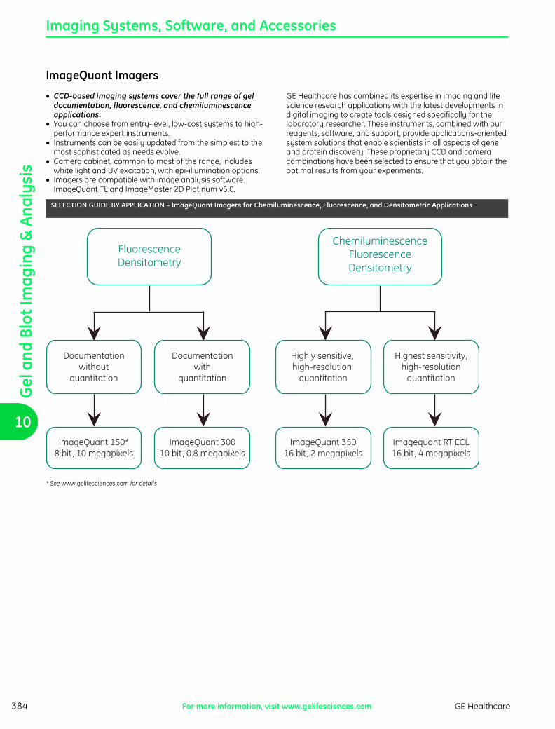
SELECTION GUIDE BY APPLICATION – ImageQuant Imagers for Chemiluminescence, Fluorescence, and Densitometric Applications
Imagequant RT ECL16 bit, 4 megapixels
ImageQuant 35016 bit, 2 megapixels
ImageQuant 30010 bit, 0.8 megapixels
ImageQuant 150*8 bit, 10 megapixels
Highest sensitivity,high-resolution
quantitation
Highly sensitive, high-resolution
quantitation
Documentationwith
quantitation
Documentationwithout
quantitation
ChemiluminescenceFluorescenceDensitometry
FluorescenceDensitometry
* See www.gelifesciences.com for details
ch10.fm Page 384 Wednesday, June 18, 2008 4:26 PM
Gel and Blot Im
aging & Analysis
10
PA
GE
: 38
5 -
CU
RR
EN
CY
: US
DP
AG
E: 3
85 -
CU
RR
EN
CY
: U
SD
PA
GE
: 3
85 -
CU
RR
EN
CY
: U
SD
PA
GE
: 38
5 -
CU
RR
EN
CY
: US
D
Imaging Systems, Software, and Accessories
GE Healthcare For more information, visit www.gelifesciences.com 385
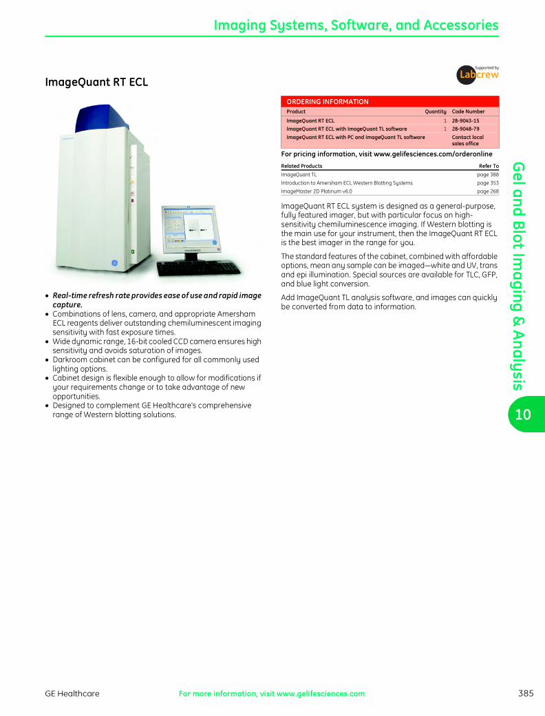
ImageQuant RT ECL
J Real-time refresh rate provides ease of use and rapid image capture.J Combinations of lens, camera, and appropriate Amersham
ECL reagents deliver outstanding chemiluminescent imaging sensitivity with fast exposure times.J Wide dynamic range, 16-bit cooled CCD camera ensures high
sensitivity and avoids saturation of images.J Darkroom cabinet can be configured for all commonly used
lighting options.J Cabinet design is flexible enough to allow for modifications if
your requirements change or to take advantage of new opportunities.J Designed to complement GE Healthcare's comprehensive
range of Western blotting solutions.
ORDERING INFORMATIONProduct Quantity Code Number
ImageQuant RT ECL 1 28-9043-15ImageQuant RT ECL with ImageQuant TL software 1 28-9048-79ImageQuant RT ECL with PC and ImageQuant TL software Contact local
sales office
For pricing information, visit www.gelifesciences.com/orderonline
Related Products Refer To
ImageQuant TL page 388
Introduction to Amersham ECL Western Blotting Systems page 353
ImageMaster 2D Platinum v6.0 page 268
ImageQuant RT ECL system is designed as a general-purpose, fully featured imager, but with particular focus on high-sensitivity chemiluminescence imaging. If Western blotting is the main use for your instrument, then the ImageQuant RT ECL is the best imager in the range for you.
The standard features of the cabinet, combined with affordable options, mean any sample can be imaged—white and UV, trans and epi illumination. Special sources are available for TLC, GFP, and blue light conversion.
Add ImageQuant TL analysis software, and images can quickly be converted from data to information.
ch10.fm Page 385 Wednesday, June 18, 2008 4:26 PM

Gel
and
Blo
t Im
agin
g &
Ana
lysi
s
10
PA
GE
: 386 - C
UR
RE
NC
Y: U
SD
PA
GE
: 386
- CU
RR
EN
CY
: US
DP
AG
E: 3
86 - C
UR
RE
NC
Y: U
SD
PA
GE
: 38
6 - CU
RR
EN
CY
: US
D
Imaging Systems, Software, and Accessories
GE HealthcareFor more information, visit www.gelifesciences.com386
ImageQuant 350
ImageQuant 350 allows high-resolution scans of monochromatic fluorescence gels, Coomassie Blue-stained gels, and other colorimetric detection methods.
J Cost-efficient, sensitive, and quantitative imaging of Western blots and other monochromatic fluorescence and colorimetric imaging applications.J 16-bit dynamic range minimizes the need for repeated
acquisitions at multiple exposures because faint and dark bands can be accurately captured and analyzed in the same image.J High resolution, 1.92 megapixel camera delivers accurate
quantitation of closely spaced bands in complex protein samples for gels and films up to 21 x 16 cm.J High sensitivity enables detection and quantitation of low-
abundant proteins on a Western blot. J Simplified workflow eliminates the time, effort, and
environmental waste of the darkroom. J Easy-to-use system allows rapid switching between
applications such as analyzing blots or imaging gels, microplates, or culture dishes.
ImageQuant 350 is a high-performance 16-bit cooled CCD camera-based system designed for a wide range of monochromatic imaging applications. ImageQuant 350 is well-suited for quantitative analysis of Western blots. The system is also fully capable of imaging monochromatic fluorescent gels and membranes, Coomassie Blue-stained gels, and other colorimetric detection methods. Cultures and microplate-based assays can also be imaged. Whereas film is valuable and convenient for certain applications, ImageQuant 350 supports a more quantitative approach for a broader range of applications without compromising sensitivity, resolution, or simplicity of operation. Most commands are performed by pushing a single button, thereby supporting many different applications in a day.
The ImageQuant 350 is a compact, easily installed system, designed for easy exchange of cameras, lenses, and filters. The standard system includes one manual fixed lens and one manual zoom lens, and the motorized system includes one motorized fixed lens and one manual zoom lens.
The standard UV transilluminator and the white light source (a fold-down table), as well as any optional light sources, are housed dust-free inside the cabinet. The UV transilluminator can be operated at either 302 nm or 365 nm at low or high intensity. For cutting sample bands from the gel, the transilluminator slides out of the cabinet.
ORDERING INFORMATIONProduct Quantity Code Number
ImageQuant 350 Standard Z 1 28-9261-75Includes: Two lenses: manual fixed f/1.8 28 mm, manual zoom f/2.8 28-70 mm, 302/365 nm
dual wavelength transilluminator, Image Capture Software, without computer and monitor
ImageQuant 350 with Motorized Fixed Lens Z 1 28-9272-95Includes: Two lenses: motorized fixed f/1.8 28 mm, manual zoom f/2.8 28-70 mm,
302/365 nm dual wavelength transilluminator, Image Capture Software, without computer and monitor
AccessoriesMotorized lensesf/2.8 zoom lens 28-70 mm Z 1 28-9265-84
(suitable for ImageQuant models 350, 400, and RT ECL)f/1.8 fixed lens 28 mm Z 1 28-9265-85
(suitable for ImageQuant models 350, 400, and RT ECL)f/1.4 fixed lens 50 mm Z 1 28-9265-86
(suitable for ImageQuant models 350, 400, and RT ECL)
Manual lensesf/1.4 fixed lens 50 mm 1 63-0057-14f/1.8 fixed lens 28 mm 1 28-9277-27f/0.95 fixed lens 50 mm Z 1 28-9266-05f/0.95 fixed lens 25 mm Z 1 28-9266-06
FiltersEmission Filter 590/55 nm Z 1 28-9272-77
for ethidium bromide, Coomassie Blue, SYPRO OrangeEmission Filter 537/35 nm Z 1 28-9272-78
for GFP, SYBR Safe, SYBR Gold, Fluorescein, SYBR GreenEmission Filter 460/40 nm Z 1 63-0056-65
for Hoechst 33258, blue dye, or carbohydrate gelsEmission Filter 620/40 nm Z 1 63-0056-64
for SYPRO Red, SYPRO Ruby, Texas Red, Rhodamine
Other AccessoriesDual Epi UV Lights, 254 nm, 110 VAC 1 63-0056-56Dual Epi UV Lights, 254 nm, 220 VAC 1 63-0057-10Dual Epi UV Lights, 365 nm, 110 VAC 1 63-0056-57Dual Epi UV Lights, 365 nm, 220 VAC 1 63-0057-11Dual Epi Blue Light Clip-ons, 460 nm 1 63-0056-58Thermal Printer 1 63-0056-69High-gloss thermal paper 5 63-0056-70
for best image qualityHigh-contrast thermal paper 4 63-0056-71
for good image quality
For pricing information, visit www.gelifesciences.com/orderonline
Related Products Refer To
ImageQuant TL page 388
Amersham ECL Western Blotting System page 357
The imager comes with an ethidum bromide (EtBr) filter as standard; other filters are available as options.
Using ImageQuant TL analysis software (see page 388) with ImageQuant 350 provides high levels of automation, accuracy and reduces analysis time to a minimum.
ch10.fm Page 386 Wednesday, June 18, 2008 4:26 PM
Gel and Blot Im
aging & Analysis
10
PA
GE
: 38
7 -
CU
RR
EN
CY
: US
DP
AG
E: 3
87 -
CU
RR
EN
CY
: U
SD
PA
GE
: 3
87 -
CU
RR
EN
CY
: U
SD
PA
GE
: 38
7 -
CU
RR
EN
CY
: US
D
Imaging Systems, Software, and Accessories
GE Healthcare For more information, visit www.gelifesciences.com 387
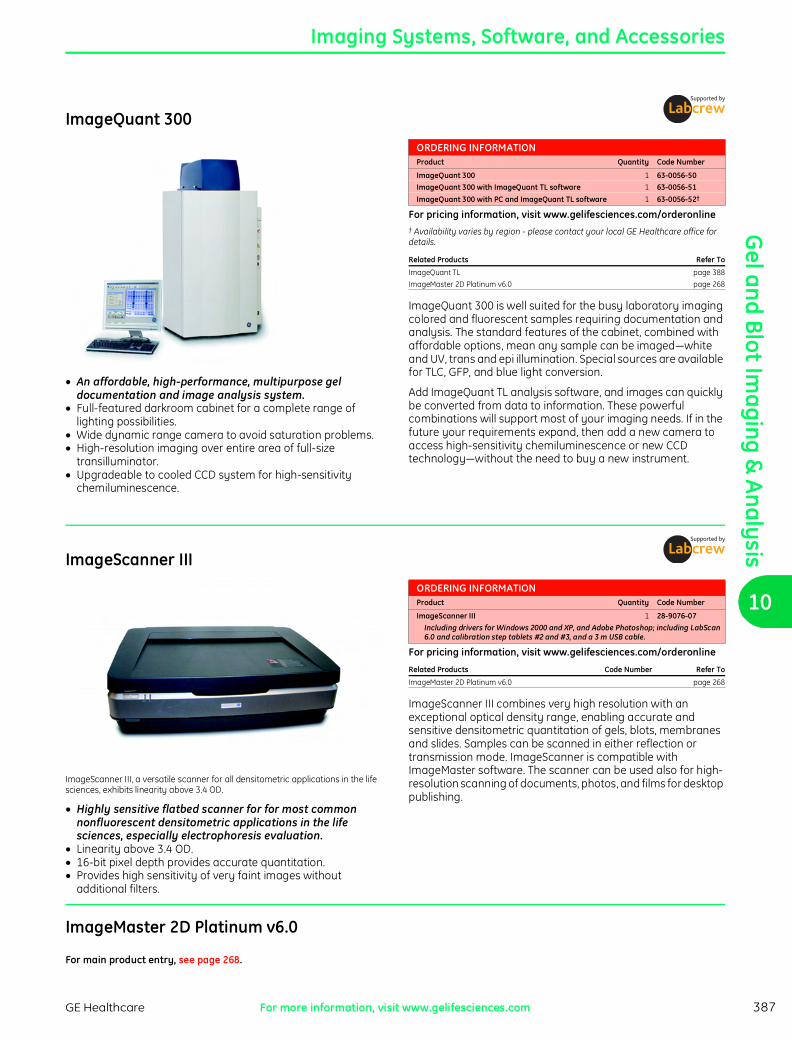
ImageQuant 300
J An affordable, high-performance, multipurpose gel documentation and image analysis system.J Full-featured darkroom cabinet for a complete range of
lighting possibilities.J Wide dynamic range camera to avoid saturation problems.J High-resolution imaging over entire area of full-size
transilluminator.J Upgradeable to cooled CCD system for high-sensitivity
chemiluminescence.
ORDERING INFORMATIONProduct Quantity Code Number
ImageQuant 300 1 63-0056-50ImageQuant 300 with ImageQuant TL software 1 63-0056-51ImageQuant 300 with PC and ImageQuant TL software 1 63-0056-52†
For pricing information, visit www.gelifesciences.com/orderonline† Availability varies by region - please contact your local GE Healthcare office for details.
Related Products Refer To
ImageQuant TL page 388
ImageMaster 2D Platinum v6.0 page 268
ImageQuant 300 is well suited for the busy laboratory imaging colored and fluorescent samples requiring documentation and analysis. The standard features of the cabinet, combined with affordable options, mean any sample can be imaged—white and UV, trans and epi illumination. Special sources are available for TLC, GFP, and blue light conversion.
Add ImageQuant TL analysis software, and images can quickly be converted from data to information. These powerful combinations will support most of your imaging needs. If in the future your requirements expand, then add a new camera to access high-sensitivity chemiluminescence or new CCD technology—without the need to buy a new instrument.
ImageScanner III
ImageScanner III, a versatile scanner for all densitometric applications in the life sciences, exhibits linearity above 3.4 OD.
J Highly sensitive flatbed scanner for for most common nonfluorescent densitometric applications in the life sciences, especially electrophoresis evaluation.J Linearity above 3.4 OD.J 16-bit pixel depth provides accurate quantitation.J Provides high sensitivity of very faint images without
additional filters.
ORDERING INFORMATIONProduct Quantity Code Number
ImageScanner III 1 28-9076-07Including drivers for Windows 2000 and XP, and Adobe Photoshop; including LabScan 6.0 and calibration step tablets #2 and #3, and a 3 m USB cable.
For pricing information, visit www.gelifesciences.com/orderonline
Related Products Code Number Refer To
ImageMaster 2D Platinum v6.0 page 268
ImageScanner III combines very high resolution with an exceptional optical density range, enabling accurate and sensitive densitometric quantitation of gels, blots, membranes and slides. Samples can be scanned in either reflection or transmission mode. ImageScanner is compatible with ImageMaster software. The scanner can be used also for high-resolution scanning of documents, photos, and films for desktop publishing.
ImageMaster 2D Platinum v6.0
For main product entry, see page 268.
ch10.fm Page 387 Wednesday, June 18, 2008 4:26 PM

Gel
and
Blo
t Im
agin
g &
Ana
lysi
s
10
PA
GE
: 388 - C
UR
RE
NC
Y: U
SD
PA
GE
: 388
- CU
RR
EN
CY
: US
DP
AG
E: 3
88 - C
UR
RE
NC
Y: U
SD
PA
GE
: 38
8 - CU
RR
EN
CY
: US
D
Imaging Systems, Software, and Accessories
GE HealthcareFor more information, visit www.gelifesciences.com388
ImageQuant TL
J Automation for fast and consistent results.J Advanced algorithms for delivery of accurate results.J Editing tools to crop, rotate, and filter images.J Support of multicolor files.J Manual override capability.
ImageQuant TL is the most automatic and easy to use general image analysis software. It offers four separate functional areas within one product:J 1-D electrophoresis gels.J Dot blots, microplates, and basic arrays.J Colony counting and measurement of 2-D spots and other
image features.J Toolbox to analyze gels with user-defined objects.
Its high level of automation throughout allows for fully automatic analysis of 1-D gels—including lane creation, background subtraction, band detection, molecular weight calibration, quantity calibration, and normalization—in a matter of a few seconds. This ensures high reproducibility.
1-D AnalysisImageQuant TL has a redesigned interface, which increases automation and ease of use. J Fully automatic lane and band detection and quantitation for
up to four channels.J Compatible with multitiered gels.J Advanced automatic background subtraction including
''rolling disk'' method.J De-smiling of gels and de-grimacing of lanes.J Normalization and quantity calibration with many regression
curves.J Molecular weight calibration across up to four channels.
Array analysisImageQuant TL can be used for dot and slot blots, microplates, and other arrays and grids. J Automatic creation of grids with up to 1536 independent cells
allows fast analysis of high throughput microplates.J Grid can be stretched or distorted.J Spot size and position adjustable for individual spots. Both
features help to analyze distorted arrays, e.g. membrane arrays or manually spotted arrays.J Normalization, presence flagging, and quality factor allow for
easy assessment of results.
Colony Counting and 2-D spot measurementWe have incorporated the 2-D spot detection algorithm of ImageMaster into ImageQuant TL to enable accurate and reproducible identification, measurement and background subtraction on images containing colonies and spots. This can be very useful for colony counting, detection, and measurement of 2-D spots and measurement of other image features. Currently, the colony counting module does not support multichannel files.
Analysis ToolboxThe Toolbox modules can be used to analyze samples that do not fit any of the above categories. ImageQuant Solutions users will find all the familiar tools they used with previous versions of ImageQuant in this module. Toolbox requires the manual definition of objects for quantitation.J Palette of tools including rectangle, ellipse, polygon, freeform
and autotracer.J Auto-tracer objects can be further edited.J Wide choice of background correction methods.J Wide choice of measurement statistics.
ORDERING INFORMATIONProduct Quantity Code Number
ImageQuant TL, DVD, and quick start guide 1 28-9194-45ImageQuant TL, single user license 1 28-9236-62ImageQuant TL, 5-user network license 1 28-9206-39ImageQuant TL, 10-user network license 1 28-9236-57ImageQuant TL User Manual 1 28-9175-41
For pricing information, visit www.gelifesciences.com/orderonlineContact your local sales office for special upgrade offerings and other packages
Related Products Code Number Refer To
Typhoon Variable Mode Imager page 376
Storm page 379
ImageScanner III 28-9076-07 page 387
Automated analysis of 1-D electrophoresis gel images.
IQTools and FluorSepThese two modules allow further image manipulations including:J Image data export to Excel for 3-D charting.J Channel flickering.J Renaming of individual channels.J Fluorescent cross-talk removal.
ImageQuant TL accepts files from all GE Healthcare imaging equipment, including Typhoon, Storm, ImageScanner III, and VDS-CL, and accepts TIFF files from third party instruments. The software fully supports multicolor files with up to 4 channels (.ds files). Operates under Microsoft Windows 2000, and XP. Contains 1 CD with software and electronic documentation, and a printed quick start manual.
For a free 14 days trial download, see www.gelifesciences.com/iqtl.
ch10.fm Page 388 Wednesday, June 18, 2008 4:26 PM

Gel and Blot Im
aging & Analysis
10
PA
GE
: 38
9 -
CU
RR
EN
CY
: US
DP
AG
E: 3
89 -
CU
RR
EN
CY
: U
SD
PA
GE
: 3
89 -
CU
RR
EN
CY
: U
SD
PA
GE
: 38
9 -
CU
RR
EN
CY
: US
D
Autoradiography Films, Screens, and Accessores
GE Healthcare For more information, visit www.gelifesciences.com 389
AutoradiographyFor more than two decades, Amersham products from GE Healthcare have been used in autoradiography applications within biomedical research. Our expertise in the labeling and detection of biomolecules has enabled us to continue to supply and support a range of innovative products, including Amersham Hyperfilm.
SELECTION GUIDE – Autoradiography Methods and Materials for Life Science ApplicationsApplication Result Required Label Method Film Support products
Southern/Northern blots; Colony/plaque blots
High speed, resolution, and sensitivity; Accurate quantitation
32P, 33P, 35S Preflash with screens at -70°C Hyperfilm MP Sensitize, Hyperscreen
Nonradioactive; quantitation ECL, ECF,CDP-Star
Preflash and direct detection Hyperfilm ECL Sensitize
Slot blots High speed and sensitivity; accurate quantitation
32P Preflash with screens at -70°C Hyperfilm MP Sensitize, Hyperscreen
Nonradioactive, quantitation ECL, ECF,CDP-Star
Preflash and direct detection Hyperfilm ECL Sensitize
Dideoxy sequencing High speed 32P Direct autoradiography Hyperfilm MP
Cycle sequencing Good resolution and speed for routine work
33P Direct autoradiography Hyperfilm MP
Protein synthesis, lysates
Maximum speed and good resolution
35S/14C/ 3H/125I
Preflash and fluorography at -70°C
Hyperfilm MP Amplify, Sensitize
Direct autoradiography Hyperfilm 3H
Western blots Maximum speedand sensitivity
125I/35S/14C Preflash and screens at -70°C Hyperfilm MP Sensitize, Hyperscreen
Nonradioactive/maximum sensitivity
ECL,ECL Advance,ECL Plus
Preflash and direct detection Hyperfilm ECL Sensitize
In situ hybridization Macroscale in situ optimization of parameters
32P Direct autoradiography Hyperfilm MP
Receptor studies Localization at cellular level– high resolution
33P/35S Micro autoradiography Hypercoat emulsion LM-1
Localization at sub-cellular level– high resolution
125I/3H Micro autoradiography Hypercoat emulsion EM-1
Localization at sub-cellular level– intermediate resolution
125I/3H Direct autoradiography Hyperfilm 3H
Whole body autoradiography
Optimum resolution 125I/3H Direct autoradiography Hyperfilm 3H
ch10.fm Page 389 Wednesday, June 18, 2008 4:26 PM
Gel
and
Blo
t Im
agin
g &
Ana
lysi
s
10
PA
GE
: 390 - C
UR
RE
NC
Y: U
SD
PA
GE
: 390
- CU
RR
EN
CY
: US
DP
AG
E: 3
90 - C
UR
RE
NC
Y: U
SD
PA
GE
: 39
0 - CU
RR
EN
CY
: US
D
Autoradiography Films, Screens, and Accessories
GE HealthcareFor more information, visit www.gelifesciences.com390
Amersham Hyperfilm ECL
J Amersham Hyperfilm ECL with improved clarity.J High-speed detection of chemiluminescent signal from
nucleic acid and protein blots.J System-tested with ECL and Gene Images kits to ensure
optimum performance.J Clear base offers high contrast for easy reading of images.J Can be processed in either automatic processors or manually
with most commonly used X-ray film developers and fixers.
The high-performance emulsion in Hyperfilm ECL is sensitive to both blue and green light chemiluminescence systems. The emulsion of this double-coated film provides improved clarity and sensitivity for the detection of chemiluminescent-labeled samples, making it suitable for ECL, ECL Plus and CDP-Star and all other chemiluminescence systems.
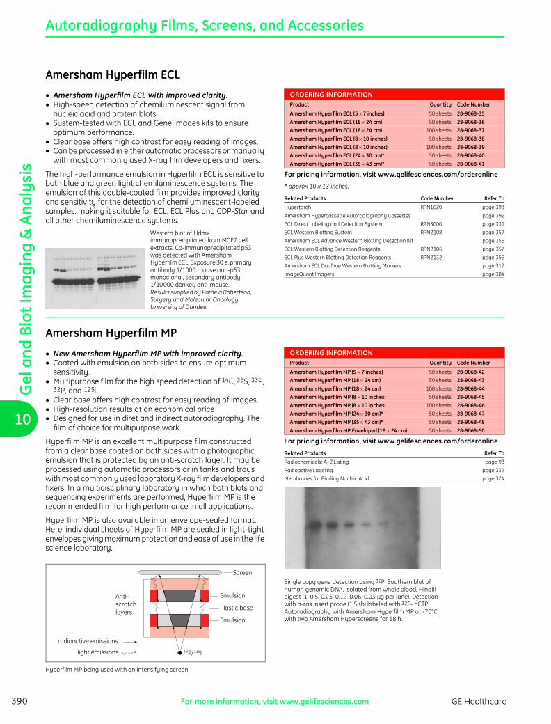
Western blot of Hdmx immunoprecipitated from MCF7 cell extracts. Co-immunoprecipitated p53 was detected with Amersham Hyperfilm ECL. Exposure 30 s, primary antibody 1/1000 mouse anti-p53 monoclonal, secondary antibody 1/10 000 donkey anti-mouse. Results supplied by Pamela Robertson, Surgery and Molecular Oncology, University of Dundee.
ORDERING INFORMATIONProduct Quantity Code Number
Amersham Hyperfilm ECL (5 u 7 inches) 50 sheets 28-9068-35Amersham Hyperfilm ECL (18 u 24 cm) 50 sheets 28-9068-36Amersham Hyperfilm ECL (18 u 24 cm) 100 sheets 28-9068-37Amersham Hyperfilm ECL (8 u 10 inches) 50 sheets 28-9068-38Amersham Hyperfilm ECL (8 u 10 inches) 100 sheets 28-9068-39Amersham Hyperfilm ECL (24 u 30 cm)* 50 sheets 28-9068-40Amersham Hyperfilm ECL (35 u 43 cm)* 50 sheets 28-9068-41
For pricing information, visit www.gelifesciences.com/orderonline
* approx 10 x 12 inches.
Related Products Code Number Refer To
Hypertorch RPN1620 page 393
Amersham Hypercassette Autoradiography Cassettes page 392
ECL Direct Labeling and Detection System RPN3000 page 331
ECL Western Blotting System RPN2108 page 357
Amersham ECL Advance Western Blotting Detection Kit page 355
ECL Western Blotting Detection Reagents RPN2106 page 357
ECL Plus Western Blotting Detection Reagents RPN2132 page 356
Amersham ECL DualVue Western Blotting Markers page 317
ImageQuant Imagers page 384
Amersham Hyperfilm MP
J New Amersham Hyperfilm MP with improved clarity.J Coated with emulsion on both sides to ensure optimum
sensitivity.J Multipurpose film for the high speed detection of 14C, 35S, 33P,
32P, and 125l.J Clear base offers high contrast for easy reading of images.J High-resolution results at an economical priceJ Designed for use in diret and indirect autoradiography. The
film of choice for multipurpose work.
Hyperfilm MP is an excellent multipurpose film constructed from a clear base coated on both sides with a photographic emulsion that is protected by an anti-scratch layer. It may be processed using automatic processors or in tanks and trays with most commonly used laboratory X-ray film developers and fixers. In a multidisciplinary laboratory in which both blots and sequencing experiments are performed, Hyperfilm MP is the recommended film for high performance in all applications.
Hyperfilm MP is also available in an envelope-sealed format. Here, individual sheets of Hyperfilm MP are sealed in light-tight envelopes giving maximum protection and ease of use in the life science laboratory.
Screen
Emulsion
Plastic base
Emulsion
Anti-scratchlayers
radioactive emissions
light emissions 32P/125I
Hyperfilm MP being used with an intensifying screen.
ORDERING INFORMATIONProduct Quantity Code Number
Amersham Hyperfilm MP (5 u 7 inches) 50 sheets 28-9068-42Amersham Hyperfilm MP (18 u 24 cm) 50 sheets 28-9068-43Amersham Hyperfilm MP (18 u 24 cm) 100 sheets 28-9068-44Amersham Hyperfilm MP (8 u 10 inches) 50 sheets 28-9068-45Amersham Hyperfilm MP (8 u 10 inches) 100 sheets 28-9068-46Amersham Hyperfilm MP (24 u 30 cm)* 50 sheets 28-9068-47Amersham Hyperfilm MP (35 u 43 cm)* 50 sheets 28-9068-48Amersham Hyperfilm MP Enveloped (18 u 24 cm) 50 sheets 28-9068-50
For pricing information, visit www.gelifesciences.com/orderonline
Related Products Refer To
Radiochemicals: A–Z Listing page 93
Radioactive Labeling page 332
Membranes for Binding Nucleic Acid page 324
Single copy gene detection using 32P. Southern blot of human genomic DNA, isolated from whole blood, HindIII digest (1, 0.5, 0.25, 0.12, 0.06, 0.03 µg per lane). Detection with n-ras insert probe (1.5Kb) labeled with 32P- dCTP. Autoradiography with Amersham Hyperfilm MP at -70°C with two Amersham Hyperscreens for 18 h.
ch10.fm Page 390 Wednesday, June 18, 2008 4:26 PM
Gel and Blot Im
aging & Analysis
10
PA
GE
: 39
1 -
CU
RR
EN
CY
: US
DP
AG
E: 3
91 -
CU
RR
EN
CY
: U
SD
PA
GE
: 3
91 -
CU
RR
EN
CY
: U
SD
PA
GE
: 39
1 -
CU
RR
EN
CY
: US
D
Autoradiography Films, Screens, and Accessories
GE Healthcare For more information, visit www.gelifesciences.com 391
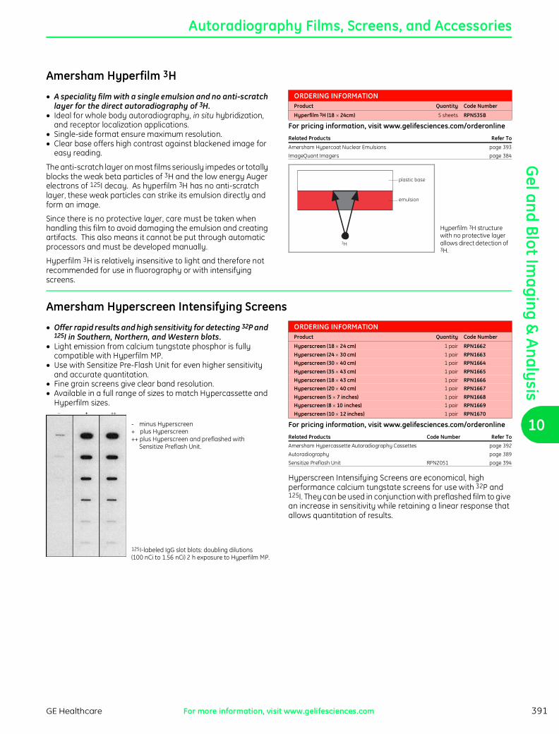
Amersham Hyperfilm 3H
J A speciality film with a single emulsion and no anti-scratch layer for the direct autoradiography of 3H.J Ideal for whole body autoradiography, in situ hybridization,
and receptor localization applications.J Single-side format ensure maximum resolution.J Clear base offers high contrast against blackened image for
easy reading.
The anti-scratch layer on most films seriously impedes or totally blocks the weak beta particles of 3H and the low energy Auger electrons of 125I decay. As hyperfilm 3H has no anti-scratch layer, these weak particles can strike its emulsion directly and form an image.
Since there is no protective layer, care must be taken when handling this film to avoid damaging the emulsion and creating artifacts. This also means it cannot be put through automatic processors and must be developed manually.
Hyperfilm 3H is relatively insensitive to light and therefore not recommended for use in fluorography or with intensifying screens.
ORDERING INFORMATIONProduct Quantity Code Number
Hyperfilm 3H (18 u 24cm) 5 sheets RPN535B
For pricing information, visit www.gelifesciences.com/orderonline
Related Products Refer To
Amersham Hypercoat Nuclear Emulsions page 393
ImageQuant Imagers page 384
emulsion
3H
plastic base
Hyperfilm 3H structure with no protective layer allows direct detection of 3H.
Amersham Hyperscreen Intensifying Screens
J Offer rapid results and high sensitivity for detecting 32P and 125I in Southern, Northern, and Western blots.J Light emission from calcium tungstate phosphor is fully
compatible with Hyperfilm MP.J Use with Sensitize Pre-Flash Unit for even higher sensitivity
and accurate quantitation.J Fine grain screens give clear band resolution.J Available in a full range of sizes to match Hypercassette and
Hyperfilm sizes.
- minus Hyperscreen+ plus Hyperscreen++ plus Hyperscreen and preflashed with
Sensitize Preflash Unit.
125I-labeled IgG slot blots: doubling dilutions (100 nCi to 1.56 nCi) 2 h exposure to Hyperfilm MP.
ORDERING INFORMATIONProduct Quantity Code Number
Hyperscreen (18 u 24 cm) 1 pair RPN1662Hyperscreen (24 u 30 cm) 1 pair RPN1663Hyperscreen (30 u 40 cm) 1 pair RPN1664Hyperscreen (35 u 43 cm) 1 pair RPN1665Hyperscreen (18 u 43 cm) 1 pair RPN1666Hyperscreen (20 u 40 cm) 1 pair RPN1667Hyperscreen (5 u 7 inches) 1 pair RPN1668Hyperscreen (8 u 10 inches) 1 pair RPN1669Hyperscreen (10 u 12 inches) 1 pair RPN1670
For pricing information, visit www.gelifesciences.com/orderonline
Related Products Code Number Refer To
Amersham Hypercassette Autoradiography Cassettes page 392
Autoradiography page 389
Sensitize Preflash Unit RPN2051 page 394
Hyperscreen Intensifying Screens are economical, high performance calcium tungstate screens for use with 32P and 125I. They can be used in conjunction with preflashed film to give an increase in sensitivity while retaining a linear response that allows quantitation of results.
ch10.fm Page 391 Wednesday, June 18, 2008 4:26 PM
Gel
and
Blo
t Im
agin
g &
Ana
lysi
s
10
PA
GE
: 392 - C
UR
RE
NC
Y: U
SD
PA
GE
: 392
- CU
RR
EN
CY
: US
DP
AG
E: 3
92 - C
UR
RE
NC
Y: U
SD
PA
GE
: 39
2 - CU
RR
EN
CY
: US
D
Autoradiography Films, Screens, and Accessories
GE HealthcareFor more information, visit www.gelifesciences.com392
Amersham Hypercassette Autoradiography Cassettes
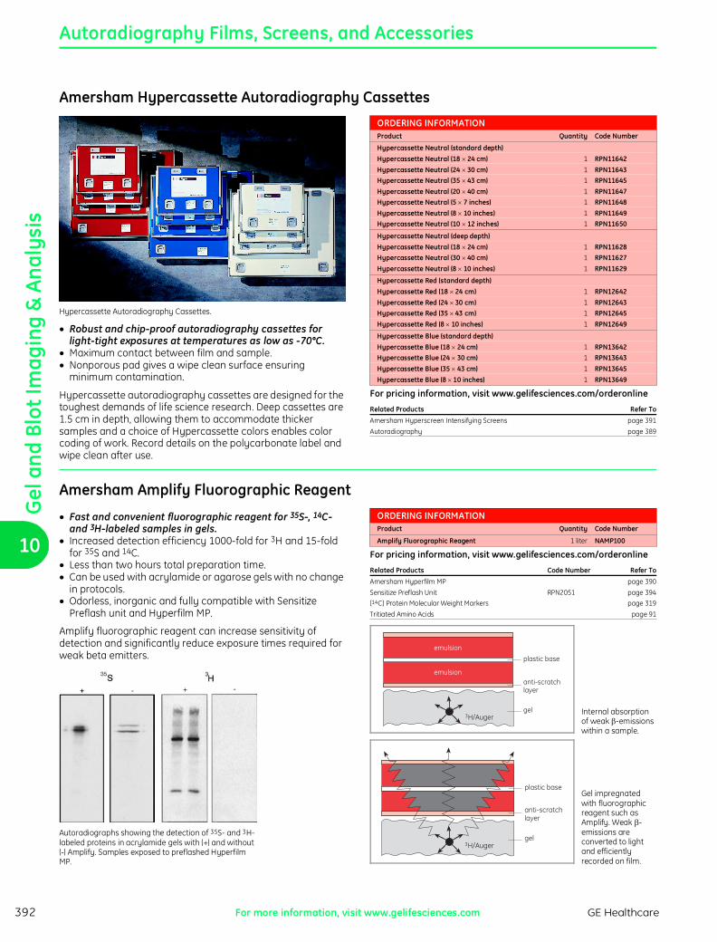
Hypercassette Autoradiography Cassettes.
J Robust and chip-proof autoradiography cassettes for light-tight exposures at temperatures as low as -70°C.J Maximum contact between film and sample.J Nonporous pad gives a wipe clean surface ensuring
minimum contamination.
Hypercassette autoradiography cassettes are designed for the toughest demands of life science research. Deep cassettes are 1.5 cm in depth, allowing them to accommodate thicker samples and a choice of Hypercassette colors enables color coding of work. Record details on the polycarbonate label and wipe clean after use.
ORDERING INFORMATIONProduct Quantity Code Number
Hypercassette Neutral (standard depth)Hypercassette Neutral (18 u 24 cm) 1 RPN11642Hypercassette Neutral (24 u 30 cm) 1 RPN11643Hypercassette Neutral (35 u 43 cm) 1 RPN11645Hypercassette Neutral (20 u 40 cm) 1 RPN11647Hypercassette Neutral (5 u 7 inches) 1 RPN11648Hypercassette Neutral (8 u 10 inches) 1 RPN11649Hypercassette Neutral (10 u 12 inches) 1 RPN11650
Hypercassette Neutral (deep depth)Hypercassette Neutral (18 u 24 cm) 1 RPN11628Hypercassette Neutral (30 u 40 cm) 1 RPN11627Hypercassette Neutral (8 u 10 inches) 1 RPN11629
Hypercassette Red (standard depth)Hypercassette Red (18 u 24 cm) 1 RPN12642Hypercassette Red (24 u 30 cm) 1 RPN12643Hypercassette Red (35 u 43 cm) 1 RPN12645Hypercassette Red (8 u 10 inches) 1 RPN12649
Hypercassette Blue (standard depth)Hypercassette Blue (18 u 24 cm) 1 RPN13642Hypercassette Blue (24 u 30 cm) 1 RPN13643Hypercassette Blue (35 u 43 cm) 1 RPN13645Hypercassette Blue (8 u 10 inches) 1 RPN13649
For pricing information, visit www.gelifesciences.com/orderonline
Related Products Refer To
Amersham Hyperscreen Intensifying Screens page 391
Autoradiography page 389
Amersham Amplify Fluorographic Reagent
J Fast and convenient fluorographic reagent for 35S-, 14C- and 3H-labeled samples in gels.J Increased detection efficiency 1000-fold for 3H and 15-fold
for 35S and 14C.J Less than two hours total preparation time.J Can be used with acrylamide or agarose gels with no change
in protocols.J Odorless, inorganic and fully compatible with Sensitize
Preflash unit and Hyperfilm MP.
Amplify fluorographic reagent can increase sensitivity of detection and significantly reduce exposure times required for weak beta emitters.
Autoradiographs showing the detection of 35S- and 3H-labeled proteins in acrylamide gels with (+) and without (-) Amplify. Samples exposed to preflashed Hyperfilm MP.
ORDERING INFORMATIONProduct Quantity Code Number
Amplify Fluorographic Reagent 1 liter NAMP100
For pricing information, visit www.gelifesciences.com/orderonline
Related Products Code Number Refer To
Amersham Hyperfilm MP page 390
Sensitize Preflash Unit RPN2051 page 394
[14C] Protein Molecular Weight Markers page 319
Tritiated Amino Acids page 91
emulsion
emulsion
3H/Auger
plastic base
anti-scratchlayer
gel Internal absorption of weak b-emissions within a sample.
3H/Auger
plastic base
anti-scratchlayer
gel
Gel impregnated with fluorographic reagent such as Amplify. Weak b-emissions are converted to light and efficiently recorded on film.
ch10.fm Page 392 Wednesday, June 18, 2008 4:26 PM

Gel and Blot Im
aging & Analysis
10
PA
GE
: 39
3 -
CU
RR
EN
CY
: US
DP
AG
E: 3
93 -
CU
RR
EN
CY
: U
SD
PA
GE
: 3
93 -
CU
RR
EN
CY
: U
SD
PA
GE
: 39
3 -
CU
RR
EN
CY
: US
D
Autoradiography Films, Screens, and Accessories
GE Healthcare For more information, visit www.gelifesciences.com 393
Amersham Hypertorch
Hypertorch—battery-powered, darkroom pen torch.
J Red light darkroom torch for localized illumination.J Minimizes film backgrounds by only illuminating local areas
of work in a darkroom.J Allows visibility in areas of darkroom where fixed safelights
cannot reach.
ORDERING INFORMATIONProduct Quantity Code Number
Hypertorch (includes 2 AAA batteries) 3 RPN1620
For pricing information, visit www.gelifesciences.com/orderonlineRelated Products Refer To
Autoradiography page 389
Amersham Hypercassette Autoradiography Cassettes page 392
Hypertorch contains a red LED which produces a safe wavelength of light for short exposures to film.
Amersham Hypercoat Nuclear Emulsions
Hypercoat nuclear emulsions and Hypercoat dipping vessel.
J Ready-to-use nuclear emulsions that can be used in the microautoradiography of radiolabels in tissues or cells.J Uniform crystal size and aggregate-free emulsions ensure
high resolution.J Use Hypercoat LM-1 (0.2 µm crystal size) for general
microautoradiography with 14C, 35S, 33P, 32P, 3H, and 125I.J Use Hypercoat EM-1 (0.13 µm crystal size) for high-resolution
light or electron microscopy with 3H-labeled samples.J High silver content allows reduced exposure times.
Hypercoat EM-1 is recommended for high-resolution results when working with 3H. Isotopes such as 14C, 35S, 33P, 32P, and 125I are best detected with Hypercoat LM-1 which has a slightly larger grain size optimized for use with higher energy emissions. Both emulsions come ready to use and do not require diluting, a process that can increase backgrounds.
ORDERING INFORMATIONProduct Quantity Code Number
Hypercoat LM-1 2 u 20 ml RPN40Hypercoat EM-1 2 u 20 ml RPN41
For pricing information, visit www.gelifesciences.com/orderonline
Related Products Code Number Refer To
Hypertorch RPN1620 this page
Amersham Hyperfilm 3H page 391
Binding of [125I]endothelin-I to artery wall of patient with ischaemic heart disease, visualized with Hypercoat LM-I.
Amersham Microscale Autoradiography StandardsFor main product entry, see page 118.
Amersham Liquid Scintillation CocktailsFor main product entry, see page 117.
ch10.fm Page 393 Wednesday, June 18, 2008 4:26 PM
Gel
and
Blo
t Im
agin
g &
Ana
lysi
s
10
PA
GE
: 394 - C
UR
RE
NC
Y: U
SD
PA
GE
: 394
- CU
RR
EN
CY
: US
DP
AG
E: 3
94 - C
UR
RE
NC
Y: U
SD
PA
GE
: 39
4 - CU
RR
EN
CY
: US
D
Autoradiography Films, Screens, and Accessories
GE HealthcareFor more information, visit www.gelifesciences.com394
Amersham Sensitize Preflash Unit
J Specially designed flashgun for use with Hyperfilm ECL or Hyperfilm MP in conjunction with Hyperscreen Intensifying Screens to improve sensitivity and quantitation.J Most effective when detecting small amounts of radioactivity
or chemiluminescent signal.J High quality orange filter included to ensure optimum
wavelength for preflashing.J Sealed unit to prevent white-light leakage.J Calibration protocol included to ensure reproducibility.
ORDERING INFORMATIONProduct Quantity Code Number
Sensitize Preflash Unit 1 RPN2051 (Batteries not included)
For pricing information, visit www.gelifesciences.com/orderonline
Related Products Code Number Refer To
ECL Western Blotting System RPN2108 page 357
ECL Western Blotting Detection Reagents RPN2106 page 357
Amersham Hyperfilm ECL page 390
Amersham Hyperfilm MP page 390
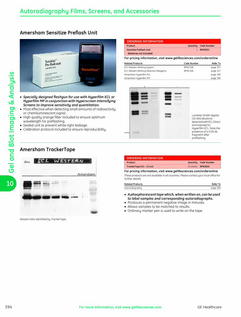
Lambda HindIII digests (10-fold dilutions) detected with ECL Direct and exposed to Hyperfilm ECL. Note the presence of a 0.56 kb fragment after preflashing.
Amersham TrackerTape
Western blot identified by TrackerTape.
ORDERING INFORMATIONProduct Quantity Code Number
TrackerTape (10 u 15 cm) 10 sheets RPN2050
For pricing information, visit www.gelifesciences.com/orderonlineThese products are not available in all countries. Please contact your local office for further details.
Related Products Refer To
Autoradiography page 389
J A phosphorescent tape which, when written on, can be used to label samples and corresponding autoradiographs.J Produces a permanent negative image in minutes.J Allows samples to be matched to results.J Ordinary marker pen is used to write on the tape.
ch10.fm Page 394 Wednesday, June 18, 2008 4:26 PM

Gel and Blot Im
aging & Analysis
10
PA
GE
: 39
5 -
CU
RR
EN
CY
: US
DP
AG
E: 3
95 -
CU
RR
EN
CY
: U
SD
PA
GE
: 3
95 -
CU
RR
EN
CY
: U
SD
PA
GE
: 39
5 -
CU
RR
EN
CY
: US
D
Autoradiography Films, Screens, and Accessories
GE Healthcare For more information, visit www.gelifesciences.com 395
Amersham Hyperprocessor Automatic Film Processor
J An advanced automatic film processor of compact bench top design, requiring less than 0.2 m2 of floor space.J Develops, fixes, washes, and dries film automatically in 2.5
min.J Internal control systems ensure consistent high performance.J Handles film sizes from 10 u 10 cm to 35 u 43 cm.J Can process 70 films, size 24 u 30 cm, per h.
ORDERING INFORMATIONProduct Quantity Code Number
Hyperprocessor 220/240 V 1 RPN1700Hyperprocessor 110/120 V 1 RPN1700AIncludes: Trolley, Two Chemical Reservoirs, Small Spares Kit, Full Operator Manual and Full
Installation Manual.
For pricing information, visit www.gelifesciences.com/orderonlineHyperprocessor is not available in all countries. Please contact your local office for further details.
Related Products Refer To
Autoradiography page 389
Amersham Hypercassette Autoradiography Cassettes page 392
Hyperprocessor is designed to give consistent high-quality autoradiographs simply and quickly with Hyperfilm MP, Hyperfilm ECL, Kodak BioMax, X-Omat AR, X-Omat LS, and most standard X-ray films. Sensors measure the film throughput and replenish chemicals as needed. Chemicals are maintained at optimum temperatures for quality results.
TECHNICAL SPECIFICATIONSSafety certifications CE 89/336/EEC (EMC directive);
CE 73/23/EEC (LV directive); EN-61010-1 (IEC1010-1)
ch10.fm Page 395 Wednesday, June 18, 2008 4:26 PM