Embed Size (px)
Citation preview
Gauging a Hydrocarbon Ruler by an Intrinsic Exciton Probe†
M. Adil Khan,‡,§ Chris Neale,| Catherine Michaux,⊥ Regis Pome`s,|,@ Gilbert G. Prive,|,⊥ Robert W. Woody,# andRussell E. Bishop*,‡,§,|
Departments of Laboratory Medicine and Pathobiology and of Biochemistry, UniVersity of Toronto, Toronto,ON, Canada M5S 1A8, Department of Medical Biophysics, UniVersity of Toronto, and DiVision of Molecular and
Structural Biology, Ontario Cancer Institute, Toronto, ON, Canada M5G 2M9, Department of Structural Biology andBiochemistry, Hospital for Sick Children, Toronto, ON, Canada M5G 1X8, Department of Biochemistry and Molecular Biology,
Colorado State UniVersity, Fort Collins, Colorado 80523, and Department of Biochemistry and Biomedical Sciences,McMaster UniVersity, Hamilton, ON, Canada L8N 3Z5
ReceiVed December 7, 2006; ReVised Manuscript ReceiVed February 21, 2007
ABSTRACT: The structural basis of lipid acyl-chain selection by membrane-intrinsic enzymes is poorlyunderstood because most integral membrane enzymes of lipid metabolism have proven refractory to structuredetermination; however, robust enzymes from the outer membranes of Gram-negative bacteria are nowproviding a first glimpse at the underlying mechanisms. The methylene unit resolution of the phospholipid:lipid A palmitoyltransferase PagP is determined by the hydrocarbon ruler, a 16-carbon saturated acyl-chain-binding pocket buried within the transmembraneâ-barrel structure. Substitution of Gly88 liningthe floor of the hydrocarbon ruler with Ala or Met makes the enzyme select specifically 15- or 12-carbonsaturated acyl chains, respectively, indicating that hydrocarbon ruler depth determines acyl-chain selection.However, the Gly88Cys PagP resolution does not diminish linearly because it selects both 14- and 15-carbon saturated acyl chains. We discovered that an exciton, emanating from a buried Tyr26-Trp66phenol-indole interaction, is extinguished by a local structural perturbation arising from the proximalGly88Cys PagP sulfhydryl group. Site-specific S-methylation of the single Cys afforded Gly88Cys-S-methyl PagP, which reasserted both the exciton and methylene unit resolution by specifically selecting13-carbon saturated acyl chains for transfer to lipid A. Unlike the other Gly88 substitutions, the Cyssulfhydryl group recedes from the hydrocarbon ruler floor and locally perturbs the subjacent Tyr26 andTrp66 aromatic rings. The resulting hydrocarbon ruler expansion thus occurs at the exciton’s expense andaccommodates an extra methylene unit in the selected acyl chain. The hydrocarbon ruler-excitonjuxtaposition endows PagP with a molecular gauge for probing the structural basis of lipid acyl-chainselection in a membrane-intrinsic environment.
The diversity of acyl chains found in membrane lipidsreflects the ability of cells to modulate membrane biophysicalstates and to employ specific lipids in signal transductionpathways (1, 2). Enzymes of lipid metabolism encounter aneffective combinatorial library in their substrates and arenecessarily endowed with mechanisms for selecting specificacyl-chain types. Most lipid-metabolizing enzymes of knownstructure represent soluble globular domains that exist eitherfree in solution or as monotopic membrane proteins (3, 4).Some of these structures indicate how enzymes interact with
soluble lipid substrates and have revealed acyl-chain-measuring devices known as hydrocarbon rulers (5, 6). Themonotopic membrane enzyme structures further indicate howglobular domains interact with their substrates at theperiphery of lipid bilayers. However, many key lipidmetabolic steps are catalyzed by integral membrane enzymes,which are embedded in their substrates and encounter a low-dielectric milieu not normally accessible to soluble globularprotein domains. The biophysical basis of integral membranelipid-protein recognition is advancing with increasingnumbers of determined structures, but integral membraneenzymes of lipid metabolism have mostly proven refractoryto structure determination (7). Exceptions are found in theouter membranes of Gram-negative bacteria, which haverecently revealed integral membrane protein structures for aphospholipase, a lipid deacylase, and a lipid acyltransferase(8-11). These integral membraneâ-barrel enzymes are morerobust than mostR-helical integral membrane enzymes oflipid metabolism, and are now providing insights into themolecular mechanisms of lipid acyl-chain selection withina lipid bilayer environment.
Many integral membraneâ-barrel proteins can be purifiedin an unfolded state and refolded at high concentrations in
† This work was supported by CIHR Operating Grants MOP-43886awarded to R.E.B. and MOP-57922 awarded to G.G.P. R.P. is a CRCPchairholder.
* To whom correspondence should be addressed: Department ofBiochemistry and Biomedical Sciences, McMaster University,Hamilton, ON, Canada L8N 3Z5. Telephone: (905) 525-9140, ext.28810. Fax: (905) 522-9033. E-mail: [email protected].
‡ Department of Laboratory Medicine and Pathobiology, Universityof Toronto.
§ McMaster University.| Department of Biochemistry, University of Toronto.⊥ Department of Medical Biophysics, University of Toronto, and
Ontario Cancer Institute.@ Hospital for Sick Children.# Colorado State University.
4565Biochemistry2007,46, 4565-4579
10.1021/bi602526k CCC: $37.00 © 2007 American Chemical SocietyPublished on Web 03/22/2007
detergent micelles, which are transparent to certain spectro-scopic analyses of protein structure and stability. Extrinsicspectroscopic probes can be introduced into proteins to studystructure-function relationships, but these studies must becarefully scrutinized to avoid artifacts arising from structuralperturbations introduced by the probe itself. If Trp and Tyrresidues are localized in a functionally interesting proteinregion, their aromatic side chains can be exploited as intrinsicprobes to prevent such artifacts. However, it can be difficultor impossible to deconvolute the signals that arise from asingle aromatic side chain if multiple copies are present ina given protein. More rarely, two or more Trp and/or Tyrresidues that interact within specific geometrical and distanceconstraints can afford a so-called exciton interaction. Theexciton effect arises from the delocalization of the excitedstates of two interacting chromophores (12). The strongπf π* transitions arising from Trp and Tyr side chains donot on their own generate Cotton effects that can be detectedby circular dichroism (CD)1 spectroscopy. Cotton effectsarise when parallel components of electric and magneticdipole transition moments are combined, and strict grouptheoretical rules show that this cannot happen when achromophore possesses centers or planes of symmetry (13).When they are placed in a chiral environment, such as thatprovided by the polypeptide backbone of a protein molecule,an exciton effect arising from a pair of interacting chro-mophores can generate two Cotton effects of equal magnitudeand opposite sign that are slightly separated by the so-calledDavydov energy (14). The resultant of these two overlappingCotton effects is a bisignate curve known as an excitoncouplet, which can be detected in the far-UV region of aprotein CD spectrum (15).
Exciton theory lies at the heart of protein CD spectroscopybecause it accounts for key spectroscopic signatures that arisebetweenπ f π* transitions in interacting peptide groupslocated within the secondary structural elements. However,exciton couplets arising from interacting aromatic side chainshave largely been ignored in protein secondary structureanalysis, despite their clear overlap in the far-UV range withthe signals that arise from secondary structure (16). Due tothe strong rotational strengths associated with theR-helix,the weaker aromatic exciton couplets in the far-UV rangeare most apparent in proteins with lowR-helical content,but their presence can significantly influence the far-UV CDspectrum of any protein (17, 18). Theoretical analyses showthat exciton couplets arising from aromatic side-chaininteractions are among the more reliably predicted CDspectroscopic signatures that can occur within a proteinmolecule (19). Algorithms that consider aromatic excitoninteractions for predicting a protein CD spectrum from agiven set of crystal structure coordinates are available. Onecan usually pinpoint interacting aromatic side chains associ-ated with an exciton couplet by systematically replacing eacharomatic amino acid with Ala in silico and subtracting thecalculated CD spectrum from that for the wild-type protein(19). Given the strict geometrical requirements of the excitoneffect, aromatic exciton couplets are anticipated to be
extraordinarily sensitive to local structural perturbations thatmight well be introduced during the experimental modifica-tion of any given protein structure. A considerable degreeof untapped potential lies in the identification and applicationof aromatic exciton interactions to study protein function inthe current era of structural proteomics. In this paper, wereport evidence of the modulation of an aromatic excitoncouplet in a lipid-metabolizing integral membrane enzyme.
We have been investigating the structure and function ofPagP, a 161-amino acid membrane protein that resides withinthe outer membranes of pathogenic Gram-negative bacteriasuch asEscherichia coli(20). PagP is an enzyme of lipidmetabolism that transfers a palmitate chain from a phospho-lipid molecule to the lipid A (endotoxin) component oflipopolysaccharide (LPS) (21). The Gram-negative outermembrane is an asymmetric bilayer that normally displaysLPS in the outer leaflet and restricts phospholipids to theinner leaflet (22). This asymmetric lipid organization createsa permeability barrier to hydrophobic antibiotics and deter-gents that are normally encountered in the host and naturalenvironments (23). PagP provides bacteria with a degree ofresistance to host-derived antimicrobial agents and attenuatesthe ability of endotoxin to activate the TLR-4 host defensepathway (24, 25). Additionally, PagP can function as anapical sensory transducer that reports perturbations of lipidasymmetry by a novel signal transduction mechanism inbacteria (S. Kim, W. Jia, E. Vinogradov, C. Gyles, and R.Bishop, unpublished observations).
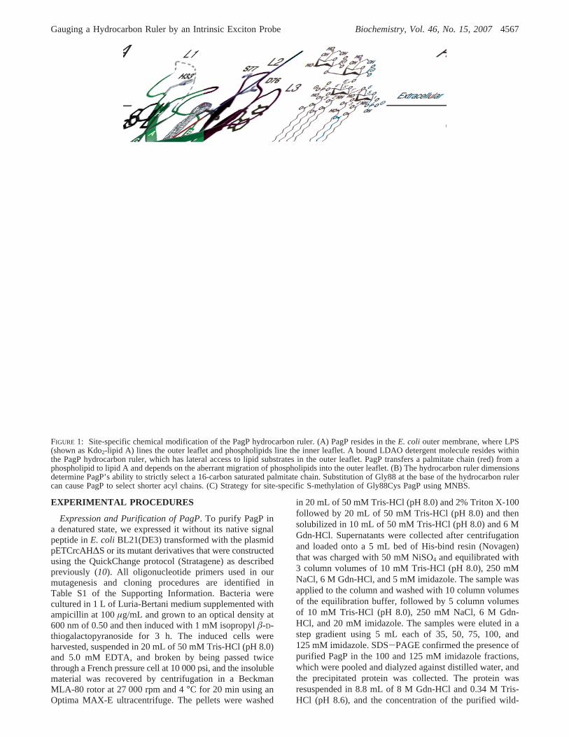
PagP is an eight-stranded antiparallelâ-barrel with a shortR-helix at its N-terminus, and it sits in the membrane withthe barrel axis tilted by roughly 25° (Figure 1A) (11). ThePagP palmitate recognition pocket, known as the hydrocarbonruler, resides within the interior of theâ-barrel and islocalized in the outer LPS-exposed region of the protein (11).A single molecule of the detergent lauroyldimethylamineN-oxide (LDAO) serves to identify the position of thehydrocarbon ruler within theâ-barrel interior (Figure 1A).Two discontinuities inâ-strand hydrogen bonding within thisLPS-exposed region provide obvious routes for lateral accessof lipids to the hydrocarbon ruler (10, 26). The implicationthat PagP depends on the aberrant migration of phospholipidsinto the outer leaflet is supported by observations that theenzyme normally remains dormant in outer membranes untilits activity is directly triggered by perturbations to lipidasymmetry (27).
A striking enzymological feature of PagP lies in its abilityto distinguish a 16-carbon saturated palmitate chain inphospholipids from all other acyl chains, even those thatdiffer by only a single methylene unit (11, 21). The depthof the PagP hydrocarbon ruler, which is lined at its base byGly88, determines this single-methylene unit resolution(Figure 1B). PagP lacks any Cys residues, but it is particu-larly rich in aromatic amino acids and includes 10 Phe, 11Tyr, and 12 Trp residues, not including the N-terminal signalpeptide. During our efforts to modulate acyl-chain selectionby site-specific chemical methylation of a Gly88Cys PagPmutant, we have discovered an exciton couplet that couldbe pinpointed to an interaction between Tyr26 and Trp66 atthe floor of the hydrocarbon ruler. We demonstrate that thisintrinsic exciton probe can provide a gauge for reportingmethylene unit resolution in the PagP hydrocarbon ruler.
1 Abbreviations: CD, circular dichroism; DSC, differential scanningcalorimetry; ESI-MS, electrospray ionization mass spectrometry;LDAO, lauroyldimethylamineN-oxide; LPS, lipopolysaccharide; MNBS,methyl p-nitrobenzenesulfonate.
4566 Biochemistry, Vol. 46, No. 15, 2007 Khan et al.
EXPERIMENTAL PROCEDURES
Expression and Purification of PagP. To purify PagP ina denatured state, we expressed it without its native signalpeptide inE. coli BL21(DE3) transformed with the plasmidpETCrcAH∆S or its mutant derivatives that were constructedusing the QuickChange protocol (Stratagene) as describedpreviously (10). All oligonucleotide primers used in ourmutagenesis and cloning procedures are identified inTable S1 of the Supporting Information. Bacteria werecultured in 1 L of Luria-Bertani medium supplemented withampicillin at 100µg/mL and grown to an optical density at600 nm of 0.50 and then induced with 1 mM isopropylâ-D-thiogalactopyranoside for 3 h. The induced cells wereharvested, suspended in 20 mL of 50 mM Tris-HCl (pH 8.0)and 5.0 mM EDTA, and broken by being passed twicethrough a French pressure cell at 10 000 psi, and the insolublematerial was recovered by centrifugation in a BeckmanMLA-80 rotor at 27 000 rpm and 4°C for 20 min using anOptima MAX-E ultracentrifuge. The pellets were washed
in 20 mL of 50 mM Tris-HCl (pH 8.0) and 2% Triton X-100followed by 20 mL of 50 mM Tris-HCl (pH 8.0) and thensolubilized in 10 mL of 50 mM Tris-HCl (pH 8.0) and 6 MGdn-HCl. Supernatants were collected after centrifugationand loaded onto a 5 mL bed ofHis-bind resin (Novagen)that was charged with 50 mM NiSO4 and equilibrated with3 column volumes of 10 mM Tris-HCl (pH 8.0), 250 mMNaCl, 6 M Gdn-HCl, and 5 mM imidazole. The sample wasapplied to the column and washed with 10 column volumesof the equilibration buffer, followed by 5 column volumesof 10 mM Tris-HCl (pH 8.0), 250 mM NaCl, 6 M Gdn-HCl, and 20 mM imidazole. The samples were eluted in astep gradient using 5 mL each of 35, 50, 75, 100, and125 mM imidazole. SDS-PAGE confirmed the presence ofpurified PagP in the 100 and 125 mM imidazole fractions,which were pooled and dialyzed against distilled water, andthe precipitated protein was collected. The protein wasresuspended in 8.8 mL of 8 M Gdn-HCl and 0.34 M Tris-HCl (pH 8.6), and the concentration of the purified wild-
FIGURE 1: Site-specific chemical modification of the PagP hydrocarbon ruler. (A) PagP resides in theE. coli outer membrane, where LPS(shown as Kdo2-lipid A) lines the outer leaflet and phospholipids line the inner leaflet. A bound LDAO detergent molecule resides withinthe PagP hydrocarbon ruler, which has lateral access to lipid substrates in the outer leaflet. PagP transfers a palmitate chain (red) from aphospholipid to lipid A and depends on the aberrant migration of phospholipids into the outer leaflet. (B) The hydrocarbon ruler dimensionsdetermine PagP’s ability to strictly select a 16-carbon saturated palmitate chain. Substitution of Gly88 at the base of the hydrocarbon rulercan cause PagP to select shorter acyl chains. (C) Strategy for site-specific S-methylation of Gly88Cys PagP using MNBS.
Gauging a Hydrocarbon Ruler by an Intrinsic Exciton Probe Biochemistry, Vol. 46, No. 15, 20074567
type protein was determined, using a calculated extinctioncoefficient (ε280) of 82 630 M-1 cm-1, to be 6.67 mg/mLfor a total yield of 58.7 mg (28). The remaining mutantproteins were obtained in similar yields.
PagP Trp66 mutant proteins that failed to refold in vitrocould be folded in vivo when they were expressed with theintact signal peptide in plasmid pETCrcAH and then purifieddirectly from LDAO-solubilized membranes in 1 mg quanti-ties from 1 L cultures as described previously (21).
Methylation of PagP. Wild-type and Gly88Cys mutantPagP in Gdn-HCl were subjected to an S-methylationprocedure using methylp-nitrobenzenesulfonate (MNBS)(29). Control reaction mixtures excluding only MNBS werealso prepared, and the four reactions were carried out incapped glass tubes. Protein in 8 M Gdn-HCl and 0.34 MTris-HCl (pH 8.6) was adjusted to 5 mg/mL in a final volumeof 5 mL containing 6 M Gdn-HCl, 0.25 M Tris-HCl(pH 8.6), 3.3 mM EDTA, and 25% (v/v) acetonitrile. Thesolutions were flushed with N2 for 1 min to create an anoxicbarrier, and 50µL of 260 mMâ-mercaptoethanol was added(10-50-fold molar excess of the protein). The tubes weretightly sealed, placed in a 50°C bath for 1 h, and thengradually cooled to 37°C. Under the N2 barrier, 0.5 mL of5.2 mM MNBS (2-fold molar excess of theâ-mercaptoet-hanol) was added, and the tubes were tightly sealed andplaced in a 37°C bath for 2 h. The reactions were quenchedvia addition of 5µL of 14 M â-mercaptoethanol. The reactionmix was dialyzed exhaustively against distilled water, andthe precipitated proteins were collected and dissolved in10 mL of 10 mM Tris-HCl (pH 8.0) and 6 M Gdn-HCl. Theprotein yield was nearly quantitative (25 mg) for all foursamples. An aliquot of each sample (0.5 mL) was dialyzedagainst water and the precipitated protein used for massspectrometry.
Electrospray Ionizatation Mass Spectrometry (ESI-MS).MS was performed at the Advanced Proteomics Facility atthe Hospital for Sick Children. Our procedure for dissolvingprecipitated PagP was adapted from a prior study of lactosepermease (30). Precipitated samples to be analyzed weredissolved in 5 mL of a 1:1 (v/v) acetonitrile/1% formic acidmixture at a concentration of∼1 ng/µL just prior to ESI-MS and were injected directly into a triple-quadrupole massspectrometer. The positive ion mode was used and the conepotential maintained at 48 eV and the collision energy at4 eV. The spectra were reconstructed using Mass Lynx 3.5.In one instance, a Sciex API III+ triple-quadrupole massspectrometer was used, but all other analyses were performedwith a Micromass Quattro Ultima LC-ESI/APCI triple-quadrupole mass spectrometer.
Refolding of PagP.Samples in 10 mM Tris-HCl (pH 8.0),6 M Gdn-HCl, and 20 mMâ-mercaptoethanol were diluteddropwise (∼1 drop per 2 s) into a 10-fold excess of 10 mMTris-HCl (pH 8.0) and 20 mMâ-mercaptoethanol containing0.5% LDAO at room temperature with vigorous stirring andleft to stir overnight at 4°C. The â-mercaptoethanol wasexcluded in subsequent refolding experiments using PagPmutants that lack any Cys residues. The refolding samplewas then applied to a 4 mL bed ofHis-bind resin (Novagen)charged with 50 mM NiSO4 and equilibrated with 10 mMTris-HCl (pH 8.0), 0.1% LDAO, and 5 mM imidazole.The column was washed with 10 column volumes of theequilibration buffer and 10 column volumes of 10 mM Tris-
HCl (pH 8.0), 0.1% LDAO, and 20 mM imidazole and theneluted with 2 mL of 10 mM Tris-HCl (pH 8.0), 0.1% LDAO,and 250 mM imidazole. The sample was dialyzed against10 mM Tris-HCl (pH 8.0) and 0.1% LDAO, and the refoldedsamples (6µg) were resolved via SDS-PAGE on 1 mmNovex 16% Tris glycine precast gels under reducing ornonreducing conditions (Invitrogen). The heated sampleswere boiled at 100°C for 10 min before loading, and proteinwas stained using Coomassie blue dye. Disulfide bonds wereencouraged to form under nonreducing conditions by uncap-ping the heated and unheated samples on the bench forseveral hours prior to loading on the gel. Refolded PagPprotein concentrations were determined using either thebicinchoninic acid assay (31) or by absorbance using anextinction coefficient (ε280) determined experimentally by theEdelhoch method (32, 33).
CD Spectroscopy.Samples to be analyzed for CD weremaintained at a concentration of 0.3-0.1 mg/mL in 10 mMTris-HCl (pH 8.0) and 0.1% LDAO and were analyzed usinga cuvette with a path length of 1 mm. The samples wereanalyzed with either a Jasco J-812 or an Aviv 215 CDspectrometer, each of which was linked to Peltier devicesfor temperature control. For each sample, three accumulationswere averaged at a data pitch of 1 nm and a scanning speedof 10 nm/min. The temperature was maintained at 25°C,and data sets were obtained from 200 to 280 nm. For thermaldenaturation profiles, samples at a concentration of 0.8-0.3mg/mL in 10 mM Tris-HCl (pH 8.0) and 0.1% LDAO wereanalyzed in a cuvette with a path length of 1 mm. Thesamples were heated from 20 to 100°C at a rate of 2°C/min, with a response time of 16 s.
Differential Scanning Calorimetry (DSC).The specificheat capacity (Cp) as a function of temperature was obtainedin an N-DSC (cell volume of 0.3 mL) at a scan rate of1.0 °C/min and a pressure of∼37 psi. The protein samplewas first dialyzed overnight at 4°C against 100 mM sodiumphosphate buffer (pH 8.0) with 0.05% LDAO. The solutionwas then degassed for 5 min before it was loaded into theDSC cells. A blank scan with buffer in both calorimeter cellswas subtracted to correct for the difference between the cells.The DSC instrument that was employed was a CSC model6100 Nano II. The software used to collect the data wasDSCRUN, N-DSC control program (version 2.5.0.29s),Calorimetry Science Corp. The software used to visualizethe data was cpcalc (version 2.1, Applied Thermodynamics),while the software used to process the data was MicrocalTM (version 5.0, Microcal Software Inc.).
Theoretical Calculations of CD.The CD spectrum forwild-type PagP was calculated using the X-ray structure (11)(PDB entry 1THQ). The methods and parameters have beendescribed previously (18, 34). Calculations were also per-formed for mutants generated in silico by deletion, one byone, of each of the Tyr and Trp side chains in the vicinityof Gly88 in the crystal structure: Tyr26, Trp60, Trp66,Tyr70, Tyr87, and Trp156. (Phe side chains were not mutatedbecause they have only the weak 260 nm band above 220nm.) In each case, a difference spectrum was calculated bysubtracting the predicted spectrum of the mutant from thatof the wild type, thus providing the contribution of themutated side chain to the wild-type CD spectrum.
Hydrocarbon Ruler Assays.Kdo2-lipid A was preparedfrom heptose-deficientE. coli WBB06 as described previ-
4568 Biochemistry, Vol. 46, No. 15, 2007 Khan et al.
ously (35) and quantified as described (36). Syntheticdiacylphosphatidylcholines were obtained from Avanti PolarLipids (Alabaster, AL). The hydrocarbon ruler assays wereperformed at 30°C by a TLC-based radiolabeling procedureas described previously (11), except that 25µCi of [32P]-orthophosphate was employed in place of sodium [14C]-acetate.
Molecular Dynamics.We have used high-temperaturesimulated annealing to probe the local conformationalchanges induced by mutation of Gly88. The same procedurehas been applied to two starting conformations. The initialconformation was the X-ray structure (11) (PDB entry1THQ) in the absence of LDAO and crystal waters. Anotherstarting conformation was obtained by equilibration of theX-ray structure containing a rebuilt L1 loop and boundLDAO, and embedded in an explicit 1,2-dimyristoyl-sn-glycero-3-phosphocholine (37) bilayer and TIP3P water (38).All nonprotein atoms were then removed. Residues His22and His102 were protonated on Nδ1, while residues His33and His67 were protonated on Nε2. All simulations werecarried out using the CHARMM software package(39) usingthe CHARMM22 all-hydrogen topology and parameter files(40). Parameters for the methylated cysteine residue weredeveloped on the basis of methionine via removal of Câ.All nonbonded interactions (Lennard-Jones and Coulombic)were switched off from 10 to 11 Å. The neglect of long-range electrostatic interactions is justified because confor-mational sampling is restricted to local side-chain rearrange-ments of neutral residues within a static protein framework.
Initial conformations of the mutants were obtained asfollows. Hydrogen atoms to be replaced by heavy atoms werechosen such that the nascent side chain was in the LDAObinding pocket and pointed toward the extracellular region.Mutants were created according to the following succes-sion: Gly88Ala (HR1 f Câ, HR2 f HR), Ala88Cys (Hâ3 fSγ), Cys88-S-methyl (Hγ f Cδ), and Cys-S-methyl88Met (Sγ
f Cγ, Cδ f Sδ, Hδ1 f Cε). A harmonic restraining potentialwas applied for C′, N′, CR, and O′ atoms of residue 88 witha force constant of 10 kcal mol-1 Å-2, and all other residueswere held fixed. Each mutant was energy minimized.
In the remaining calculations, a harmonic restrainingpotential was applied to the backbone heavy atoms ofresidues Trp66, Gly68, Tyr70, X88, Thr108, and Leu128with a force constant of 10 kcal mol-1 Å-2 (greater by 1order of magnitude for calculations starting from the X-raystructure). All other residues were held fixed. Each of theconformations resulting from the procedure outlined abovewas simulated at 3000 K for 75 ps. The temperature wascontrolled by Langevin dynamics with a friction coefficientof 2.0 ps-1. High-temperature seed conformations were takenafter 15, 25, 35, 45, 55, 65, and 75 ps. Each seed was thencooled to 206 K over a minimum of 1.3 ns by reducing thetemperature to 0.8 of its previous value after each segment.Dynamics at and below 3000 K were produced with timesteps of 0.5 and 1.0 fs, respectively. The final cooling stepwas followed by energy minimization.
Figures were generated with VMD version 1.8.3 (41) andrendered with POV-Ray version 3.6 (Persistence of VisionPty. Ltd.). Trp66 and X88 are overlaid for all seven replicas.Contouring of theâ-barrel interior was performed by HOLEwith a cutoff radius of 1.4 Å (42). For the wild type andeach mutant, contours were generated on the basis of the
initial wild-type structure plus the coordinates of residue 88overlaid from all seven replicas. All other figures derivedfrom the PagP crystal structure coordinates were renderedusing PyMOL (43).
RESULTS
In our prior investigation, we analyzed membrane-derivedwild-type PagP and its site-specific mutants Gly88Ala,Gly88Cys, and Gly88Met (11). To improve the quality ofthese initial studies, we removed the signal peptide from eachof the mutant proteins for expression and purification in adenatured state to be followed by refolding. Since PagP lacksany Cys residues, the single Cys in Gly88Cys PagP wasavailable for a chemical methylation procedure that occursin Gdn-HCl needed to unfold PagP, which is necessary toexpose the Cys residue and to normalize the reactivity ofother functional groups (29) (Figure 1B,C). By synthesizingGly88Cys-S-methyl PagP, we predicted that we could createa suite of hydrocarbon ruler mutants that select 15-carbon(Gly88Ala), 14-carbon (Gly88Cys), 13-carbon (Gly88Cys-S-methyl), and 12-carbon (Gly88Met) saturated acyl chains.The hydrocarbon ruler hypothesis predicts that the substitutedamino acid side chain at position 88 will plug the floor ofthe hydrocarbon ruler and make it shallower by the samelength as the introduced side chain. This change in hydro-carbon ruler dimensions should afford a correspondingshortening of the acyl chain that is selected in enzymaticassays.
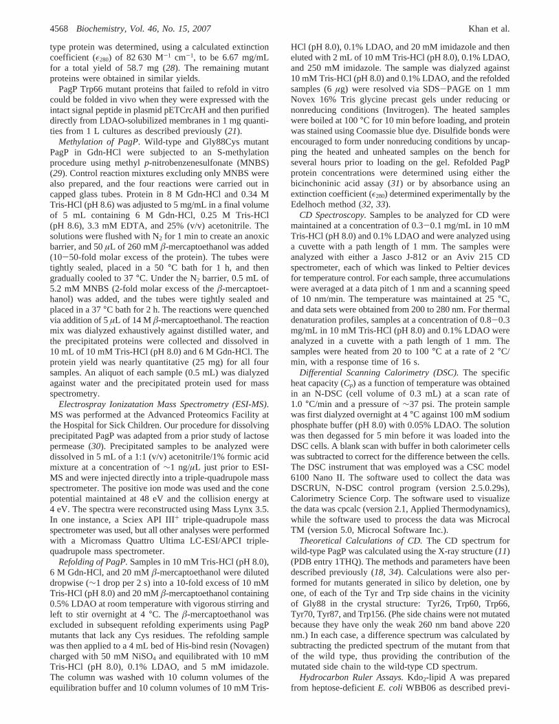
Site-Specific Chemical Modification of Gly88Cys PagP.We subjected the unfolded wild-type and Gly88Cys PagPproteins to methylation with MNBS (Figure 1C) as describedpreviously (29). Control reactions were also performedwithout MNBS. To detect the incorporation of a singlemethylene group (14.03 Da) into an∼20 kDa protein, wechose to determine the mass by ESI-MS. This method isincompatible with the Gdn-HCl or LDAO used to keep PagPin solution, but we were able to develop a mild procedure,adapted from a much harsher one using formic acid toperform ESI-MS on lactose permease (30); our adaptationinvolves removing denaturants by dialysis against water anddissolving the precipitated protein in a 1:1 acetonitrile/1%formic acid solution immediately prior to ESI-MS analysis.We could demonstrate that MNBS leads to a 14 Da increasein the mass of the Gly88Cys mutant, but not in the wild-type protein, which lacks any Cys residues (Table 1). Themass spectra reveal that the Gly88Cys mutant in the absenceof MNBS treatment forms two species, the major one beinga disulfide-linked dimer (Figure 2). MNBS treatment of theGly88Cys mutant reveals a single molecular species corre-sponding to a monomeric S-methylated protein. Thesefindings validate the claim that the methylation procedureis both quantitative and highly selective for Cys under thespecified conditions (29). We were also able to verify themasses of the remaining PagP mutants by ESI-MS(Table 1).
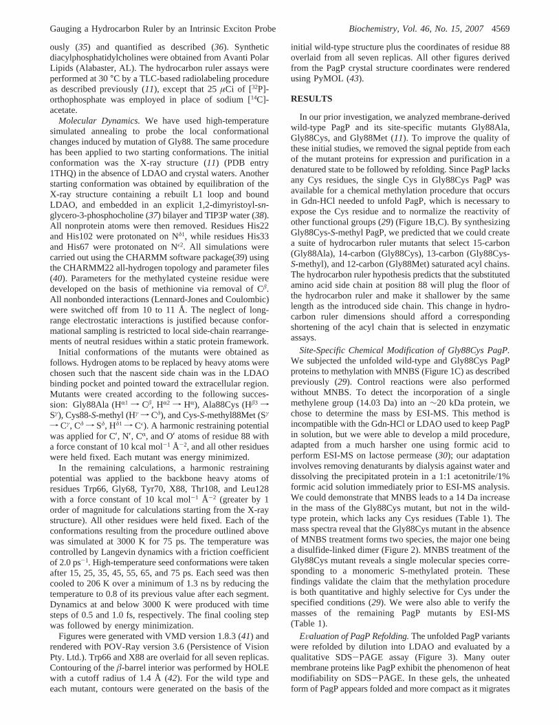
EValuation of PagP Refolding.The unfolded PagP variantswere refolded by dilution into LDAO and evaluated by aqualitative SDS-PAGE assay (Figure 3). Many outermembrane proteins like PagP exhibit the phenomenon of heatmodifiability on SDS-PAGE. In these gels, the unheatedform of PagP appears folded and more compact as it migrates
Gauging a Hydrocarbon Ruler by an Intrinsic Exciton Probe Biochemistry, Vol. 46, No. 15, 20074569
slightly ahead of its expected molecular mass of 20 kDa (21).However, after irreversible heat denaturation, PagP migratesat its expected molecular mass. This explanation for the
observed heat modifiability is reinforced by the observationthat only the heat-denatured Gly88Cys PagP is capable ofmigrating as a disulfide-linked dimer in the nonreducing gel(Figure 3A). The single Cys in this mutant appears to beunexposed in the unheated sample, consistent with the faster-migrating species reflecting a folded state of the protein.Wild-type PagP and its Gly88 mutant derivatives all appearedto be adequately folded by this SDS-PAGE criterion.
Spectroscopic Examination of Refolded PagP Mutants.Wewanted to determine whether subtler structural details couldbe ascertained by a more refined CD spectroscopic examina-tion of the refolded PagP proteins. The LDAO micelles aretransparent down to 200 nm in CD analysis of the refoldedPagP proteins. The far-UV CD spectrum of wild-type PagP(Figure 4A,B) displays a negative CD band at 218 nm thatis characteristic of the nf π* transition derived from peptidebonds in a largelyâ-sheet conformation (44). This observa-tion is consistent with theâ-barrel structure of PagP.However, the positive ellipticity at 232 nm is likely the firstCotton effect of an exciton couplet, which becomes fullyapparent only by its absence in Gly88Cys PagP (Figure 4A).The second Cotton effect of this exciton couplet is largelysuperimposed on theâ-sheet n f π* transition. TheGly88Cys PagP sulfhydryl group appears to be responsible
Table 1: Selective S-Methylation of Gly88Cys PagP Treated withMNBS
PagP proteina theoretical mass (Da) ESI-MS (Da)
Gly88Cys control 20221.58 20220.15( 0.6(Gly88Cys S-S dimer) (40441.14) (40439.32( 1.0)Gly88Cys with MNBS 20235.61 20234.53( 0.5∆ with S-methyl 14.03 14.38( 1.1
wild-type control 20175.49 20174.53( 0.4wild type with MNBS 20175.49 20174.57( 0.4∆ with S-methyl 0 -0.04( 0.7
Gly88Ala 20189.52 20190.63( 1.7Gly88Met 20249.64 20249.41( 0.7
Tyr26Phe 20159.49 20161.80( 2.3Trp66Phe 20136.45 20135.17( 1.9Trp66His 20126.42 20128.01( 2.2
a The wild-typeE. coli PagP protein used in this study lacks theN-terminal signal peptide and contains a C-terminal His6 tag. The aminoacid sequence with Gly88 underlined is as follows: MNADEWMT-TFRENIAQTWQQPEHYDLYIPAITWHARFAYDKEKTDRYNE R-PWGGGFGLSRWDEKGNWHGLYAMAFKDSWNKWEPIAGYG-WESTWRPLADENFHLGLGFTAGVTARDNWNYIPLPVLLPLA-SVGYGPVTFQMTYIPGTYNNGNVYFAWMRFQFLEHHHHHH.
FIGURE 2: ESI-MS confirmation of Gly88Cys-S-methyl PagP. (A)Wild-type PagP treated with MNBS exhibits a single mass thatcorresponds to an unmodified monomeric species. (B) In theabsence of MNBS, Gly88Cys PagP exhibits two masses thatcorrespond to an unmodified monomer and a disulfide-linked dimer.(C) MNBS treatment of Gly88Cys PagP results in a 14 Da massincrease for the monomeric species and a loss of the disulfide-linked dimer.
FIGURE 3: SDS-PAGE analysis of PagP refolding. PagP requiresheat treatment to become denatured via SDS-PAGE and migratesanomalously fast in the absence of heat treatment. (A) Analysis ofwild-type and Gly88Cys PagP under reducing and nonreducingconditions, in the presence and absence of heat treatment, and withor without prior exposure to MNBS. (B) Analysis of Gly88Ala andGly88Met PagP as described above, but without exposure to MNBS.
4570 Biochemistry, Vol. 46, No. 15, 2007 Khan et al.
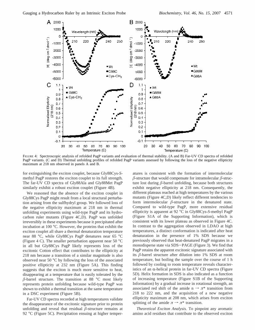
for extinguishing the exciton couplet, because Gly88Cys-S-methyl PagP restores the exciton couplet to its full strength.The far-UV CD spectra of Gly88Ala and Gly88Met PagPsimilarly exhibit a robust exciton couplet (Figure 4B).
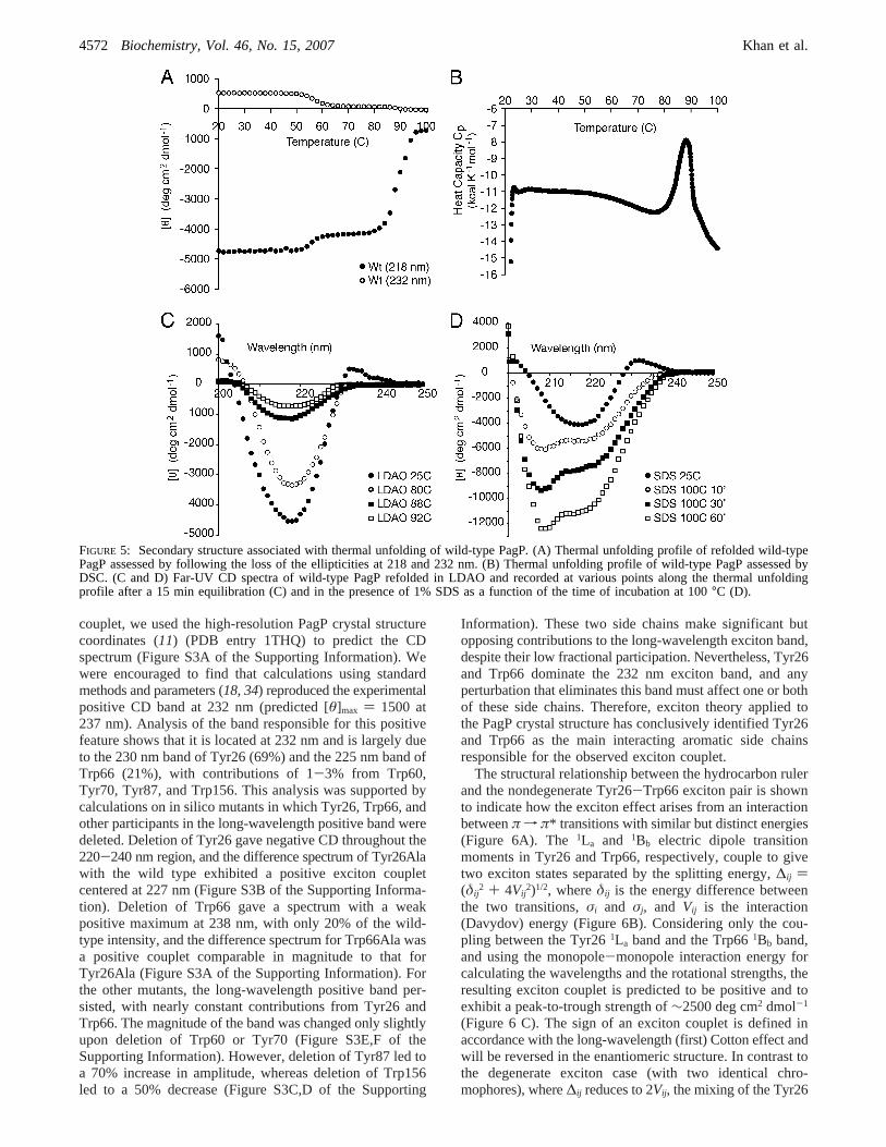
We reasoned that the absence of the exciton couplet inGly88Cys PagP might result from a local structural perturba-tion arising from the sulfhydryl group. We followed loss ofthe negative ellipticity maximum at 218 nm in thermalunfolding experiments using wild-type PagP and its hydro-carbon ruler mutants (Figure 4C,D). PagP was unfoldedirreversibly in these experiments because it precipitated afterincubation at 100°C. However, the proteins that exhibit theexciton couplet all share a thermal denaturation temperaturenear 88°C, while Gly88Cys PagP denatures near 65°C(Figure 4 C). The smaller perturbation apparent near 50°Cin all but Gly88Cys PagP likely represents loss of theexcitonic Cotton effect that contributes to the ellipticity at218 nm because a transition of a similar magnitude is alsoobserved near 50°C by following the loss of the associatedpositive ellipticity at 232 nm (Figure 5A). This findingsuggests that the exciton is much more sensitive to heat,disappearing at a temperature that is easily tolerated by theâ-barrel structure. The transition at 88°C most likelyrepresents protein unfolding because wild-type PagP wasshown to exhibit a thermal transition at the same temperaturein a DSC experiment (Figure 5B).
Far-UV CD spectra recorded at high temperatures validatethe disappearance of the excitonic signature prior to proteinunfolding and reveal that residualâ-structure remains at92 °C (Figure 5C). Precipitation ensuing at higher temper-
atures is consistent with the formation of intermolecularâ-structure that would compensate for intramolecularâ-struc-ture lost duringâ-barrel unfolding, because both structuresexhibit negative ellipticity at 218 nm. Consequently, thedifferent plateaus reached at high temperatures by the variousmutants (Figure 4C,D) likely reflect different tendencies toform intermolecularâ-structure in the denatured state.Compared to wild-type PagP, more extensive residualellipticity is apparent at 92°C in Gly88Cys-S-methyl PagP(Figure S1A of the Supporting Information), which isconsistent with its lower plateau as observed in Figure 4C.In contrast to the aggregation observed in LDAO at hightemperatures, a distinct conformation is indicated after heatdenaturation in the presence of 1% SDS because wepreviously observed that heat-denatured PagP migrates in amonodisperse state via SDS-PAGE (Figure 3). We find thatPagP retains the apparent excitonic signature associated withits â-barrel structure after dilution into 1% SDS at roomtemperature, but boiling the sample over the course of 1 hfollowed by cooling to room temperature reveals character-istics of anR-helical protein in far-UV CD spectra (Figure5D). Helix formation in SDS is also indicated as a functionof increasing temperature (Figure S1B of the SupportingInformation) by a gradual increase in rotational strength, anassociated red shift of the amide nf π* transition from218 to 222 nm, and the acquisition of a new negativeellipticity maximum at 208 nm, which arises from excitonsplitting of the amideπ f π* transition.
Theoretical Exciton Analysis.To pinpoint any aromaticamino acid residues that contribute to the observed exciton
FIGURE 4: Spectroscopic analysis of refolded PagP variants and evaluation of thermal stability. (A and B) Far-UV CD spectra of refoldedPagP variants. (C and D) Thermal unfolding profiles of refolded PagP variants assessed by following the loss of the negative ellipticitymaximum at 218 nm observed in panels A and B.
Gauging a Hydrocarbon Ruler by an Intrinsic Exciton Probe Biochemistry, Vol. 46, No. 15, 20074571
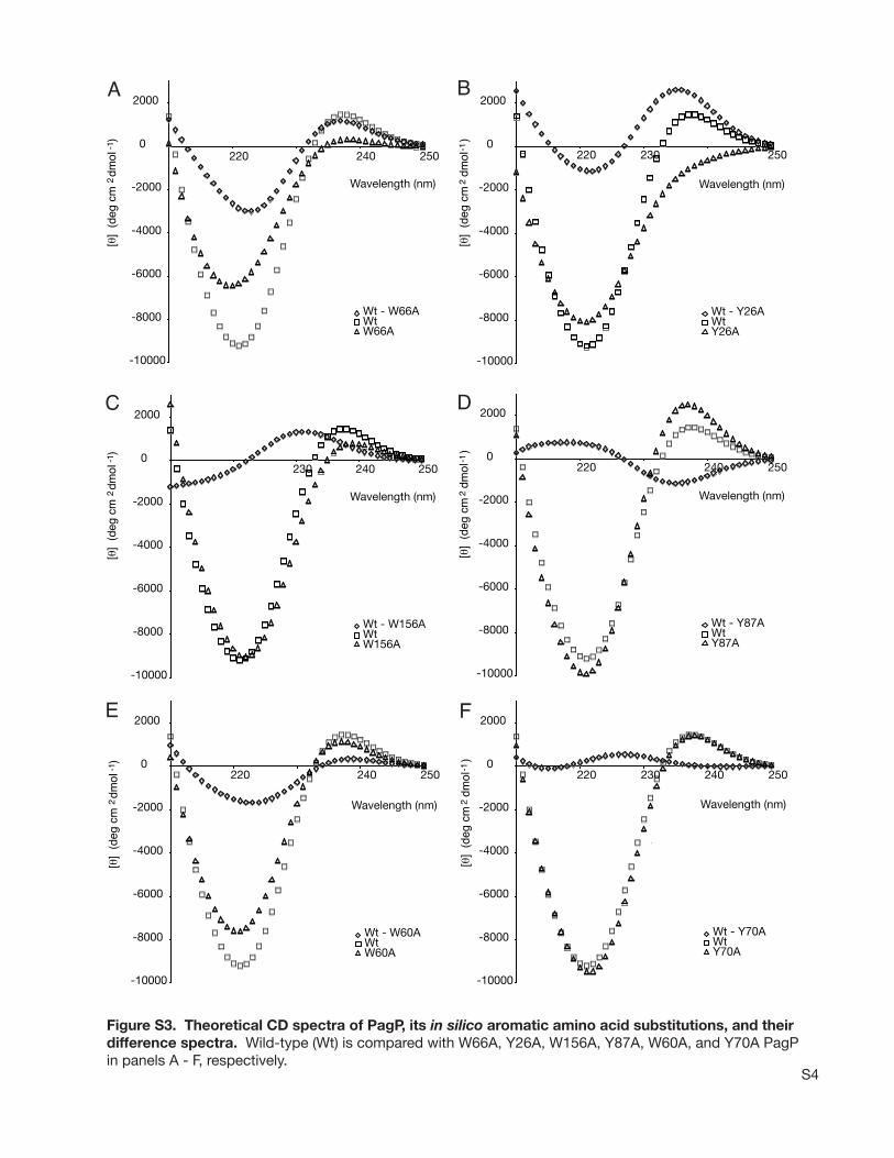
couplet, we used the high-resolution PagP crystal structurecoordinates (11) (PDB entry 1THQ) to predict the CDspectrum (Figure S3A of the Supporting Information). Wewere encouraged to find that calculations using standardmethods and parameters (18, 34) reproduced the experimentalpositive CD band at 232 nm (predicted [θ]max ) 1500 at237 nm). Analysis of the band responsible for this positivefeature shows that it is located at 232 nm and is largely dueto the 230 nm band of Tyr26 (69%) and the 225 nm band ofTrp66 (21%), with contributions of 1-3% from Trp60,Tyr70, Tyr87, and Trp156. This analysis was supported bycalculations on in silico mutants in which Tyr26, Trp66, andother participants in the long-wavelength positive band weredeleted. Deletion of Tyr26 gave negative CD throughout the220-240 nm region, and the difference spectrum of Tyr26Alawith the wild type exhibited a positive exciton coupletcentered at 227 nm (Figure S3B of the Supporting Informa-tion). Deletion of Trp66 gave a spectrum with a weakpositive maximum at 238 nm, with only 20% of the wild-type intensity, and the difference spectrum for Trp66Ala wasa positive couplet comparable in magnitude to that forTyr26Ala (Figure S3A of the Supporting Information). Forthe other mutants, the long-wavelength positive band per-sisted, with nearly constant contributions from Tyr26 andTrp66. The magnitude of the band was changed only slightlyupon deletion of Trp60 or Tyr70 (Figure S3E,F of theSupporting Information). However, deletion of Tyr87 led toa 70% increase in amplitude, whereas deletion of Trp156led to a 50% decrease (Figure S3C,D of the Supporting
Information). These two side chains make significant butopposing contributions to the long-wavelength exciton band,despite their low fractional participation. Nevertheless, Tyr26and Trp66 dominate the 232 nm exciton band, and anyperturbation that eliminates this band must affect one or bothof these side chains. Therefore, exciton theory applied tothe PagP crystal structure has conclusively identified Tyr26and Trp66 as the main interacting aromatic side chainsresponsible for the observed exciton couplet.
The structural relationship between the hydrocarbon rulerand the nondegenerate Tyr26-Trp66 exciton pair is shownto indicate how the exciton effect arises from an interactionbetweenπ f π* transitions with similar but distinct energies(Figure 6A). The 1La and 1Bb electric dipole transitionmoments in Tyr26 and Trp66, respectively, couple to givetwo exciton states separated by the splitting energy,∆ij )(δij
2 + 4Vij2)1/2, whereδij is the energy difference between
the two transitions,σi and σj, and Vij is the interaction(Davydov) energy (Figure 6B). Considering only the cou-pling between the Tyr261La band and the Trp661Bb band,and using the monopole-monopole interaction energy forcalculating the wavelengths and the rotational strengths, theresulting exciton couplet is predicted to be positive and toexhibit a peak-to-trough strength of∼2500 deg cm2 dmol-1
(Figure 6 C). The sign of an exciton couplet is defined inaccordance with the long-wavelength (first) Cotton effect andwill be reversed in the enantiomeric structure. In contrast tothe degenerate exciton case (with two identical chro-mophores), where∆ij reduces to 2Vij, the mixing of the Tyr26
FIGURE 5: Secondary structure associated with thermal unfolding of wild-type PagP. (A) Thermal unfolding profile of refolded wild-typePagP assessed by following the loss of the ellipticities at 218 and 232 nm. (B) Thermal unfolding profile of wild-type PagP assessed byDSC. (C and D) Far-UV CD spectra of wild-type PagP refolded in LDAO and recorded at various points along the thermal unfoldingprofile after a 15 min equilibration (C) and in the presence of 1% SDS as a function of the time of incubation at 100°C (D).
4572 Biochemistry, Vol. 46, No. 15, 2007 Khan et al.
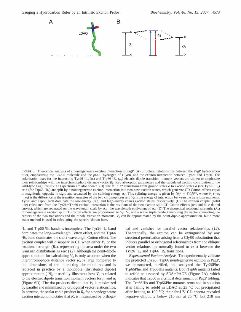
1La and Trp661Bb bands is incomplete. The Tyr261La banddominates the long-wavelength Cotton effect, and the Trp661Bb band dominates the short-wavelength Cotton effect. Theexciton couplet will disappear in CD when eitherVij or therotational strength (Ro), representing the area under the twoGaussian distributions, is zero (12). Although the point-dipoleapproximation for calculatingVij is only accurate when theinterchromophore distance vectorRij is large compared tothe dimensions of the interacting chromophores and isreplaced in practice by a monopole (distributed dipole)approximation (19), it usefully illustrates howVij is relatedto the electric dipole transition moment vectors forµi andµj
(Figure 6D). The dot products dictate thatVij is maximizedby parallel and minimized by orthogonal vector relationships.In contrast, the scalar triple product inRo for a nondegenerateexciton interaction dictates thatRo is maximized by orthogo-
nal and vanishes for parallel vector relationships (12).Theoretically, the exciton can be extinguished by anystructural perturbation arising from a Gly88 substitution thatinduces parallel or orthogonal relationships from the obliquevector relationships normally found to exist between theTyr26 1La and Trp661Bb transitions.
Experimental Exciton Analysis.To experimentally validatethe predicted Tyr26-Trp66 nondegenerate exciton in PagP,we constructed, purified, and analyzed the Tyr26Phe,Trp66Phe, and Trp66His mutants. Both Trp66 mutants failedto refold as assessed by SDS-PAGE (Figure 7A), whichindicates that Trp66 is a critical determinant of PagP folding.The Trp66His and Trp66Phe mutants remained in solutionafter failing to refold in LDAO at 25°C but precipitatedafter heating to 100°C; their far-UV CD spectra revealednegative ellipticity below 210 nm at 25°C, but 218 nm
FIGURE 6: Theoretical analysis of a nondegenerate exciton interaction in PagP. (A) Structural relationships between the PagP hydrocarbonruler, emphasizing the LDAO molecule and thepro-L hydrogen of Gly88, and the exciton interaction between Tyr26 and Trp66. Thepolarization axes for the interacting Tyr261La (µi) and Trp661Bb (µj) electric dipole transition moment vectors are shown to emphasizetheir relationships with the interchromophore distance vectorRij. Key absorption parameters and the calculated exciton contribution to thewild-type PagP far-UV CD spectrum are also shown. (B) Theπ f π* transitions from ground stateso to excited statesa (for Tyr26 1La)or b (for Trp66 1Bb) are split by a nondegenerate exciton interaction into two new exciton states, which generate CD Cotton effects equalin magnitude, opposite in sign, and separated by the splitting energy∆ij. This splitting energy is given by (δij
2 + 4Vij2)1/2, whereδij ()σj
- σi) is the difference in the transition energies of the two chromophores andVij is the energy of interaction between the transition moments.Tyr26 and Trp66 each dominate the low-energy (red) and high-energy (blue) exciton states, respectively. (C) The exciton couplet (solidline) calculated from the Tyr26-Trp66 exciton interaction is the resultant of the two exciton-split CD Cotton effects (red and blue dottedcurves), which are separated on the wavelength scale by∆ij ′, the wavelength equivalent of∆ij. (D) The theoretical rotational strengths (Ro)of nondegenerate exciton split CD Cotton effects are proportional toVij, ∆ij, and a scalar triple product involving the vector connecting thecenters of the two transitions and the dipole transition moments.Vij can be approximated by the point-dipole approximation, but a moreexact method is used in calculating the spectra shown here.
Gauging a Hydrocarbon Ruler by an Intrinsic Exciton Probe Biochemistry, Vol. 46, No. 15, 20074573
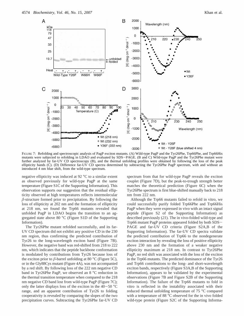
negative ellipticity was induced at 92°C to a similar extentas observed previously for wild-type PagP at the sametemperature (Figure S1C of the Supporting Information). Thisobservation supports our suggestion that the residual ellip-ticity observed at high temperatures reflects intermolecularâ-structure formed prior to precipitation. By following theloss of ellipticity at 202 nm and the formation of ellipticityat 218 nm, we found the Trp66 mutants revealed thatunfolded PagP in LDAO begins the transition to an ag-gregated state above 80°C (Figure S1D of the SupportingInformation).
The Tyr26Phe mutant refolded successfully, and its far-UV CD spectrum did not exhibit any positive CD in the 230nm region, thus confirming the predicted contribution ofTyr26 to the long-wavelength exciton band (Figure 7B).However, the negative band was red-shifted from 218 to 222nm, which indicates that the peptide backbone conformationis modulated by contributions from Tyr26 because loss ofthe exciton prior toâ-barrel unfolding at 80°C (Figure 5C),or in the Gly88Cys mutant (Figure 4A), was not accompaniedby a red shift. By following loss of the 222 nm negative CDband in Tyr26Phe PagP, we observed an 8°C reduction inthe thermal transition temperature when compared to the 218nm negative CD band lost from wild-type PagP (Figure 7C);only the latter displays loss of the exciton in the 40-50 °Crange, and an apparent contribution of Tyr26 to foldingcooperativity is revealed by comparing the slopes of the twoprecipitation curves. Subtracting the Tyr26Phe far-UV CD
spectrum from that for wild-type PagP reveals the excitoncouplet (Figure 7D), but the peak-to-trough strength bettermatches the theoretical prediction (Figure 6C) when theTyr26Phe spectrum is first blue-shifted manually back to 218nm from 222 nm.
Although the Trp66 mutants failed to refold in vitro, wecould successfully purify folded Trp66Phe and Trp66HisPagP when they were expressed in vivo with an intact signalpeptide (Figure S2 of the Supporting Information) asdescribed previously (21). The in vivo-folded wild-type andTrp66 mutant PagP proteins appeared folded by both SDS-PAGE and far-UV CD criteria (Figure S2A,B of theSupporting Information). The far-UV CD spectra validatethe predicted contribution of Trp66 to the nondegenerateexciton interaction by revealing the loss of positive ellipticityabove 230 nm and the formation of a weaker negativeellipticity maximum at 218 nm. In contrast to Tyr26PhePagP, no red shift was associated with the loss of the excitonin the Trp66 mutants. The predicted dominance of the Tyr26and Trp66 contributions to the long- and short-wavelengthexciton bands, respectively (Figure S3A,B of the SupportingInformation), appears to be validated by the experimentalobservations (Figure 7B and Figure S2B of the SupportingInformation). The failure of the Trp66 mutants to fold invitro is reflected in the instability associated with theirreduced thermal unfolding temperature of 75°C comparedwith a temperature of 88°C observed for the in vivo foldedwild-type protein (Figure S2C of the Supporting Informa-
FIGURE 7: Refolding and spectroscopic analysis of PagP exciton mutants. (A) Wild-type PagP and the Tyr26Phe, Trp66Phe, and Trp66Hismutants were subjected to refolding in LDAO and evaluated by SDS-PAGE. (B and C) Wild-type PagP and the Tyr26Phe mutant werefurther analyzed by far-UV CD spectroscopy (B), and the thermal unfolding profiles were obtained by following the loss of the peakellipticity bands (C). (D) Difference far-UV CD spectra determined by subtracting the Tyr26Phe PagP spectrum, with and without anintroduced 4 nm blue shift, from the wild-type spectrum.
4574 Biochemistry, Vol. 46, No. 15, 2007 Khan et al.
tion). However, the Gly88Cys PagP mutant was successfullyfolded in vitro despite the fact it displays a thermal unfoldingtemperature of only 65°C (Figure 4C). Periplasmic chap-erones are known to assist the folding of outer membraneâ-barrel proteins inE. coli and likely help to overcome thefolding defect associated with the PagP Trp66 mutations (45).The observation of reduced thermal unfolding temperaturesfor both the Tyr26 (Figure 7C) and Trp66 (Figure S2C ofthe Supporting Information) PagP mutants suggests thatdestabilizing effects will likely be associated with proximalgroups that perturb these aromatic side chains.
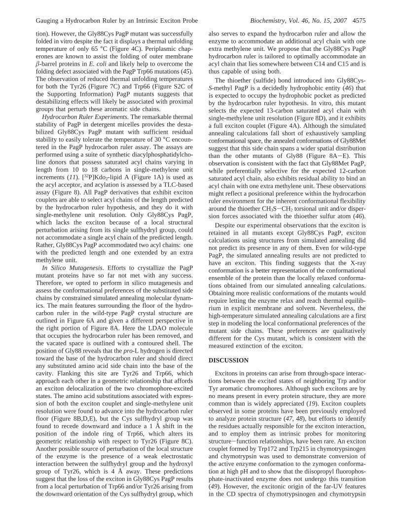
Hydrocarbon Ruler Experiments.The remarkable thermalstability of PagP in detergent micelles provides the desta-bilized Gly88Cys PagP mutant with sufficient residualstability to easily tolerate the temperature of 30°C encoun-tered in the PagP hydrocarbon ruler assay. The assays areperformed using a suite of synthetic diacylphosphatidylcho-line donors that possess saturated acyl chains varying inlength from 10 to 18 carbons in single-methylene unitincrements (11). [32P]Kdo2-lipid A (Figure 1A) is used asthe acyl acceptor, and acylation is assessed by a TLC-basedassay (Figure 8). All PagP derivatives that exhibit excitoncouplets are able to select acyl chains of the length predictedby the hydrocarbon ruler hypothesis, and they do it withsingle-methylene unit resolution. Only Gly88Cys PagP,which lacks the exciton because of a local structuralperturbation arising from its single sulfhydryl group, couldnot accommodate a single acyl chain of the predicted length.Rather, Gly88Cys PagP accommodated two acyl chains: onewith the predicted length and one extended by an extramethylene unit.
In Silico Mutagenesis. Efforts to crystallize the PagPmutant proteins have so far not met with any success.Therefore, we opted to perform in silico mutagenesis andassess the conformational preferences of the substituted sidechains by constrained simulated annealing molecular dynam-ics. The main features surrounding the floor of the hydro-carbon ruler in the wild-type PagP crystal structure areoutlined in Figure 6A and given a different perspective inthe right portion of Figure 8A. Here the LDAO moleculethat occupies the hydrocarbon ruler has been removed, andthe vacated space is outlined with a contoured shell. Theposition of Gly88 reveals that thepro-L hydrogen is directedtoward the base of the hydrocarbon ruler and should directany substituted amino acid side chain into the base of thecavity. Flanking this site are Tyr26 and Trp66, whichapproach each other in a geometric relationship that affordsan exciton delocalization of the two chromophore-excitedstates. The amino acid substitutions associated with expres-sion of both the exciton couplet and single-methylene unitresolution were found to advance into the hydrocarbon rulerfloor (Figure 8B,D,E), but the Cys sulfhydryl group wasfound to recede downward and induce a 1 Å shift in theposition of the indole ring of Trp66, which alters itsgeometric relationship with respect to Tyr26 (Figure 8C).Another possible source of perturbation of the local structureof the enzyme is the presence of a weak electrostaticinteraction between the sulfhydryl group and the hydroxylgroup of Tyr26, which is 4 Å away. These predictionssuggest that the loss of the exciton in Gly88Cys PagP resultsfrom a local perturbation of Trp66 and/or Tyr26 arising fromthe downward orientation of the Cys sulfhydryl group, which
also serves to expand the hydrocarbon ruler and allow theenzyme to accommodate an additional acyl chain with oneextra methylene unit. We propose that the Gly88Cys PagPhydrocarbon ruler is tailored to optimally accommodate anacyl chain that lies somewhere between C14 and C15 and isthus capable of using both.
The thioether (sulfide) bond introduced into Gly88Cys-S-methyl PagP is a decidedly hydrophobic entity (46) thatis expected to occupy the hydrophobic pocket as predictedby the hydrocarbon ruler hypothesis. In vitro, this mutantselects the expected 13-carbon saturated acyl chain withsingle-methylene unit resolution (Figure 8D), and it exhibitsa full exciton couplet (Figure 4A). Although the simulatedannealing calculations fall short of exhaustively samplingconformational space, the annealed conformations of Gly88Metsuggest that this side chain spans a wider spatial distributionthan the other mutants of Gly88 (Figure 8A-E). Thisobservation is consistent with the fact that Gly88Met PagP,while preferentially selective for the expected 12-carbonsaturated acyl chain, also exhibits residual ability to bind anacyl chain with one extra methylene unit. These observationsmight reflect a positional preference within the hydrocarbonruler environment for the inherent conformational flexibilityaround the thioether CH3S-CH2 torsional unit and/or disper-sion forces associated with the thioether sulfur atom (46).
Despite our experimental observations that the exciton isretained in all mutants except Gly88Cys PagP, excitoncalculations using structures from simulated annealing didnot predict its presence in any of them. Even for wild-typePagP, the simulated annealing results are not predicted tohave an exciton. This finding suggests that the X-rayconformation is a better representation of the conformationalensemble of the protein than the locally relaxed conforma-tions obtained from our simulated annealing calculations.Obtaining more realistic conformations of the mutants wouldrequire letting the enzyme relax and reach thermal equilib-rium in explicit membrane and solvent. Nevertheless, thehigh-temperature simulated annealing calculations are a firststep in modeling the local conformational preferences of themutant side chains. These preferences are qualitativelydifferent for the Cys mutant, which is consistent with themeasured extinction of the exciton.
DISCUSSION
Excitons in proteins can arise from through-space interac-tions between the excited states of neighboring Trp and/orTyr aromatic chromophores. Although such excitons are byno means present in every protein structure, they are morecommon than is widely appreciated (19). Exciton coupletsobserved in some proteins have been previously employedto analyze protein structure (47, 48), but efforts to identifythe residues actually responsible for the exciton interaction,and to employ them as intrinsic probes for monitoringstructure-function relationships, have been rare. An excitoncouplet formed by Trp172 and Trp215 in chymotrypsinogenand chymotrypsin was used to demonstrate conversion ofthe active enzyme conformation to the zymogen conforma-tion at high pH and to show that the diisopropyl fluorophos-phate-inactivated enzyme does not undergo this transition(49). However, the excitonic origin of the far-UV featuresin the CD spectra of chymotrypsinogen and chymotrypsin
Gauging a Hydrocarbon Ruler by an Intrinsic Exciton Probe Biochemistry, Vol. 46, No. 15, 20074575
was not demonstrated until much later (19). Exciton couplingbetween Trp47 and Trp74 inE. coli dihydrofolate reductasewas identified as the source of the first transient that can bedetected by stopped flow in the refolding of the urea-denatured enzyme (50). More recently, a Gly95Ala mutationhas been shown to abolish the Trp-Trp exciton, whereasbinding of methotrexate restores it (51). Given the forecastedoutput of current structural proteomics consortia, it is likely
that the pool of proteins exhibiting clearly discernible excitoncouplets will continue to grow rapidly into the future. Suchknowledge will provide investigators with highly sensitiveprobes for monitoring the functional effects of locallyintroduced structural alterations.
A case in point is revealed by our current study of PagP,which has a number of favorable features that made the studypossible. (1) The compactâ-barrel structure of PagP produces
FIGURE 8: Hydrocarbon ruler analysis of refolded PagP variants. Synthetic diacylphosphatidylcholines with saturated acyl chains varyingin single methylene increments from 10 to 18 carbon atoms were used as donors for the enzymatic acylation of Kdo2-lipid A. Panels A-Epresent acyl-chain selection profiles for the refolded PagP variants. The structure of PagP (PDB entry 1THQ) emphasizing the positions ofGly88 and the exciton interaction between Tyr26 and Trp66 is shown at the right of panel A. Contouring reveals theâ-barrel interior regionafter removal of the LDAO detergent molecule. In silico mutagenesis followed by simulated annealing was performed to reveal a predictedorientation of the PagP Gly88 substitutions, which are shown at the right of panels B-E. ND means not detected.
4576 Biochemistry, Vol. 46, No. 15, 2007 Khan et al.
a relatively weak backbone CD that makes obvious theoverlapping exciton couplet. (2) PagP can resolve methyleneunits in the acyl chains of its lipid substrate by using amolecular measuring device known as a hydrocarbon ruler,the floor of which is located in the proximity of theinteracting exciton partners. (3) PagP is an extremely stablesmall protein that can be unfolded and refolded in vitro,enabling the transient exposure of buried functional groupsfor chemical modification. The fact that PagP is an integralmembrane protein makes these favorable features all themore remarkable. Consequently, PagP beautifully demon-strates both the sensitivity of the exciton and how it can beemployed as a spectroscopic probe to monitor a localstructural perturbation.
The PagP hydrocarbon ruler was identified in a prior X-raystructure determination (11) and validated in that study byusing enzyme that had been purified directly from mem-branes (21). However, the LDAO detergent used to purifythe enzyzme is a competitive inhibitor (11) and must beexchanged by dilution of the enzyme into a reaction cocktailthat includesn-dodecylâ-D-maltoside or another detergentthat supports PagP activity (21). While PagP purified directlyfrom membranes is obtained at∼1 mg/mL (21), PagP canalso be expressed without its N-terminal signal peptide,purified in an unfolded state, and reliably refolded in LDAOto reach a concentration of>10 mg/mL (10, 11). Conse-quently, LDAO can be removed more effectively by dilutionwhen using refolded PagP, which gives more reliable resultsin the enzyme assays. By using refolded PagP derivativesin this investigation, we observed for the first time that Gly88substitutions, with the exception of the Cys mutation,modulate hydrocarbon ruler acyl-chain selection with me-thylene unit precision.
The predicted interactions of the Cys sulfhydryl group withthe Trp66 and Tyr26 aromatic side chains can explain ourmain experimental observations that Gly88Cys PagP is bothdevoid of the exciton and compromised in terms of acyl-chain selection. Our simulated annealing experiments dem-onstrate that the amino acid substitutions observed experi-mentally to retain the exciton are advancing upward into thehydrocarbon ruler floor, while the one side chain that is ableto recede downward (Cys) is observed to extinguish theexciton. The question of why the Cys sulfhydryl group wouldprefer the position between Tyr26 and Trp66 instead of insidethe hydrophobic hydrocarbon ruler pocket remains. Thehydrocarbon ruler floor in PagP marks the boundary betweenthe upper hydrophobic and lower polar interior regions oftheâ-barrel (11), which might indicate that the Cys sulfhy-dryl group has an inherent tendency to seek out a more polarmicroenvironment. Hydrophobicity scales derived primarilyfrom R-helical transmembrane domains typically identify Cysas being hydrophobic (52), but Cys in anR-helix normallydonates a hydrogen bond to the carbonyl oxygen atom ofthe preceding peptide bond (53). In contrast, the sulfhydrylgroup of Gly88Cys PagP is forced into aâ-structuredconformation where it is faced above with nonpolar andbelow with polar environments. In the absence of a suitablehydrogen bond acceptor, the Cys sulfhydryl group appearsto have rotated about itsø1 dihedral angle to establish a weakelectrostatic interaction with the Tyr26 hydroxyl group and/or a so-calledπ (aromatic)-lone pair (sulfur) attraction withTrp66 (54). The polarizability of the sulfur atom and the
acidity of the sulfhydryl group are reported to promote itsinteraction with aromatic partners (55). Although our forcefield contains only partial point charges on each nucleus,which is but a rough approximation to the electronicdistribution associated with (pure and hybrid) p orbitals, theforce field may be capable of capturing these effectsqualitatively.
The lactose permease provides an example of anR-helicaltransmembrane protein in which substitutions of Cys for Glyhave had unpredictable effects on protein stability. Thereplacement of Cys154 with Gly within a transmembranedomain of the lactose permease has been shown to havestabilizing effects that are incompatible with active transportbut necessary to obtain crystals for determining the structureby X-ray diffraction (56, 57). Only Cys at position 154 wascapable of introducing a local instability that was necessaryfor normal transport (58). The lactose permease in this regionincludes local distortions of helical conformation and closepacking interactions with several aromatic partners. Perhapsthe unusual physical properties associated with Cys154 inlactose permease have in common with Gly88Cys PagP theneed to establish interactions with aromatic side chains.Clearly, the PagP hydrocarbon ruler affords unique op-portunities to evaluate the physical forces at work indetermining membrane protein stability. Our ability toenzymatically synthesize endotoxin analogues with definedacyl-chain substitutions might also reveal pharmacologicallyinteresting derivatives, because acyl-chain length is knownto be an important factor in determining endotoxin structure-activity relationships (59).
ACKNOWLEDGMENT
We gratefully acknowledge Walid Houry and Avi Chakra-bartty (University of Toronto) for sharing their CD spec-trometers, Anthony Mittermaier (now at McGill University,Montreal, QC) for setting up the initial PagP structure formolecular dynamics studies, and Fraser Hof (University ofVictoria, Victoria, BC), Charles Deber (University of Tor-onto), and Richard and Raquel Epand (McMaster University)for helpful discussions.
SUPPORTING INFORMATION AVAILABLE
Oligonucleotide primers used for site-directed mutagenesisand cloning (Table S1), far-UV CD spectra of PagP atdifferent temperatures (Figure S1), analysis of PagP Trp66mutants folded in vivo (Figure S2), and theoretical CDspectra of PagP, its in silico aromatic amino acid substitu-tions, and their difference spectra (Figure S3). This materialis available free of charge via the Internet at http://pubs.acs.org.
REFERENCES
1. Vance, D. E., and Vance, J. E. (2002)Biochemistry of Lipids,Lipoproteins, and Membranes, Elsevier, Amsterdam.
2. Dowhan, W. (1997) Molecular basis for membrane phospholipiddiversity: Why are there so many lipids?Annu. ReV. Biochem.66, 199-232.
3. Zhang, Y. M., White, S. W., and Rock, C. O. (2006) Inhibitingbacterial fatty acid synthesis,J. Biol. Chem. 281, 17541-17544.
4. Bracey, M. H., Cravatt, B. F., and Stevens, R. C. (2004) Structuralcommonalities among integral membrane enzymes,FEBS Lett.567, 159-165.
Gauging a Hydrocarbon Ruler by an Intrinsic Exciton Probe Biochemistry, Vol. 46, No. 15, 20074577
5. Long, S. B., Casey, P. J., and Beese, L. S. (1998) Cocrystalstructure of protein farnesyltransferase complexed with a farnesyldiphosphate substrate,Biochemistry 37, 9612-9618.
6. Wyckoff, T. J., Lin, S., Cotter, R. J., Dotson, G. D., and Raetz,C. R. (1998) Hydrocarbon rulers in UDP-N-acetylglucosamineacyltransferases,J. Biol. Chem. 273, 32369-32372.
7. White, S. H., Ladokhin, A. S., Jayasinghe, S., and Hristova, K.(2001) How membranes shape protein structure,J. Biol. Chem.276, 32395-32398.
8. Snijder, H. J., Ubarretxena-Belandia, I., Blaauw, M., Kalk, K. H.,Verheij, H. M., Egmond, M. R., Dekker, N., and Dijkstra, B. W.(1999) Structural evidence for dimerization-regulated activationof an integral membrane phospholipase,Nature 401, 717-721.
9. Rutten, L., Geurtsen, J., Lambert, W., Smolenaers, J. J., Bonvin,A. M., de Haan, A., van der Ley, P., Egmond, M. R., Gros, P.,and Tommassen, J. (2006) Crystal structure and catalytic mech-anism of the LPS 3-O-deacylase PagL fromPseudomonasaeruginosa, Proc. Natl. Acad. Sci. U.S.A. 103, 7071-7076.
10. Hwang, P. M., Choy, W. Y., Lo, E. I., Chen, L., Forman-Kay, J.D., Raetz, C. R., Prive, G. G., Bishop, R. E., and Kay, L. E. (2002)Solution structure and dynamics of the outer membrane enzymePagP by NMR,Proc. Natl. Acad. Sci. U.S.A. 99, 13560-13565.
11. Ahn, V. E., Lo, E. I., Engel, C. K., Chen, L., Hwang, P. M., Kay,L. E., Bishop, R. E., and Prive, G. G. (2004) A hydrocarbon rulermeasures palmitate in the enzymatic acylation of endotoxin,EMBO J. 23, 2931-2941.
12. Harada, N., and Nakanishi, K. (1983)Circular dichroic spectros-copy: Exciton coupling in organic stereochemistry, UniversityScience Books, Mill Valley, CA.
13. Schellman, J. A. (1968) Symmetry rules for optical rotation,Acc.Chem. Res. 1, 144-151.
14. Davydov, A. S. (1971)Theory of molecular excitons. Translatedfrom Russian by Stephen B. Dresner, New American ed., PlenumPress, New York.
15. Woody, R. W. (1995) Circular dichroism,Methods Enzymol. 246,34-71.
16. Woody, R. W. (1994) Contributions of tryptophan side chains tothe far-ultraviolet circular dichroism of proteins,Eur. Biophys. J.23, 253-262.
17. Sreerama, N., and Woody, R. W. (2004) On the analysis ofmembrane protein circular dichroism spectra,Protein Sci. 13,100-112.
18. Sreerama, N., and Woody, R. W. (2004) Computation and analysisof protein circular dichroism spectra,Methods Enzymol. 383, 318-351.
19. Grishina, I. B., and Woody, R. W. (1994) Contributions oftryptophan side chains to the circular dichroism of globularproteins: Exciton couplets and coupled oscillators,FaradayDiscuss., 245-262.
20. Bishop, R. E. (2005) The lipid A palmitoyltransferase PagP:Molecular mechanisms and role in bacterial pathogenesis,Mol.Microbiol. 57, 900-912.
21. Bishop, R. E., Gibbons, H. S., Guina, T., Trent, M. S., Miller, S.I., and Raetz, C. R. (2000) Transfer of palmitate from phospho-lipids to lipid A in outer membranes of Gram-negative bacteria,EMBO J. 19, 5071-5080.
22. Kamio, Y., and Nikaido, H. (1976) Outer membrane ofSalmonellatyphimurium: Accessibility of phospholipid head groups tophospholipase c and cyanogen bromide activated dextran in theexternal medium,Biochemistry 15, 2561-2570.
23. Nikaido, H. (2003) Molecular basis of bacterial outer membranepermeability revisited,Microbiol. Mol. Biol. ReV. 67, 593-656.
24. Guo, L., Lim, K. B., Poduje, C. M., Daniel, M., Gunn, J. S.,Hackett, M., and Miller, S. I. (1998) Lipid A acylation andbacterial resistance against vertebrate antimicrobial peptides,Cell95, 189-198.
25. Kawasaki, K., Ernst, R. K., and Miller, S. I. (2004) 3-O-Deacylation of lipid A by PagL, a PhoP/PhoQ-regulated deacylaseof Salmonella typhimurium, modulates signaling through toll-likereceptor 4,J. Biol. Chem. 279, 20044-20048.
26. Hwang, P. M., Bishop, R. E., and Kay, L. E. (2004) The integralmembrane enzyme PagP alternates between two dynamicallydistinct states,Proc. Natl. Acad. Sci. U.S.A. 101, 9618-9623.
27. Jia, W., Zoeiby, A. E., Petruzziello, T. N., Jayabalasingham, B.,Seyedirashti, S., and Bishop, R. E. (2004) Lipid trafficking controlsendotoxin acylation in outer membranes ofEscherichia coli, J.Biol. Chem. 279, 44966-44975.
28. Gill, S. C., and von Hippel, P. H. (1989) Calculation of proteinextinction coefficients from amino acid sequence data,Anal.Biochem. 182, 319-326.
29. Heinrikson, R. L. (1971) The selectiveS-methylation of sulfhydrylgroups in proteins and peptides with methyl-p-nitrobenzene-sulfonate,J. Biol. Chem. 246, 4090-4096.
30. Whitelegge, J. P., le Coutre, J., Lee, J. C., Engel, C. K., Prive, G.G., Faull, K. F., and Kaback, H. R. (1999) Toward the bilayerproteome, electrospray ionization-mass spectrometry of large,intact transmembrane proteins,Proc. Natl. Acad. Sci. U.S.A. 96,10695-10698.
31. Smith, P. K., Krohn, R. I., Hermanson, G. T., Mallia, A. K.,Gartner, F. H., Provenzano, M. D., Fujimoto, E. K., Goeke, N.M., Olson, B. J., and Klenk, D. C. (1985) Measurement of proteinusing bicinchoninic acid,Anal. Biochem. 150, 76-85.
32. Edelhoch, H. (1967) Spectroscopic determination of tryptophanand tyrosine in proteins,Biochemistry 6, 1948-1954.
33. Pace, C. N., Vajdos, F., Fee, L., Grimsley, G., and Gray, T. (1995)How to measure and predict the molar absorption coefficient ofa protein,Protein Sci. 4, 2411-2423.
34. Woody, R. W., and Sreerama, N. (1999) Comment on ‘Improvingprotein circular dichroism calculations in the far-ultraviolet throughreparameterizing the amide chromophore’ [J. Chem. Phys. 109,782 (1998)],J. Chem. Phys. 111, 2844-2845.
35. Kanipes, M. I., Lin, S., Cotter, R. J., and Raetz, C. R. (2001) Ca2+-induced phosphoethanolamine transfer to the outer 3-deoxy-D-manno-octulosonic acid moiety ofEscherichia colilipopolysac-charide. A novel membrane enzyme dependent upon phosphatidyl-ethanolamine,J. Biol. Chem. 276, 1156-1163.
36. Lee, C. H., and Tsai, C. M. (1999) Quantification of bacteriallipopolysaccharides by the purpald assay: Measuring formalde-hyde generated from 2-keto-3-deoxyoctonate and heptose at theinner core by periodate oxidation,Anal. Biochem. 267, 161-168.
37. Feller, S. E., and MacKerell, A. D. (2000) An improved empiricalpotential energy function for molecular simulations of phospho-lipids, J. Phys. Chem. B 104, 7510-7515.
38. Jorgensen, W. L., Chandrasekhar, J., Madura, J. D., Impey, R.W., and Klein, M. L. (1983) Comparison of simple potentialfunctions for simulating liquid water,J. Chem. Phys. 79, 926-935.
39. Brooks, B. R., Bruccoleri, R. E., Olafson, B. D., States, D. J.,Swaminathan, S., and Karplus, M. (1983) Charmm: A Programfor Macromolecular Energy, Minimization, and Dynamics Cal-culations,J. Comput. Chem. 4, 187-217.
40. MacKerell, A. D., Bashford, D., Bellott, M., Dunbrack, R. L.,Evanseck, J. D., Field, M. J., Fischer, S., Gao, J., Guo, H., Ha,S., Joseph-McCarthy, D., Kuchnir, L., Kuczera, K., Lau, F. T.K., Mattos, C., Michnick, S., Ngo, T., Nguyen, D. T., Prodhom,B., Reiher, W. E., Roux, B., Schlenkrich, M., Smith, J. C., Stote,R., Straub, J., Watanabe, M., Wiorkiewicz-Kuczera, J., Yin, D.,and Karplus, M. (1998) All-atom empirical potential for molecularmodeling and dynamics studies of proteins,J. Phys. Chem. B 102,3586-3616.
41. Humphrey, W., Dalke, A., and Schulten, K. (1996) VMD: Visualmolecular dynamics,J. Mol. Graphics 14, 33-38, 27-28.
42. Smart, O. S., Goodfellow, J. M., and Wallace, B. A. (1993) Thepore dimensions of gramicidin A,Biophys. J. 65, 2455-2460.
43. DeLano, W. L. (2002)The PyMOL Molecular Graphics System,DeLano Scientific, San Carlos, CA.
44. Saxena, V. P., and Wetlaufer, D. B. (1971) A new basis forinterpreting the circular dichroic spectra of proteins,Proc. Natl.Acad. Sci. U.S.A. 68, 969-972.
45. Mogensen, J. E., and Otzen, D. E. (2005) Interactions betweenfolding factors and bacterial outer membrane proteins,Mol.Microbiol. 57, 326-346.
46. Gellman, S. H. (1991) On the role of methionine residues in thesequence-independent recognition of nonpolar protein surfaces,Biochemistry 30, 6633-6636.
47. Gasymov, O. K., Abduragimov, A. R., Yusifov, T. N., andGlasgow, B. J. (2002) RET and anisotropy measurements establishthe proximity of the conserved Trp17 to Ile98 and Phe99 of tearlipocalin, Biochemistry 41, 8837-8848.
48. Nakao, M., Maki, K., Arai, M., Koshiba, T., Nitta, K., andKuwajima, K. (2005) Characterization of kinetic folding inter-mediates of recombinant canine milk lysozyme by stopped-flowcircular dichroism,Biochemistry 44, 6685-6692.
4578 Biochemistry, Vol. 46, No. 15, 2007 Khan et al.
49. McConn, J., Fasman, G. D., and Hess, G. P. (1969) Conformationof the high pH form of chymotrypsin,J. Mol. Biol. 39, 551-562.
50. Kuwajima, K., Garvey, E. P., Finn, B. E., Matthews, C. R., andSugai, S. (1991) Transient intermediates in the folding ofdihydrofolate reductase as detected by far-ultraviolet circulardichroism spectroscopy,Biochemistry 30, 7693-7703.
51. Svensson, A. K., O’Neill, J. C., Jr., and Matthews, C. R. (2003)The coordination of the isomerization of a conserved non-prolylcis peptide bond with the rate-limiting steps in the folding ofdihydrofolate reductase,J. Mol. Biol. 326, 569-583.
52. Bowie, J. U. (2005) Solving the membrane protein foldingproblem,Nature 438, 581-589.
53. Gray, T. M., and Matthews, B. W. (1984) Intrahelical hydrogenbonding of serine, threonine and cysteine residues withinR-helicesand its relevance to membrane-bound proteins,J. Mol. Biol. 175,75-81.
54. Duan, G., Smith, V. H., Jr., and Weaver, D. F. (2001) Charac-terization of aromatic-thiolπ-type hydrogen bonding and pheny-lalanine-cysteine side chain interactions throughab initio calcu-lations and protein database analyses,Mol. Phys. 99, 1689-1699.
55. Meyer, E. A., Castellano, R. K., and Diederich, F. (2003)Interactions with aromatic rings in chemical and biologicalrecognition,Angew. Chem., Int. Ed. 42, 1210-1250.
56. Smirnova, I. N., and Kaback, H. R. (2003) A mutation in thelactose permease ofEscherichia colithat decreases conformationalflexibility and increases protein stability,Biochemistry 42, 3025-3031.
57. Abramson, J., Smirnova, I., Kasho, V., Verner, G., Kaback, H.R., and Iwata, S. (2003) Structure and mechanism of the lactosepermease ofEscherichia coli, Science 301, 610-615.
58. Ermolova, N. V., Smirnova, I. N., Kasho, V. N., and Kaback, H.R. (2005) Interhelical packing modulates conformational flexibilityin the lactose permease ofEscherichia coli, Biochemistry 44,7669-7677.
59. Stover, A. G., Da Silva Correia, J., Evans, J. T., Cluff, C. W.,Elliott, M. W., Jeffery, E. W., Johnson, D. A., Lacy, M. J.,Baldridge, J. R., Probst, P., Ulevitch, R. J., Persing, D. H., andHershberg, R. M. (2004) Structure-activity relationship ofsynthetic toll-like receptor 4 agonists,J. Biol. Chem. 279,4440-4449.
BI602526K
Gauging a Hydrocarbon Ruler by an Intrinsic Exciton Probe Biochemistry, Vol. 46, No. 15, 20074579
SUPPORTING INFORMATION:
Gauging a Hydrocarbon Ruler by an Intrinsic Exciton Probe
M. Adil Khan, Chris Neale, Catherine Michaux, Régis Pomès, Gilbert G. Privé, Robert W. Woody,
and Russell E. Bishop
Table S1: Oligonucleotide primers used for site-directed mutagenesis and cloning1
G88A.top 5'-CCGATTGCCGGATACGCTTGGGAAAGTACCTG-3'
G88A.bot 5'-CAGGTACTTTCCCAAGCGTATCCGGCAATCGG-3'
G88C.top 5'-CCGATTGCCGGATACTGCTGGGAAAGTACCTG-3'
G88C.bot 5'-CAGGTACTTTCCCAGCAGTATCCGGCAATCGG-3'
G88M.top 5'-CCGATTGCCGGATACATGTGGGAAAGTACCTGG-3'
G88M.bot 5'-CCAGGTACTTTCCCACATGTATCCGGCAATCGG-3'
W66H.top 5'-GAAAAAGGAAACCACCATGGCCTGTATGCCATGGC-3'
W66H.bot 5'-GCCATGGCATACAGGCCATGGTGGTTTCCTTTTTC-3'
W66F.top 5'-GAAAAAGGAAACTTCCATGGCCTGTATGCCATGGC-3'
W66F.bot 5'-GCCATGGCATACAGGCCATGGAAGTTTCCTTTTTC-3'
Y26F.top2 5'-GGAACATTATGATTTATTCATTCCTGCCATCACCTGG-3'
Y26F.bot2 5'-CCAGGTGATGGCAGGAATGAATAAATCATAATGTTCC-3'
Forward 5'-GGTAAAGCATATGAACGCAGATGAGTGGAT-3'
Reverse 5'-GCTCAGCGGTGGCAGCAG-3'
1 Mutagenized codons are underlined.
2 The Y26F mutation was generated in pETCrcAH!S for direct expression without the N-terminal
signal peptide. The remaining mutations were generated first in pETCrcAH with the intact signal
peptide, and then amplified by PCR using the primers Forward and Reverse; subcloning into
pETCrcAH!S by NdeI/XhoI digestion and ligation was then verified by double strand sequencing.
S1
Wt 25CWt 92CCys-CH 25CCys-CH 92C
-12000
-10000
-8000
-6000
-4000
-2000
0
2000
4000
200 210 220 240 250
Wt SDS 25CWt SDS 80CWt SDS 88CWt SDS 92C
[θ]
(deg
cm
d
mol
)
-12
A Wavelength (nm)
[θ]
(deg
cm
d
mol
)
-12
B Wavelength (nm)
Figure S1. Far-UV CD spectra of PagP at different temperatures. Spectra were taken at the indicated temperatures after a 15 minute equilibration using PagP derivatives that were refolded in LDAO as indicated in Experimental Procedures. Panel A shows spectra for wild-type (Wt) and G88C-S-methyl PagP. Panel B shows spectra for Wt PagP refolded in LDAO and diluted into 1% SDS. Panel C shows W66H and W66FPagP, whereas Panel D shows their thermal precipitation curves.
-6000
-5000
-4000
-3000
-2000
-1000
0
1000
2000
200 220 240 250
-6000
-5000
-4000
-3000
-2000
-1000
0
1000
2000
200 220 230 240 250
W66H 25CW66H 92CW66F 25C W66F 92C3
3
[θ]
(deg
cm
d
mol
)
-12
C Wavelength (nm)
-6000
-5000
-4000
-3000
-2000
-1000
0
1000
20 40 60 80 100
Temperature (C)
W66H 202nmW66H 218nmW66F 202nmW66F 218nm
[θ]
(deg
cm
d
mol
)
-12
D
S2
kDa
79
47
33
25
19.7
118A
folded
unfolded
Wild-Type W66H W66F
Heat + - + - + -
-5000
-4500
-4000
-3500
-3000
-2500
-2000
-1500
-1000
-500
030 40 50 60 70 80 90 100
Temperature (C)
Wt 218 nmW66F 218 nm W66H 218 nm
-7000
-6000
-5000
-4000
-3000
-2000
-1000
0
1000
2000
3000
200 210 220 230 240 250
Wavelength (nm)
WtW66F W66H
B
C
Figure S2. Analysis of PagP W66 mutants folded in vivo. Wild-type (Wt) is compared with W66H and W66F PagP mutants. Panel A shows SDS-PAGE, Panel B shows far-UV CD, and Panel C shows thermalprecipitation curves following the loss of ellipticity at 218 nm.
[θ]
(deg
cm
d
mol
)
-12
[θ]
(deg
cm
d
mol
)
-12
S3
Wt - W66AWtW66A
[θ] (
deg c
m d
mol )
-12 Wavelength (nm)
Figure S3. Theoretical CD spectra of PagP, its in silico aromatic amino acid substitutions, and their difference spectra. Wild-type (Wt) is compared with W66A, Y26A, W156A, Y87A, W60A, and Y70A PagPin panels A - F, respectively.
[θ] (
deg c
m d
mol )
-12 Wavelength (nm)
Wt - Y26AWtY26A
-10000
-8000
-6000
-4000
-2000
0
2000
220 240 250
-10000
-8000
-6000
-4000
-2000
0
2000
220 230 250
-10000
-8000
-6000
-4000
-2000
0
2000
220 240 250
Wt - W60AWtW60A
[θ] (
deg c
m d
mol )
-12 Wavelength (nm)
-10000
-8000
-6000
-4000
-2000
0
2000
220 230 240 250
\
-10000
-8000
-6000
-4000
-2000
0
2000
220 240 250[θ
] (
deg c
m d
mol )
-12 Wavelength (nm)
Wt - Y87AWtY87A
[θ] (
deg c
m d
mol )
-12 Wavelength (nm)
Wt - Y70AWtY70A
-10000
-8000
-6000
-4000
-2000
0
2000
230 240 250
Wt - W156AWtW156A
[θ] (
deg c
m d
mol )
-12 Wavelength (nm)
A B
C D
E F
S4