Embed Size (px)
Citation preview

Gastrointestinal Gastrointestinal System System ORAL CAVITYORAL CAVITY
Dr. Zainab H.H.Dr. Zainab H.H.
Dept. of physiologyDept. of physiology
College of medicineCollege of medicine
Al-Nahrain UniversityAl-Nahrain University

ORAL CAVITY
There are 3 main structures in the oral cavity
these are:
1.Teeth.1.Teeth.
2.Tongue.2.Tongue.
3.Salivary glands3.Salivary glands.

TEETH: they are important for the process of mastication
(chewing).
Mastication is vital because: 1. result in the opening or destruction of the cellulose
covering of most fruits and raw vegetables. 2. aids in the digestion of food by digestive enzymes
act only on the surface of the food particles.
3. bolus formed will be reduced into a paste form.
SALIVARY GLANDSSALIVARY GLANDS
There are 3 chief paired salivary glands
these are:1) Parotid.2) Submandibular (submaxillary). 3) Sublingual.
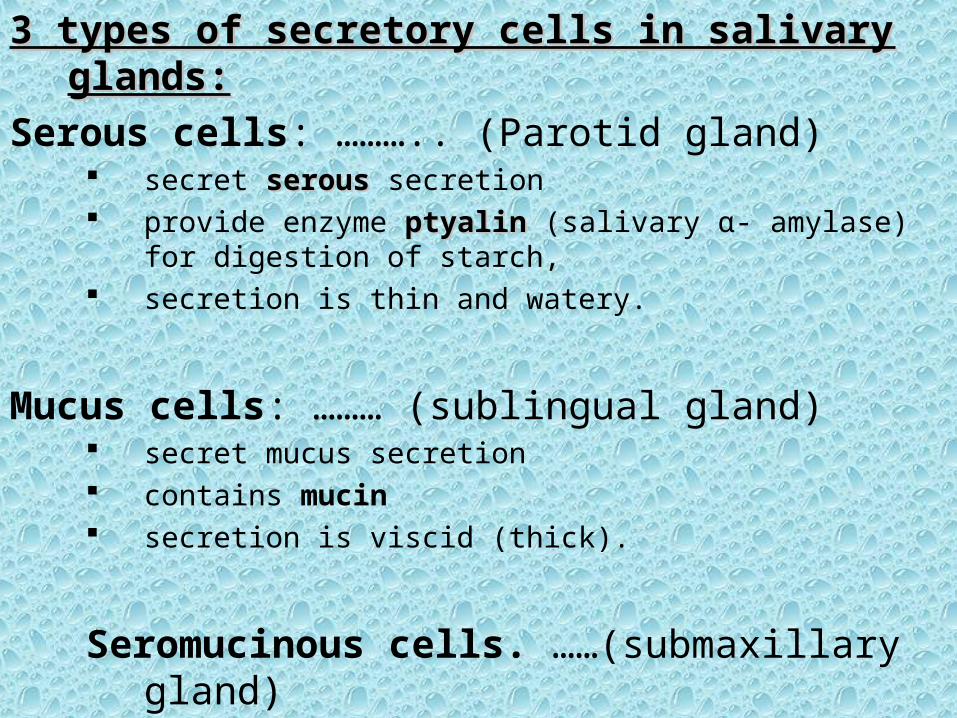
3 types of secretory cells in salivary glands:3 types of secretory cells in salivary glands:
Serous cells: ……….. (Parotid gland) secret serousserous secretion provide enzyme ptyalin ptyalin (salivary α- amylase) for
digestion of starch, secretion is thin and watery.
Mucus cells: ……… (sublingual gland) secret mucus secretion contains mucin secretion is viscid (thick).
Seromucinous cells. ……(submaxillary gland)

SALIVARY GLANDS: structure of gland is similar to a"bunch of grapes.
acinus,the blind end of each duct,
lined with acinar cells
secretes the initial saliva.
branching duct system is lined with columnar epithelial cells, modify the saliva.


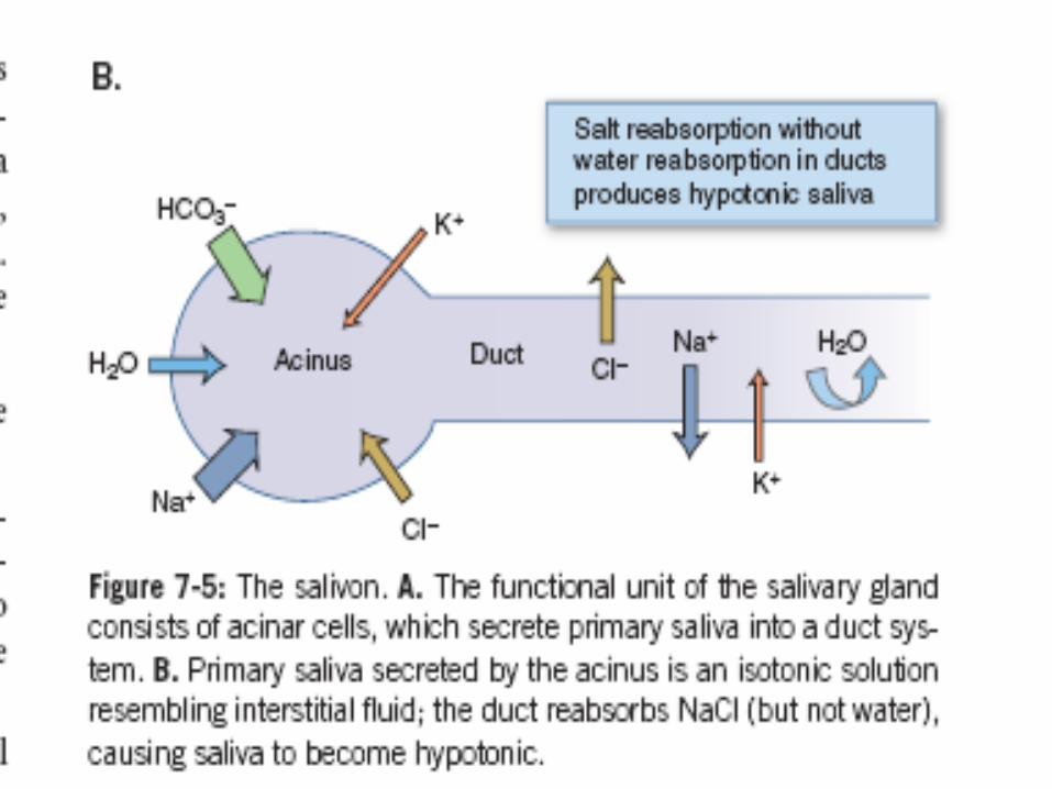
a- acinus– produces an initial saliva
– initial salivainitial saliva• composition is about the same as plasma.• is isotonic • has the same Na, k, Cl, and HCO concentrations as
plasma
Myoepithelial cells in acinus and initial ducts contract when saliva production is stimulated eject saliva into the mouth.

b- ductsmodify the saliva by the following processes:
1-reabsorb Na and Cl, makes the concentrations of these ions lower than plasma
concentrations
2-secrete K and HCO3 ,makes the concentrations of these ions higher than plasma
concentrations..
3- are relatively impermeable to water.
Saliva:…. Becomes: » hypotonic» dilute relative to plasma.

Aldosterone:Aldosterone: similar to kidney:
increases the absorption of Na and Cl⁺Na and Cl⁺ from the saliva ⁻secretion of K ⁺ K ⁺ to the saliva
Addison disease:?

Aldosterone:Aldosterone: similar to kidney:
increases the absorption of Na and Cl⁺Na and Cl⁺ from the saliva ⁻secretion of K ⁺ K ⁺ to the saliva
Addison disease:? there is a high Na /K ratio in saliva. ⁺ ⁺

COMPOSITION OF SALIVAdaily secretion of saliva is about 1-1.5 liter per day.
contains:1.1. Water 99.5%.Water 99.5%.
2.2. Solids 0.5%.Solids 0.5%.
solid materials are:1.1. OrganicOrganic
2.2. InorganicInorganic

Organic constituents of saliva:1. Protein mucin.
2. Ptyalin or α-amylase for the digestion of starch.
3. Lingual lipase plays an important role in the hydrolysis of triglycerides. differs from pancreatic lipase in that it does not need a
detergent for its action. digest as much as 30% of dietary triglycerides

4.Specific blood group antigen (ABO system): are present in 80% of the people are called (secretors) is important in medico-legal significance .
5. Immunoglobulin A can destroy the bacteria including those that
cause the dental caries.

Inorganic constituents of saliva:
ANIONS such as chloride, phosphate, bicarbonate, Floride
Floride is important to prevent dental caries,
CATIONS such as calcium, sodium and potassium.
calcium salts :might be the source of tartar deposits on the teeth.

PH of salivaPH of saliva

PH of saliva
is between (6-7.4). is quiet favorable for action of enzyme ptyalin.
At PH 7:At PH 7:saliva is saturated with calcium
so that teeth do not lose calcium to the saliva.
at more acidic PH at more acidic PH calcium will be lost from the teeth to saliva.

INNERVATION INNERVATION
OF OF
SALIVARY GLANDSSALIVARY GLANDS

INNERVATION OF THE SALIVARY GLANDSare supplied with:
efferent fibers from both parasympathetic sympathetic division of the autonomic nervous
system.
parasympathetic stimulation causes: Increase salivation (copious secretion). produces a rapid flow of large amount of watery
saliva from the gland.
Sympathetic stimulation causes:
1. Mainly vasoconstriction.
2. Some secretory response which is more variable than that of the parasympathetic
depends on the species and the gland, submandibular gland causes the secretion of
small amounts of thick viscid saliva rich in organic constituents
has no effect on parotid secretion.
3.Contraction of the myoepithelial cells.


FUNCTIONS OF SALIVA
1. moisten, lubricate and soften food,
2. keeps the mouth wet and facilitates speech.
3. important for the taste sensationit acts as a solvent.
4. contains 3 buffering systems are:a. bicarbonate,
b. phosphonate,
c. mucin.
5. has a digestive function through its enzyme ptyalin and lingual lipase.
6. Oral hygiene flow of saliva plays a very important role in maintaining
healthy oral tissues.
Disturbance of salivary secretion:Xerostomia (Deficiency of salivary secretion)
Due to:
• Emotional state such as fear or anxiety. ? ?• Dehydration. • Fever. • Anticholinergic drugs.
sialorhoea (Hyper salivation) due to:
• Pregnancy.• Tumours of the mouth or tongue or even a carious tooth
(reflex stimulation of salivary secretion due to local irritation).
• Diseases of the esophagus, stomach, pancreas such as tumor of the esophagus or spasm, gastric or duodenal ulcer, pancreatitis, (esophago-salivary reflex).

EsophagusEsophagus

Functions of the Esophagus:secretes mucus to prevent excoriation in upper esophagus and
to protect lower esophagus from acid.
• transports food into the stomach.• It does not produce digestive enzymes, and it does not carry
on absorption. • The passage of food from the laryngopharynx into the
esophagus is regulated at the entrance to the esophagus by a
sphincter (a circular band or ring of muscle that is normally contracted) called the upper esophageal sphincter or valve, it consists of skeletal muscle
• The elevation of the larynx causes the sphincter to relax, allowing the bolus to enter the esophagus.
This sphincter also relaxes during exhalation.• esophagus is controlled by the medulla oblongata and it is
innervated by vagus and sympathetic NS

• Just superior to the level of the diaphragm, the esophagus narrows slightly.
• This narrowing is a physiological sphincter in the inferior part of the esophagus composed of smooth muscle known as the lower esophageal sphincter (LES) or valve.
• in this case the esophagus, which functions
like a sphincter even though no sphincter muscle is actually present.)

mechanism of swallowing :mechanism of swallowing :
a-nasopharynx closes breathing is inhibited.
b- laryngeal muscles contract to close glottis elevate the larynx.
c- Peristalsis begins in pharynx to propel food toward esophagus.
upper esophageal sphincter relaxes:to permit entry of the food bolus into the esophagus.

mechanism of esophageal motility:a- upper esophageal sphincter relaxes to permit the swallowed food bolus to enter the esophagus.
b- upper esophageal sphincter then contracts so that food will not reflux.
c- A primary peristaltic contraction creates an area of increased pressure just behind the food bolus.
d- peristaltic contraction moves down the esophagus, propelling the food bolus along.
GravityGravity accelerates the movement.
e- A secondary peristaltic contraction clears the esophagus of any food remaining.
f- lower esophageal sphincter relaxes as the food bolus approaches it.
orad region of stomach relaxes (“receptive relaxation”),
allowing the food bolus to enter the stomach.

Esophagus
• Peristalsis: – Produced by a series of
localized reflexes in response to distention of wall by bolus.
• Wave-like muscular contractions:– Circular smooth muscle
contract behind, relaxes in front of the bolus.
• Followed by longitudinal contraction (shortening) of
• smooth muscle below the bolus and pushing its walls outward
• Rate of 2-4 cm/sec.
– After food passes into stomach, LES constricts.
Insert 18.4a

Clinical correlations of esophageal motility
a-Gastric reflux (Heartburn) :decreased tone of the lower esophageal sphincter (gastric contents reflux into esophagus), or
secondary peristalsis does not completely clear the esophagus of food.
b- Achalasia:•lower esophageal sphincter does not relax during swallowing,
•food accumulates in the esophagus.