Embed Size (px)
Citation preview
Note: This copy is for your personal non-commercial use only. To order presentation-ready copies for distribution to your colleagues or clients, contact us at www.rsna.org/rsnarights.
1053GASTROINTESTINAL IMAGING
Verghese George, MBBS, FRCR • Varaha Sairam Tammisetti, MD Venkateswar Rao Surabhi, MD • Alampady K. Shanbhogue, MD
Chronic fibrosing conditions of the abdomen are relatively poorly under-stood and involve varied and often multiple organ systems. At histopatho-logic analysis, they share the unifying features of proliferative fibrosis and chronic inflammation. Different conditions in this group are often found in association with each other and with other fibrosing conditions out-side the abdomen. Some of the confusion about these conditions stems from their complex nomenclature, which includes a gamut of alternate terms and eponyms. Many of them can be categorized within two large subgroups: the fibromatoses and immunoglobulin G4 (IgG4)–related disorders. While many of these entities are of uncertain etiology, some, especially the IgG4-associated conditions, appear to have an immune-mediated pathogenesis. Nephrogenic systemic fibrosis, sclerosing perito-nitis, and retroperitoneal fibrosis have iatrogenic associations, while some of the fibromatoses are genetically inherited. Imaging differentiation of these conditions is difficult due to considerable overlap in their radiologic findings. However, certain conditions such as penile fibromatosis and sclerosing peritonitis may have unique imaging features that can help the radiologist make the diagnosis. Others such as deep fibromatoses and in-flammatory pseudotumor demonstrate fibroproliferative mass formation and cannot be differentiated from neoplastic conditions at imaging. Thus, histopathologic correlation is often required to confirm their diagnosis.
IntroductionThe conditions described in this article manifest as infiltrating, often locally invasive fibroproliferative processes involving diverse sites in the abdomen and pelvis. Pro-gressive compressive encasement of abdominopelvic viscera by the infiltrating fibro-sis typically leads to mass effect and potentially fatal organ dysfunction. While some of the conditions are discrete, unrelated entities, many of them occur concurrently and in association with other fibrosing conditions involving the rest of the body.
Chronic Fibrosing Conditions in Abdominal Imaging1
Abbreviations: AIP = autoimmune pancreatitis, ANCA = anti–neutrophil cytoplasmic antibody, CAPD = continuous ambulatory peritoneal dialy-sis, CBD = common bile duct, FAP = familial adenomatous polyposis, FDG = fluorine 18 fluorodeoxyglucose, IgG4 = immunoglobulin G4, IMT = inflammatory myofibroblastic tumor, IPT = inflammatory pseudotumor, ISD = IgG4-related sclerosing disease, MIP = maximum intensity projec-tion, MOE = massive ovarian edema, NSF = nephrogenic systemic fibrosis, PSC = primary sclerosing cholangitis, RPF = retroperitoneal fibrosis, SEP = sclerosing encapsulating peritonitis, SSC = secondary sclerosing cholangitis
RadioGraphics 2013; 33:1053–1080 • Published online 10.1148/rg.334125081 • Content Codes: 1From the Department of Diagnostic and Interventional Imaging, University of Texas Health Science Center at Houston, MSB 2.130B, 6431 Fannin St, Houston, TX 77030 (V.G., V.S.T., V.R.S.); and Department of Radiology, University of Texas Health Science Center at San Antonio, San Antonio, Tex (A.K.S.). Presented as an education exhibit at the 2011 RSNA Annual Meeting. Received April 18, 2012; revision requested July 9; final revision received January 4, 2013; accepted January 11. For this journal-based SA-CME activity, the authors, editor, and reviewers have no financial relation-ships to disclose. Address correspondence to V.G. (e-mail: [email protected]).
©RSNA, 2013 • radiographics.rsna.org
INVITED COMMENTARY
See discussion on this article by Butler (pp 1080–1082).
SA-CME
See www.rsna .org/education
/search/RG
LEARNING OBJECTIVES FOR TEST 5
After completing this journal-based SA-
CME activity, partic-ipants will be able to:
■ Discuss the nomenclature of chronic fibrosing conditions of the abdomen.
■ Describe the pathophysiology and associations of these conditions.
■ List the imaging findings and differ-ential diagnoses of these conditions.
1054 July-August 2013 radiographics.rsna.org
Many of the entities described in this article can be categorized under one of two large sub-groups: the fibromatoses and immunoglobulin G4 (IgG4)–related disorders. The fibromatoses subgroup comprises a wide range of conditions that are characterized by infiltrative fibrous tis-sue proliferation and a tendency to recur locally. IgG4-related sclerosing disease (ISD), also known as hyper-IgG4 disease, is a recently described constellation of conditions that share the common histopathologic findings of lymphoplasmacytic inflammation with IgG4-positive cells and marked fibrosis. Many discrete conditions described in this article such as sclerosing mesenteritis, retroperito-neal fibrosis (RPF), sclerosing cholangitis, inflam-matory pseudotumor (IPT), and autoimmune pancreatitis (AIP) are now thought to fall under the IgG4-related umbrella. For the sake of clarity, however, these conditions have been described in-dividually, with a brief overview of ISD toward the end of the article.
Nomenclature relating to these conditions is dealt with in some detail, since it can be a source of confusion, resulting predominantly from the multiple alternate terms employed in the litera-ture to describe them. The condition previously known as desmoid is a typical case in point. Now identified as aggressive fibromatosis, desmoid belongs to the aforementioned subgroup called the fibromatoses. The multiple subtypes of fibro-matoses can involve different parts of the body; to further add to the confusion, many of these subtypes also have eponyms, such as Peyronie disease for penile fibromatosis and Ledderhose disease for plantar fibromatosis. Similar-sounding names for unrelated conditions, such as scleros-ing mesenteritis and sclerosing peritonitis, can also contribute to the lack of clarity in nomencla-ture. On a similar note, ovarian fibromatosis and neurofibromatosis may be mistakenly identified as entities within the fibromatoses subgroup.
The pathophysiology of many of these pro-cesses is complex and often poorly understood. A significant number of these entities are idiopathic. While nephrogenic systemic fibrosis (NSF) is the most notable iatrogenic condition in the group, others such as sclerosing peritonitis and RPF also have iatrogenic associations. Histopathologic findings of ISD suggest an immune-mediated mechanism for the components of that subgroup. The fibromatoses are unique among chronic fibrosing conditions in that a subset of them is genetically inherited.
Imaging findings of these conditions follow gen-eral radiologic characteristics of infiltrative fibrous tissue and hence often show considerable overlap;
their differentiation may thus be difficult without the aid of histopathologic correlation. However, a few conditions may have unique radiologic find-ings that can enable a diagnosis based on imaging findings alone. These include sclerosing encapsulat-ing peritonitis (SEP), penile fibromatosis, ovarian fibromatosis, and AIP. Some, such as deep fibroma-tosis and IPT, demonstrate fibroproliferative mass formation (as opposed to the more commonly seen infiltrative fibrosis) and cannot be differentiated from neoplasia without biopsy correlation.
After an overview of imaging features of fibrous tissue, chronic fibrosing conditions of the abdo-men are reviewed in the following order: sclerosing mesenteritis; sclerosing peritonitis; primary scle-rosing cholangitis (PSC); RPF; the fibromatoses (abdominal fibromatoses, penile fibromatosis); ovarian fibromatosis; IPT; NSF; AIP; and ISD.
Imaging Features of Fibrous TissueRegardless of location, fibrosis displays certain general radiologic characteristics. Fibrous tis-sue is hypoechoic at ultrasonography (US), often showing acoustic shadowing, with relative hy-povascularity at Doppler imaging. At computed tomography (CT), it appears isoattenuating to muscle, with corresponding isointensity noted at T1-weighted magnetic resonance (MR) imag-ing. It shows markedly low signal intensity at T2-weighted imaging. It typically does not show sig-nificant restriction at diffusion-weighted imaging due to reduced cellularity. Contrast-enhanced CT and MR imaging show relative paucity of enhance-ment in the parenchymal phase, with character-istic delayed phase enhancement. Variations from these appearances may be seen, depending on the degree of cellularity of the lesions and associated inflammatory changes.
Sclerosing MesenteritisThe term sclerosing mesenteritis refers to a spectrum (1) of uncommon, idiopathic, chronic inflamma-tory conditions affecting the mesentery (2). The condition predominantly affects men between the 5th and 7th decades of life and can lead to a vari-ety of nonspecific symptoms, including abdominal pain, nausea and vomiting, weight loss, and fever.
NomenclatureOwing to a multitude of alternate names for this condition, the nomenclature of sclerosing mesen-teritis has been mired in confusion. The following terms have been used to describe it in the past: systemic nodular panniculitis, liposclerosis mesenteri-tis, mesenteric Weber-Christian disease, mesenteric lipogranuloma, and xanthogranulomatous mesen-teritis (2). In addition, the subtypes of the condi-tion—namely, mesenteric panniculitis, mesenteric
RG • Volume 33 Number 4 George et al 1055
lipodystrophy, and retractile mesenteritis—can further add to the confusion.
Pathophysiology and AssociationsAs mentioned earlier, sclerosing mesenteritis is an umbrella term for a spectrum of conditions. Sclerosing mesenteritis is categorized into three subtypes—mesenteric panniculitis, mesenteric lipodystrophy, and retractile mesenteritis—on the basis of the predominant histopathologic finding in the affected mesentery. Although most patients with the condition have a range of mesenteric findings, usually one histopathologic feature is predominant at a given time: chronic inflamma-tion in mesenteric panniculitis, fat necrosis in mesenteric lipodystrophy, and fibrosis in retrac-tile mesenteritis. The condition most commonly involves the small bowel mesentery but may oc-casionally involve the mesocolon and rarely the omentum or retroperitoneum (3).
As mentioned earlier, sclerosing mesenteritis is now considered to be one of the abdominal com-ponents of ISD and hence can coexist with the
other conditions in that subgroup, such as scleros-ing cholangitis, RPF, and IPT. It is also associated with other fibrosing conditions such as Riedel thyroiditis and orbital pseudotumor (3). An as-sociation with malignancy—especially lymphoma, melanoma, and breast, lung, and colon cancers—has also been reported (4), most commonly with the mesenteric panniculitis subtype (5). Reports of associations with infection, trauma, and surgery also exist in the literature (2).
Imaging FindingsCross-sectional imaging findings of sclerosing mesenteritis range from haziness of the mesentery to a soft-tissue mass at the mesenteric root (3). The following findings and named signs character-ize the disorder:
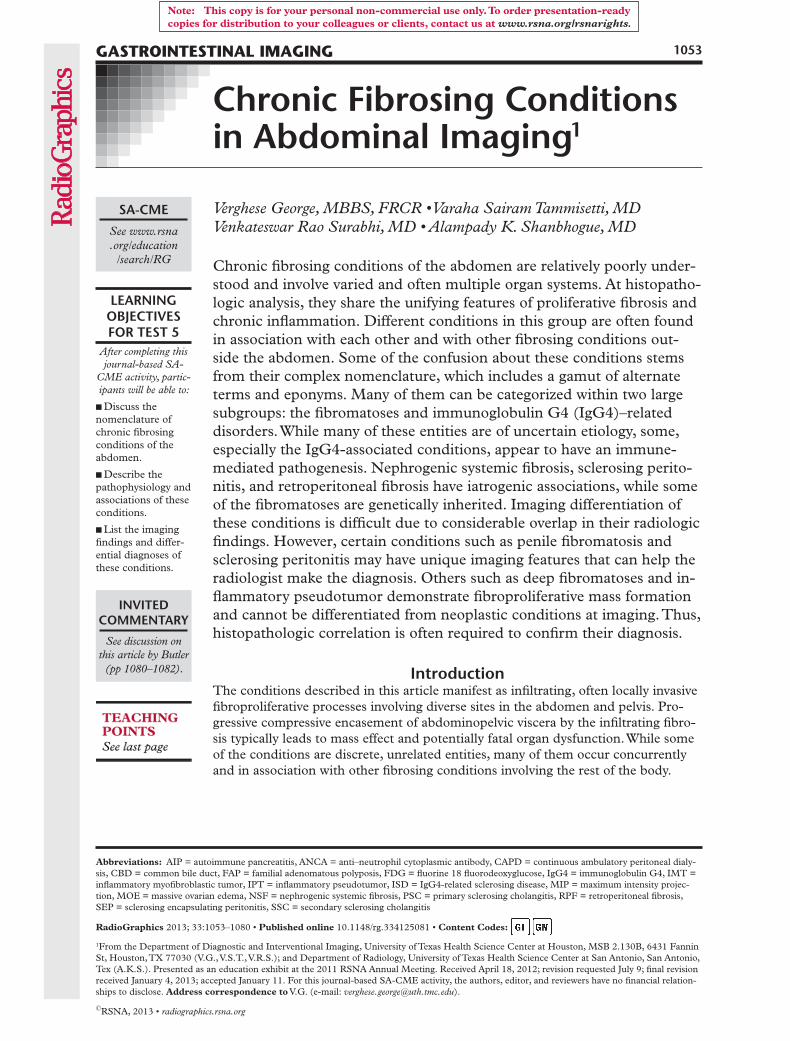
Misty Mesentery Sign.—The misty mesentery sign refers to the presence of subtle increased attenuation in the mesentery at CT, often associ-ated with small to borderline-sized nodes (Fig 1).
Figure 1. Mesenteric panniculitis. (a) Axial contrast-enhanced CT image shows the misty mesentery sign as haziness of the affected mesentery (arrow). Note the multiple mesenteric nodes. (b) Axial contrast-enhanced CT image shows the fat-ring sign as preservation of fatty attenuation around vessels in the in-volved mesentery (arrow). (c) Axial contrast-enhanced CT image shows mesenteric haziness and a tumoral pseudocapsule (arrow) at the interface between normal and affected mesentery. (d) Coronal contrast-enhanced CT image shows mesenteric haziness, the fat-ring sign, and a tumoral pseudocapsule (arrow).
1056 July-August 2013 radiographics.rsna.org
This finding represents chronic inflammation of the mesentery and hence is most commonly as-sociated with the mesenteric panniculitis subtype. However, misty mesentery is a nonspecific sign and can be seen in any infiltrating process involv-ing the mesentery, such as hemorrhage, edema, or malignancy (especially lymphoma) (3).
Fat-Ring Sign.—The fat-ring sign appears at CT as preservation of fatty attenuation, in the form of a low-attenuation halo, around the vessels of the involved mesentery (Fig 1b, 1d). Typically associ-ated with mesenteric panniculitis and mesenteric lipodystrophy, this sign can help differentiate sclerosing mesenteritis from other mesenteric infiltrating processes.
Tumoral Pseudocapsule.—A band of soft-tissue attenuation called the tumoral pseudocapsule at the edge of the affected mesentery (Fig 1c, 1d) has been reported in up to 50% of cases of sclerosing mesenteritis, mainly of the mesenteric panniculitis and mesenteric lipodystrophy sub-types (2). As with the fat-ring sign, the tumoral pseudocapsule is an important distinguishing feature of sclerosing mesenteritis.
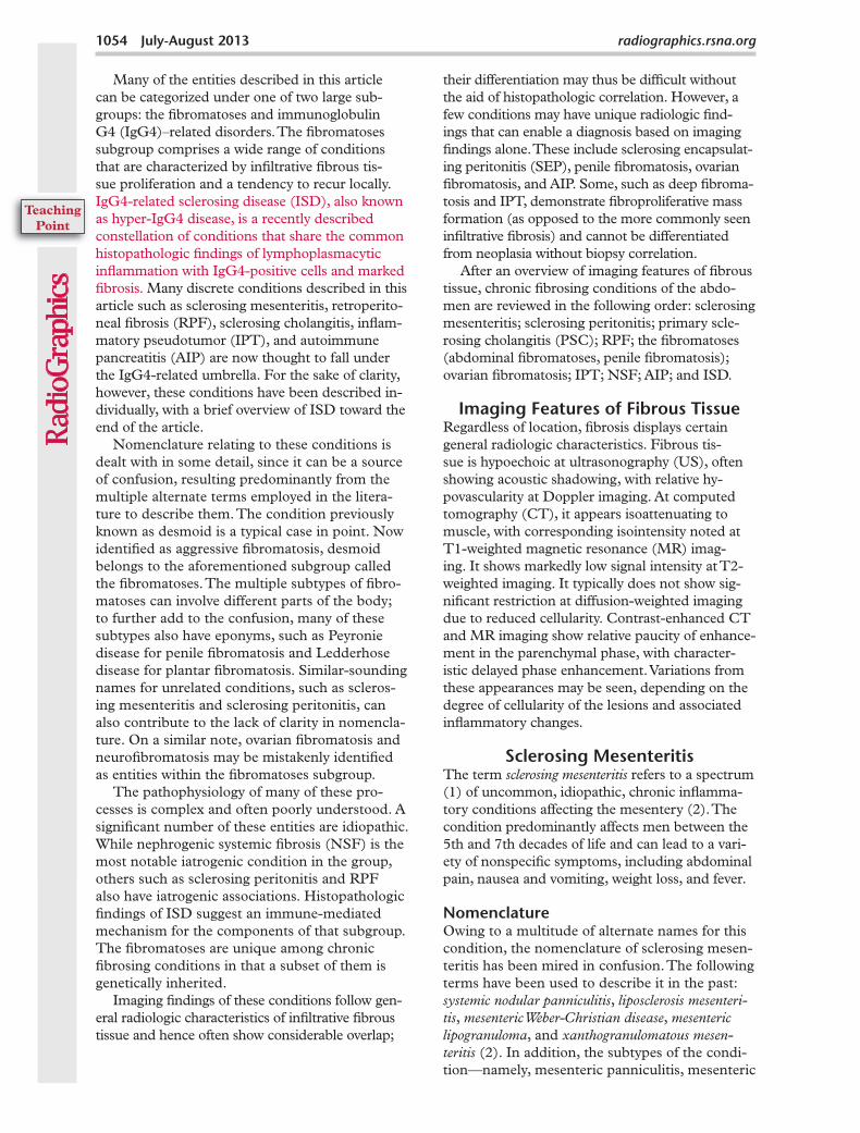
Mesenteric Root Mass.—The most common imaging appearance of sclerosing mesenteritis is a mass at the root of the small bowel mesentery. While the mass is typically ill-defined, heteroge-neous, and of relatively reduced attenuation in mesenteric panniculitis, it is usually denser, more homogeneous, and better-defined in retractile mesenteritis. As the name suggests, retractile mesenteritis is often associated with desmoplastic response and spiculation in relation to the mass (Fig 2). Calcification (Fig 2b) and cystic changes may also be seen in the mass (3).
As the disease progresses, findings related to progressive fibrosis and shortening of the mesen-tery may be seen. These include compression of the mesenteric vessels with formation of collateral vessels and possible bowel ischemia. Bowel may also be more directly involved by progressive en-casement, although bowel involvement is much less frequent than in lymphoma (2,3), the main differential diagnosis for sclerosing mesenteritis.
Differential DiagnosisOwing to the nonspecific nature of its imaging findings, sclerosing mesenteritis has a wide differ-ential diagnosis.
Lymphoma is probably the most important condition mimicking sclerosing mesenteritis. As with sclerosing mesenteritis, lymphoma can also manifest as a mesenteric mass with nodes; typically, however, no mesenteric calcification is noted in lymphoma (except after treatment). Also unlike sclerosing mesenteritis, lymphoma does not typically cause compressive narrowing of the mesenteric vessels despite encasing them. While treated lymphoma can also produce a misty mes-entery appearance, the presence of a fat-ring sign and tumoral pseudocapsule can help differentiate mesenteric panniculitis from lymphoma.
The other differential diagnoses for mesenteric panniculitis include IPT, Crohn disease with fi-
Figure 2. Retractile mesenteritis. Coronal contrast-enhanced (a) and axial nonenhanced (b) CT images in different patients with retractile mesenteritis show spiculated mesenteric masses with markedly different degrees of calcification.
RG • Volume 33 Number 4 George et al 1057
brofatty proliferation, and mesenteric lipogenic liposarcoma (2).
The primary differential diagnosis for retractile mesenteritis is mesenteric carcinoid, which also appears as a spiculated mesenteric root mass with possible calcification. Carcinoid may be differ-entiated from retractile mesenteritis by means of uptake at octreotide and somatostatin scintigra-phy, elevated levels of blood serotonin and urine 5-hydroxyindoleacetic acid, and typical clinical features, especially carcinoid syndrome with liver metastases. The other differential diagnoses for retractile mesenteritis include desmoid (mesen-teric fibromatosis) and carcinomatosis.
Sclerosing PeritonitisSclerosing peritonitis is a condition marked by chronic fibrotic thickening of the peritoneum (6). In the more severe version of the condition—SEP—the thickened abnormal peritoneum can progress to encase small bowel loops in a fibrocol-lagenous “abdominal cocoon” (7), eventually re-sulting in recurrent small bowel obstruction.
NomenclatureSclerosing peritonitis has been referred to in the literature by multiple alternate terms, including encapsulating peritoneal sclerosis, peritonitis chronica fibrosa incapsulata, and peritoneal fibrosing syn-drome (8–10).
Pathophysiology and AssociationsWhile the exact etiology of the condition is un-clear, chronic irritation of the peritoneum has been postulated as the cause (11). This is best
demonstrated by the well-recognized association of SEP with continuous ambulatory peritoneal dialysis (CAPD). The overall prevalence of SEP in patients receiving CAPD has been reported to be 0.7%, but it increases to 19.4% in patients receiving CAPD for more than 8 years (6,12). A similar association also exists with ventriculoperi-toneal and peritoneovenous shunts (9,13).
Other associations include tuberculosis (14), sarcoidosis (15), familial Mediterranean fever, gastrointestinal malignancy, protein S defi-ciency, liver transplantation, fibrogenic foreign material, and luteinized ovarian thecoma (9). It has also been reported with use of the b-blocker practolol (16). Foo et al (7) reported an idiopathic form of the condition in adolescent females from tropical and subtropical countries; however, the same form has now been described in male and pediatric patients from nontropical regions as well (9,17).
Imaging FindingsThe main imaging features of sclerosing peritoni-tis pertain to appearances of the abnormal perito-neum and of its encasing effects on the bowel (6).
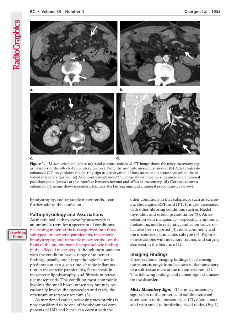
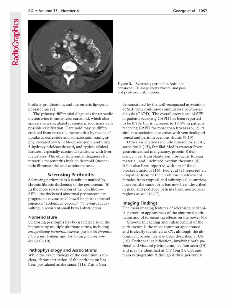
Smooth thickening and enhancement of the peritoneum is the most common appearance and is clearly identified at CT, although the ab-dominal cocoon has also been described at US (18). Peritoneal calcification, involving both pa-rietal and visceral peritoneum, is often seen (19) and may be identified at CT (Fig 3), US, and plain radiography. Although diffuse peritoneal
Figure 3. Sclerosing peritonitis. Axial non-enhanced CT image shows visceral and pari-etal peritoneal calcification.
1058 July-August 2013 radiographics.rsna.org
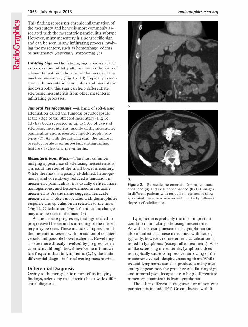
calcification is typically associated with advanced encapsulating peritonitis, severe encapsulation may also occur in the absence of peritoneal calci-fication (Fig 4a) (6).
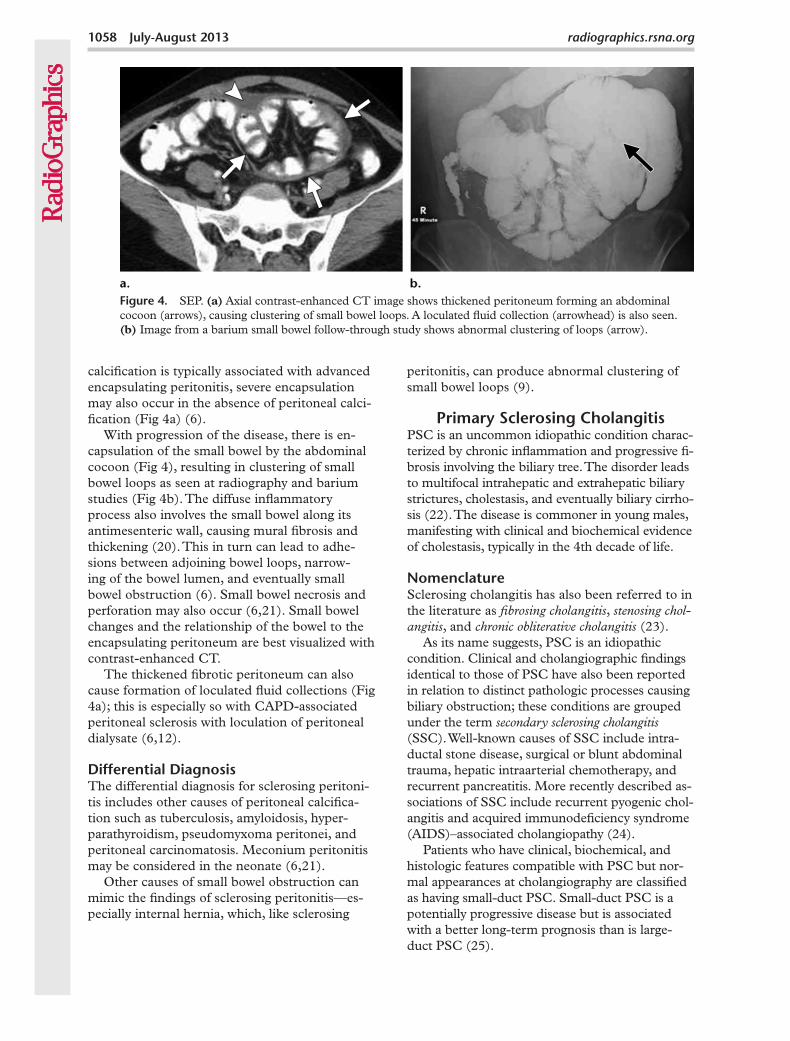
With progression of the disease, there is en-capsulation of the small bowel by the abdominal cocoon (Fig 4), resulting in clustering of small bowel loops as seen at radiography and barium studies (Fig 4b). The diffuse inflammatory process also involves the small bowel along its antimesenteric wall, causing mural fibrosis and thickening (20). This in turn can lead to adhe-sions between adjoining bowel loops, narrow-ing of the bowel lumen, and eventually small bowel obstruction (6). Small bowel necrosis and perforation may also occur (6,21). Small bowel changes and the relationship of the bowel to the encapsulating peritoneum are best visualized with contrast-enhanced CT.
The thickened fibrotic peritoneum can also cause formation of loculated fluid collections (Fig 4a); this is especially so with CAPD-associated peritoneal sclerosis with loculation of peritoneal dialysate (6,12).
Differential DiagnosisThe differential diagnosis for sclerosing peritoni-tis includes other causes of peritoneal calcifica-tion such as tuberculosis, amyloidosis, hyper-parathyroidism, pseudomyxoma peritonei, and peritoneal carcinomatosis. Meconium peritonitis may be considered in the neonate (6,21).
Other causes of small bowel obstruction can mimic the findings of sclerosing peritonitis—es-pecially internal hernia, which, like sclerosing
peritonitis, can produce abnormal clustering of small bowel loops (9).
Primary Sclerosing CholangitisPSC is an uncommon idiopathic condition charac-terized by chronic inflammation and progressive fi-brosis involving the biliary tree. The disorder leads to multifocal intrahepatic and extrahepatic biliary strictures, cholestasis, and eventually biliary cirrho-sis (22). The disease is commoner in young males, manifesting with clinical and biochemical evidence of cholestasis, typically in the 4th decade of life.
NomenclatureSclerosing cholangitis has also been referred to in the literature as fibrosing cholangitis, stenosing chol-angitis, and chronic obliterative cholangitis (23).
As its name suggests, PSC is an idiopathic condition. Clinical and cholangiographic findings identical to those of PSC have also been reported in relation to distinct pathologic processes causing biliary obstruction; these conditions are grouped under the term secondary sclerosing cholangitis (SSC). Well-known causes of SSC include intra-ductal stone disease, surgical or blunt abdominal trauma, hepatic intraarterial chemotherapy, and recurrent pancreatitis. More recently described as-sociations of SSC include recurrent pyogenic chol-angitis and acquired immunodeficiency syndrome (AIDS)–associated cholangiopathy (24).
Patients who have clinical, biochemical, and histologic features compatible with PSC but nor-mal appearances at cholangiography are classified as having small-duct PSC. Small-duct PSC is a potentially progressive disease but is associated with a better long-term prognosis than is large-duct PSC (25).
Figure 4. SEP. (a) Axial contrast-enhanced CT image shows thickened peritoneum forming an abdominal cocoon (arrows), causing clustering of small bowel loops. A loculated fluid collection (arrowhead) is also seen. (b) Image from a barium small bowel follow-through study shows abnormal clustering of loops (arrow).
RG • Volume 33 Number 4 George et al 1059
Pathophysiology and AssociationsAs mentioned earlier, the exact etiology of PSC is unclear. Along with autoimmune pancreatitis, scle-rosing mesenteritis, and RPF, PSC is now consid-ered a key abdominal component of the recently described group of IgG4-related sclerosing dis-orders. Even earlier, the association of PSC with autoimmune disorders such as Sjögren syndrome, systemic lupus erythematosus, and rheumatoid arthritis, and the response of PSC to treatment with steroids, had led to theories that PSC is an immune-mediated obliterative cholangitis (24). In addition, a large number of autoantibodies such as anti–neutrophil cytoplasmic antibody (ANCA) and antinuclear antibody have been detected in patients with PSC, but the specificity and titer of these antibodies are generally low (26).
PSC is intimately associated with inflam-matory bowel disease, with an 80% association
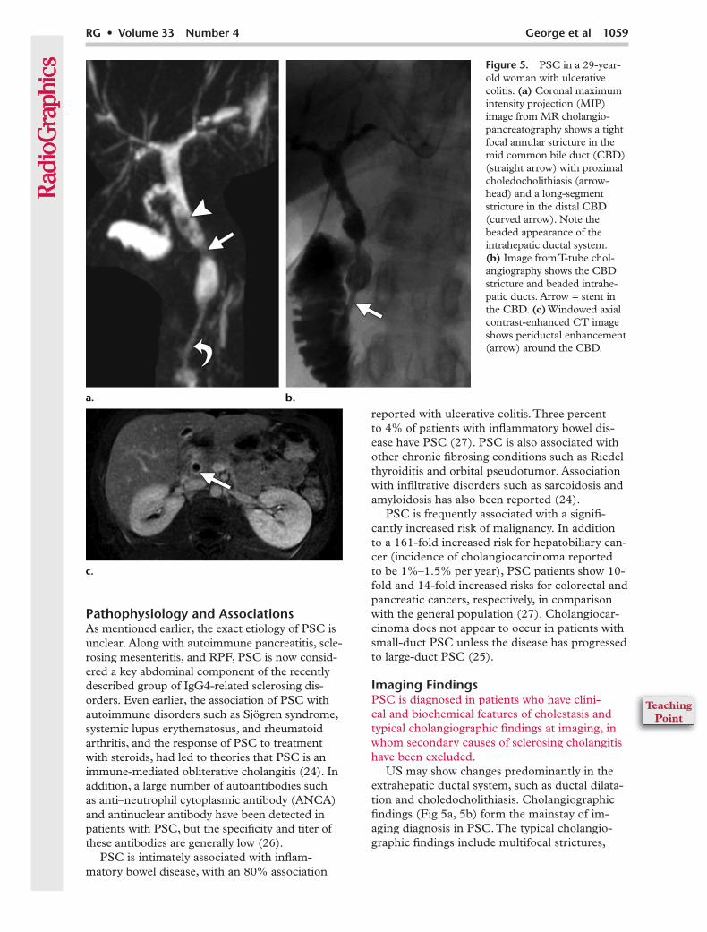
Figure 5. PSC in a 29-year-old woman with ulcerative colitis. (a) Coronal maximum intensity projection (MIP) image from MR cholangio-pancreatography shows a tight focal annular stricture in the mid common bile duct (CBD) (straight arrow) with proximal choledocholithiasis (arrow-head) and a long-segment stricture in the distal CBD (curved arrow). Note the beaded appearance of the intrahepatic ductal system. (b) Image from T-tube chol-angiography shows the CBD stricture and beaded intrahe-patic ducts. Arrow = stent in the CBD. (c) Windowed axial contrast-enhanced CT image shows periductal enhancement (arrow) around the CBD.
reported with ulcerative colitis. Three percent to 4% of patients with inflammatory bowel dis-ease have PSC (27). PSC is also associated with other chronic fibrosing conditions such as Riedel thyroiditis and orbital pseudotumor. Association with infiltrative disorders such as sarcoidosis and amyloidosis has also been reported (24).
PSC is frequently associated with a signifi-cantly increased risk of malignancy. In addition to a 161-fold increased risk for hepatobiliary can-cer (incidence of cholangiocarcinoma reported to be 1%–1.5% per year), PSC patients show 10-fold and 14-fold increased risks for colorectal and pancreatic cancers, respectively, in comparison with the general population (27). Cholangiocar-cinoma does not appear to occur in patients with small-duct PSC unless the disease has progressed to large-duct PSC (25).
Imaging FindingsPSC is diagnosed in patients who have clini-cal and biochemical features of cholestasis and typical cholangiographic findings at imaging, in whom secondary causes of sclerosing cholangitis have been excluded.
US may show changes predominantly in the extrahepatic ductal system, such as ductal dilata-tion and choledocholithiasis. Cholangiographic findings (Fig 5a, 5b) form the mainstay of im-aging diagnosis in PSC. The typical cholangio-graphic findings include multifocal strictures,
1060 July-August 2013 radiographics.rsna.org
segmental dilatation, ductal webs and diverticula, and cholestasis-related choledocholithiasis. Wall thickening and enhancement of the extrahepatic ductal system may be seen (28) (Fig 5c), al-though significant periductal soft tissue (>1.5 cm thick) with delayed enhancement should raise the possibility of cholangiocarcinoma (22).
Early in the disease, short, randomly distributed annular biliary strictures are noted, which can involve the intrahepatic and extrahepatic ducts. Ductal dilatation is often only minimal, despite the degree of stenosis related to the strictures. Multi-focal strictures alternating with minimally dilated intervening ductal segments give rise to the classic beaded appearance of PSC (22).
Localized high-grade strictures of the larger ducts, called dominant strictures, may be super-imposed on diffuse ductal changes and are an im-portant imaging finding of PSC, found in approx-imately 45% of patients. A dominant stricture has been defined as ductal stenosis with a luminal di-ameter of 1.5 mm or less in the CBD and 1 mm or less in the right and left hepatic ducts (29). As the disease progresses, worsening obliterative cholangitis results in the characteristic “pruned tree” appearance caused by the obliteration and subsequent lack of visualization of the peripheral intrahepatic ducts at cholangiography.
While endoscopic retrograde cholangiopan-creatography (ERCP) has traditionally been con-sidered the standard of reference in diagnosing PSC, MR cholangiopancreatography has rapidly gained acceptance, mainly due to its noninva-sive nature. At MR cholangiopancreatography, ductal stenosis is usually inferred on the basis of prestenotic dilatation, which is typically mild in PSC, as described earlier. Thus, ERCP is usually better in detecting early stenosis; however, visu-alization of peripheral ducts may be better at MR cholangiopancreatography due to the difficulty of distending peripheral ducts past a central-duct dominant stricture at ERCP (Fig 5a). ERCP also has the added advantage of the ability to perform concurrent therapeutic interventions in the ducts, such as dilatation or stent placement.
Differential DiagnosisImaging findings of PSC are indistinguishable from those of SSC. As mentioned earlier, second-ary causes of sclerosing cholangitis need to be
excluded by nonimaging means before a diagno-sis of PSC can be made.
The key differential diagnosis for PSC is chol-angiocarcinoma, especially the periductal infil-trating type (30). Cholangiographic features sug-gestive of cholangiocarcinoma include an irregu-lar high-grade ductal stricture with shouldered margins, rapid progression of strictures, marked ductal dilatation proximal to strictures (as men-tioned earlier, PSC produces only minimal ductal dilatation), and significant periductal soft tissue, typically with delayed enhancement (22). While a high-grade ductal stricture is more often benign than malignant, a dominant stricture should al-ways raise the possibility of cholangiocarcinoma, especially since cholangiocarcinoma may develop in 10%–15% of PSC cases.
Retroperitoneal FibrosisRPF is characterized by development of aggres-sive confluent fibroproliferative masses in the retroperitoneal space, leading to progressive encasement of the retroperitoneal structures, especially the ureters (31). A rare condition with a prevalence of 1 in 200,000 (32), it is typically seen in men (two to three times more often than in women) between the 5th and 7th decades of life (31,33).
NomenclatureAlternate names for RPF include Ormond disease (34), Gerota fasciitis, periureteritis fibrosa, perirenal fasciitis, sclerosing lipogranuloma (35), sclerosing retroperitoneal granuloma (36), nonspecific retroperi-toneal inflammation, sclerosing retroperitonitis, ret-roperitoneal vasculitis with perivascular fibrosis, and chronic periaortitis (31,37).
Pathophysiology and AssociationsRPF is idiopathic in more than 70% of cases (31). Up to 15% of patients have additional fi-brotic processes outside the retroperitoneum, the most notable being mediastinal fibrosis, Riedel thyroiditis, sclerosing cholangitis, sclerosing mes-enteritis, and orbital pseudotumor (33,37).
RPF is often seen in association with athero-sclerosis, the fibrotic mass encasing a severely atherosclerotic vessel, usually the aorta (Fig 6a, 6b). In these cases, it has been postulated that the fibrosis develops as an immune response to leakage of an insoluble lipid—ceroid—from the atheromatous plaque into the perivascular tis-
RG • Volume 33 Number 4 George et al 1061
sues (38). On the same lines, the coexistence of RPF in association with autoimmune disorders such as p-ANCA–associated polyarteritis no-dosa (39) and IgG4-related sclerosing disorders (40,41) and its response to steroids have raised the possibility that RPF is of autoimmune origin. As mentioned earlier, RPF is now considered an important abdominal component of the group of IgG4-related sclerosing disorders.
The most common known cause of RPF is use of methysergide, an ergot derivative, in treat-ment of migraine. Other ergot derivatives linked to RPF include lysergic acid diethylamide (LSD)
and bromocriptine (42). Beta blockers, methyl-dopa, and hydralazine have also been reported to be associated with RPF (37). RPF is also associ-ated with infectious and inflammatory processes, retroperitoneal hemorrhage (37), smoking, and asbestos exposure (43).
Malignancy is associated with up to 8% of RPF cases (33). It is postulated that desmoplas-tic response elicited from small retroperitoneal metastatic deposits results in development of a confluent fibrotic process that is difficult to
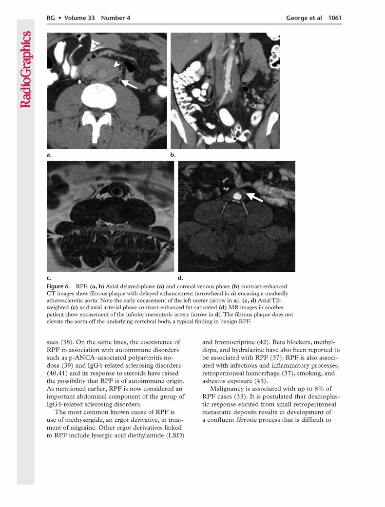
Figure 6. RPF. (a, b) Axial delayed-phase (a) and coronal venous phase (b) contrast-enhanced CT images show fibrous plaque with delayed enhancement (arrowhead in a) encasing a markedly atherosclerotic aorta. Note the early encasement of the left ureter (arrow in a). (c, d) Axial T2-weighted (c) and axial arterial phase contrast-enhanced fat-saturated (d) MR images in another patient show encasement of the inferior mesenteric artery (arrow in d). The fibrous plaque does not elevate the aorta off the underlying vertebral body, a typical finding in benign RPF.
1062 July-August 2013 radiographics.rsna.org
differentiate from RPF due to other causes, es-pecially in patients with no known primary ma-lignancy (37). Malignancies reported with this association include breast, lung, thyroid, gastro-intestinal, and genitourinary cancers, as well as lymphomas and some sarcomas (33,37).
Imaging FindingsRPF begins as a confluent fibrotic plaque, typi-cally below the aortic bifurcation at the L4-5 level. It can then extend contiguously along the midline retroperitoneum, usually in a superior direction, encasing the aorta, IVC, and eventually the ureters, causing hydronephroureterosis. Typi-cally, the process does not show lateral extension beyond the lateral margins of the psoas muscles (37); also, the fibrotic plaque characteristically does not cause anterior displacement of the aorta and IVC from the spine (31,37). Further pro-gression may show extension toward the renal hila, duodenum, pancreas, porta hepatis, and spleen. Extension cranially into the mediastinum and caudally into the pelvis can occur.
Before the advent of cross-sectional imaging, excretory urography was the imaging modality of choice for diagnosis of RPF. Thus, the condition had to have progressed to involve the renal tracts for a diagnosis to be made at urography. The findings include medial deviation and tapering of the middle third of the ureters at the L4-5 level, with proximal hydronephroureterosis and delayed excretion of contrast material (31,44).
Cross-sectional imaging, particularly CT and MR imaging, has now replaced urography in diagnosis and follow-up of RPF. These modali-ties rely on demonstration of the fibrotic plaque and the effects of progressive encasement by the plaque. At CT, the plaque typically has the same attenuation as muscle (Fig 6a, 6b). At MR imag-ing (Fig 6c, 6d), it has low signal intensity on T1- and T2-weighted images.
High T2 signal may be seen in the plaque, a finding indicative of inflammatory edema and as
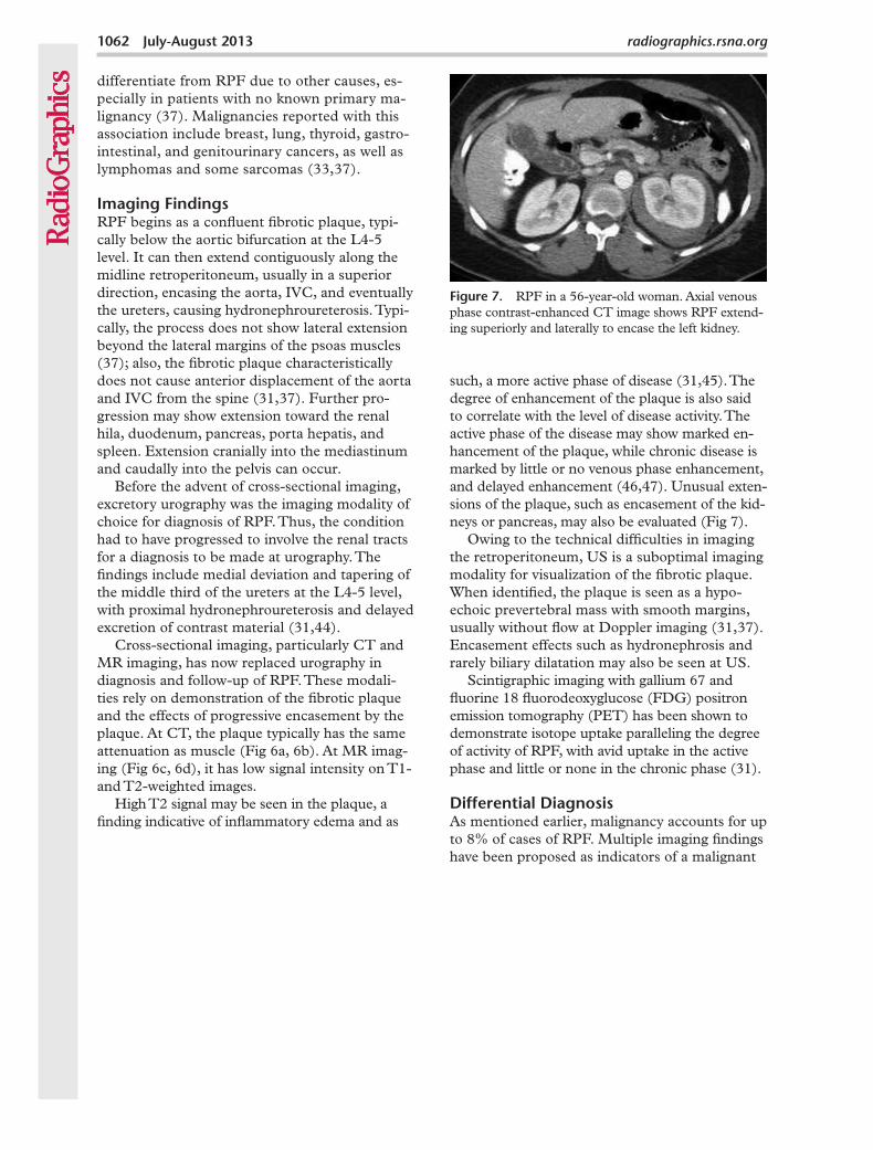
such, a more active phase of disease (31,45). The degree of enhancement of the plaque is also said to correlate with the level of disease activity. The active phase of the disease may show marked en-hancement of the plaque, while chronic disease is marked by little or no venous phase enhancement, and delayed enhancement (46,47). Unusual exten-sions of the plaque, such as encasement of the kid-neys or pancreas, may also be evaluated (Fig 7).
Owing to the technical difficulties in imaging the retroperitoneum, US is a suboptimal imaging modality for visualization of the fibrotic plaque. When identified, the plaque is seen as a hypo-echoic prevertebral mass with smooth margins, usually without flow at Doppler imaging (31,37). Encasement effects such as hydronephrosis and rarely biliary dilatation may also be seen at US.
Scintigraphic imaging with gallium 67 and fluorine 18 fluorodeoxyglucose (FDG) positron emission tomography (PET) has been shown to demonstrate isotope uptake paralleling the degree of activity of RPF, with avid uptake in the active phase and little or none in the chronic phase (31).
Differential DiagnosisAs mentioned earlier, malignancy accounts for up to 8% of cases of RPF. Multiple imaging findings have been proposed as indicators of a malignant
Figure 7. RPF in a 56-year-old woman. Axial venous phase contrast-enhanced CT image shows RPF extend-ing superiorly and laterally to encase the left kidney.
RG • Volume 33 Number 4 George et al 1063
cause of RPF, including anterior displacement of the aorta by the retroperitoneal mass, marginal lobulation of the mass, and edema, Doppler flow, contrast enhancement, and isotope uptake in relation to the mass (48). However, results of im-aging are often suboptimal in making this distinc-tion, and biopsy may be required to definitively exclude malignancy (31,37).
Metastatic adenopathy, especially confluent adenopathy as in lymphoma, is an important dif-ferential diagnosis for RPF. As with malignant RPF, metastatic retroperitoneal adenopathy typi-cally elevates the aorta off the underlying verte-bral body (49), although exceptions to this guide-line have been reported (50). Other differential diagnoses include retroperitoneal amyloidosis and occasionally subacute retroperitoneal hemor-rhage (37). Atypical locations of RPF can widen the differential diagnosis. For example, RPF in the female pelvis has been reported to mimic cer-vical carcinoma (51).
The FibromatosesThe term fibromatosis refers to a wide range of clinicopathologic conditions with benign pro-liferation of fibrous tissue, characterized by in-filtrative growth pattern and frequent tendency
to recur locally, although lacking in metastatic potential (52,53).
Nomenclature and ClassificationNo single entity in this article better illustrates the nomenclature-associated confusion of chronic fibrosing conditions than the fibromatoses. The group is divided into many subcategories con-taining multiple entities, most of them with alter-nate names and eponyms.
Fibromatoses are broadly categorized as super-ficial (fascial) and deep (musculoaponeurotic). Superficial fibromatoses are generally small and slow-growing (typically <5 cm), while deep fibro-matoses are larger, more aggressive masses that may show rapid growth (54,55). Superficial fibro-matoses rarely involve deeper structures, while deep fibromatoses have a variable tendency to infiltrate surrounding visceral organs. Superficial and deep fibromatoses are further subclassified as in the Table.
Owing to their clinical behavior, deep fibroma-toses are now more commonly referred to as ag-gressive fibromatoses. However, they continue to be frequently referred to by the older term desmoid (Greek desmoid meaning tendon-like), as used in 1838 by Johannes Mueller (56). In this article, the terms aggressive fibromatosis and desmoid are used interchangeably to describe deep fibromatoses. In 2002, the Committee for Classification of Soft Tis-sue Tumors of the World Health Organization des-ignated the term desmoid-type fibromatosis for deep fibromatoses (57); this term encompasses extraab-dominal, abdominal wall, and intrabdominal fibro-matoses (52). Use of terms like nonmetastasizing fibrosarcoma and fibrosarcoma grade 1 to describe deep fibromatoses has been phased out of the lit-erature due to their unpleasant connotations (58).
Among the fibromatoses, abdominal imaging deals with essentially two entities: abdominal fi-bromatosis and penile fibromatosis.
Abdominal FibromatosesDesmoid-type fibromatoses or aggressive fibroma-toses in the abdomen are further classified on the
Classification of Fibromatoses
Superficial (fascial) Palmar fibromatosis (Dupuytren disease) Plantar fibromatosis (Ledderhose disease) Juvenile aponeurotic fibroma Infantile digital fibromatosis Penile fibromatosis (Peyronie disease)Deep (musculoaponeurotic) Extraabdominal fibromatosis Abdominal fibromatosis Abdominal wall fibromatosis Intraabdominal fibromatosis Mesenteric fibromatosis Retroperitoneal fibromatosis Pelvic fibromatosis Fibromatosis associated with Gardner syndrome Infantile myofibromatosis Fibromatosis colli
1064 July-August 2013 radiographics.rsna.org
trauma (including surgery), and genetic abnor-malities have been described as potential caus-ative factors (52).
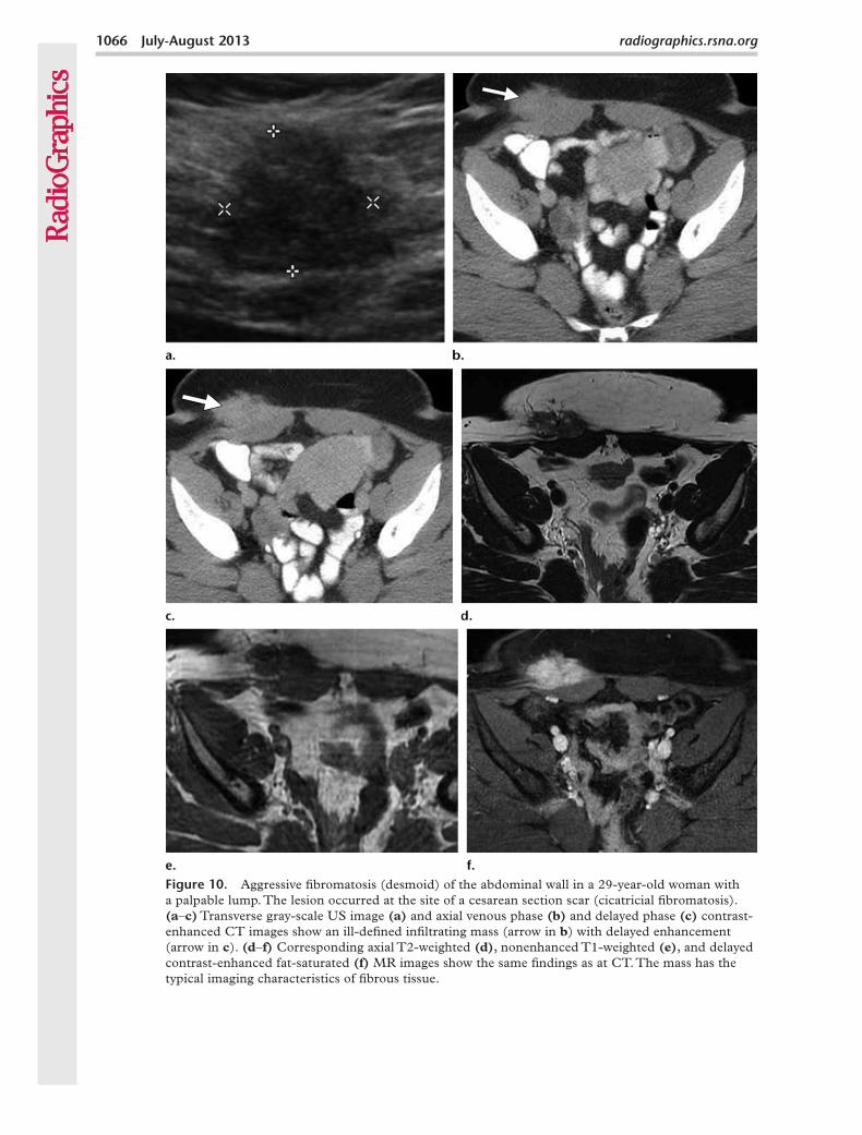
The pattern of occurrence implicates estrogen as a growth factor for fibroblasts, and regression of the lesions after menopause or oophorectomy has been reported (52,60). Abdominal wall des-moids may also arise in areas of previous abdomi-nal surgery such as cesarean section scars, indi-cating that trauma is an important contributory factor, as in other desmoid-type fibromatoses (52). Such lesions arising from scars have been described as cicatricial fibromatoses (61).
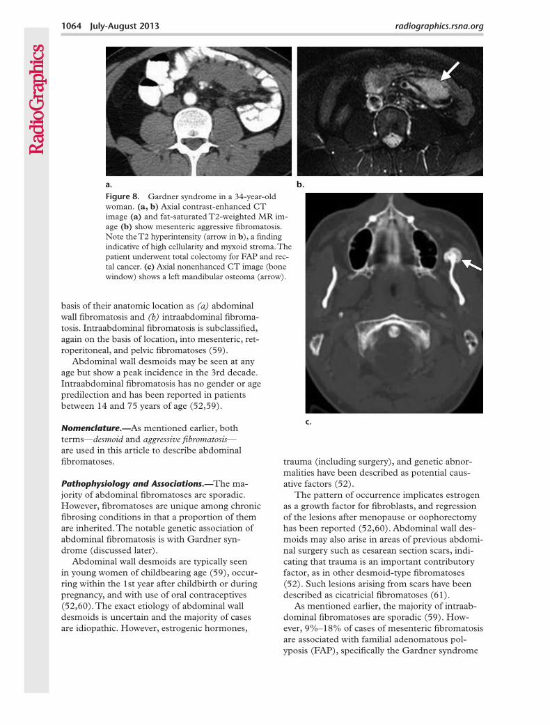
As mentioned earlier, the majority of intraab-dominal fibromatoses are sporadic (59). How-ever, 9%–18% of cases of mesenteric fibromatosis are associated with familial adenomatous pol-yposis (FAP), specifically the Gardner syndrome
Figure 8. Gardner syndrome in a 34-year-old woman. (a, b) Axial contrast-enhanced CT image (a) and fat-saturated T2-weighted MR im-age (b) show mesenteric aggressive fibromatosis. Note the T2 hyperintensity (arrow in b), a finding indicative of high cellularity and myxoid stroma. The patient underwent total colectomy for FAP and rec-tal cancer. (c) Axial nonenhanced CT image (bone window) shows a left mandibular osteoma (arrow).
basis of their anatomic location as (a) abdominal wall fibromatosis and (b) intraabdominal fibroma-tosis. Intraabdominal fibromatosis is subclassified, again on the basis of location, into mesenteric, ret-roperitoneal, and pelvic fibromatoses (59).
Abdominal wall desmoids may be seen at any age but show a peak incidence in the 3rd decade. Intraabdominal fibromatosis has no gender or age predilection and has been reported in patients between 14 and 75 years of age (52,59).
Nomenclature.—As mentioned earlier, both terms—desmoid and aggressive fibromatosis—are used in this article to describe abdominal fibromatoses.
Pathophysiology and Associations.—The ma-jority of abdominal fibromatoses are sporadic. However, fibromatoses are unique among chronic fibrosing conditions in that a proportion of them are inherited. The notable genetic association of abdominal fibromatosis is with Gardner syn-drome (discussed later).
Abdominal wall desmoids are typically seen in young women of childbearing age (59), occur-ring within the 1st year after childbirth or during pregnancy, and with use of oral contraceptives (52,60). The exact etiology of abdominal wall desmoids is uncertain and the majority of cases are idiopathic. However, estrogenic hormones,
RG • Volume 33 Number 4 George et al 1065
variant of FAP (59,62); there is no gender predi-lection in these patients. Gardner syndrome, also known as familial colorectal polyposis, is charac-terized by the presence of multiple polyps in the colon together with extracolonic disease that may include osteomas of the skull, thyroid malignan-cies, epidermoid cysts, fibromas, sebaceous cysts, and desmoids (Fig 8). Inheritance of FAP is au-tosomal dominant with variable penetrance (63).
Desmoids associated with FAP are multiple and small and may be intraabdominal or involve the abdominal wall, whereas sporadic desmoids are generally singular (64). In addition, FAP-associated desmoids have a higher rate of recur-rence after resection than do sporadic desmoids. Prior abdominal surgery, especially total colec-tomy (usually performed prophylactically to pre-vent development of colorectal cancer), is an im-portant risk factor for development of mesenteric fibromatosis in patients with FAP (59); the con-dition typically develops within 4 years of surgery (Fig 8a, 8b). Eighty-three percent of patients with FAP and mesenteric fibromatosis have a history of prior surgery, while only 10% of those with sporadic mesenteric fibromatosis have undergone previous surgery (59).
Imaging Findings.—CT and MR imaging are the preferred modalities for imaging assessment of abdominal desmoid. Appearances at CT and MR imaging are variable, nonspecific, and reflective of the underlying histologic characteristics, vascu-larity, and amount of collagenous and myxoid stroma in the lesion (58). The masses may appear well-circumscribed or ill-defined and infiltrating.
At CT, they may be homogeneous and isoat-tenuating to muscle when the stroma is predom-inantly collagenous; myxoid stroma produces a typically hypoattenuating appearance. The masses may appear striated or whorled when alternating collagenous and myxoid stroma is present (59). Contrast enhancement is variable; mild to moderate parenchymal phase enhance-ment is typically noted, and delayed enhance-ment may be seen.
At MR imaging, most lesions are hypoin-tense to muscle on T1-weighted images; on T2-weighted images, signal intensity is variable and the lesions can have heterogeneous inter-mediate or high signal intensity. Bands of low signal intensity in the lesion are a typical finding, seen in 62% of patients, and correspond to col-lagen bundles (55,59). High signal intensity on T2-weighted images (Fig 8b) is related to high cellularity and myxoid stroma (55,59,62). It has been suggested that actively growing desmoids have higher T2 signal intensity due to increased cellularity (65).
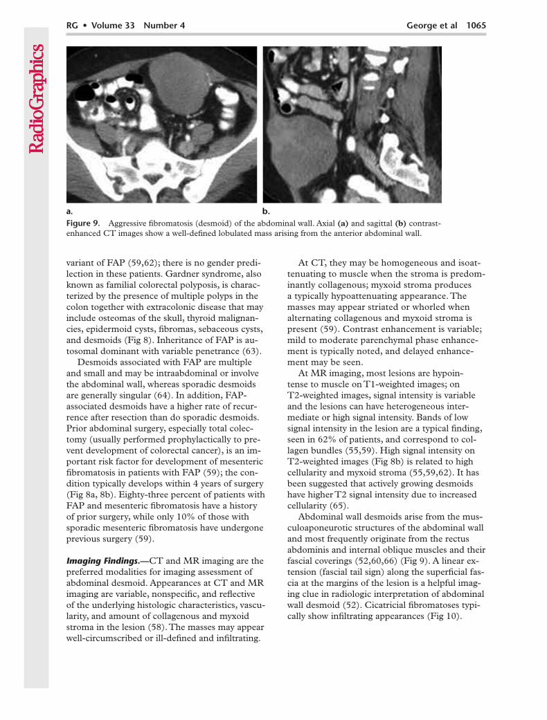
Abdominal wall desmoids arise from the mus-culoaponeurotic structures of the abdominal wall and most frequently originate from the rectus abdominis and internal oblique muscles and their fascial coverings (52,60,66) (Fig 9). A linear ex-tension (fascial tail sign) along the superficial fas-cia at the margins of the lesion is a helpful imag-ing clue in radiologic interpretation of abdominal wall desmoid (52). Cicatricial fibromatoses typi-cally show infiltrating appearances (Fig 10).
Figure 9. Aggressive fibromatosis (desmoid) of the abdominal wall. Axial (a) and sagittal (b) contrast-enhanced CT images show a well-defined lobulated mass arising from the anterior abdominal wall.
1066 July-August 2013 radiographics.rsna.org
Figure 10. Aggressive fibromatosis (desmoid) of the abdominal wall in a 29-year-old woman with a palpable lump. The lesion occurred at the site of a cesarean section scar (cicatricial fibromatosis). (a–c) Transverse gray-scale US image (a) and axial venous phase (b) and delayed phase (c) contrast-enhanced CT images show an ill-defined infiltrating mass (arrow in b) with delayed enhancement (arrow in c). (d–f) Corresponding axial T2-weighted (d), nonenhanced T1-weighted (e), and delayed contrast-enhanced fat-saturated (f) MR images show the same findings as at CT. The mass has the typical imaging characteristics of fibrous tissue.
RG • Volume 33 Number 4 George et al 1067
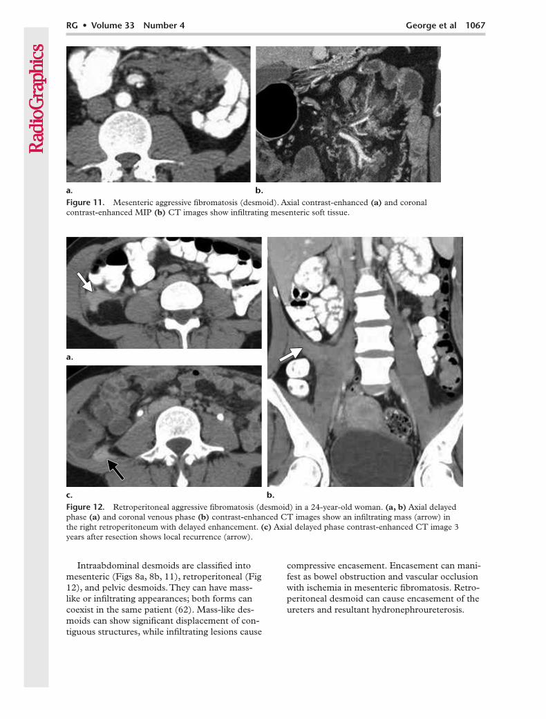
Intraabdominal desmoids are classified into mesenteric (Figs 8a, 8b, 11), retroperitoneal (Fig 12), and pelvic desmoids. They can have mass-like or infiltrating appearances; both forms can coexist in the same patient (62). Mass-like des-moids can show significant displacement of con-tiguous structures, while infiltrating lesions cause
compressive encasement. Encasement can mani-fest as bowel obstruction and vascular occlusion with ischemia in mesenteric fibromatosis. Retro-peritoneal desmoid can cause encasement of the ureters and resultant hydronephroureterosis.
Figure 11. Mesenteric aggressive fibromatosis (desmoid). Axial contrast-enhanced (a) and coronal contrast-enhanced MIP (b) CT images show infiltrating mesenteric soft tissue.
Figure 12. Retroperitoneal aggressive fibromatosis (desmoid) in a 24-year-old woman. (a, b) Axial delayed phase (a) and coronal venous phase (b) contrast-enhanced CT images show an infiltrating mass (arrow) in the right retroperitoneum with delayed enhancement. (c) Axial delayed phase contrast-enhanced CT image 3 years after resection shows local recurrence (arrow).
1068 July-August 2013 radiographics.rsna.org
Differential Diagnosis.—Regardless of location, malignancy is an important differential diagno-sis in evaluation of desmoids. Abdominal wall desmoids should be differentiated from malig-nant processes such as lymphoma, metastases, and soft-tissue sarcomas. Scar endometriosis is an important differential diagnosis in patients with desmoids at the site of a cesarean section scar (67).
In intraabdominal desmoids, the main differ-ential diagnoses for mesenteric fibromatoses are malignant neoplasms of the mesentery such as lymphoma, metastases, and soft-tissue sarcomas such as leiomyosarcoma and malignant fibrous histiocytoma. Other differential diagnoses include IPT, extrapleural solitary fibrous tumor, and mes-enteric gastrointestinal stromal tumor (GIST) (59). RPF and IPT may show imaging appear-ances similar to that of retroperitoneal desmoid. Benign and malignant pelvic neoplasms are the most important mimics of pelvic fibromatosis.
Penile FibromatosisPenile fibromatosis (Peyronie disease) is a be-nign acquired fibrotic condition characterized by chronic inflammation leading to fibrosis and focal thickening of the penile tunica albuginea (68,69). It is a surprisingly common disorder with a prevalence of 3%; the average age at pre-sentation is 40–60 years. Patients with penile fibromatosis present with painful penile indura-tion and palpable albugineal plaques along the corpora cavernosa, typically on the dorsal aspect of the penis. The condition causes abnormal pe-nile curvature and deformity of the penis during
erection (70,71) and is an important cause of erectile dysfunction.
Nomenclature.—Penile fibromatosis, also known as Peyronie disease and induratio penis plastica, is categorized as a superficial fibromatosis (69).
Pathophysiology and Associations.—Although the etiology of the condition is poorly under-stood, the typical dorsal location of the plaques and the presence of fibrin likely represent aber-rant healing response to minor penile trauma (71). There is a reported association of penile fibromatosis in 16%–20% of cases with pal-mar fibromatosis (Dupuytren contracture), an autosomal dominant condition, thus indicating a possible genetic basis for the disease (69,71). As-sociations with other conditions such as plantar fibromatosis, Paget disease of bone, diabetes mel-litus, and gout have also been reported (72).
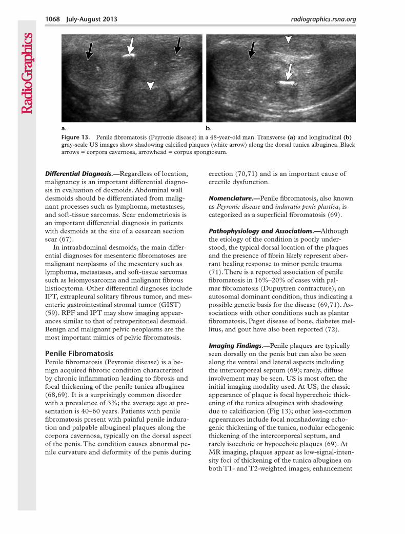
Imaging Findings.—Penile plaques are typically seen dorsally on the penis but can also be seen along the ventral and lateral aspects including the intercorporeal septum (69); rarely, diffuse involvement may be seen. US is most often the initial imaging modality used. At US, the classic appearance of plaque is focal hyperechoic thick-ening of the tunica albuginea with shadowing due to calcification (Fig 13); other less-common appearances include focal nonshadowing echo-genic thickening of the tunica, nodular echogenic thickening of the intercorporeal septum, and rarely isoechoic or hypoechoic plaques (69). At MR imaging, plaques appear as low-signal-inten-sity foci of thickening of the tunica albuginea on both T1- and T2-weighted images; enhancement
Figure 13. Penile fibromatosis (Peyronie disease) in a 48-year-old man. Transverse (a) and longitudinal (b) gray-scale US images show shadowing calcified plaques (white arrow) along the dorsal tunica albuginea. Black arrows = corpora cavernosa, arrowhead = corpus spongiosum.
RG • Volume 33 Number 4 George et al 1069
of the plaque can be seen in the phase of active inflammation (72).
The sensitivities of US (68%) and MR im-aging (61%) for identifying the plaques are relatively similar (73). While US is superior to MR imaging in detection of calcific plaque, MR imaging is superior in demonstrating plaque enhancement and also accurately depicts the penile deformity (72). US is useful in identify-ing the relationship of plaque to surrounding structures such as encasement of dorsal arteries and the neurovascular bundle (may result in pe-nile numbness after surgical correction) and the cavernosal artery (arteriogenic cause of erectile dysfunction) (70). The condition may be inciden-tally detected at abdominal CT (Fig 14). Imaging plays a key role in patient selection before surgery and also in preoperative definition of the extent of plaque and the deformity.
Differential Diagnosis.—While the majority of patients with focal or diffuse penile indura-tion have penile fibromatosis, other differential diagnoses to consider include congenital penile curvature, dorsal vein thrombosis (thrombosed dorsal vein identified at imaging), penile calci-phylaxis (acute life-threatening condition in patients with end-stage renal disease, with oc-clusion of flow to penile vessels and extensive tunica and cavernosal arterial calcifications), and other causes of penile fibrosis such as prolonged priapism, trauma, removal of penile prostheses, and prior intracavernosal injections for erectile dysfunction. Penile malignancies may also mimic penile fibromatosis (70).
Ovarian FibromatosisOvarian fibromatosis is a rare benign condition causing tumor-like enlargement of the ovaries. Typically seen in younger patients (average age at presentation, 25 years), it manifests as pain, menstrual abnormalities, and occasionally viril-ization (74).
NomenclatureDespite similarity in nomenclature, ovarian fibro-matosis is not considered one of the fibromatoses, as mentioned earlier, although it is notable that pelvic aggressive fibromatosis (desmoid) can sec-ondarily involve the ovary (75). Conversely, ovar-ian fibromatosis is confined to the ovaries and does not demonstrate the locally invasive features typical of the fibromatoses (74).
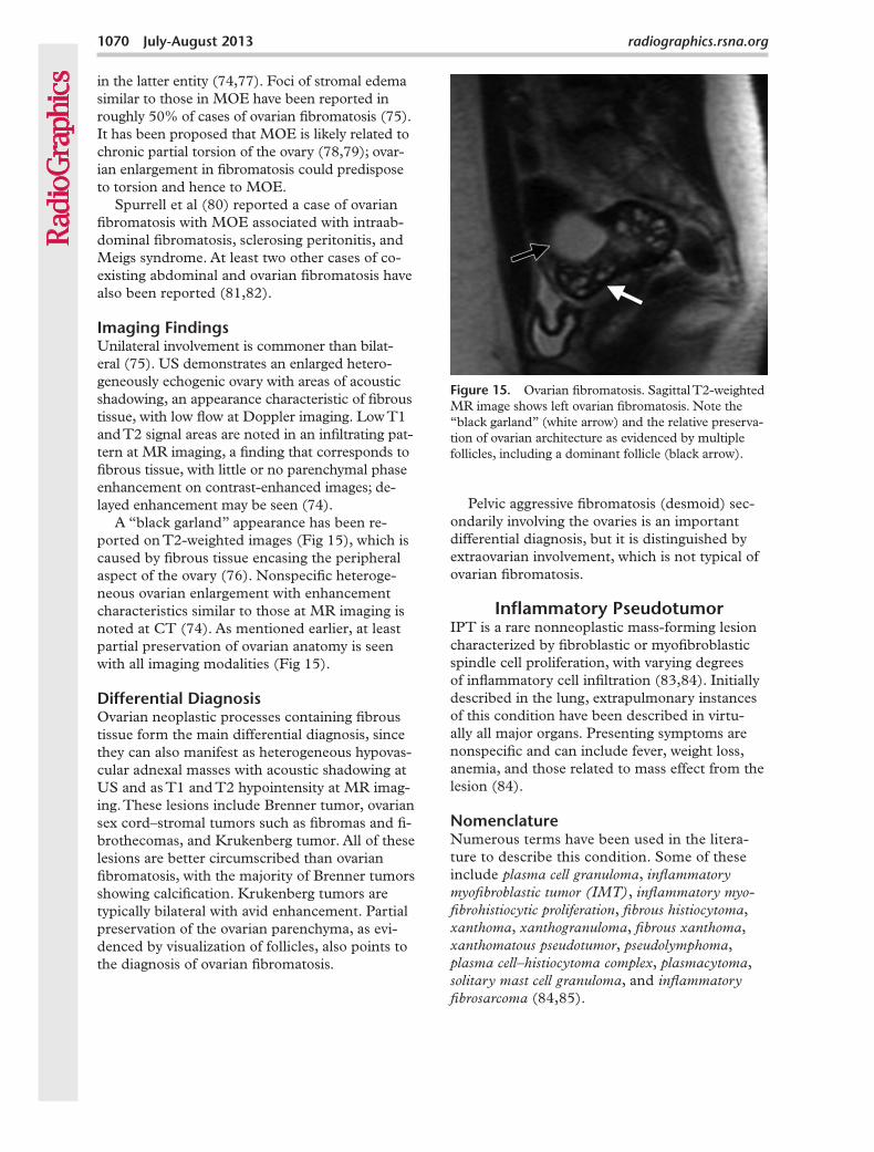
Pathophysiology and AssociationsOvarian fibromatosis is characterized by prolif-eration of collagen-producing spindle cells sur-rounding normal ovarian structures (76), with collagenous thickening of the superficial cortex (75). Despite these changes, there is partial pres-ervation of normal ovarian structures within the fibrous mass, a feature that has been reported to allow differentiation of ovarian fibromatosis from similar conditions (75,76).
Ovarian fibromatosis appears to be related to massive ovarian edema (MOE) (77). MOE is characterized by ovarian enlargement and stromal edema; it differs from ovarian fibromatosis because of the presence of edema, as opposed to fibrosis
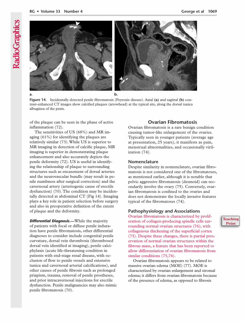
Figure 14. Incidentally detected penile fibromatosis (Peyronie disease). Axial (a) and sagittal (b) con-trast-enhanced CT images show calcified plaques (arrowhead) at the typical site, along the dorsal tunica albuginea of the penis.
1070 July-August 2013 radiographics.rsna.org
in the latter entity (74,77). Foci of stromal edema similar to those in MOE have been reported in roughly 50% of cases of ovarian fibromatosis (75). It has been proposed that MOE is likely related to chronic partial torsion of the ovary (78,79); ovar-ian enlargement in fibromatosis could predispose to torsion and hence to MOE.
Spurrell et al (80) reported a case of ovarian fibromatosis with MOE associated with intraab-dominal fibromatosis, sclerosing peritonitis, and Meigs syndrome. At least two other cases of co-existing abdominal and ovarian fibromatosis have also been reported (81,82).
Imaging FindingsUnilateral involvement is commoner than bilat-eral (75). US demonstrates an enlarged hetero-geneously echogenic ovary with areas of acoustic shadowing, an appearance characteristic of fibrous tissue, with low flow at Doppler imaging. Low T1 and T2 signal areas are noted in an infiltrating pat-tern at MR imaging, a finding that corresponds to fibrous tissue, with little or no parenchymal phase enhancement on contrast-enhanced images; de-layed enhancement may be seen (74).
A “black garland” appearance has been re-ported on T2-weighted images (Fig 15), which is caused by fibrous tissue encasing the peripheral aspect of the ovary (76). Nonspecific heteroge-neous ovarian enlargement with enhancement characteristics similar to those at MR imaging is noted at CT (74). As mentioned earlier, at least partial preservation of ovarian anatomy is seen with all imaging modalities (Fig 15).
Differential DiagnosisOvarian neoplastic processes containing fibrous tissue form the main differential diagnosis, since they can also manifest as heterogeneous hypovas-cular adnexal masses with acoustic shadowing at US and as T1 and T2 hypointensity at MR imag-ing. These lesions include Brenner tumor, ovarian sex cord–stromal tumors such as fibromas and fi-brothecomas, and Krukenberg tumor. All of these lesions are better circumscribed than ovarian fibromatosis, with the majority of Brenner tumors showing calcification. Krukenberg tumors are typically bilateral with avid enhancement. Partial preservation of the ovarian parenchyma, as evi-denced by visualization of follicles, also points to the diagnosis of ovarian fibromatosis.
Pelvic aggressive fibromatosis (desmoid) sec-ondarily involving the ovaries is an important differential diagnosis, but it is distinguished by extraovarian involvement, which is not typical of ovarian fibromatosis.
Inflammatory PseudotumorIPT is a rare nonneoplastic mass-forming lesion characterized by fibroblastic or myofibroblastic spindle cell proliferation, with varying degrees of inflammatory cell infiltration (83,84). Initially described in the lung, extrapulmonary instances of this condition have been described in virtu-ally all major organs. Presenting symptoms are nonspecific and can include fever, weight loss, anemia, and those related to mass effect from the lesion (84).
NomenclatureNumerous terms have been used in the litera-ture to describe this condition. Some of these include plasma cell granuloma, inflammatory myofibroblastic tumor (IMT), inflammatory myo-fibrohistiocytic proliferation, fibrous histiocytoma, xanthoma, xanthogranuloma, fibrous xanthoma, xanthomatous pseudotumor, pseudolymphoma, plasma cell–histiocytoma complex, plasmacytoma, solitary mast cell granuloma, and inflammatory fibrosarcoma (84,85).
Figure 15. Ovarian fibromatosis. Sagittal T2-weighted MR image shows left ovarian fibromatosis. Note the “black garland” (white arrow) and the relative preserva-tion of ovarian architecture as evidenced by multiple follicles, including a dominant follicle (black arrow).
RG • Volume 33 Number 4 George et al 1071
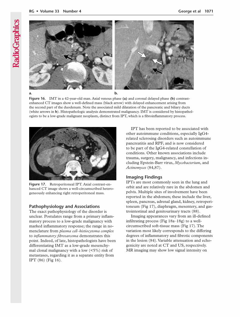
Pathophysiology and AssociationsThe exact pathophysiology of the disorder is unclear. Postulates range from a primary inflam-matory process to a low-grade malignancy with marked inflammatory response; the range in no-menclature from plasma cell–histiocytoma complex to inflammatory fibrosarcoma demonstrates this point. Indeed, of late, histopathologists have been differentiating IMT as a low-grade mesenchy-mal clonal malignancy with a low (<5%) risk of metastases, regarding it as a separate entity from IPT (86) (Fig 16).
IPT has been reported to be associated with other autoimmune conditions, especially IgG4-related sclerosing disorders such as autoimmune pancreatitis and RPF, and is now considered to be part of the IgG4-related constellation of conditions. Other known associations include trauma, surgery, malignancy, and infections in-cluding Epstein-Barr virus, Mycobacterium, and Actinomyces (84,87).
Imaging FindingsIPTs are most commonly seen in the lung and orbit and are relatively rare in the abdomen and pelvis. Multiple sites of involvement have been reported in the abdomen; these include the liver, spleen, pancreas, adrenal gland, kidney, retroperi-toneum (Fig 17), diaphragm, mesentery, and gas-trointestinal and genitourinary tracts (88).
Imaging appearances vary from an ill-defined infiltrating process (Fig 18a–18g) to a well-circumscribed soft-tissue mass (Fig 17). The variation most likely corresponds to the differing degrees of inflammatory and fibrotic components in the lesion (84). Variable attenuation and echo-genicity are noted at CT and US, respectively. MR imaging may show low signal intensity on
Figure 16. IMT in a 42-year-old man. Axial venous phase (a) and coronal delayed phase (b) contrast-enhanced CT images show a well-defined mass (black arrow) with delayed enhancement arising from the second part of the duodenum. Note the associated mild dilatation of the pancreatic and biliary ducts (white arrows in b). Histopathologic analysis demonstrated malignancy. IMT is considered by histopathol-ogists to be a low-grade malignant neoplasm, distinct from IPT, which is a fibroinflammatory process.
Figure 17. Retroperitoneal IPT. Axial contrast-en-hanced CT image shows a well-circumscribed hetero-geneously enhancing right retroperitoneal mass.
1072 July-August 2013 radiographics.rsna.org
RG • Volume 33 Number 4 George et al 1073
T1- and T2-weighted images due to the fibrotic component (Fig 18d, 18e), along with absence of restricted diffusion (Fig 18h).
Calcification may be seen at CT (Fig 18b). Variable enhancement and Doppler flow are also noted; delayed enhancement may be seen in the fibrotic component (Fig 18g) (84,88). In general, hepatic, splenic, retroperitoneal, and genitouri-nary lesions are well-circumscribed, while those involving the gastrointestinal tract and biliary tree are ill-defined and infiltrating. Mesenteric lesions may manifest in either of the two forms (84).
Differential DiagnosisIPT has a wide imaging differential diagnosis due to its varied radiologic appearances. IPT should be considered in the differential diagnosis of any soft-tissue mass in the abdomen and pelvis. Important entities in the differential diagnosis include malignancies such as hepatocellular car-cinoma, gastric carcinoma, cholangiocarcinoma, pancreatic carcinoma, and carcinomatosis (88) and other fibrosing conditions such as RPF, scle-rosing mesenteritis, and abdominal fibromatosis.
Nephrogenic Systemic FibrosisNSF is a chronic fibrosing condition similar to scleroderma, identified predominantly in the skin and subcutaneous tissues, although it can involve multiple other organs including skeletal muscle, bone, and the lung, heart, pericardium, testis, and esophagus (89). The disease usually mani-fests as skin thickness and pruritis but may prog-ress to cause contractures and joint immobility. Death may result in some patients, presumably due to visceral organ involvement.
NSF does not show gender or race predilec-tion. NSF has been reported in children and the elderly but tends to affect the middle-aged most commonly (90).
NomenclatureThe condition was initially described in 2000 as one that manifests as thickening and hardening of the skin, predominantly in patients with end-stage renal disease, especially those receiving di-
alysis (91). This accounted for the disorder being initially named nephrogenic fibrosing dermopathy. Later reports showing that it also affects skeletal muscle, joints, and the viscera prompted a change in nomenclature to NSF (89).
Pathophysiology and AssociationsThe exact pathophysiology of NSF is unclear. As mentioned earlier, the first report of NSF was on 15 patients with end-stage renal disease (91), with multiple subsequent studies confirming the association of NSF with chronic kidney disease, especially dialysis.
As mentioned, NSF shows similarities to scleroderma. However, unlike scleroderma, NSF does not show a gender predilection and does not involve the face or have associated serologic markers (89).
Associations of NSF with hypercoagulability and thrombotic events, surgical procedures, and hepatic and pulmonary disease have been reported (92). In 2006, gadolinium was identified as a pos-sible trigger for NSF (93), followed by a report in 2007 of detection of gadolinium in the skin and soft tissues of patients with NSF (94). Worldwide reevaluation of the use of gadolinium contrast ma-terial ensued as a result of those studies.
In 2007, the U.S. Food and Drug Administra-tion mandated a boxed warning about the risk of NSF on all gadolinium-based contrast agents. Use of these contrast agents in patients with renal disease is now more controlled, with most clinical practices following prescribed guidelines (95) and departmental protocols designed to limit the dose of gadolinium contrast material, based on degrees of renal impairment.
Imaging FindingsNSF can show variable imaging findings, most of them relating to skin and subcutaneous soft-tissue thickening as well as joint contractures. Conventional radiographs and mammograms may show skin thickening with occasional calcific foci. CT and US show changes of skin thickening
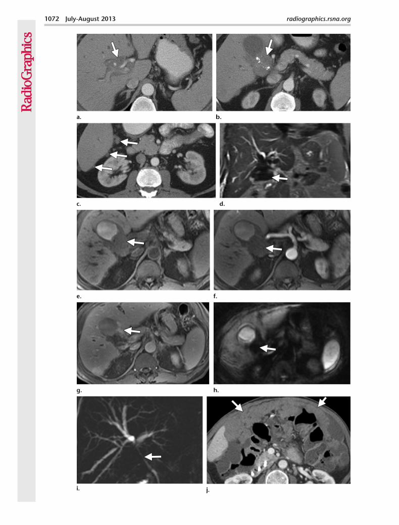
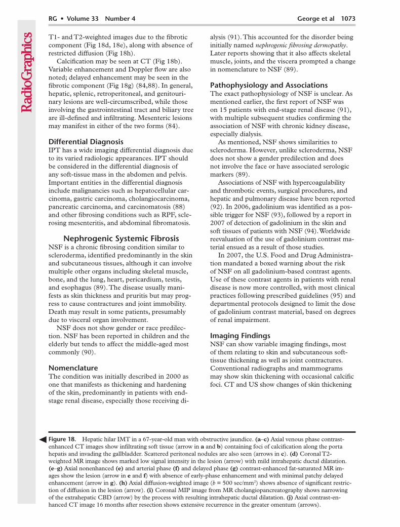
Figure 18. Hepatic hilar IMT in a 67-year-old man with obstructive jaundice. (a–c) Axial venous phase contrast-enhanced CT images show infiltrating soft tissue (arrow in a and b) containing foci of calcification along the porta hepatis and invading the gallbladder. Scattered peritoneal nodules are also seen (arrows in c). (d) Coronal T2-weighted MR image shows marked low signal intensity in the lesion (arrow) with mild intrahepatic ductal dilatation. (e–g) Axial nonenhanced (e) and arterial phase (f) and delayed phase (g) contrast-enhanced fat-saturated MR im-ages show the lesion (arrow in e and f) with absence of early-phase enhancement and with minimal patchy delayed enhancement (arrow in g). (h) Axial diffusion-weighted image (b = 500 sec/mm2) shows absence of significant restric-tion of diffusion in the lesion (arrow). (i) Coronal MIP image from MR cholangiopancreatography shows narrowing of the extrahepatic CBD (arrow) by the process with resulting intrahepatic ductal dilatation. (j) Axial contrast-en-hanced CT image 16 months after resection shows extensive recurrence in the greater omentum (arrows).
1074 July-August 2013 radiographics.rsna.org
The presenting symptoms include abdominal dis-comfort and jaundice; 33% of patients with AIP have been reported to have symptoms of pancre-atitis at presentation (99).
NomenclatureMultiple alternate terms for the condition are found in the literature. These include primary in-flammatory sclerosis of the pancreas, chronic sclerosing pancreatitis, lymphoplasmacytic sclerosing pancreatitis, nonalcoholic duct-destructive pancreatitis, sclerosing pancreatocholangitis, idiopathic duct-centric chronic pancreatitis, idiopathic tumefactive chronic pancreati-tis, and pancreatic pseudolymphoma (100).
Pathophysiology and AssociationsCharacteristic lymphoplasmacytic inflammatory infiltration at histologic analysis, elevated IgG4 levels, frequently positive results on serologic tests, response to steroids, and association with other
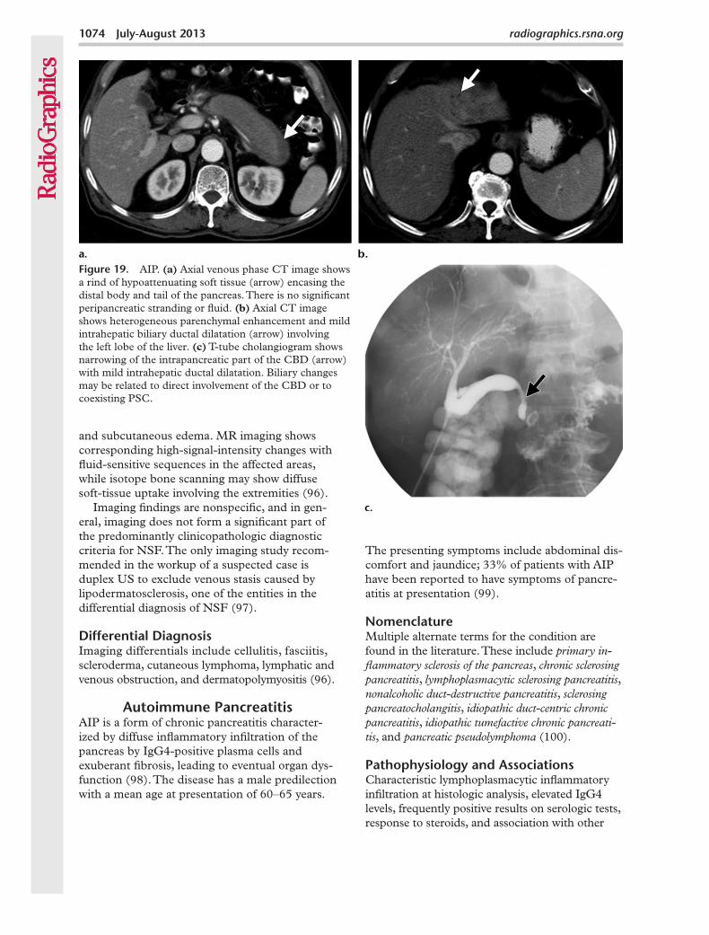
Figure 19. AIP. (a) Axial venous phase CT image shows a rind of hypoattenuating soft tissue (arrow) encasing the distal body and tail of the pancreas. There is no significant peripancreatic stranding or fluid. (b) Axial CT image shows heterogeneous parenchymal enhancement and mild intrahepatic biliary ductal dilatation (arrow) involving the left lobe of the liver. (c) T-tube cholangiogram shows narrowing of the intrapancreatic part of the CBD (arrow) with mild intrahepatic ductal dilatation. Biliary changes may be related to direct involvement of the CBD or to coexisting PSC.
and subcutaneous edema. MR imaging shows corresponding high-signal-intensity changes with fluid-sensitive sequences in the affected areas, while isotope bone scanning may show diffuse soft-tissue uptake involving the extremities (96).
Imaging findings are nonspecific, and in gen-eral, imaging does not form a significant part of the predominantly clinicopathologic diagnostic criteria for NSF. The only imaging study recom-mended in the workup of a suspected case is duplex US to exclude venous stasis caused by lipodermatosclerosis, one of the entities in the differential diagnosis of NSF (97).
Differential DiagnosisImaging differentials include cellulitis, fasciitis, scleroderma, cutaneous lymphoma, lymphatic and venous obstruction, and dermatopolymyositis (96).
Autoimmune PancreatitisAIP is a form of chronic pancreatitis character-ized by diffuse inflammatory infiltration of the pancreas by IgG4-positive plasma cells and exuberant fibrosis, leading to eventual organ dys-function (98). The disease has a male predilection with a mean age at presentation of 60–65 years.
RG • Volume 33 Number 4 George et al 1075
autoimmune disorders such as Sjögren syndrome have led to suggestions that AIP is an immune- mediated fibroinflammatory condition. To reiter-ate, AIP is one of the most important components of the recently described group of conditions called IgG4-related sclerosing disorders, which can cause extensive multiorgan involvement.
Imaging FindingsInvolved areas of the pancreas typically show hypoechogenicity at US, low attenuation at CT, and mild hyperintensity at MR imaging. In the most commonly seen diffuse pattern of involvement, the pancreas is enlarged and sau-sage-shaped, with loss of its normal lobulated contour (Figs 19a, 20a). The pancreas shows relatively reduced enhancement in the parenchy-mal phase; delayed enhancement may be noted. A thin capsule-like rim or peripancreatic halo, hypoattenuating at CT and hypointense at MR imaging, may be noted around the body and tail
of the pancreas (Fig 19a) and likely corresponds to inflammatory tissue.
The focal form of the disease usually shows the abnormal findings confined typically to the pancreatic head with evidence of mass effect, es-pecially upstream dilatation of the main pancreatic duct. More than one focus of discrete pancreatic involvement may be seen in the multifocal form of the disease (98).
Cholangiography shows pancreatic ductal nar-rowing and irregularities in the involved segments. Coexisting biliary ductal abnormalities may also be seen (Fig 19b, 19c), usually related to associ-ated IgG4-related sclerosing cholangitis (101).
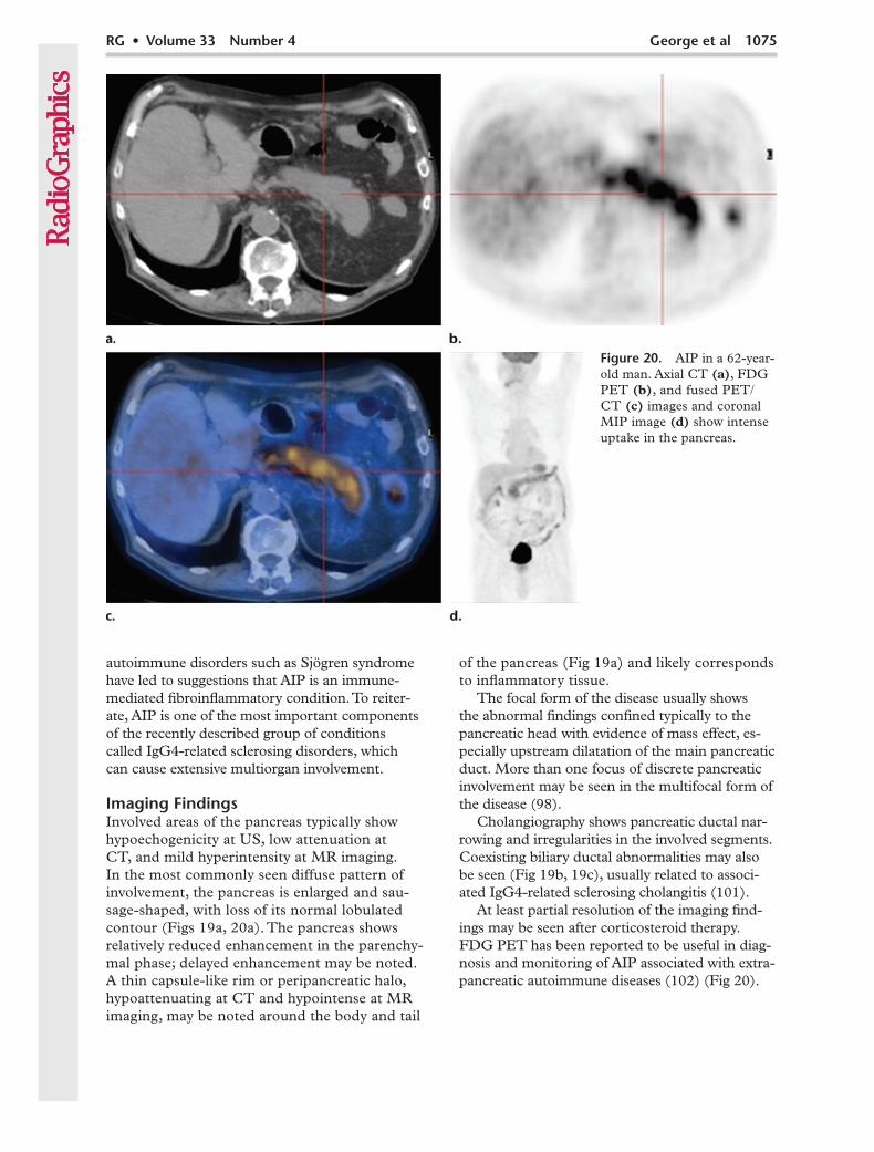
At least partial resolution of the imaging find-ings may be seen after corticosteroid therapy. FDG PET has been reported to be useful in diag-nosis and monitoring of AIP associated with extra-pancreatic autoimmune diseases (102) (Fig 20).
Figure 20. AIP in a 62-year-old man. Axial CT (a), FDG PET (b), and fused PET/CT (c) images and coronal MIP image (d) show intense uptake in the pancreas.
1076 July-August 2013 radiographics.rsna.org
Differential DiagnosisThe most important differential diagnosis for the diffuse form of the disease is acute pancre-atitis. Imaging findings favoring a diagnosis of diffuse AIP over acute pancreatitis include mini-mal or no peripancreatic stranding, the afore-mentioned peripancreatic halo, and absence of peripancreatic fat necrosis (98).
The focal form of the disease may be indis-tinguishable from pancreatic cancer. Although presence of associated IgG4-related conditions and serologic findings may help corroborate a suspicion of AIP, biopsy is often required for de-finitive diagnosis.
IgG4-related Sclerosing DiseaseAs mentioned earlier, ISD is a recently described assortment of conditions that share the common histopathologic findings of inflammation with IgG4-positive cells and proliferative fibrosis.
NomenclatureThe condition was described in 2006, when it was called hyper-IgG4 disease (103). It has also been described as IgG4-related systemic disease.
Pathophysiology and AssociationsISD has been proposed as a unifying concept to bring multiple fibroinflammatory conditions in-volving various anatomic sites under one umbrella. The common features of these conditions involve multiorgan involvement, infiltration of the af-
fected organs by T-lymphocytes and IgG4-positive plasma cells, and a favorable response to steroids.
The prototype ISD in the abdomen is AIP. Multiple other conditions that are components of ISD in the abdomen include RPF, sclerosing mesenteritis, and sclerosing cholangitis. ISD also includes sclerosing cholecystitis, sclerosing sialad-enitis (Küttner tumor), renal disease, interstitial pneumonia, prostatitis, lymphadenopathy, Riedel thyroiditis, and IPT (104).
Imaging Findings and Differential DiagnosisMost of the important components of ISD in the abdomen—namely, AIP, sclerosing cholangitis, RPF, IPT, and sclerosing mesenteritis—have been described earlier.
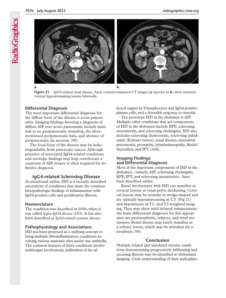
Renal involvement with ISD can manifest as cortical lesions or renal pelvic thickening. Corti-cal lesions may be nodular or wedge-shaped and are typically hypoattenuating at CT (Fig 21) and hypointense at T1- and T2-weighted imag-ing. They may show mild delayed enhancement; the main differential diagnoses for this appear-ance are pyelonephritis, infarcts, and renal me-tastases. Renal disease may rarely manifest as a solitary lesion, which may be mistaken for a neoplasm (98).
ConclusionMultiple related and unrelated chronic condi-tions demonstrating progressively infiltrating and encasing fibrosis may be identified at abdominal imaging. Clear understanding of their pathophys-
Figure 21. IgG4-related renal disease. Axial contrast-enhanced CT images (a superior to b) show extensive cortical hypoattenuating lesions bilaterally.
RG • Volume 33 Number 4 George et al 1077
iology and nomenclature is required for optimal radiologic interpretation. However, overlapping imaging findings often result in the need for his-topathologic assessment for definitive diagnosis of these conditions.
References 1. Emory TS, Monihan JM, Carr NJ, Sobin LH.
Sclerosing mesenteritis, mesenteric panniculitis and mesenteric lipodystrophy: a single entity? Am J Surg Pathol 1997;21(4):392–398.
2. Sabaté JM, Torrubia S, Maideu J, Franquet T, Monill JM, Pérez C. Sclerosing mesenteritis: imag-ing findings in 17 patients. AJR Am J Roentgenol 1999;172(3):625–629.
3. Horton KM, Lawler LP, Fishman EK. CT findings in sclerosing mesenteritis (panniculitis): spectrum of disease. RadioGraphics 2003;23(6):1561–1567.
4. Daskalogiannaki M, Voloudaki A, Prassopoulos P, et al. CT evaluation of mesenteric panniculi-tis: prevalence and associated diseases. AJR Am J Roentgenol 2000;174(2):427–431.
5. Zissin R, Metser U, Hain D, Even-Sapir E. Mes-enteric panniculitis in oncologic patients: PET-CT findings. Br J Radiol 2006;79(937):37–43.
6. Ti JP, Al-Aradi A, Conlon PJ, Lee MJ, Morrin MM. Imaging features of encapsulating peritoneal sclerosis in continuous ambulatory peritoneal dialysis patients. AJR Am J Roentgenol 2010;195 (1):W50–W54.
7. Foo KT, Ng KC, Rauff A, Foong WC, Sinniah R. Unusual small intestinal obstruction in adolescent girls: the abdominal cocoon. Br J Surg 1978;65(6): 427–430.
8. Holland P. Sclerosing encapsulating peritonitis in chronic ambulatory peritoneal dialysis. Clin Radiol 1990;41(1):19–23.
9. Demir MK, Akinci O, Onur E, Koksal N. Case 108: sclerosing encapsulating peritonitis. Radiology 2007;242(3):937–939.
10. Dobbie JW. Pathogenesis of peritoneal fibrosing syndromes (sclerosing peritonitis) in peritoneal dialysis. Perit Dial Int 1992;12(1):14–27.
11. Lee RG. Sclerosing peritonitis. Dig Dis Sci 1989;34 (9):1473–1476.
12. Rigby RJ, Hawley CM. Sclerosing peritonitis: the experience in Australia. Nephrol Dial Transplant 1998;13(1):154–159.
13. Stanley MM, Reyes CV, Greenlee HB, Nem-chausky B, Reinhardt GF. Peritoneal fibrosis in cir-rhotics treated with peritoneovenous shunting for ascites: an autopsy study with clinical correlations. Dig Dis Sci 1996;41(3):571–577.
14. Kaushik R, Punia RPS, Mohan H, Attri AK. Tu-berculous abdominal cocoon: a report of 6 cases and review of the literature. World J Emerg Surg 2006;1:18.
15. Ngô Y, Messing B, Marteau P, et al. Peritoneal sarcoidosis: an unrecognized cause of sclerosing peritonitis. Dig Dis Sci 1992;37(11):1776–1780.
16. Eltringham WK, Espiner HJ, Windsor CW, et al. Sclerosing peritonitis due to practolol: a report on 9 cases and their surgical management. Br J Surg 1977;64(4):229–235.
17. Sahoo SP, Gangopadhyay AN, Gupta DK, Gopal SC, Sharma SP, Dash RN. Abdominal cocoon in children: a report of four cases. J Pediatr Surg 1996; 31(7):987–988.
18. Vijayaraghavan SB, Palanivelu C, Sendhilkumar K, Parthasarathi R. Abdominal cocoon: sonographic features. J Ultrasound Med 2003;22(7):719–721.
19. Kawaguchi Y, Kawanishi H, Mujais S, Topley N, Oreopoulos DG. Encapsulating peritoneal sclero-sis: definition, etiology, diagnosis, and treatment. International Society for Peritoneal Dialysis Ad Hoc Committee on Ultrafiltration Management in Peritoneal Dialysis. Perit Dial Int 2000;20(suppl 4): S43–S55.
20. George C, Al-Zwae K, Nair S, Cast JEI. Computed tomography appearances of sclerosing encapsulat-ing peritonitis. Clin Radiol 2007;62(8):732–737.
21. Loughrey GJ, Hawnaur JM, Sambrook P. Case report: computed tomographic appearance of scle-rosing peritonitis with gross peritoneal calcifica-tion. Clin Radiol 1997;52(7):557–558.
22. Vitellas KM, Keogan MT, Freed KS, et al. Radio-logic manifestations of sclerosing cholangitis with emphasis on MR cholangiopancreatography. Ra-dioGraphics 2000;20(4):959–975.
23. Tinckler L. Primary sclerosing cholangitis. Post-grad Med J 1971;47(552):666–670.
24. Abdalian R, Heathcote EJ. Sclerosing cholangitis: a focus on secondary causes. Hepatology 2006;44 (5):1063–1074.
25. Björnsson E, Olsson R, Bergquist A, et al. The nat-ural history of small-duct primary sclerosing chol-angitis. Gastroenterology 2008;134(4):975–980.
26. Hov J-R, Boberg K-M, Karlsen T-H. Autoantibod-ies in primary sclerosing cholangitis. World J Gas-troenterol 2008;14(24):3781–3791.
27. Weismüller TJ, Wedemeyer J, Kubicka S, Strassburg CP, Manns MP. The challenges in primary scleros-ing cholangitis: aetiopathogenesis, autoimmunity, management and malignancy. J Hepatol 2008;48 (suppl 1):S38–S57.
28. Ito K, Mitchell DG, Outwater EK, Blasbalg R. Pri-mary sclerosing cholangitis: MR imaging features. AJR Am J Roentgenol 1999;172(6):1527–1533.
29. Björnsson E, Lindqvist-Ottosson J, Asztely M, Olsson R. Dominant strictures in patients with primary sclerosing cholangitis. Am J Gastroenterol 2004;99(3):502–508.
30. Menias CO, Surabhi VR, Prasad SR, Wang HL, Narra VR, Chintapalli KN. Mimics of cholangiocar-cinoma: spectrum of disease. RadioGraphics 2008; 28(4):1115–1129.
31. Cronin CG, Lohan DG, Blake MA, Roche C, Mc-Carthy P, Murphy JM. Retroperitoneal fibrosis: a review of clinical features and imaging findings. AJR Am J Roentgenol 2008;191(2):423–431.
32. Kottra JJ, Dunnick NR. Retroperitoneal fibrosis. Radiol Clin North Am 1996;34(6):1259–1275.
33. Lepor H, Walsh PC. Idiopathic retroperitoneal fi-brosis. J Urol 1979;122(1):1–6.
34. Kovács T, Besznyák I, Köves I, Petri K. Ormond’s disease. Acta Chir Hung 1995-1996;35(3-4): 339–350.
1078 July-August 2013 radiographics.rsna.org
51. Bramm HG, Griffith RP, Griffith TH, Shasteen WJ. Retroperitoneal fibrosis simulating carcinoma of the cervix. Obstet Gynecol 1979;53(3 suppl):77S–78S.
52. Murphey MD, Ruble CM, Tyszko SM, Zbojnie-wicz AM, Potter BK, Miettinen M. Musculoskel-etal fibromatoses: radiologic-pathologic correlation. RadioGraphics 2009;29(7):2143–2173.
53. McDonald ES, Yi ES, Wenger DE. Extraabdomi-nal desmoid-type fibromatosis. RadioGraphics 2008;28(3):901–906.
54. Robbin MR, Murphey MD, Temple HT, Kransdorf MJ, Choi JJ. Imaging of musculoskeletal fibromato-sis. RadioGraphics 2001;21(3):585–600.
55. Lee JC, Thomas JM, Phillips S, Fisher C, Mos-kovic E. Aggressive fibromatosis: MRI features with pathologic correlation. AJR Am J Roentgenol 2006;186(1):247–254.
56. Rosen RS, Kimball W. Extra-abdominal desmoid tumor. Radiology 1966;86(3):534–540.
57. Fletcher CDM, Unni KK, Mertens F. Pathology and genetics of tumours of soft tissue and bone. Geneva, Switzerland: World Health Organization, 2002. http://www.iarc.fr/en/publications/pdfs-online/pat-gen/bb5/BB5.pdf. Accessed April 4, 2012.
58. Mackenzie DH. The fibromatoses: a clinicopatho-logical concept. BMJ 1972;4(5835):277–281.
59. Levy AD, Rimola J, Mehrotra AK, Sobin LH. Be-nign fibrous tumors and tumorlike lesions of the mesentery: radiologic-pathologic correlation. Ra-dioGraphics 2006;26(1):245–264.
60. Dinauer PA, Brixey CJ, Moncur JT, Fanburg-Smith JC, Murphey MD. Pathologic and MR imaging fea-tures of benign fibrous soft-tissue tumors in adults. RadioGraphics 2007;27(1):173–187.
61. Okamoto K, Ito J, Sakai K. Cicatricial fibromatosis mimics metastatic medulloblastoma. AJNR Am J Neuroradiol 1999;20(3):472–473.
62. Azizi L, Balu M, Belkacem A, Lewin M, Tubiana J-M, Arrivé L. MRI features of mesenteric desmoid tumors in familial adenomatous polyposis. AJR Am J Roentgenol 2005;184(4):1128–1135.
63. Lips DJ, Barker N, Clevers H, Hennipman A. The role of APC and beta-catenin in the aetiology of aggressive fibromatosis (desmoid tumors). Eur J Surg Oncol 2009;35(1):3–10.
64. Kawashima A, Goldman SM, Fishman EK, et al. CT of intraabdominal desmoid tumors: is the tu-mor different in patients with Gardner’s disease? AJR Am J Roentgenol 1994;162(2):339–342.
65. Shinagare AB, Ramaiya NH, Jagannathan JP, et al. A to Z of desmoid tumors. AJR Am J Roentgenol 2011;197(6):W1008–W1014.
66. Casillas J, Sais GJ, Greve JL, Iparraguirre MC, Mo-rillo G. Imaging of intra- and extraabdominal des-moid tumors. RadioGraphics 1991;11(6):959–968.
67. Stein L, Elsayes KM, Wagner-Bartak N. Subcutane-ous abdominal wall masses: radiological reasoning. AJR Am J Roentgenol 2012;198(2):W146–W151.
35. Pathak LR, Renjhen RC, Chandra CP. Idiopathic retroperitoneal fibrosis. Postgrad Med J 1971;47 (552):675–678.
36. Dehner LP, Coffin CM. Idiopathic fibrosclerotic disorders and other inflammatory pseudotumors. Semin Diagn Pathol 1998;15(2):161–173.
37. Amis ES Jr. Retroperitoneal fibrosis. AJR Am J Roentgenol 1991;157(2):321–329.
38. Bullock N. Idiopathic retroperitoneal fibrosis. BMJ 1988;297(6643):240–241.
39. van Bommel EF, Brouwers AJW, Makkus AC, van Vliet AC. Retroperitoneal fibrosis and p-ANCA-asso-ciated polyarteritis nodosa: coincidental or common etiology? Eur J Intern Med 2002;13(6):392–395.
40. Matsuki Y, Sato K, Fujikawa A, Kyoto Y, Hashimoto H, Hakozaki Y. A case of incidentally detected IgG4-related sclerosing disease involving inflammatory abdominal aortic aneurysm and autoimmune pan-creatitis. Mod Rheumatol 2010;20(3):306–310.
41. Sohn J-H, Byun JH, Yoon SE, et al. Abdominal ex-trapancreatic lesions associated with autoimmune pancreatitis: radiological findings and changes after therapy. Eur J Radiol 2008;67(3):497–507.
42. Stecker JF Jr, Rawls HP, Devine CJ Jr, Devine PC. Retroperitoneal fibrosis and ergot derivatives. J Urol 1974;112(1):30–32.
43. Uibu T, Oksa P, Auvinen A, et al. Asbestos exposure as a risk factor for retroperitoneal fibrosis. Lancet 2004;363(9419):1422–1426.
44. Vivas I, Nicolás AI, Velázquez P, Elduayen B, Fernández-Villa T, Martínez-Cuesta A. Retroperito-neal fibrosis: typical and atypical manifestations. Br J Radiol 2000;73(866):214–222.
45. Yuh WT, Barloon TJ, Sickels WJ, Kramolowsky EV, Williams RD. Magnetic resonance imaging in the diagnosis and followup of idiopathic retroperito-neal fibrosis. J Urol 1989;141(3):602–605.
46. Feinstein RS, Gatewood OM, Goldman SM, Co-peland B, Walsh PC, Siegelman SS. Computerized tomography in the diagnosis of retroperitoneal fibrosis. J Urol 1981;126(2):255–259.
47. Burn PR, Singh S, Barbar S, Boustead G, King CM. Role of gadolinium-enhanced magnetic reso-nance imaging in retroperitoneal fibrosis. Can As-soc Radiol J 2002;53(3):168–170.
48. Arrivé L, Hricak H, Tavares NJ, Miller TR. Malig-nant versus nonmalignant retroperitoneal fibrosis: differentiation with MR imaging. Radiology 1989; 172(1):139–143.
49. Degesys GE, Dunnick NR, Silverman PM, Cohan RH, Illescas FF, Castagno A. Retroperitoneal fi-brosis: use of CT in distinguishing among possible causes. AJR Am J Roentgenol 1986;146(1):57–60.
50. Brooks AP, Reznek RH, Webb JA. Aortic displace-ment on computed tomography of idiopathic retro-peritoneal fibrosis. Clin Radiol 1989;40(1):51–52.
RG • Volume 33 Number 4 George et al 1079
84. Patnana M, Sevrukov AB, Elsayes KM, Viswa-nathan C, Lubner M, Menias CO. Inflammatory pseudotumor: the great mimicker. AJR Am J Roentgenol 2012;198(3):W217–W227.
85. Bisceglia M, Fusilli S, Zaffarano L, Fiorentino F, Tardio B. Inflammatory pseudotumor of the breast: report of a case and review of the literature [in Ital-ian]. Pathologica 1995;87(1):59–64.
86. Gleason BC, Hornick JL. Inflammatory myofibro-blastic tumours: where are we now? J Clin Pathol 2008;61(4):428–437.
87. Androulaki A, Papathomas TG, Liapis G, et al. Inflammatory pseudotumor associated with Myco-bacterium tuberculosis infection. Int J Infect Dis 2008;12(6):607–610.
88. Narla LD, Newman B, Spottswood SS, Narla S, Kolli R. Inflammatory pseudotumor. RadioGraph-ics 2003;23(3):719–729.
89. Broome DR, Girguis MS, Baron PW, Cottrell AC, Kjellin I, Kirk GA. Gadodiamide-associated nephrogenic systemic fibrosis: why radiologists should be concerned. AJR Am J Roentgenol 2007;188(2):586–592.
90. Cowper SE. Nephrogenic systemic fibrosis. ICNSFR Web site. 2001–2009. http://www .icnsfr.org/. Accessed March 17, 2012.
91. Cowper SE, Robin HS, Steinberg SM, Su LD, Gupta S, LeBoit PE. Scleromyxoedema-like cu-taneous diseases in renal-dialysis patients. Lancet 2000;356(9234):1000–1001.
92. Cowper SE. Nephrogenic fibrosing dermopathy: the first 6 years. Curr Opin Rheumatol 2003;15 (6):785–790.
93. Grobner T. Gadolinium: a specific trigger for the development of nephrogenic fibrosing dermopathy and nephrogenic systemic fibrosis? Nephrol Dial Transplant 2006;21(4):1104–1108.
94. High WA, Ayers RA, Chandler J, Zito G, Cowper SE. Gadolinium is detectable within the tissue of patients with nephrogenic systemic fibrosis. J Am Acad Dermatol 2007;56(1):21–26.
95. ACR manual on contrast media, version 7. http://www.acr.org/SecondaryMainMenuCategories/quality_safety/contrast_manual/NephrogenicSys-temicFibrosis.aspx. Accessed March 16, 2012.
96. Morris MF, Zhang Y, Zhang H, et al. Features of nephrogenic systemic fibrosis on radiology examina-tions. AJR Am J Roentgenol 2009;193(1):61–69.
97. Girardi M, Kay J, Elston DM, Leboit PE, Abu-Alfa A, Cowper SE. Nephrogenic systemic fibro-sis: clinicopathological definition and workup recommendations. J Am Acad Dermatol 2011;65 (6):1095–1106, e7.
68. Pretorius ES, Siegelman ES, Ramchandani P, Ban-ner MP. MR imaging of the penis. RadioGraphics 2001;21(Spec No):S283–S298; discussion S298–S299.
69. Prando D. New sonographic aspects of Peyronie disease. J Ultrasound Med 2009;28(2):217–232.
70. Bertolotto M, Pavlica P, Serafini G, Quaia E, Zap-petti R. Painful penile induration: imaging findings and management. RadioGraphics 2009;29(2): 477–493.
71. Jalkut M, Gonzalez-Cadavid N, Rajfer J. Peyronie’s disease: a review. Rev Urol 2003;5(3):142–148.
72. Kirkham APS, Illing RO, Minhas S, Minhas S, Al-len C. MR imaging of nonmalignant penile lesions. RadioGraphics 2008;28(3):837–853.
73. Hauck EW, Hackstein N, Vosshenrich R, et al. Diagnostic value of magnetic resonance imaging in Peyronie’s disease: a comparison both with pal-pation and ultrasound in the evaluation of plaque formation. Eur Urol 2003;43(3):293–299; discus-sion 299–300.
74. Bazot M, Salem C, Cortez A, Antoine J-M, Da-raï E. Imaging of ovarian fibromatosis. AJR Am J Roentgenol 2003;180(5):1288–1290.
75. Sternberg SS, Mills SE. Sternberg’s diagnostic sur-gical pathology. Philadelphia, Pa: Lippincott Wil-liams & Wilkins, 2004.
76. Takeuchi M, Matsuzaki K, Sano N, Furumoto H, Nishitani H. Ovarian fibromatosis: magnetic reso-nance imaging findings with pathologic correlation. J Comput Assist Tomogr 2008;32(5):776–777.
77. Young RH, Scully RE. Fibromatosis and massive edema of the ovary, possibly related entities: a report of 14 cases of fibromatosis and 11 cases of massive edema. Int J Gynecol Pathol 1984;3(2): 153–178.
78. Coakley FV, Anwar M, Poder L, Wang ZJ, Yeh BM, Joe BN. Magnetic resonance imaging of massive ovarian edema in pregnancy. J Comput Assist To-mogr 2010;34(6):865–867.
79. Lee AR, Kim KH, Lee BH, Chin SY. Massive edema of the ovary: imaging findings. AJR Am J Roentgenol 1993;161(2):343–344.
80. Spurrell EL, Yeo YC, Rollason TP, Judson IR. A case of ovarian fibromatosis and massive ovarian oedema associated with intra-abdominal fibroma-tosis, sclerosing peritonitis and Meig’s syndrome. Sarcoma 2004;8(4):113–121.
81. Antoniuk P, Tjandra JJ, Lavery IC. Diffuse intra-abdominal fibromatosis in association with bilateral ovarian fibromatosis and oedema. Aust N Z J Surg 1993;63(4):315–318.
82. Roche WR, du Boulay CE. A case of ovarian fibro-matosis with disseminated intra-abdominal fibro-matosis. Histopathology 1989;14(1):101–107.
83. Oshiro H, Nomura M, Yamanaka S, Watanabe S, Inayama Y. Splenic inflammatory pseudotumor (inflammatory myofibroblastic tumor). J Clin Exp Hematop 2007;47(2):83–88.
1080 July-August 2013 radiographics.rsna.org
98. Vlachou PA, Khalili K, Jang H-J, Fischer S, Hirschfield GM, Kim TK. IgG4-related scleros-ing disease: autoimmune pancreatitis and extra-pancreatic manifestations. RadioGraphics 2011; 31(5):1379–1402.
99. Sah RP, Pannala R, Chari ST, et al. Prevalence, diagnosis, and profile of autoimmune pancreati-tis presenting with features of acute or chronic pancreatitis. Clin Gastroenterol Hepatol 2010;8 (1):91–96.
100. Lara LP, Chari ST. Autoimmune pancreatitis. Curr Gastroenterol Rep 2005;7(2):101–106.
101. Sahani DV, Kalva SP, Farrell J, et al. Autoim-mune pancreatitis: imaging features. Radiology 2004;233(2):345–352.
102. Nakajo M, Jinnouchi S, Noguchi M, et al. FDG PET and PET/CT monitoring of autoimmune pancreatitis associated with extrapancreatic autoimmune disease. Clin Nucl Med 2007;32 (4):282–285.
103. Neild GH, Rodriguez-Justo M, Wall C, Connolly JO. Hyper-IgG4 disease: report and characterisa-tion of a new disease. BMC Med 2006;4:23.
104. Kamisawa T, Okamoto A. IgG4-related scleros-ing disease. World J Gastroenterol 2008;14(25): 3948–3955.
This journal-based SA-CME activity has been approved for AMA PRA Category 1 CreditTM. See www.rsna.org/education/search/RG.
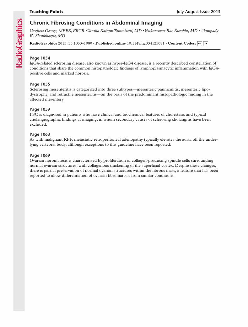
Teaching Points July-August Issue 2013
Chronic Fibrosing Conditions in Abdominal ImagingVerghese George, MBBS, FRCR • Varaha Sairam Tammisetti, MD • Venkateswar Rao Surabhi, MD • Alampady K. Shanbhogue, MD
RadioGraphics 2013; 33:1053–1080 • Published online 10.1148/rg.334125081 • Content Codes:
Page 1054IgG4-related sclerosing disease, also known as hyper-IgG4 disease, is a recently described constellation of conditions that share the common histopathologic findings of lymphoplasmacytic inflammation with IgG4-positive cells and marked fibrosis.
Page 1055Sclerosing mesenteritis is categorized into three subtypes—mesenteric panniculitis, mesenteric lipo-dystrophy, and retractile mesenteritis—on the basis of the predominant histopathologic finding in the affected mesentery.
Page 1059PSC is diagnosed in patients who have clinical and biochemical features of cholestasis and typical cholangiographic findings at imaging, in whom secondary causes of sclerosing cholangitis have been excluded.
Page 1063As with malignant RPF, metastatic retroperitoneal adenopathy typically elevates the aorta off the under-lying vertebral body, although exceptions to this guideline have been reported.
Page 1069Ovarian fibromatosis is characterized by proliferation of collagen-producing spindle cells surrounding normal ovarian structures, with collagenous thickening of the superficial cortex. Despite these changes, there is partial preservation of normal ovarian structures within the fibrous mass, a feature that has been reported to allow differentiation of ovarian fibromatosis from similar conditions.