Embed Size (px)
Citation preview

Gastrointestinal Block Pathology lecture
2013
Dr. Maha ArafahDr. Ahmed Al Humaidi
Malabsorption

1. Know major malabsorption syndromes and its causes.2. Know the many organ systems affected by the
consequences of malabsorption3. Know the following aspects of celiac disease:
a. definition b. pathogenesisc. clinical featuresd. pathology (gross and microscopic features)e. complications (T-cell lymphoma and GI tract carcinoma)
4. Know the cause and types of Lactose intolerance.
Malabsorption

Malabsorption Syndrome
Inability of the intestine to absorb nutrients adequately into the bloodstream
Impairment can be of single or multiple nutrients depending on the abnormality

Physiology
– The main purpose of the gastrointestinal tract is to digests and absorbs nutrients (fat, carbohydrate, and protein), micronutrients (vitamins and trace minerals), water, and electrolytes.

Mechanisms and Causes of Malabsorption Syndrome1. Inadequate digestion
2 .Deficient bile salt
3. Primary mucosal abnormalities
4. Inadequate small intestine
5. Lymphatic obstruction

Mechanisms and Causes of Malabsorption SyndromeInadequate digestion
Postgastrectomy Deficiency of pancreatic lipase
Chronic pancreatitis Cystic fibrosis
Pancreatic resection Zollinger-Ellison syndrome
Deficient bile salt Obstructive jaundice Bacterial overgrowth
Stasis in blind loops, diverticula Fistulas
Hypomotility states (diabetes) Terminal ileal resection
Crohns' disease Precipitation of bile salts (neomycin)
Primary mucosal abnormalities Celiac disease Tropical sprue
Whipple's disease Amyloidosis
Radiation enteritis Abetalipoproteinemia
GiardiasisInadequate small intestine
Intestinal resection Crohn's disease
Mesenteric vascular disease with infarction
Jejunoileal bypassLymphatic obstruction
Intestinal lymphangiectasia Malignant lymphoma
Macroglobulinemia
Man
y cau
ses

Pathophysiology
Small intestine abnormalitiesInadequate digestion Or Malabsorption =

Pathophysiology
Inadequate digestion
Small intestine abnormalities
Pancreas
Bile
Stomach
mucosa
Inadequate small intestine
Lymphatic obstruction
Postgastrectomy
Deficiency of pancreatic lipaseChronic pancreatitisCystic fibrosisPancreatic resection
Obstructive jaundiceTerminal ileal resection

PathophysiologyInadequate digestion
Small intestine abnormalities
Pancreas
Bile
Stomach
mucosa
Inadequate small intestine
Lymphatic obstruction
Celiac diseaseTropical sprueWhipple's disease, (caused by Tropheryma whipplei)Giardiasis
Intestinal resectionCrohn's disease
Intestinal lymphangiectasia Malignant lymphoma

Pathophysiology
Pancreas
Bile
mucosa

Malabsorption Syndrome Clinical features
• Abnormal stools
• Failure to thrive or poor growth in most but not all cases (Weight loss despite increased oral intake of nutrients).
• Specific nutrient deficiencies, either singly or in combination.
Quantitative stool for fat(1) Best screening test(2) 72-hour collection of stool(3) Positive test > 7 g of fat/24 hours.
Stools become soft, yellow, malodorous, greasy and floated at the top of the water in the toilet
There is increased fecal excretion of fat (steatorrhea)

Clinical features

Malabsorption Syndrome Clinical features
Protein
Swelling or edemaMuscle wasting
B12, folic acid and iron deficiency Anemia
(fatigue and weakness
vitamin D, calcium Muscle cramp
Osteomalacia and osteoporosis
vitamin K and other coagulation factor Bleeding tendencies
Depend on the deficient nutrient

Diagnosis
There is no specific test for malabsorption. Investigation is guided by symptoms and signs.1. Fecal fat study to diagnose steatorrhoea2. Blood tests3. Stool studies
4. Endoscopy Biopsy of small bowel

Malabsorption Syndrome Celiac disease
An immune reaction to gliadin fraction of the wheat protein gluten in genetically predisposed persons more in European white
Usually diagnosed in childhood – mid adult.
Patients have raised antibodies to gluten autoantibodies
Highly specific association with class II HLA DQ2 (haplotypes DR-17 or DR5/7) and, to a lesser extent, DQ8 (haplotype DR-4).

a 33-amino acid gliadin peptide that is resistant to degradation by gastric, pancreatic, and small intestinal proteases
Gliadin is deamidated by tissue transglutaminase
deamidated gliadin peptides induce epithelial cells to produce the cytokine IL-15, which in turn triggers activation and proliferation of CD8+ intraepithelial lymphocytes

a 33-amino acid peptide
Clinical features Celiac disease
Typical presentationGI symptoms that characteristically appear at age 9-24 months. Symptoms begin at various times after the introduction of foods that contain gluten. A relationship between the age of onset and the type of presentation; Infants and toddlers….GI symptoms and failure to thriveChildhood…………………minor GI symptoms, inadequate rate of
weight gain, Young adults……………anemia is the most common form of
presentation. Adults and elderly…...GI symptoms are more prevalent
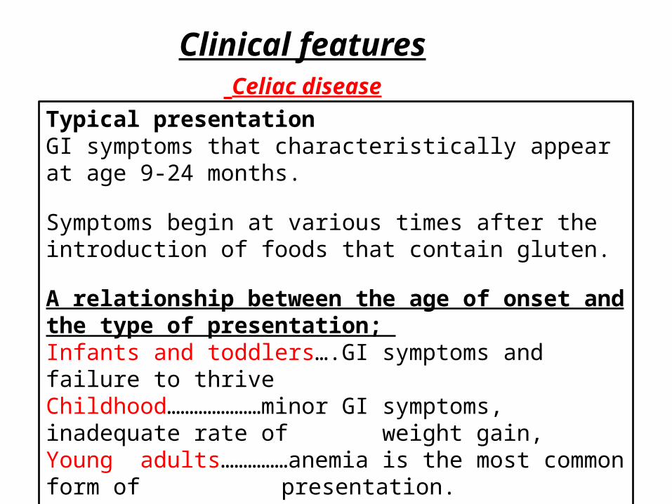
Endoscopy Normal
Celiac disease
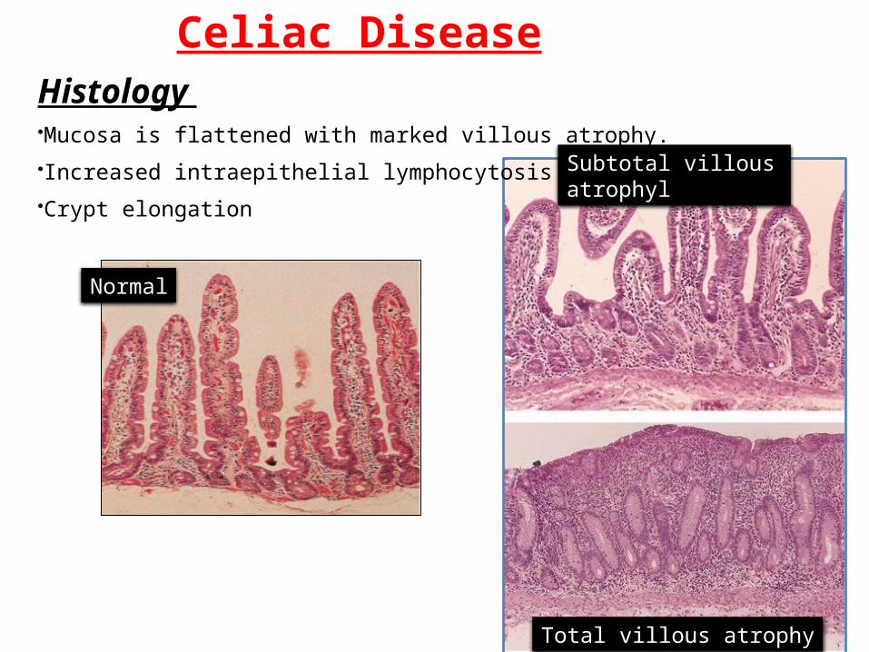
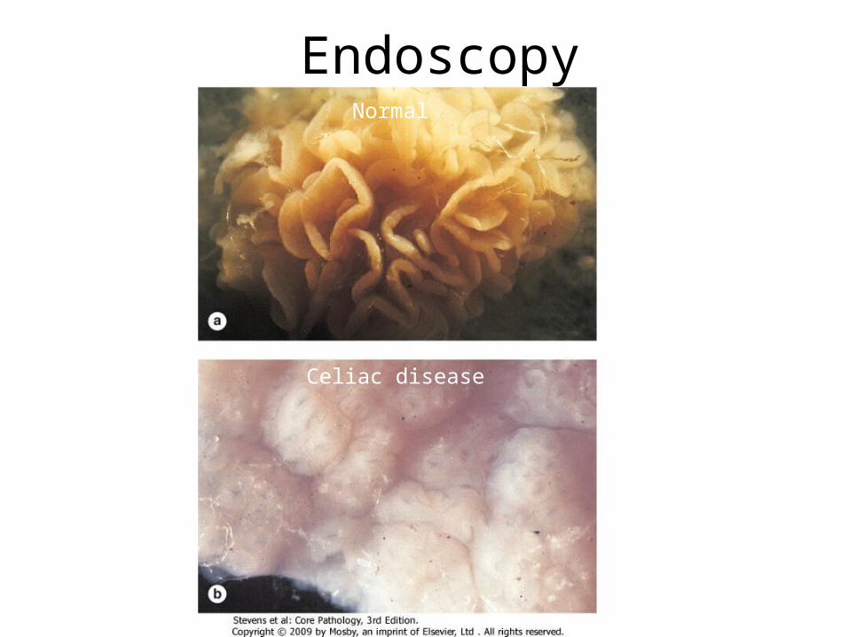
Histology •Mucosa is flattened with marked villous atrophy.
•Increased intraepithelial lymphocytosis
•Crypt elongation
Celiac Disease
Normal
Subtotal villous atrophyl
Total villous atrophy

Celiac DiseaseDiagnosisClinical documentations of malabsorption.Stool ………. FatSerology is +ve for IgA to tissue transglutaminase or IgG to
deamidated gliadin or anti-endomysial antibodiesSmall intestine biopsy demonstrate villous atrophy.Improvement of symptom and mucosal histology on gluten
withdrawal from diet.
wheat, barley, flour Other grains, such as rice and corn flour, do not have such an effect.


Celiac Disease
ComplicationsOsteopenia , osteoporosisInfertility in women Short stature, delayed puberty, anemia, Malignancies,[intestinal T-cell lymphoma]10 to 15% risk of developing GI lymphoma.• DDX Tropical sprue

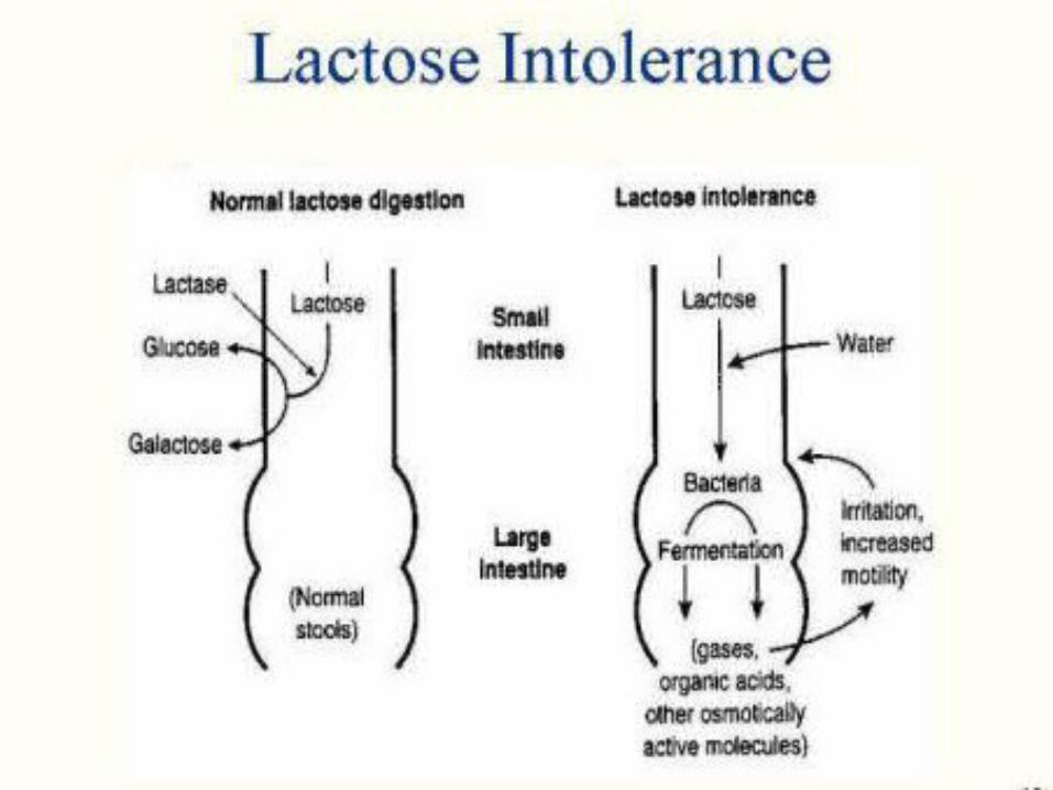
Lactose Intolerance

Lactose Intolerance Pathophysiology
Lactose glucose + galactose At the brush border of enterocytes
lactase
Low or absent activity of the enzyme lactaseLactose Intolerance

Lactose Intolerancecauses
Congenital lactase deficiency Childhood-onset and adult-onset lactase deficiency
Genetically programmed progressive loss of the activity of the small intestinal enzyme lactase.
Secondary lactase deficiency due to intestinal mucosal injury by an infectious, allergic, or inflammatory process
Acquired lactase deficiency
Inherited lactase deficiency
extremely rare common
Transient

Clinical
Bloating, abdominal discomfort, and flatulence ……………1 hour to a few hours after ingestion of milk products

Lactose Intolerance Diagnosis
Empirical treatment with a lactose-free diet, which results in resolution of symptoms;
Hydrogen breath test

Hydrogen breath test
• An oral dose of lactose is administered • The sole source of H2 is bacterial
fermentation; • Unabsorbed lactose makes its way to
colonic bacteria, resulting in excess breath H2.
• Increased exhaled H2 after lactose ingestion suggests lactose malabsorption.

A 3-week trial of a diet that is free of milk and milk products is a satisfactory trial to diagnose lactose intolerance

Lactose Intolerancesummary
• Deficiency/absence of the enzyme lactase in the brush border of the intestinal mucosa → maldigestion and malabsorption of lactose
• Unabsorbed lactose draws water in the intestinal lumen
• In the colon, lactose is metabolized by bacteria to organic acid, CO2 and H2; acid is an irritant and exerts an osmotic effect
• Causes diarrhea, gaseousness, bloating and abdominal cramps

Case scenario• A 44-year-old white male presented with a seven-month history of
increasing diarrhea. The frequency of his bowel movements had increased to 5-7 per day, and his stools were yellow and malodorous and floated at the top of the water in the toilet. He had occasional abdominal cramping, but no tenesmus, melena, or bleeding. His appetite was good, but he had experienced gradual weight loss. His bowel movement frequency would decrease upon fasting and would increase with food intake. A family history of a brother with chronic diarrhea was elicited.
• Stool tests revealed a stool output of 4128 g/d (nl 100-200 g/d) with fat excretion of 17 g/d (nl <5 g/d). Microscopic examination for ova and parasites and cultures for bacterial pathogens and acid-fast bacilli were negative. Blood testing showed mild anemia (Hct 36), hypoproteinemia (4.9 mg/dL), and hypoalbuminemia (3.4 mg/dL). Colonoscopy showed normal mucosa. Esophagogastroduodenoscopy (EGD) showed flat-appearing mucosa in the duodenum. Biopsies were performed. Serum tests for anti-endomysial and antigliadin antibodies were positive.
• The patient was discharged with appropriate dietary instructions. His diarrhea resolved, and his weight began to increase.

Duodenal Biopsy
• What effect does this process have on the surface area available for absorption?
Flattening of the villi greatly decreases the surface area available for absorption.

• Exposure to what dietary antigen is thought to be the cause of these changes?
What food components contain this antigen?
• Will these histologic changes resolve with dietary modification?
Gluten (specifically, the gliaden constituent of this protein).
Wheat, barley and rye
Yes.

Small bowel, Whipple disease (Mucosal disease)
• Numerous macrophages are seen throughout the lamina propria.
• A PAS stain highlights the cytoplasmic inclusions, staining them a deep red.
• An electron micrograph reveals that these inclusions are rod-shaped bacilli.