Embed Size (px)
Citation preview


• Gastrointestinal bleeding describe every form of haemorrhage in the GIT, from the pharynx to the rectum.
• Can be divided into 2 clinical syndromes:-- upper GI bleed
(pharynx to ligament of Treitz)- lower GI bleed
(ligament of Treitz to rectum)

3/81
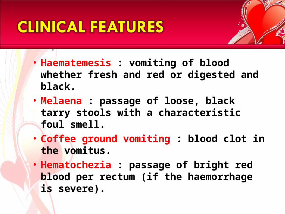
• Haematemesis : vomiting of blood whether fresh and red or digested and black.
• Melaena : passage of loose, black tarry stools with a characteristic foul smell.
• Coffee ground vomiting : blood clot in the vomitus.
• Hematochezia : passage of bright red blood per rectum (if the haemorrhage is severe).

• Haematemesis without malaena is generally due to lesions proximal to the ligament of Treitz, since blood entering the GIT below the duodenum rarely enters the stomach.
• Malaena without haematemesis is usually due to lesions distal to the pylorus
• Approximately 60mL of blood is required to produced a single black stool.

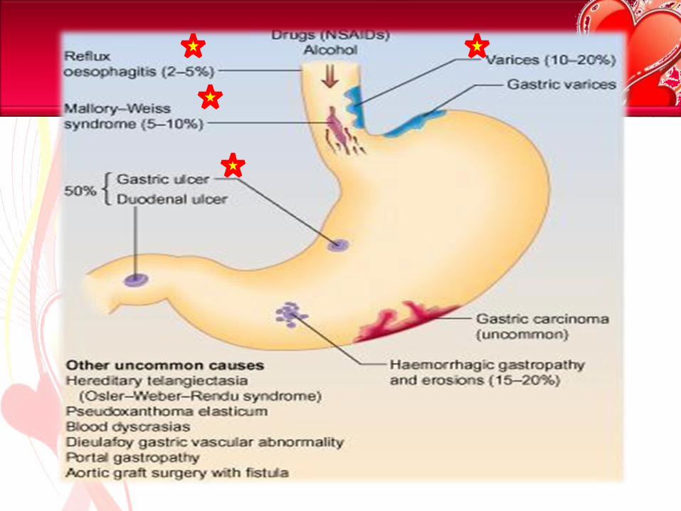
Oesophagus-Oesophageal varices-Oesophageal CA-Mallory-Weiss syndrome
-Haemophilia-Leukemia-Thrombocytopenia-Anti-coagulant therapy
Stomach-Gastric ulcer-Erosive gastritis-Gastric CA
Duodenum-Duodenal ulcer-Duodenitis
LOCAL
GENERAL

• Abnormal dilatation of subepithelial and submucosal veins due to increased venous pressure from portal hypertension (collateral exist between portal system and azygous vein via lower oesophageal venous plexus).
• Most commonly : lower esophagus.

Esophageal varices: a view of the everted
esophagus and gastroesophageal junction, showing
dilated submucosal veins (varices).


(Deflate every 4
hours for 15
minutes )

• Mallory-Weiss syndrome refers to bleeding from tears (a Mallory-Weiss tear) in the mucosa at the junction of the stomach and esophagus, usually caused by severe retching, coughing, or vomiting.
• Mallory-Weiss tears account for 5% to 10% of cases of upper GI bleeding.

- Bleeding from MWTs stops spontaneously in 80-90% of patients
- Endoscopic band ligation (use of elastic bands )- Endoscopic hemoclipping (a metallic mechanical
device used in endoscopy in order to close two
mucosal surfaces without the need for surgery )


15/81

• 8th most common cancer seen throughout the world.
• 40% occur in the middle 3rd of the oesophagus and are squamous carcinomas.
• adenoCA (45%) occur in the lower 3rd of the oesophagus and at the cardia.

1) Dysphagia2) Odynophagia : retrosternal
pain on swallowing.3) Regurgitation4) Weight loss5) Anorexia6) Anemia

• gastric ulcer & duodenal ulcer
• Caused by imbalance between secretion of acid and pepsin, and mucosal defence mechanism.
-Helicobacter pylori infection-NSAIDs-others: stress, smoking,alcohol, steroid
- epigastric pain- haematemesis- Melaena- heartburn
AETIOLOGY
SIGNS & SYMPTOMS

Feature Gastric ulcer Duodenal ulcer
Onset Soon after eating 2-3 hours after eating
Relieving factor vomiting Eating
Precipitating factor
eating Missing a meal, anxiety, stress
Duration of attack
A few weeks A month or two

20/81

Crohn’s disease
Rectal carcinoma
Carcinoma of colon
HaemorrhoidsAnal carcinoma

• Crohn's disease (also spelled Crohn disease) is a chronic inflammatory disease of the intestines. It primarily causes ulcerations (breaks in the lining) of the small and large intestines
• The cause of Crohn's disease is unknown. Some scientists suspect that infection by certain bacteria, such as strains of mycobacterium

• abdominal pain, diarrhea, and weight loss. Less common symptoms include poor appetite, fever, night sweats, rectal pain, and occasionally rectal bleeding.

• There is no medication that can cure Crohn's disease. Patients with Crohn's disease typically will experience periods of relapse (worsening of inflammation) followed by periods of remission (lessening of inflammation) lasting months to years.
• Medications for treating Crohn's disease include anti-inflammatory agents and corticosteroids, topical antibiotics, and immuno-modulators.

• Rare < 50 years old, Common > 60 years old
• Common site- sigmoid colon, rectum
• Clinical features: -altered bowel habit &
large bowel obstruction -rectal bleeding -iron deficiency
anaemia -tenesmus -perforation -anorexia & weight
loss

• M > F• Female- late pregnancy,
puerperium• Supine lithotomy position- 3 ,7,
11 o’clock positions
• Classification: 1st degree : never prolapse 2nd degree: prolapse during
defaecation but return spontaneously
3rd degree : remain prolapse but can be reduced
digitally 4th degree: long-standing
prolapse cannot be reduced

• Rectal bleeding• Perianal irritation & itching• Mucus leakage• Mild incontinence of flatus • Prolapse• Acute pain• Skin tags at anal margin
• Rapid history and examination. • Monitor the pulse and blood
pressure half-hourly.
MANAGEMENT OF ACUTE GASTROINTESTINAL BLEEDING

- when?- have u vomited blood/passed black tarry stools?- had both haematemesis & malaena?- have u had, bleeding from the nose? Bloody expectoration? A dental extraction?
- what is the color, the appearance of the vomited blood?- red? Dark red? Brown? Black?- ‘coffee ground appearance?- bright red & frothy?- what is the color of the stool? Bright red? Black tarry?
- have u vomited blood only once/several times?- has the bleeding been abrupt/massive?- have u had >1 black, tarry stool within a 24-h period?- for how long have the tarry stools persisted?

- retching & severe nonbloody vomiting?- lightheadedness? Nausea? Thirst? Sweating?- faintness when lying down/when standing/syncope?- following the haemorrhage did you have diarrhea?
- aspirin? anticoagulant therapy? iron preparation?- age of the patient?- what is your smoke/alcohol intake?
- have there been similar episode in the past? When? Diagnosis?- were u hospitalized on this occasion? Did u receive a transfusion?- are there any other members of your family who have intestinal disease/bleeding tendency/peptic ulcer/liver disease, History of Malignancy?

Anaemic Bruishing/ Purpura Cachexic Dehydrated Jaundice
Inspection - distension, scar, prominent vein.
Palpation - tenderness, mass/ organomegaly
Percussion - shifting dullness, fluid thrill.
Auscultation - hyperactive bowel sound.
Perianal Skin Lesion Masses Melaena
Confusion ( Shock, liver failure….)
Neurological Deficit

• Clinical shock• Systolic BP < 100mmHg• Pulse rate > 100 bpm• Postural sign: patient place in a
upright position – pulse rate rises 25% or more- systolic BP alls 20mmHg or
more• Sign of liver disease & portal
hypertension• Sign of GI disease• Sign of bleeding abnormalities• Bloody / black stools on per rectal
examination.

• Take blood for haemoglobin, urea, electrolytes, ,liver functions ,blood grouping and crossmatching .
• Establish intravenous access - central line if brisk bleed.
• Stop drugs, e.g. NSAIDs, warfarin

-Full Blood Count- Hb, Platelet- PCV*-Coagulation Profile-Liver Function tests-Serum urea and electrolytes-Blood urea nitrogen-Cross matching of blood.-Serial ECG
- Barium meal / Double- contrast barium meal-Ultrasound-CT scan

• Oxygen therapy for shocked patients.
• Urgent endoscopy in shocked patients/liver disease.
• Continue to monitor pulse and BP.
• Re-endoscope for continued bleeding/hypovolaemia.
• Surgery if bleeding persists.

• Urgent resuscitation is required in patients with large bleeds and the clinical signs of shock.
• Oxygen should be given by face mask and the patient should be kept by mouth until endoscopy has been performed.
• The major principle is to rapidly restore the blood volume to normal. This can be best achieved by transfusion of whole blood via one or more large-bore intravenous cannulae; physiological saline is given until the blood becomes available .
• The rate of blood transfusion must be monitored carefully to avoid overtransfusion and consequent heart failure.
• The pulse rate and venous pressure are the best guides to transfusion rates.

• airway and oxygen• Insert 2 large-bore (14-16G) IV cannulate
take blood• IV colloid - crossmatched. • haemodynamically stable.• Correct clotting abnormalities• Monitor• Insert urinary catheter and monitor
hourly urine output if shocked.• Consider a CVP line to monitor CVP and
guide fluid replacement.• Organize a ECG, and check arterial blood
gases in high-risk patient.• Arrange an urgent endoscopy.• Notify surgeon of all severe bleeds on
admision.

Endoscopy• should be performed within 24 hours in most
patients. Early endoscopy helps to make a diagnosis and to make decisions regarding discharge from hospital, particularly in patients with minor bleeds and under 60 years of age.
• Urgent endoscopy (i.e. after resuscitation) should be performed in patients with shock, suspected liver disease or with continued bleeding.
• Endoscopy can detect the cause of the haemorrhage in 80% or more of cases. In patients with a peptic ulcer, if the stigmata of a recent bleed are seen (i.e. a spurting artery, active oozing, fresh or organized blood clot or black spots) the patient is more likely to re-bleed.

• Most important component of investigation
• 90% accuracy In diagnosis if done with in 24 hours


• all bleeding ulcers should be either injected with epinephrine (adrenaline), the vessel coagulated either with a heater probe or with laser therapy or metallic clips applied. Epinephrine injection -reduces or stops bleeding via a mechanism of vasoconstriction and tamponade
• These methods reduce the incidence of re-bleeding, although they do not significantly improve mortality as re-bleeding still occurs in 20% within 72 hours.
• Intravenous omeprazole 80 mg followed by infusion 8 mg/h for 72 hours should be given to all patients in this group, as it reduces re-bleeding rates and the need for surgery.

• Antacid – aluminium/Mg hydroxide, Mg Trisiclate
• Mucosal protective agents – sucralfate• H2 receptor antagonist – cimetidine &
ranitidine• Proton pump inhibitor – omeprazole &
lansoprazole• Somatostatin (which reduces the
splanchnic blood flow as well as acid secretion) can be given as an infusion if the bleeding is difficult to stop
There is little evidence that H2-receptor antagonists or proton-pump inhibitors (PPIs) affect the mortality rate of GI haemorrhage, but PPIs are usually given to all patients with ulcers because of their longer-term benefits.

– Haemoglobin - May be normal during the acute stages until haemodilution occurs
– Urea and electrolytes - Elevated blood urea suggests severe bleeding
– Cross match for transfusion - Two units of blood are sufficient unless bleeding is extreme.
– If the transfusion is not needed urgently, group the blood and save the serum
– LFT and coagulation profile
1.Systolic BP < 110 mmHg
2.Postural hypotension
3.Pulse > 110/min
4.Haemoglobin <8g/dl
5.Angina or cardiovascular disease with a Haemoglobin <10g/dl