Embed Size (px)
Citation preview
8/3/2019 Fuster 2001
http://slidepdf.com/reader/full/fuster-2001 1/15
Neuron, Vol. 30, 319–333, May, 2001, Copyright ©2001 by Cell Press
ReviewThe Prefrontal Cortex—An Update:Time Is of the Essence
many of the principles discussed below apply also to
the PFC of nonprimate species.
Joaquı n M. Fuster*
Neuropsychiatric Institute and Brain Research
InstituteUniversity of California, Los Angeles Anatomy and Connections
The PFC is the association cortex of the frontal lobe. InLos Angeles, California 90095
primates, it comprises areas 8–13, 24, 32, 46, and 47
according to the cytoarchitectonic map of BrodmannThe physiology of the cerebral cortex is organized in
(1909), recently updated for the monkey by Petrides andhierarchical manner. At the bottom of the cortical organi-
Pandya (Figure 1). Phylogenetically, it is one of thelatestzation, sensory and motor areas support specific sen-
cortices to develop, having attained maximum relativesory and motor functions. Progressively higher areas—of
growth in the human brain (Brodmann, 1912; Jerison,later phylogenetic and ontogenetic development—support
1994), where it constitutes nearly one-third of the neocor-functions that are progressively more integrative. The
tex. Furthermore, the PFC undergoes late development inprefrontal cortex (PFC) constitutes the highest level of
the course of ontogeny. In the human, by myelogenic andthe cortical hierarchy dedicated to the representation
synaptogenic criteria, the PFC is clearly late-maturingand execution of actions.
cortex (Flechsig, 1920; Conel, 1939; Huttenlocher, 1990;
The PFC can be subdivided in three major regions: Huttenlocher and Dabholkar, 1997). In the monkey’sorbital, medial, and lateral. The orbital and medial re-
PFC, myelogenesis also seems to develop late (Gibson,gions are involved in emotional behavior. The lateral
1991). However, the assumption that the synaptic struc-region, which is maximally developed in thehuman, pro-
ture of the PFC lags behind that of other neocorticalvides the cognitive support to the temporal organization
areas has been challenged with morphometric dataof behavior, speech, and reasoning. This function of
(Bourgeois et al., 1994). In any case, imaging studiestemporal organization is served by several subordinate
indicate that, in thehuman, prefrontal areas do notattainfunctions that are closely intertwined (e.g., temporal in-
full maturity until adolescence (Chugani et al., 1987;tegration,working memory, set).Whateverareal special-
Paus et al., 1999; Sowell et al., 1999). This conclusionization can be discerned in the PFC is not so much
is consistent with the behavioral evidence that theseattributable to the topographical distribution of those
areas are critical for those higher cognitive functionsfunctions as to the nature of the cognitive information
that develop late, such as propositional speech andwith which they operate. Much of the prevalent confu-
reasoning.sion in the PFC literature derives from two common
The profuse variety of connections of the PFC is obvi-
errors. The first is to argue for one particular prefrontalfunction while opposing or neglecting others that com-
plement it; the second is to localize any of them within
a discrete portion of PFC.
The functions of the PFC rely closely on its connec-
tions with a vast array of other cerebral structures. None
of its cognitive functions can be understood if taken
out of a broad connectionist context. Any hypothetical
modularity of the PFC is functionally meaningless if
taken out of wide-ranging networks that extend far be-
yond the confines of any given prefrontal area. This is
the reason why the discussion of the operations of the
PFC is here preceded by the placement of the PFC in a
cortical connectionist map of cognitive representations.
After reviewing the anatomy and connectivity of thePFC, I discuss its highest and most general functions,
which are inferred mainly from neuropsychologicalstud-
ies. Then I proceed with a conceptual model of the
cognitive organization of the neocortex, which derives
from those studies as well as from our knowledge of
cortical connectivity. Next, I deal with the dynamics of
the PFC in cognitive operations and with current evi-
dence on the functional specificity of its areas. The re-
view concludes with recent insights from physiological
research on monkeys into the prefrontal mechanisms Figure 1. Cytoarchitectonic Map of the Monkey’s Frontal Cortex
of temporal integration. Although the focus here is on (A) Lateral view.
(B) Medial view.the PFC of the primate, it is reasonable to assume that(C) Inferior (orbital) view.
CC, corpus callosum; PS, principal sulcus. From Petrides and Pan-dya (1994), slightly modified, with permission.* Correspondence: [email protected]
8/3/2019 Fuster 2001
http://slidepdf.com/reader/full/fuster-2001 2/15
Neuron320
Figure 2. Cortex of the Human
Prefrontal areas are numbered according to
Brodmann’s cytoarchitectural map.
ously related to the variety of the information it inte- of the major prefrontal regions—medial, orbital, and lat-
eral—is connected with itself and with the other twogrates. For detailed accounts of PFC connections in the
primate, the reader may wish to consult other reviews (Jacobson and Trojanowski, 1977a, 1977b; Pandya and
Yeterian, 1985). Some of the corticocortical connectivity(Pandya and Yeterian, 1985; Fuster, 1997; Mesulam, 1998;
Barbas, 2000). Here, I will only consider some of the of the PFC is interhemispheric, and almost all of it is
reciprocal and topologically organized (Pandya and Yet-extrinsic prefrontal connections.
The PFC is connected with the brainstem, the thala- erian, 1985;Cavada and Goldman-Rakic, 1989a, 1989b).
In general, connections between association corticesmus, the basal ganglia, and the limbic system. Much of
that connectivity with subcortical structures is recipro- both originate and terminate in upper cortical layers,
especially II and III (Jones, 1981; Andersen et al., 1985).cal. Especially well organized topologically are the con-
nections between the PFC and the thalamus. The prefron- Those connections presumably constitute the structural
frame of cognitive networks (Fuster, 1995). As the mem-tal connections with the mediodorsal thalamic nucleus
have been used as a criterion for identifying the PFC in ory networks of posterior cortex acquire associations
with action, they extend into PFC to shape the networksa wide variety of species (Fuster, 1997).
The functional role of the afferent connections of the of executive memory.
PFC can be broadly inferred from the functions of the
contributing structures. In the aggregate, the afferent Neuropsychology of the PFC
What we know about the higher integrative functionsconnections from the brainstem, the diencephalon and
the limbic system convey to the PFC information about of the PFC is inferred mainly from neuropsychological
studies in the human. Because of large variations in thethe internal environment, the level of arousal, the drives
andmotivesof theanimal,and thevisceral concomitants location, extent, andclinical manifestations of prefrontal
damage, that knowledge is the distillate of a vast litera-of emotion. Especially relevant for the behavioral integ-
rative functions of the PFC are its afferent connections ture. In essence, three distinct clusters of symptoms
can be observed after lesions of the three major regionsfrom the amygdala and the hypothalamus. The amygdala
projects to the ventral and medial aspects of the PFC of the PFC: orbital or inferior, medial/cingulate, and lat-
eral (Figure 2). Although many reported cases are mixed(Porrino et al., 1981; Ray and Price, 1993), and so does
the hypothalamus (Kievit and Kuypers, 1975; Jacobson in terms of cortex affected and clinical picture, the three
prefrontal “syndromes” provide insights into the majoret al., 1978). In all likelihood, these connections carry to
thefrontal lobe information notonly about internal states and most general functions of those regions.
Ever since Harlow (1848) described the famous casebut about the motivational significance of sensory stim-
uli. These connections probably play a major role in the of Phineas Gage, it has been known that lesions of
orbital PFC often induce dramatic changes of personal-representation and enactment of emotional behavior (Le
Doux, 1993). Theconnections of the PFCwith thehippo- ity (Damasio et al., 1994; Fuster, 1997). Subjects with such
lesions areimpulsive anddisinhibited in a host of instinc-campus are also of major behavioral relevance. All pre-
frontal regions receive projections from the hippocam- tual behaviors. They are irritable and contentious, with
a characteristic tendency to coarse humor and disre-pus, either directly or indirectly (Rosene and Van Hoesen,
1977; Amaral, 1987; Barbas and Blatt, 1995). gardforsocial and moral principles. Their impulsiveness
frequently leads them to reckless high-risk behavior andThe PFC is connected with other cortices of associa-
tion, but not with primary sensory or motor cortices. Each conflicts with the law. In addition, orbitofrontal patients
8/3/2019 Fuster 2001
http://slidepdf.com/reader/full/fuster-2001 3/15
Review321
almost uniformly exhibit a severe disorder of attention. Onthebasis ofa large body ofanatomical,electrophysi-
ological, and neuropsychological evidence reviewedThis disorder is characterized by the failure to withstand
interference from distraction. Monkeys with orbitofron- elsewhere (Fuster, 1995), it is reasonable to treat the
cortex behind the Rolandic fissure as the substrate fortal damage show similar difficulties as do people with
comparable damage in the control of instinctual im- perceptual memory, and the frontal cortex for executive
memory (Figure 3). According to this view, neuronal net-pulses and internal or external distraction. The orbito-
frontal cortex exerts its functions of inhibitory control works of perceptual memory are formed in postrolandiccortex and organized hierarchically over a base layervia its efferents to the hypothalamus, the basal ganglia,
and other neocortical areas, some in the PFC itself. of primary sensory cortices (phyletic sensory memory).
Progressively higher areas accommodate progressivelyThemedial regionof the PFC, which includes themost
anterior portion of the cingulate gyrus, also appears more general categories of memory, including episodic
and semantic memory—which together constitute de-involved in general motility, attention, and emotion. Le-
sions of this regioncommonlylead to loss of spontaneity clarative memory. That upward expansion of perceptual
memory networks occurs along well-identified paths ofand difficulty in the initiation of movements and speech
(Verfaellie and Heilman, 1987; Cummings, 1993). Large corticocortical connection. Although the general cate-
gories of both memory and knowledge arehierarchicallyand bilateral lesions lead to akinetic mutism. Patients
with medial/cingulate lesions are commonly apathetic, organized, individual items of memory or knowledge are
to some degree heterarchical. Autobiographical mem-disinterested in the environment, and unable to concen-
trate their attention on behavioral or cognitive tasks. ory contains both semantic and sensory components
intermixed. Thus, the memory of an episode in one’sConversely, the neuroimaging of normal subjects by
positron emission tomography (PET) or functional mag- life is probably represented by a cortical network thatspans several levels of the perceptual hierarchy.netic resonance (fMRI) shows marked activations of the
anterior cingulate in tasks that demand sustained effort The counterpart of the perceptual memory hierarchy
in posterior cortex is an executive memory hierarchy inand concentrated attention (Posner et al., 1988; Raichle,
1994). Such evidence led to the formulation of an “ante- frontal cortex. Motor networks grow toward cortex of
association (PFC) on a base of primary motor cortexrior attentional system” (Posner and Petersen, 1990), of
which theanterior cingulateregion would be anessential (phyleticmotormemory).At thelower level of that hierar-
chy, movements are defined mainly by the action ofpart (further discussion below).
Lesions of the lateral region induce the most charac- muscles or muscle groups. At higher levels, in premotor
cortex, executive networks represent acts and pro-teristic cognitive deficits from frontal lobe injury. In hu-
mans with large lateral prefrontal damage, the most grams of movement defined by goal and trajectory. Net-
works in some premotor areas represent elementarycommon disorder is the inability to formulate and to
carry out plans and sequences of actions. Luria (1966) linguistic structures (notably “Broca’s area,” area 45).
From the neuropsychological research briefly reviewedwas the first to investigate and describe this disorder
in a large number of patients. The deficit in planning, in theprevioussection, thelateralPFC appears to harborwhich extends to the representation and construction networks representing schemas, plans, and concepts
of sequences of spoken and written language (Luria of action. In sum, the neuroanatomy and neuropsychol-1970), is now widely recognized as a constant manifes- ogy of the frontal lobe strongly suggest the upward
tation of large lateral PFC damage. It has two major hierarchical layering of executive memory, from the in-
aspects: one is the difficulty to consciously represent nate representation of elementary movement in motor
sequences of speech or behavior, especially if they are cortex to the representation of sequential action in the
novel or complex; the other is the difficulty to initiate PFC. That upward layering appears to take place in
them and to execute them in orderly manner. These several interrelated domains of action (e.g., skeletal, lin-difficulties constitute what has been termed the dys- guistic), each containing executive representations ofexecutive syndrome (Baddeley, 1986), which is usually varying degrees of specificity and abstraction. As inaccompanied by a severe attention disorder that Shal- posterior cortex, however, the organization of represen-
lice (1988) characterizes as a loss of “supervisory atten- tations in frontal cortex does not appear strictly hierar-
tional control.” In sum, from the neuropsychological evi- chical, at least inasmuch as hierarchical representation
dence, it can be concluded that the lateral PFC plays a implies serial processing from the top down. In the exe-crucial role in the organization and execution of behav- cution of complex actions, the activations of prefrontal,
ior, speech, and reasoning. premotor, and motor networks do not always follow a
strict temporal sequence (Kalaska et al., 1998). Even at
the lowest cortical level (motor cortex), actions seem toThe PFC in the Cortical Cognitive Map
The cortex of the human appears divided by the Rolan- be represented, to a degree, in terms of direction of
movement (Georgopoulos et al., 1982). Thus, it is rea-dic fissure into two major parts, each dedicated to a
separate broad category of functions: the cortex of the sonable to suppose that executive networks, like per-
ceptual networks, are to some degree heterarchical.occipital, temporal, and parietal lobes, dedicated to
“sensory” functions; and the cortex of the frontal lobe, Imaging studies show that lateral prefrontal and pre-
motor areas are activated at the beginning of the learn-dedicated to “motor” functions. This dichotomy of corti-
ces seems to represent the evolutionary expansion into ing of a motor sequence; with practice and repetition,
however, thatactivation subsides, whilethat of subcorti-the telencephalon of the dichotomy of structures that
spans the entire length of the nerve axis from the spinal cal structures, notably the basal ganglia, increases (Graf-
ton et al., 1992; Jenkins et al., 1994; Iacoboni et al., 1996;cord upwards: a posterior sensory moiety and an ante-
rior motor one. Petersen et al., 1998). Thus, as sequences become over-
8/3/2019 Fuster 2001
http://slidepdf.com/reader/full/fuster-2001 4/15
Neuron322
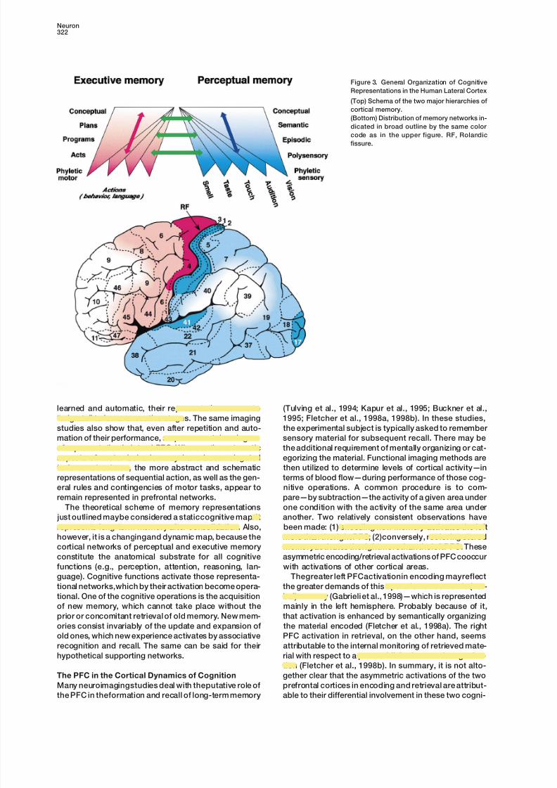
Figure 3. General Organization of Cognitive
Representations in the Human Lateral Cortex
(Top) Schema of the two major hierarchies of
cortical memory.
(Bottom) Distribution of memory networks in-
dicated in broad outline by the same color
code as in the upper figure. RF, Rolandicfissure.
learned and automatic, their representation seems to (Tulving et al., 1994; Kapur et al., 1995; Buckner et al.,
1995; Fletcher et al., 1998a, 1998b). In these studies,“migrate” to lower executive stages. The same imaging
studies also show that, even after repetition and auto- the experimental subject is typically asked to remember
sensory material for subsequent recall. There may bemation of their performance, sequences retain a degree
of representation in lateral PFC. Whereas the automatic the additional requirement of mentally organizing or cat-
egorizing the material. Functional imaging methods areaspects of motor behavior may have been relegated
to lower structures, the more abstract and schematic then utilized to determine levels of cortical activity—in
terms of blood flow—during performance of those cog-representations of sequential action, as well as the gen-
eral rules and contingencies of motor tasks, appear to nitive operations. A common procedure is to com-
pare—by subtraction—the activity of a given area underremain represented in prefrontal networks.
The theoretical scheme of memory representations one condition with the activity of the same area under
another. Two relatively consistent observations have just outlined maybe considered a staticcognitive map. It
represents long-term memory after consolidation. Also, been made: (1) encoding new memory activates the left
more than theright PFC; (2)conversely, retrieving storedhowever, it is a changingand dynamic map, because the
cortical networks of perceptual and executive memory memoryactivates therightmorethan theleftPFC. These
asymmetric encoding/retrieval activations of PFC cooccurconstitute the anatomical substrate for all cognitive
functions (e.g., perception, attention, reasoning, lan- with activations of other cortical areas.
Thegreater left PFCactivationin encoding mayreflectguage). Cognitive functions activate those representa-
tional networks,which by their activation become opera- the greater demands of this operation on semantic (ver-
bal) memory (Gabrieli et al., 1998)—which is representedtional. One of the cognitive operations is the acquisition
of new memory, which cannot take place without the mainly in the left hemisphere. Probably because of it,
that activation is enhanced by semantically organizingprior or concomitant retrieval of old memory. New mem-
ories consist invariably of the update and expansion of the material encoded (Fletcher et al., 1998a). The right
PFC activation in retrieval, on the other hand, seemsold ones, which new experience activates by associative
recognition and recall. The same can be said for their attributable to the internal monitoring of retrieved mate-
rial with respect to a preestablished semantic organiza-hypothetical supporting networks.
tion (Fletcher et al., 1998b). In summary, it is not alto-
gether clear that the asymmetric activations of the twoThe PFC in the Cortical Dynamics of Cognition
Many neuroimagingstudies deal with theputative role of prefrontal cortices in encoding and retrieval are attribut-
able to their differential involvement in these two cogni-the PFC in theformation and recall of long-term memory
8/3/2019 Fuster 2001
http://slidepdf.com/reader/full/fuster-2001 5/15
Review323
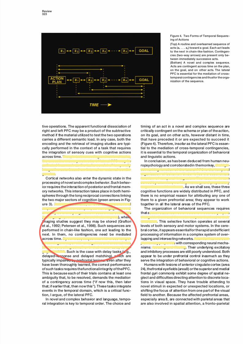
Figure 4. Two Forms of Temporal Sequenc-
ing of Actions
(Top) A routine and overlearned sequence of
acts (a1 . . . an ) toward a goal. Each act leads
to the next in chain-like fashion. Contingen-
cies (two-way arrows) are present only be-
tween immediately successive acts.(Bottom) A novel and complex sequence.
Acts are contingent across time on the plan,
on the goal, and on other acts. The lateral
PFC is essential for the mediation of cross-
temporal contingencies and thusfor the orga-
nization of the sequence.
tive operations. The apparent functional dissociation of timing of an act in a novel and complex sequence are
critically contingent on the schema or plan of the action,right and left PFC may be a product of the subtractive
method if the material utilized to test the two operations on its goal, and on other acts, however distant in time,
that have preceded it or are expected to succeed itcarries a different semantic load. In any case, both the
encoding and the retrieval of imaging studies are typi- (Figure 4). Therefore, insofar as the lateral PFC is essen-
tial to the mediation of cross-temporal contingencies,cally performed in the context of a task that requires
the integration of sensory cues with cognitive actions it is essential to the temporal organization of behavioral
and linguistic actions.across time. Thus, in both encoding and retrieval tasks,
the executive memory networks of lateral PFC may be In conclusion, as has been deduced from human neu-
ropsychology and corroboratedin themonkey, thehigh-activated inasmuch as the tasks consist of temporally
integrative acts based on internal or external contin- est-ranking function of the lateral PFC is the temporal
organization of behavior. Under it lies the subordinategencies.
Cortical networks also enter the dynamic state in the function of temporal integration. The latter is in turn
served by attention, working memory, and preparatoryprocessing of novel andcomplex behavior. Such behav-
ior requires the interaction of posterior and frontal mem- set, to be considered next. As we shall see, these three
cognitive functions are widely distributed in PFC, andory networks. This interaction takes place in both hemi-
spheres through the long reciprocal connections linking there is no empirical reason for attributing any one of
them to a given prefrontal area; they appear to workthe two major sectors of cognition (green arrows in Fig-
ure 3). Routine, automatic, or overlearned behavioral together in all the lateral areas of the PFC.
The organization of behavioral sequences requiressequences, however complex, do not engage the PFC
and may be entirely organized in subcortical structures that selectiveattentionbe directed to the sensory inputs
that guide them and the motor outputs that lead to their(basal ganglia, cerebellum, lateral thalamus, etc.), where
imaging studies suggest they may be stored (Grafton execution. This selective function operates at several
levels of both sensory and motor systems. In the cere-et al., 1992; Petersen et al., 1998). Such sequences are
performed in chain-like fashion, one act leading to the bral cortex, it appears essentialfor therapid andefficient
processing of information in a complex system of over-next. In them, no contingencies need be mediated
across time. By contrast, sequences with cross-tempo- lapping and intersecting networks. Attention has two com-
plementary properties with corresponding neural mecha-ral contingencies, or with ambiguities and uncertainties
in their controlling stimuli or in their motor acts, do en- nisms: focusing and filtering. Their underlying excitatory
and inhibitory processes are still poorly understood. Bothgage the PFC. Such is the case with delay tasks (e.g.,
delayed-response and delayed matching), which are appear to be under prefrontal control inasmuch as they
serve the integration of behavioral or cognitive actions.typically impaired by prefrontal lesions. Even after they
have been thoroughly learned, the correct performance Humans with lesions of anterior cingulate cortex (area
24), thefrontal eyefields (area8) or the superior and medialof such tasks requires thefunctionalintegrity of thePFC.
This is because each of their trials contains at least one frontal gyri commonly exhibit some degree of spatial ne-
glect and difficulties directing attention to discrete loca-ambiguity that, to be resolved, demands the mediation
of a contingency across time (“if now this, then later tions in visual space. They have trouble attending to
novel stimuli in expected or unexpected locations, orthat; if earlier that, then now this”). These tasks integrate
events in the temporal domain, which is a critical func- shifting the focus of attention from one part of the visual
field to another. Because the affected prefrontal areas,tion, I argue, of the lateral PFC.
In novel and complex behavior and language, tempo- especially area 8, are connected with parietal areas that
are also involved in spatial attention, a fronto-parietalral integration is key to temporal order. The choice and

8/3/2019 Fuster 2001
http://slidepdf.com/reader/full/fuster-2001 6/15
Neuron324
“network” for spatial attention has been postulated
(Mesulam, 1981; Posner and Petersen, 1990). In support
of this view, imaging studies show that tasks demanding
high levelsof spatial attention activatethosesame areas
of prefrontal and parietal cortex (Pardo et al., 1990; Cor-
betta et al., 1993;Nobreet al., 1997;Kastner et al., 1999).
Some studies also show, however, prefrontal activa-tions in tasks that demand attention to nonspatially de-
fined stimuli (Vandenberghe et al., 1997; Duncan and
Owen, 2000). The anterior cingulate region, in particular,
is activated in a broad variety of attentional states and
tasks that do not require spatial attention. Perhaps the
close connections of this region with limbic structures
account for its activation in situations that demand con-
centrated effort and close attention to detail (Pardo et
al., 1990; Posner and Petersen,1990; Taylor et al., 1994).
Human and animal neuropsychology (Fuster, 1997) indi-
cates that the orbital PFC plays a major role in the exclu-
sionary aspect of attention, inhibiting or filtering out cogni-
tive information that is extraneous to the task at hand.
In general, therefore, the involvement of the PFC inattention, and thus in selective information processing,
is inseparable from its role in organizing goal-directed
actions. The anterior cingulate seems involved in the
motivation to perform them, the orbitofrontal cortex in Figure 5. Frontal Activations under Five Cognitive Demandsthe suppression of distractions that interfere with them, Colored squares mark theestimated loci of peak activation of frontaland the lateral cortex in the mediation of their cross- cortex during performance of cognitive tasks that test the effect
temporal contingencies. All three aspects of attention of response conflict (green), novelty (pink), working-memory load
(yellow), working-memory delay (red), and perceptual difficultyare supported by the PFC. This is most apparent in(blue). The estimates of peak location result from metaanalysis ofspatial tasks, where temporal integration depends onseveral neuroimaging studies. Note that assorted tasks and de-
gaze control and thus on the frontal eye fields of areasmands activate three prefrontal regions bilaterally. From Duncan
6 and 8. In any event, attention, goal-directedness, and and Owen (2000), slightly modified, with permission.temporal integration are common elements of most all
the task paradigms utilized to explore thePFC by means
of functional imaging. Not surprisingly, therefore, those delay periods, that is, periods when short-term memoryparadigms activate several prefrontal areas in common was required, than during intertrial (baseline) periods.(Figure 5). Temporal integration is present in practically That kind of firing activity related to spatial memory,all of those paradigms (though the subject’s need to which persisted through delays of tens of seconds (upmentally integrate the experimenter’s instructions with to 2 or 3 min), had four important properties: (1) it wassubsequent performance is neglected in most studies). absent after stimuli that did not call for prospective ac-Thus, the presumed tests of attention utilized in imaging tion; (2) it was absent in the mere expectation of reward;studies are essentially tests of temporal integration, and (3) it was correlated with the accuracy of the animal’sit appears that for this reason they activate lateral pre- performance (i.e., efficiency of short-term memory); andfrontal areas. After all, both working memory and prepa- (4) it could be diminished or aborted by distracting stim-ratory set, the two basic cognitive supports of temporal uli occurring in the delay period (e.g., taped playbackintegration in the lateral PFC, essentially consist of at- of monkey voices). These properties of their firing, espe-tention directed to internal representations. In any case, cially during long delays (10 s), strongly implicatedsuch related psychological functions as attentional se- those cells in the retention of the memorandum. Argua-lectivity, resolution of ambiguities, and suppression of bly, memory cells opened systems neuroscience to thedistractions are essentially necessary for temporal inte- study of mental representation.gration and, therefore, can be considered essential parts Spatial memory cells such as those just describedof itsprocess. They depend, most likely, on theselective were subsequently observed in thePFC by many investi-activation of posterior cortical areas under prefrontal gators (e.g., Niki, 1974; Nikiand Watanabe, 1976; Josephcontrol (Desimone and Duncan, 1995; Fuster, 1995). and Barone, 1987; Quintanaet al., 1988). Goldman-Rakic
andher colleagues (Funahashi et al., 1989), in a delayed-
response task with brief delays and eye movement asMemory and Set, for the Two Sides of Time
Working memory is the first temporal integrative func- thebehavioral indicator, demonstrated memory cells for
discrete light-spot positions in the four quadrants of thetion of the PFCto have been substantiated electrophysi-
ologically. Cells in the PFC of the monkey were found two-dimensional visual field.
In delayed-response tests, memory cells of the kindto fire persistently at high rates while the animal retained
an item of visual information in short term memory (Fuster just described ostensively retain the position of the vi-
sual cue or memorandum that the animal must retainand Alexander, 1971; Fuster, 1973); in delayed-response
tasks, such “memory cells” fired more frequently during for subsequent motor response. By using various other
8/3/2019 Fuster 2001
http://slidepdf.com/reader/full/fuster-2001 7/15
Review325
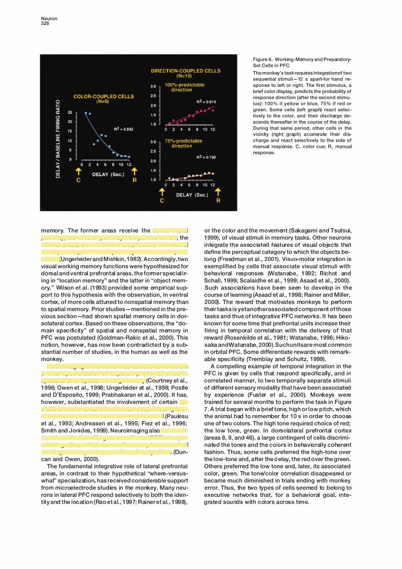
types of delay tasks, especially delayed matching to sam- gradually toward baseline level (Figure 6); these sen-
sory-coupled units behaved like conventional working-ple, memory cells have also been found—in lateral PFC-
that are attuned to visual-nonspatial (Fuster et al., 1982; memory cells for color. Intermixed with them in thesame
area, direction-coupled cells were found, which showedMiller et al., 1996), auditory (Bodner et al., 1996), or tactile
(Romo et al., 1999) memoranda. In anycase,the involve- a temporally reciprocal behavior. Their discharge in-
creased in the course of the delay, and they fired atment of prefrontal cells in working memory appears
strictly related to the need to retain information for an differentrates atthe time of themotor choice, dependingon whether it was the right or the left. Further, the slopeimpending action that is in some way dependent on that
information. Prefrontal cells “remember for action.” of accelerating dischargein thedelay periodwas greater
when the monkey could predict side with certaintyFunctional neuroimaging reveals PFC activations in a
variety of working memory tasks. Such activations pre- (100%) than when he could not.
Thus, neuropsychological, electrical, and imaging datasumably reflect the excitation of large assemblies of
memory cells. Thus, lateral activation has been demon- point to the coexistence in lateral PFC of two neural
substrates of active representation, one for the recentstrated when the memorandum is visuospatial (Jonides
et al., 1993; McCarthy et al., 1994), visual-nonspatial (Co- past and the other for the anticipated future. There is
no evidence of anatomical segregation of those twohen et al., 1994; Swartz et al., 1995), or verbal (Grasby et
al., 1993; Petrides et al., 1993b; Smith et al., 1996). None- substrates. Quite the contrary, there is microelectrode
evidence that the two overlap to a considerable extent.theless, current imaging methods lackthe sufficient spa-
tial and temporal resolution to detect topographic differ- Some prefrontal cells seem to “look” to the past and
others, nearby, to the future. It is reasonable to assumeences related to specific memoranda or the time course
of cellular excitability in memory periods. that the two populations of cells are part of the samecortical network of long-term memory that representsIt has long been known that the PFC, especially its
lateral region, is involvedin the expectation of, and prep- the associated sensory and motor components of a
given task or behavioral sequence. That network is pre-aration for, anticipated events. This prospective involve-
ment of the PFC is most probably related to the proven sumably activated for the task, its neurons to bridge tem-
poral contingencies and to guide-through motor sys-deficit of frontal patients in planning. Electrophysiologi-
cally, the anticipatory aspects of prefrontal integration tems-the behavior to its goal. Thus, the two temporal
perspectives of a behavioral sequence would be in-are at the root of the surface-negative field potentials—
notably the “expectancy wave”—recorded over frontal tegrated by the sustained activation of the network’s
components. Working memory would consist of the acti-regions in the time between a stimulus and a response
contingent on it (Kutas and Donchin, 1980; Brunia et vation of theperceptual components. A temporally sym-
metric and complementary function of preparatory set,al., 1985; Singh and Knight, 1990). Another electrical
manifestation of anticipation in the PFC is the presence or “motorattention,”would activatethe network’s motor
components and thus prime executive systems for theof cells that, during the delay of a delay task, fire at high
levelsin theapparent expectationof themotorresponse anticipated action.or another stimulus related to it (Niki and Watanabe,
1979; Fuster et al., 1982; Boch and Goldberg, 1989; Areal Specificity in PFC: The “What,” the “Where,”Sawaguchi et al., 1989; Rainer et al., 1999). That kind and the “When”of preparatory activity has also been inferred from the Beginning in the 1930’s (Jacobsen, 1931), a long seriesresults of neuroimaging during planning (Partiot et al., of lesion studies on the monkey showed that the lateral1995; Baker et al., 1996). In sum, considerable evidence PFC was essential for delay tasks. In these tasks, thefrom several methodologies supports a prospective role animal must memorize items of information through pe-of thePFC, in addition to itsretrospective role of working riods of forced delay. From deficits induced in thosememory. Ingvar (1985) dubbed that prospective role the tasks by ablating lateral PFC, its role was deduced in“memory of the future.” thekind of short-term memorywhichis nowconvention-
Quintana and Fuster (1999) found two types of units, ally called working memory—applying to the monkey aworking-memory cells and preparatory-set cells, topo- term with somewhat different meaning in human cogni-graphically intermingled in the dorsolateral PFC of mon- tion (Baddeley 1986). Since the tasks (e.g., delayed-keys performing a delay task with variable probabilities response) commonly required memorization of a spa-of contingency between color and direction of manual tially defined visual stimulus, some researchers furtherchoice. A trial began with the brief presentation of a assumed that thelateral PFCcortex was thestorage sitecolor, which the animal had to retain for pairing it with for memory of spatial location. However, the reversiblea second stimulus some 12 s later. Depending on the lesion of this cortex—by local cooling—induced a tem-combination of those two stimuli, the animal had to porary deficit in delayed color matching (a nonspatialchoose a right or a left location. After the animal had memory task) that was of the same magnitude as thebeen extensively trained to perform the task, each color deficit in delayed response (Bauer and Fuster, 1976).carried a given probability that the second stimulus Furthermore, microelectrode recording in monkeys per-would demand a response to one side or to the other. forming both tasks revealed spatial and nonspatial
Thus, some colors predicted response location with memory cells throughout the lateral PFC (Fuster et al.,
100% probability and others with 75%. In the cortex 1982).
above the principal sulcus (areas 9 and 46), some cells Nonetheless, some investigators used an anatomical
discriminated the colors (by differences in firing fre- rationale in support of a functional dissociation between
dorsal and ventral areas of the lateral PFC in visualquency), and their activity during the delay descended
8/3/2019 Fuster 2001
http://slidepdf.com/reader/full/fuster-2001 8/15
Neuron326
Figure 6. Working-Memory and Preparatory-
Set Cells in PFC
The monkey’s task requires integrationof two
sequential stimuli—12 s apart-for hand re-
sponse to left or right. The first stimulus, a
brief color display, predicts the probability of
response direction (after the second stimu-lus): 100% if yellow or blue, 75% if red or
green. Some cells (left graph) react selec-
tively to the color, and their discharge de-
scends thereafter in the course of the delay.
During that same period, other cells in the
vicinity (right graph) accelerate their dis-
charge and react selectively to the side of
manual response. C, color cue; R, manual
response.
memory. The former areas receive the dorsal visual or the color and the movement (Sakagami and Tsutsui,
1999), of visual stimuli in memory tasks. Other neuronspathway, the “where” pathway from parietal cortex; the
ventral areas, on the other hand, receive the ventral integrate the associated features of visual objects that
define the perceptual category to which the objects be-visual pathway, the “what” pathway from inferotemporal
cortex (Ungerleider and Mishkin, 1982). Accordingly, two long (Freedman et al., 2001). Visuo-motor integration is
exemplified by cells that associate visual stimuli withvisual working memory functions were hypothesized for
dorsal and ventral prefrontal areas, the former specializ- behavioral responses (Watanabe, 1992; Bichot and
Schall, 1999; Scalaidhe et al., 1999; Asaad et al., 2000).ing in “location memory” and the latter in “object mem-
ory.” Wilson et al. (1993) provided some empirical sup- Such associations have been seen to develop in the
course of learning (Asaad et al., 1998; Rainer and Miller,port to this hypothesis with the observation, in ventral
cortex, of more cells attuned to nonspatial memory than 2000). The reward that motivates monkeys to perform
their tasks is yetanotherassociated component of thoseto spatial memory. Prior studies—mentioned in the pre-
vious section—had shown spatial memory cells in dor- tasks and thus of integrative PFC networks. It has been
known for some time that prefrontal units increase theirsolateral cortex. Based on these observations, the “do-
main specificity” of spatial and nonspatial memory in firing in temporal correlation with the delivery of that
reward (Rosenkilde et al., 1981; Watanabe, 1996; Hiko-PFC was postulated (Goldman-Rakic et al., 2000). This
notion, however, has now been contradicted by a sub- saka andWatanabe, 2000).Suchunitsare most common
in orbital PFC. Some differentiate rewards with remark-stantial number of studies, in the human as well as the
monkey. able specificity (Tremblay and Schultz, 1999).
A compelling example of temporal integration in theNeuroimaging in the human has failed to demonstrate
a clear specialization of separate prefrontal areas in PFC is given by cells that respond specifically, and in
correlated manner, to two temporally separate stimulispatial and nonspatial working memory (Courtney et al.,
1996; Owen et al., 1998; Ungerleider et al., 1998; Postle of different sensory modality that have been associated
by experience (Fuster et al., 2000). Monkeys wereand D’Esposito, 1999; Prabhakaran et al., 2000). It has,
however, substantiated the involvement of certain lat- trained for several months to perform the task in Figure
7. A trial began with a brief tone, high or low pitch, whicheral and inferior areas of the frontal lobe, including Bro-
ca’s, in the memorization of semantic material (Paulesu the animal had to remember for 10 s in order to choose
one of two colors. The high tone required choice of red;et al., 1993; Andreasen et al., 1995; Fiez et al., 1996;
Smith and Jonides, 1999). Neuroimaging also substanti- the low tone, green. In dorsolateral prefrontal cortex
(areas 8, 9, and 46), a large contingent of cells discrimi-ates theactivationof large areas of lateral PFCin tempo-
ral integration, which is a uniform requirement of all nated the tones and the colors in behaviorally coherent
fashion. Thus, some cells preferred the high-tone overworking-memory tasks, as well as of many others (Dun-
can and Owen, 2000). the low-tone and, after the delay, the red over the green.
Others preferred the low tone and, later, its associatedThe fundamental integrative role of lateral prefrontal
areas, in contrast to their hypothetical “where-versus- color, green. The tone/color correlation disappeared or
became much diminished in trials ending with monkeywhat” specialization, has received considerable support
from microelectrode studies in the monkey. Many neu- error. Thus, the two types of cells seemed to belong to
executive networks that, for a behavioral goal, inte-rons in lateral PFC respond selectively to both the iden-
tity and the location (Rao et al., 1997; Rainer et al., 1998), grated sounds with colors across time.

8/3/2019 Fuster 2001
http://slidepdf.com/reader/full/fuster-2001 9/15
Review327
These views do not exclude new information from work-
ing memory. Indeed, new stimuli can be readily incorpo-
rated into an activated network of long-term memory
by processes of categorization, perceptual constancy,
and contextual association.
Cortical Mechanisms of Temporal Integration
The mechanisms of temporal integration and the role of
the PFC in them are still poorly understood, although
we know enough about them for some reasonable con-
jectures. Here are two critical questions to be resolved:
(1) How are the components of an executive cortical
network timely and selectively activated in the execution
of a goal-directed sequence of behavior? (2) How is a
cortical network maintained active in the process of
bridging temporally separate components of the se-
quence?
Humans with prefrontal damage, as already men-
tioned, have difficulties focusing and maintaining atten-
tion; these difficulties are accompanied by a diminution
of the potentials evoked by sensory stimuli in posterior
cortical areas (Knight 1984; Daffner et al., 2000). Thus, to
process perceptual information in the focus of attention,
those areas seem to require excitatory modulation from
the PFC. That kind of modulation would be essential for
the retrieval of context-dependent perceptual informa-
tion in posteriorcortex. In turn, feedback from this cortex
would activate frontal executive networks. These as-
sumptions are supported by evidence that, in height-
ened visual search and attentive set-shifting, visual as
well as prefrontal cortical areas are activated (Kastner
et al., 1999; Konishi et al., 1999). Recent microelectrodeFigure 7. Cross-Temporal Integration of Sound and Color in Frontal studies in monkeys with callosal severance point to theCortex
importance of the prefrontal modulation of inferotem-(Top, left) Sequence of events in the behavioral task: (1) brief tone poral cells for visual memory retrieval (Hasegawa et al.,from overhead loudspeaker, (2) 10 s delay, (3) two colors simultane-
1998; Tomita et al., 1999).ously in two buttons, (4) animal rewarded for choosing the color (c)
For the retrieval of an executive memory, inputs fromthat matches the tone—see text. (Tone and color position changeposterior cortex and the internal environment activateat random between trials.)
(Top, right) Diagram of monkey’s brain. Numbers indicate cytoarchi- a network of neurons in the PFC. That network repre-tectonic areas; in blue, frontal region from which tone- and color- sents in its connectional architecture a schema of se-reactive cells were recorded. quential action—e.g., a trial in a delay task. With the(Below) Firing frequency histograms of two cells, one selective for
retrieval of the memory of that schema, the networkhigh-tone and red (top), and the other for low-tone and green (bot-
becomes operational. From then on, the focus and tim-tom). Histograms are from 1 s period beginning with tone onseting of sensory and motor processing are presumably(left), and from 1 s period immediately preceding choice of color
(right). 1 marks 200 ms bin of maximal cell discrimination of tones, controlled by selective serial activations of the compo-and 4, of colors. Note the correlation of preferential cell reactions nents of that prefrontal network in cooperation with sub-to tones and colors according to the task rule. From Fuster et al. cortical structures (e.g., thalamus, basal ganglia) and(2000), modified.
with posterior associative cortices. Each prefrontal acti-
vation is subject to feedback from internal and external
environment. Hence, the so-called monitoring functionNeurons such as those just described uphold an
emerging principle of prefrontal function already ad- of the PFC, which has been postulated on the basis of
lesion experiments (Petrides, 1991) and supported byvanced in the previous section. The networks that in
the course of behavior integrate information in a timely neuroimaging (Petrides et al., 1993a; Fletcher et al.,
1998b).manner are essentially the same networks that represent
that information in long-term executive memory. Conse- When there is a temporal discontinuity in the execu-
tion of a behavioral sequence, prefrontal mechanismsquently, executive working memory seems to be essen-
tially based on the ad hoc activation of executive net- are necessary and come into play for integrating infor-
mation across the discontinuity. That some of that inte-works of long-term memory (Fuster, 1995). Accordingly,
those networks associate, and thus encode, all the sen- gration takes place locally in discrete PFC areas—by
joint operation of working memory and preparatorysory and motor components of a task. Higher prefrontal
networks represent the temporal contingencies be- set—is suggested by the evidence of neighboring PFC
cells that are attuned either to the memory of a sensorytween those components, the “rules” of performance
of the task (Passingham, 1993; White and Wise, 1999). stimulus or to the prospective response to it (Figure 6).

8/3/2019 Fuster 2001
http://slidepdf.com/reader/full/fuster-2001 10/15
Neuron328
Figure 8. Working Memory Discharge At-
tuned to Sound/Color Association in Long-
Term Memory
Average frequency histograms of three cells
that, in the task of Figure 7, prefer the low
tone. Their discharge during the delay is
higher in the time between low-tone andgreen choicethanbetween high-toneand red
choice. At color presentation, thesecond and
third cells from top show clear preference for
the color matching the low tone, green. From
Fuster et al. (2000), modified.
Even more suggestive of local temporal integration is of working memory for color, and (2) a decrease in the
ability of some inferotemporal cells to discriminate col-the stimulus-selective discharge of some PFC cells in
the time between two associated stimuli of different ors in the memory period (delay). Both effects occurred
in the absence of perceptual or motor deficit. Similarmodality, a sound and a color, that the monkey must
integrate across that time (Fuster et al., 2000). Not only effects were observed by inferotemporal cooling on be-
havior and on PFC cells. These findings suggest thatdo those cells respond similarly to the two stimuli, but
their discharge during the interposed delay is also at- thecooling of eithercortex, prefrontal or inferotemporal,
interrupts loops of reverberating activity between themtuned to both (Figure 8). This implies that the first stimu-
lus (sound) activates the network that associates it with that are necessary for visual working memory. Also by
cooling, cellular interactions have been exposed be-the second (color), and the network stays activated
through the time between the two. Thus, the cellular tween PFC and parietal cortex (Quintana et al., 1989;
Chafee and Goldman-Rakic, 2000).activation in short-term memory is not so much related
to one stimulus or to the other as to the association The reverberation through recurrent neuronal circuits
suggestedby cooling experiments is a likely mechanismbetween the two, which is in long-term memory.
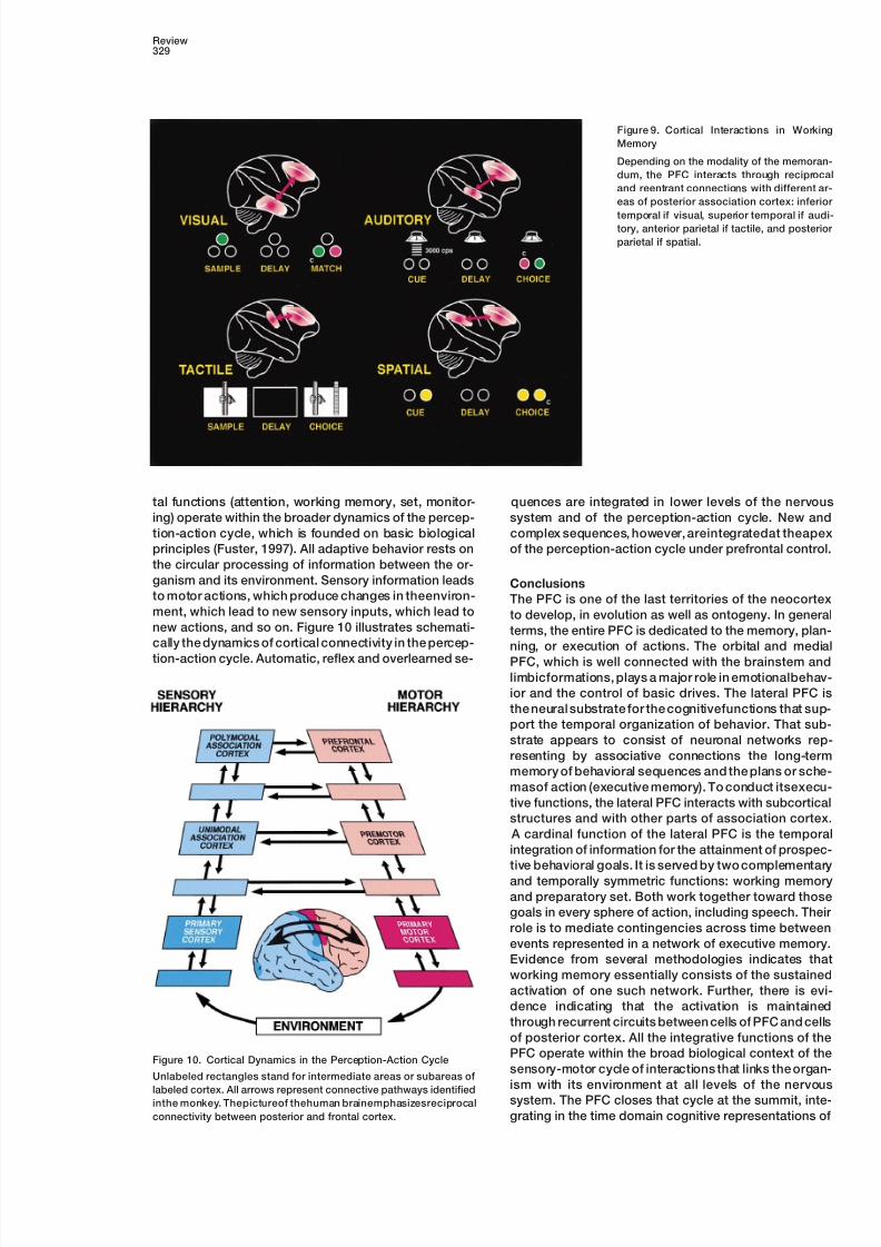
Temporal integration also seems to result, however, of working memory, and thus of temporal integration.
Such a mechanism was postulated by Hebb (1949) forfrom the cooperation of the PFC with other cortical ar-
eas. Working memory is as widely distributed as the short-term memory in local cortical circuitry. In a fully
recurrent computer network model trained to performlong-term memory that supports it. Cells in posterior
cortex of sensory association show working-memory sample-and-hold operations as delay tasks require,
Zipser et al. (1993) observed “cells” (hidden units) thatproperties for stimuli of the specific modality they pro-
cess. Thus, there are memory cells for vision in infero- behaved much like real working-memory cells of PFC
or inferotemporal cortex during delay tasks. Therefore,temporal cortex (Fuster and Jervey, 1982; Miller et al.,
1993), for touch in somatosensory cortex (Koch and working memory is emerging as a mechanism of tempo-
ral integration essentially based on the concurrent andFuster, 1989; Zhou and Fuster, 1996), and for location
in posterior parietal cortex (Andersen et al., 1990). There recurrent activation of cell assemblies in long-term
memory networks of frontal and posterior cortex. Duringis also mounting evidence that, in the maintenance of
working memory, the PFC interacts with those cortices. the delay period of delay tasks, a different recurrent
loop of cortical connectivity would be active dependingBy the cooling of lateral PFC in monkeys performing a
color delayed matching task (Fuster et al., 1985), two on the modality of the memorandum (Figure 9).
Finally, temporal integration and its ancillary prefron-correlated effects were obtained: (1) a behavioral deficit
8/3/2019 Fuster 2001
http://slidepdf.com/reader/full/fuster-2001 11/15
Review329
Figure 9. Cortical Interactions in Working
Memory
Depending on the modality of the memoran-
dum, the PFC interacts through reciprocal
and reentrant connections with different ar-
eas of posterior association cortex: inferior
temporal if visual, superior temporal if audi-tory, anterior parietal if tactile, and posterior
parietal if spatial.
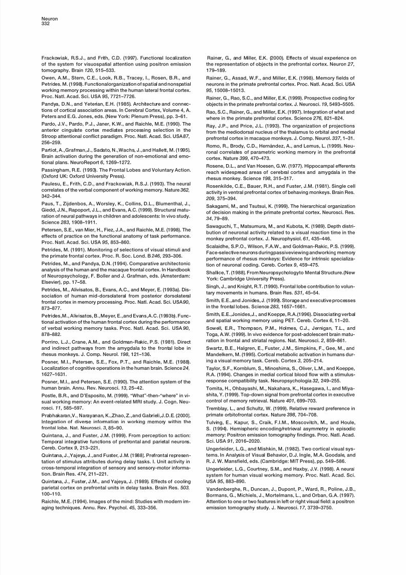
tal functions (attention, working memory, set, monitor- quences are integrated in lower levels of the nervous
system and of the perception-action cycle. New anding) operate within the broader dynamics of the percep-
tion-action cycle, which is founded on basic biological complex sequences, however, areintegratedat theapex
of the perception-action cycle under prefrontal control.principles (Fuster, 1997). All adaptive behavior rests on
the circular processing of information between the or-
ganism and its environment. Sensory information leads Conclusionsto motor actions, which produce changes in theenviron- The PFC is one of the last territories of the neocortexment, which lead to new sensory inputs, which lead to to develop, in evolution as well as ontogeny. In generalnew actions, and so on. Figure 10 illustrates schemati- terms, the entire PFC is dedicated to the memory, plan-cally the dynamics of cortical connectivity in the percep- ning, or execution of actions. The orbital and medialtion-action cycle. Automatic, reflex and overlearned se- PFC, which is well connected with the brainstem and
limbicformations, plays a major role in emotionalbehav-
ior and the control of basic drives. The lateral PFC is
the neural substrate for the cognitivefunctions that sup-
port the temporal organization of behavior. That sub-
strate appears to consist of neuronal networks rep-
resenting by associative connections the long-term
memory of behavioral sequences and the plans or sche-
masof action (executive memory). To conduct itsexecu-
tive functions, the lateral PFC interacts with subcortical
structures and with other parts of association cortex.
A cardinal function of the lateral PFC is the temporal
integration of information for the attainment of prospec-
tive behavioral goals. It is served by two complementary
and temporally symmetric functions: working memory
and preparatory set. Both work together toward those
goals in every sphere of action, including speech. Their
role is to mediate contingencies across time between
events represented in a network of executive memory.
Evidence from several methodologies indicates that
working memory essentially consists of the sustained
activation of one such network. Further, there is evi-
dence indicating that the activation is maintained
through recurrent circuits between cells of PFC and cells
of posterior cortex. All the integrative functions of the
PFC operate within the broad biological context of theFigure 10. Cortical Dynamics in the Perception-Action Cycle
sensory-motor cycle of interactions that links the organ-Unlabeled rectangles stand for intermediate areas or subareas of
ism with its environment at all levels of the nervouslabeled cortex. All arrows represent connective pathways identified system. The PFC closes that cycle at the summit, inte-inthe monkey. Thepictureof thehuman brainemphasizesreciprocal
connectivity between posterior and frontal cortex. grating in the time domain cognitive representations of

8/3/2019 Fuster 2001
http://slidepdf.com/reader/full/fuster-2001 12/15
Neuron330
Cavada, C., and Goldman-Rakic, P.S. (1989a). Posterior parietalperception and of action as required in goal-directedcortexin rhesusmonkey:I. Parcellationof areas based ondistinctivebehavior.limbic and sensory corticocortical connections. J. Comp. Neurol.
287 , 393–421. Acknowledgments
Cavada, C., and Goldman-Rakic, P.S. (1989b). Posterior parietal
cortex in rhesus monkey: II. Evidence for segregated corticocorticalI wish to thank Eric Kandel, James McGaugh, Earl Miller, and
networks linking sensory and limbic areas with the frontal lobe. J.Thomas O’Dell for their comments on the manuscript.
Comp. Neurol. 287 , 422–445.
References Chafee, M.V., and Goldman-Rakic, P.S. (2000). Inactivation of pari-
etal and prefrontal cortex reveals interdependence of neural activity Amaral, D.G. (1987). Memory: anatomical organization of candidate during memory-guided saccades. J. Neurophysiol. 83, 1550–1566.brain regions. In Handbook of Physiology: Nervous System, Volume
Chugani, H.T., Phelps, M.E., and Mazziotta, J.C. (1987). Positron V: Higher Functions of the Brain, Part 1, F. Plum, ed. (Bethesda:
emission tomographystudy of humanbrain functionaldevelopment. Amer. Physiol. Soc.), pp. 211–294.
Ann. Neurol. 22, 487–497. Andersen, R.A.,Asanuma, C., and Cowan,W.M. (1985). Callosal and
Cohen, J.D., Forman, S.D., Braver, T.S., Casey, B.J., Servan-prefrontal associational projecting cell populations in area 7A of the
Schreiber, D., and Noll, D.C. (1994). Activation of the prefrontal cor-macaque monkey: A study using retrogradely transported fluores-
tex in a nonspatial working memory task with functional MRI. Hum.cent dyes. J. Comp. Neurol. 232, 443–455.
Brain Map. 1, 293–304. Andersen, R.A., Bracewell, R.M., Barash, S., Gnadt, J.W., and Fo-
Conel, J.L. (1939). The Postnatal Development of the Human Cere-gassi, L. (1990). Eye position effects on visual, memory, and sac-bral Cortex, Volumes 1–6. (Cambridge, MA: Harvard Universitycade-related activity in areas LIP and 7a of macaque. J. Neurosci-Press).ence 10, 1176–1196.
Corbetta, M., Miezin, F.M.,Shulman, G.L., and Petersen, S.E. (1993). Andreasen, N.C., O’Leary, D.S., Arndt, S., Cizadlo, T., Hurtig, R., A PET study of visuospatial attention. J. Neurosci. 13, 1202–1226.Rezai, K., Watkins, G.L., Boles Ponto, L.L., and Hichwa, R.D. (1995).
Short-term and long-term verbal memory: A positron emission to- Courtney, S.M., Ungerleider, L.G., Keil, K., and Haxby, J.V. (1996).
mography study. Proc. Natl. Acad. Sci. 92, 5111–5115. Object and spatial visual working memory activate separate neural
systems in human cortex. Cereb. Cortex 6, 39–49. Asaad, W.F., Rainer, G., and Miller, E.K. (1998). Neural activity in
the primate prefrontal cortex during associative learning. Neuron Cummings, J.L. (1993). Frontal-subcortical circuits and human be- 21, 1399–1407. havior. Arch. Neurol. 50, 873–880.
Asaad, W.F., Rainer, G., and Miller, E.K. (2000). Task-specific neural Daffner, K.R., Mesulam, M.-M., Scinto, L.F.M., Acar, D., Calvo, V.,activity in the primate prefrontal cortex. J. Neurophysiology 84, Faust, R., Chabrerie, A., Kennedy, B., and Holcomb, P. (2000). The451–459. central role of the prefrontal cortex in directing attention to novel
events. Brain 123, 927–939.Baddeley, A. (1986). Working Memory (Oxford: Clarendon Press).
Baker, S.C.,Rogers, R.D.,Owen, A.M.,Frith, C.D.,Dolan, R.J.,Frack- Damasio, H., Grabowski, T., Frank, R., Galaburda, A.M., and Da-
owiak, R.S.J., and Robbins, T.W. (1996). Neural systems engaged masio, A.R. (1994). The return of Phineas Gage: Clues about the
by planning:a PETstudyof theTowerof Londontask.Neuropsycho- brain from the skull of a famous patient. Science 264, 1102-1105.
logia 34, 515–526.Desimone, R., and Duncan, J. (1995). Neural mechanisms of selec-
Barbas, H. (2000). Connections underlying the synthesis of cogni- tive visual attention. Annu. Rev. Neurosci. 18, 193–222.tion, memory, and emotion in primate prefrontal cortices. Brain Res.
Duncan, J., and Owen, A.M. (2000). Common regions of the humanBull. 52, 319–330.
frontal lobe recruited by diverse cognitive demands. Trends Neu-Barbas, H., and Blatt, G.J. (1995). Topographically specific hippo- rosci. 23, 475–483.campal projections target functionally distinct prefrontal areas in
Fiez, J.A., Raife, E.A., Balota, D.A., Schwarz, J.P., Raichle, M.E., andthe rhesus monkey. Hippocampus 5, 511–533.
Petersen, S.E. (1996). A positron emission tomography study ofBauer, R.H.,and Fuster, J.M.(1976). Delayed-matchingand delayed- the short-term maintenance of verbal information. J. Neurosci. 16,response deficit from cooling dorsolateral prefrontal cortex in mon- 808–822.keys. J. Comp. Physiol. Psychol. 90, 293–302.
Flechsig, P. (1920). Anatomie des Menschlichen Gehirns undBichot, N.P., and Schall, J.D. (1999). Effects of similarity and history
Rckenmarks auf Myelogenetischer Grundlage (Leipzig: Thieme).on neural mechanismsof visualselection. Nat.Neurosci. 2, 549–554.
Fletcher, P.C., Shallice, T., and Dolan, R.J. (1998a). The functionalBoch, R.A., and Goldberg, M.E. (1989). Participation of prefrontal
roles of prefrontalcortex in episodic memory. I. Encoding Brain 121,neurons in the preparation of visually guided eye movements in the
1239–1248.rhesus monkey. J. Neurophysiol. 61, 1064–1084.
Fletcher, P.C.,Shallice, T., Frith, C.D.,Frackowiak, R.S.J., and Dolan,Bodner, M., Kroger, J., and Fuster, J.M. (1996). Auditory memory
R.J. (1998b). The functional roles of prefrontal cortex in episodic
cells in dorsolateral prefrontal cortex. NeuroReport7
, 1905–1908. memory. II. Retrieval Brain 121, 1249–1256.Bourgeois,J.P., Goldman-Rakic,P.S., and Rakic, P. (1994). Synapto-
Freedman, D.F., Riesenhuber, M., Poggio, T., and Miller, E.K. (2001).genesis in the prefrontal cortex of rhesus monkeys. Cereb. CortexCategorical representation of visual stimuli in the primate prefrontal
4, 78–96.cortex. Science 291, 312–316.
Brodmann, K. (1909). Vergleichende Lokalisationslehre der Gross-Funahashi, S., Bruce, C.J., and Goldman-Rakic, P.S. (1989). Mne-hirnrinde in ihren Prinzipien dargestellt auf Grund des Zellenbauesmonic codingof visualspacein themonkey’sdorsolateral prefrontal(Leipzig: Barth).cortex. J. Neurophysiol. 61, 331–349.
Brodmann, K. (1912). Neue Ergebnisseuber die vergleichende histo-Fuster, J.M. (1973). Unit activity in prefrontal cortex during delayed-logische Lokalisation der Grosshirnrinde mit besonderer Beruck-response performance: Neuronal correlates of transient memory.sichtigung des Stirnhirns. Anat. Anz. Suppl. 41, 157–216.J.Neurophysiol. 36, 61–78.
Brunia, C.H.M., Haagh, S.A.V.M., and Scheirs, J.G.M. (1985). WaitingFuster, J.M. (1995). Memory in the Cerebral Cortex—An Empiricalto respond: Electrophysiological measurements in man during prep- Approach to Neural Networks in the Human and Nonhuman Primatearation for a voluntary movement. In Motor Behavior, H. Heuer, U.
(Cambridge, MA: MIT Press).Kleinbeck, and K.-H. Schmidt, eds. (New York: Springer).
Buckner, R.L., Petersen, S.E., Ojemann, J.G., Miezin, F.M., Squire, Fuster, J.M. (1997). The Prefrontal Cortex-Anatomy Physiology, and
Neuropsychology of the Frontal Lobe, Third Edition (Philadelphia:L.R., and Raichle, M.E. (1995). Functional anatomical studies ofexplicit and implicit memory retrieval tasks. J. Neurosci. 15, 12–29. Lippincott-Raven).
8/3/2019 Fuster 2001
http://slidepdf.com/reader/full/fuster-2001 13/15
Review331
Fuster, J.M., and Alexander, G.E. (1971). Neuron activity related to Jenkins, I.H., Brooks, D.J., Nixon, P.D., Frackowiak, R.S.J., and
Passingham, R.E. (1994). Motor sequence learning: A study withshort-term memory. Science 173, 652–654.
positron emission tomography. J. Neurosci. 14, 3775–3790.Fuster, J.M., and Jervey, J.P. (1982). Neuronal firing in the infero-
Jerison, H.J. (1994). Evolution of the brain. In Neuropsychology,temporal cortex of the monkey in a visual memory task. J. Neurosci.
D.W. Zaidel, ed. (San Diego: Academic Press, Inc.), pp. 53–81. 2, 361–375.
Jones, E.G. (1981). Anatomy of cerebral cortex: Columnar input-Fuster, J.M., Bauer, R.H., and Jervey, J.P. (1982). Cellular discharge
outputorganization. In TheOrganization of theCerebralCortex, F.O.in thedorsolateral prefrontal cortexof themonkey incognitivetasks. Schmitt, F.G. Worden, G. Adelman, and S.G. Dennis, eds. (Cam-Exp. Neurol. 77 , 679–694.
bridge, MA: MIT Press), pp. 199–235.Fuster, J.M., Bauer, R.H.,and Jervey, J.P. (1985). Functionalinterac-
Jonides, J., Smith, E.E., Koeppe, R.A., Awh, E., Minoshima, S., andtions between inferotemporal and prefrontal cortex in a cognitive
Mintun, M.A. (1993). Spatial working memory in humans as revealedtask. Brain Res. 330, 299–307.
by PET. Nature 363, 623–625.Fuster, J.M., Bodner, M., and Kroger, J. (2000). Cross-modal andJoseph, J.-P., and Barone, P. (1987). Prefrontal unit activity during across-temporal association in neurons of frontal cortex. Nature 405,delayed oculomotor task inthe monkey. Exp. BrainRes.67 , 460–468.347–351.
Kalaska, J.F., Sergio, L.E., and Cisek, P. (1998). Cortical control ofGabrieli, J.D.E., Poldrack, R.A., and Desmond, J.E. (1998). The rolewhole-arm motor tasks. Novartis Foundation Symp. 218, 176–190.of left prefrontal cortex in language and memory. Proc. Natl. Acad.
Sci. USA 95, 906–913. Kapur, S., Craik, F.I.M., Jones, C., Brown, G.M., Houle, S., and Tul-
ving, E. (1995). Functional role of the prefrontal cortex in retrievalGeorgopoulos, A.P., Kalaska, J.F., Caminiti, R., and Massey, J.T.of memories: a PET study. NeuroReport 6, 1880–1884.(1982). On the relations between the direction of two-dimensional
arm movements and cell discharge in primate motor cortex. J. Neu- Kastner, S., Pinsk, M.A., De Weerd, P., Desimone, R., and Unger-rosci. 2, 1527–1537. leider, L.G. (1999). Increased activity in human visual cortex during
directed attention in the abscence of visual stimulation. Neuron 22,Gibson, K.R. (1991). Myelination and behavioral development: a 751–761.comparative perspective on questions of neoteny, altriciality and
intelligence. In Brain Maturation and Cognitive Development, K.R. Kievit, J., and Kuypers, H.G.J.M. (1975). Basal forebrain and hypo-
Gibson and A.C. Petersen, eds. (New York: Aldine de Gruyter), pp. thalamic connections to frontal and parietal cortex in the rhesus
29–63. monkey. Science 187 , 660–662.
Goldman-Rakic, P.S., Scalaidhe, S.P.O., and Chafee, M.V. (2000). Knight, R.T. (1984). Decreased response to novel stimuli after pre-
Domain specificity in cognitive systems. In The New Cognitive Neu- frontal lesions in man. Electroencephalogr. Clin. Neurophysiol. 59,
rosciences, M.S. Gazzaniga, ed. (Cambridge, MA.: MIT Press), pp. 9–20.
733–742. Koch, K.W., and Fuster, J.M. (1989). Unit activity in monkey parietal
Grafton, S.T., Mazziotta, J.C., Presty, S., Friston, K.J., Frackowiak, cortex related to haptic perception and temporary memory. Exp.
R.S.J., andPhelps, M.E.(1992). Functionalanatomy of humanproce- Brain Res. 76, 292–306.
durallearning determined withregional cerebral bloodflow and PET. Konishi, S., Kawazu, M., Uchida, I., Kikyo, H., Asakura, I., and Miya-J. Neurosci. 12, 2542–2548. shita, Y. (1999). Contribution of working memory to transient activa-
Grasby, P.M.,Frith, C.D.,Friston,K.J., Bench, C., Frackowiak,R.S.J., tion in human inferior prefrontal cortex during performance of the
andDolan, R.J. (1993). Functional mapping of brain areas implicated Wisconsin card sorting test. Cereb. Cortex 9, 745–753.
in auditory-verbal memory function. Brain 116, 1–20. Kutas, M., and Donchin, E. (1980). Preparation to respond as mani-
fested by movement-related brain potentials. Brain Res. 202,Harlow, J.M. (1848). Passage of aniron rodthroughthe head. Boston
95–115.Med. Surg. J. 39, 389–393.
Le Doux, J.E. (1993). Emotionalmemory systems in thebrain. Behav.Hasegawa, I., Fukushima, T., Ihara, T., and Miyashita, Y. (1998).
Brain Res. 58, 69–79.Callosal window between prefrontal cortices: Cognitive interaction
to retrieve long-term memory. Science 281, 814–818. Luria,A.R. (1966). HigherCorticalFunctions in Man(New York: Basic
Books).Hebb, D.O. (1949). The Organization of Behavior (New York: John
Wiley & Sons). Luria, A.R. (1970). Traumatic Aphasia (The Hague: Mouton).
Hikosaka, K., and Watanabe, M. (2000). Delay activity of orbital McCarthy,G., Blamire, A.M., Puce, A.,Nobre, A.C., Bloch,G., Hyder,and lateral prefrontal neurons of the monkey varying with different F., Goldman-Rakic, P., and Shulman, R.G. (1994). Functional mag-rewards. Cereb. Cortex 10, 263–271. netic resonance imaging of human prefrontal cortex activation dur-
ing a spatial working memory task. Proc. Natl. Acad. Sci. USA 91,Huttenlocher, P.R. (1990). Morphometric study of human cerebral8690–8694.cortex development. Neuropsychologia 28, 517–527.
Mesulam, M.-M. (1981). A cortical network for directed attentionHuttenlocher, P.R., and Dabholkar, A.S. (1997). Regional differencesand unilateral neglect. Neurology 10, 309–325.in synaptogenesis in human cerebral cortex. J. Comp. Neurol. 387 ,
167–178. Mesulam, M.-M. (1998). From sensation to cognition. Brain 121,
1013-1052.Iacoboni, M., Woods, R.P., and Mazziotta, J.C. (1996). Brain-behav-
ior relationships: Evidence from practice effects in spatial stimulus- Miller, E.K., Li, L., and Desimone, R. (1993). Activity of neurons inresponse compatibility. J. Neurophysiol. 76, 321–331. anterior inferior temporal cortex during a short-term memory task.
J. Neurosci. 13, 1460–1478.Ingvar, D.H. (1985). “Memory ofthe future”:An essayon thetemporal
organization of conscious awareness. Hum. Neurobiol. 4, 127–136. Miller, E.K., Erickson, C.A., and Desimone, R. (1996). Neural mecha-
nisms of visual working memory in the prefrontal cortex of the ma-Jacobsen, C.F. (1931). A study of cerebral function in learning: Thecaque. J. Neurosci. 16, 5154–5167.frontal lobes. J. Comp. Neurol. 52, 271–340.
Niki, H. (1974). Differential activity of prefrontal units during rightJacobson, S., and Trojanowski, J.Q. (1977a). Prefrontal granularand left delayed response trials. Brain Res. 70, 346–349.cortex of the rhesus monkey. I. Intrahemispheric cortical afferents.
Brain Res. 132, 209–233. Niki, H., and Watanabe, M. (1976). Prefrontal unit activity and de-
layed response: relation to cue location versus direction of re-Jacobson, S., and Trojanowski, J.Q. (1977b). Prefrontal granularsponse. Brain Res. 105, 79–88.cortex of the rhesus monkey: II. Interhemispheric cortical afferents.
Brain Res. 132, 235–246. Niki, H., and Watanabe, M. (1979). Prefrontal and cingulate unit
activity during timing behavior in the monkey. Brain Res. 171,Jacobson, S., Butters, N., and Tovsky, N.J. (1978). Afferent and213–224.
efferent subcortical projections of behaviorally defined sectors ofprefrontal granular cortex. Brain Res. 159, 279–296. Nobre, A.C., Sebestyen, G.N., Gitelman, D.R., Mesulam, M.-M.,
8/3/2019 Fuster 2001
http://slidepdf.com/reader/full/fuster-2001 14/15
Neuron332
Frackowiak, R.S.J., and Frith, C.D. (1997). Functional localization Rainer, G., and Miller, E.K. (2000). Effects of visual experience on
the representation of objects in the prefrontal cortex. Neuron 27 ,of the system for visuospatial attention using positron emission
tomography. Brain 120, 515–533. 179–189.
Owen, A.M., Stern, C.E., Look, R.B., Tracey, I., Rosen, B.R., and Rainer, G., Assad, W.F., and Miller, E.K. (1998). Memory fields of
Petrides, M. (1998). Functionalorganization of spatial and nonspatial neurons in the primate prefrontal cortex. Proc. Natl. Acad. Sci. USA
working memory processing within the human lateral frontal cortex. 95, 15008–15013.
Proc. Natl. Acad. Sci. USA 95, 7721–7726. Rainer, G., Rao, S.C., and Miller, E.K. (1999). Prospective coding for
Pandya, D.N., and Yeterian, E.H. (1985). Architecture and connec- objects in the primate prefrontal cortex. J. Neurosci. 19, 5493–5505.
tions of cortical association areas. In Cerebral Cortex, Volume 4, A. Rao, S.C., Rainer, G., and Miller, E.K. (1997). Integration of what andPeters and E.G. Jones, eds. (New York: Plenum Press), pp. 3–61. where in the primate prefrontal cortex. Science 276, 821–824.
Pardo, J.V., Pardo, P.J., Janer, K.W., and Raichle, M.E. (1990). The Ray, J.P., and Price, J.L. (1993). The organization of projectionsanterior cingulate cortex mediates processing selection in the from the mediodorsal nucleus of the thalamus to orbital and medialStroop attentional conflict paradigm. Proc. Natl. Acad. Sci. USA 87 , prefrontal cortex in macaque monkeys. J. Comp. Neurol. 337 , 1–31.256–259.
Romo, R., Brody, C.D., Herna ndez, A., and Lemus, L. (1999). Neu-Partiot, A.,Grafman,J., Sadato, N.,Wachs, J.,and Hallett, M. (1995). ronal correlates of parametric working memory in the prefrontalBrain activation during the generation of non-emotional and emo- cortex. Nature 399, 470–473.tional plans. NeuroReport 6, 1269–1272.
Rosene, D.L., and Van Hoesen, G.W. (1977). Hippocampal efferentsPassingham, R.E. (1993). The Frontal Lobes and Voluntary Action. reach widespread areas of cerebral cortex and amygdala in the(Oxford UK: Oxford University Press). rhesus monkey. Science 198, 315–317.Paulesu, E., Frith, C.D., and Frackowiak, R.S.J. (1993). The neural
Rosenkilde, C.E., Bauer, R.H., and Fuster, J.M. (1981). Single cellcorrelates of the verbal component of working memory. Nature 362,
activity in ventral prefrontal cortex of behaving monkeys. Brain Res.342–344.
209, 375–394.Paus, T., Zijdenbos, A., Worsley, K., Collins, D.L., Blumenthal, J.,
Sakagami, M., and Tsutsui, K. (1999). The hierarchical organizationGiedd, J.N., Rapoport, J.L., and Evans, A.C. (1999). Structural matu-
of decision making in the primate prefrontal cortex. Neurosci. Res.ration of neural pathways in children and adolescents: In vivo study.
34, 79–89.Science 283, 1908–1911.
Sawaguchi, T., Matsumura, M., and Kubota, K. (1989). Depth distri-Petersen, S.E., van Mier, H., Fiez, J.A., and Raichle, M.E. (1998). The
bution of neuronal activity related to a visual reaction time in theeffects of practice on the functional anatomy of task performance.
monkey prefrontal cortex. J. Neurophysiol. 61, 435–446.Proc. Natl. Acad. Sci. USA 95, 853–860.
Scalaidhe, S.P.O., Wilson, F.A.W., and Goldman-Rakic, P.S. (1999).Petrides, M. (1991). Monitoring of selections of visual stimuli and
Face-selective neurons duringpassiveviewing andworking memorythe primate frontal cortex. Proc. R. Soc. Lond. B 246, 293–306.
performance of rhesus monkeys: Evidence for intrinsic specializa-Petrides, M., and Pandya, D.N. (1994). Comparative architectonic tion of neuronal coding. Cereb. Cortex 9, 459–475.analysis of the human and the macaque frontal cortex. In Handbook
Shallice, T. (1988). From Neuropsychologyto Mental Structure.(Newof Neuropsychology, F. Boller and J. Grafman, eds. (Amsterdam:
York: Cambridge University Press).Elsevier), pp. 17–58.
Singh, J., and Knight, R.T. (1990). Frontal lobe contribution to volun-Petrides, M., Alivisatos, B., Evans, A.C., and Meyer, E. (1993a). Dis-
tary movements in humans. Brain Res. 531, 45–54.sociation of human mid-dorsolateral from posterior dorsolateral
Smith, E.E.,and Jonides, J. (1999). Storage and executive processesfrontal cortex in memory processing. Proc. Natl. Acad. Sci. USA 90,in the frontal lobes. Science 283, 1657–1661.873–877.
Smith, E.E.,Jonides,J., and Koeppe, R.A.(1996). Dissociating verbalPetrides,M., Alivisatos, B.,Meyer, E.,and Evans,A.C. (1993b). Func-and spatial working memory using PET. Cereb. Cortex 6, 11–20.tional activation of the human frontal cortex during the performance
of verbal working memory tasks. Proc. Natl. Acad. Sci. USA 90, Sowell, E.R., Thompson, P.M., Holmes, C.J., Jernigan, T.L., and
Toga, A.W. (1999). In vivo evidence for post-adolescent brain matu-878–882.
ration in frontal and striatal regions. Nat. Neurosci. 2, 859–861.Porrino, L.J., Crane, A.M., and Goldman-Rakic, P.S. (1981). Direct
and indirect pathways from the amygdala to the frontal lobe in Swartz, B.E., Halgren, E., Fuster, J.M., Simpkins, F., Gee, M., and
rhesus monkeys. J. Comp. Neurol. 198, 121–136. Mandelkern, M. (1995). Cortical metabolic activation in humans dur-
ing a visual memory task. Cereb. Cortex 3, 205–214.Posner, M.I., Petersen, S.E., Fox, P.T., and Raichle, M.E. (1988).
Localization of cognitive operations in the human brain. Science 24, Taylor, S.F., Kornblum, S., Minoshima, S., Oliver, L.M., and Koeppe,
1627–1631. R.A. (1994). Changes in medial cortical blood flow with a stimulus-
response compatibility task. Neuropsychologia 32, 249–255.Posner, M.I., and Petersen, S.E. (1990). The attention system of the
human brain. Annu. Rev. Neurosci. 13, 25–42. Tomita, H., Ohbayashi, M., Nakahara, K., Hasegawa, I., and Miya-
shita, Y. (1999). Top-down signal from prefrontal cortex in executivePostle, B.R., and D’Esposito, M. (1999). “What”-then-“where” in vi-
control of memory retrieval. Nature401
, 699–703.sual working memory: An event-related MRI study. J. Cogn. Neu-rosci. 11, 585–597. Tremblay, L., and Schultz, W. (1999). Relative reward preference in
primate orbitofrontal cortex. Nature 398, 704–708.Prabhakaran,V., Narayanan, K.,Zhao, Z.,and Gabrieli,J.D.E. (2000).
Integration of diverse information in working memory within the Tulving, E., Kapur, S., Craik, F.I.M., Moscovitch, M., and Houle,frontal lobe. Nat. Neurosci. 3, 85–90. S. (1994). Hemispheric encoding/retrieval asymmetry in episodic
memory: Positron emission tomography findings. Proc. Natl. Acad.Quintana, J., and Fuster, J.M. (1999). From perception to action:Sci. USA 91, 2016–2020.Temporal integrative functions of prefrontal and parietal neurons.
Cereb. Cortex 9, 213–221. Ungerleider, L.G., and Mishkin, M. (1982). Two cortical visual sys-
tems. In Analysis of Visual Behavior, D.J. Ingle, M.A. Goodale, andQuintana, J.,Yajeya, J.,and Fuster, J.M. (1988). Prefrontal represen-
R. J. W. Mansfield, eds. (Cambridge: MIT Press), pp. 549–586.tation of stimulus attributes during delay tasks. I. Unit activity in
cross-temporal integration of sensory and sensory-motor informa- Ungerleider, L.G., Courtney, S.M., and Haxby, J.V. (1998). A neuraltion. Brain Res. 474, 211–221. system for human visual working memory. Proc. Natl. Acad. Sci.
USA 95, 883–890.Quintana, J., Fuster, J.M., and Yajeya, J. (1989). Effects of cooling
parietal cortex on prefrontal units in delay tasks. Brain Res. 503, Vandenberghe, R., Duncan, J., Dupont, P., Ward, R., Poline, J.B.,100–110. Bormans, G., Michiels, J., Mortelmans, L., and Orban, G.A. (1997).
Attention to one or two features in left or right visual field: a positronRaichle, M.E. (1994). Images of the mind: Studies with modern im-aging techniques. Annu. Rev. Psychol. 45, 333–356. emission tomography study. J. Neurosci. 17 , 3739–3750.

8/3/2019 Fuster 2001
http://slidepdf.com/reader/full/fuster-2001 15/15
Review333
Verfaellie, M., and Heilman, K.M. (1987). Response preparation and
response inhibition after lesions of the medial frontal lobe. Arch.
Neurol. 44, 1265–1271.
Watanabe, M. (1992). Frontal units of the monkey coding the asso-
ciative significance of visual and auditory stimuli. Exp. Brain Res.
89, 233–247.
Watanabe, M. (1996). Reward expectancy in primate prefrontal neu-
rons. Nature 382, 629–632.
White, I.M., and Wise, S.P. (1999). Rule-dependent neuronal activity
in the prefrontal cortex. Exp. Brain Res. 126, 315–335.
Wilson, F.A.W., Scalaidhe, S.P.O., and Goldman-Rakic, P.S. (1993).
Dissociation of object and spatial processing domains in primate
prefrontal cortex. Science 260, 1955–1958.
Zhou, Y.-D., and Fuster, J.M. (1996). Mnemonic neuronal activity in
somatosensory cortex. Proc. Natl. Acad. Sci. USA 93, 10533–10537.
Zipser, D., Kehoe, B., Littlewort, G., and Fuster, J. (1993). A spiking
network model of short-term active memory. J. Neurosci. 13, 3406–
3420.