Embed Size (px)
Citation preview
Fusion Visualization for Cardiac Anatomical and Ischemic Models with Depth Weighted Optic Radiation Function
Fei Yang
1, Weigang Lu
2 , Lei Zhang3, Wangmeng Zuo4, Kuanquan Wang4, Henggui Zhang4,5
1 Shandong University, Weihai, China
2 Ocean University of China, Qingdao, China
3Harbin University, Harbin, China 4
Harbin Institute of Technology, Harbin, China 5University of Manchester, Manchester, UK
Abstract
Despite the emerging of various innovations of visualization methods, highlighting and exploring the important function of interest of computational cardiac model still remain a challenging problem. Due to the complex and fine geometries of heart, the silhouette of the whole volume structure may be destroyed when there is a necessity to demonstrate cardiac electrophysiological behaviors. In this paper, we proposed a fusion visualization framework, which combines the electrophysiology pattern with the anatomy pattern through a novel multi-dimensional fusion transfer function. A depth weighted optic radiation model is thus presented in the framework to inspect the occluded cardiac ischemia information. According to the depth to the tissue boundary, the hidden ischemia region of pathological tissue can be revealed from complex overlapping features. Experiment results on the cardiac anatomy and electrophysiological simulation data verifies the effectiveness of the proposed method for intuitively exploring and inspecting electrophysiological activities with the preserved authentic context of anatomical structure, which is found to be useful in analyzing and explaining cardiac functioning phenomena.
1. Introduction
Cardiac diseases, such as myocardial infarction, cardiomyopathy and heart rhythm problem, are stated as the leading cause of death and disability in the world. There is the evidence that functional abnormity of heart may lead to the severe problem of heart failure and embolism with increased morbidity and mortality [1].
Though the functional abnormity of reentry can be induced in a healthy heart, in most cases, it often occurs in a functionally abnormal heart, typically an ischemic
heart. The electrophysiological spatial inconsistency in the ischemic tissue eventually leads to the critical impairment of both its electrical and mechanical function and induces ventricular fibrillation [2-3]. It is thus considered to play an important role in cardiac arrhythmia genesis.
A vast amount of experimental and clinical data on the underlying ionic, cellular and tissue substrates have been acquired, while the precise electrical mechanisms of ischemia arising from the functional interactions aren’t well understood. Any advances in finding and tracking the pathophysiological feature, especially advances that might help analyze and treat more effectively are, therefore, of great significance. Shenai [4] presented the visualization of normal and ischemic propagation and found intra-QRS changes in and around the ischemic region, which proved that ischemia may cause depolarization changes detectable by both action potentials and unipolar leads. To analyze the spatio-temporal deformation parameters for the myocardial contraction, Han etc. [5] proposed the visualization tools and a strategy for the automatic detection of dysfunctional regions of cardiac ischemic pathologies, which is proved very useful for quantitatively demonstrating the main properties of the left ventricle myocardial contraction. Trejos etc. [6] proposed the mechanism of automatic detecting ischemic events using ECG signals, which allows a better interpretation of cardiac ischemic behavior and results in an increase in the discrimination capability for ischemia detection. Lu etc. [7] developed a 3D human ventricular ischemic model combining a detailed biophysical description of the excitation kinetics of human ventricular cells with an integrated geometry of human ventricular tissue. Through the model, the functional consequences and mechanisms underlying the arrhythmias in early acute global ischemia are investigated to analyze the influence of acute global ischemia on cardiac electrical activity and subsequently on reentrant arrhythmogenesis.
937ISSN 2325-8861 Computing in Cardiology 2015; 42:937-940.
In this paper, we proposed a fusion visualization framework, which combines the electrophysiology pattern with the anatomy pattern through a novel depth fusion transfer function. To delineate the complicated relation between the tissue structure and its functions under cardiac ischemic condition, we construct a depth weighted optic radiation model through the fusion transfer function and inspect the occluded cardiac ischemia information. According to the depth to the tissue boundary, the hidden ischemia region of pathological tissue can be revealed from complex overlapping features.
The reminder of the paper is organized as follows. Section 2 presents the fusion visualization framework, including fusion transfer function design and the depth weighted optic radiation model construction. Section 3 provides experimental results and discussions. Finally, Section 4 gives our conclusion and future work.
2. Fusion visualization
To explore organs of interest from mass of cardiac tissues and intertwined cardiac vessels, Zhang et al. [8] proposed a novel approach for revealing detailed structures in the human heart anatomy via perception-based lighting enhancement. While different from the intensity scalar value, those voxels with electro- physiological value in the human heart volume data represent propagation patterns of excitation waves of various tissues of the heart rather than those tissues themselves. Current rendering model cannot explore those occluded functions of the heart at physiological or pathological conditions reliably with the complex and fine anatomical structure.
In this section, we first induce the Euclidean distance transform and design the depth fusion transfer function, which maps the Euclidean distance value of a voxel close to the boundary to a lower opacity. Then the fusion transfer function is implemented to construct the depth weighted optic radiation model for fusion visualization of cardiac anatomical and ischemic model.
2.1. Euclidean distance transform
Distance transform (DT) maps each image pixel into its smallest distance to regions of interest [9], wherein Exact Euclidean distance is the preferable model to numerous geometrical facts of the human-scale world, especially to medical visualization for the high accuracy requirement.
The central problem of EDT (Euclidean Distance Transform) is to compute the Euclidean distance of each point of the plane to a given subset of it. In image processing terminology, this is rephrased in the following way: Let 2: 0,1I Z be a binary image where
0, ,1 0, ,1 . By convention, 0 is associated to
black and 1 to white. Hence we have an object O represented by all the white pixels:
| ( ) 1O p I p , as shown in Fig. 1. The set O is
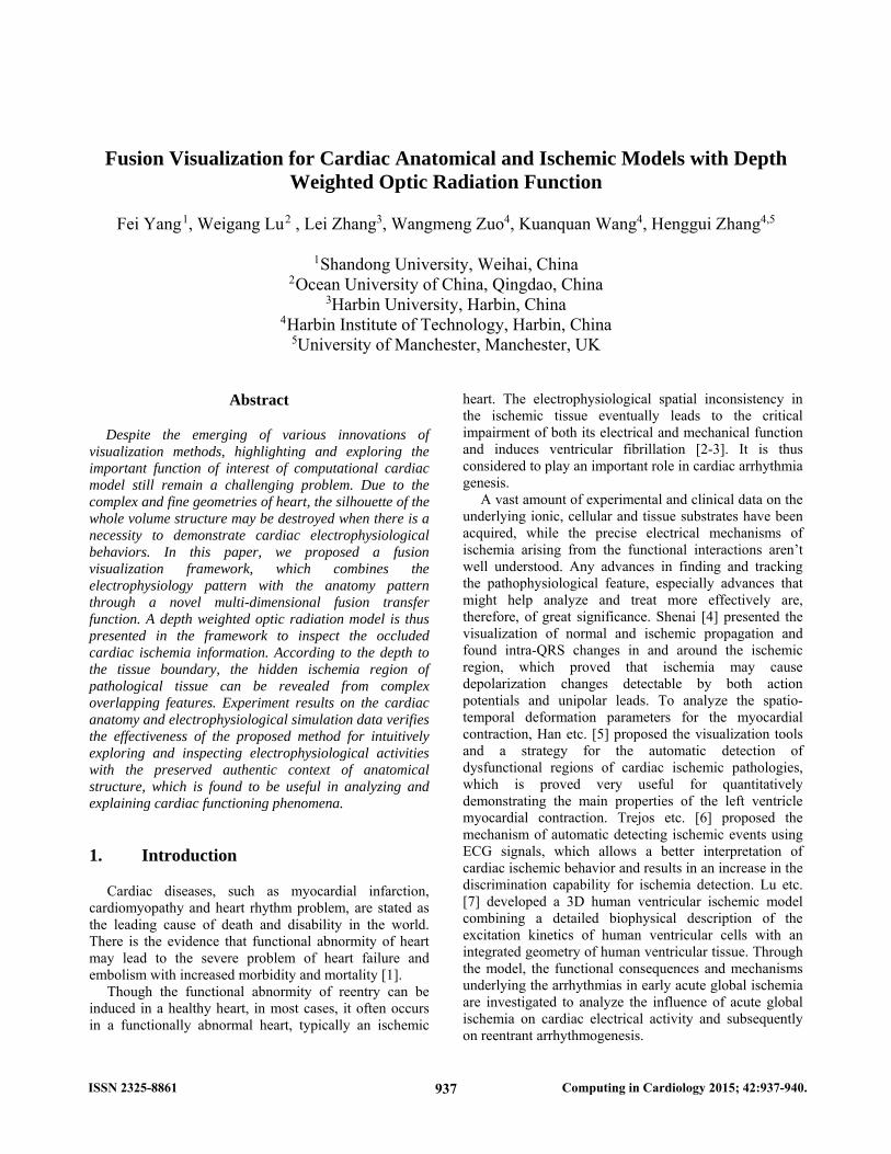
called object or foreground and can consist of any subset of the image domain, including disjoint sets. The elements of its complement, Oc, the set of black pixels in Ω, are called background. From the DT point of view, the background pixels are called the interest points or feature points.
Figure 1. Elements defined in the field of Euclidean distance transform.
Definition 1. The distance transform (DT) is the transformation that generates a map D whose value in each pixel p is the smallest distance from this pixel to Oc:
( ) min ( , ) | min ( , ) | ( ) 0cD p d p q q O d p q I q (1)
The image D is called the distance map of I. D itself
can also be called a distance transform. Moreover, d(p, q) is generally taken as the Euclidean distance, given by:
2 2( , ) ( ) ( )i i j jd p q p q p q (2)
Extend the 2D binary image I to 3D space, we let
33 3: 0,1D DI Z
be a set of 2D binary images
where 3 0, ,1 0, ,1 0, ,1D . 0 and 1 are the
same as in 2D binary image. 3DO and 3c
DO are object
set and the set of black pixels in 3D respectively. 3D
distance map of each pixel p in 3DI is defined as:
3 3 3( ) : min ( , ) | cD D DD p d p q q O
and 3D Euclidean distance d3D(p, q) is given by:
2 2 23 ( , ) ( ) ( ) ( )D i i j j k kd p q p q p q p q
2.2. Depth fusion transfer function design
We consider the calculated 3D Euclidean distance transform of a voxel sample xi as the depth to the boundary of tissues that xi belongs to at the position of xi. Then the resulting opacity of a voxel sample xi in cardiac volume data can then be acquired by the depth fusion
p∈O
q∈Oc
Oc
O
938
transfer function as
( ) ( ) ( ) ( )fusion i anatomy i electrophy i EDT ix x x x
Here ( )anatomy ix is generated by selecting various
regions based on the scalar value of xi and assigning opacity to them. Determining value of ( )electrophy ix in this
equation depends on electrophysiological value which reflects the physiological or pathological statement at different positions in volume data. ( )EDT ix is associated
to unit normalized 3D Euclidean distance transform result, which is thought to be the depth of xi. Thus the opacity of a sample voxel will increase when it has a larger depth to the boundary, which means that a sample voxel is more opaque when it is farther from the boundary.
2.3. Depth weighted optic radiation model
According to the volume rendering integral [10], the optic radiation function for visualizing the cardiac volume data is
0( )
0( ) ( )
tD s ds
C C t t e dt
where ( )C t is the radiance or color and ( )t is the
attenuation of a sample t along the view direction. We implement the depth fusion transfer function in section 2.2 in the optic radiation function and construct the depth weighted optic radiation model as
0( )
0( ) ( )
t
fusionD s ds
fusionC C t t e dt
(7)
where attenuation ( )t is replaced by the fusion transfer
function ( )fusion ix . Thus cardiac physiology model is
fused with anatomy model and the hidden ischemia region of pathological tissue can be revealed from complex overlapping features.
3. Results
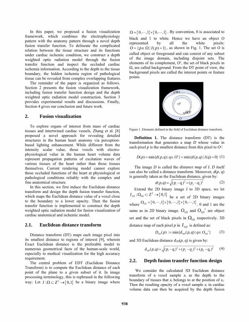
Through the 3D exact Euclidean distance transform on volume data, the depth of inner voxels to boundary is represented by distance transformation of those voxels. Fig. 2 shows the effect of exact Euclidean distance transformation in 3D space of left ventricle muscles papillaris. The value in each pixel which represents the smallest distance from this pixel to the “black” pixels is mapped to color which changes from blue to red with increasing of the distance to the boundary.
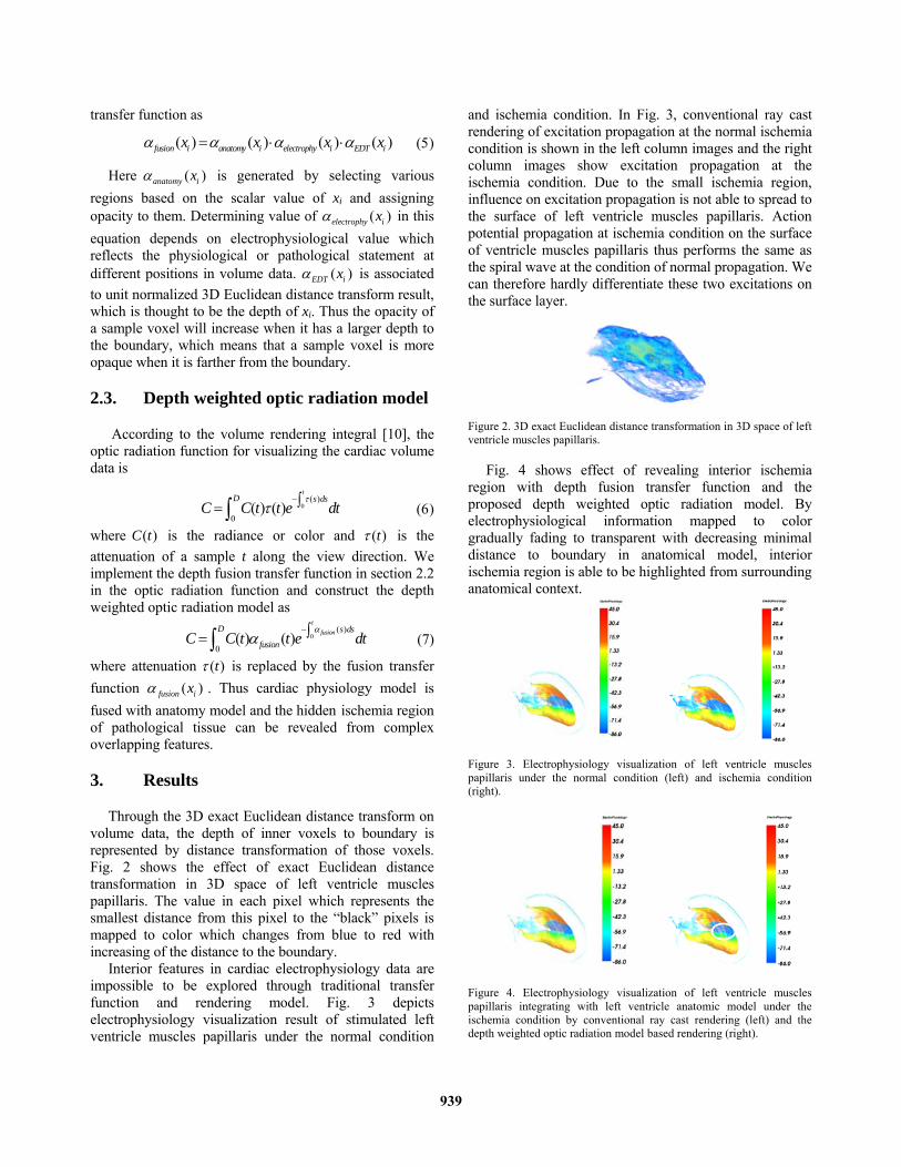
Interior features in cardiac electrophysiology data are impossible to be explored through traditional transfer function and rendering model. Fig. 3 depicts electrophysiology visualization result of stimulated left ventricle muscles papillaris under the normal condition
and ischemia condition. In Fig. 3, conventional ray cast rendering of excitation propagation at the normal ischemia condition is shown in the left column images and the right column images show excitation propagation at the ischemia condition. Due to the small ischemia region, influence on excitation propagation is not able to spread to the surface of left ventricle muscles papillaris. Action potential propagation at ischemia condition on the surface of ventricle muscles papillaris thus performs the same as the spiral wave at the condition of normal propagation. We can therefore hardly differentiate these two excitations on the surface layer.
Figure 2. 3D exact Euclidean distance transformation in 3D space of left ventricle muscles papillaris.
Fig. 4 shows effect of revealing interior ischemia region with depth fusion transfer function and the proposed depth weighted optic radiation model. By electrophysiological information mapped to color gradually fading to transparent with decreasing minimal distance to boundary in anatomical model, interior ischemia region is able to be highlighted from surrounding anatomical context.
Figure 3. Electrophysiology visualization of left ventricle muscles papillaris under the normal condition (left) and ischemia condition (right).
Figure 4. Electrophysiology visualization of left ventricle muscles papillaris integrating with left ventricle anatomic model under the ischemia condition by conventional ray cast rendering (left) and the depth weighted optic radiation model based rendering (right).
939
4. Conclusions
In this paper, we proposed a depth fusion transfer function, and presented a depth weighted optic radiation model for visualizing cardiac behavior under the normal condition and ischemia condition. To explore the important function of interest of computational cardiac model, we first used a 3D Euclidean distance transform to acquire the depth of a sample in the model to the boundary it belongs to, and then proposed a depth fusion transfer function for the fusion optical property mapping for cardiac anatomical and electrophysiological model. We then constructed an optic radiation model with the fusion transfer function and effectively visualized hiding ischemia region with complex anatomical and electro- physiological context. Acknowledgements
The work was supported by the National Natural Science Foundation of China (NSFC) under Grant Nos. 61502275 and the Fundamental Research Funds for the Central Universities (No. 2015ZQXM004). This work was also supported in part by the National Natural Science Foundation of China (NSFC) (No. 61173086), the Fundamental Research Funds for the Central Universities (No. 201413012), Youth Foundation of Harbin University (No. HUYF2013-025), the Higher Education and Teaching Reform project (No. JG2014011155) and Natural Science Foundation of Shandong Province, China under Grants No. ZR2015AM015.
References
[1] Ten Tusscher KH, Hren R, Panfilov AV. Organization of ventricular fibrillation in the human heart. Circulation Research 2007;100(12):87-101
[2] Gray RA, Pertsov AM, Jalife J. Spatial and temporal organization during cardiac fibrillation. Nature 1998;39:75-8.
[3] Gray RA, Jalife J, Panfilov AV, Baxter WT, Cabo C, Davidenko JM, Pertsov AM. Mechanisms of cardiac fibrillation. Science 1995;270:1222-3.
[4] Shenai M, Gramatikov B, Thakor N V. Computer Modeling of Depolarization Changes Induced by Myocardial Ischemia. Computers in Cardiology 1998;25:321-4.
[5] Han M, Clarysse P, Croisille P, Magnin I E, Revel D. Computer aided diagnosis of the myocardial ischemia based on a spatio-temporal deformation features analysis. Computers in Cardiology 1998;25:749-52.
[6] Trejos ED, Lluna AP, Ferrer MV, Magrans PC, Dominguez GC. Dimensionality Reduction Oriented Toward the Feature Visualization for Ischemia Detection. IEEE Transactions on Information Technology in Biomedicine 2009;13(4):590-8.
[7] Lu WG, Li J, Yang F, Luo CJ, Wang KQ, Adeniran I, Zhang HG. Effectsof acute global ischemia on re-entrant arrhythmogenesis: a simulation study. Journal of Biological Systems 2015;23(2):213-30.
[8] Zhang L, Wang KQ, Zhang HG, Zuo, WM, Liang XQ, Shi J. Illustrative cardiac visualization via perception-based lighting enhancement. Journal of Medical Imaging and Health Informatics 2014;4(2):312-6.
[9] Rosenfeld A, Pfaltz JL. Sequential operations in digital picture processing. Journal of the Association for Computing Machinery 1966;13(4):471-94
[10] Max N. Optical models for direct volume rendering. IEEE Transactions on Visualization and Computer Graphics 1995;1(2):99–108.
Address for correspondence. Weigang Lu Department of Educational Technology Ocean University of China Qingdao 266100, China E-mail: [email protected]
940