Embed Size (px)
Citation preview
Nogo proteins were discovered, and have been exten-sively studied, in the context of injury and repair of fibre tracts in the CNS1 — a topic of great research interest and clinical relevance. However, much less in known about the physiological functions of Nogo proteins in development and in the intact adult organism, including in the brain. Through a number of recent publications, Nogo proteins have emerged as important regulators of cell motility and growth — for example, for develop-ing neurons and blood vessels. Recent studies have also strengthened the evidence that they have intracellular roles — for example, as regulators of secretases, endoplas-mic reticulum (ER) composition and cell survival. This Review summarizes the current knowledge of the com-plex receptor interactions of Nogo proteins, their tissue expression and their diverse roles in the developing and adult nervous system, and other tissues.
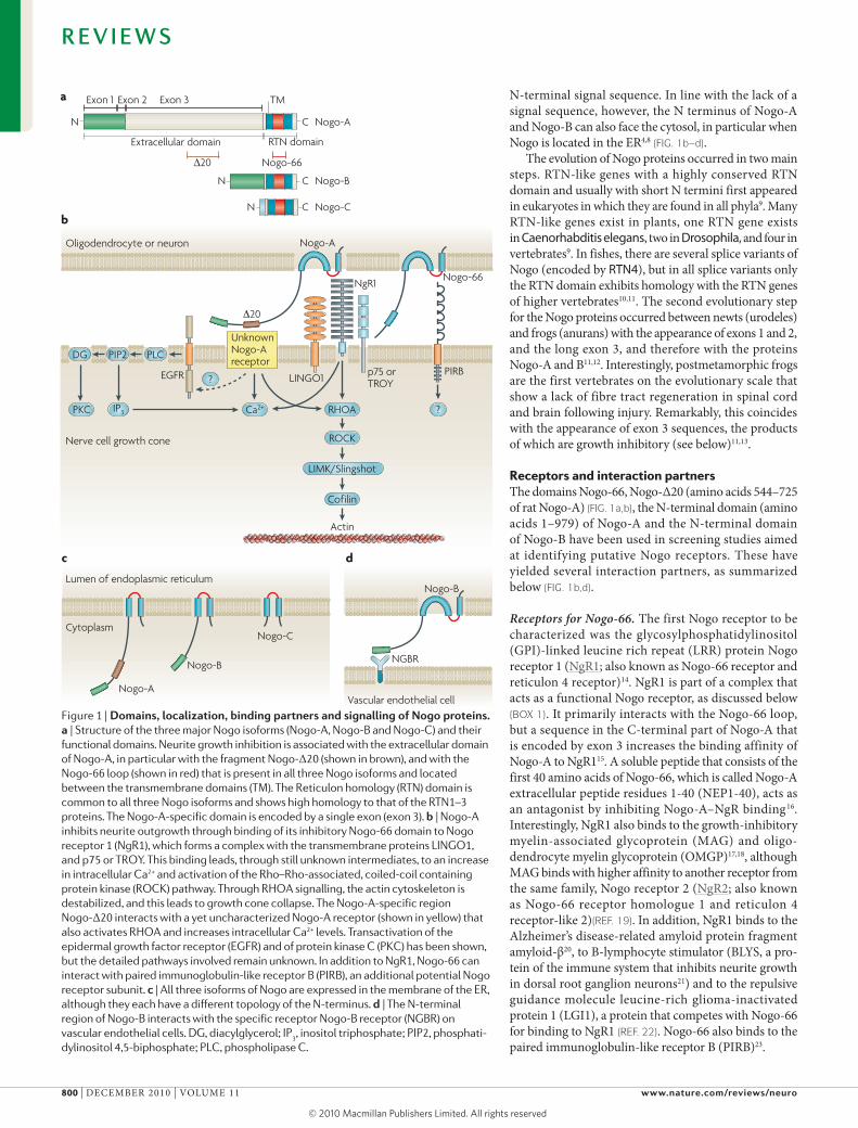
Nogo proteinsNogo-A, Nogo-B and Nogo-C are the three main protein products of the reticulon 4 (RTN4; also known as NOGO) gene2–4 (FIG. 1a). Only the last 188 amino acids in the carboxyl terminus, the so-called Reticulon homology (RTN) domain, are common to the three isoforms. This RTN domain also shows high similarity to the RTN domain in the proteins encoded by the other three Reticulon genes — RTN1, RTN2 and RTN3 (ReFs 3,5) — whose functions in the nervous system and in other organs are mostly unknown. The RTN domain of Nogo proteins contains two long hydrophobic stretches, each of which is long enough to span the cell membrane twice. They are linked together by a 66-amino acid segment called Nogo-66 (ReF. 3) (FIG. 1b).
The amino-terminal segments of the proteins encoded by the different RTN genes have differing lengths and there is no homology between them3,5. The N termini of the RTN4 products Nogo-A and Nogo-B are identical, consisting of a 172-amino acid sequence that is encoded by a single exon (exon 1) that is followed by a short exon 2 and, in Nogo-A, by the very long exon 3 encoding 800 amino acids2,6 (FIG. 1a). This 800-amino acid insert, as well as the N terminus of Nogo-A and Nogo-B, are rich in proline and contain large unstruc-tured regions7. No homologies of any of these exons with known protein sequences have been found so far.
The shortest Nogo isoform, Nogo-C, has an N terminus of just a few amino acids that is directly followed by the RTN domain. This N terminus is encoded by a primary transcript that is generated from a different promoter to the one that generates the primary transcript for the N termini of Nogo-A and Nogo-B6. In all three Nogo isoforms, the N terminus lacks a signal sequence for ER translocation.
Nogo proteins are present in the ER and at the cell surface. They can have several membrane topologies, in particular with regard to a cytoplasmic versus extra-cellular position of the N termini. On the cell surface, the N termini of Nogo-A and Nogo-B, and the exon 3 sequence of Nogo-A have been found to face the extra-cellular space, even though they do not contain a con-ventional signal peptide for ER translocation. This fact classifies Nogo-A and Nogo-B as unconventional mem-brane proteins, similar to cystic fibrosis transport regu-lator (CFTR) or the secreted proteins fibroblast growth factor (FGF), interleukin (IL)-1b and ciliary neuro-trophic factor (CNTF), all of which lack a conventional
University of Zurich and ETH, Zurich, Winterthurerstrasse 190, CH‑8057 Zurich, Switzerland. e‑mail: [email protected]:10.1038/nrn2936Published online 3 November 2010
Functions of Nogo proteins and their receptors in the nervous systemMartin E. Schwab
Abstract | The membrane protein Nogo-A was initially characterized as a CNS-specific inhibitor of axonal regeneration. Recent studies have uncovered regulatory roles of Nogo proteins and their receptors — in precursor migration, neurite growth and branching in the developing nervous system — as well as a growth-restricting function during CNS maturation. The function of Nogo in the adult CNS is now understood to be that of a negative regulator of neuronal growth, leading to stabilization of the CNS wiring at the expense of extensive plastic rearrangements and regeneration after injury. In addition, Nogo proteins interact with various intracellular components and may have roles in the regulation of endoplasmic reticulum (ER) structure, processing of amyloid precursor protein and cell survival.
REVIEWS
NATuRE REvIEwS | NeuroscieNce vOLumE 11 | dECEmBER 2010 | 799
© 20 Macmillan Publishers Limited. All rights reserved10
Nature Reviews | Neuroscience
a
b
c d
Exon 1 Exon 2 Exon 3 TM
N C Nogo-A
C Nogo-B
C
N
N Nogo-C
Extracellular domain RTN domain
∆20 Nogo-66
PKC
DG PIP2 PLC
IP3 Ca2+ ?
?
ROCK
LIMK/Slingshot
Cofilin
Actin
p75 or TROYLINGO1
NgR1
PIRBEGFR
Nogo-AOligodendrocyte or neuron
Nerve cell growth cone
20
UnknownNogo-Areceptor
Nogo-66
Nogo-B
Nogo-A
Nogo-C
Nogo-B
Lumen of endoplasmic reticulum
Cytoplasm
NGBR
Vascular endothelial cell
RHOA
N-terminal signal sequence. In line with the lack of a signal sequence, however, the N terminus of Nogo-A and Nogo-B can also face the cytosol, in particular when Nogo is located in the ER4,8 (FIG. 1b–d).
The evolution of Nogo proteins occurred in two main steps. RTN-like genes with a highly conserved RTN domain and usually with short N termini first appeared in eukaryotes in which they are found in all phyla9. many RTN-like genes exist in plants, one RTN gene exists in Caenorhabditis elegans, two in Drosophila, and four in vertebrates9. In fishes, there are several splice variants of Nogo (encoded by RTN4), but in all splice variants only the RTN domain exhibits homology with the RTN genes of higher vertebrates10,11. The second evolutionary step for the Nogo proteins occurred between newts (urodeles) and frogs (anurans) with the appearance of exons 1 and 2, and the long exon 3, and therefore with the proteins Nogo-A and B11,12. Interestingly, postmetamorphic frogs are the first vertebrates on the evolutionary scale that show a lack of fibre tract regeneration in spinal cord and brain following injury. Remarkably, this coincides with the appearance of exon 3 sequences, the products of which are growth inhibitory (see below)11,13.
Receptors and interaction partnersThe domains Nogo-66, Nogo-Δ20 (amino acids 544–725 of rat Nogo-A) (FIG. 1a,b), the N-terminal domain (amino acids 1–979) of Nogo-A and the N-terminal domain of Nogo-B have been used in screening studies aimed at identifying putative Nogo receptors. These have yielded several interaction partners, as summarized below (FIG. 1b,d).
Receptors for Nogo‑66. The first Nogo receptor to be characterized was the glycosylphosphatidylinositol (GPI)-linked leucine rich repeat (LRR) protein Nogo receptor 1 (NgR1; also known as Nogo-66 receptor and reticulon 4 receptor)14. NgR1 is part of a complex that acts as a functional Nogo receptor, as discussed below (BOX 1). It primarily interacts with the Nogo-66 loop, but a sequence in the C-terminal part of Nogo-A that is encoded by exon 3 increases the binding affinity of Nogo-A to NgR115. A soluble peptide that consists of the first 40 amino acids of Nogo-66, which is called Nogo-A extracellular peptide residues 1-40 (NEP1-40), acts as an antagonist by inhibiting Nogo-A–NgR binding16. Interestingly, NgR1 also binds to the growth-inhibitory myelin-associated glycoprotein (mAG) and oligo-dendrocyte myelin glycoprotein (OmGP)17,18, although mAG binds with higher affinity to another receptor from the same family, Nogo receptor 2 (NgR2; also known as Nogo-66 receptor homologue 1 and reticulon 4 receptor-like 2)(ReF. 19). In addition, NgR1 binds to the Alzheimer’s disease-related amyloid protein fragment amyloid-β20, to B-lymphocyte stimulator (BLYS, a pro-tein of the immune system that inhibits neurite growth in dorsal root ganglion neurons21) and to the repulsive guidance molecule leucine-rich glioma-inactivated protein 1 (LGI1), a protein that competes with Nogo-66 for binding to NgR1 (ReF. 22). Nogo-66 also binds to the paired immunoglobulin-like receptor B (PIRB)23.
Figure 1 | Domains, localization, binding partners and signalling of Nogo proteins. a | Structure of the three major Nogo isoforms (Nogo-A, Nogo-B and Nogo-C) and their functional domains. Neurite growth inhibition is associated with the extracellular domain of Nogo-A, in particular with the fragment Nogo-Δ20 (shown in brown), and with the Nogo-66 loop (shown in red) that is present in all three Nogo isoforms and located between the transmembrane domains (TM). The Reticulon homology (RTN) domain is common to all three Nogo isoforms and shows high homology to that of the RTN1–3 proteins. The Nogo-A-specific domain is encoded by a single exon (exon 3). b | Nogo-A inhibits neurite outgrowth through binding of its inhibitory Nogo-66 domain to Nogo receptor 1 (NgR1), which forms a complex with the transmembrane proteins LINGO1, and p75 or TROY. This binding leads, through still unknown intermediates, to an increase in intracellular Ca2+ and activation of the Rho–Rho-associated, coiled-coil containing protein kinase (ROCK) pathway. Through RHOA signalling, the actin cytoskeleton is destabilized, and this leads to growth cone collapse. The Nogo-A-specific region Nogo-Δ20 interacts with a yet uncharacterized Nogo-A receptor (shown in yellow) that also activates RHOA and increases intracellular Ca2+ levels. Transactivation of the epidermal growth factor receptor (EGFR) and of protein kinase C (PKC) has been shown, but the detailed pathways involved remain unknown. In addition to NgR1, Nogo-66 can interact with paired immunoglobulin-like receptor B (PIRB), an additional potential Nogo receptor subunit. c | All three isoforms of Nogo are expressed in the membrane of the ER, although they each have a different topology of the N-terminus. d | The N-terminal region of Nogo-B interacts with the specific receptor Nogo-B receptor (NGBR) on vascular endothelial cells. DG, diacylglycerol; IP
3, inositol triphosphate; PIP2, phosphati-
dylinositol 4,5-biphosphate; PLC, phospholipase C.
R E V I E W S
800 | dECEmBER 2010 | vOLumE 11 www.nature.com/reviews/neuro
© 20 Macmillan Publishers Limited. All rights reserved10
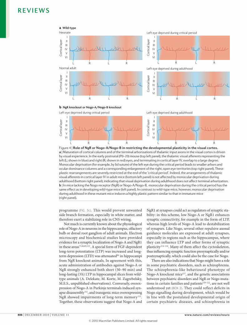
The biological functions of these potential receptors for Nogo-66 and their roles in specific cell types are not fully understood. disruption of Nogo–NgR1 interac-tions increased neurite growth in culture dishes coated with Nogo-66 or CNS myelin14,24. The Nogo-66–NgR1 interaction induced growth cone collapse but was not required for inhibition of long-term neurite growth25. NgR1 phosphorylation by extracellular casein kinase 2 inhibited NgR1 binding to Nogo-66, mAG and OmGP, and increased neurite outgrowth on CNS myelin in vitro26. In vivo, acute blockade of NgR1 enhanced sprouting, regeneration and plastic rearrangements of fibre connections after CNS injury in adult rats, but NgR1 knockout mice showed similar effects only in some studies16,24,27–30. mice lacking both Nogo-A and Nogo-B, or NgR1 or PIRB show prolonged plasticity for the formation of ocular dominance columns (see below): although plasticity after monocular deprivation is pro-gressively restricted in wild type mice at 30 days of age and almost absent after 3 months, mutant mice retained a high level of plasticity well beyond 100 days31,32. These results suggest that both NgR1 and PIRB are functionally relevant receptors for growth- and plasticity-restricting actions of Nogo proteins (see below).
Because NgR1 has no transmembrane domain14 it is thought that, in order to induce Nogo signalling, NgR1 must be associated with membrane proteins involved in sig-nal transduction. The low-affinity neurotrophin receptor p75 and its relative, tumour necrosis factor-α (TNFα) recep-tor superfamily member 19 (TROY), have been found to be signal transducing NgR1-associated components18,33–37. Another LRR protein, LINGO1, can bind to NgR1 extracellularly35,38. Blockade of LINGO1 resulted in enhanced regenerative fibre growth, neuroprotection
and functional recovery after spinal cord injury in adult rats39,40. However, the distribution of LINGO1 in the developing and adult mouse and chicken CNS is much wider than that of NgRs41,42, indicating that NgRs are probably not its only binding partners. The NgR1 receptor complex has also been observed to interact with integrins, resulting in decreased integrin activity, decreased cell–substrate adhesion and a transactivation of the epidermal growth factor (EGF) receptor, which is involved in many aspects of cellular growth and migration43,44.
Nogo‑A‑specific receptors. The extracellular part of Nogo-A (in rats, this includes amino acids 1–979, often called ‘amino Nogo’), the part encoded by exon 3 (amino acids 173–979) and the fragment of the peptide sequence encoded by exon 3 called Nogo-Δ20 (amino acids 544–725) (FIG. 1a) exert strong inhibitory effects on growing neur-ites and growth cones in vitro and, unlike Nogo-66, also on the migration of non-neuronal cells such as fibrob-lasts, in vitro2–4. Finding the binding site (or sites) and receptors for these active regions of Nogo-A has been difficult; so far, the G protein coupled orphan receptor GPR50 is a candidate, but current data are incomplete45. In vitro, overexpression of GPR50 increased neurite length in a neuronal cell line, but results from GPR50 knockdown or neutralization experiments, or from in vivo regeneration studies are not available yet, and the binding affinity of GPR50 to Nogo-A has not been deter-mined. The search for additional Nogo-A receptors is on going. Considering the known interaction of Nogo-66 with NgR1 and PIRB, it is likely that Nogo-A interacts with a multisubunit receptor complex that is similar to those described for neurotrophins, wnts and the axonal guidance molecules netrin and semaphorins46–49 (BOX 1).
Box 1 | Multisubunit receptors for neurite growth regulators and guidance molecules
The search for Nogo binding partners and receptors has not been easy and is not yet complete. The Nogo binding proteins Nogo receptor 1 (NgR1; also known as Nogo‑66 receptor and reticulon 4 receptor) and paired immuno globulin‑like receptor B (PIRB) interact with all three Nogo isoforms but also with other ligands, only some of which are known to inhibit neurite outgrowth14,23,34,35,151. In addition, the identity of the interaction partners of the most strongly inhibitory Nogo‑A fragments (‘amino Nogo’ and Nogo‑Δ20) has not been determined yet. The recently characterized G protein‑coupled orphan receptor GPR50 is the first candidate for a Nogo‑A‑specific receptor45. In addition, NgR1 forms a complex with proteins such as LINGO1 and the presumed signal transducers p75 and Troy33,35. The concerted action of these receptor components is still poorly understood.
Interestingly, the picture of a multisubunit Nogo receptor complex that emerges resembles that described for other ligands such as neurotrophic factors — for example, nerve growth factor (NGF), brain‑derived neurotrophic factor (BDNF) and ciliary neurotrophic factor (CNTF) — and repulsive or attractive guidance molecules (semaphorins, netrins and Wnt proteins)46–49,152. Neurotrophins interact with three different binding sites or receptor subunits: p75, high affinity nerve growth factor receptors (Trks) and sortilin. Different neurotrophin‑responsive cell types express different combinations of these receptor constituents and respond to the same neurotrophin in different ways, leading in some cases to cell survival and in others to apoptosis46,153. Whether the guidance molecule netrin exerts attractive or repulsive functions depends on the expression of uncoordinated 5 (UNC5), deleted in colorectal cancer (DCC) and Down syndrome cell adhesion molecule (DSCAM), providing another example of triple interactions of a ligand with subunits of a receptor complex48,154,155. Semaphorin 3 molecules interact with plexins and neuropilins as co‑receptors, and neuropilins are also constituents of vascular endothelial growth factor (VEGF) receptors49,156. An even more complex situation exists for Wnt receptors, with different combinations of binding partners and receptor subunits determining which of various intracellular signalling pathways is activated152.
Nogo receptors, therefore, seem to fit a concept of multisubunit receptors that applies to many regulators of neurite growth. Different cell types at different developmental stages could respond differently to Nogo signalling depending on the expression of receptor subunits. Elucidating the evolution of the Nogo receptor composition will also shed light on the functional evolution of Nogo signalling.
R E V I E W S
NATuRE REvIEwS | NeuroscieNce vOLumE 11 | dECEmBER 2010 | 801
© 20 Macmillan Publishers Limited. All rights reserved10
Compartmentalized culturesNeurons are grown in the middle chamber of a three‑chamber culture system. Their neurites are guided into the side chambers under a Teflon ring divider or through microfluidic channels. Neurites can be treated with substances and subsequently analysed separately from the cell bodies.
Tangential migrationA mode of neuron migration that is non‑radial. Most interneurons immigrate tangentially into the forebrain cortex from a proliferation zone in the basal ganglia region.
Nogo‑B‑specific receptors. A single Nogo-B specific receptor (NGBR) has been identified in blood vessels50. This 30-kd receptor binds to Nogo-B sequences spanning the splice junction between the N terminus of Nogo-B and the beginning of the RTN domain50,51. NGBR medi-ates the chemotactic, pro-migratory actions of Nogo-B on human and mouse vascular endothelial cells and is involved in vascular remodelling and repair after injury in mice50,51. Its role under physiological conditions and in development remains to be studied. A recent study described an intracellular role of NGBR in cholesterol trafficking in a human hepatocyte cell line52.
Second messenger and effector pathwaysmany studies have shown that Nogo-66 and other active fragments of Nogo-A, such as Nogo-Δ20 and the extra-cellular domain (amino acids 1–979), can trigger activa-tion of the small GTPase RHOA and its effector protein Rho-associated, coiled-coil containing protein kinase 1 (ROCK) in different neuronal cell types33,35,53–57 (FIG. 1b). In agreement with this, pharmacological blockade of RHOA or of ROCK activation prevents the inhibitory effects of Nogo on neurite outgrowth in vitro and allows regeneration and compensatory sprouting to occur in the mechanically injured spinal cord, optic nerve or brain of rats and mice in vivo. These data indicate that Rho GTPases have a crucial role in Nogo signalling33,35,56 (FIG. 1b). Furthermore, it has been reported that inactiva-tion of Rac — a small GTPase with cytoskeletal regu-latory functions opposite to those of Rho — occurs in response to Nogo-Δ20 (ReF. 58).
Elevated levels of Nogo proteins can also increase intracellular Ca2+ levels59,60 and influence the activation of integrins43, protein kinase C (PKC)61, mammalian tar-get of rapamycin (mTOR), signal transducer and acti-vator of transcription 3 (STAT3)62,63 and the epidermal growth factor receptor (EGFR)44. moreover, high levels of cyclic AmP can override the inhibitory effects of Nogo signalling on neurite growth in vitro and possibly in vivo, suggesting a convergence of cAmP and RHOA signalling pathways57,64.
How signals are transmitted from the Nogo recep-tor complex to Rho is not entirely understood65. downstream of Rho, the phosphatase slingshot, the LIm domain kinase 1 (LImK1) and the actin regulator cofilin have been shown to mediate the effect of Nogo-A (FIG. 1b) on the destabilization of the actin cytoskeleton that leads to growth cone collapse and growth arrest66,67. Interestingly, Nogo that is bound to the receptor complex has to be internalized through endocytosis for growth cone collapse to occur57, suggesting that Nogo bind-ing leads to the formation of a molecular complex — comprising the activated Nogo receptor, signal transduc-tion components, probably adaptor molecules and endo-somal components — that then triggers the effects on the cytoskeleton57. A similar phenomenon has recently been described for ephrin-induced growth cone collapse68.
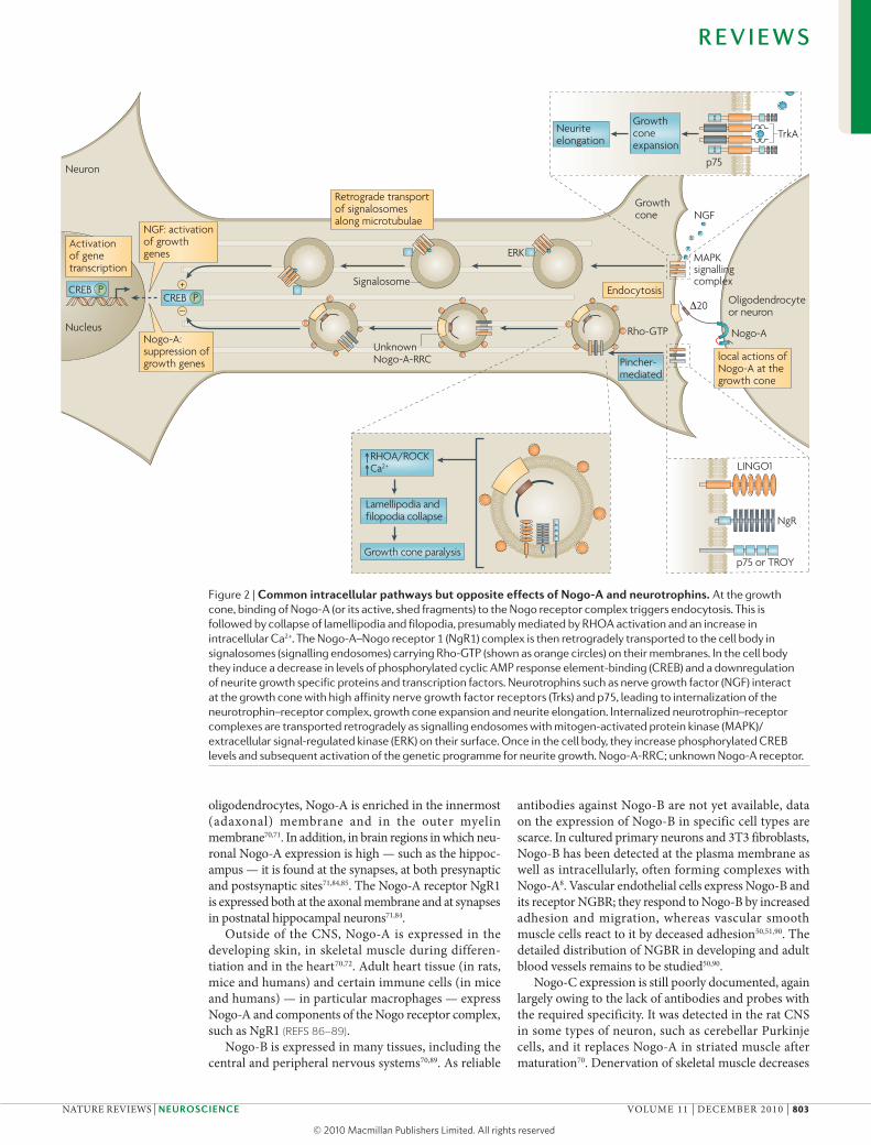
dorsal root ganglion neurons have been used to study the internalization events following binding of Nogo to its cell surface receptors. when dorsal root ganglion cells from newborn rats were grown in compartmentalized
cultures and the neurites exposed to the Nogo-Δ20 fragment, the fragment was internalized and subse-quently transported retrogradely to the cell bodies57. Rho activation was increased in the cell bodies upon arrival of the Nogo-containing endosomes and Rho-GTP colo-calized with Nogo-containing endosomes, as measured by immunofluorescence. In contrast to the increase in cAmP-responsive element-binding protein (CREB) phosphorylation that has been observed upon arrival of growth-promoting factors such as neurotrophins after retrograde axonal transport57,64,69, CREB phosphoryla-tion was decreased after arrival of Nogo-containing endosomes57. These results suggest that growth sup-pressors such as Nogo-A and growth stimulators such as neurotrophins are internalized as ligand–receptor complexes and subsequently transported to the cell body in signalling endosomes (signalosomes) (FIG. 2). Thus, Nogo and neurotrophins seem to act in opposite direc-tions at the level of the growth cone, and subsequently, at the level of the cell body, where they cause changes in the transcriptional machinery, with Nogo proteins act-ing as growth suppressors and neurotrophins as growth enhancers (FIG. 2). This yin–yang mechanism may have a role in the regulation of stability and plasticity in the developing CNS and adult CNS (described below).
Expression of Nogo proteinsThe Nogo isoforms Nogo-A, Nogo-B and Nogo-C have very different distribution patterns throughout the body and the nervous system of rats, mice and chickens70–73. Nogo-A is largely, but not exclusively, expressed in the nervous system, with three distinct expression windows during development70–73. First, Nogo-A is expressed in migrating neuroblasts and immature neurons in the neural tube during early stages of development73–75. In the developing cortex, radially migrating neurons and tangentially migrating interneurons express Nogo-A, whereas cortical radial glial cells show weak Nogo-A staining74,75. Cultured neural precursor cells express Nogo-A on the cell surface75. In the optic chiasm and in the floor plate of the early spinal cord, Nogo-A is also expressed by radial glial cells76,77. The Nogo-A receptor NgR1 is expressed by subpopulations of neurons, for example retinal ganglion cells, at different developmental stages72,73,75–79.
Second, during the main outgrowth phase of central and peripheral neurons, Nogo-A is expressed by many neuron types, especially those with long axons70,72,73,80–82. This expression decreases to low, often undetectable, lev-els after birth70,72. However, some neuron types retain high levels of Nogo-A expression, in particular those in the olfactory bulb, pyramidal cells and interneurons in the hippocampus, spinal motor neurons and dorsal root ganglion cells70,78,81–83. All these neuronal types are characterized by the high plasticity of their connections, suggesting that neuronal Nogo could have a role in synaptic plasticity.
Finally, in the postnatal CNS, Nogo-A is mainly expressed in oligodendrocytes70–72. Interestingly, the analogous cell type in the peripheral nervous sys-tem (Schwann cells) does not express Nogo-A70,71. In
R E V I E W S
802 | dECEmBER 2010 | vOLumE 11 www.nature.com/reviews/neuro
© 20 Macmillan Publishers Limited. All rights reserved10
Nogo-A
Nature Reviews | Neuroscience
20
UnknownNogo-A-RRC
Rho-GTP
LINGO1
p75 or TROY
NgR
Signalosome
Neuron
Nucleus
MAPKsignallingcomplex
Oligodendrocyteor neuron
Growthcone
ERK
–
+PCREB
PCREB
p75
TrkA
NGF
Growthconeexpansion
Neuriteelongation
Activationof genetranscription
Retrograde transportof signalosomesalong microtubulae
local actions ofNogo-A at thegrowth cone
Endocytosis
Pincher-mediated
RHOA/ROCKCa2+
Lamellipodia andfilopodia collapse
Growth cone paralysis
NGF: activation of growth genes
Nogo-A:suppression ofgrowth genes
oligodendrocytes, Nogo-A is enriched in the innermost (adaxonal) membrane and in the outer myelin membrane70,71. In addition, in brain regions in which neu-ronal Nogo-A expression is high — such as the hippoc-ampus — it is found at the synapses, at both presynaptic and postsynaptic sites71,84,85. The Nogo-A receptor NgR1 is expressed both at the axonal membrane and at synapses in postnatal hippocampal neurons71,84.
Outside of the CNS, Nogo-A is expressed in the developing skin, in skeletal muscle during differen-tiation and in the heart70,72. Adult heart tissue (in rats, mice and humans) and certain immune cells (in mice and humans) — in particular macrophages — express Nogo-A and components of the Nogo receptor complex, such as NgR1 (ReFs 86–89).
Nogo-B is expressed in many tissues, including the central and peripheral nervous systems70,89. As reliable
antibodies against Nogo-B are not yet available, data on the expression of Nogo-B in specific cell types are scarce. In cultured primary neurons and 3T3 fibroblasts, Nogo-B has been detected at the plasma membrane as well as intracellularly, often forming complexes with Nogo-A8. vascular endothelial cells express Nogo-B and its receptor NGBR; they respond to Nogo-B by increased adhesion and migration, whereas vascular smooth muscle cells react to it by deceased adhesion50,51,90. The detailed distribution of NGBR in developing and adult blood vessels remains to be studied50,90.
Nogo-C expression is still poorly documented, again largely owing to the lack of antibodies and probes with the required specificity. It was detected in the rat CNS in some types of neuron, such as cerebellar Purkinje cells, and it replaces Nogo-A in striated muscle after maturation70. denervation of skeletal muscle decreases
Figure 2 | common intracellular pathways but opposite effects of Nogo-A and neurotrophins. At the growth cone, binding of Nogo-A (or its active, shed fragments) to the Nogo receptor complex triggers endocytosis. This is followed by collapse of lamellipodia and filopodia, presumably mediated by RHOA activation and an increase in intracellular Ca2+. The Nogo-A–Nogo receptor 1 (NgR1) complex is then retrogradely transported to the cell body in signalosomes (signalling endosomes) carrying Rho-GTP (shown as orange circles) on their membranes. In the cell body they induce a decrease in levels of phosphorylated cyclic AMP response element-binding (CREB) and a downregulation of neurite growth specific proteins and transcription factors. Neurotrophins such as nerve growth factor (NGF) interact at the growth cone with high affinity nerve growth factor receptors (Trks) and p75, leading to internalization of the neurotrophin–receptor complex, growth cone expansion and neurite elongation. Internalized neurotrophin–receptor complexes are transported retrogradely as signalling endosomes with mitogen-activated protein kinase (MAPK)/extracellular signal-regulated kinase (ERK) on their surface. Once in the cell body, they increase phosphorylated CREB levels and subsequent activation of the genetic programme for neurite growth. Nogo-A-RRC; unknown Nogo-A receptor.
R E V I E W S
NATuRE REvIEwS | NeuroscieNce vOLumE 11 | dECEmBER 2010 | 803
© 20 Macmillan Publishers Limited. All rights reserved10
Nature Reviews | Neuroscience
Growth conecollapse andgrowth arrest
Balancedrepulsion
Glial scar
Extension ofregeneratingaxons
Compensatorycollateralsprouting
Nerve fibres Surface Nogo-A
Surface Nogo-A
Oligodendrocytea
b c
Nogo-Aon myelinsuppressesbranching
Downregulationof neuronalgrowthprogramme
Spinal cord
Strokelesion
Cortex
Midline
d Spinal cord lesion e Cortical lesion
Spontaneouscollateralsprouting
Control Ab
α-Nogo-A Ab 11C7
α-NgR Ab
Nogo-C — and increases Nogo-A — expression91. Indeed, as patients with amyotrophic lateral sclerosis have increased Nogo-A expression in muscle in the early stages of the disease, Nogo-A expression in muscle has been proposed as a diagnostic marker for the disease92.
Roles of Nogo in CNS developmentA role for Nogo in neuronal migration. In the developing forebrain cortex, Nogo-A expressing post mitotic neu-ronal precursors migrate along Nogo-A positive radial glia75. Cells that will become part of the cortical plate pass between Nogo-A expressing neurons in the subplate and intermediate zone, and in mice lacking Nogo-A this migration through the intermediate zone is enhanced75. The early tangential migration of cortical interneu-rons from the median eminence, on the other hand, is delayed in mice lacking all three forms of Nogo, sug-gesting that Nogo proteins can slow down (in the case of radial migrating cells) or enhance (in the case of tan-gentially migrating interneurons) migrating neurons74. In vitro, migration of cortical neural precursors was enhanced in cells derived from Nogo-A knockout ani-mals and in wild-type neurons cultured in the presence of antibodies against Nogo-A, NgR1 or LINGO1, sug-gesting that Nogo-A at the cell surface regulates neu-ronal migration in the early developing CNS via an NgR1–LINGO1 containing receptor complex75. many details regarding the role of Nogo-A and Nogo-B in the migration of neurons and non-neuronal (for example, vascular) cells remain to be studied.
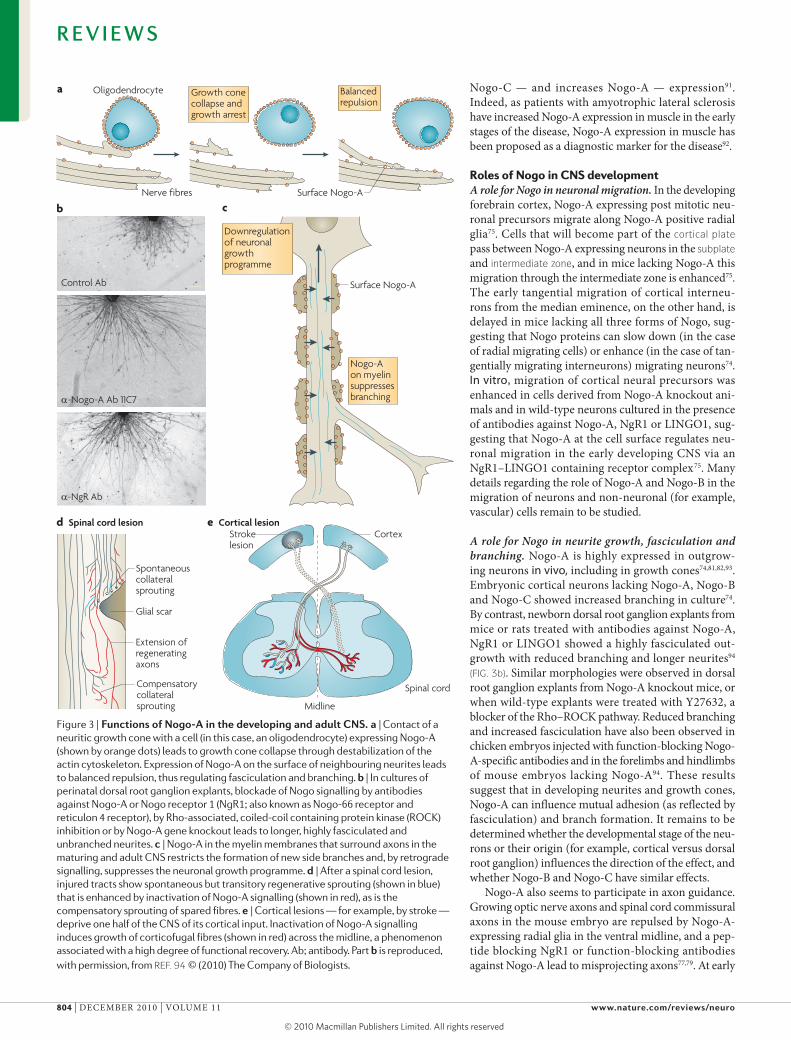
A role for Nogo in neurite growth, fasciculation and branching. Nogo-A is highly expressed in outgrow-ing neurons in vivo, including in growth cones74,81,82,93. Embryonic cortical neurons lacking Nogo-A, Nogo-B and Nogo-C showed increased branching in culture74. By contrast, newborn dorsal root ganglion explants from mice or rats treated with antibodies against Nogo-A, NgR1 or LINGO1 showed a highly fasciculated out-growth with reduced branching and longer neurites94 (FIG. 3b). Similar morphologies were observed in dorsal root ganglion explants from Nogo-A knockout mice, or when wild-type explants were treated with Y27632, a blocker of the Rho–ROCK pathway. Reduced branching and increased fasciculation have also been observed in chicken embryos injected with function-blocking Nogo-A-specific antibodies and in the forelimbs and hindlimbs of mouse embryos lacking Nogo-A94. These results suggest that in developing neurites and growth cones, Nogo-A can influence mutual adhesion (as reflected by fasciculation) and branch formation. It remains to be determined whether the developmental stage of the neu-rons or their origin (for example, cortical versus dorsal root ganglion) influences the direction of the effect, and whether Nogo-B and Nogo-C have similar effects.
Nogo-A also seems to participate in axon guidance. Growing optic nerve axons and spinal cord commissural axons in the mouse embryo are repulsed by Nogo-A-expressing radial glia in the ventral midline, and a pep-tide blocking NgR1 or function-blocking antibodies against Nogo-A lead to misprojecting axons77,79. At early
Figure 3 | Functions of Nogo-A in the developing and adult cNs. a | Contact of a neuritic growth cone with a cell (in this case, an oligodendrocyte) expressing Nogo-A (shown by orange dots) leads to growth cone collapse through destabilization of the actin cytoskeleton. Expression of Nogo-A on the surface of neighbouring neurites leads to balanced repulsion, thus regulating fasciculation and branching. b | In cultures of perinatal dorsal root ganglion explants, blockade of Nogo signalling by antibodies against Nogo-A or Nogo receptor 1 (NgR1; also known as Nogo-66 receptor and reticulon 4 receptor), by Rho-associated, coiled-coil containing protein kinase (ROCK) inhibition or by Nogo-A gene knockout leads to longer, highly fasciculated and unbranched neurites. c | Nogo-A in the myelin membranes that surround axons in the maturing and adult CNS restricts the formation of new side branches and, by retrograde signalling, suppresses the neuronal growth programme. d | After a spinal cord lesion, injured tracts show spontaneous but transitory regenerative sprouting (shown in blue) that is enhanced by inactivation of Nogo-A signalling (shown in red), as is the compensatory sprouting of spared fibres. e | Cortical lesions — for example, by stroke — deprive one half of the CNS of its cortical input. Inactivation of Nogo-A signalling induces growth of corticofugal fibres (shown in red) across the midline, a phenomenon associated with a high degree of functional recovery. Ab; antibody. Part b is reproduced, with permission, from ReF. 94 © (2010) The Company of Biologists.
R E V I E W S
804 | dECEmBER 2010 | vOLumE 11 www.nature.com/reviews/neuro
© 20 Macmillan Publishers Limited. All rights reserved10
Cortical plateThe upper part of the developing cerebral cortex, where neurons end their migration and start to assemble into the distinct neuronal layers that will form the future adult cortex.
Subplate A transient layer of cells in the fetal brain that lies beneath the cortical plate.
Intermediate zone A transient layer in the developing cortex through which neurons migrate on their way from the proliferative zone to the cortical plate. With maturation, this zone is replaced by the subcortical white matter.
Morpholinos Antisense oligonucleotides that block gene expression.
postnatal stages of development, the myelinating, Nogo-A-positive, ascending sensory tracts in the dorsal column of the rat spinal cord seem to channel the late-growing corticospinal tract into its characteristic territory and prevent it from mixing with the surrounding sensory tracts95. Antibodies against Nogo-A or the ablation of oligodendrocytes by irradiation lead to aberrant, wide-spread distribution of corticospinal tract fibres beyond the normal tract boundaries95,96. Thus, Nogo proteins — and Nogo-A in particular — expressed by glial cells and neurons can exert guidance effects in the developing CNS by repulsion of growing fibres.
Developmental roles of Nogo in zebrafish. Nogo proteins affecting axonal guidance and fasciculation have also been documented in the developing peripheral nervous system of zebrafish embryos. Fish Nogo proteins have short N termini that have no resemblance to those of Nogo-A, Nogo-B or Nogo-C in higher vertebrates, but the RTN domains are very similar to those in mammals and include an NgR-binding Nogo-66 domain10,97. An NgR1 homologue is also present in fish98,99. Addition of mammalian Nogo-66 to cultured zebrafish neu-rons induced growth cone collapse but, astonishingly, Nogo-66 derived from zebrafish proved to be growth enhancing instead97. Indeed, in vivo knock down of Nogo or NgR in the fish embryo impaired growth of the lat-eral line nerve, suggesting a growth-enhancing function of Nogo proteins in fish99. By contrast, regeneration of lesioned trigeminal axons in the skin of zebrafish larvae is normally restricted to the area formerly occupied by the cut nerves, but this restriction was absent in zebrafish expressing dominant-negative forms of NgR, LINGO or RHOA, or in zebrafish in which NgR expression was suppressed by antisense morpholinos100. This suggests that at later developmental stages the Nogo-positive axonal fragments of the lesioned nerve inhibit the growth of new axons in their vicinity100.
These observations in zebrafish are interesting from an evolutionary point of view. Short Nogo proteins that consist mostly of the RTN domain and contain the Nogo-66 sequence are expressed in the developing fish nervous system and are involved in growth regulation of neurites. The presence of NgR in fish suggests that at least some of these effects are mediated by a Nogo-66–NgR interaction. However, the effects can be growth enhancing or growth inhibitory, depending on fac-tors that are presently unknown. In higher vertebrates, new N-terminal sequences, in particular the Nogo-A-specific Nogo-Δ20 domain, have made the molecule predominantly growth-inhibitory, presumably through interaction with a second receptor subunit.
Role of Nogo in myelin formation. At late developmental stages of the CNS of higher vertebrates, axons and their surrounding myelin-forming oligodendrocytes express Nogo proteins and Nogo receptor components70,71,101, suggesting a possible role of Nogo proteins in axon–oligodendrocyte cross-talk. Indeed, it was found that Nogo-A and mAG double knockout mice showed a delay in myelin formation and had myelin malformations101.
One or more receptors from the NgR family, to which both Nogo proteins and mAG can bind, are likely to be involved — as is the NgR-binding protein LINGO1, which has been shown to inhibit oligodendrocyte differentiation and myelin formation102.
Roles of Nogo in the adult CNSDownregulation of growth, stabilization of wiring and restriction of plasticity in the adult CNS. Adult CNS tissue, in particular white matter, is a largely non- permissive environment for neurite growth. Nogo-A was originally described as one of the main neurite growth-inhibitory components of oligodendrocytes and myelin in the CNS103–105. myelin is formed dur-ing the final phase of CNS maturation; in many CNS regions this phase is characterized by the refinement of the neuronal connections and ends with the closure of the so-called ‘critical period’ for major plastic rear-rangements of axons and dendrites106. Examples of such postnatal periods of plasticity include the ocu-lar dominance column plasticity in the visual cortex and, in the spinal cord, the compensatory sprouting of neighbouring sensory roots into denervated seg-ments after dorsal root section. Axonal rearrange-ments and growth are greatly reduced at specific time points after birth (around 3–5 weeks in mice and rats in these areas)107,108. Notably, this coincides with myelin formation in these areas and with a local downregulation of growth-promoting proteins such as growth-associated protein 43 (GAP43; also known as neuromodulin)32,108,109. when myelin formation was prevented in the spinal cord of postnatal rats, sprout-ing of spared root fibres into neighbouring denervated segments continued much beyond the end of the plastic period108. In the visual cortex, a relationship between the termination of this highly plastic period and Nogo signalling was suggested by the finding that mice lacking both Nogo-A and Nogo-B, or mutant mice lacking NgR1 or PIRB, showed levels of ocular dominance plasticity in adulthood that were compa-rable to those in immature mice32,31 (FIG. 4). moreover, adult mice lacking Nogo-A showed an upregulation of cytoskeletal and growth-related mRNAs, and proteins in the spinal cord and cortex67. Furthermore, addition of function-blocking Nogo-A-specific antibodies to mature (3- to 5-week-old) organotypic hippocampal slice cultures induced both upregulation of growth-specific proteins and pronounced neurite sprouting in the absence of lesions110. An in vivo growth-restricting effect of myelin, preventing aberrant sprouting of cholinergic septal fibres that was mediated by p75 and Rho, was recently shown in the intact adult rodent brain111. Together, these observations suggest that Nogo proteins — and Nogo-A in particular — may be act-ing as a tonic negative growth regulator in the adult CNS. From a developmental perspective, a possible sequence of events could be that the maturing axon induces oligodendrocyte differentiation, resulting in its myelination. Nogo-A expressed on the myelin may then suppress further branching locally and formation of collaterals, by downregulating the neuronal growth
R E V I E W S
NATuRE REvIEwS | NeuroscieNce vOLumE 11 | dECEmBER 2010 | 805
© 20 Macmillan Publishers Limited. All rights reserved10
Left eye deprived during critical period
Cor
tical
laye
r
IIIIIIIVVVI
L R L R
IIIIIIIVVVI
IIIIIIIVVVI
LC
ortic
al la
yer
Cor
tical
laye
r
Cor
tical
laye
rC
ortic
al la
yer
R L R
L R L R
I
I
IIIIIIVVVI
L R L R
IIIIIIVVVI
L R L R
a Wild-typeNeonate
Normal adult
Left eye deprived during critical period
Cor
tical
laye
r
IIIIIIIVVVI
L R L R
Left eye deprived during adulthood
Left eye deprived during adulthood
b NgR knockout or Nogo-A/Nogo-B knockout
Nature Reviews | Neuroscience
programme (FIG. 3c). This would prevent unwanted side branch formation, especially in white matter, and therefore exert a stabilizing role in CNS wiring.
Not much is currently known about the physiological role of Nogo-A in neurons in the hippocampus, olfactory bulb or dorsal root ganglion of adult animals. Electron microscopy and biochemical studies have provided evidence for a synaptic localization of Nogo-A and NgR1 in these areas71,84,85,112. A special form of FGF-dependent long-term potentiation (LTP) was increased and long-term depression (LTd) was attenuated84 in hippocampi from NgR knockout animals. In agreement with this, acute administration of antibodies against Nogo-A or NgR strongly enhanced both short (30–90 min) and long-lasting (3 h) LTP in hippocampal slices from wild-type animals (A. delekate, m. Korte, m. Zagrebelsky, m.E.S., unpublished observations). Conversely, overex-pression of Nogo-A in Purkinje terminals induced syn-apse disassembly112, and transgenic mice overexpressing NgR showed impairments of long-term memory113. Together, these observations suggest that Nogo-A and
NgR1 at synapses could act as regulators of synaptic sta-bility: in this scheme, low Nogo-A or NgR1 enhances synaptic connectivity, for example in the form of LTP, whereas high levels of Nogo-A lead to destabilization of synapses. Like Nogo, several other repulsive axonal guidance molecules are expressed at adult synapses, especially in regions such as the hippocampus, where they can influence LTP and other forms of synaptic plasticity114–116. many of them affect the cyctoskeleton, thus influencing synaptic function presynaptically and/or postsynaptically, which could also be the case for Nogo.
There are also indications that Nogo might have a role in some psychiatric disorders, such as schizophrenia. The schizophrenia-like behavioural phenotype of Nogo-A knockout mice117, and the genetic associations between psychiatric disorders and NgR or Nogo muta-tions in certain families and patients118–122, are not well understood yet (BOX 2). They could reflect deficits in Nogo signalling during development, which would be in line with the postulated developmental origin of certain psychiatric diseases, and schizophrenia in
Figure 4 | role of Ngr or Nogo-A/Nogo-B in restricting the developmental plasticity in the visual cortex. a | Maturation of cortical columns and of the terminal arborizations of thalamic input axons in the visual cortex is driven by visual experience. In the early postnatal (P0–20) mouse (top left panel), the thalamic visual afferents representing the left (L; shown in blue) and right (R; shown in red) eyes, and terminating in cortical layer IV, overlap to a large degree. Monocular deprivation (for example, by lid suture) of the left eye during the critical period leads to smaller arbors and ocular dominance columns and a corresponding enlargement of the right, open eye territories (top right panel). These plastic rearrangements are severely restricted at the end of the ‘critical period’. Indeed, the arrangements of thalamic visual afferents in cortical layer IV in adult mice (bottom left panel) is not affected by monocular deprivation during adulthood (bottom right panel), indicating that visual deprivation during adulthood does not affect terminal arborization. b | In mice lacking the Nogo receptor (NgR) or Nogo-A/Nogo-B, monocular deprivation during the critical period has the same effect as in developing wild-type mice (left panel). In contrast to wild-type mice, however, monocular deprivation during adulthood in these mutant mice induces a highly plastic pattern similar to that in immature mice (right panel).
R E V I E W S
806 | dECEmBER 2010 | vOLumE 11 www.nature.com/reviews/neuro
© 20 Macmillan Publishers Limited. All rights reserved10
Tubular ERA major part of the endoplasmic reticulum (eR) of cells that is characterized by a tubular shape, as opposed to the flat eR cisterns that compose the nuclear membrane, or the Nissl bodies in synthetically highly active neurons.
particular123. On the other hand, the evidence discussed above suggests that Nogo signalling might also have a role in these disorders through dysregulation of circuit functions in adulthood.
Specific roles of Nogo-B and its receptor NGBR in the CNS have not yet been defined. In blood vessels, they influence vessel repair after injury and also macrophage infiltration51,90.
In conclusion, much remains to be learnt of the roles played by Nogo proteins in the adult nervous system. The downregulation of the neuronal growth programme mainly by Nogo-A expressed in myelin may contribute to the termination of the plastic period for large-scale circuit rearrangements, such as in ocular dominance plasticity. Nogo-A-mediated growth suppression may help to stabi-lize the highly complex neuronal wiring of the adult CNS, preventing aberrant fibre growth. Neuronal Nogo and its receptor components are also expressed at synapses, in particular in regions that show synaptic plasticity and are involved in learning. Current evidence points to a role of synaptic Nogo and NgR as negative regulators of syn-aptic stability, but the precise site of action (presynaptic versus postsynaptic), the postreceptor signalling cascades involved, and the key effectors (for example, actin, pre-synaptic or postsynaptic cytoskeleton, and extracellular adhesion mechanisms) remain to be studied.
Intracellular functions of Nogo proteinsThe relatively few studies addressing the intracellular functions of Nogo — of which a large number are based on molecular interactions — suggest that Nogo proteins could have intracellular functions in the adult CNS5,124. By binding to and inhibiting the enzyme β-secretase (BACE), Nogo-A, Nogo-B or Nogo-C, as well as RTN3, can inhibit the production of amyloid-β peptides, which are linked to Alzheimer’s disease125–127. Interestingly, amyloid-β peptides bind to NgR20,128. However, there is currently no direct evidence for a crucial role of
Nogo-A or Nogo-B in the development or progression of Alzheimer’s disease, except for the demonstration that increased fibre sprouting occurs around amyloid plaques in Nogo knockout mice, which could be due to the absence of the growth-inhibitory effect of Nogo-A129.
Several studies have found an upregulation of Nogo-A under conditions of cellular stress5,124, for example in neu-rons and astrocytes surrounding stroke lesions or amyloid plaques130,131. In a superoxide dismutase (SOd) mutant mouse model of amyotrophic lateral sclerosis, genetic deletion of Nogo-A and Nogo-B accelerated disease pro-gression and decreased spinal motor neuron survival, pos-sibly as a result of a lack of regulatory effects of Nogo-A on the ER chaperone protein disulphide isomerase in these mice. Overexpression or deletion of Nogo-A changes the intracellular location of the enzyme, which in turn might compromise its chaperone functions132.
Another intracellular interaction partner of Nogo proteins and of the reticulon protein RTN3 is the anti-apoptotic protein BCL2 (ReFs 133,134), suggesting a link between Nogo or RTN3 and apoptosis in neuroprotection or neurodegeneration. Indeed, a role of Nogo-B in regu-lating the survival of cancer cells has been proposed134–136. A possible mechanism could be that Nogo or RTN3 changes the intracellular localization of BCL2, but this remains to be confirmed by additional studies135,136.
All Rtn proteins, including Nogo-A, Nogo-B and Nogo-C, are enriched in the ER, suggesting additional functions of Nogo proteins that may be specific to this subcellular compartment5,124. Indeed, overexpression of Nogo-A or RTN3 led to an increase in the proportion of tubular eR137, but prevented the assembly of the nuclear membrane after cell division — a disturbance that was also observed after neutralization of Nogo-A by antibodies138,139. These effects were presumably mediated by interactions of the Rtn proteins with membrane pro-teins like the ER protein deleted in polyposis 1 (dP1), leading to complexes that increase the curvature of the ER membrane and thereby help to shape the ER and the nuclear membrane137–140. Nogo-B and NGBR also play a part in intracellular cholesterol transport52.
The results described above suggest that, through their phylogenetically ancient C-terminal RTN domain, Nogo proteins function as interaction partners of specific intracellular proteins. They have also been proposed to have structural roles, especially for the tubular part of the ER and the nuclear membrane, and to be involved in mechanisms of cell survival and ER stress. Still, most of these functions have not been well studied yet. Functional compensation by other Rtn family members in knockout mice has made the study of these intracellular roles more difficult141. Nevertheless, it seems clear that Nogo proteins are an interesting example of multidomain, multipurpose proteins in which domains were added during evolution, resulting in novel functions for different isoforms.
Role of Nogo in CNS repairThe bulk of the Nogo and Nogo receptor literature deals with the role of these molecules in the injured spinal cord and brain, and many reviews are available on this topic1,33,35,142–147.
Box 2 | Nogo and its receptor in psychiatric diseases
Human genetic linkage studies and examination of the post‑mortem brain tissue of patients with psychiatric disorders suggest a link between Nogo signalling and bipolar disorder and schizophrenia118–122,157–159. However, the number of families and individuals that have been examined so far is limited, and contradictory reports have been published. Two recent studies support the hypothesis that there is a link between Nogo‑A–Nogo receptor signalling and schizophrenia117,122. One study122 described point mutations in Nogo receptor 1 (NgR1; also known as Nogo‑66 receptor and reticulon 4 receptor) in individuals affected by schizophrenia from different families. Some of these mutations directly affected Nogo binding122. Moreover, mice lacking NgR1 showed mild behavioural alterations that mimic some symptoms of schizophrenia122.
Two studies have analysed the behavioural phenotype of mice lacking Nogo‑A117,160. Reflexes and locomotor behaviour in these mice were normal, but they showed enhanced locomotion in response to amphetamine administration as well as abnormalities in prepulse inhibition and latent inhibition, indicative of attentional deficits117,160. Similar abnormalities in analogous tests for humans are typically seen in patients with schizophrenia161,162. Nogo‑A knockout mice also showed neurochemical abnormalities — particularly in the level, turnover and receptor levels for dopamine and serotonin in the striatum and prefrontal cortex — that paralleled changes observed in patients with schizophrenia117. The mechanisms and possible developmental time course of the Nogo‑A or NgR1‑related defects leading to abnormal adult behaviour remain to be investigated.
R E V I E W S
NATuRE REvIEwS | NeuroscieNce vOLumE 11 | dECEmBER 2010 | 807
© 20 Macmillan Publishers Limited. All rights reserved10
Suppression of Nogo or Nogo receptor function — by antibodies, soluble receptor fragments, antagonistic peptides, gene knockout or knockdown — or block-ade of Rho or ROCK are all treatments that have been shown to enhance regenerative sprouting and growth of lesioned fibres after spinal cord or brain injury. In addi-tion, these treatments induce compensatory collateral sprouting of intact fibres in the adult CNS after injury. These pro cesses result in increased plasticity and a cer-tain degree of regeneration, depending on the size and type of the lesion (FIGs 3d,e). They induce reorganization of CNS circuits in the spinal cord, brainstem and cortex. Transitory, short-distance regenerative sprouting and a limited degree of plastic reorganization occurs sponta-neously in many tracts after a lesion. The higher level of fibre growth and functional recovery seen after inactiva-tion of Nogo-A signalling is best explained by the sup-pression of its growth-inhibitory effects. These occur at the level of the growing fibres through inactivation of a growth-inhibitory interaction with myelin, oligodendro-cytes and myelin debris, and at the level of the cell body through suppression of the retrograde, tonic Nogo-A-mediated growth-inhibitory signals, which are present in the adult CNS. Thus, Nogo-A neutralization may shift the adult, stabilized CNS back into a more plastic, quasi-developmental stage.
For most of the anti-Nogo or anti-Nogo receptor treatments mentioned above, enhanced recovery of lost functions has also been shown behaviourally. Treatments
targeting Nogo signalling resulted in the most consistent and extensive structural and functional recoveries com-pared to other experimental interventions after spinal cord or stoke lesions, such as scar-reducing treatments, growth factor injections and cell transplantations1,35. Importantly, very similar functional and structural improvements were observed in studies that used dif-ferent methods of blocking Nogo-A or NgR function, whereby acute treatments that interrupt Nogo-A signal-ling at the time of CNS injury were more efficient than conventional chronic gene knockouts (BOX 3). Although most of these results were obtained in rats or mice, proof-of-principle experiments have also been performed on monkeys148–150. A clinical study in patients with acute injuries in the spinal cord with a human Nogo-A antibody (ATI-355; Novartis) is currently underway (ClincalTrials.gov: NCT00406016). Together, these results support the concept that Nogo-A is a key stabilizer and negative growth regulator in the adult CNS of higher vertebrates.
Conclusions and future directionsNogo proteins on cell membranes interact with multisub-unit receptors consisting of ligand binding, signal trans-duction and associated proteins. Some key components of the Nogo-A receptor still remain to be characterized, and, in spite of the known role of Rho and Ca2+, the full complexity of the second messenger cascades generated downstream of the activated Nogo receptor remains to be unravelled. In the CNS of developing and adult animals
Box 3 | Multiple methods to block Nogo function: acute blockade versus gene knockout
A traditional way to block the function of signalling molecules is the use of agents (‘blockers’) that interfere with ligand binding to a receptor, with receptor activation or with the downstream intracellular signalling cascade. To interfere with Nogo signalling, the peptide Nogo‑A extracellular peptide residues 1‑40 (NEP1‑40) can be used as an antagonist of Nogo receptors (NgRs)1,16,35. Pharmacological blockers of RHOA and of the Rho effector kinase Rho‑associated, coiled‑coil containing protein kinase (ROCK) blocked Nogo‑A functions in vivo and in vitro33. The Rho inactivating enzyme C3‑transferase (Cethrin; BioAxone Therapeutic) is currently being tested in clinical trials with patients affected by spinal cord injuries163.
Antibodies binding to, and sterically inhibiting access to, active sites of ligands or receptors have been successfully used in the study of Nogo‑A functions1,142,146. ‘Receptor bodies’ are fusion proteins that consist of receptor fragments containing the ligand binding site and Fc antibody sequences. Such an NgR–Fc fusion protein was shown to be a potent blocker of inhibitory NgR ligands, including Nogo proteins164. A remarkable congruence of the effects of different blocking agents for Nogo‑A signalling — including antibodies, receptor bodies and small molecule blockers — has been found in a number of regeneration and plasticity paradigms1,35,142. An antibody that blocks Nogo‑A function has reached the stage of clinical trial as a novel treatment for spinal cord injury (ClinicalTrials.gov: NCT00406016).
An alternative to small molecular weight blockers or antibodies are technologies that allow gene silencing or gene deletion. Acute knock down of NgR in optic nerve fibres enhanced regeneration30. However, the systemic knock out of Nogo‑A, Nogo‑A and B, or Nogo‑A, B and C simultaneously, and also the recent triple knockouts of Nogo‑A, myelin associated protein (MAG) and oligodendrocyte myelin glycoprotein (OMGP), performed in different laboratories, resulted in rather mild and seemingly inconsistent phenotypes141,165,166–168. More detailed analyses revealed several factors that affected the outcome. First, SV129 mice showed a significantly higher intrinsic growth and regeneration potential than C57BL/6 mice169, demonstrating the important influence of genetic background and the pitfalls of the conventional mixed‑strain technology (that is, unknown proportions of C57BL/6 and SV129 genes in different mutant offspring lines). Second, the absence of Nogo proteins has different effects according to the neuroanatomical structures and types of lesions being considered32,170. Third, functional compensation often occurs in knockout models — demonstrated, for example, by the increased levels of semaphorins and ephrins and their receptors in the CNS of mice lacking Nogo‑A171. The literature contains many examples of mouse mutants in which physiologically important molecules were knocked out but that nevertheless had only mild or even absent phenotypes, probably owing to compensatory effects172. Indeed, the many mutations present in the human population that do not dramatically affect viability are a good example of this compensatory capacity172. Therefore, inducible conditional knockouts and well‑defined knockdowns can be expected to yield much more relevant and informative phenotypes than models generated with conventional knockout techniques.
R E V I E W S
808 | dECEmBER 2010 | vOLumE 11 www.nature.com/reviews/neuro
© 20 Macmillan Publishers Limited. All rights reserved10

Nogo-A exerts repulsive and neurite growth-inhibitory functions. migration of neuronal precursors can also be affected. In the embryonic forebrain cortex Nogo pro-teins affect tangential and radial neuronal migration, but the generality of this phenomenon and its cell biologi-cal mechanistic basis remain to be studied. In develop-ing peripheral neurons Nogo-A also restricts the growth of neurites, counteracts the neurons’ mutual adhesive interactions during fasciculation and influences branch-ing. Nogo-A, therefore, appears as a new player in the orchestra of repulsive or inhibitory, and attractive or growth-promoting cues that steer axons and govern the formation of the highly complex neuronal circuits and connections during development. The various levels at which Nogo-A-mediated growth regulation takes place — for example, on the level of the growth cone versus cell body, or cytoskeleton assembly versus gene transcription — remain to be elucidated in detail, as do the potential interactions between Nogo-A and the different growth regulators and guidance factors.
In the maturing and adult CNS, myelin-derived Nogo-A has stabilizing functions for the neurite network by acting as a growth suppressor. The recently discovered role of neuronal Nogo-A and NgR1 at synapses
— suppression of Nogo-A and/or NgR1 function enhances LTP and synaptic stability — points to an additional role of Nogo-A as a regulator of neuronal connections. The growth-suppressive function of myelin Nogo-A sets a conceptual framework for understanding the observed enhancement of axonal growth and regeneration and the functional recovery after spinal cord or brain lesions by inactivation of Nogo-A–NgR1 signalling. If this enhance-ment of CNS fibre growth and reparative processes by blockade of the Nogo-A pathway proves to be a general phenomenon, therapeutic windows might open for CNS diseases beyond spinal trauma and stroke.
Although the largest Nogo protein — Nogo-A — is well studied, much less information is available on Nogo-B and Nogo-C, in particular with regard to func-tions in non-neural tissues, where these Nogo isoforms are frequently found. The role of Nogo-B and its spe-cific receptor NGRB in vascular repair is interesting but begs the question of the function of Nogo-B in the normal patterning and function of the blood vessel sys-tem. much more data are also needed on the potential intracellular functions of Nogo proteins, some of which may be related to basic biological processes such as ER structure and function, and cell survival.
1. Schwab, M. E. Nogo and axon regeneration. Curr. Opin. Neurobiol. 14, 118–124 (2004).
2. Chen, M. S. et al. Nogo-A is a myelin-associated neurite outgrowth inhibitor and an antigen form monoclonal antibody IN-1. Nature 403, 434–439 (2000).
3. GrandPre, T., Nakamura, F., Vartanian, T. & Strittmatter, S. M. Identification of the Nogo inhibitor of axon regeneration as a reticulon protein. Nature 403, 439–444 (2000).
4. Oertle, T. et al. Nogo-A inhibits neurite outgrowth and cell spreading with three discrete regions. J. Neurosci. 23, 5393–5406 (2003).
5. Oertle, T. & Schwab, M. E. Nogo and its paRTNers. Trends Cell Biol. 13, 187–194 (2003).
6. Oertle, T., Huber, C., van der Putten, H. & Schwab, M. E. Genomic structure and functional characterisation of the promoters of human and mouse nogo/rtn4. J. Mol. Biol. 325, 299–323 (2003).
7. Li, M. & Song, J. The N- and C-termini of the human Nogo molecules are intrinsically unstructured: bioinformatics, CD, NMR characterization, and functional implications. Proteins 68, 100–108 (2007).
8. Dodd, D. A. et al. Nogo-A, -B and -C are found on the cell surface and interact together in many different cell types. J. Biol. Chem. 280, 12494–12502 (2005).
9. Oertle, T., Klinger, M., Stuermer, C. A. O. & Schwab, M. E. A reticular rhapsody: phylogenic evolution and nomenclature of the RTN/Nogo gene family. FASEB J. 17, 1238–1247 (2003).
10. Diekmann, H. et al. Analysis of the reticulon gene family demonstrates the absence of the neurite growth inhibitor Nogo-A in fish. Mol. Biol. Evol. 22, 1635–1648 (2005).
11. Schweigreiter, R. The natural history of the myelin-derived nerve growth inhibitor Nogo-A. Neuron Glia Biol. 4, 83–89 (2008).
12. Klinger, M. et al. Identification of two nogo/rtn4 genes and analysis of Nogo-A expression in Xenopus laevis. Mol. Cell. Neurosci. 25, 205–216 (2004).
13. Ferretti, P., Zhang, F. & O’Neill, P. Changes in spinal cord regenerative ability through phylogenesis and development: lessons to be learnt. Dev. Dyn. 226, 245–256 (2003).
14. Fournier, A. E., GrandPre, T. & Strittmatter, S. M. Identification of a receptor mediating Nogo-66 inhibition of axonal regeneration. Nature 409, 341–346 (2001).
15. Hu, F. et al. Nogo-A interacts with the Nogo-66 receptor throuth multiple sites to create an isoform-selective subnanomolar agonist. J. Neurosci. 25 5298–5304 (2005).
16. GrandPre, T., Li, S. & Strittmatter, S. M. Nogo-66 receptor antagonist peptide promotes axonal regeneration. Nature 417, 547–551 (2002).
17. Domeniconi, M. et al. Myelin-associated glycoprotein interacts with the Nogo66 receptor to inhibit neurite outgrowth. Neuron 35, 283–290 (2002).
18. Wang, K. C., Kim, J. A., Sivasankaran, R., Segal, R. & He, Z. p75 interacts with the Nogo receptor as a co-receptor for Nogo, MAG and OMgp. Nature 420, 74–78 (2002).
19. Venkatesh, K. et al. The Nogo-66 receptor homolog NgR2 is a sialic acid-dependent receptor selective for myelin-associated glycoprotein. J. Neurosci. 25, 808–822 (2005).
20. Park, J. H. et al. Alzheimer precursor protein interaction with the Nogo-66 receptor reduces amyloid-β plaque deposition. J. Neurosci. 26, 1386–1395 (2006).
21. Zhang, L. et al. Identification of BLyS (B lymphocyte stimulator), a non-myelin-associated protein, as a functional ligand for Nogo-66 receptor. J. Neurosci. 29, 6348–6352 (2009).
22. Thomas, R. et al. LGI1 is a Nogo receptor 1 ligand that antagonizes myelin-based growth inhibition. J. Neurosci. 30, 6607–6612 (2010).
23. Atwal, J. K. et al. PirB is a functional receptor for myelin inhibitors of axonal regeneration. Science 322, 967–970 (2008).
24. Fournier, A. E., Gould, G. C., Liu, B. P. & Strittmatter, S. M. Truncated soluble Nogo receptor binds Nogo-66 and blocks inhibition of axon growth by myelin. J. Neurosci. 22, 8876–8883 (2002).
25. Chivatakarn, O., Kaneko, S., He, Z., Tessier-Lavigne, M. & Giger, R. J. The Nogo-66 receptor NgR1 is required only for the acute growth cone-collapsing but not the chronic growth-inhibitory actions of myelin inhibitors. J. Neurosci. 27, 7117–7124 (2007).
26. Takei, Y. Phosphorylation of Nogo receptors suppresses Nogo signaling, allowing neurite regeneration. Sci. Signal 2, ra14 (2009).
27. Lee, J. K., Kim, J. E., Sivula, M. & Strittmatter, S. M. Nogo receptor antagonism promotes stroke recovery by enhancing axonal plasticity. J. Neurosci. 24, 6209–6217 (2004).
28. Kim, J. E., Liu, B. P., Park, H. J. & Strittmatter, S. M. Nogo-66 receptor prevents raphespinal and rubrospinal axon regeneration and limits functional recovery from spinal cord injury. Neuron 44, 439–451 (2004).
29. Zheng, B. et al. Genetic deletion of the Nogo receptor does not reduce neurite inhibition in vitro or promote corticospinal tract regeneration in vivo. Proc. Natl Acad. Sci. USA 102, 1205–1210 (2005).
30. Fischer, D., He, Z. & Benowitz, L. I. Counteracting the Nogo receptor enhances optic nerve regeneration if retinal ganglion cells are in an active growth state. J. Neurosci. 24, 1651 (2004).
31. Syken, J., Grandpre, T., Kanold, P. O. & Shatz, C. J. PirB restricts ocular-dominance plasticity in visual cortex. Science 313, 1795–1800 (2006).
32. McGee, A. W., Yang, Y., Fischer, Q. S., Daw, N. W. & Strittmatter, S. M. Experience-driven plasticity of visual cortex limited by myelin and Nogo receptor. Science 309, 2222–2226 (2006).This paper shows that the developmental plasticity in the well-known paradigm of visual cortex ocular dominance columns is restricted at the end of the critical period by mechanisms involving NgR, and Nogo-A and Nogo-B.
33. Nash, M., Pribiag, H., Fournier, A. E. & Jacobson, C. Central nervous system regeneration inhibitors and their intracellular substrates. Mol. Neurobiol. 40, 224–235 (2009).A recent review on the intracellular pathways, in particular RHOA signalling, that have key roles in Nogo signalling.
34. Spencer, T., Domeniconi, M., Cao, Z. & Filbin, M. T. New roles for old proteins in adult CNS axonal regeneration. Curr. Opin. Neurobiol. 13, 133–139 (2003).
35. Yiu, G. & He, Z. Glial inhibition of CNS axon regeneration. Nature Rev. Neurosci. 7, 617–627 (2006).
36. Shao, Z. et al. TAJ/TROY, an orphan TNR receptor family member, binds Nogo-66 receptor 1 and regulates axonal regeneration. Neuron 45, 353–359 (2005).
37. Park, J. B. et al. A TNF receptor family member, TROY, is a coreceptor with Nogo receptor in mediating the inhibitory activity of myelin inhibitors. Neuron 45, 345–351 (2005).
38. Mi, S. et al. LINGO-1 is a component of the Nogo-66 receptor/p75 signaling complex. Nature Neurosci. 7, 221–228 (2004).
39. Ji, B. et al. LINGO-1 antagonist promotes functional recovery and axonal sprouting after spinal cord injury. Mol. Cell. Neurosci. 33, 311–320 (2006).
40. Lv, J. et al. Passive immunization with LINGO-1 polyclonal antiserum afforded neuroprotection and promoted functional recovery in a rat model of spinal cord injury. Neuroimmunomodulation 17, 270–278 (2010).
41. Okafuji, T. & Tanaka, H. Expression patern of LINGO-1 in the developing nervous system of the chick embryo. Gene Expr. Patterns 6, 57–62 (2005).
R E V I E W S
NATuRE REvIEwS | NeuroscieNce vOLumE 11 | dECEmBER 2010 | 809
© 20 Macmillan Publishers Limited. All rights reserved10
42. Barrette, B., Vallieres, N., Dube, M. & Lacroix, S. Expression profile of receptors for myelin-associated inhibitors of axonal regeneration in the intact and injured mouse central nervous system. Mol. Cell. Neurosci. 34, 519–538 (2007).
43. Hu, F. & Strittmatter, S. M. The N-terminal domain of Nogo-A inhibits cell adhesion and axonal outgrowth by an integrin-specific mechanism. J. Neurosci. 28, 1262–1269 (2008).
44. Koprivica, V. et al. EGFR activation mediates inhibition of axon regeneration by myelin and chondroitin sulfate proteoglycans. Science 310, 106–110 (2005).
45. Grunewald, E., Kinnell, H. L., Porteous, D. J. & Thomson, P. A. GPR50 interacts with neuronal NOGO-A and affects neurite outgrowth. Mol. Cell. Neurosci. 42, 363–371 (2009).
46. Chao, M. V. Neurotrophins and their receptors: a convergence point for many signalling pathways. Nature Rev. Neurosci. 4, 299–309 (2003).
47. Inestrosa, N. C. & Arenas, E. Emerging roles of Wnts in the adult nervous system. Nature Rev. Neurosci. 11, 77–86 (2010).
48. Rajasekharan, S. & Kennedy, T. E. The netrin protein family. Genome Biol. 10, 239 (2009).
49. Jackson, R. E. & Eickholt, B. J. Semaphorin signalling. Curr. Biol. 19, R504–R507 (2009).
50. Miao, R. Q. et al. Identification of a receptor necessary for Nogo-B stimulated chemotaxis and morphogenesis of endothelial cells. Proc. Natl Acad. Sci. USA 103, 10997–11002 (2006).
51. Acevedo, L. et al. A new role for Nogo as a regulator of vascular remodeling. Nature Med. 10, 382–388 (2004).The first demonstration of a role of Nogo proteins, in particular Nogo-B, in vascular endothelial cells and smooth muscle cells, and in blood vessel repair.
52. Harrison, K. D. et al. Nogo-B receptor stabilizes Niemann-Pick type C2 protein and regulates intracellular cholesterol trafficking. Cell Metab. 10, 208–218 (2009).
53. Fournier, A. E., Takizawa, B. T. & Strittmatter, S. M. Rho kinase inhibition enhances axonal regeneration in the injured CNS. J. Neurosci. 23, 1416–1423 (2003).
54. Lehmann, M. et al. Inactivation of Rho signaling pathway promotes CNS axon regeneration. J. Neurosci. 19, 7537–7547 (1999).
55. Niederost, B., Oertle, T., Fritsche, J., McKinney, R. A. & Bandtlow, C. E. Nogo-A and myelin-associated glycoprotein mediate neurite growth inhibition by antagonistic regulation of RhoA and Rac1. J. Neurosci. 22, 10368–10376 (2002).
56. Kubo, T., Yamaguchi, A., Iwata, N. & Yamashita, T. The therapeutic effects of Rho-ROCK inhibitors on CNS disorders. Ther. Clin. Risk Manag. 4, 605–615 (2008).
57. Joset, A., Dodd, D. A., Halegoua, S. & Schwab, M. E. Pincher-generated Nogo-A endosomes mediate growth cone collapse and retrograde signaling. J. Cell Biol. 188, 271–285 (2010).
58. Deng, K. et al. Overcoming amino-Nogo-induced inhibition of cell spreading and neurite outgrowth by 12-O-tetradecanoylphorbol-13-acetate-type tumor promoters. J. Biol. Chem. 285, 6425–6433 (2010).
59. Bandtlow, C. E., Schmidt, M. F., Hassinger, T. D., Schwab, M. E. & Kater, S. B. Role of intracellular calcium in NI-35-evoked collapse of neuronal growth cones. Science 259, 80–83 (1993).
60. Wong, S. T. et al. A p75 NTR and Nogo receptor complex mediates repulsive signaling by myelin-associated glycoprotein. Nature Neurosci. 5, 1302–1308 (2002).
61. Sivasankaran, R. et al. PKC mediates inhibitory effects of myelin and chondroitin sulfate proteoglycans on axonal regeneration. Nature Neurosci. 7, 261–268 (2004).
62. Wang, B. et al. Nogo-66 promotes the differentiation of neural progenitors into astroglial lineage cells through mTOR–STAT3 pathway. PLoS ONE 3, e1856 (2008).
63. Gao, Y. et al. Nogo-66 regulates nanog expression through stat3 pathway in murine embryonic stem cells. Stem Cells Dev. 19, 53–60 (2010).
64. Hannila, S. S. & Filbin, M. T. The role of cyclic AMP signaling in promoting axonal regeneration after spinal cord injury. Exp. Neurol. 209, 321–332 (2008).
65. Yamashita, T. & Tohyama, M. The p75 receptor acts as a displacement factor that releases Rho from Rho–GDI. Nature Neurosci. 6, 461–467 (2003).
66. Hsieh, S. H. K., Ferraro, G. B. & Fournier, A. E. Myelin-associated inhibitors regulate cofilin phosphorylation and neuronal inhibition through LIM kinase and slingshot phosphatase. J. Neurosci. 26, 1006–1015 (2006).
67. Montani, L. et al. Neuronal Nogo-A modulates growth cone motility via Rho-GTP/LIMK1/cofilin in the unlesioned adult nervous system. J. Biol. Chem. 284, 10793–10807 (2009).
68. Cowan, C. W. et al. Vav family GEFs link activated Ephs to endocytosis and axon guidance. Neuron 46, 205–217 (2005).
69. Ibanez, C. F. Message in a bottle: long-range retrograde signaling in the nervous system. Trends Cell Biol. 17, 519–528 (2007).
70. Huber, A. B., Weinmann, O., Brösamle, C., Oertle, T. & Schwab, M. E. Patterns of Nogo mRNA and protein expression in the developing and adult rat and after CNS lesions. J. Neurosci. 22, 3553–3567 (2002).
71. Wang, X. et al. Localization of Nogo-A and Nogo-66 receptor proteins at sites of axon-myelin and synaptic contact. J. Neurosci. 22, 5505–5515 (2002).
72. O’Neill, P., Whalley, K. & Ferretti, P. Nogo and Nogo-66 receptor in human and chick: implications for development and regeneration. Dev. Dyn. 231, 109–121 (2004).
73. Caltharp, S. et al. Nogo-A induction and localization during chick brain development indicate a role disparate from neurite outgrowth inhibition. BMC Dev. Biol. 7, 32 (2007).
74. Mingorance-Le Meur, A., Zheng, B., Soriano, E. & Del Rio, J. A. Involvement of the myelin-associated inhibitor Nogo-A in early cortical development and neuronal maturation. Cereb Cortex 17, 2375–2386 (2007).
75. Mathis, C., Schroter, A., Thallmair, M. & Schwab, M. E. Nogo-A regulates neural precursor migration in the embryonic mouse cortex. Cereb Cortex 20, 2380–2390 (2010).
76. Wang, J., Chan, C. K., Taylor, J. S. & Chan, S. O. Localization of Nogo and its receptor in the optic pathway of mouse embryos. J. Neurosci. Res. 86, 1721–1733 (2008).
77. Wang, J., Wang, L., Zhao, H. & Chan, S. O. Localization of an axon growth inhibitory molecule Nogo and its receptor in the spinal cord of mouse embryos. Brain Res. 1306, 8–17 (2010).
78. Mingorance, A. et al. Regulation of Nogo and Nogo receptor during the development of the entorhino-hippocampal pathway and after adult hippocampal lesions. Mol. Cell Neurosci. 26, 34–49 (2004).
79. Wang, J., Chan, C. K., Taylor, J. S. & Chan, S. O. The growth-inhibitory protein Nogo is involved in midline routing of axons in the mouse optic chiasm. J. Neurosci. Res. 86, 2581–2590 (2008).
80. Josephson, A., Widenfalk, J., Widmer, H. W., Olson, L. & Spenger, C. Nogo mRNA expression in adult and fetal human and rat nervous tissue and in weight drop injury. Exp. Neurol. 169, 319–328 (2001).
81. Richard, M. et al. Neuronal expression of Nogo-A mRNA and protein during neurite outgrowth in the developing rat olfacotry system. Europ. J. Neurosci. 22, 2145–2158 (2005).
82. Hunt, D., Coffin, R. S., Prinjha, R. K., Campbell, G. & Anderson, P. N. Nogo-A expression in the intact and injured nervous system. Mol. Cell Neurosci. 24, 1083–1102 (2003).
83. Meier, S. et al. Molecular analysis of Nogo expression in the hippocampus during development and following lesion and seizure. FASEB J. 17, 1153–1155 (2003).
84. Lee, H. et al. Synaptic function for the Nogo-66 receptor NgR1: regulation of dendritic spine morphology and activity-dependent synaptic strength. J. Neurosci. 28, 2753–2765 (2008).The first comprehensive analysis of Nogo-A and its receptor components localized at synapses, and the possible role of NgR1 in synaptic plasticity
85. Liu, Y. Y., Jin, W. L., Liu, H. L. & Ju, G. Electron. microscopic localization of Nogo-A at the postsynaptic active zone of the rat. Neurosci. Lett. 346, 153–156 (2003).
86. Fry, E. J., Ho, C. & David, S. A role for Nogo receptor in macrophage clearance from injured peripheral nerve. Neuron 53, 649–662 (2007).
87. David, S., Fry, E. J. & Lopez-Vales, R. Novel roles for Nogo receptor in inflammation and disease. Trends Neurosci. 31, 221–226 (2008).
88. Pool, M. et al. Myelin regulates immune cell adhesion and motility. Exp. Neurol. 217, 371–377 (2009).
89. Bullard, T. A. et al. Identification of Nogo as a novel indicator of heart failure. Physiol. Genomics 32, 182–189 (2008).
90. Yu, J. et al. Reticulon 4B (Nogo-B) is necessary for macrophage infiltration and tissue repair. Proc. Natl Acad. Sci. USA 106, 17511–17516 (2009).
91. Magnusson, C., Svensson, A., Christerson, U. & Tagerud, S. Denervation-induced alterations in gene expression in mouse skeletal muscle. Eur. J. Neurosci. 21, 577–580 (2005).
92. Jokic, N. et al. Nogo expression in muscle correlates with amyotrophic lateral sclerosis severity. Annals Neurology 57, 553–556 (2005).
93. Tozaki, H., Kawasaki, T., Takagi, Y. & Hirata, T. Expression of Nogo protein by growing axons in the developing nervous system. Molec. Brain Res. 104, 111–119 (2002).
94. Petrinovic, M. M. et al. Neuronal Nogo-A regulates neurite fasciculation, branching and extension in the developing nervous system. Development 137, 2539–2550 (2010).Using antibodies against Nogo-A or NgR as well as using Nogo-A knockout mice, this paper shows a role of Nogo-A expressed by developing neurons in neurite fasciculation and branching in vitro, and in chicken and mouse embryos.
95. Schwab, M. E. & Schnell, L. Channelling of developing rat corticospinal tract axons by myelin-associated neurite growth inhibitors. J. Neurosci. 11, 709–722 (1991).
96. Schwab, M. E. Nerve fiber regeneration after traumatic lesions of the CNS; progress and problems. Phil. Trans. R. Soc. B 331, 303–306 (1991).
97. Abdesselem, H., Shypitsyna, A., Solis, G. P., Bodrikov, V. & Stuermer, C. A. No Nogo-66- and NgR-mediated inhibition of regenerating axons in the zebrafish optic nerve. J. Neurosci. 29, 15489–15498 (2009).
98. Klinger, M. et al. Identification of Nogo-66 receptor (NgF) and homologous genes in fish. Mol. Biol. Evol. 21, 76–85 (2004).
99. Brosamle, C. & Halpern, M. E. Nogo–Nogo receptor signalling in PNS axon outgrowth and pathfinding. Mol. Cell Neurosci. 40, 401–409 (2009).
100. O’Brien, G. S. et al. Developmentally regulated impediments to skin reinnervation by injured peripheral sensory axon terminals. Curr. Biol. 19, 2086–2090 (2009).This paper shows that zebrafish embryos express short Nogo forms as well as NgR, which play a part in neurite growth and target reinnervation.
101. Pernet, V., Joly, S., Christ, F., Dimou, L. & Schwab, M. E. Nogo-a and myelin-associated glycoprotein differently regulate oligodendrocyte maturation and myelin formation. J. Neurosci. 28, 7435–7444 (2008).
102. Mi, S. et al. LINGO-1 negatively regulates myelination by oligodendrocytes. Nature Neurosci. 8, 745–751 (2005).
103. Caroni, P. & Schwab, M. E. Two membrane protein fractions from rat central myelin with inhibitory properties for neurite growth and fibroblast spreading. J. Cell Biol. 106, 1281–1288 (1988).
104. Caroni, P. & Schwab, M. E. Antibody against myelin-associated inhibitor of neurite growth neutralizes nonpermissive substrate properties of CNS white matter. Neuron 1, 85–96 (1988).One of three papers published in 1988 showing that CNS myelin is inhibitory for neurite growth, that a high molecular weight myelin protein (‘NI-250’, later called Nogo-A) is a key component of this activity and that it can be neutralized by antibodies. One of these antibodies was shown 2 years later to enhance long-distance regeneration in the injured rat spinal cord (see also references 103 and 105).
105. Schwab, M. E. & Caroni, P. Oligodendrocytes and CNS myelin are nonpermissive substrates for neurite growth and fibroblast spreading in vitro. J. Neurosci. 8, 2381–2393 (1988).
106. Maffei, A. & Turrigiano, G. The age of plasticity: developmental regulation of synaptic plasticity in neocortical microcircuits. Prog. Brain Res. 169, 211–223 (2008).
107. Hooks, B. M. & Chen, C. Critical periods in the visual system: changing views for a model of experience-dependent plasticity. Neuron 56, 312–326 (2007).
108. Schwegler, G., Schwab, M. E. & Kapfhammer, J. P. Increased collateral sprouting of primary afferents in the myelin-free spinal cord. J. Neurosci. 15, 2756–2767 (1995).
109. Kapfhammer, J. P. & Schwab, M. E. Inverse pattern of myelination and GAP-43 expression in the adult CNS: neurite growth inhibitors as regulators of neuronal plasticity? J. Comp. Neurol. 340, 194–206 (1994).
110. Craveiro, L. M. et al. Neutralization of the membrane protein Nogo-A enhances growth and reactive sprouting in established organotypic hippocampal slice cultures. Eur. J. Neurosci. 28, 1808–1824 (2008).
R E V I E W S
810 | dECEmBER 2010 | vOLumE 11 www.nature.com/reviews/neuro
© 20 Macmillan Publishers Limited. All rights reserved10
111. Park, K. J., Grosso, C. A., Aubert, I., Kaplan, D. R. & Miller, F. D. p75NTR-dependent, myelin-mediated axonal degeneration regulates neural connectivity in the adult brain. Nature Neurosci. 13, 559–566 (2010).
112. Aloy, E. M. et al. Synaptic destabilization by neuronal Nogo-A. Brain Cell Biol. 35, 137–156 (2006).
113. Karlen, A. et al. Nogo receptor 1 regulates formation of lasting memories. Proc. Natl Acad. Sci. USA 106, 20476–20481 (2009).The first demonstration that NgR is linked to specific aspects of memory in vivo.
114. Lai, K. O. & Ip, N. Y. Synapse development and plasticity: roles of ephrin/Eph receptor signaling. Curr. Opin. Neurobiol. 19, 275–283 (2009).
115. Yoshihara, Y., De Roo, M. & Muller, D. Dendritic spine formation and stabilization. Curr. Opin. Neurobiol. 19, 146–153 (2009).
116. Pasterkamp, R. J. & Giger, R. J. Semaphorin function in neural plasticity and disease. Curr. Opin. Neurobiol. 19, 263–274 (2009).
117. Willi, R. et al. Constitutive genetic deletion of the growth regulator Nogo-A induces schizophrenia-related endophenotypes. J. Neurosci. 30, 556–567 (2010).
118. Novak, G., Kim, D., Seeman, P. & Tallerico, T. Schizophrenia and Nogo: elevated mRNA in cortex, in high prevalence of a homozygous CAA insert. Molecular Brain Res. 107, 183–189 (2002).
119. Tan, E. C., Chong, S. A., Wang, H., Lim, E. C. P. & Teo, Y. Y. Gender-specific association of insertion/deletoin polymorphisms in the nogo gene and chronic schizophrenia. Molec. Brain Res. 139 212–216 (2005).
120. Sinibaldi, L. et al. Mutations of the Nogo-66 receptor (RTN4R) gene in schizophrenia. Hum. Mutat. 24, 534–535 (2004).
121. Hsu, R. et al. Nogo Receptor 1 (RTN4R) as a candidate gene for schizophrenia: analysis using human and mouse genetic approaches. PLoS ONE 2, e1234 (2007).
122. Budel, S. et al. Genetic variants of Nogo-66 receptor with possible association to schizophrenia block myelin inhibition of axon growth. J. Neurosci. 28, 13161–13172 (2008).Some families affected with psychiatric diseases show mutations in NgR1 that affect its interaction with Nogo-66. Knock-in mice show weak schizophrenia-like phenotypes.
123. Lewis, D. A. & Levitt, P. Schizophrenia as a disorder of neurodevelopment. Annu. Rev. Neurosci. 25, 409–432 (2002).
124. Teng, F. Y. & Tang, B. L. Cell autonomous function of Nogo and reticulons: the emerging story at the endoplasmic reticulum. J. Cell Physiol. 216, 303–308 (2008).
125. He, W. et al. Reticulon family members modulate BACE1 activity and amyloid-beta peptide generation. Nature Med. 10, 959–965 (2004).The first demonstration that Rtn proteins, including Nogo, interact with BACE, suggesting that Nogo proteins could be physiological regulators of BACE activity and amyloid production.
126. Murayama, K. S. et al. Reticulons RTN3 and RTN4-B/C interact with BACE1 and inhibit its ability to produce amyloid beta-protein. Eur. J. Neurosci. 24, 1237–1244 (2006).
127. He, W. et al. Mapping of interaction domains mediating binding between BACE1 and RTN/Nogo proteins. J. Mol. Biol. 363, 625–634 (2006).
128. Park, J.H. et al. Alzheimer precursor protein interaction with the Nogo-66 receptor reduces amyloid-beta plaque deposition. J. Neurosci. 26, 1386–1395 (2006).
129. Masliah, E. et al. Genetic deletion of Nogo/Rtn4 ameliorates behavioral and neuropathological outcomes in amyloid precursor protein transgenic mice. Neuroscience 169, 488–494 (2010).
130. Gil, V. et al. Nogo-A expression in the human hippocampus in normal aging and in Alzheimer disease. J. Neuropathol. Exp. Neurol. 65, 433–444 (2006).
131. Cheatwood, J. L., Emerick, A. J., Schwab, M. E. & Kartje, G. L. Nogo-A expression after focal ischemic stroke in the adult rat. Stroke 39, 2091–2098 (2008).
132. Yang, Y. S., Harel, N. Y. & Strittmatter, S. M. Reticulon-4A (Nogo-A) redistributes protein disulfide isomerase to protect mice from SOD1-dependent amyotrophic lateral sclerosis. J. Neurosci. 29, 13850–13859 (2009).
133. Tagami, S., Eguchi, Y., Kinoshita, M., Takeda, M. & Tsujimoto, Y. A novel protein, RTN-xs, interacts with both Bcl-xL, and Bcl-2 on endoplasmic reticulum and reduces their anti-apoptotic activity. Oncogene 19, 5736–5746 (2000).
134. Wan, Q. et al. Reticulon 3 mediates Bcl-2 accumulation in mitochondria in response to endoplasmic reticulum stress. Apoptosis 12, 319–328 (2007).
135. Li, Q. et al. Link of a new type of apoptosis-inducing gene ASY/Nogo-B to human cancer. Oncogene 20, 3929–3936 (2001).
136. Oertle, T., Merkler, D. & Schwab, M. E. Do cancer cells die because of Nogo-B? Oncogene 22, 1390–1399 (2003).
137. Voeltz, G. K., Prinz, W. A., Shibata, Y., Rist, J. M. & Rapoport, T. A. A class of membrane proteins shaping the tubular endoplasmic reticulum. Cell 124, 573–586 (2006).A demonstration of the role of Nogo proteins in structuring specific parts of the ER and its interaction with other ER membrane proteins that induce membrane curvature.
138. Kiseleva, E., Morozova, K. N., Voeltz, G. K., Allen, T. D. & Goldberg, M. W. Reticulon 4a/Nogo-A locates to regions of high membrane curvature and may have a role in nuclear envelope growth. J. Struct. Biol. 160, 224–235 (2007).
139. Anderson, D. J. & Hetzer, M. W. Reshaping of the endoplasmic reticulum limits the rate for nuclear envelope formation. J. Cell Biol. 182, 911–924 (2008).
140. Shibata, Y. et al. The reticulon and DP1/Yop1p proteins form immobile oligomers in the tubular endoplasmic reticulum. J. Biol. Chem. 283, 18892–18904 (2008).
141. Simonen, M. et al. Systemic deletion of the myelin-associated outgrowth inhibitor Nogo-A improves regenerative and plastic responses after spinal cord injury. Neuron 38, 201–211 (2003).
142. Gonzenbach, R. R. & Schwab, M. E. Disinhibition of neurite growth to repair the injured adult CNS: focusing on Nogo. Cell. Mol. Life Sci. 65, 161–176 (2008).A review summarizing the various methods of Nogo-A blockade and its consequences for the treatment of spinal cord and brain injuries.
143. Rossignol, S., Schwab, M., Schwartz, M. & Fehlings, M. G. Spinal cord injury: time to move? J. Neurosci. 27, 11782–11792 (2007).
144. Xie, F. & Zheng, B. White matter inhibitors in CNS axon regeneration failure. Exp. Neurol. 209, 302–312 (2008).
145. Rowland, J. W., Hawryluk, G. W., Kwon, B. & Fehlings, M. G. Current status of acute spinal cord injury pathophysiology and emerging therapies: promise on the horizon. Neurosurg. Focus 25, e2 (2008).
146. Cheatwood, J. L., Emerick, A. J. & Kartje, G. L. Neuronal plasticity and functional recovery after ischemic stroke. Top. Stroke Rehabil. 15, 42–50 (2008).
147. Liu, B. P., Cafferty, W. B., Budel, S. O. & Strittmatter, S. M. Extracellular regulators of axonal growth in the adult central nervous system. Phil. Trans. R. Soc. Lond. B 361, 1593–1610 (2006).
148. Fouad, K., Klusman, I. & Schwab, M. E. Regenerating corticospinal fibers in the marmoset (Callitrix jacchus) after spinal cord lesion and treatment with the anti-Nogo-A antibody IN-1. Eur. J. Neurosci. 20, 2479–2482 (2004).
149. Freund, P. et al. Nogo-A-specific antibody treatment enhances sprouting and functional recovery after cervical lesion in adult primates. Nature Med. 12, 790–792 (2006).
150. Freund, P. et al. Anti-Nogo-A antibody treatment promotes recovery of manual dexterity after unilateral cervical lesion in adult primates--re-examination and extension of behavioral data. Eur. J. Neurosci. 29, 983–996 (2009).
151. Barton, W. A. et al. Structure and axon outgrowth inhibitor binding of the Nogo-66 receptor and related proteins. EMBO J. 22, 3291–3302 (2003).
152. Gordon, M. D. & Nusse, R. Wnt signaling: multiple pathways, multiple receptors, and multiple transcription factors. J. Biol. Chem. 281, 22429–22433 (2006).
153. Teng, K. K., Felice, S., Kim, T. & Hempstead, B. L. Understanding proneurotrophin actions: Recent advances and challenges. Dev. Neurobiol. 70, 350–359 (2010).
154. Moore, S. W., Tessier-Lavigne, M. & Kennedy, T. E. Netrins and their receptors. Adv. Exp. Med. Biol. 621, 17–31 (2007).
155. Ly, A. et al. DSCAM is a netrin receptor that collaborates with DCC in mediating turning responses to netrin-1. Cell 133, 1241–1254 (2008).
156. Carmeliet, P. & Tessier-Lavigne, M. Common mechanisms of nerve and blood vessel wiring. Nature 436, 193–200 (2005).
157. Coon, H. et al. Evidence for a chromosome 2p13–14 schizophrenia susceptibility locus in families from Palau, Micronesia. Mol. Psychiatry 3, 521–527 (1998).
158. Liu, H. et al. Genetic variation in the 22q11 locus and susceptibility to schizophrenia. Proc. Natl Acad. Sci. USA 99, 16859–16864 (2002).
159. Shaw, S. H. et al. A genome-wide search for schizophrenia susceptibility genes. Am. J. Med. Genet. 81, 364–376 (1998).
160. Willi, R., Aloy, E. M., Yee, B. K., Feldon, J. & Schwab, M. E. Behavioral characterization of mice lacking the neurite outgrowth inhibitor Nogo-A. Genes Brain Behav. 8, 181–192 (2009).
161. Braff, D. L., Geyer, M. A. & Swerdlow, N. R. Human studies of prepulse inhibition of startle: normal subjects, patient groups, and pharmacological studies. Psychopharmacology 156, 234–258 (2001).
162. Baruch, I., Hemsley, D. R. & Gray, J. A. Differential performance of acute and chronic schizophrenics in a latent inhibition task. J. Nerv. Ment. Dis. 176, 598–606 (1988).
163. Baptiste, D. C., Tighe, A. & Fehlings, M. G. Spinal cord injury and neural repair: focus on neuroregenerative approaches for spinal cord injury. Expert Opin. Investig. Drugs 18, 663–673 (2009).
164. Li, S. et al. Blockade of Nogo-66, myelin-associated glycoprotein, and oligodendrocyte myelin glycoprotein by soluble nogo-66 receptor promotes axonal sprouting and recovery after spinal cord. J. Neurosci. 24, 10511–10520 (2004).
165. Lee, J. K. et al. Assessing spinal axon regeneration and sprouting in Nogo-, MAG-, and OMgp-deficient mice. Neuron 66, 663–670 (2010).
166. Kim, J. E., Li, S., GrandPre, T., Qiu, D. & Strittmatter, S. M. Axon regeneration in young adult mice lacking Nogo-A/B. Neuron 38, 187–199 (2003).
167. Zheng, B. et al. Lack of enhanced spinal regeneration in Nogo-deficient mice. Neuron 38, 213–224 (2003).
168. Cafferty, W. B., Duffy, P., Huebner, E. & Strittmatter, S. M. MAG and OMgp synergize with Nogo-A to restrict axonal growth and neurological recovery after spinal cord trauma. J. Neurosci. 30, 6825–6837 (2010).
169. Dimou, L. et al. Nogo-A-deficient mice reveal strain-dependent differences in axonal regeneration. J. Neurosci. 26, 5591–5603 (2006).
170. Cafferty, W. B. & Strittmatter, S. M. The Nogo–Nogo receptor pathway limits a spectrum of adult CNS axonal growth. J. Neurosci. 26, 12242–12250 (2006).
171. Kempf, A., Montani, L., Petrinovic, M., Patrignani, A., Dimou, L., Schwab, M. E. EphrinA3 and ephrinB2 act as novel myelin-based neurite outgrowth inhibitors. Soc. Neurosci. Abstr. 411.9 (Chicago, IL, 17–21 October 2009).
172. Barbaric, I., Miller, G. & Dear, T. N. Appearances can be deceiving: phenotypes of knockout mice. Brief Funct. Genomic Proteomic 6, 91–103 (2007).
AcknowledgementsThe author thanks his laboratory colleagues; in particular A. Buchli, A. Joset, V. Pernet, B. Tews and R. Willi for critically reading the manuscripts. The author’s work is supported by grants from the Swiss National Science Foundation (3100AO-122,527/1), the National Center for Competence
in Research ‘Neural Plasticity & Repair’ of the Swiss National Science Foundation, the Spinal Cord Consortium of the Christopher and Dana Reeve Foundation, several EU Framework 7 projects, and the International Foundation of Research in Paraplegia.
Competing interests statement The author declares no competing financial interests.
DATABASESClinicalTrials.gov: http://clinicaltrials.govNCT00406016 UniprotKB: http://www.uniprot.org NgR1 | NgR2
FURTHER INFORMATIONMartin E. Schwab’s homepage: http://www.hifo.uzh.ch/research/neuromorphology/schwab.html
All liNks Are Active iN the oNliNe pDF
R E V I E W S
NATuRE REvIEwS | NeuroscieNce vOLumE 11 | dECEmBER 2010 | 811
© 20 Macmillan Publishers Limited. All rights reserved10