Embed Size (px)
Citation preview
Genetic information transmitted from parents to their offspring underpins the inheritance of traits across generations. Nevertheless, a number of examples of heritable phenotypic variation in different organisms as well as clinical cases in humans cannot be fully explained by Mendelian genetics, which encompasses DNA sequencebased inheritance1–3. Apart from DNA sequence, gene regulation determinants that can be transmitted through mitosis and meiosis, such as covalent chemical modifications to the DNA, histone post translational modifications (PTMs) and diverse RNA species, can also be transmitted from parental gametes to the zygote (Fig. 1). Such factors, which comprise the layer of regulatory information that is superimposed on the DNA sequence and imparts cell typespecific function, are often referred to as ‘epigenetic’ factors4. These epigenetic factors are frequently invoked when discussing potentially heritable cellular responses to environmental signals in the absence of detectable DNA sequence alterations.
The idea that particular characteristics acquired in response to environmental exposure can be transferred from parents to progeny has its roots in the doctrines of Hippocrates5, which were later propagated by Jean Baptiste Lamarck6 and others. Work from Conrad Waddington7,8 demonstrated that the exposure of Drosophila melanogaster pupae to ether vapour or heat shock resulted in phenotypes that through selection had
become genetically assimilated; the induced phenotypic traits had eventually manifested at high frequency in the selected population even without the exposure to the initial environmental agent. Such genetic assimilation of environmentally induced phenotypes could therefore act as a driver of evolutionary change9 and possibly contribute to the origin of new species. Although now we know that the phenomena observed by Waddington are underpinned by a genetic basis rather than by transgenerational propagation of an acquired trait, the precise mechanisms of such genetic assimilation remain unknown. Such experiments, however, raise the question of which molecular components are implicated in the complex interactions between the genome and the environment and in their potential heritability. A number of studies published in the past couple of decades have postulated that traumatic experiences, exposure to chemicals and deficiency in nutrients can alter cellular epigenetic states10–15 and that, in some cases, such altered states might be transmitted to the offspring2,16–21. In this Review, we explore the principles of meiotic epigenetic inheritance (MEI) in animals, which we define as epigenetic inheritance through meiotic products that spans at least two generations (that is, from F0 to F1 and beyond). MEI includes both intergenerational epigenetic inheritance and transgenerational epigenetic inheritance (TEI). Comprehensive reviews of TEI in microorganisms,
TransgenerationalSpanning more than two generations, from F0 to F2 and beyond.
Intergenerational epigenetic inheritanceinheritance of an epigenetic trait across two generations, from F0 to F1.
Functions and mechanisms of epigenetic inheritance in animalsKsenia Skvortsova1, Nicola Iovino2* and Ozren Bogdanović1,3*
Abstract | The idea that epigenetic determinants such as DNA methylation, histone modifications or RNA can be passed to the next generation through meiotic products (gametes) is long standing. Such meiotic epigenetic inheritance (MEI) is fairly common in yeast, plants and nematodes, but its extent in mammals has been much debated. Advances in genomics techniques are now driving the profiling of germline and zygotic epigenomes, thereby improving our understanding of MEI in diverse species. Whereas the role of DNA methylation in MEI remains unclear, insights from genome- wide studies suggest that a previously underappreciated fraction of mammalian genomes bypass epigenetic reprogramming during development. Notably , intergenerational inheritance of histone modifications, tRNA fragments and microRNAs can affect gene regulation in the offspring. It is important to note that MEI in mammals rarely constitutes transgenerational epigenetic inheritance (TEI), which spans multiple generations. In this Review , we discuss the examples of MEI in mammals, including mammalian epigenome reprogramming, and the molecular mechanisms of MEI in vertebrates in general. We also discuss the implications of the inheritance of histone modifications and small RNA for embryogenesis in metazoans, with a particular focus on insights gained from genome- wide studies.
1Genomics and Epigenetics Division, Garvan Institute of Medical Research, Sydney, New South Wales, Australia.2Department of Chromatin Regulation, Max Planck Institute of Immunobiology and Epigenetics, Freiburg, Germany.3St Vincent’s Clinical School, University of New South Wales–Sydney, Sydney, New South Wales, Australia.
*e- mail: iovino@ ie- freiburg.mpg.de; [email protected]
https://doi.org/10.1038/ s41580-018-0074-2
www.nature.com/nrm
R e v i e w s
774 | december 2018 | volume 19
DNMT2
a DNA methylation
b Histone modifications
c Small non-coding RNAs
Maternal allele
Paternal allele
CpG island Gene body Enhancer Repeat ICR Imprintedgene
DNMT3A DNMT3B
DNMT1 Unmethylated CpG
Methylated CpG
Constitutive heterochromatin Facultative heterochromatin Transcriptionally permissive chromatin
H3K9me3 H3K27me3H3K4me1H3K4me2H3K4me3
SETDB1HP1
Dicer
Dicer
RdRP
AGO PRG-1
Target RNA
Target RNA
SUV39H1/2SUZ12
EZH2EED
SETD1A,SETD1B
(COMPASS)
KMT2A,KMT2AB
(COMPASS-like)
KMT2C,KMT2AD
(COMPASS-like)
PRE
MTF2
miRNA genespiRNA genes(C. elegans)
tRNA genesExogenous andendogenous dsRNAs
pri-miRNA dsRNA tRNAs
tRFs
piRNA
AGO RDE-1ERGO-1siRNA
pre-miRNAsiRNA
miRNA
Drosha
AGO WAGO AGO
?
SecondarysiRNAs
Transcriptional and post-transcriptional regulation
PRC2
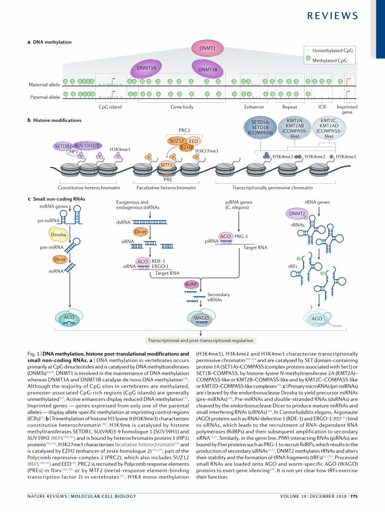
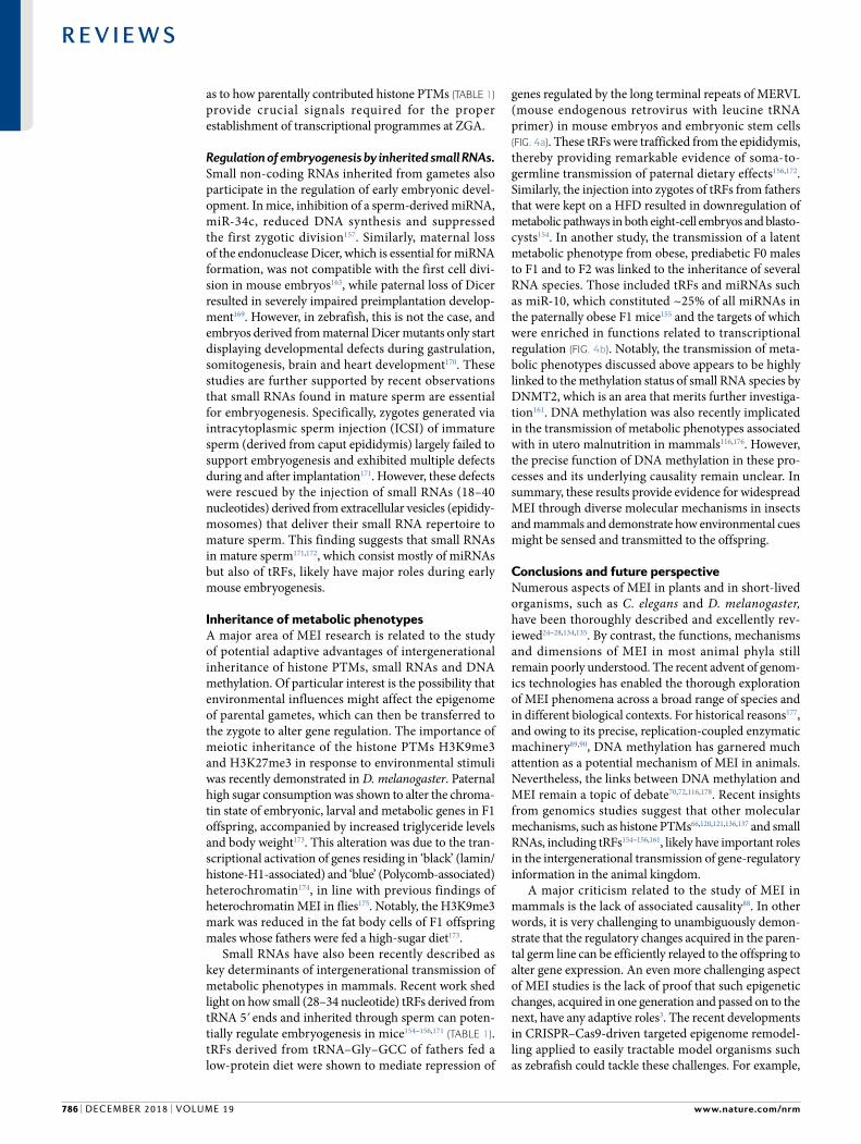
Fig. 1 | DNA methylation, histone post- translational modifications and small non- coding RNAs. a | DNA methylation in vertebrates occurs primarily at CpG dinucleotides and is catalysed by DNA methyltransferases (DNMTs)89,90. DNMT1 is involved in the maintenance of DNA methylation whereas DNMT3A and DNMT3B catalyse de novo DNA methylation186. Although the majority of CpG sites in vertebrates are methylated, promoter- associated CpG- rich regions (CpG islands) are generally unmethylated187. Active enhancers display reduced DNA methylation132. Imprinted genes — genes expressed from only one of the parental alleles — display allele- specific methylation at imprinting control regions (ICRs)58. b | Trimethylation of histone H3 lysine 9 (H3K9me3) characterizes constitutive heterochromatin188. H3K9me is catalysed by histone methyltransferases SETDB1, SU(VAR)3-9 homologue 1 (SUV39H1) and SUV39H2 (reFS189,190) and is bound by heterochromatin protein 1 (HP1) proteins191,192. H3K27me3 characterizes facultative heterochromatin193 and is catalysed by EZH2 (enhancer of zeste homologue 2)194,195, part of the Polycomb repressive complex 2 (PRC2), which also includes SUZ12 (reFS196,197) and EED198. PRC2 is recruited by Polycomb response elements (PREs) in flies199,200 or by MTF2 (metal- response element- binding transcription factor 2) in vertebrates201. H3K4 mono- methylation
(H3K4me1), H3K4me2 and H3K4me3 characterize transcriptionally permissive chromatin202–204 and are catalysed by SET domain- containing protein 1A (SET1A)–COMPASS (complex proteins associated with Set1) or SET1B–COMPASS, by histone- lysine N- methyltransferase 2A (KMT2A)–COMPASS- like or KMT2B–COMPASS- like and by KMT2C–COMPASS- like or KMT2D–COMPASS- like complexes205. c | Primary microRNAs (pri- miRNAs) are cleaved by the endoribonuclease Drosha to yield precursor miRNAs (pre- miRNAs)206. Pre- miRNAs and double- stranded RNAs (dsRNAs) are cleaved by the endoribonuclease Dicer to produce mature miRNAs and small interfering RNAs (siRNAs)206. In Caenorhabditis elegans, Argonaute (AGO) proteins such as RNAi- defective 1 (RDE-1) and ERGO-1 (reF.33) bind to siRNAs, which leads to the recruitment of RNA- dependent RNA polymerases (RdRPs) and their subsequent amplification to secondary siRNA34,35. Similarly , in the germ line, PIWI- interacting RNAs (piRNAs) are bound by Piwi proteins such as PRG-1 to recruit RdRPs, which results in the production of secondary siRNAs42,43. DNMT2 methylates tRNAs and alters their stability and the formation of tRNA fragments (tRFs)161,207. Processed small RNAs are loaded onto AGO and worm- specific AGO (WAGO) proteins to exert gene silencing208. It is not yet clear how tRFs exercise their function.
NATure revIeWs | MOLeCuLAR CeLL BIOLOGy
R e v i e w s
volume 19 | december 2018 | 775
yeast, plants and in the nematode Caenorhabditis elegans have been published previously22–29. Here, we discuss examples of MEI and epigenome reprogramming in vertebrates and elaborate on the mechanisms of MEI, namely through DNA methylation, histone modifications, tRNA fragments (tRFs) and other small RNAs. Finally, we discuss the implications of parental transmission of histone modifications and small RNAs for embryonic development in metazoans.
MEI in Caenorhabditis elegansOne of the best studied mechanisms of epigenetic inheritance in animals is RNAi, which is thoroughly described in the nematode C. elegans. Apart from RNAi responses that are heritable through many generations, C. elegans also displays robust transgenerational inheritance of both active and repressive histone PTMs.
RNAi pathways. In C. elegans, double stranded RNA (dsRNA) triggers RNAi, which promotes systemic mRNA degradation and is heritable through the germ line30. The RNAi response in C. elegans is dependent on dsRNA cleavage by the RNase III family nuclease Dicer31,32, which results in the production of small interfering RNAs (siRNAs) (Fig. 1c). siRNAs are subsequently bound by the Argonaute (AGO) proteins, such as RNAi defective 1 (RDE1)33. This results in the recruitment of RNA dependent RNA polymerase (RdRP) and the production of secondary siRNAs34,35. Secondary siRNAs are loaded onto worm specific AGO (WAGO) proteins36 that localize to the nucleus and initiate silencing33,37. The transgenerational effect of RNAi in C. elegans is dependent on the inheritance of small RNA molecules and their amplification, as demonstrated by virus derived small RNAs38. The heritable maintenance of silencing is dependent on nuclear RNAi pathways39. Additionally, very recent work has identified a highly conserved RNA helicase, ZNFX1, that together with WAGO proteins forms phase separated nuage granules in the cytoplasm of germ cells and that has a role in the propagation of heritable RNAi40,41. Endogenously derived PiWi- interacting rNAs (piRNAs), which suppress the expression and activity of transposons in the germ line, can also trigger heritable RNAi42,43. Together with PIWI like protein PRG1, piRNAs exert their function through the generation of secondary siRNAs42,43 (Fig. 1c). Such piRNA initiated silencing is heritable across generations and is dependent on nuclear RNAi pathways44–46. Chromatin remodelling activity and repressive chromatin pathways, including the histone H3 lysine 9 (H3K9) methyltransferase SET25, also participate in the maintenance of long term silencing44,46–48. Interestingly, transgenerational inheritance of RNAi can continue in the absence of H3K9 trimethylation49,50 and the H3K9 methyltransferase MET2 was recently shown to inhibit the biogenesis of heritable siRNAs, thereby constraining transgenerational transmission of RNAi50.
Histone PTM inheritance in Caenorhabditis elegans. TEI of both active and repressive histone PTMs occurs in the germ line of the nematode C. elegans. Mutations in proteins of the Trithorax methyltransferase complex (myeloid/lymphoid or mixed lineage leukaemia, in
mammals) WD repeat containing protein 5 (WDR5), ASH2 and SET2 decreased levels of H3K4 trimethylation (H3K4me3; a positive regulator of gene expression) and resulted in approximately 20% extension of the C. elegans lifespan, which was inherited across several generations51. Depletion of the H3K4me3 demethylase RBR2 abolished the transmission of extended lifespan in WDR5deficient worms, suggesting that TEI of prolonged lifespan due to deficiency in H3K4 trimethylation depends on histone demethylation. Similarly, knockouts of spr-5, which codes for an orthologue of the H3K4 dimethylation (H3K4me2) lysine specific histone demethylase 1A LSD1 (also known as KDM1A), result in a phenotype characterized by an increased incidence of sterility across generations (germline mortality)52. This phenotype is driven by the accumulation of H3K4me2, causing the misregulation of genes expressed during spermatogenesis, and is indicative of TEI of H3K4me2 patterns. Repressive histone PTMs such as H3K27me3 and H3K9me3 can also be inherited in C. elegans. X chromosome inactivation through Polycomb repressive complex 2 (PRC2)mediated H3K27 trimethylation can be intergenerationally transmitted from both oocytes and sperm to the embryos53. Also, a recent study demonstrated TEI of exposure to high temperature, which directly inhibited the H3K9 methyltransferase SET25; this resulted in derepression of SET25 target loci in the germ line and was inherited for multiple generations of worms raised at normal temperature54 (TAble 1). This effect persisted for up to 14 generations on an integrated transgene array, whereas for endogenous repetitive elements, considerable changes in expression levels were observed for up to 6 generations. How small RNAs cooperate with repressive histone PTMs in epigenetic inheritance is currently unclear44,46,48,50. Although the inactivation of RNAi components such as HRDE1 or NRDE2 did not affect the transmission of this H3K9me3mediated epigenetic memory54, very recent data argue that H3K9me3 might be required for siRNA dependent TEI that specifically targets newly evolved genes and is particularly sensitive to transgenes55.
MEI in mammalsAlthough MEI is well documented in plants (Supplementary Box 1) and in C. elegans, in mammals such phenomena remain more of an exception than the rule. This is partly because in mammals, the epigenome inherited from the gametes is heavily reprogrammed upon fertilization and during the formation of primordial germ cells (PGCs)56,57 (see below). However, although these epigenome remodelling processes reduce DNA methylation to its lowest levels during the mammalian life cycle, methylation is not erased and re established with the same efficiency at all sequences, thereby constituting a potential route for MEI. For clarity, we use the term ‘intergenerational’ epigenetic inheritance if the potentially heritable effect experienced by the pregnant female (F0) was observed in F1 and F2, and we use the term ‘transgenerational’ epigenetic inheritance if the effect persisted to F3 or beyond. Accordingly, we use ‘intergenerational’ if the effect experienced by the male (F0) or a non pregnant female (F0)
Facultative heterochromatinCondensed, transcriptionally silent chromatin that retains the ability to decondense and license transcription within temporal and spatial contexts.
PIWI- interacting RNAs(pirNAs). A class of endogenous small non- coding rNAs that interact with Piwi- domain-containing proteins and have a role in retrotransposon silencing in the germ line.
Primordial germ cells(PgCs). Primary germ cells that give rise to gametes.
www.nature.com/nrm
R e v i e w s
776 | december 2018 | volume 19
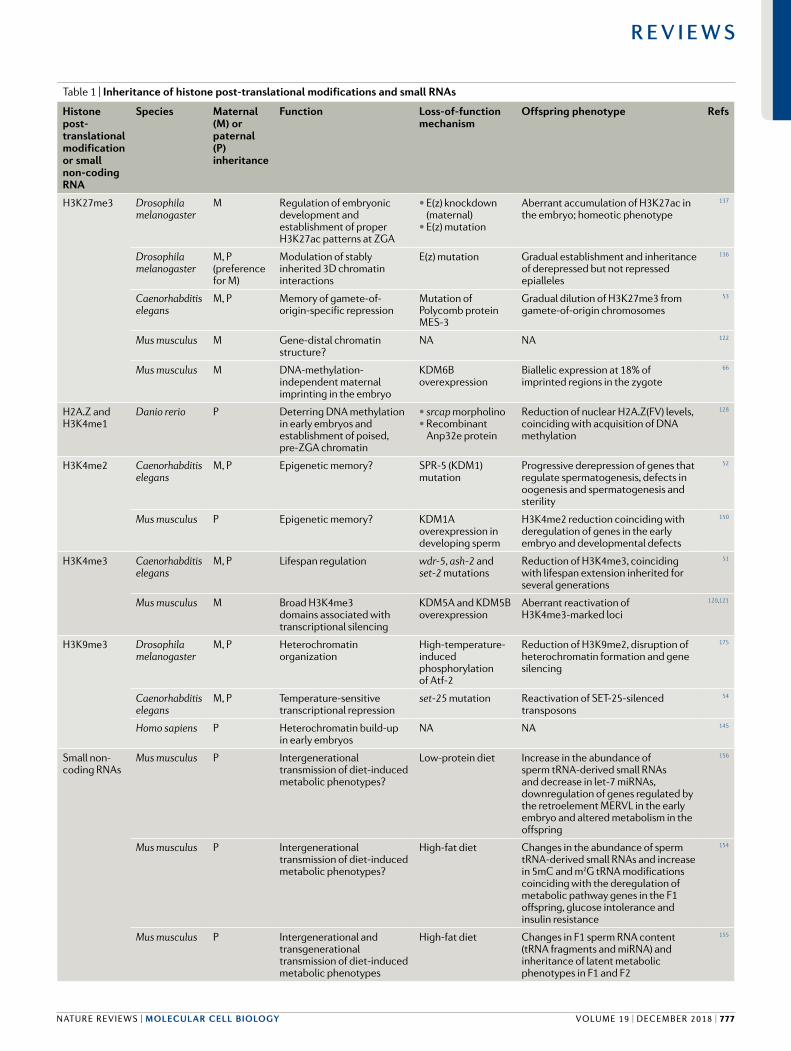
Table 1 | Inheritance of histone post- translational modifications and small RNAs
Histone post- translational modification or small non- coding RNA
Species Maternal (M) or paternal (P) inheritance
Function Loss- of-function mechanism
Offspring phenotype Refs
H3K27me3 Drosophila melanogaster
M Regulation of embryonic development and establishment of proper H3K27ac patterns at ZGA
• E(z) knockdown (maternal)
• E(z) mutation
Aberrant accumulation of H3K27ac in the embryo; homeotic phenotype
137
Drosophila melanogaster
M, P (preference for M)
Modulation of stably inherited 3D chromatin interactions
E(z) mutation Gradual establishment and inheritance of derepressed but not repressed epialleles
136
Caenorhabditis elegans
M, P Memory of gamete-of- origin-specific repression
Mutation of Polycomb protein MES-3
Gradual dilution of H3K27me3 from gamete- of-origin chromosomes
53
Mus musculus M Gene- distal chromatin structure?
NA NA 122
Mus musculus M DNA- methylation- independent maternal imprinting in the embryo
KDM6B overexpression
Biallelic expression at 18% of imprinted regions in the zygote
66
H2A.Z and H3K4me1
Danio rerio P Deterring DNA methylation in early embryos and establishment of poised, pre- ZGA chromatin
• srcap morpholino• Recombinant
Anp32e protein
Reduction of nuclear H2A.Z(FV) levels, coinciding with acquisition of DNA methylation
128
H3K4me2 Caenorhabditis elegans
M, P Epigenetic memory? SPR-5 (KDM1) mutation
Progressive derepression of genes that regulate spermatogenesis, defects in oogenesis and spermatogenesis and sterility
52
Mus musculus P Epigenetic memory? KDM1A overexpression in developing sperm
H3K4me2 reduction coinciding with deregulation of genes in the early embryo and developmental defects
150
H3K4me3 Caenorhabditis elegans
M, P Lifespan regulation wdr-5, ash-2 and set-2 mutations
Reduction of H3K4me3, coinciding with lifespan extension inherited for several generations
51
Mus musculus M Broad H3K4me3 domains associated with transcriptional silencing
KDM5A and KDM5B overexpression
Aberrant reactivation of H3K4me3-marked loci
120,121
H3K9me3 Drosophila melanogaster
M, P Heterochromatin organization
High-temperature-induced phosphorylation of Atf-2
Reduction of H3K9me2, disruption of heterochromatin formation and gene silencing
175
Caenorhabditis elegans
M, P Temperature- sensitive transcriptional repression
set-25 mutation Reactivation of SET-25-silenced transposons
54
Homo sapiens P Heterochromatin build- up in early embryos
NA NA 145
Small non- coding RNAs
Mus musculus P Intergenerational transmission of diet- induced metabolic phenotypes?
Low- protein diet Increase in the abundance of sperm tRNA- derived small RNAs and decrease in let-7 miRNAs, downregulation of genes regulated by the retroelement MERVL in the early embryo and altered metabolism in the offspring
156
Mus musculus P Intergenerational transmission of diet- induced metabolic phenotypes?
High- fat diet Changes in the abundance of sperm tRNA- derived small RNAs and increase in 5mC and m2G tRNA modifications coinciding with the deregulation of metabolic pathway genes in the F1 offspring, glucose intolerance and insulin resistance
154
Mus musculus P Intergenerational and transgenerational transmission of diet- induced metabolic phenotypes
High- fat diet Changes in F1 sperm RNA content (tRNA fragments and miRNA) and inheritance of latent metabolic phenotypes in F1 and F2
155
NATure revIeWs | MOLeCuLAR CeLL BIOLOGy
R e v i e w s
volume 19 | december 2018 | 777
was observed in F1 and ‘transgenerational’ if the effect persisted to F2 and beyond (box 1).
Genomic imprinting. In mammals, a small number of genes (~125 in mice and ~100 in humans; Geneimprint) are expressed in a parent oforigin specific manner, a phenomenon termed genomic imprinting58. This differential allelic expression is the result of epigenetic silencing of the inactive allele, a process that involves DNA methylation59–62. Such imprinted genes are regulated through imprinting control regions (Fig. 1a), which are resistant to post fertilization DNA methylation reprogramming, thereby forming a platform for intergenerational epigenetic inheritance. Many imprinted genes are expressed during prenatal development and are required for proper placental function63,64. Errors occurring during the establishment of imprinting in the parental germ line were shown to account for a number of behavioural and neurodevelopmental disorders, such as Prader–Willi syndrome and Angelman syndrome65. Interestingly, recent work in mouse embryos provided evidence for imprinting that occurs independently of DNA methylation and is associated with maternal inheritance of the repressive modification H3K27me3 (see below)66. The number of genes (n = 76) regulated by such non canonical imprinting mechanisms appears to be similar to the number of genes regulated by canonical, DNA methylationdependent imprinting.
Epigenetic reprogramming of intracisternal A par-ticles. Intracisternal A particles (IAPs) are retrotransposons present in many copies (~1,000) in the mouse genome67. To suppress their capacity for retrotransposition in the germ line, IAPs are predominantly methylated and silenced in germline cells. Similarly to genomic imprints, IAPs can largely resist embryonic DNA methylation reprogramming, which makes them potentially attractive targets for MEI studies67. One of the best examples of MEI in mammals is the agouti viable yellow (Avy) locus in mice, which harbours an insertion of the IAP retrotransposon upstream of the agouti gene that determines coat colour68. Avy expression is highly variable among littermates and is dependent on the
DNA methylation state of the IAP inserted in the agouti locus68,69. While the phenotype of the silent, methylated allele (pseudoagouti) is wild type dark brown coat colour, the hypomethylated, active allele gives rise to a yellow coat. The spectrum of agouti phenotypes in the offspring is dependent upon the phenotype of the dam. For example, a dam with the yellow coat phenotype is more likely to have yellow coat offspring68. Interestingly, dietary supplementation of methyl donors during mid gestation results in the shift of agouti phenotypes towards pseudoagouti in both F1 and F2, suggestive of increased IAP methylation in the germ line and the intergenerational retention of this altered epigenetic state16. Nevertheless, bisulfite sequencing data suggest that both the maternally contributed and paternally contributed Avy associated IAPs are reprogrammed during early embryogenesis, albeit with different dynamics70. This suggestion implies that DNA methylation might not be the primary inherited mark controlling the spectrum of agouti phenotypes in F1. It is currently not clear whether histone modifications or RNA based mechanisms have a role in the inheritance of the Avy epialleles; however, recent studies have demonstrated robust intergenerational inheritance of H3K4me3 and H3K27me3 histone modifications through the maternal germ line71. Methyl donor supplementation during mid gestation affects the percentage of agouti phenotypes only when the Avy allele is contributed through the paternal germ line. This finding suggests that the paternal allele might undergo more extensive epigenetic reprogramming and is thus affected by environmental cues during the period of PGC methylome reprogramming (embryonic day 8.5 (E8.5)–E.15.5)16. Therefore, it is possible that the inheritance of the maternal Avy locus is more tightly regulated, perhaps through the coordinated action of diverse gene regulatory mechanisms, including histone modifications, and is not limited only to DNA methylation.
Contribution of MEI to human diseases. Considerable efforts have been made in identifying the contribution of epigenetic modifications to the heritability of complex diseases. In particular, the possibility that epimutations confer predisposition to cancer has garnered much
Histone post- translational modification or small non- coding RNA
Species Maternal (M) or paternal (P) inheritance
Function Loss- of-function mechanism
Offspring phenotype Refs
Small noncoding RNAs (cont.)
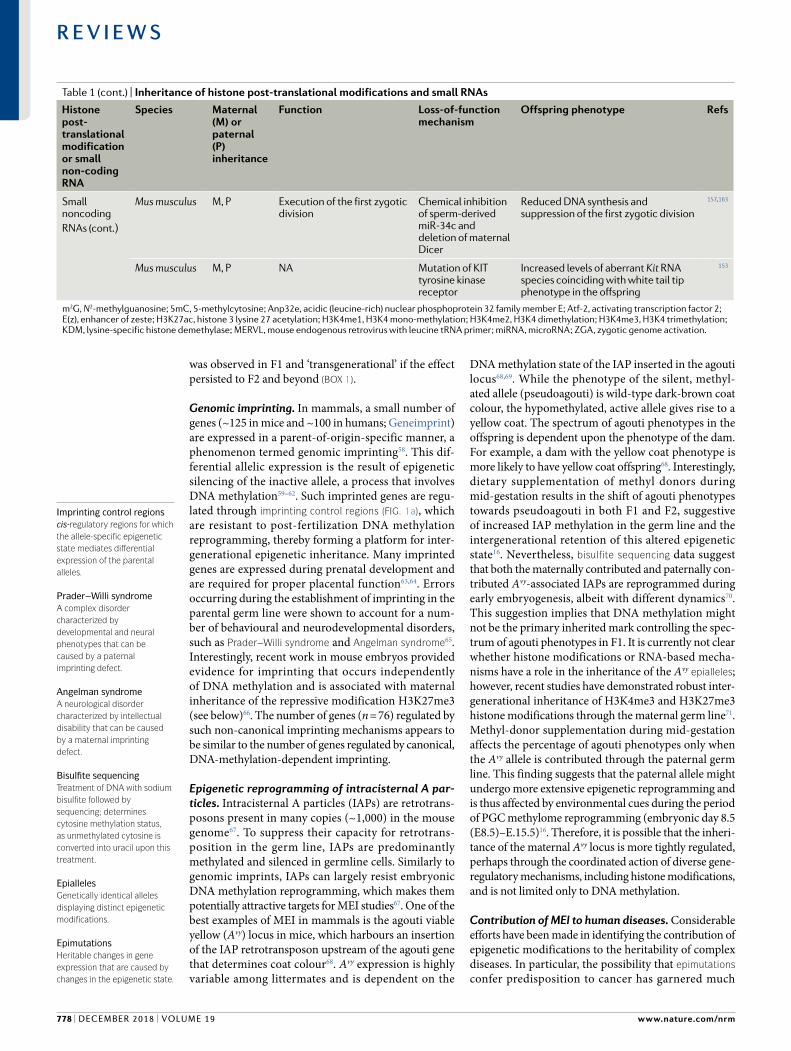
Mus musculus M, P Execution of the first zygotic division
Chemical inhibition of sperm- derived miR-34c and deletion of maternal Dicer
Reduced DNA synthesis and suppression of the first zygotic division
157,163
Mus musculus M, P NA Mutation of KIT tyrosine kinase receptor
Increased levels of aberrant Kit RNA species coinciding with white tail tip phenotype in the offspring
153
m2G, N2-methylguanosine; 5mC, 5-methylcytosine; Anp32e, acidic (leucine- rich) nuclear phosphoprotein 32 family member E; Atf-2, activating transcription factor 2; E(z), enhancer of zeste; H3K27ac, histone 3 lysine 27 acetylation; H3K4me1, H3K4 mono- methylation; H3K4me2, H3K4 dimethylation; H3K4me3, H3K4 trimethylation; KDM, lysine- specific histone demethylase; MERVL , mouse endogenous retrovirus with leucine tRNA primer ; miRNA , microRNA ; ZGA , zygotic genome activation.
Table 1 (cont.) | Inheritance of histone post- translational modifications and small RNAs
Imprinting control regionscis- regulatory regions for which the allele- specific epigenetic state mediates differential expression of the parental alleles.
Prader–Willi syndromeA complex disorder characterized by developmental and neural phenotypes that can be caused by a paternal imprinting defect.
Angelman syndromeA neurological disorder characterized by intellectual disability that can be caused by a maternal imprinting defect.
Bisulfite sequencingTreatment of DNA with sodium bisulfite followed by sequencing; determines cytosine methylation status, as unmethylated cytosine is converted into uracil upon this treatment.
Epiallelesgenetically identical alleles displaying distinct epigenetic modifications.
EpimutationsHeritable changes in gene expression that are caused by changes in the epigenetic state.
www.nature.com/nrm
R e v i e w s
778 | december 2018 | volume 19
attention over the past few decades72. One of the best examples of such an epimutation is the silencing of the mutL homologue 1 (MLH1) gene, which encodes a DNA mismatch repair factor, through hypermethylation of its promoter. The MLH1 epimutation is widespread in somatic tissues derived from all three major embryonic lineages (ectoderm, mesoderm and endoderm), suggesting that it has an embryonic or germline origin73,74. Such MLH1 epimutations contribute to Lynch syndrome, which is characterized by early onset of multiple cancers, including colorectal cancer and endometrial cancer, and in general cancers displaying microsatellite instability75. In the majority of cases, MLH1 epimutations arise de novo in affected individuals and are reprogrammed to the unmethylated state in their offspring72. Nevertheless, to date at least one case was
reported in which the methylated state was inherited from the affected mother to one of her three sons; however, it was then erased in his germ line74. This observation demonstrates the possibility that epimutations might be passed on from parents to their offspring, albeit infrequently, likely owing to the DNA methylation reprogramming that occurs post fertilization and in PGCs. Although additional data support the (rare) occurrence of intergenerational epigenetic inheritance at the MLH1 locus76,77, evidence for this type of heritability in humans remains very limited. Finally, it should also be stressed that, on the basis of currently available data, it is very difficult to unequivocally rule out the involvement of other cis- acting or even trans- acting factors in the intergenerational heritability of disease causing epimutations in humans.
MEI of environmental cues in humans. Understanding how the environment affects the human epigenome and how potentially acquired traits can be propagated through generations remains an area of intense investigation78–81. One of the most noted studies on this topic is the Dutch Hunger Winter study79,82,83. DNA methylation profiling of blood samples extracted from individuals who were conceived during the Dutch Hunger Winter (1944–1945) revealed small DNA methylation differences in the differentially methylated region (DMR) associated with the insulin like growth factor 2 (IGF2) gene82. Five CpG dinucleotides located within the IGF2 DMR were assessed by quantitative mass spectrometry to reveal on average 5.2% lower DNA methylation in the affected siblings. These results were successfully validated (5.6% lower in the affected individuals) by locus specific bisulfite sequencing approaches. Notably, only the cohort exposed to maternal malnutrition during early gestation but not during later stages of pregnancy displayed these changes in DNA methylation at the IGF2 locus82. Although this understandably invites speculation about epigenetic mechanisms of adaptation to the environment, one has to keep in mind that MEI studies in humans frequently suffer from serious methodological flaws. A major limitation, even in the presence of a testable hypothesis, is the fact that such analyses are often performed on a complex tissue such as blood, which consists of different cell types. Therefore, it is difficult to exclude the possibility that the observed differences in DNA methylation merely reflect the differences in cell type composition between samples. Furthermore, even if those differences were truly caused by the proposed environmental stress, it remains challenging to prove that such modest changes could result in an adaptive phenotype. Finally, a major confounding factor that is often disregarded in MEI studies is the difference in DNA sequence composition between individuals, which is well known to be a major contributor to the inheritance of epigenetic states84–87.
Whereas the Dutch Hunger Winter study aimed to assess the potential impacts of early intrauterine exposure to famine on DNA methylation during adult life, a more recent study tried to address whether intergenerational inheritance of stress effects, such as those caused
Microsatellite instabilitygenetic instability in short tandem repeats due to impaired DNA mismatch repair.
Differentially methylated region(DMr). A genomic region displaying statistically significant change in DNA methylation between at least two samples.
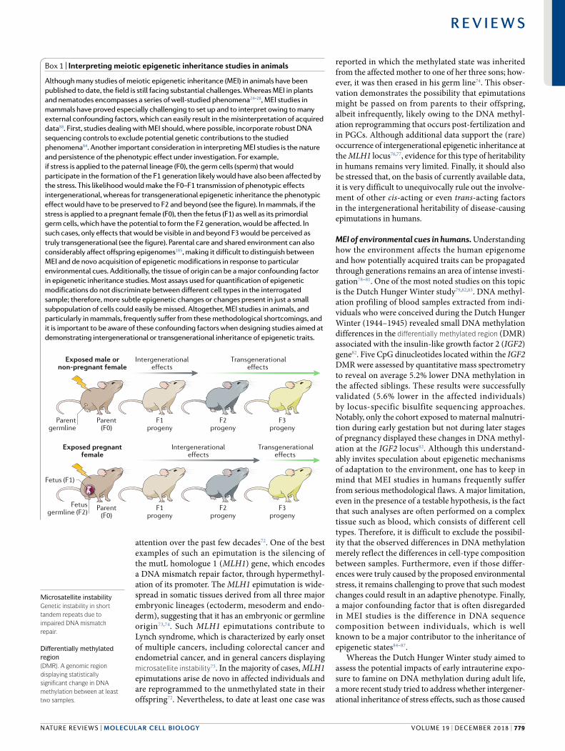
Box 1 | Interpreting meiotic epigenetic inheritance studies in animals
Although many studies of meiotic epigenetic inheritance (meI) in animals have been published to date, the field is still facing substantial challenges. Whereas meI in plants and nematodes encompasses a series of well- studied phenomena24–28, meI studies in mammals have proved especially challenging to set up and to interpret owing to many external confounding factors, which can easily result in the misinterpretation of acquired data88. First, studies dealing with meI should, where possible, incorporate robust dNA sequencing controls to exclude potential genetic contributions to the studied phenomena84. Another important consideration in interpreting meI studies is the nature and persistence of the phenotypic effect under investigation. For example, if stress is applied to the paternal lineage (F0), the germ cells (sperm) that would participate in the formation of the F1 generation likely would have also been affected by the stress. This likelihood would make the F0–F1 transmission of phenotypic effects intergenerational, whereas for transgenerational epigenetic inheritance the phenotypic effect would have to be preserved to F2 and beyond (see the figure). In mammals, if the stress is applied to a pregnant female (F0), then the fetus (F1) as well as its primordial germ cells, which have the potential to form the F2 generation, would be affected. In such cases, only effects that would be visible in and beyond F3 would be perceived as truly transgenerational (see the figure). Parental care and shared environment can also considerably affect offspring epigenomes185, making it difficult to distinguish between MEI and de novo acquisition of epigenetic modifications in response to particular environmental cues. Additionally, the tissue of origin can be a major confounding factor in epigenetic inheritance studies. most assays used for quantification of epigenetic modifications do not discriminate between different cell types in the interrogated sample; therefore, more subtle epigenetic changes or changes present in just a small subpopulation of cells could easily be missed. Altogether, meI studies in animals, and particularly in mammals, frequently suffer from these methodological shortcomings, and it is important to be aware of these confounding factors when designing studies aimed at demonstrating intergenerational or transgenerational inheritance of epigenetic traits.
Exposed pregnantfemale
Parent(F0)
F1progeny
F2progeny
F3progeny
Fetus (F1)
Fetusgermline (F2)
Transgenerationaleffects
Intergenerationaleffects
Exposed male ornon-pregnant female
Parent(F0)
F1progeny
F2progeny
F3progeny
Transgenerationaleffects
Intergenerationaleffects
Parentgermline
NATure revIeWs | MOLeCuLAR CeLL BIOLOGy
R e v i e w s
volume 19 | december 2018 | 779
to survivors of the Holocaust, can be demonstrated in humans81. The offspring of Holocaust survivors displayed diminished DNA methylation of the FKBP5 gene, which was previously linked to post traumatic stress disorder, whereas this gene was marginally more methylated in the Holocaust survivors themselves than in the non affected controls. The concept of non genetic trauma inheritance has previously been postulated in mice20; however, drawing any definite conclusions regarding the potential intergenerational epigenetic inheritance of trauma in humans would be very challenging (box 1). Apart from the methodological issues discussed above, many of which apply also to this study, it is very difficult to distinguish between the effects of parental upbringing on a growing child and epigenetic inheritance per se. Owing to the genetically hetero geneous nature of the human population and long human lifespan, longitudinal studies that would span multiple generations and that would be able to properly distinguish genotype from phenotype remain very difficult to set up88. In the absence of those, the published research on the topic of MEI in humans requires highly circumspect treatment.
MEI through DNA methylationDNA methylation has often been proposed as a MEI carrier in animals owing to its relative stability and well defined mechanisms of de novo deposition and maintenance89,90 (Fig. 1a). Although DNA methylation is present in fungi, plant and animal kingdoms, its genomic abundance and distribution vary considerably between species91,92. For example, species with easily demonstrable MEI, such as D. melanogaster and C. elegans, are virtually devoid of DNA methylation93,94. Thus, it is unclear to what extent DNA methylation contributes to MEI in animals and in what biological contexts. However, recent insights from genome wide methylome studies suggest that a considerable fraction of mammalian genomes might potentially bypass the removal of DNA methylation that occurs during preimplantation and PGC reprogramming (TAble 2).
Reprogramming of the preimplantation methylome. DNA methylation is mostly static in adult somatic tissues; however, global DNA methylation patterns are reprogrammed twice during the mammalian life cycle56. The first reprogramming event takes place
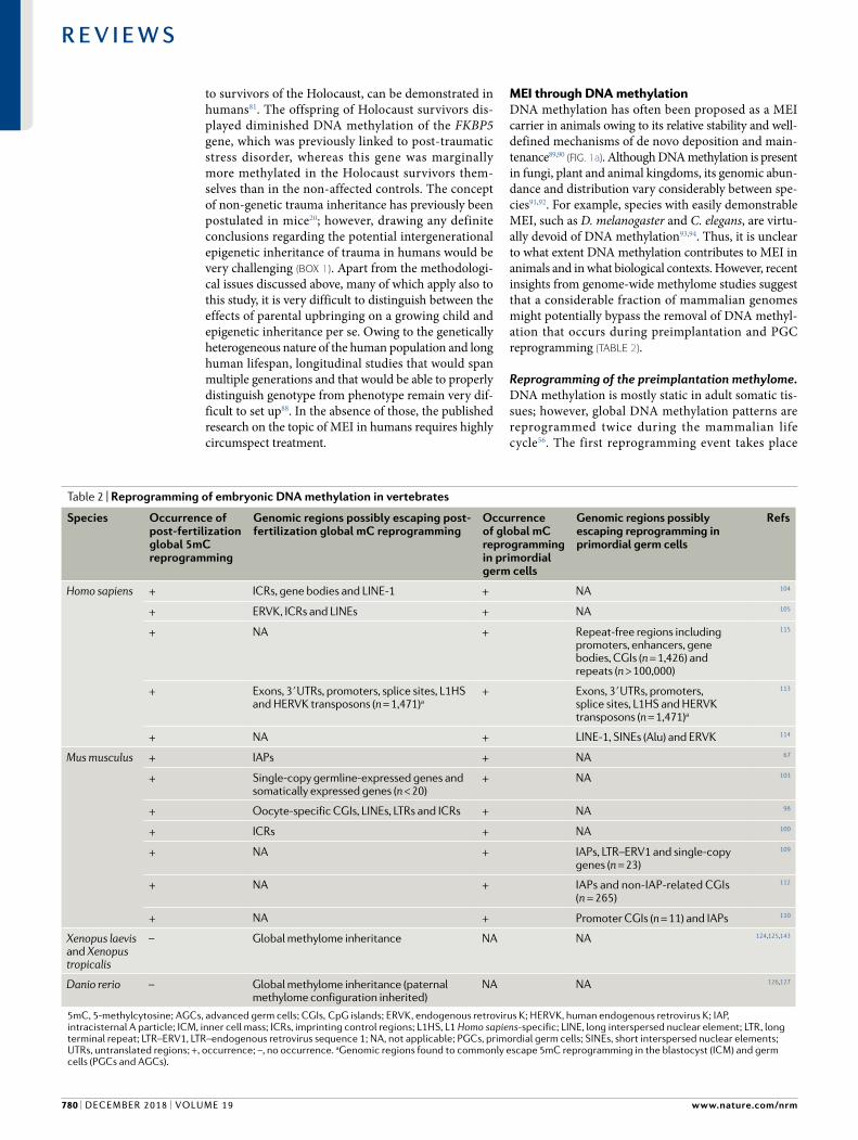
Table 2 | Reprogramming of embryonic DNA methylation in vertebrates
Species Occurrence of post- fertilization global 5mC reprogramming
Genomic regions possibly escaping post- fertilization global mC reprogramming
Occurrence of global mC reprogramming in primordial germ cells
Genomic regions possibly escaping reprogramming in primordial germ cells
Refs
Homo sapiens + ICRs, gene bodies and LINE-1 + NA 104
+ ERVK , ICRs and LINEs + NA 105
+ NA + Repeat- free regions including promoters, enhancers, gene bodies, CGIs (n = 1,426) and repeats (n > 100,000)
115
+ Exons, 3ʹ UTRs, promoters, splice sites, L1HS and HERVK transposons (n = 1,471)a
+ Exons, 3ʹ UTRs, promoters, splice sites, L1HS and HERVK transposons (n = 1,471)a
113
+ NA + LINE-1, SINEs (Alu) and ERVK 114
Mus musculus + IAPs + NA 67
+ Single- copy germline- expressed genes and somatically expressed genes (n < 20)
+ NA 103
+ Oocyte- specific CGIs, LINEs, LTRs and ICRs + NA 98
+ ICRs + NA 100
+ NA + IAPs, LTR–ERV1 and single- copy genes (n = 23)
109
+ NA + IAPs and non-IAP-related CGIs (n = 265)
112
+ NA + Promoter CGIs (n = 11) and IAPs 110
Xenopus laevis and Xenopus tropicalis
– Global methylome inheritance NA NA 124,125,143
Danio rerio – Global methylome inheritance (paternal methylome configuration inherited)
NA NA 126,127
5mC, 5-methylcytosine; AGCs, advanced germ cells; CGIs, CpG islands; ERVK , endogenous retrovirus K; HERVK, human endogenous retrovirus K; IAP, intracisternal A particle; ICM, inner cell mass; ICRs, imprinting control regions; L1HS, L1 Homo sapiens- specific; LINE, long interspersed nuclear element; LTR , long terminal repeat; LTR–ERV1, LTR–endogenous retrovirus sequence 1; NA, not applicable; PGCs, primordial germ cells; SINEs, short interspersed nuclear elements; UTRs, untranslated regions; +, occurrence; –, no occurrence. aGenomic regions found to commonly escape 5mC reprogramming in the blastocyst (ICM) and germ cells (PGCs and AGCs).
www.nature.com/nrm
R e v i e w s
780 | december 2018 | volume 19
post fertilization and involves widespread DNA demethylation of the paternal pronucleus, followed by a progressive drop in global DNA methylation in the zygote, which reaches its lowest point in the blastocyst95–98. The precise mechanisms of embryonic DNA methylome remodelling remain a topic of debate99–101; however, it is likely that both active (enzymatic) and passive (replication coupled dilution) demethylation mechanisms have a role in this process. Active DNA demethylation is mediated by ten- eleven translocation (TeT) enzymes, which oxidize 5methylcytosine, whereas passive reduction in DNA methylation levels is dependent on the inhibition of DNA (cytosine5)methyltransferase 1 (DNMT1) activity in the nucleus56. Whereas rapid post fertilization demethylation of the paternal pronucleus was observed in mouse, rat, pig, bovine and human embryos, sheep and rabbit preimplantation development involves less demethylation of the paternal genome, indicative of species specific differences in mammalian embryonic epigenome remodelling102. The embryonic genome is remethylated following implantation, coinciding with the loss of cellular pluripotency97,98,103. Nevertheless, not all genomic loci are reprogrammed with the same efficiency. Apart from imprinted loci and IAPs, single baseresolution studies in mouse embryos revealed the persistence of DNA methylation on oocyte hypermethylated DMRs and a number of long interspersed nuclear element transposons during the initial wave of global hypomethylation98. Similarly, a number of DMRs identified as hypermethylated in the oocytes were found to be intermediately methylated during human preimplantation development, suggestive of maternal DNA methylome inheritance104. Another important feature of human preimplantation development is the persistence of DNA methylation within gene bodies104,105. As gene bodies are often enriched in enhancer elements106,107, such persistence of DNA methylation might allow for the intergenerational inheritance of cis- regulatory states. In summary, these observations provide evidence for locus specific retention of DNA methylation (TAble 2) during a regulatory phase in which DNA methylation maintenance is largely inactive. This evidence suggests that a previously underappreciated fraction of mammalian genomes, including promoters, gene bodies and repeats, confer resistance to early DNA methylation reprogramming and thus constitute a potential platform for MEI.
DNA methylation reprogramming in PGCs. The second wave of mammalian DNA methylation reprogramming takes place in PGCs and also involves a combination of active and passive mechanisms108–111. The hallmark of this process is erasure of imprinting followed by gender specific re establishment of imprinting in the gonads. As in the post fertilization demethylation process, not all genomic sequences are reprogrammed with the same efficiency109,112. Studies in mice revealed that the feature most resilient to reprogramming are IAPs, which remain predominantly hypermethylated throughout PGC demethylation109,112 (TAble 2). Human PGCs exhibit similar genomic DNA methylation erasure
patterns to those observed in mice113–115. However, more than 116,000 genomic regions were identified that remained significantly hypermethylated following methylome reprogramming in human PGCs115. Of those, more than 7,000 regions were repeat poor and mostly coincided with CpG rich regions known as Cpg islands, promoters, enhancers and gene bodies. Many of these regions were associated with obesity, multiple sclerosis and schizophrenia. Comparisons with mouse bisulfite sequencing data revealed 794 such escapees that were shared between mice and humans (TAble 2). Of note, many of these loci were linked to neural and metabolic functions and could thus be of potential interest for further exploration within the context of disease related risk inheritance.
In mice, metabolic deficiencies coinciding with perturbed DNA methylation in the germ line of adults can be inherited intergenerationally116. Undernourishment of pregnant F0 dames during a critical phase of embryonic PGC reprogramming resulted in metabolic phenotypes and modest DNA hypomethylation at discrete loci in adult F1 sperm (F1 DMRs). Whereas these altered methylation patterns were not detectable in brain and liver tissues obtained from E16.5 F2 embryos, genes found in the vicinity of a small number of F1 DMRs had significantly altered transcriptional profiles in those tissues in F2. Indeed, several of these genes were attributed with metabolic functions, and pancreatic islets isolated from 4month old F2 mice displayed impaired insulin secretion. The altered transcriptional state of those genes in E16.5 embryos, in the absence of corresponding DNA methylation changes, suggests that F1 sperm DMRs might exercise an effect during early F2 embryogenesis. This effect would not be detectable in terms of altered DNA methylation; however, it would be conveyed into an altered transcriptional output detectable at E16.5. Alternatively, DNA methylation might not be the primary regulatory mechanism responsible for the formation of F1 sperm DMRs as well as for the observed transcriptional changes in F2. In mammalian sperm, the majority of nucleosomes are replaced with protamines. However, certain loci of developmental importance, such as Hox genes, promoters of developmental genes, imprinted genes and microRNA (miRNA) clusters, can retain nucleosomes and associated histone PTMs, such as H3K27me3, H3K4me2 and H3K4me3 (reFS117,118). Interestingly, 21% of the identified F1 DMRs (n = 23) were found within such nucleosomal regions116. This finding raises the possibility that the observed differences in F1 sperm DNA methylation might merely be secondary effects of altered chromatin structure, potentially caused by defects in regulatory mechanisms other than DNA methylation, such as nucleosome positioning or histone PTMs, both of which have potential for intergenerational heritability119–122. In light of these findings, it is worthwhile mentioning that in many cases epigenetic inheritance might be limited only to certain genomic contexts. For example, targeting trimethylation of both H3K27 and H3K9 requires input from specific DNA sequences85–87. Thus, it is plausible that sequences that escape various rounds of epigenetic reprogramming might be genetically defined.
Ten- eleven translocation (TET) enzymesenzymes required for active DNA demethylation, which catalyse a series of iterative oxidations of 5-methylcytosine to 5-hydroxymethylcytosine and further to 5-formylcytosine and 5-carboxylcytosine.
CpG islandsgenomic regions of high gC content and high frequency of Cpg sites relative to the genome average; often associated with gene promoters and maintained in an unmethylated state.
NATure revIeWs | MOLeCuLAR CeLL BIOLOGy
R e v i e w s
volume 19 | december 2018 | 781
Methylome reprogramming in anamniotes. Unlike mammals, anamniote vertebrates, such as fish and frogs, do not undergo global DNA methylation remodelling during embryogenesis123–127. Instead, zebrafish remodel their methylomes before zygotic genome activation (ZGA) in a fascinating fashion: the maternal methylome is reconfigured to match the paternal methylome pattern, thus making the early embryonic methylome adopt a paternal like configuration at ZGA126,127. This harmonization of paternally and maternally inherited epigenetic states before ZGA is achieved through the usage of ‘placeholder’ nucleosomes that repel DNA methyl ation but, on the other hand, are incompatible with transcriptional activation128 (see below). Other notable differences in early embryonic reprogramming also exist between zebrafish and mammals, including the absence of early TET expression and of active demethylation in zebrafish127,129,130. The lack of global embryonic demethyl ation and remethylation makes zebrafish a potentially useful model organism for MEI studies in vertebrates131. However, whether such principles of embryonic reprogramming are common to teleosts and other anamniotes is currently unclear. Similarily, it is currently unknown whether the erasure and re establishment of DNA methyl ation observed in mammalian PGCs is conserved more broadly.
MEI through histone PTMsDespite the increasingly precise classification of regions that might escape DNA methylome reprogramming in mammals (TAble 2), direct evidence for the role of DNA methylation in meiotic inheritance in animals is largely lacking70,116. This is perhaps not surprising considering that in many cases DNA methylation merely anti correlates with the activity of gene regulatory regions, suggestive of its role as a secondary, reinforcing regulatory mark132,133. Instead, an increasing number of recent studies have demonstrated intergenerational epigenetic inheritance and TEI of histone PTMs (Fig. 1b) in diverse animal species. We have already discussed the inheritance of histone PTMs in C. elegans above.
Histone PTM inheritance in Drosophila melanogaster. D. melanogaster has a long history of exploration of MEI phenomena134,135. The role of histone PTMs in TEI in this organism was recently demonstrated by the formation of long range chromatin interactions accompanied by PRC2mediated H3K27me3 deposition, which deli neated the inheritance of repressed or derepressed epialleles through multiple generations136. Interestingly, the establishment of these epigenetic states was shown to be sensitive to environmental stimuli such as temperature, thereby demonstrating the interplay between the environment and the epigenome. In line with these observations of stable Polycomb TEI, the maternal inheritance of H3K27me3 and enhancer of zeste (E(z)) protein in D. melanogaster was recently shown to have instructive roles for gene expression at ZGA137 (see below). Taken together, these examples demonstrate the requirement of meiotic histone PTM inheritance for responses to environmental stimuli and the supervision of early embryogenesis in insects.
Histone PTM inheritance in anamniotes. The degree of nucleosome retention in vertebrate sperm varies greatly. Whereas zebrafish retain their entire nucleosomal content138, in Xenopus laevis, only ~10% of the nucleosomes are retained in the mature sperm139. This level is similar to that in humans, whereas in mice the degree of nucleosome retention is close to 1%118. The sperm chromatin of both zebrafish and mammals is characterized by the presence of active (H3K4me2 and H3K4me3) and repressive (H3K27me3) histone PTMs, thereby allowing for the possibility of intergenerational transmission of these marks117,118,138. In X. laevis, sperm maturation entails a global loss of H3K4me2 and H3K4me3 and the retention of H3K27me3, and this process appears to be important for the execution of correct gene expression patterns during embryogenesis139. In zebrafish, the presence of both H3K27me3 and H3K4me3 at a number of developmental genes pre marked in sperm by the same histone PTMs was observed before the ZGA140. Those observations notwithstanding, the major chromatin signatures of embryonic development are established at or after the ZGA141,142. Moreover, the deposition of histone marks at the ZGA seems to be largely maternally defined, as recently described in Xenopus tropicalis143.
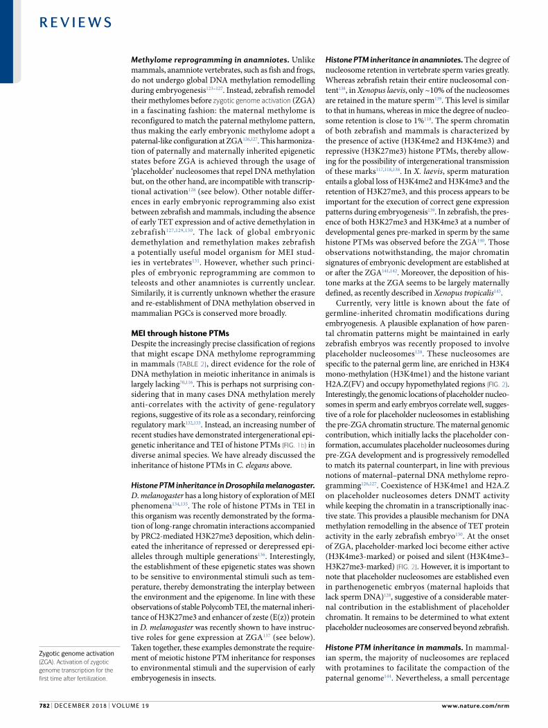
Currently, very little is known about the fate of germline inherited chromatin modifications during embryogenesis. A plausible explanation of how parental chromatin patterns might be maintained in early zebrafish embryos was recently proposed to involve placeholder nucleosomes128. These nucleosomes are specific to the paternal germ line, are enriched in H3K4 mono methylation (H3K4me1) and the histone variant H2A.Z(FV) and occupy hypomethylated regions (Fig. 2). Interestingly, the genomic locations of placeholder nucleosomes in sperm and early embryos correlate well, suggestive of a role for placeholder nucleosomes in establishing the pre ZGA chromatin structure. The maternal genomic contribution, which initially lacks the placeholder conformation, accumulates placeholder nucleosomes during pre ZGA development and is progressively remodelled to match its paternal counterpart, in line with previous notions of maternal–paternal DNA methylome reprogramming126,127. Coexistence of H3K4me1 and H2A.Z on placeholder nucleosomes deters DNMT activity while keeping the chromatin in a transcriptionally inactive state. This provides a plausible mechanism for DNA methylation remodelling in the absence of TET protein activity in the early zebrafish embryo130. At the onset of ZGA, placeholder marked loci become either active (H3K4me3marked) or poised and silent (H3K4me3–H3K27me3marked) (Fig. 2). However, it is important to note that placeholder nucleosomes are established even in parthenogenetic embryos (maternal haploids that lack sperm DNA)128, suggestive of a considerable maternal contribution in the establishment of placeholder chromatin. It remains to be determined to what extent placeholder nucleosomes are conserved beyond zebrafish.
Histone PTM inheritance in mammals. In mammalian sperm, the majority of nucleosomes are replaced with protamines to facilitate the compaction of the paternal genome144. Nevertheless, a small percentage
Zygotic genome activation(ZgA). Activation of zygotic genome transcription for the first time after fertilization.
www.nature.com/nrm
R e v i e w s
782 | december 2018 | volume 19
of nucleosomes and their associated histone PTMs are retained, thereby forming a potential platform for the intergenerational transmission of regulatory states117,118. Both active (H3K4me2 and H3K4me3) and repressive (H3K27me3, H3K9me3 and H4K20me3) histone PTMs have been detected in mammalian sperm; however, their distribution and dynamics vary considerably between species. For example, in humans, H3K9me3marked and H4K20me3marked nucleosomes are transmitted through the sperm into the zygote, where they participate in the build up of constitutive heterochromatin145. In mice, where only ~1% of the nucleosomes are retained in sperm118, the build up of paternal heterochromatin in the zygote is initiated through maternally contributed PRC1 (reF.146). Until recently, genome wide studies of MEI in mammals were very challenging to orchestrate owing to the limited amounts of embryonic material that can be obtained for investigation. This limitation is now being overcome by the advent of novel genomics technologies, which allow for low input transcriptome and epigenome profiling, often at single cell resolution147–149. Recent genome wide studies revealed the existence of robust inheritance of H3K4me3 and H3K27me3 patterns through oocytes in mice and their role in the regulation of embryonic development66,71,120–122. In line with these
findings, overexpression of human LSD1 in the developing mouse sperm resulted in the reduction of H3K4me2 at promoters of genes regulating developmental and metabolic processes and was accompanied by deregulation of gene expression in early F1 embryos150. Notably, these changes promoted developmental defects in the offspring and were transmitted across three generations, indicative of TEI. Thus, both active and repressive histone PTMs can be transferred from the gametes to embryos in mammals (TAble 1), with potential implications for the regulation of embryonic development (see below).
MEI through small RNAsThe meiotic inheritance of silencing through small non coding RNAs has been widely studied in nematodes30,38,39,44–48 and flies151,152 but much less so in vertebrates153–156 (TAble 1). A wealth of data suggest that distinct RNA species present in sperm and oocytes can be carried into the zygote upon fertilization20,153–158. Nonetheless, the molecular mechanisms by which these RNA species might act in the embryo to potentially regulate early development remain largely unknown. Different external cues, such as viral exposure38, nutrition154–156,159, psychological stress20,21 and high temperature160, can alter the abundance and composition of RNAs in the germ line, affect embryogenesis
H3K4me1 H3K4me1
H3K4me3H3K4me3
Fertilization
ZGA
Reprogramming of maternally contributedDNA methylation patterns
Poised, silent state(developmental genes)
Active state(housekeeping genes)
Sperm
Pre-ZGAembryo
Post-ZGAembryo
DNMTs
Placeholdernucleosome(H2A.Z(FV))
H3K27me3
mC
Fig. 2 | The role of placeholder nucleosomes in germline- to-embryo epigenetic reprogramming in zebrafish. In zebrafish sperm and embryos before zygotic genome activation (ZGA), DNA hypomethylated regions are occupied by ‘placeholder’ nucleosomes bearing the histone variant H2A.Z(FV)128 and mono- methylated histone H3 lysine 4 (H3K4me1). The presence of placeholder nucleosomes mediates the maintenance of paternal hypomethylated DNA patterns through the transcriptionally quiescent cleavage stages by deterring de novo DNA methylation without activating transcription. The maternal contribution to the embryonic epigenome, which initially lacks the placeholder conformation, is remodelled through the accumulation of placeholder nucleosomes to match the sperm epigenome in pre- ZGA embryos. Upon ZGA , placeholder nucleosomes at DNA hypomethylated regions resolve into either active (H3K4 trimethylated (H3K4me3)-marked) or poised and inactive (H3K27me3-marked and H3K4me3-marked) nucleosomes128. DNMTs, DNA (cytosine-5)-methyltransferases; mC, methylcytosine.
Constitutive heterochromatinHighly condensed, permanently transcriptionally silent late- replicating chromatin.
NATure revIeWs | MOLeCuLAR CeLL BIOLOGy
R e v i e w s
volume 19 | december 2018 | 783
and induce phenotypic changes in diverse species (see below). Recent work also suggests that RNA modifications might have an important role in the heritability of metabolic phenotypes. DNMT2, which is a tRNA methyltransferase, might have a major role in the transmission of paternally acquired metabolic disorders in mice161. Specifically, injection of total RNA from sperm of obese (high fat diet (HFD)fed) males into zygotes resulted in a metabolic phenotype that was not observed upon injection of RNA extracted from Dnmt2−/− HFD fed males. It is currently unknown whether the loss of tRNA methylation can have a broader, indirect effect on other RNA species present in the sperm161.
piRNAs (Fig. 1c) are found in a broad range of organisms, such as C. elegans44–46, D. melanogaster151,152, zebrafish162 and mammals, in which they have major roles in maintaining the stability of germline genomes. However, unlike in C. elegans and D. melanogaster45–47,151, where piRNAs also have a role in germline inherited epigenetic silencing (Fig. 1c), in mammals such a function for piRNAs remains unproved. Examples of intergenerational transmission of small RNAs in mammals with clear implications to offspring phenotypes involve miRNAs153,157,163 and tRFs154–156,161. Intergenerationally inheri ted miRNAs have a role in embryonic development157,163, transmission of stress effects20,21 and paramutation of the KIT gene153, whereas tRFs have mostly been linked to the transmission of metabolic phenotypes154–156. Altogether, these findings highlight a role for small RNAs in heritable silencing of genomic elements in different species, regulation of embryonic development and transmission of acquired phenotypes. Although many of these examples await independent experimental validation, in the future, it will be of great interest to unravel the contribution of RNA chemical modifications164 to their MEI potential161 and to explore the evolutionary conservation of such mechanisms and their potential relevance to humans.
MEI and embryogenesisA number of studies published over the past few years have suggested that intergenerational transmission of histone PTMs and small RNAs might have important implications for the regulation of early embryogenesis of the F1 offspring. These observations have been made in both vertebrate and invertebrate organisms and have revealed novel modes of embryonic gene regulation.
Regulation of embryogenesis by inherited histone PTMs. In D. melanogaster, H3K27me3 can be transmitted from oocytes to the embryo together with maternally supplied E(z), which is the methyltransferase component of the PRC2 complex137. Ablation of maternally inherited E(z) in D. melanogaster as well as its homologue enhancer of zeste homologue 2 in mice compromises embryonic development by causing homeotic transformation137,165. Therefore, by establishing appropriate chromatin states in the maternal germ line, Polycomb proteins and H3K27me3 ensure intergenerational control of early embryonic development before ZGA137,165,166. Specifically, oocyte specific depletion of E(z) results in the aberrant accumulation of the active genes histone
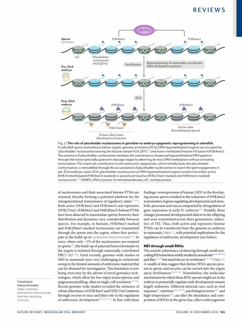
PTM H3K27 acetylation (H3K27ac) at enhancers during ZGA, thereby linking maternally inherited, Polycomb mediated gene silencing with proper enhancer usage in the early embryo137 (Fig. 3a).
In mammalian embryos, H3K27me3 appears to have an important role in DNA methylationindependent maternal imprinting66. Mapping open chromatin regi ons using the DNase I hypersensitivity assay in mouse zygotes revealed several hundred parental allele specific (imprinted) DNase I hypersensitive sites (DHSs), which are indicative of gene expression, including novel imprinted regions. Of note, only ~20% of paternal allele specific DHSs showed DNA methylation at the maternal allele, implying the existence of an alternative mechanism for reducing chromatin accessibility at the maternal allele (Fig. 3b). One fifth of these maternal allele imprints were lost upon overexpression of lysine specific demethylase 6B (KDM6B), which in the zygote is H3K27me3specific and forms open chromatin at the maternal alleles. H3K27me3regulated maternal alleles were hypomethylated in the oocyte, suggesting that it is the repressive histone PTM H3K27me3 rather than DNA methylation that constrains maternal allele accessibility at these imprinted loci66. Moreover, highly sensitive chromatin immunoprecipitation followed by sequencing (ChIP–seq) profiling of 14 stages of mouse embryogenesis and gametogenesis revealed the existence of inheritance of maternally provided H3K27me3, which persists on thousands of distal regulatory regions until the blastocyst stage122. This finding is in line with the proposed role of maternally contributed H3K27me3 in enhancer regulation at ZGA in D. melanogaster137. However, unlike in D. melanogaster, in mice, H3K27me3 is erased from Hox genes upon fertilization and the repressive state of the Hox cluster is maintained through alternative mechanisms; H3K27me3 is reestablished in post implantation embryos122 (Fig. 3b). Other Polycomb targets in mouse embryos displayed similar H3K27me3 dynamics.
In mouse oocytes, non canonically broad H3K4me3 domains occupy almost one quarter of the genome and coincide with DNA hypomethylation120,121. The great majority of genes activated in the major ZGA — the major burst of zygotic transcription by RNA polymerase II — as well as distal regulatory elements involved in ZGA and in the establishment of totipotency, reside within these broad H3K4me3 domains120,121. However, ZGA is associated with the decomposition of these broad H3K4me3 domains into sharper, transcription start site associated H3K4me3 ‘peaks’ in late two cell embryos, which is accompanied by the deposition of H3K27ac120 (Fig. 3c). Establishment of broad H3K4me3 domains in oocytes, which is mediated by the histone lysine N methyltransferase 2B (KMT2B), and their resolution upon ZGA, which is mediated by the histone demethylases KDM5A and KDM5B, are crucial for H3K4me3associated genome silencing during oogenesis and reactivation in late two cell embryos, respectively120,121. Depletion of KMT2B, KDM5A or KDM5B compromised early embryonic development120,121,167,168. Together with the recent observations of intergenerational H3K27me3 inheritance66,137, these findings provide intriguing clues
ParamutationHeritable change in gene expression of a (paramutable) allele, which is mediated by trans- interaction with the homologous (paramutagenic) allele.
Homeotic transformationThe formation of a body structure or an organ in place of another in an abnormal location.
www.nature.com/nrm
R e v i e w s
784 | december 2018 | volume 19
Oocyte
Totipotent nuclei
Pluripotent nuclei
ZGA
ZGAWT
ZGAE(z) KD
Hox genecluster
Hox genecluster Enhancers
a Drosophila melanogaster b Mus musculus
c Mus musculus
Gene-distal regions
Polycomb-targetedpromoters H3K27me3-
imprinted regions
Gametes
Zygote
Blastocyst
E6.5
E6.5
E9.5
M
P
M
P
M
P
M
P
M
P
M
P
M
P
Epiblast
ICM
Extraembryonic ectoderm
Visceral endoderm
Placenta
ZGA genepromoters
ZGA genepromoters
Distal regulatoryelements
Gametes
Zygote
Early two-cellembryo
Late two-cellembryo(ZGA)
Eight-cellembryo
M
P
M
P
M
P
M
P
M
P
KDM5A and KDM5BH3K27me3
H3K27ac
H3K4me3
Closed chromatin
Open chromatin (DHS)
Gene repression
Gene expression
Trophoectoderm
Nucleus
Fig. 3 | Intergenerational inheritance of histone post- translational modifications contributes to gene regulation during embryogenesis. a | In Drosophila melanogaster, histone H3 lysine 27 trimethylation (H3K27me3), which is a repressive post- translational modification, is maternally transmitted to the embryo and maintained until zygotic gene activation (ZGA), including at Hox loci137. Depletion of the maternally inherited Polycomb protein enhancer of zeste (E(z)) results in loss of H3K27me3 and aberrant H3K27 acetylation (H3K27ac) at enhancers, leading to erroneous gene activation during ZGA. b | In mouse gametes, Hox loci and the majority of promoters targeted by Polycomb proteins are marked by H3K27me3; however, in the zygote and two- cell embryos, H3K27me3 is reduced at the maternal allele and is completely lost at the paternal allele. H3K27me3 at these regions emerges in the inner cell mass (ICM) of the blastocyst and is re- established in the epiblast at embryonic day 6.5 (E6.5). H3K27me3 at gene- distal regions is inherited from oocytes and propagated on the maternal allele in the zygote, two- cell embryos and ICM122. DNA methylation- independent imprinted loci, characterized by the presence of DNase I- hypersensitive sites (DHSs) at the paternal allele
and H3K27me3 at the maternal allele, are inherited by the zygote and propagated to the blastocyst stage66. Their nearest genes (n = 76) display paternal- allele-specific gene expression that is largely maintained in the blastocyst (more so in the trophectoderm than in the ICM). In post- implantation embryos, H3K27me3-dependent imprinting is erased in the epiblast but is partially maintained in extra- embryonic ectoderm and visceral endoderm at E6.5 and in the placenta until E9.5. c | Non- canonically broad H3K4me3 domains in mouse oocytes are transmitted to the zygote upon fertilization and are transformed into canonical H3K4me3 peaks in late two- cell embryos, where they characterize genes and regulatory elements activated during ZGA. Decomposition of non- canonical H3K4me3 domains coincides with the appearance of H3K27ac at ZGA genes in late two- cell and eight- cell stages. Compared with the sperm, the paternal pronucleus has considerably less H3K4me3, which is re- established by lysine- specific histone demethylase 5A (KDM5A) and KDM5B in late two- cell embryos and onwards120,121. KD, knockdown; M, maternal; P, paternal; WT, wild type.
NATure revIeWs | MOLeCuLAR CeLL BIOLOGy
R e v i e w s
volume 19 | december 2018 | 785
as to how parentally contributed histone PTMs (TAble 1) provide crucial signals required for the proper establishment of transcriptional programmes at ZGA.
Regulation of embryogenesis by inherited small RNAs. Small non coding RNAs inherited from gametes also participate in the regulation of early embryonic development. In mice, inhibition of a sperm derived miRNA, miR34c, reduced DNA synthesis and suppressed the first zygotic division157. Similarly, maternal loss of the endonuclease Dicer, which is essential for miRNA formation, was not compatible with the first cell division in mouse embryos163, while paternal loss of Dicer resulted in severely impaired preimplantation development169. However, in zebrafish, this is not the case, and embryos derived from maternal Dicer mutants only start displaying developmental defects during gastrulation, somitogenesis, brain and heart development170. These studies are further supported by recent observations that small RNAs found in mature sperm are essential for embryogenesis. Specifically, zygotes generated via intracytoplasmic sperm injection (ICSI) of immature sperm (derived from caput epididymis) largely failed to support embryo genesis and exhibited multiple defects during and after implantation171. However, these defects were rescued by the injection of small RNAs (18–40 nucleotides) derived from extracellular vesicles (epididymosomes) that deliver their small RNA repertoire to mature sperm. This finding suggests that small RNAs in mature sperm171,172, which consist mostly of miRNAs but also of tRFs, likely have major roles during early mouse embryogenesis.
Inheritance of metabolic phenotypesA major area of MEI research is related to the study of potential adaptive advantages of intergenerational inheritance of histone PTMs, small RNAs and DNA methylation. Of particular interest is the possibility that environmental influences might affect the epigenome of parental gametes, which can then be transferred to the zygote to alter gene regulation. The importance of meiotic inheritance of the histone PTMs H3K9me3 and H3K27me3 in response to environmental stimuli was recently demonstrated in D. melanogaster. Paternal high sugar consumption was shown to alter the chromatin state of embryonic, larval and metabolic genes in F1 offspring, accompanied by increased triglyceride levels and body weight173. This alteration was due to the transcriptional activation of genes residing in ‘black’ (lamin/histone H1associated) and ‘blue’ (Polycomb associated) heterochromatin174, in line with previous findings of heterochromatin MEI in flies175. Notably, the H3K9me3 mark was reduced in the fat body cells of F1 offspring males whose fathers were fed a high sugar diet173.
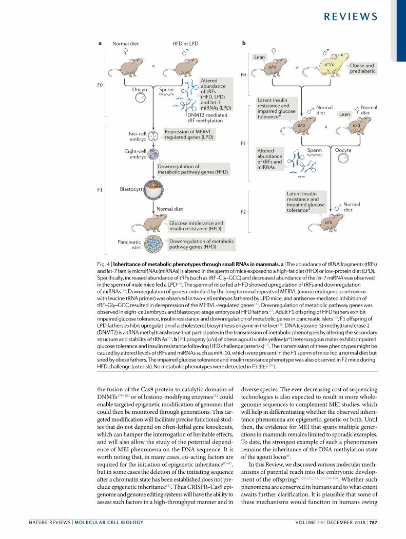
Small RNAs have also been recently described as key determinants of intergenerational transmission of metabolic phenotypes in mammals. Recent work shed light on how small (28–34 nucleotide) tRFs derived from tRNA 5ʹ ends and inherited through sperm can potentially regulate embryogenesis in mice154–156,171 (TAble 1). tRFs derived from tRNA–Gly–GCC of fathers fed a low protein diet were shown to mediate repression of
genes regulated by the long terminal repeats of MERVL (mouse endogenous retrovirus with leucine tRNA primer) in mouse embryos and embryonic stem cells (Fig. 4a). These tRFs were trafficked from the epididymis, thereby providing remarkable evidence of soma to germline transmission of paternal dietary effects156,172. Similarly, the injection into zygotes of tRFs from fathers that were kept on a HFD resulted in downregulation of metabolic pathways in both eight cell embryos and blastocysts154. In another study, the transmission of a latent metabolic phenotype from obese, prediabetic F0 males to F1 and to F2 was linked to the inheritance of several RNA species. Those included tRFs and miRNAs such as miR10, which constituted ~25% of all miRNAs in the paternally obese F1 mice155 and the targets of which were enriched in functions related to transcriptional regulation (Fig. 4b). Notably, the transmission of metabolic phenotypes discussed above appears to be highly linked to the methylation status of small RNA species by DNMT2, which is an area that merits further investigation161. DNA methylation was also recently implicated in the transmission of metabolic phenotypes associated with in utero malnutrition in mammals116,176. However, the precise function of DNA methylation in these processes and its underlying causality remain unclear. In summary, these results provide evidence for widespread MEI through diverse molecular mechanisms in insects and mammals and demonstrate how environmental cues might be sensed and transmitted to the offspring.
Conclusions and future perspectiveNumerous aspects of MEI in plants and in short lived organisms, such as C. elegans and D. melanogaster, have been thoroughly described and excellently reviewed24–28,134,135. By contrast, the functions, mechanisms and dimensions of MEI in most animal phyla still remain poorly understood. The recent advent of genomics technologies has enabled the thorough exploration of MEI phenomena across a broad range of species and in different biological contexts. For historical reasons177, and owing to its precise, replication coupled enzymatic machinery89,90, DNA methylation has garnered much attention as a potential mechanism of MEI in animals. Nevertheless, the links between DNA methylation and MEI remain a topic of debate70,72,116,178. Recent insights from genomics studies suggest that other molecular mechanisms, such as histone PTMs66,120,121,136,137 and small RNAs, including tRFs154–156,161, likely have important roles in the intergenerational transmission of gene regulatory information in the animal kingdom.
A major criticism related to the study of MEI in mammals is the lack of associated causality88. In other words, it is very challenging to unambiguously demonstrate that the regulatory changes acquired in the parental germ line can be efficiently relayed to the offspring to alter gene expression. An even more challenging aspect of MEI studies is the lack of proof that such epigenetic changes, acquired in one generation and passed on to the next, have any adaptive roles3. The recent developments in CRISPR–Cas9driven targeted epigenome remodelling applied to easily tractable model organisms such as zebrafish could tackle these challenges. For example,
www.nature.com/nrm
R e v i e w s
786 | december 2018 | volume 19
the fusion of the Cas9 protein to catalytic domains of DNMTs179–181 or of histone modifying enzymes182 could enable targeted epigenetic modification of genomes that could then be monitored through generations. This targeted modification will facilitate precise functional studies that do not depend on often lethal gene knockouts, which can hamper the interrogation of heritable effects, and will also allow the study of the potential dependence of MEI phenomena on the DNA sequence. It is worth noting that, in many cases, cis- acting factors are required for the initiation of epigenetic inheritance85–87, but in some cases the deletion of the initiating sequence after a chromatin state has been established does not preclude epigenetic inheritance183. Thus CRISPR–Cas9 epigenome and genome editing systems will have the ability to assess such factors in a high throughput manner and in
diverse species. The ever decreasing cost of sequencing technologies is also expected to result in more whole genome sequences to complement MEI studies, which will help in differentiating whether the observed inheritance phenomena are epigenetic, genetic or both. Until then, the evidence for MEI that spans multiple generations in mammals remains limited to sporadic examples. To date, the strongest example of such a phenomenon remains the inheritance of the DNA methylation state of the agouti locus68.
In this Review, we discussed various molecular mechanisms of parental reach into the embryonic development of the offspring66,120,121,136,137,154–156. Whether such phenomena are conserved in humans and to what extent awaits further clarification. It is plausible that some of these mechanisms would function in humans owing
Normal diet
Normaldiet
Normaldiet
HFD or LPDa b
× ×
×
SpermOocyte
Alteredabundanceof tRFs(HFD, LPD)and let-7miRNAs (LPD)
Latent insulinresistance andimpaired glucosetolerance*
F0
F0
F1
F2
F1
Sperm Oocyte
DNMT2-mediated tRF methylation
Two-cellembryo
Eight-cellembryo
Blastocyst
Normal diet
Repression of MERVL-regulated genes (LPD)
Glucose intolerance andinsulin resistance (HFD)
Pancreaticislet
Downregulation of metabolicpathway genes (HFD)
Downregulation ofmetabolic pathway genes (HFD)
a/a
a/a
Normaldiet
a/a
a/a
avy/a Obese andprediabetic
Lean
Latent insulinresistance andimpaired glucosetolerance*
Lean
Alteredabundanceof tRFs andmiRNAs
Fig. 4 | Inheritance of metabolic phenotypes through small RNAs in mammals. a | The abundance of tRNA fragments (tRFs) and let-7 family microRNAs (miRNAs) is altered in the sperm of mice exposed to a high- fat diet (HFD) or low- protein diet (LPD). Specifically , increased abundance of tRFs (such as tRF–Gly–GCC) and decreased abundance of the let-7 miRNA was observed in the sperm of male mice fed a LPD156. The sperm of mice fed a HFD showed upregulation of tRFs and downregulation of miRNAs154. Downregulation of genes controlled by the long terminal repeats of MERVL (mouse endogenous retrovirus with leucine tRNA primer) was observed in two- cell embryos fathered by LPD mice, and antisense-mediated inhibition of tRF–Gly–GCC resulted in derepression of the MERVL- regulated genes156. Downregulation of metabolic pathway genes was observed in eight- cell embryos and blastocyst- stage embryos of HFD fathers154. Adult F1 offspring of HFD fathers exhibit impaired glucose tolerance, insulin resistance and downregulation of metabolic genes in pancreatic islets154. F1 offspring of LPD fathers exhibit upregulation of a cholesterol biosynthesis enzyme in the liver156. DNA (cytosine-5)-methyltransferase 2 (DNMT2) is a tRNA methyltransferase that participates in the transmission of metabolic phenotypes by altering the secondary structure and stability of tRNAs161. b | F1 progeny (a/a) of obese agouti viable yellow (avy) heterozygous males exhibit impaired glucose tolerance and insulin resistance following HFD challenge (asterisk)155. The transmission of these phenotypes might be caused by altered levels of tRFs and miRNAs such as miR-10, which were present in the F1 sperm of mice fed a normal diet but sired by obese fathers. The impaired glucose tolerance and insulin resistance phenotype was also observed in F2 mice during HFD challenge (asterisk). No metabolic phenotypes were detected in F3 (reF.155).
NATure revIeWs | MOLeCuLAR CeLL BIOLOGy
R e v i e w s
volume 19 | december 2018 | 787
1. Richards, E. J. Inherited epigenetic variation — revisiting soft inheritance. Nat. Rev. Genet. 7, 395–401 (2006).
2. Daxinger, L. & Whitelaw, E. Understanding transgenerational epigenetic inheritance via the gametes in mammals. Nat. Rev. Genet. 13, 153–162 (2012).
3. Miska, E. A. & Ferguson- Smith, A. C. Transgenerational inheritance: models and mechanisms of non- DNA sequence- based inheritance. Science 354, 59–63 (2016).
4. Greally, J. M. A user’s guide to the ambiguous word ‘epigenetics’. Nat. Rev. Mol. Cell Biol. 19, 207–208 (2018).
5. Adams, F. The Genuine Works of Hippocrates (William Wood, NY, USA, 1891).
6. Lamarck, J. B. Philosophie Zoologique (JB Baillière, Paris, 1809).
7. Waddington, C. H. Genetic assimilation of an acquired character. Int. J. Org. Evol. 7, 118–126 (1953).
8. Waddington, C. H. Genetic assimilation of the bithorax phenotype. Evolution 10, 1 (1956).
9. Rutherford, S. L. & Lindquist, S. Hsp90 as a capacitor for morphological evolution. Nature 396, 336–342 (1998). This work describes the key role of Hsp90 in the buffering of naturally occurring phenotypic variation in fruit flies.
10. Zhao, C. Q., Young, M. R., Diwan, B. A., Coogan, T. P. & Waalkes, M. P. Association of arsenic- induced malignant transformation with DNA hypomethylation and aberrant gene expression. Proc. Natl Acad. Sci. USA 94, 10907–10912 (1997).
11. James, S. J. et al. Mechanisms of DNA damage, DNA hypomethylation, and tumor progression in the folate/methyl- deficient rat model of hepatocarcinogenesis. J. Nutr. 133, 3740S–3747S (2003).
12. Baccarelli, A. & Bollati, V. Epigenetics and environmental chemicals. Curr. Opin. Pediatr. 21, 243–251 (2009).
13. Blaschke, K. et al. Vitamin C induces Tet- dependent DNA demethylation and a blastocyst- like state in ES cells. Nature 500, 222–226 (2013).
14. Vickers, M. H. Early life nutrition, epigenetics and programming of later life disease. Nutrients 6, 2165–2178 (2014).
15. Klengel, T. & Binder, E. B. Epigenetics of stress- related psychiatric disorders and gene x environment interactions. Neuron 86, 1343–1357 (2015).
16. Cropley, J. E., Suter, C. M., Beckman, K. B. & Martin, D. I. Germ- line epigenetic modification of the murine A vy allele by nutritional supplementation. Proc. Natl Acad. Sci. USA 103, 17308–17312 (2006).
17. Dunn, G. A. & Bale, T. L. Maternal high- fat diet effects on third- generation female body size via the paternal lineage. Endocrinology 152, 2228–2236 (2011).
18. Crews, D. et al. Epigenetic transgenerational inheritance of altered stress responses. Proc. Natl Acad. Sci. USA 109, 9143–9148 (2012).
19. Padmanabhan, N. et al. Mutation in folate metabolism causes epigenetic instability and transgenerational effects on development. Cell 155, 81–93 (2013).
20. Gapp, K. et al. Implication of sperm RNAs in transgenerational inheritance of the effects of early trauma in mice. Nat. Neurosci. 17, 667–669 (2014).
21. Rodgers, A. B., Morgan, C. P., Leu, N. A. & Bale, T. L. Transgenerational epigenetic programming via sperm microRNA recapitulates effects of paternal stress. Proc. Natl Acad. Sci. USA 112, 13699–13704 (2015).
22. Rusche, L. N., Kirchmaier, A. L. & Rine, J. The establishment, inheritance, and function of
silenced chromatin in Saccharomyces cerevisiae. Annu. Rev. Biochem. 72, 481–516 (2003).
23. Casadesus, J. & Low, D. Epigenetic gene regulation in the bacterial world. Microbiol. Mol. Biol. Rev. 70, 830–856 (2006).
24. Quadrana, L. & Colot, V. Plant transgenerational epigenetics. Annu. Rev. Genet. 50, 467–491 (2016).
25. Hollick, J. B. Paramutation and related phenomena in diverse species. Nat. Rev. Genet. 18, 5–23 (2017).
26. Houri- Zeevi, L. & Rechavi, O. A. Matter of time: small RNAs regulate the duration of epigenetic inheritance. Trends Genet. 33, 46–57 (2017).
27. Minkina, O. & Hunter, C. P. Intergenerational transmission of gene regulatory information in Caenorhabditis elegans. Trends Genet. 34, 54–64 (2017).
28. Serobyan, V. & Sommer, R. J. Developmental systems of plasticity and trans- generational epigenetic inheritance in nematodes. Curr. Opin. Genet. Dev. 45, 51–57 (2017).
29. Springer, N. M. & Schmitz, R. J. Exploiting induced and natural epigenetic variation for crop improvement. Nat. Rev. Genet. 18, 563–575 (2017).
30. Fire, A. et al. Potent and specific genetic interference by double- stranded RNA in Caenorhabditis elegans. Nature 391, 806–811 (1998).
31. Bernstein, E., Caudy, A. A., Hammond, S. M. & Hannon, G. J. Role for a bidentate ribonuclease in the initiation step of RNA interference. Nature 409, 363–366 (2001).
32. Ketting, R. F. et al. Dicer functions in RNA interference and in synthesis of small RNA involved in developmental timing in C. elegans. Genes Dev. 15, 2654–2659 (2001).
33. Yigit, E. et al. Analysis of the C. elegans Argonaute family reveals that distinct Argonautes act sequentially during RNAi. Cell 127, 747–757 (2006).
34. Sijen, T. et al. On the role of RNA amplification in dsRNA- triggered gene silencing. Cell 107, 465–476 (2001).
35. Pak, J. & Fire, A. Distinct populations of primary and secondary effectors during RNAi in C. elegans. Science 315, 241–244 (2007).
36. Swarts, D. C. et al. The evolutionary journey of Argonaute proteins. Nat. Struct. Mol. Biol. 21, 743–753 (2014).
37. Guang, S. et al. An Argonaute transports siRNAs from the cytoplasm to the nucleus. Science 321, 537–541 (2008).
38. Rechavi, O., Minevich, G. & Hobert, O. Transgenerational inheritance of an acquired small RNA- based antiviral response in C. elegans. Cell 147, 1248–1256 (2011).
39. Burton, N. O., Burkhart, K. B. & Kennedy, S. Nuclear RNAi maintains heritable gene silencing in Caenorhabditis elegans. Proc. Natl Acad. Sci. USA 108, 19683–19688 (2011).
40. Ishidate, T. et al. ZNFX-1 functions within perinuclear nuage to balance epigenetic signals. Mol. Cell 70, 639–649 (2018).
41. Wan, G. et al. Spatiotemporal regulation of liquid- like condensates in epigenetic inheritance. Nature 557, 679–683 (2018).
42. Bagijn, M. P. et al. Function, targets, and evolution of Caenorhabditis elegans piRNAs. Science 337, 574–578 (2012).
43. Lee, H. C. et al. C. elegans piRNAs mediate the genome- wide surveillance of germline transcripts. Cell 150, 78–87 (2012).
44. Ashe, A. et al. piRNAs can trigger a multigenerational epigenetic memory in the germline of C. elegans. Cell 150, 88–99 (2012).
45. Luteijn, M. J. et al. Extremely stable Piwi- induced gene silencing in Caenorhabditis elegans. EMBO J. 31, 3422–3430 (2012).
46. Shirayama, M. et al. piRNAs initiate an epigenetic memory of nonself RNA in the C. elegans germline. Cell 150, 65–77 (2012). References 44–46 describe how piRNAs contribute to transgenerational epigenetic memory in Caenorhabditis elegans.
47. Grishok, A., Tabara, H. & Mello, C. C. Genetic requirements for inheritance of RNAi in C. elegans. Science 287, 2494–2497 (2000).
48. Vastenhouw, N. L. et al. Gene expression: long- term gene silencing by RNAi. Nature 442, 882 (2006).
49. Kalinava, N., Ni, J. Z., Peterman, K., Chen, E. & Gu, S. G. Decoupling the downstream effects of germline nuclear RNAi reveals that H3K9me3 is dispensable for heritable RNAi and the maintenance of endogenous siRNA- mediated transcriptional silencing in Caenorhabditis elegans. Epigenetics Chromatin 10, 6 (2017).
50. Lev, I. et al. MET-2-dependent H3K9 methylation suppresses transgenerational small RNA inheritance. Curr. Biol. 27, 1138–1147 (2017). This work demonstrates how induction of RNAi in nematodes deficient in the H3K9 methyltransferase MET-2 results in permanent RNA responses that persist even after 30 generations.
51. Greer, E. L. et al. Transgenerational epigenetic inheritance of longevity in Caenorhabditis elegans. Nature 479, 365–371 (2011).
52. Katz, D. J., Edwards, T. M., Reinke, V. & Kelly, W. G. A. C. elegans LSD1 demethylase contributes to germline immortality by reprogramming epigenetic memory. Cell 137, 308–320 (2009).
53. Gaydos, L. J., Wang, W. & Strome, S. Gene repression. H3K27me and PRC2 transmit a memory of repression across generations and during development. Science 345, 1515–1518 (2014).
54. Klosin, A., Casas, E., Hidalgo- Carcedo, C., Vavouri, T. & Lehner, B. Transgenerational transmission of environmental information in C. elegans. Science 356, 320–323 (2017). This report demonstrates H3K9me3-associated, temperature- induced changes in gene expression, which are heritable for at least 14 generations in the nematode Caenorhabditis elegans.
55. Lev, I., Gingold, H. & Rechavi, O. H3K9me3 is required for transgenerational inheritance of small RNAs that target a unique subset of newly evolved genes. bioRxiv https://doi.org/10.1101/338582 (2018).
56. Lee, H. J., Hore, T. A. & Reik, W. Reprogramming the methylome: erasing memory and creating diversity. Cell Stem Cell 14, 710–719 (2014).
57. Eckersley- Maslin, M. A., Alda- Catalinas, C. & Reik, W. Dynamics of the epigenetic landscape during the maternal- to-zygotic transition. Nat. Rev. Mol. Cell Biol. 19, 436–450 (2018).
58. Ferguson- Smith, A. C. Genomic imprinting: the emergence of an epigenetic paradigm. Nat. Rev. Genet. 12, 565–575 (2011).
59. Reik, W., Collick, A., Norris, M. L., Barton, S. C. & Surani, M. A. Genomic imprinting determines methylation of parental alleles in transgenic mice. Nature 328, 248–251 (1987).
60. Sapienza, C., Peterson, A. C., Rossant, J. & Balling, R. Degree of methylation of transgenes is dependent on gamete of origin. Nature 328, 251–254 (1987).
61. Bartolomei, M. S., Zemel, S. & Tilghman, S. M. Parental imprinting of the mouse H19 gene. Nature 351, 153–155 (1991).
62. Bourc’his, D., Xu, G. L., Lin, C. S., Bollman, B. & Bestor, T. H. Dnmt3L and the establishment of maternal genomic imprints. Science 294, 2536–2539 (2001).
to the high conservation of the underlying enzymatic machinery184. On the other hand, it is worth noting that considerable differences in sperm nucleosome retention and early embryonic chromatin establishment have been observed between humans and mice118,145. Studying epigenetic inheritance in humans will remain a problem owing to long generation times, which restrict the collection of biological specimens, and owing to the genetic diversity of the human population. Nevertheless, with the reduced amounts of input material required for
genome wide assays and the increased frequency of studies carried out on donated human embryonic material, it is reasonable to expect such proof ofprinciple studies sooner rather than later. Overall, the recent revolution in genomics technologies, including targeted genome and epigenome manipulation, is expected to provide long sought answers to fundamental questions about the functions and extent of MEI in animals.
Published online 13 November 2018
www.nature.com/nrm
R e v i e w s
788 | december 2018 | volume 19
63. Coan, P. M., Burton, G. J. & Ferguson- Smith, A. C. Imprinted genes in the placenta — a review. Placenta 26, S10–S20 (2005).
64. Frost, J. M. & Moore, G. E. The importance of imprinting in the human placenta. PLOS Genet. 6, e1001015 (2010).
65. Nicholls, R. D. & Knepper, J. L. Genome organization, function, and imprinting in Prader–Willi and Angelman syndromes. Annu. Rev. Genom. Hum. Genet. 2, 153–175 (2001).
66. Inoue, A., Jiang, L., Lu, F., Suzuki, T. & Zhang, Y. Maternal H3K27me3 controls DNA methylation- independent imprinting. Nature 547, 419–424 (2017). This work demonstrates the existence of H3K27me3-dependent imprinting independently of DNA methylation during mammalian embryogenesis.
67. Lane, N. et al. Resistance of IAPs to methylation reprogramming may provide a mechanism for epigenetic inheritance in the mouse. Genesis 35, 88–93 (2003).
68. Morgan, H. D., Sutherland, H. G., Martin, D. I. & Whitelaw, E. Epigenetic inheritance at the agouti locus in the mouse. Nat. Genet. 23, 314–318 (1999).
69. Waterland, R. A. & Jirtle, R. L. Transposable elements: targets for early nutritional effects on epigenetic gene regulation. Mol. Cell. Biol. 23, 5293–5300 (2003).
70. Blewitt, M. E., Vickaryous, N. K., Paldi, A., Koseki, H. & Whitelaw, E. Dynamic reprogramming of DNA methylation at an epigenetically sensitive allele in mice. PLOS Genet. 2, e49 (2006).
71. Xu, Q. & Xie, W. Epigenome in early mammalian development: inheritance, reprogramming and establishment. Trends Cell Biol. 28, 237–253 (2018).
72. Hitchins, M. P. Constitutional epimutation as a mechanism for cancer causality and heritability? Nat. Rev. Cancer 15, 625–634 (2015).
73. Suter, C. M., Martin, D. I. & Ward, R. L. Germline epimutation of MLH1 in individuals with multiple cancers. Nat. Genet. 36, 497–501 (2004).
74. Hitchins, M. P. et al. Inheritance of a cancer- associated MLH1 germ- line epimutation. N. Engl. J. Med. 356, 697–705 (2007).
75. Lynch, H. T. et al. Review of the Lynch syndrome: history, molecular genetics, screening, differential diagnosis, and medicolegal ramifications. Clin. Genet. 76, 1–18 (2009).
76. Morak, M. et al. Further evidence for heritability of an epimutation in one of 12 cases with MLH1 promoter methylation in blood cells clinically displaying HNPCC. Eur. J. Hum. Genet. 16, 804–811 (2008).
77. Crepin, M. et al. Evidence of constitutional MLH1 epimutation associated to transgenerational inheritance of cancer susceptibility. Hum. Mutat. 33, 180–188 (2012).
78. Kaati, G., Bygren, L. O. & Edvinsson, S. Cardiovascular and diabetes mortality determined by nutrition during parents’ and grandparents’ slow growth period. Eur. J. Hum. Genet. 10, 682–688 (2002).
79. Roseboom, T., de Rooij, S. & Painter, R. The Dutch famine and its long- term consequences for adult health. Early Hum. Dev. 82, 485–491 (2006).
80. Bygren, L. O. et al. Change in paternal grandmothers’ early food supply influenced cardiovascular mortality of the female grandchildren. BMC Genet. 15, 12 (2014).
81. Yehuda, R. et al. Holocaust exposure induced intergenerational effects on FKBP5 methylation. Biol. Psychiatry 80, 372–380 (2016).
82. Heijmans, B. T. et al. Persistent epigenetic differences associated with prenatal exposure to famine in humans. Proc. Natl Acad. Sci. USA 105, 17046–17049 (2008).
83. Painter, R. C. et al. Transgenerational effects of prenatal exposure to the Dutch famine on neonatal adiposity and health in later life. BJOG 115, 1243–1249 (2008).
84. Lappalainen, T. & Greally, J. M. Associating cellular epigenetic models with human phenotypes. Nat. Rev. Genet. 18, 441–451 (2017).
85. Coleman, R. T. & Struhl, G. Causal role for inheritance of H3K27me3 in maintaining the OFF state of a Drosophila HOX gene. Science 356, eaai8236 (2017).
86. Laprell, F., Finkl, K. & Muller, J. Propagation of Polycomb- repressed chromatin requires sequence- specific recruitment to DNA. Science 356, 85–88 (2017).
87. Wang, X. & Moazed, D. DNA sequence- dependent epigenetic inheritance of gene silencing and histone H3K9 methylation. Science 356, 88–91 (2017).
88. Bohacek, J. & Mansuy, I. M. A guide to designing germline- dependent epigenetic inheritance
experiments in mammals. Nat. Methods 14, 243–249 (2017).
89. Bestor, T. H. The DNA methyltransferases of mammals. Hum. Mol. Genet. 9, 2395–2402 (2000).
90. Jurkowska, R. Z., Jurkowski, T. P. & Jeltsch, A. Structure and function of mammalian DNA methyltransferases. Chembiochem 12, 206–222 (2011).
91. Suzuki, M. M. & Bird, A. DNA methylation landscapes: provocative insights from epigenomics. Nat. Rev. Genet. 9, 465–476 (2008).
92. Schubeler, D. Function and information content of DNA methylation. Nature 517, 321–326 (2015).
93. Simpson, V. J., Johnson, T. E. & Hammen, R. F. Caenorhabditis elegans DNA does not contain 5-methylcytosine at any time during development or aging. Nucleic Acids Res. 14, 6711–6719 (1986).
94. Lyko, F., Ramsahoye, B. H. & Jaenisch, R. DNA methylation in Drosophila melanogaster. Nature 408, 538–540 (2000).
95. Mayer, W., Niveleau, A., Walter, J., Fundele, R. & Haaf, T. Demethylation of the zygotic paternal genome. Nature 403, 501–502 (2000).
96. Oswald, J. et al. Active demethylation of the paternal genome in the mouse zygote. Curr. Biol. 10, 475–478 (2000).
97. Santos, F., Hendrich, B., Reik, W. & Dean, W. Dynamic reprogramming of DNA methylation in the early mouse embryo. Dev. Biol. 241, 172–182 (2002).
98. Smith, Z. D. et al. A unique regulatory phase of DNA methylation in the early mammalian embryo. Nature 484, 339–344 (2012).
99. Wossidlo, M. et al. 5-Hydroxymethylcytosine in the mammalian zygote is linked with epigenetic reprogramming. Nat. Commun. 2, 241 (2011).
100. Wang, L. et al. Programming and inheritance of parental DNA methylomes in mammals. Cell 157, 979–991 (2014).
101. Amouroux, R. et al. De novo DNA methylation drives 5hmC accumulation in mouse zygotes. Nat. Cell Biol. 18, 225–233 (2016).
102. Morgan, H. D., Santos, F., Green, K., Dean, W. & Reik, W. Epigenetic reprogramming in mammals. Hum. Mol. Genet. 1, R47–R58 (2005).
103. Borgel, J. et al. Targets and dynamics of promoter DNA methylation during early mouse development. Nat. Genet. 42, 1093–1100 (2010).
104. Guo, H. et al. The DNA methylation landscape of human early embryos. Nature 511, 606–610 (2014).
105. Smith, Z. D. et al. DNA methylation dynamics of the human preimplantation embryo. Nature 511, 611–615 (2014).
106. Birnbaum, R. Y. et al. Coding exons function as tissue- specific enhancers of nearby genes. Genome Res. 22, 1059–1068 (2012).
107. Blattler, A. et al. Global loss of DNA methylation uncovers intronic enhancers in genes showing expression changes. Genome Biol. 15, 469 (2014).
108. Popp, C. et al. Genome- wide erasure of DNA methylation in mouse primordial germ cells is affected by AID deficiency. Nature 463, 1101–1105 (2010).
109. Guibert, S., Forne, T. & Weber, M. Global profiling of DNA methylation erasure in mouse primordial germ cells. Genome Res. 22, 633–641 (2012).
110. Hackett, J. A. et al. Germline DNA demethylation dynamics and imprint erasure through 5-hydroxymethylcytosine. Science 339, 448–452 (2013).
111. Hill, P. W. S. et al. Epigenetic reprogramming enables the transition from primordial germ cell to gonocyte. Nature 555, 392–396 (2018).
112. Seisenberger, S. et al. The dynamics of genome- wide DNA methylation reprogramming in mouse primordial germ cells. Mol. Cell 48, 849–862 (2012).
113. Gkountela, S. et al. DNA demethylation dynamics in the human prenatal germline. Cell 161, 1425–1436 (2015).
114. Guo, F. et al. The transcriptome and DNA methylome landscapes of human primordial germ cells. Cell 161, 1437–1452 (2015).
115. Tang, W. W. et al. A unique gene regulatory network resets the human germline epigenome for development. Cell 161, 1453–1467 (2015).
116. Radford, E. J. et al. In utero effects. In utero undernourishment perturbs the adult sperm methylome and intergenerational metabolism. Science 345, 1255903 (2014).
117. Hammoud, S. S. et al. Distinctive chromatin in human sperm packages genes for embryo development. Nature 460, 473–478 (2009).
118. Brykczynska, U. et al. Repressive and active histone methylation mark distinct promoters in human and mouse spermatozoa. Nat. Struct. Mol. Biol. 17, 679–687 (2010).
119. Erkek, S. et al. Molecular determinants of nucleosome retention at CpG- rich sequences in mouse spermatozoa. Nat. Struct. Mol. Biol. 20, 868–875 (2013). References 117–119 describe potentially heritable DNA and histone modifications in the mammalian sperm.
120. Dahl, J. A. et al. Broad histone H3K4me3 domains in mouse oocytes modulate maternal- to-zygotic transition. Nature 537, 548–552 (2016).
121. Zhang, B. et al. Allelic reprogramming of the histone modification H3K4me3 in early mammalian development. Nature 537, 553–557 (2016). References 120 and 121 describe the intergenerational inheritance of the histone modification H3K4me3 in mammals and its roles during early mammalian embryogenesis.
122. Zheng, H. et al. Resetting epigenetic memory by reprogramming of histone modifications in mammals. Mol. Cell 63, 1066–1079 (2016).
123. Macleod, D., Clark, V. H. & Bird, A. Absence of genome- wide changes in DNA methylation during development of the zebrafish. Nat. Genet. 23, 139–140 (1999).
124. Veenstra, G. J. & Wolffe, A. P. Constitutive genomic methylation during embryonic development of Xenopus. Biochim. Biophys. Acta 1521, 39–44 (2001).
125. Bogdanovic, O. et al. Temporal uncoupling of the DNA methylome and transcriptional repression during embryogenesis. Genome Res. 21, 1313–1327 (2011).
126. Jiang, L. et al. Sperm, but not oocyte, DNA methylome is inherited by zebrafish early embryos. Cell 153, 773–784 (2013).
127. Potok, M. E., Nix, D. A., Parnell, T. J. & Cairns, B. R. Reprogramming the maternal zebrafish genome after fertilization to match the paternal methylation pattern. Cell 153, 759–772 (2013).
128. Murphy, P. J., Wu, S. F., James, C. R., Wike, C. L. & Cairns, B. R. Placeholder nucleosomes underlie germline- to-embryo DNA methylation reprogramming. Cell 172, 993–1006 (2018). This work describes placeholder nucleosomes, marked by H3K4me1 and H2AZ, which have a role in intergenerational inheritance of paternal epigenetic modifications in zebrafish.
129. Almeida, R. D. et al. 5-Hydroxymethyl- cytosine enrichment of non- committed cells is not a universal feature of vertebrate development. Epigenetics 7, 383–389 (2012).
130. Bogdanovic, O. et al. Active DNA demethylation at enhancers during the vertebrate phylotypic period. Nat. Genet. 48, 417–426 (2016).
131. Kamstra, J. H., Sales, L. B., Alestrom, P. & Legler, J. Differential DNA methylation at conserved non- genic elements and evidence for transgenerational inheritance following developmental exposure to mono(2-ethylhexyl) phthalate and 5-azacytidine in zebrafish. Epigenetics 10, 20 (2017).
132. Stadler, M. B. et al. DNA- binding factors shape the mouse methylome at distal regulatory regions. Nature 480, 490–495 (2011).
133. Thurman, R. E. et al. The accessible chromatin landscape of the human genome. Nature 489, 75–82 (2012).
134. Cavalli, G. & Paro, R. The Drosophila Fab-7 chromosomal element conveys epigenetic inheritance during mitosis and meiosis. Cell 93, 505–518 (1998).
135. Cavalli, G. & Paro, R. Epigenetic inheritance of active chromatin after removal of the main transactivator. Science 286, 955–958 (1999).
136. Ciabrelli, F. et al. Stable polycomb- dependent transgenerational inheritance of chromatin states in Drosophila. Nat. Genet. 49, 876–886 (2017). This work demonstrates how nuclear organization and Polycomb group proteins contribute to the formation of stable and transgenerationally heritable epialleles in fruit flies.
137. Zenk, F. et al. Germ line- inherited H3K27me3 restricts enhancer function during maternal- to-zygotic transition. Science 357, 212–216 (2017). This study reports regulation of enhancer activity at the onset of zygotic genome activation through maternally- inherited H3K27me3 in fruit fly embryos.
138. Wu, S. F., Zhang, H. & Cairns, B. R. Genes for embryo development are packaged in blocks of multivalent chromatin in zebrafish sperm. Genome Res. 21, 578–589 (2011).
NATure revIeWs | MOLeCuLAR CeLL BIOLOGy
R e v i e w s
volume 19 | december 2018 | 789
139. Teperek, M. et al. Sperm is epigenetically programmed to regulate gene transcription in embryos. Genome Res. 26, 1034–1046 (2016).
140. Lindeman, L. C. et al. Prepatterning of developmental gene expression by modified histones before zygotic genome activation. Dev. Cell 21, 993–1004 (2011).
141. Akkers, R. C. et al. A hierarchy of H3K4me3 and H3K27me3 acquisition in spatial gene regulation in Xenopus embryos. Dev. Cell 17, 425–434 (2009).
142. Vastenhouw, N. L. et al. Chromatin signature of embryonic pluripotency is established during genome activation. Nature 464, 922–926 (2010).
143. Hontelez, S. et al. Embryonic transcription is controlled by maternally defined chromatin state. Nat. Commun. 6, 10148 (2015).
144. Balhorn, R. The protamine family of sperm nuclear proteins. Genome Biol. 8, 227 (2007).
145. van de Werken, C. et al. Paternal heterochromatin formation in human embryos is H3K9/HP1 directed and primed by sperm- derived histone modifications. Nat. Commun. 5, 5868 (2014).
146. Puschendorf, M. et al. PRC1 and Suv39h specify parental asymmetry at constitutive heterochromatin in early mouse embryos. Nat. Genet. 40, 411–420 (2008).
147. Brind’Amour, J. et al. An ultra- low-input native ChIP-seq protocol for genome- wide profiling of rare cell populations. Nat. Commun. 6, 6033 (2015).
148. Rotem, A. et al. Single- cell ChIP- seq reveals cell subpopulations defined by chromatin state. Nat. Biotechnol. 33, 1165–1172 (2015).
149. Clark, S. J., Lee, H. J., Smallwood, S. A., Kelsey, G. & Reik, W. Single- cell epigenomics: powerful new methods for understanding gene regulation and cell identity. Genome Biol. 17, 72 (2016).
150. Siklenka, K. et al. Disruption of histone methylation in developing sperm impairs offspring health transgenerationally. Science 350, aab2006 (2015). This work demonstrates how overexpression of the H3K4 methylase LSD1 during mouse spermatogenesis results in transgenerationally heritable developmental phenotypes.
151. Brennecke, J. et al. An epigenetic role for maternally inherited piRNAs in transposon silencing. Science 322, 1387–1392 (2008).
152. de Vanssay, A. et al. Paramutation in Drosophila linked to emergence of a piRNA- producing locus. Nature 490, 112–115 (2012).
153. Rassoulzadegan, M. et al. RNA- mediated non- mendelian inheritance of an epigenetic change in the mouse. Nature 441, 469–474 (2006).
154. Chen, Q. et al. Sperm tsRNAs contribute to intergenerational inheritance of an acquired metabolic disorder. Science 351, 397–400 (2016).
155. Cropley, J. E. et al. Male- lineage transmission of an acquired metabolic phenotype induced by grand- paternal obesity. Mol. Metab. 5, 699–708 (2016).
156. Sharma, U. et al. Biogenesis and function of tRNA fragments during sperm maturation and fertilization in mammals. Science 351, 391–396 (2016). References 154–156 demonstrate the importance of small RNAs in the intergenerational transmission of metabolic phenotypes in mice.
157. Liu, W. M. et al. Sperm- borne microRNA-34c is required for the first cleavage division in mouse. Proc. Natl Acad. Sci. USA 109, 490–494 (2012).
158. Yang, Q. et al. Highly sensitive sequencing reveals dynamic modifications and activities of small RNAs in mouse oocytes and early embryos. Sci. Adv. 2, e1501482 (2016).
159. Rechavi, O. et al. Starvation- induced transgenerational inheritance of small RNAs in C. elegans. Cell 158, 277–287 (2014).
160. Schott, D., Yanai, I. & Hunter, C. P. Natural RNA interference directs a heritable response to the environment. Sci. Rep. 4, 7387 (2014).
161. Zhang, Y. et al. Dnmt2 mediates intergenerational transmission of paternally acquired metabolic disorders through sperm small non- coding RNAs. Nat. Cell Biol. 20, 535–540 (2018). This study describes the key role of the tRNA methyltransferase DNMT2 in the transmission of metabolic phenotypes in mice.
162. Houwing, S. et al. A role for Piwi and piRNAs in germ cell maintenance and transposon silencing in zebrafish. Cell 129, 69–82 (2007).
163. Tang, F. et al. Maternal microRNAs are essential for mouse zygotic development. Genes Dev. 21, 644–648 (2007).
164. Jonkhout, N. et al. The RNA modification landscape in human disease. RNA 23, 1754–1769 (2017).
165. Erhardt, S. et al. Consequences of the depletion of zygotic and embryonic enhancer of zeste 2 during preimplantation mouse development. Development 130, 4235–4248 (2003).
166. Posfai, E. et al. Polycomb function during oogenesis is required for mouse embryonic development. Genes Dev. 26, 920–932 (2012).
167. Andreu- Vieyra, C. V. et al. MLL2 is required in oocytes for bulk histone 3 lysine 4 trimethylation and transcriptional silencing. PLOS Biol. 8, e1000453 (2010).
168. Liu, X. et al. Distinct features of H3K4me3 and H3K27me3 chromatin domains in pre- implantation embryos. Nature 537, 558–562 (2016).
169. Yuan, S. et al. Sperm- borne mi- RNAs and endo- siRNAs are important for fertilization and preimplantation embryonic development. Development 143, 635–647 (2016).
170. Giraldez, A. J. et al. MicroRNAs regulate brain morphogenesis in zebrafish. Science 308, 833–838 (2005).
171. Conine, C. C., Sun, F., Song, L., Rivera- Perez, J. A. & Rando, O. J. Small RNAs gained during epididymal transit of sperm are essential for embryonic development in mice. Dev. Cell 46, 470–480 (2018).
172. Sharma, U. et al. Small RNAs are trafficked from the epididymis to developing mammalian sperm. Dev. Cell 46, 481–494 (2018).
173. Ost, A. et al. Paternal diet defines offspring chromatin state and intergenerational obesity. Cell 159, 1352–1364 (2014).
174. Filion, G. J. et al. Systematic protein location mapping reveals five principal chromatin types in Drosophila cells. Cell 143, 212–224 (2010).
175. Seong, K. H., Li, D., Shimizu, H., Nakamura, R. & Ishii, S. Inheritance of stress- induced, ATF-2-dependent epigenetic change. Cell 145, 1049–1061 (2011).
176. Martinez, D. et al. In utero undernutrition in male mice programs liver lipid metabolism in the second- generation offspring involving altered Lxra DNA methylation. Cell Metab. 19, 941–951 (2014).
177. Cubas, P., Vincent, C. & Coen, E. An epigenetic mutation responsible for natural variation in floral symmetry. Nature 401, 157–161 (1999).
178. Shea, J. M. et al. Genetic and epigenetic variation, but not diet, shape the sperm methylome. Dev. Cell 35, 750–758 (2015).
179. Liu, X. S. et al. Editing DNA methylation in the mammalian genome. Cell 167, 233–247 (2016).
180. Vojta, A. et al. Repurposing the CRISPR- Cas9 system for targeted DNA methylation. Nucleic Acids Res. 44, 5615–5628 (2016).
181. Pflueger, C. et al. A modular dCas9-SunTag DNMT3A epigenome editing system overcomes pervasive off-target activity of direct fusion dCas9-DNMT3A constructs. Genome Res. 28, 1193–1206 (2018).
182. Kwon, D. Y., Zhao, Y. T., Lamonica, J. M. & Zhou, Z. Locus- specific histone deacetylation using a synthetic CRISPR- Cas9-based HDAC. Nat. Commun. 8, 15315 (2017).
183. Ragunathan, K., Jih, G. & Moazed, D. Epigenetics. Epigenetic inheritance uncoupled from sequence- specific recruitment. Science 348, 1258699 (2015).
184. Bannister, A. J. & Kouzarides, T. Regulation of chromatin by histone modifications. Cell Res. 21, 381–395 (2011).
185. Meaney, M. J. Maternal care, gene expression, and the transmission of individual differences in stress reactivity across generations. Annu. Rev. Neurosci. 24, 1161–1192 (2001).
186. Goll, M. G. & Bestor, T. H. Eukaryotic cytosine methyltransferases. Annu. Rev. Biochem. 74, 481–514 (2005).
187. Bird, A. DNA methylation patterns and epigenetic memory. Genes Dev. 16, 6–21 (2002).
188. Peters, A. H. et al. Loss of the Suv39h histone methyltransferases impairs mammalian heterochromatin and genome stability. Cell 107, 323–337 (2001).
189. Rea, S. et al. Regulation of chromatin structure by site- specific histone H3 methyltransferases. Nature 406, 593–599 (2000).
190. Schultz, D. C., Ayyanathan, K., Negorev, D., Maul, G. G. & Rauscher, F. J. 3rd. SETDB1: a novel KAP-1-associated histone H3, lysine 9-specific methyltransferase that contributes to HP1-mediated silencing of euchromatic genes by KRAB zinc- finger proteins. Genes Dev. 16, 919–932 (2002).
191. Bannister, A. J. et al. Selective recognition of methylated lysine 9 on histone H3 by the HP1 chromo domain. Nature 410, 120–124 (2001).
192. Lachner, M., O’Carroll, D., Rea, S., Mechtler, K. & Jenuwein, T. Methylation of histone H3 lysine 9 creates a binding site for HP1 proteins. Nature 410, 116–120 (2001).
193. Bernstein, E. et al. Mouse polycomb proteins bind differentially to methylated histone H3 and RNA and are enriched in facultative heterochromatin. Mol. Cell. Biol. 26, 2560–2569 (2006).
194. Czermin, B. et al. Drosophila enhancer of Zeste/ESC complexes have a histone H3 methyltransferase activity that marks chromosomal Polycomb sites. Cell 111, 185–196 (2002).
195. Kuzmichev, A., Nishioka, K., Erdjument- Bromage, H., Tempst, P. & Reinberg, D. Histone methyltransferase activity associated with a human multiprotein complex containing the Enhancer of Zeste protein. Genes Dev. 16, 2893–2905 (2002).
196. Birve, A. et al. Su(z)12, a novel Drosophila Polycomb group gene that is conserved in vertebrates and plants. Development 128, 3371–3379 (2001).
197. Pasini, D., Bracken, A. P., Jensen, M. R., Lazzerini Denchi, E. & Helin, K. Suz12 is essential for mouse development and for EZH2 histone methyltransferase activity. EMBO J. 23, 4061–4071 (2004).
198. Margueron, R. et al. Role of the polycomb protein EED in the propagation of repressive histone marks. Nature 461, 762–767 (2009).
199. Simon, J., Chiang, A., Bender, W., Shimell, M. J. & O’Connor, M. Elements of the Drosophila bithorax complex that mediate repression by Polycomb group products. Dev. Biol. 158, 131–144 (1993).
200. Chan, C. S., Rastelli, L. & Pirrotta, V. A. Polycomb response element in the Ubx gene that determines an epigenetically inherited state of repression. EMBO J. 13, 2553–2564 (1994).
201. Perino, M. et al. MTF2 recruits Polycomb Repressive Complex 2 by helical- shape-selective DNA binding. Nat. Genet. 50, 1002–1010 (2018).
202. Santos- Rosa, H. et al. Active genes are tri-methylated at K4 of histone H3. Nature 419, 407–411 (2002).
203. Bernstein, B. E. et al. Genomic maps and comparative analysis of histone modifications in human and mouse. Cell 120, 169–181 (2005).
204. Koch, C. M. et al. The landscape of histone modifications across 1% of the human genome in five human cell lines. Genome Res. 17, 691–707 (2007).
205. Shilatifard, A. The COMPASS family of histone H3K4 methylases: mechanisms of regulation in development and disease pathogenesis. Annu. Rev. Biochem. 81, 65–95 (2012).
206. Kim, V. N., Han, J. & Siomi, M. C. Biogenesis of small RNAs in animals. Nat. Rev. Mol. Cell Biol. 10, 126–139 (2009).
207. Goll, M. G. et al. Methylation of tRNAAsp by the DNA methyltransferase homolog Dnmt2. Science 311, 395–398 (2006).
208. Hock, J. & Meister, G. The Argonaute protein family. Genome Biol. 9, 210 (2008).
AcknowledgementsThe authors thank J. Cropley, F. Zenk, A. Ashe and R. Lister for comments on the manuscript. The authors apologize to colleagues whose work could not be cited owing to space limitations.
Author contributionsAll authors contributed to researching data for the article, discussion of content and the writing and editing of the manuscript.
Competing interestsThe authors declare no competing interests.
Publisher’s noteSpringer Nature remains neutral with regard to jurisdictional claims in published maps and institutional affiliations.
Reviewer informationNature Reviews Molecular Cell Biology thanks O. Rechavi and the other anonymous reviewer(s) for their contribution to the peer review of this work.
Supplementary informationSupplementary information is available for this paper at https://doi.org/10.1038/s41580-018-0074-2.
RElATED lINksGeneimprint: http://www.geneimprint.com
www.nature.com/nrm
R e v i e w s
790 | december 2018 | volume 19