Embed Size (px)
Citation preview
DENTAL TECHNIQUE
Supported byTechniques aaDoctoral canand MaterialStomatologybProfessor, CMaterial TechStomatologycProfessor, DStomatologydProfessor, CMaterial TechStomatology
THE JOURNA
Functionally suitable digital removable completedentures: A dental technique
Kehui Deng, BM,a Yong Wang, MS, SCI,b Yongsheng Zhou, DDS, PhD,c and Yuchun Sun, DDS, PhDd
ABSTRACTThis article describes a technique for fabricating removable complete dentures by using digitaltechnology which aims to produce an individually designed, diagnostic, complete denture. Thistechnology could reduce the number of appointments compared with traditional completedenture treatment and has a wide range of applications for different types of edentulouspatients, including those with severe resorption of the alveolar ridge or a high occlusal force.Furthermore, the low cost of 3D printers, compared with expensive milling machines, may makethe approach more accessible. (J Prosthet Dent 2020;123:795-9)
Traditional removable com-plete dentures have a longhistory; however, limitationsinclude the long treatmentperiod,1,2 a difficult procedurethat depends heavily on theexperience and skill of thedentist, and a definitive den-ture that typically requires
repeated adjustment or even reworking after denturedelivery.3 Few dentists have the ability to fabricate asuitable removable complete denture, and many youngerdentists are reluctant to provide complete dentures.4In the recent years, digital technology has been usedin a more efficient manner than traditional methods forcomplete dentures.5 Commercial digital complete den-ture systems, such as AvaDent (Global Dental ScienceLLC), Dentca (Dentca Inc), and Baltic (Merz DentalGmbH), can provide the denture at the second visit (notincluding a clinical evaluation), which is the most expe-dient of the existing commercially available systems.3
However, the 1-step impression method those systemsuse may not be suitable for all patients, such as for thosewith severe alveolar resorption. In such patients, a 2-stepimpression method is recommended to acquire more
the National Key R&D Program of China (2018YFB1106900); Capital’s Funnd Therapies of Peking University School and Hospital of Stomatology (PKdidate, Center of Digital Dentistry, Faculty of Prosthodontics, Peking UniversTechnology of Stomatology & Research Center of Engineering and Techno& National Clinical Research Center for Oral Diseases, Beijing, PR China.enter of Digital Dentistry, Faculty of Prosthodontics, Peking University Schnology of Stomatology & Research Center of Engineering and Technologyand National Clinical Research Center for Oral Diseases, Beijing, PR Chinaepartment of Prosthodontics, Peking University School and Hospital of Sto& Beijing Key Laboratory of Digital Stomatology, Beijing, PR China.enter of Digital Dentistry, Faculty of Prosthodontics, Peking University Schnology of Stomatology & Research Center of Engineering and Technologyand National Clinical Research Center for Oral Diseases, Beijing, PR China
L OF PROSTHETIC DENTISTRY
accurate and better border extension of the impression soas to improve retention and comfort of the denture.6
Moreover, a clinical evaluation of the denture is highlyrecommended in those systems to reduce the possibilityof inappropriate fit or poor esthetics, requiring anotherappointment visit.7 The protocol for a milled denturebase bonded to artificial teeth is widely used incommercially available complete denture systems. Theadvantage is the lack of polymerization shrinkage of themilled blank compared with traditional denture bases.However, it is not suitable for patients with a strongocclusal force who require a metal reinforcing baseplatein their denture or those with reduced vertical di-mensions, in whom there is insufficient interalveolarspace to generate the integrated teeth location hole onthe denture base in the software program.8
ds for Health Improvement and Research (2018-2-4103); Program for New ClinicalUSSNCT-18G01).ity School and Hospital of Stomatology & National Engineering Laboratory for Digitallogy for Digital Dentistry of Ministry of Health & Beijing Key Laboratory of Digital
ool and Hospital of Stomatology & National Engineering Laboratory for Digital andfor Digital Dentistry of Ministry of Health & Beijing Key Laboratory of Digital.matology & National Engineering Laboratory for Digital and Material Technology of
ool and Hospital of Stomatology & National Engineering Laboratory for Digital andfor Digital Dentistry of Ministry of Health & Beijing Key Laboratory of Digital.
795
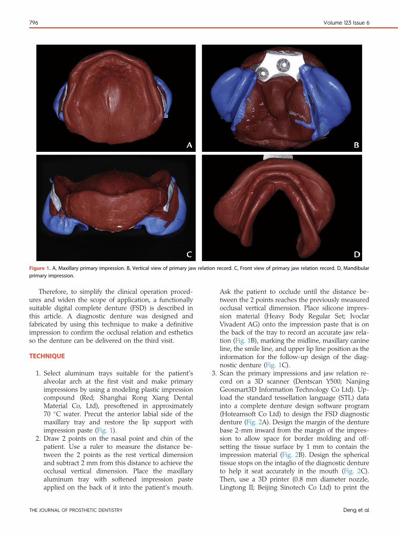
Figure 1. A, Maxillary primary impression. B, Vertical view of primary jaw relation record. C, Front view of primary jaw relation record. D, Mandibularprimary impression.
796 Volume 123 Issue 6
Therefore, to simplify the clinical operation proced-ures and widen the scope of application, a functionallysuitable digital complete denture (FSD) is described inthis article. A diagnostic denture was designed andfabricated by using this technique to make a definitiveimpression to confirm the occlusal relation and estheticsso the denture can be delivered on the third visit.
TECHNIQUE
1. Select aluminum trays suitable for the patient’salveolar arch at the first visit and make primaryimpressions by using a modeling plastic impressioncompound (Red; Shanghai Rong Xiang DentalMaterial Co, Ltd), presoftened in approximately70 �C water. Precut the anterior labial side of themaxillary tray and restore the lip support withimpression paste (Fig. 1).
2. Draw 2 points on the nasal point and chin of thepatient. Use a ruler to measure the distance be-tween the 2 points as the rest vertical dimensionand subtract 2 mm from this distance to achieve theocclusal vertical dimension. Place the maxillaryaluminum tray with softened impression pasteapplied on the back of it into the patient’s mouth.
THE JOURNAL OF PROSTHETIC DENTISTRY
Ask the patient to occlude until the distance be-tween the 2 points reaches the previously measuredocclusal vertical dimension. Place silicone impres-sion material (Heavy Body Regular Set; IvoclarVivadent AG) onto the impression paste that is onthe back of the tray to record an accurate jaw rela-tion (Fig. 1B), marking the midline, maxillary canineline, the smile line, and upper lip line position as theinformation for the follow-up design of the diag-nostic denture (Fig. 1C).
3. Scan the primary impressions and jaw relation re-cord on a 3D scanner (Dentscan Y500; NanjingGeosmart3D Information Technology Co Ltd). Up-load the standard tessellation language (STL) datainto a complete denture design software program(Hoteamsoft Co Ltd) to design the FSD diagnosticdenture (Fig. 2A). Design the margin of the denturebase 2-mm inward from the margin of the impres-sion to allow space for border molding and off-setting the tissue surface by 1 mm to contain theimpression material (Fig. 2B). Design the sphericaltissue stops on the intaglio of the diagnostic dentureto help it seat accurately in the mouth (Fig. 2C).Then, use a 3D printer (0.8 mm diameter nozzle,Lingtong II; Beijing Sinotech Co Ltd) to print the
Deng et al
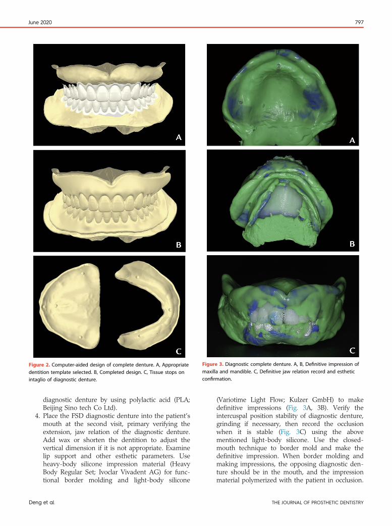
Figure 2. Computer-aided design of complete denture. A, Appropriatedentition template selected. B, Completed design. C, Tissue stops onintaglio of diagnostic denture.
Figure 3. Diagnostic complete denture. A, B, Definitive impression ofmaxilla and mandible. C, Definitive jaw relation record and estheticconfirmation.
June 2020 797
De
diagnostic denture by using polylactic acid (PLA;Beijing Sino tech Co Ltd).
4. Place the FSD diagnostic denture into the patient’smouth at the second visit, primary verifying theextension, jaw relation of the diagnostic denture.Add wax or shorten the dentition to adjust thevertical dimension if it is not appropriate. Examinelip support and other esthetic parameters. Useheavy-body silicone impression material (HeavyBody Regular Set; Ivoclar Vivadent AG) for func-tional border molding and light-body silicone
ng et al
(Variotime Light Flow; Kulzer GmbH) to makedefinitive impressions (Fig. 3A, 3B). Verify theintercuspal position stability of diagnostic denture,grinding if necessary, then record the occlusionwhen it is stable (Fig. 3C) using the abovementioned light-body silicone. Use the closed-mouth technique to border mold and make thedefinitive impression. When border molding andmaking impressions, the opposing diagnostic den-ture should be in the mouth, and the impressionmaterial polymerized with the patient in occlusion.
THE JOURNAL OF PROSTHETIC DENTISTRY
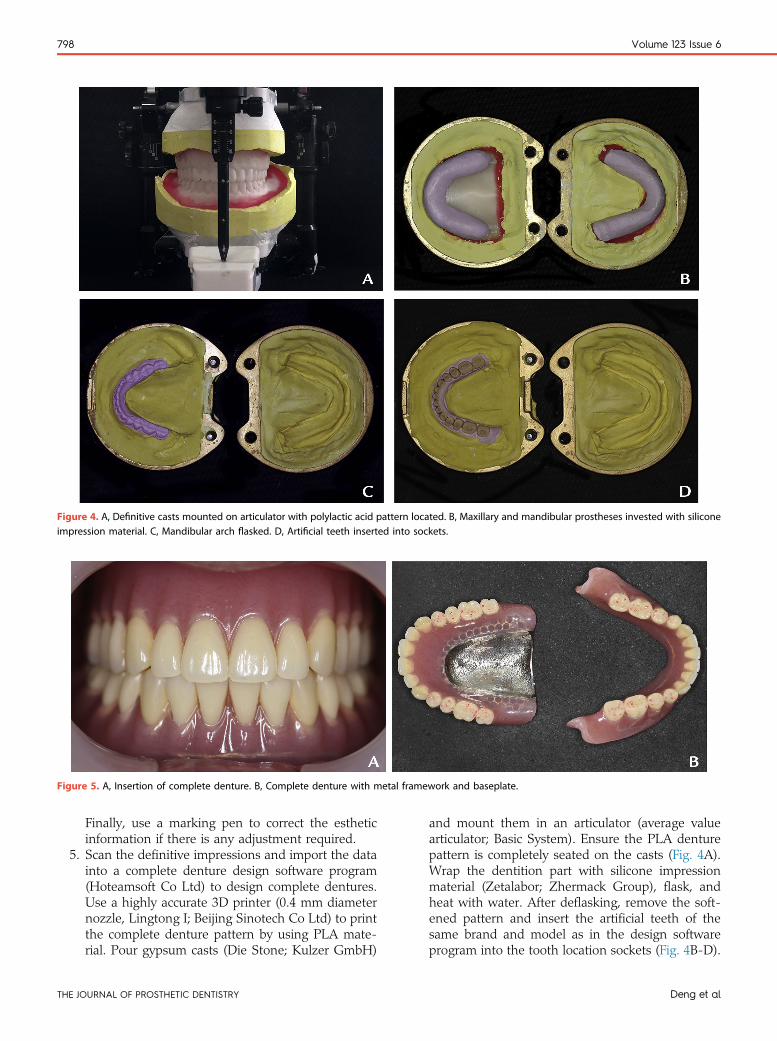
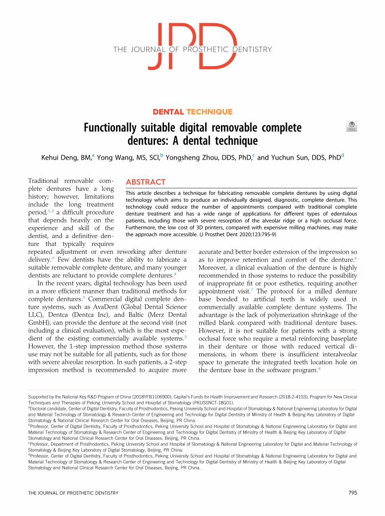
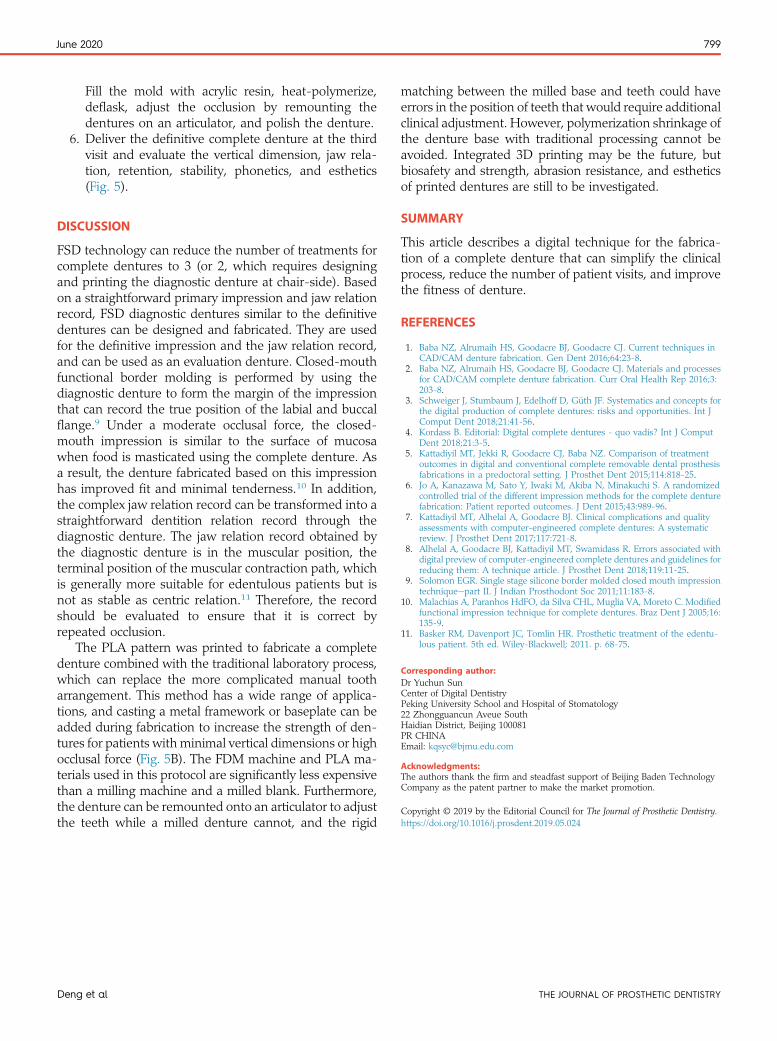
Figure 4. A, Definitive casts mounted on articulator with polylactic acid pattern located. B, Maxillary and mandibular prostheses invested with siliconeimpression material. C, Mandibular arch flasked. D, Artificial teeth inserted into sockets.
Figure 5. A, Insertion of complete denture. B, Complete denture with metal framework and baseplate.
798 Volume 123 Issue 6
TH
Finally, use a marking pen to correct the estheticinformation if there is any adjustment required.
5. Scan the definitive impressions and import the datainto a complete denture design software program(Hoteamsoft Co Ltd) to design complete dentures.Use a highly accurate 3D printer (0.4 mm diameternozzle, Lingtong I; Beijing Sinotech Co Ltd) to printthe complete denture pattern by using PLA mate-rial. Pour gypsum casts (Die Stone; Kulzer GmbH)
E JOURNAL OF PROSTHETIC DENTISTRY
and mount them in an articulator (average valuearticulator; Basic System). Ensure the PLA denturepattern is completely seated on the casts (Fig. 4A).Wrap the dentition part with silicone impressionmaterial (Zetalabor; Zhermack Group), flask, andheat with water. After deflasking, remove the soft-ened pattern and insert the artificial teeth of thesame brand and model as in the design softwareprogram into the tooth location sockets (Fig. 4B-D).
Deng et al
June 2020 799
De
Fill the mold with acrylic resin, heat-polymerize,deflask, adjust the occlusion by remounting thedentures on an articulator, and polish the denture.
6. Deliver the definitive complete denture at the thirdvisit and evaluate the vertical dimension, jaw rela-tion, retention, stability, phonetics, and esthetics(Fig. 5).
DISCUSSION
FSD technology can reduce the number of treatments forcomplete dentures to 3 (or 2, which requires designingand printing the diagnostic denture at chair-side). Basedon a straightforward primary impression and jaw relationrecord, FSD diagnostic dentures similar to the definitivedentures can be designed and fabricated. They are usedfor the definitive impression and the jaw relation record,and can be used as an evaluation denture. Closed-mouthfunctional border molding is performed by using thediagnostic denture to form the margin of the impressionthat can record the true position of the labial and buccalflange.9 Under a moderate occlusal force, the closed-mouth impression is similar to the surface of mucosawhen food is masticated using the complete denture. Asa result, the denture fabricated based on this impressionhas improved fit and minimal tenderness.10 In addition,the complex jaw relation record can be transformed into astraightforward dentition relation record through thediagnostic denture. The jaw relation record obtained bythe diagnostic denture is in the muscular position, theterminal position of the muscular contraction path, whichis generally more suitable for edentulous patients but isnot as stable as centric relation.11 Therefore, the recordshould be evaluated to ensure that it is correct byrepeated occlusion.
The PLA pattern was printed to fabricate a completedenture combined with the traditional laboratory process,which can replace the more complicated manual tootharrangement. This method has a wide range of applica-tions, and casting a metal framework or baseplate can beadded during fabrication to increase the strength of den-tures for patients with minimal vertical dimensions or highocclusal force (Fig. 5B). The FDM machine and PLA ma-terials used in this protocol are significantly less expensivethan a milling machine and a milled blank. Furthermore,the denture can be remounted onto an articulator to adjustthe teeth while a milled denture cannot, and the rigid
ng et al
matching between the milled base and teeth could haveerrors in the position of teeth that would require additionalclinical adjustment. However, polymerization shrinkage ofthe denture base with traditional processing cannot beavoided. Integrated 3D printing may be the future, butbiosafety and strength, abrasion resistance, and estheticsof printed dentures are still to be investigated.
SUMMARY
This article describes a digital technique for the fabrica-tion of a complete denture that can simplify the clinicalprocess, reduce the number of patient visits, and improvethe fitness of denture.
REFERENCES
1. Baba NZ, Alrumaih HS, Goodacre BJ, Goodacre CJ. Current techniques inCAD/CAM denture fabrication. Gen Dent 2016;64:23-8.
2. Baba NZ, Alrumaih HS, Goodacre BJ, Goodacre CJ. Materials and processesfor CAD/CAM complete denture fabrication. Curr Oral Health Rep 2016;3:203-8.
3. Schweiger J, Stumbaum J, Edelhoff D, Güth JF. Systematics and concepts forthe digital production of complete dentures: risks and opportunities. Int JComput Dent 2018;21:41-56.
4. Kordass B. Editorial: Digital complete dentures - quo vadis? Int J ComputDent 2018;21:3-5.
5. Kattadiyil MT, Jekki R, Goodacre CJ, Baba NZ. Comparison of treatmentoutcomes in digital and conventional complete removable dental prosthesisfabrications in a predoctoral setting. J Prosthet Dent 2015;114:818-25.
6. Jo A, Kanazawa M, Sato Y, Iwaki M, Akiba N, Minakuchi S. A randomizedcontrolled trial of the different impression methods for the complete denturefabrication: Patient reported outcomes. J Dent 2015;43:989-96.
7. Kattadiyil MT, Alhelal A, Goodacre BJ. Clinical complications and qualityassessments with computer-engineered complete dentures: A systematicreview. J Prosthet Dent 2017;117:721-8.
8. Alhelal A, Goodacre BJ, Kattadiyil MT, Swamidass R. Errors associated withdigital preview of computer-engineered complete dentures and guidelines forreducing them: A technique article. J Prosthet Dent 2018;119:11-25.
9. Solomon EGR. Single stage silicone border molded closed mouth impressiontechniquedpart II. J Indian Prosthodont Soc 2011;11:183-8.
10. Malachias A, Paranhos HdFO, da Silva CHL, Muglia VA, Moreto C. Modifiedfunctional impression technique for complete dentures. Braz Dent J 2005;16:135-9.
11. Basker RM, Davenport JC, Tomlin HR. Prosthetic treatment of the edentu-lous patient. 5th ed. Wiley-Blackwell; 2011. p. 68-75.
Corresponding author:Dr Yuchun SunCenter of Digital DentistryPeking University School and Hospital of Stomatology22 Zhongguancun Aveue SouthHaidian District, Beijing 100081PR CHINAEmail: [email protected]
Acknowledgments:The authors thank the firm and steadfast support of Beijing Baden TechnologyCompany as the patent partner to make the market promotion.
Copyright © 2019 by the Editorial Council for The Journal of Prosthetic Dentistry.https://doi.org/10.1016/j.prosdent.2019.05.024
THE JOURNAL OF PROSTHETIC DENTISTRY