Embed Size (px)
Citation preview
Journal of American Science 2013;9(1) http://www.jofamericanscience.org
508
Effect of Different Clasps on Asymmetric Distal Extension Base Supporting Structures
Fatma ELzahraa Awad1, Asma Gadallah2, Hana’a Gamal Youssef 3 ,Yasser Shawky3
1- Professor of Removable Prosthodontics, Faculty of Oral and Dental Medicine, Ain Shams University, Cairo, Egypt.
2- Professor of Removable Prosthodontics, Faculty of Oral and Dental Medicine, Misr International University, Cairo, Egypt.
3- Lecturer of Removable Prosthodontics, Faculty of Oral and Dental Medicine, Misr International University, Cairo, Egypt.
Corresponding author: [email protected] Abstract: The aim of this study was to assess the stresses induced by RPI, I-bar and RPL clasps and their effect on bone height of the structures supporting asymmetric distal extension partial dentures. Materials and methods: The stresses induced by the three partial denture designs and their effect on marginal and crestal bone loss were compared. In-vitro strain gauge analysis was used to measure the stresses induced by the three direct retainers. The change in marginal and crestal bone height was measured using radiovisography (RVG). Results: Comparing the micro-strains induced by placement of the RPL clasp on the abutments, revealed statistically significant less micro-strains induced to abutments on both short and long sides of the ridge compared to RPI and I-bar clasps. The placement of I-bar clasp on short side abutment induced the greatest & statistically significant micro-strains to the short side of the ridge but the least statistically significant micro-strains on the long side of the ridge, while the stresses induced by placement of the RPI clasp on the short and long sides of the ridge were statistically insignificant. Statistically significant less value of marginal bone loss of abutments bearing RPL clasps was detected at the end of the study period compared to that caused by RPI and I-bar clasps. However, no statistical significant difference was noted between the effect of RPI and I-bar clasps at the end of the study period. Conclusion: The results revealed that asymmetric partial dentures retained on the short side by RPL clasp induced less stresses and caused less bone resorption compared to dentures retained by RPI clasp and I-bar clasp. [Fatma ELzahraa Awad, Asma Gadallah, Hana’a Gamal Youssef, Yasser Shawky. Effect of Different Clasps on Asymmetric Distal Extension Base Supporting Structures. J Am Sci 2013;9(1):508-519]. (ISSN: 1545-1003). http://www.jofamericanscience.org. 74
Key words: Asymmetric distal extension removable partial denture, RPI clasp, I-bar clasp, RPL clasp, strain gauge analysis, bone height. Introduction Distal extension removable partial dentures (RPDs) are supported and retained against functionally displacing forces by two structures that differ markedly in their visco-elastic nature; the abutment teeth and the residual ridge. The result is rotation of the partial denture about its most distal abutments during function causing trauma to the supporting structures, Monteith(1984); Budtz-Jorgnsen and Bochet(1998); McGivney, and Carr (2000). Hence, rehabilitation with distal extension RPD necessitates precise partial denture design following biomechanical principles and considerations. Forces that produce torque on abutment teeth should thus be controlled and minimized when designing RPDs, DeFranco(1984); Hindels(2001). An axis of rotation is created through the most distally placed occlusal rests when a distal extension RPD is loaded. In the symmetric bilateral distal extension RPD, the axis of rotation is perpendicular to the longitudinal axis of the denture base, McGivney, and Carr(2000).While in asymmetric bases, this axis of rotation is not perpendicular to the residual ridge and is
shifted distally towards the shorter side, Aviv et al., (1989).
Rotation of the denture induces torque effect comparable to the action of class I lever and causes mobility of the clasped teeth especially if a rigid clasp is used or if the clasp tip is placed mesial to the axis of rotation, Clayton and Jaslow (1971); Tebrock et al.,(1979).The selection of suitable direct retainers is thus a key factor contributing to the preservation of RPD supporting structures Ben-Ur et al.,(1996); Igrashi et al.,(1999). The RPI clasp and the I-bar clasp exhibiting a retentive tip placed distal to the fulcrum of rotation of the RPD are commonly used for retaining distal extension bases (DEBs), Kratochvil(1963). This is due to the stress releasing effect and tooth disengagement provided by applying the mechanical base of a class II lever, Krol(1973). However, in asymmetric DEBs, the retentive tip of the RPI clasp is too close to the fulcrum axis on the short edentulous side. This in turn causes DEBs to function as a class I lever, Browning et al.,(1986). For this reason, the RPL clasp exhibiting a

Journal of American Science 2013;9(1) http://www.jofamericanscience.org
509
retentive tip engaging a more distal undercut placed farther from the mesially placed rest could be a more effective retainer in these cases. The L-bar arm is thus proposed to disengage the tooth when rotation of the denture occurs around the mesio-occlusal rest, Ben-Ur(1988;1996).
Functional forces transmitted to DEBs are transferred to the denture supporting structures .This stimulates bone remodeling ;however, pressure beyond the physiological tolerance of bone may lead to bone resorption, Klemetti(1996);Wyatt(1998). The effect of the applied force depends on the distribution and magnitude of the applied force, condition of abutments, quality of the ridge and partial denture design, Wyatt(1998). The study of induced stresses is thus vital for providing valid data to aid in proper biomechanical design. This could be achieved by in-vitro studies including the strain gauge technology, Kratochvil et al., (1981); Perry and Lissner(1992).
Intra-oral digital radiography plays an important role in the diagnosis of the periodontal structure of teeth and in assessment of the condition of the residual ridge. Subsequent radiographs are usually used for comparing changes in linear, volumetric and area changes in bone support, Ben-Ur(1988). Aim of the Study The aim of this study was to assess the stresses induced by RPI, I-bar and RPL clasps and compare their effect on bone height of the structures supporting asymmetric distal extension partial dentures.
This study was first carried out in-vitro to obtain valid and accurate experimental data that were further assessed clinically inside the patients' mouth. The goal was first to obtain data away from the effect of differences in human factors as well as the anatomic and physiologic variations that may occur in the same patient and finally confirm these data in the oral environment. 2.Materials and Methods: 2.1.Materials 2.1.1. In-vitro stress analysis Samples: This study was conducted on an acrylic model representing asymmetric Kennedy class I, ;Wyatt(1998). The abutments were canine on one side and second premolar on the other side. The model was modified to simulate the anatomy and the visco-elastic properties of the structures supporting the tested partial dentures. The roots of abutments and the surface of the model were thus covered by a resilient silicon material whose thickness was 0.3 and 2mm respectively. 2.1.2. In-vivo stress analysis Samples: Twelve patients* were selected from the outpatient clinic, Prosthodontic Department, Faculty of Dentistry, Misr International University. Patients were selected, thoroughly examined and prepared to eliminate any factor that may adversely affect the results of this
study. The selected patients exhibited asymmetric bilateral free end edentulous areas. (*All patients participating in this study had good intention for prosthodontic treatment and the motivation for maintaining oral hygiene, hence, they responded regularly to the frequent recall appointments and to oral and denture hygiene instructions). Criteria of Patients selection: 1-Patients having an opposing dentate maxillary arch were selected in an attempt to reduce the effect of variation in forces that could be induced by different restorations on bone supporting the mandibular partial dentures ,Plotnik et al.,(1975); Wyatt (1998).
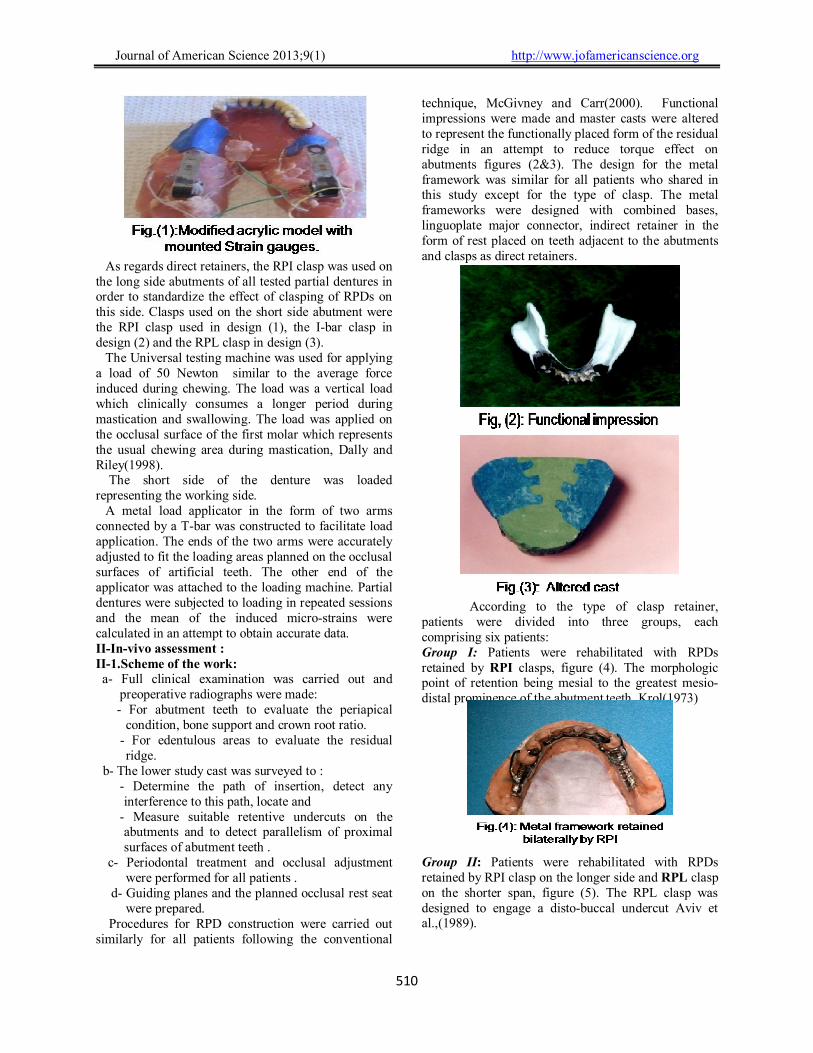
2-Patients with severe positional changes in the remaining natural dentition were excluded to avoid the effect of irregular or improperly inclined occlusal plane on abutments, residual ridge and on the stability of the partial dentures, McGivney and Carr (2000). 3-Patients exhibiting abnormal jaw relation-ship and abnormal oral habits were excluded to avoid the effect of the resulting off vertical forces on the partial denture supporting structures. 2.2.Methods: 2.2.I In Vitro studies: The acrylic model was modified to receive strain gauges figure (1). The strain gauges were cautiously mounted on the cast bilaterally at the distal ends of the edentulous ridge. Dummy gauges were also used as previously recommended in an attempt to equalize changes in ambient temperature of the main gauge thus allowing for calibration of micro-strains induced by load application rather than by change in ambient temperature ,Aydilink and Tekkaya(1992); Asundi and Kishen(2000). In an attempt to reduce the effect of differences in the properties of materials, and changes in partial denture designs on the strains induced to denture supporting structures ;all RPDs had the same design and were constructed using the same materials and the same construction technique. They only varied in the type of clasp used on the short side of the partial denture. These clasps were the RPI, I-bar and RPL clasps. The RPI and I-bar clasps were tested as they are most commonly used for retaining DEBs due to their reported stress releasing effect, Krol (1973).The RPL clasp was used in order to assess the effect of using a clasp engaging an undercut comparatively more distal to the fulcrum line & proposed to provide a better biomechanical effect, Ben-Ur(1996).Partial dentures were designed with bilaterally placed combined bases supporting cross linked acrylic teeth set on the crest of the ridge, cingulum rests as indirect retainers and lingual plate major connector.
Journal of American Science 2013;9(1) http://www.jofamericanscience.org
510
As regards direct retainers, the RPI clasp was used on the long side abutments of all tested partial dentures in order to standardize the effect of clasping of RPDs on this side. Clasps used on the short side abutment were the RPI clasp used in design (1), the I-bar clasp in design (2) and the RPL clasp in design (3). The Universal testing machine was used for applying a load of 50 Newton similar to the average force induced during chewing. The load was a vertical load which clinically consumes a longer period during mastication and swallowing. The load was applied on the occlusal surface of the first molar which represents the usual chewing area during mastication, Dally and Riley(1998). The short side of the denture was loaded representing the working side. A metal load applicator in the form of two arms connected by a T-bar was constructed to facilitate load application. The ends of the two arms were accurately adjusted to fit the loading areas planned on the occlusal surfaces of artificial teeth. The other end of the applicator was attached to the loading machine. Partial dentures were subjected to loading in repeated sessions and the mean of the induced micro-strains were calculated in an attempt to obtain accurate data. II-In-vivo assessment : II-1.Scheme of the work: a- Full clinical examination was carried out and
preoperative radiographs were made: - For abutment teeth to evaluate the periapical
condition, bone support and crown root ratio. - For edentulous areas to evaluate the residual
ridge. b- The lower study cast was surveyed to :
- Determine the path of insertion, detect any interference to this path, locate and
- Measure suitable retentive undercuts on the abutments and to detect parallelism of proximal surfaces of abutment teeth .
c- Periodontal treatment and occlusal adjustment were performed for all patients .
d- Guiding planes and the planned occlusal rest seat were prepared.
Procedures for RPD construction were carried out similarly for all patients following the conventional
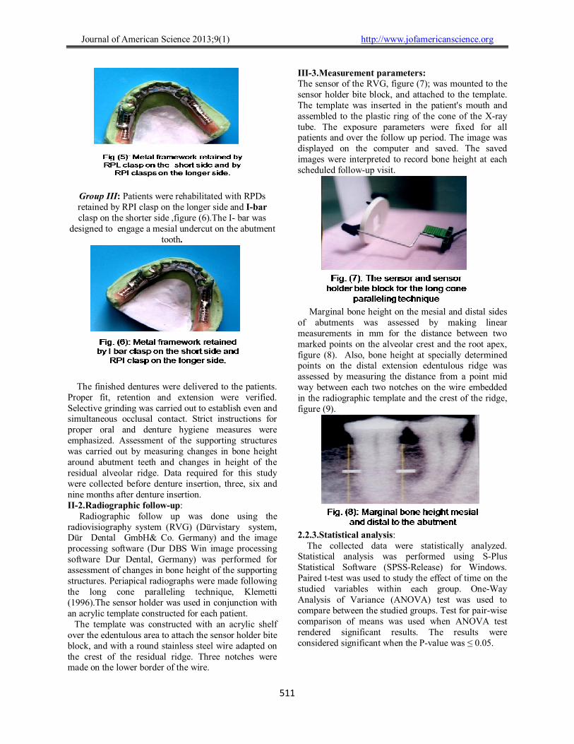
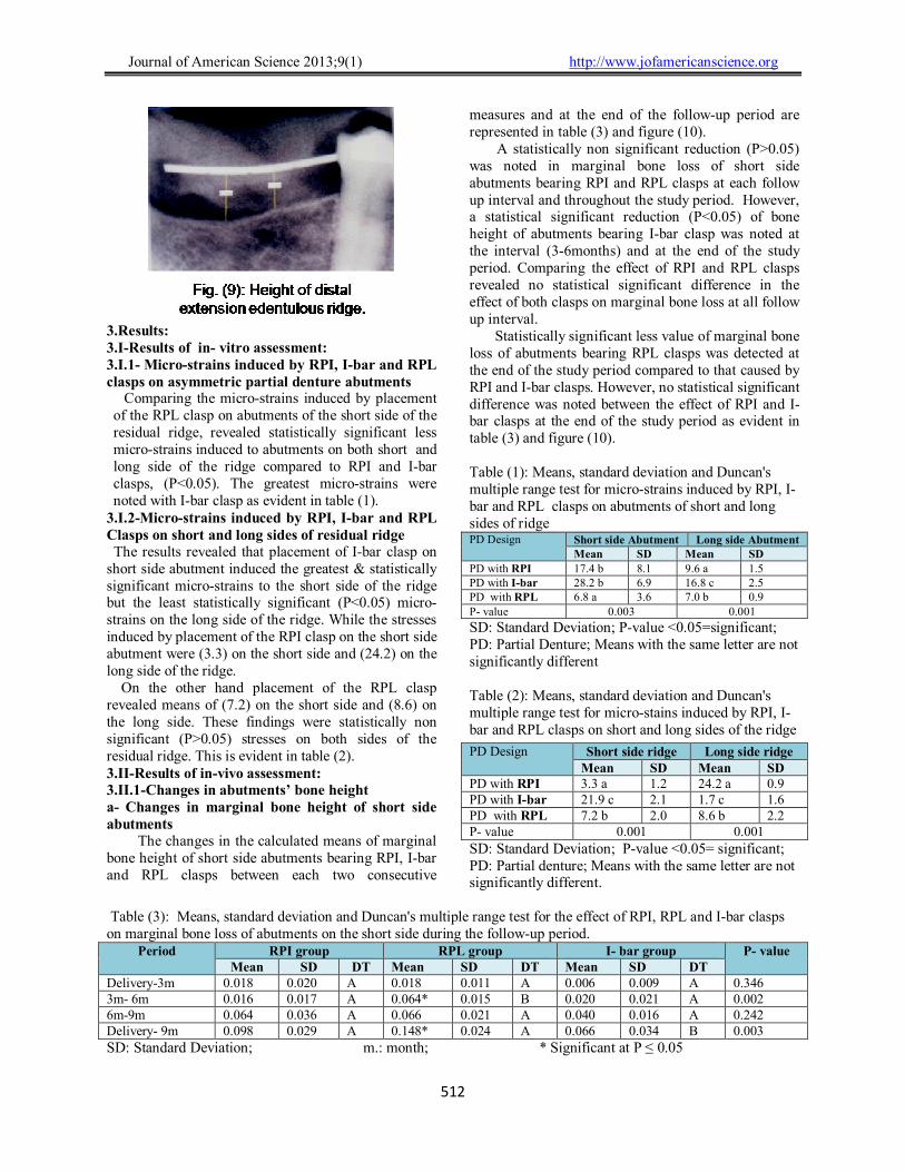
technique, McGivney and Carr(2000). Functional impressions were made and master casts were altered to represent the functionally placed form of the residual ridge in an attempt to reduce torque effect on abutments figures (2&3). The design for the metal framework was similar for all patients who shared in this study except for the type of clasp. The metal frameworks were designed with combined bases, linguoplate major connector, indirect retainer in the form of rest placed on teeth adjacent to the abutments and clasps as direct retainers.
According to the type of clasp retainer,
patients were divided into three groups, each comprising six patients: Group I: Patients were rehabilitated with RPDs retained by RPI clasps, figure (4). The morphologic point of retention being mesial to the greatest mesio-distal prominence of the abutment teeth, Krol(1973)
Group II: Patients were rehabilitated with RPDs retained by RPI clasp on the longer side and RPL clasp on the shorter span, figure (5). The RPL clasp was designed to engage a disto-buccal undercut Aviv et al.,(1989).
Journal of American Science 2013;9(1) http://www.jofamericanscience.org
511
Group III: Patients were rehabilitated with RPDs retained by RPI clasp on the longer side and I-bar clasp on the shorter side ,figure (6).The I- bar was
designed to engage a mesial undercut on the abutment tooth.
The finished dentures were delivered to the patients. Proper fit, retention and extension were verified. Selective grinding was carried out to establish even and simultaneous occlusal contact. Strict instructions for proper oral and denture hygiene measures were emphasized. Assessment of the supporting structures was carried out by measuring changes in bone height around abutment teeth and changes in height of the residual alveolar ridge. Data required for this study were collected before denture insertion, three, six and nine months after denture insertion. II-2.Radiographic follow-up: Radiographic follow up was done using the radiovisiography system (RVG) (Dürvistary system, Dür Dental GmbH& Co. Germany) and the image processing software (Dur DBS Win image processing software Dur Dental, Germany) was performed for assessment of changes in bone height of the supporting structures. Periapical radiographs were made following the long cone paralleling technique, Klemetti (1996).The sensor holder was used in conjunction with an acrylic template constructed for each patient. The template was constructed with an acrylic shelf over the edentulous area to attach the sensor holder bite block, and with a round stainless steel wire adapted on the crest of the residual ridge. Three notches were made on the lower border of the wire.
III-3.Measurement parameters: The sensor of the RVG, figure (7); was mounted to the sensor holder bite block, and attached to the template. The template was inserted in the patient's mouth and assembled to the plastic ring of the cone of the X-ray tube. The exposure parameters were fixed for all patients and over the follow up period. The image was displayed on the computer and saved. The saved images were interpreted to record bone height at each scheduled follow-up visit.
Marginal bone height on the mesial and distal sides of abutments was assessed by making linear measurements in mm for the distance between two marked points on the alveolar crest and the root apex, figure (8). Also, bone height at specially determined points on the distal extension edentulous ridge was assessed by measuring the distance from a point mid way between each two notches on the wire embedded in the radiographic template and the crest of the ridge, figure (9).
2.2.3.Statistical analysis: The collected data were statistically analyzed. Statistical analysis was performed using S-Plus Statistical Software (SPSS-Release) for Windows. Paired t-test was used to study the effect of time on the studied variables within each group. One-Way Analysis of Variance (ANOVA) test was used to compare between the studied groups. Test for pair-wise comparison of means was used when ANOVA test rendered significant results. The results were considered significant when the P-value was ≤ 0.05.
Journal of American Science 2013;9(1) http://www.jofamericanscience.org
512
3.Results: 3.I-Results of in- vitro assessment: 3.I.1- Micro-strains induced by RPI, I-bar and RPL clasps on asymmetric partial denture abutments Comparing the micro-strains induced by placement of the RPL clasp on abutments of the short side of the residual ridge, revealed statistically significant less micro-strains induced to abutments on both short and long side of the ridge compared to RPI and I-bar clasps, (P<0.05). The greatest micro-strains were noted with I-bar clasp as evident in table (1).
3.I.2-Micro-strains induced by RPI, I-bar and RPL Clasps on short and long sides of residual ridge The results revealed that placement of I-bar clasp on short side abutment induced the greatest & statistically significant micro-strains to the short side of the ridge but the least statistically significant (P<0.05) micro-strains on the long side of the ridge. While the stresses induced by placement of the RPI clasp on the short side abutment were (3.3) on the short side and (24.2) on the long side of the ridge. On the other hand placement of the RPL clasp revealed means of (7.2) on the short side and (8.6) on the long side. These findings were statistically non significant (P>0.05) stresses on both sides of the residual ridge. This is evident in table (2). 3.II-Results of in-vivo assessment: 3.II.1-Changes in abutments’ bone height a- Changes in marginal bone height of short side abutments The changes in the calculated means of marginal bone height of short side abutments bearing RPI, I-bar and RPL clasps between each two consecutive
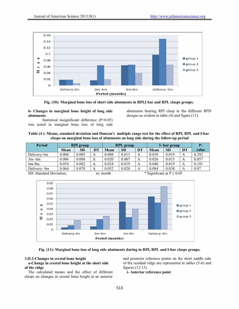
measures and at the end of the follow-up period are represented in table (3) and figure (10). A statistically non significant reduction (P>0.05) was noted in marginal bone loss of short side abutments bearing RPI and RPL clasps at each follow up interval and throughout the study period. However, a statistical significant reduction (P<0.05) of bone height of abutments bearing I-bar clasp was noted at the interval (3-6months) and at the end of the study period. Comparing the effect of RPI and RPL clasps revealed no statistical significant difference in the effect of both clasps on marginal bone loss at all follow up interval. Statistically significant less value of marginal bone loss of abutments bearing RPL clasps was detected at the end of the study period compared to that caused by RPI and I-bar clasps. However, no statistical significant difference was noted between the effect of RPI and I-bar clasps at the end of the study period as evident in table (3) and figure (10). Table (1): Means, standard deviation and Duncan's multiple range test for micro-strains induced by RPI, I-bar and RPL clasps on abutments of short and long sides of ridge PD Design Short side Abutment Long side Abutment
Mean SD Mean SD PD with RPI 17.4 b 8.1 9.6 a 1.5 PD with I-bar 28.2 b 6.9 16.8 c 2.5 PD with RPL 6.8 a 3.6 7.0 b 0.9 P- value 0.003 0.001
SD: Standard Deviation; P-value <0.05=significant; PD: Partial Denture; Means with the same letter are not significantly different
Table (2): Means, standard deviation and Duncan's multiple range test for micro-stains induced by RPI, I-bar and RPL clasps on short and long sides of the ridge
SD: Standard Deviation; P-value <0.05= significant; PD: Partial denture; Means with the same letter are not significantly different.
Table (3): Means, standard deviation and Duncan's multiple range test for the effect of RPI, RPL and I-bar clasps on marginal bone loss of abutments on the short side during the follow-up period.
Period RPI group RPL group I- bar group P- value Mean SD DT Mean SD DT Mean SD DT
Delivery-3m 0.018 0.020 A 0.018 0.011 A 0.006 0.009 A 0.346 3m- 6m 0.016 0.017 A 0.064* 0.015 B 0.020 0.021 A 0.002 6m-9m 0.064 0.036 A 0.066 0.021 A 0.040 0.016 A 0.242 Delivery- 9m 0.098 0.029 A 0.148* 0.024 A 0.066 0.034 B 0.003
SD: Standard Deviation; m.: month; * Significant at P ≤ 0.05
PD Design Short side ridge Long side ridge Mean SD Mean SD
PD with RPI 3.3 a 1.2 24.2 a 0.9 PD with I-bar 21.9 c 2.1 1.7 c 1.6 PD with RPL 7.2 b 2.0 8.6 b 2.2 P- value 0.001 0.001
Journal of American Science 2013;9(1) http://www.jofamericanscience.org
513
Fig. (10): Marginal bone loss of short side abutments in RPI,I bar and RPL clasps groups. b- Changes in marginal bone height of long side abutments
Statistical insignificant difference (P>0.05) was noted in marginal bone loss of long side
abutments bearing RPI clasp in the different RPD designs as evident in table (4) and figure (11).
Table (4 ): Means, standard deviation and Duncan's multiple range test for the effect of RPI, RPL and I-bar
clasps on marginal bone loss of abutments on long side during the follow-up period
SD: Standard Deviation; m: month * Significant at P ≤ 0.05
Fig. (11): Marginal bone loss of long side abutments during in RPI, RPL and I-bar clasps groups. 3.II.2-Changes in crestal bone height
a-Change in crestal bone height at the short side of the ridge
The calculated means and the effect of different clasps on changes in crestal bone height at an anterior
and posterior reference points on the short saddle side of the residual ridge are represented in tables (5-6) and figures (12-13).
i- Anterior reference point
Period RPI group RPL group I- bar group P- value Mean SD DT Mean SD DT Mean SD DT
Delivery-3m 0.004 0.005 A 0.008 0.013 A 0.018 0.019 A 0.292 3m- 6m 0.006 0.008 A 0.020 0.007 A 0.026 0.015 A 0.057 6m-9m 0.054 0.082 A 0.024 0.019 A 0.040 0.019 A 0.191 Delivery- 9m 0.064 0.078 A 0.052 0.028 A 0.084 0.038 A 0.07
Journal of American Science 2013;9(1) http://www.jofamericanscience.org
514
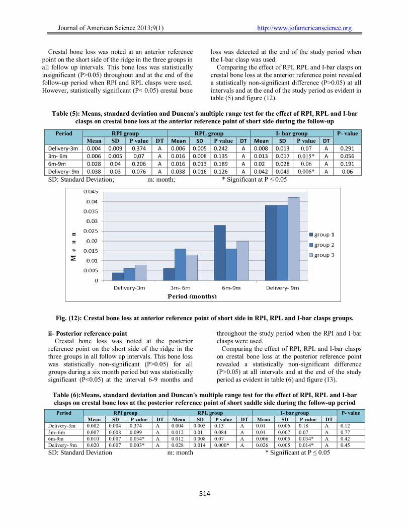
Crestal bone loss was noted at an anterior reference point on the short side of the ridge in the three groups in all follow up intervals. This bone loss was statistically insignificant (P>0.05) throughout and at the end of the follow-up period when RPI and RPL clasps were used. However, statistically significant (P< 0.05) crestal bone
loss was detected at the end of the study period when the I-bar clasp was used.
Comparing the effect of RPI, RPL and I-bar clasps on crestal bone loss at the anterior reference point revealed a statistically non-significant difference (P>0.05) at all intervals and at the end of the study period as evident in table (5) and figure (12).
Table (5): Means, standard deviation and Duncan's multiple range test for the effect of RPI, RPL and I-bar
clasps on crestal bone loss at the anterior reference point of short side during the follow-up
SD: Standard Deviation; m: month; * Significant at P ≤ 0.05
Fig. (12): Crestal bone loss at anterior reference point of short side in RPI, RPL and I-bar clasps groups.
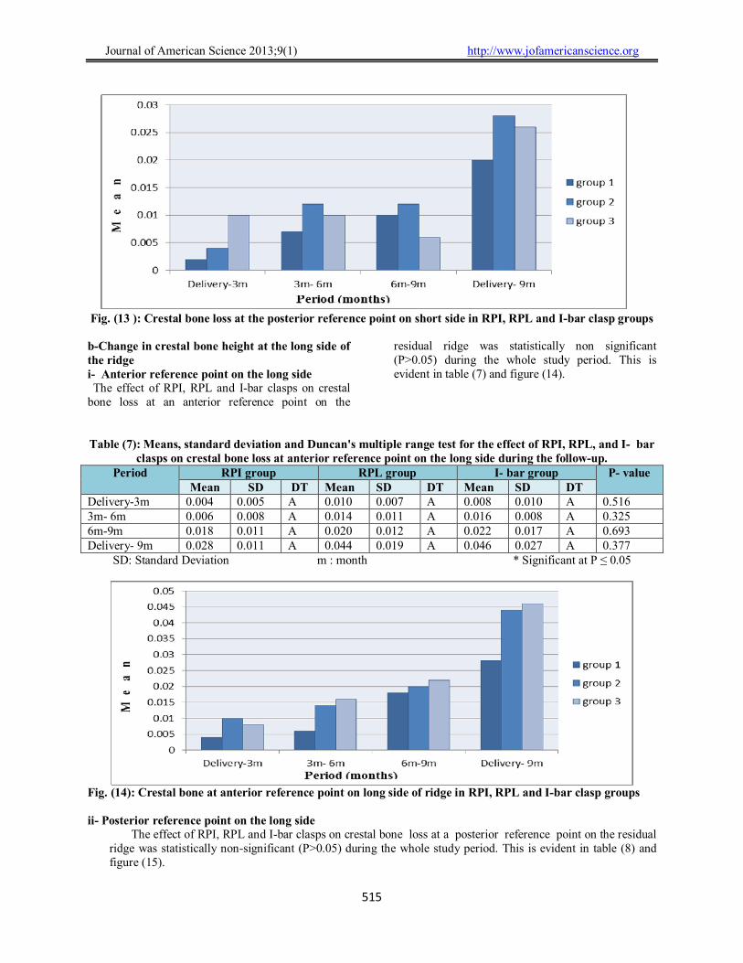
ii- Posterior reference point Crestal bone loss was noted at the posterior reference point on the short side of the ridge in the three groups in all follow up intervals. This bone loss was statistically non-significant (P>0.05) for all groups during a six month period but was statistically significant (P<0.05) at the interval 6-9 months and
throughout the study period when the RPI and I-bar clasps were used. Comparing the effect of RPI, RPL and I-bar clasps on crestal bone loss at the posterior reference point revealed a statistically non-significant difference (P>0.05) at all intervals and at the end of the study period as evident in table (6) and figure (13).
Table (6):Means, standard deviation and Duncan's multiple range test for the effect of RPI, RPL and I-bar clasps on crestal bone loss at the posterior reference point of short saddle side during the follow-up period
SD: Standard Deviation m: month * Significant at P ≤ 0.05
Period RPI group RPL group I- bar group P- value Mean SD P value DT Mean SD P value DT Mean SD P value DT
Delivery-3m 0.004 0.009 0.374 A 0.006 0.005 0.242 A 0.008 0.013 0.07 A 0.291
3m- 6m 0.006 0.005 0,07 A 0.016 0.008 0.135 A 0.013 0.017 0.015* A 0.056
6m-9m 0.028 0.04 0.206 A 0.016 0.013 0.189 A 0.02 0.028 0.06 A 0.191
Delivery- 9m 0.038 0.03 0.076 A 0.038 0.016 0.126 A 0.042 0.049 0.006* A 0.06
Period RPI group RPL group I- bar group P- value Mean SD P value DT Mean SD P value DT Mean SD P value DT
Delivery-3m 0.002 0.004 0.374 A 0.004 0.005 0.13 A 0.01 0.006 0.18 A 0.12 3m- 6m 0.007 0.008 0.099 A 0.012 0.01 0.084 A 0.01 0.007 0.07 A 0.77 6m-9m 0.010 0.007 0.034* A 0.012 0.008 0.07 A 0.006 0.005 0.034* A 0.42 Delivery- 9m 0.020 0.007 0.003* A 0.028 0.014 0.000* A 0.026 0.005 0.014* A 0.45
Journal of American Science 2013;9(1) http://www.jofamericanscience.org
515
Fig. (13 ): Crestal bone loss at the posterior reference point on short side in RPI, RPL and I-bar clasp groups b-Change in crestal bone height at the long side of the ridge i- Anterior reference point on the long side The effect of RPI, RPL and I-bar clasps on crestal
bone loss at an anterior reference point on the
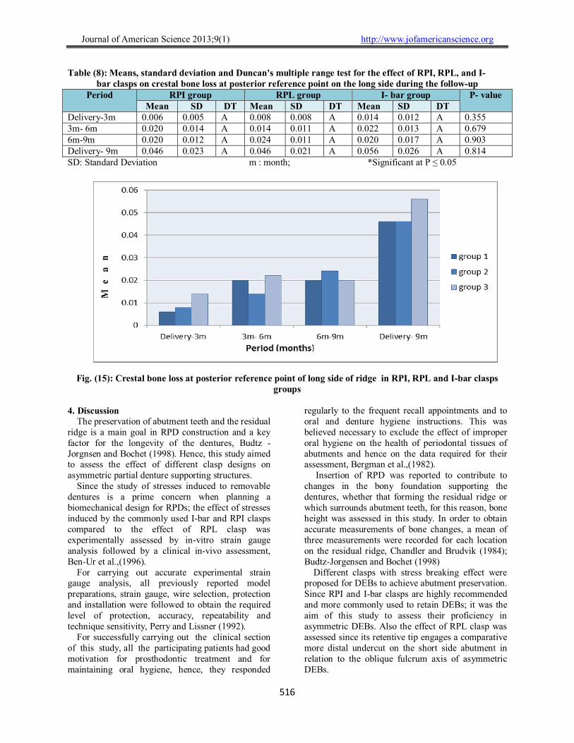
residual ridge was statistically non significant (P>0.05) during the whole study period. This is evident in table (7) and figure (14).
Table (7): Means, standard deviation and Duncan's multiple range test for the effect of RPI, RPL, and I- bar clasps on crestal bone loss at anterior reference point on the long side during the follow-up.
Period RPI group RPL group I- bar group P- value Mean SD DT Mean SD DT Mean SD DT
Delivery-3m 0.004 0.005 A 0.010 0.007 A 0.008 0.010 A 0.516 3m- 6m 0.006 0.008 A 0.014 0.011 A 0.016 0.008 A 0.325 6m-9m 0.018 0.011 A 0.020 0.012 A 0.022 0.017 A 0.693 Delivery- 9m 0.028 0.011 A 0.044 0.019 A 0.046 0.027 A 0.377
SD: Standard Deviation m : month * Significant at P ≤ 0.05
Fig. (14): Crestal bone at anterior reference point on long side of ridge in RPI, RPL and I-bar clasp groups ii- Posterior reference point on the long side
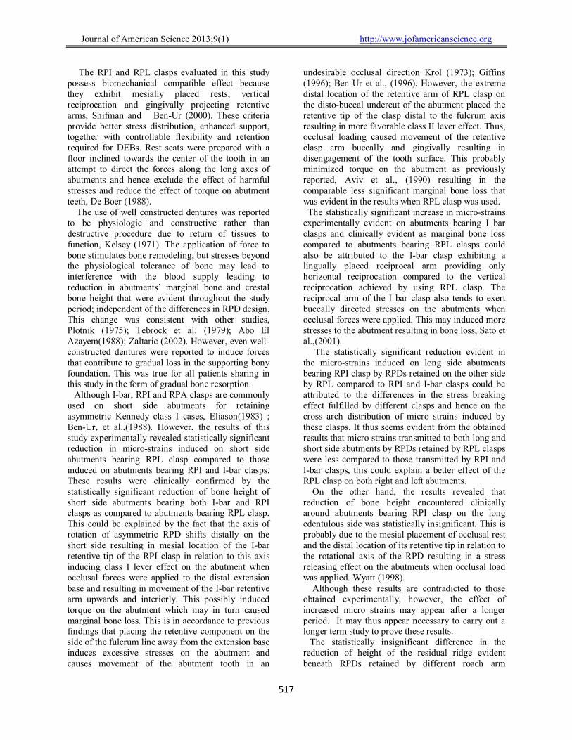
The effect of RPI, RPL and I-bar clasps on crestal bone loss at a posterior reference point on the residual ridge was statistically non-significant (P>0.05) during the whole study period. This is evident in table (8) and figure (15).
Journal of American Science 2013;9(1) http://www.jofamericanscience.org
516
Table (8): Means, standard deviation and Duncan's multiple range test for the effect of RPI, RPL, and I- bar clasps on crestal bone loss at posterior reference point on the long side during the follow-up
Period RPI group RPL group I- bar group P- value Mean SD DT Mean SD DT Mean SD DT
Delivery-3m 0.006 0.005 A 0.008 0.008 A 0.014 0.012 A 0.355 3m- 6m 0.020 0.014 A 0.014 0.011 A 0.022 0.013 A 0.679 6m-9m 0.020 0.012 A 0.024 0.011 A 0.020 0.017 A 0.903 Delivery- 9m 0.046 0.023 A 0.046 0.021 A 0.056 0.026 A 0.814 SD: Standard Deviation m : month; *Significant at P ≤ 0.05
Fig. (15): Crestal bone loss at posterior reference point of long side of ridge in RPI, RPL and I-bar clasps
groups 4. Discussion The preservation of abutment teeth and the residual ridge is a main goal in RPD construction and a key factor for the longevity of the dentures, Budtz -Jorgnsen and Bochet (1998). Hence, this study aimed to assess the effect of different clasp designs on asymmetric partial denture supporting structures. Since the study of stresses induced to removable dentures is a prime concern when planning a biomechanical design for RPDs; the effect of stresses induced by the commonly used I-bar and RPI clasps compared to the effect of RPL clasp was experimentally assessed by in-vitro strain gauge analysis followed by a clinical in-vivo assessment, Ben-Ur et al.,(1996). For carrying out accurate experimental strain gauge analysis, all previously reported model preparations, strain gauge, wire selection, protection and installation were followed to obtain the required level of protection, accuracy, repeatability and technique sensitivity, Perry and Lissner (1992). For successfully carrying out the clinical section of this study, all the participating patients had good motivation for prosthodontic treatment and for maintaining oral hygiene, hence, they responded
regularly to the frequent recall appointments and to oral and denture hygiene instructions. This was believed necessary to exclude the effect of improper oral hygiene on the health of periodontal tissues of abutments and hence on the data required for their assessment, Bergman et al.,(1982). Insertion of RPD was reported to contribute to changes in the bony foundation supporting the dentures, whether that forming the residual ridge or which surrounds abutment teeth, for this reason, bone height was assessed in this study. In order to obtain accurate measurements of bone changes, a mean of three measurements were recorded for each location on the residual ridge, Chandler and Brudvik (1984); Budtz-Jorgensen and Bochet (1998) Different clasps with stress breaking effect were proposed for DEBs to achieve abutment preservation. Since RPI and I-bar clasps are highly recommended and more commonly used to retain DEBs; it was the aim of this study to assess their proficiency in asymmetric DEBs. Also the effect of RPL clasp was assessed since its retentive tip engages a comparative more distal undercut on the short side abutment in relation to the oblique fulcrum axis of asymmetric DEBs.
Journal of American Science 2013;9(1) http://www.jofamericanscience.org
517
The RPI and RPL clasps evaluated in this study possess biomechanical compatible effect because they exhibit mesially placed rests, vertical reciprocation and gingivally projecting retentive arms, Shifman and Ben-Ur (2000). These criteria provide better stress distribution, enhanced support, together with controllable flexibility and retention required for DEBs. Rest seats were prepared with a floor inclined towards the center of the tooth in an attempt to direct the forces along the long axes of abutments and hence exclude the effect of harmful stresses and reduce the effect of torque on abutment teeth, De Boer (1988). The use of well constructed dentures was reported to be physiologic and constructive rather than destructive procedure due to return of tissues to function, Kelsey (1971). The application of force to bone stimulates bone remodeling, but stresses beyond the physiological tolerance of bone may lead to interference with the blood supply leading to reduction in abutments’ marginal bone and crestal bone height that were evident throughout the study period; independent of the differences in RPD design. This change was consistent with other studies, Plotnik (1975); Tebrock et al. (1979); Abo El Azayem(1988); Zaltaric (2002). However, even well-constructed dentures were reported to induce forces that contribute to gradual loss in the supporting bony foundation. This was true for all patients sharing in this study in the form of gradual bone resorption. Although I-bar, RPI and RPA clasps are commonly used on short side abutments for retaining asymmetric Kennedy class I cases, Eliason(1983) ; Ben-Ur, et al.,(1988). However, the results of this study experimentally revealed statistically significant reduction in micro-strains induced on short side abutments bearing RPL clasp compared to those induced on abutments bearing RPI and I-bar clasps. These results were clinically confirmed by the statistically significant reduction of bone height of short side abutments bearing both I-bar and RPI clasps as compared to abutments bearing RPL clasp. This could be explained by the fact that the axis of rotation of asymmetric RPD shifts distally on the short side resulting in mesial location of the I-bar retentive tip of the RPI clasp in relation to this axis inducing class I lever effect on the abutment when occlusal forces were applied to the distal extension base and resulting in movement of the I-bar retentive arm upwards and interiorly. This possibly induced torque on the abutment which may in turn caused marginal bone loss. This is in accordance to previous findings that placing the retentive component on the side of the fulcrum line away from the extension base induces excessive stresses on the abutment and causes movement of the abutment tooth in an
undesirable occlusal direction Krol (1973); Giffins (1996); Ben-Ur et al., (1996). However, the extreme distal location of the retentive arm of RPL clasp on the disto-buccal undercut of the abutment placed the retentive tip of the clasp distal to the fulcrum axis resulting in more favorable class II lever effect. Thus, occlusal loading caused movement of the retentive clasp arm buccally and gingivally resulting in disengagement of the tooth surface. This probably minimized torque on the abutment as previously reported, Aviv et al., (1990) resulting in the comparable less significant marginal bone loss that was evident in the results when RPL clasp was used. The statistically significant increase in micro-strains experimentally evident on abutments bearing I bar clasps and clinically evident as marginal bone loss compared to abutments bearing RPL clasps could also be attributed to the I-bar clasp exhibiting a lingually placed reciprocal arm providing only horizontal reciprocation compared to the vertical reciprocation achieved by using RPL clasp. The reciprocal arm of the I bar clasp also tends to exert buccally directed stresses on the abutments when occlusal forces were applied. This may induced more stresses to the abutment resulting in bone loss, Sato et al.,(2001). The statistically significant reduction evident in the micro-strains induced on long side abutments bearing RPI clasp by RPDs retained on the other side by RPL compared to RPI and I-bar clasps could be attributed to the differences in the stress breaking effect fulfilled by different clasps and hence on the cross arch distribution of micro strains induced by these clasps. It thus seems evident from the obtained results that micro strains transmitted to both long and short side abutments by RPDs retained by RPL clasps were less compared to those transmitted by RPI and I-bar clasps, this could explain a better effect of the RPL clasp on both right and left abutments. On the other hand, the results revealed that reduction of bone height encountered clinically around abutments bearing RPI clasp on the long edentulous side was statistically insignificant. This is probably due to the mesial placement of occlusal rest and the distal location of its retentive tip in relation to the rotational axis of the RPD resulting in a stress releasing effect on the abutments when occlusal load was applied. Wyatt (1998). Although these results are contradicted to those obtained experimentally, however, the effect of increased micro strains may appear after a longer period. It may thus appear necessary to carry out a longer term study to prove these results. The statistically insignificant difference in the reduction of height of the residual ridge evident beneath RPDs retained by different roach arm

Journal of American Science 2013;9(1) http://www.jofamericanscience.org
518
retainers could simply be attributed to the fact that all clasps used in this study followed the same stress releasing principle. Thus, it could be deduced that the change in roach arm clasp design affected the abutments especially on the short edentulous side and had similar effect on the residual ridge. However, more time may be required to assess bone changes and to formulate such findings as previously advised, Plotnik et al.,(1973). Conclusions Based on the results obtained from this study, the following conclusions could be formulated: 1- Asymmetric distal extension bases retained by RPL clasp on the short side and RPI clasp on the long side induced less micro-strain to short and long side abutments, less residual ridge resorption and less bone reduction around short side abutments compared to dentures retained by RPI clasp or I-bar clasp on the short side. They also caused an insignificant change in the marginal bone height of the long side abutments. Therefore, the RPL clasp is more preservative to short and long side abutments of asymmetric Kennedy class I cases. 2 - Asymmetric partial dentures retained by I-bar clasp on the short side and RPI clasp on the long side induced more micro-strains and caused significantly more marginal bone loss around short side abutments and more reduction in crestal bone height compared to those retained by RPL or RPI clasps. 3- Asymmetric partial dentures retained by RPI or I-bar clasp on the short side and RPI clasp on the long side resulted in asymmetric distribution of micro-strains on both sides of the residual ridge. While those retained by RPL clasp on the short side resulted in equal distribution of micro- strains to both sides of the asymmetric residual ridge. References 1. Abo El Azayem, M.A.Y.: Comparative study
on the effect of gingivally and occlusaly approaching clasps on bone height and density around the principle abutments in extension base partial denture. Ph. D. Thesis, Cairo University, 1988.
2. Asundi A. and Kishen A.: A strain gauge and photoelastic analysis of an in-vivo strain and in-vitro stress distribution in human dental supporting structures. Arch. Oral Biol., 2000 ; 45(7):543-50.
3. Aviv, I.; Ben-Ur, Z. and Cardash, H.S.: An analysis of rotational movement of asymmetrical distal-extension removable partial dentures. J. Prosthet. Dent., 1989; 61(2):211- 4.
4. Aviv, I.; Ben-Ur, Z.; Cardash, H.S. and Fateal, H.: RLS the lingually retained clasp
assembly for distal extension removable partial dentures. Quint. Int., 1990;21(3):221-3.
5. Aydilink A.K. and Tekkaya A.E.: Stress induced by different loading around weak abutments. J. Prosthet. Dent., 1992 ; 68(6):879-84.
6. Baylink, D.J.; Wergedal, J.E.;Yamamotos, K. and Manzke, E.: Systemic factors in alveolar bone loss. J. Prosthet. Dent 1974 ; 31(5):486-505.
7. Benn, D.K.: A review of the reliability of radiographic measurements in estimating alveolar bone changes. J. Clin. Periodontol., 1990 ;17(1):14-21
8. Ben-Ur, Z.; Aviv, I. and Cardash, H.S.: A modified direct retainer design for distal-extension removable partial dentures. J. Prosthet. Dent., 1988; 60(3):342-4.
9. Ben-Ur, Z.; Gorfil, C. and Shifman, A.: Designing clasps for the asymmetric distal extension removable partial denture. Int. J. Prosthodont., 1996; 9(4):374-8.
10. Bergman, B.; Hugson, A. and Olsson, C-O.: Caries, periodontal and prosthetic findings in patients with removable partial dentures: A ten years longitudinal study. J. Prosthet. Dent., 1982; 48(5):506-14.
11. Browning, J.D.; Jameson, W.E.; Stewart, C.D.; McGarrah, H.E. and Eick, J.D.: Effect of positional loading of three removable partial denture clasp assemblies on movement of abutment teeth. J. Prosthet. Dent., 1986; 55(3): 347-51.
12. Budtz-Jorgensen, E. and Bochet, G.: Alternate frame work designs for removable partial denture. J. Prosthet. Dent., 1998; 80(1):58-66.
13. Clayton, J.A. and Jaslow, C.: A measurement of clasp forces on teeth. J. Prosthet. Dent., 1971; 25(1):21-43.
14. Chandler, J.A. and Brudvik, J.S.: Clinical evaluation of patients eight to nine years after placement of removable partial dentures. J. Prosthet. Dent., 1984; 51(6):736-43.
15. Dally J.W. and Riley W.F.: Experimental Stress Analysis, McGraw Hill Co., London, 2nd ed., p. 88, 1998
16. DeFranco, R.l.: Designing removable partial dentures. Dent. Clin. N. Amr., 1984 Apr; 28(2):307-25.
17. DeBoer, J.: The effects on function of distal extension removable partial dentures as determined by occlusal rest position. J. Prosthet. Dent., 1988; 60(6):693-6.
Journal of American Science 2013;9(1) http://www.jofamericanscience.org
519
18. Eliason, C.M.: RPA clasp design for distal-extension removable partial dentures. J. Prosthet. Dent., 1983; 49(1):25-7.
19. Giffins, K.M.: Solving the distal extension removable partial denture base movement dilemma. A clinical report. J.Prosthet. Dent., 1996; 76(4):347-9.
20. Gutteridge, D.L.: Review article. The use of radiographic techniques in the diagnosis and management of periodontal diseases. J. Dento-maxillofac. Radiol., 1995;24(2):107-13.
21. Hindels, G.W.: Load distribution in extension saddle partial dentures. J. Prosthet. Dent., 2001; 85(4):324-9.
22. Igrashi, Y.; Ogata, A.; Uroiwa, A. and Wang, C.H.: Stress distribution and abutment tooth mobility of distal-extension removable partial dentures with different retainers: An in vivo study. J. Oral. Rehabil. 1999 Feb; 26(2):111-6.
23. Kelsey, C.C.: Alveolar bone resorption under complete dentures. J. Prosthet. Dent., 1971; 25(3):152-61.
24. Kratochvil, F.J.: Influence of occlusal rest position and clasp design on movement of abutment teeth. J. Prosthet. Dent., 1963, 13: 114-17.
25. Kratochvil, F.J.; Thompson, W.D. and Caputo, A.A.: Analysis of stress patterns on teeth and bone with retainers for removable partial dentures. J. Prosthet. Dent., 1981; 46(1):21-8.
26. Krol, A.J.: Clasp design for extension base removable partial dentures. J. Prosthet. Dent., 1973; 29(4):408-15.
27. Klemetti, E.: A review of residual ridge resorption and bone density. J. Prosthet. Dent., 1996; 75(5):512-4.
28. Liu, C.C.; Baylink, D.J.; Wergedal, J.E.; Allenbach, H.M. and Sipe, J.: Pore size measurements and some age related changes in human alveolar bone and rat femur. J. Dent. Rest., 1977 ;56(2):143-50.
29. Monteith, B.D.: Management of loading forces on mandibular distal-extension prostheses. Part 1: Evaluation of concepts for design. J. Prosthet. Dent., 1984;52(5):673-81.
30. McGivney, G.P. and Carr, A.B.: McCracken’s Removable Partial Prosthodontics, 10th ed. Mosby, Inc., St. Louis., pp. 97-146, 174-187, 267- 353, 2000.
31. Page, M.E.: Systemic and prosthodontic treatment to prevent bone resorption in edentulous patients. J. Prosthet. Dent., 1975; 33(5):483-8
32. Perry C.C. and Lissner H.R.: The Strain Gauge Primer, McGraw Hill Book Co., London, 4th ed., p. 103, 1992.
33. Plotnick, I.J.; Beresin, V.E. and Simkins, A.B.: A technique for standardized serial dental radiographs. J. Periodontol., 1971; 42(5):297-9.
34. Plotnik, I.J.; Beresin, V.E. and Simkins, A.B.: The effect of variations in the opposing dentition on changes in the partially edentulous mandible. Part I. Bone changes observed in serial radiographs. J. Prosthet. Dent., 1975; 33(3):278-86.
35. Rowe, D.J.: Bone loss in elderly. J. Prosthet. Dent., 1983; 50(5):607-10.
36. Sato, Y.; Tsugar, K.; Abe, Y.; Asahar, S. and Akagawa, Y.: Analysis of stiffness and stress in I-bar clasps. J. Oral Rehab. 2001; 28(6):596-600.
37. Shifman, A. and Ben-Ur, Z.: The mandibular first premolar as an abutment for distal-extension removable partial dentures: A modified clasp assembly design. British. Dent. J., 2000 Mar 11; 188(5):246-8.
38. Tebrock, O.C.; Rohen, R.M.; Fenster, R.K. and Pelleu, G.B.: The effect of various clasping systems on the mobility of abutment teeth for distal-extension removable partial dentures. J. Prosthet. Dent., 1979;41(5):511-6.
39. Versteeg, C.H.; Sanderink, G.C.H. and Vanderstelt, P.F.: Efficacy of digital intra-oral radiography in clinical dentistry. J. Dent., 1997; 25(3-4):215-24.
40. White, S.C. and Pharoah, M.J.: Oral radiology: Principles and interpretation. 4th ed. Mosby, Inc, pp. 122-217, 2000.
41. Wyatt, C.C.L.: The effect of prosthodontic treatment on alveolar bone loss: a review of the literature. J. Prosthet. Dent 1998; 80(3):362-6.
42. Zaltaric, D.K.; Celebic, A. and Valentic-Peruzovic,M.: The effect of removable partial dentures on periodontal health of abutment and non abutment teeth. J. Periodontol., 2002 ; 73(2):137-44.
12/27/2012


![4 Composite Materials [Zbigniew D Jastrzebski] · interface displacing the adhesive from the surface. Moisture and oxygen are the classic displacing agents and may cause swelling](https://img.pdfslide.us/doc/110x75/60cc0595a85190360a1196b3/4-composite-materials-zbigniew-d-jastrzebski-interface-displacing-the-adhesive.jpg)