Embed Size (px)
Citation preview
Functional Proteomics of Arabidopsis thaliana Guard CellsUncovers New Stomatal Signaling Pathways W OA
Zhixin Zhao,a Wei Zhang,a Bruce A. Stanley,b and Sarah M. Assmanna,1
1 Biology Department, Penn State University, University Park, Pennsylvania, 16802b Section of Research Resources, Pennsylvania State University College of Medicine, Hershey, Pennsylvania 17033
We isolated a total of 3 3 108 guard cell protoplasts from 22,000 Arabidopsis thaliana plants and identified 1734 unique
proteins using three complementary proteomic methods: protein spot identification from broad and narrow pH range two-
dimensional (2D) gels, and 2D liquid chromatography–matrix assisted laser desorption/ionization multidimensional protein
identification technology. This extensive single-cell-type proteome includes 336 proteins not previously represented in
transcriptome analyses of guard cells and 52 proteins classified as signaling proteins by Gene Ontology analysis, of which
only two have been previously assessed in the context of guard cell function. THIOGLUCOSIDE GLUCOHYDROLASE1
(TGG1), a myrosinase that catalyzes the production of toxic isothiocyanates from glucosinolates, showed striking
abundance in the guard cell proteome. tgg1 mutants were hyposensitive to abscisic acid (ABA) inhibition of guard cell
inward K+ channels and stomatal opening, revealing that the glucosinolate-myrosinase system, previously identified only as
a defense against biotic invaders, is required for key ABA responses of guard cells. Our results also suggest a mechanism
whereby exposure to abiotic stresses may enhance plant defense against subsequent biotic stressors and exemplify how
enhanced knowledge of the signaling networks of a specific cell type can be gained by proteomics approaches.
INTRODUCTION
Multicellular organisms develop specialized cell types, each with
unique functions with regard to its specific role in the organism.
The importance of single-cell-type transcriptomics studies in
elucidating the functions of specialized cell types is uncontested
(Dinneny et al., 2008). Single-cell-type proteomics studies are
also essential to unravel the functions of specialized cells,
particularly for cell types, like the guard cell (GC), where essential
responses to stimuli can occur within seconds (Assmann and
Grantz, 1990) and thus are unlikely to be mediated by tran-
scriptomic changes. However, there have been very few single-
cell-type proteomics studies in either plant or metazoan systems
to date, in part owing to the greater complexity of the proteome
and the greater technical challenges of proteomic methodolo-
gies (Tyers and Mann, 2003).
The most common subjects for single-cell-type proteomic
studies have been cultured mammalian cell lines, where material
is not limiting for proteomic analyses (Schirle et al., 2003; Diks
and Peppelenbosch, 2004). In addition, studies have been done
on human red blood cells (Pasini et al., 2006) and mouse red
blood cells (Pasini et al., 2008), where 593 and 668 proteins were
identified. By contrast, for plant cells, only the proteomes of
Arabidopsis thaliana and tobacco (Nicotiana tabacum) trichomes
(Wienkoop et al., 2004; Amme et al., 2005), Arabidopsis epider-
mal cells (Wienkoop et al., 2004), and soybean (Glycinemax) root
hairs (Wan et al., 2005) have been assessed, and in each case
only a handful of proteins were identified: 63 from Arabidopsis
trichomes, 35 from tobacco trichomes, 26 from Arabidopsis
epidermal cells, and 36 from root hairs. Arabidopsis pollen (a
tricell microspore) has been more widely studied using proteo-
mics because of the relative ease of isolation; however, only 135
(Holmes-Davis et al., 2005) and 121 (Noir et al., 2005) proteins
have been identified from these studies. In contrast with previ-
ously published studies that reported at most hundreds of
proteins in single-cell proteomes from a multicellular organism,
here we report the identification of 1734 proteins in the GC
proteome of Arabidopsis.
Carbon dioxide (CO2) uptake for photosynthesis, as well as
water vapor loss, occur through stomatal pores and are con-
trolled by GC movements, regulated by changes in the turgor
pressure and volume of GCs. The study of GC function is
particularly topical given that climate change models predict
that global warming will result in more frequent and more severe
droughts (Breshears et al., 2005; Schroter et al., 2005) and that
stomata have been implicated in physiological forcing of the
global water cycle (Hetherington and Woodward, 2003; Betts
et al., 2007). In addition to the central importance of GC function
to terrestrial vegetation, theGCsystemhas also become amodel
system in plant cell biology. GCs respond autonomously, di-
rectly, rapidly, and reversibly to diverse environmental cues,
including light, CO2, oxidative stress, humidity, and pathogens
(Blatt, 2000; Fan et al., 2004, 2008; Israelsson et al., 2006;
Pandey et al., 2007; Shimazaki et al., 2007; Acharya and
Assmann, 2008). Stomatal apertures can be easily observed
under the microscope, and direct measurement of ion channel
1 Address correspondence to [email protected] author responsible for distribution of materials integral to thefindings presented in this article in accordance with the policy describedin the Instructions for Authors (www.plantcell.org) is: Sarah M. Assmann([email protected]).WOnline version contains Web-only data.OAOpen Access articles can be viewed online without a subscription.www.plantcell.org/cgi/doi/10.1105/tpc.108.063263
This article is a Plant Cell Advance Online Publication. The date of its first appearance online is the official date of publication. The article has been
edited and the authors have corrected proofs, but minor changes could be made before the final version is published. Posting this version online
reduces the time to publication by several weeks.
The Plant Cell Preview, www.aspb.org ã 2008 American Society of Plant Biologists 1 of 17
and pump activity can be attained by electrophysiological
assays. Ions (Ca2+, K+, Cl2, malate22, and NO32), signaling
elements (e.g., G-proteins, phospholipase C [PLC], phospholi-
pase D [PLD], inositol 1,4,5-trisphosphate, nitric oxide [NO], and
reactive oxygen species [ROS]), and plant hormones (e.g.,
abscisic acid [ABA], auxin, and ethylene) regulate stomatal
movements. With the advent of systems biology techniques, a
dynamic model for induction of stomatal closure by the drought
and stress hormone, ABA, has been developed (Li et al., 2006).
However, despite these achievements, basic questions re-
main unanswered: Are there proteins or sets of proteins that are
preferentially expressed in GCs? How does the GC proteome
compare with that of other plant cell types, and, importantly, how
can proteomics inform studies of GC signaling and reveal new
functions and relationships? These questions are addressed by
this study.
RESULTS
Proteomic Methods and GC Proteins
A major challenge in single-cell-type proteomics is obtaining a
sufficient quantity of highly pure cells. For GCs, as for some other
plant cell types (Birnbaum et al., 2003), such purity can only be
achieved by isolation of protoplasts (Figure 1A). Previous studies
have validated that GC protoplasts (GCPs) retain key physiolog-
ical responses present in GC in situ, including responsiveness to
environmental signals such as light, ABA, and CO2. To obtain
enough GCPs for our proteomic study, we adapted our prepa-
ration method (Pandey et al., 2002) by increasing the plant
material from 100 to 300 leaves per isolation and performed a
total of 100 GCP isolations.
GC proteins were isolated from GCPs and subjected to three
complementary proteomics methods (Figure 1B): broad pH
range two-dimensional (2D) gels (BR; Figure 1C), narrow pH
range 2D gels (NR; Figure 1D), and 2D liquid chromatography–
matrix assisted laser desorption/ionization multidimensional
protein identification technology (2D LC-MALDI MudPIT). Gel-
based and gel-free methods both have advantages and draw-
backs. Gel-based methods can simultaneously separate and
visualize hundreds of proteins on a single gel but have poor
resolving power formembrane and low abundance proteins (Roe
and Griffin, 2006). Gel-free methods have greater dynamic range
in peptide identification compared with gel-based methods;
however, these methods are biased against identification of
small proteins (Baerenfaller et al., 2008).
A total of 1712 unique proteins (see Supplemental Table
1 online) was identified from two independent biological samples
of GCP subjected to LC-MALDI MudPIT using Mascot with
global false discovery rate (FDR) < 0.015 and Protein Pilot with
local FDR < 0.05 (see Methods). Protein identification accep-
tance criteria were confidence interval (CI) $ 98% for proteins
identified from multiple peptides and CI $ 99.9% for proteins
identified from a single peptide. In total, 1489 proteins were
identified by multiple peptides (nonbold proteins in Supplemen-
tal Table 2 online), 19 proteins were identified from a single
peptide but from two independent replicates (bold proteins in
Supplemental Table 2 online), and 204 unique proteins were
identified from single peptides and from only one MudPIT rep-
licate (peptide sequences are provided in Supplemental Table 3
online). For any of the 204 singletons that appear in Tables 2 or 3
(discussed below), protein identity was verified by manual in-
spection of spectra (see Supplemental Figure 3 online for these
spectra). There were 13 peptides that could be assigned to a
small group of homologous proteins but could not be assigned
unambiguously to one particular protein within that group (pep-
tide sequences are provided in Supplemental Table 3 online).
Since no single proteomic method is able to provide a com-
prehensive picture of the proteome (Kleffmann et al., 2007), we
also pursued protein identifications using two gel-based
methods that can provide information complementary to gel-
free methods such as LC-MALDI MudPIT. For these methods,
stained spots were in-gel digested by trypsin, identified by
MALDI-tandem time-of-flight (TOF/TOF), and analyzed with
Mascot software (see Methods). In two independent biological
replicates of BR 2D gels, 138 spots were consistently detected,
and from these, 85 spots, representing 58 unique proteins, were
accepted as identified, meeting a very stringent acceptance
criterion of requiring CI > 99.5% for identification.
To identify a greater number of proteins, GCP proteins were
first prefractionated into five fractionswith pH ranges 3 to 4.6, 4.6
to 5.4, 5.4 to 6.2, 6.2 to 7.0, and 7.0 to 10.0 using an isoelectric
focusing (IEF) fractionator. Each fraction was applied to a
corresponding single pH 2D gel (see Methods). A total of 250
spots was consistently detected from two biological replicates,
from which 120 spots were identified with CI > 99.5%; these
spots represent 59 unique proteins. By far the largest protein
spot on either BR or NR 2D gels was the myrosinase THIOGLU-
COSIDE GLUCOHYDROLASE1 (TGG1) (Figure 1C, spot 12, and
Figure 1D3, spot 3E12). TGG1 was also detected in 11 and 36
more spots on the BR and NR 2D gels, respectively (see
Supplemental Tables 4 and 5 online). Fourteen and 19 proteins
were identified in multiple spots on the BR and NR 2D gels,
respectively, representing a total of 28 unique proteins (see
Supplemental Table 5 online).
In total, 1734 unique Arabidopsis GC proteins were identified
from the combined application of all methods (see Figure 1B for
summary; see Supplemental Tables 1 and 4 online for all iden-
tified GC proteins and Supplemental Table 6 online for the
proteins identified from any two of the three methods). Of these
proteins, 336 proteins are not represented in previously reported
GC transcriptomes (Leonhardt et al., 2004; Yang et al., 2008),
illustrating the complementarity of proteomic and transcriptomic
approaches.
Global Bioinformatic Analyses of the GC Proteome
Enrichment of Gene Ontology (GO) Biological Process (http://
www.Arabidopsis.org/tools/bulk/go/index.jsp) categories in our
GC proteome compared with the conceptually predicted whole
Arabidopsis proteome was determined using the topGO pack-
age (Alexa et al., 2006; Baerenfaller et al., 2008). The numbers of
proteins present in the published pavement epidermal cell and
trichome proteomes are too low for valid topGO analysis, but it
was possible to perform topGO analysis of the leaf proteome
2 of 17 The Plant Cell
Figure 1. Three Proteomic Methods Identified 1734 Unique Proteins of the Arabidopsis GC Proteome.
Functional Proteomics of Arabidopsis Guard Cells 3 of 17
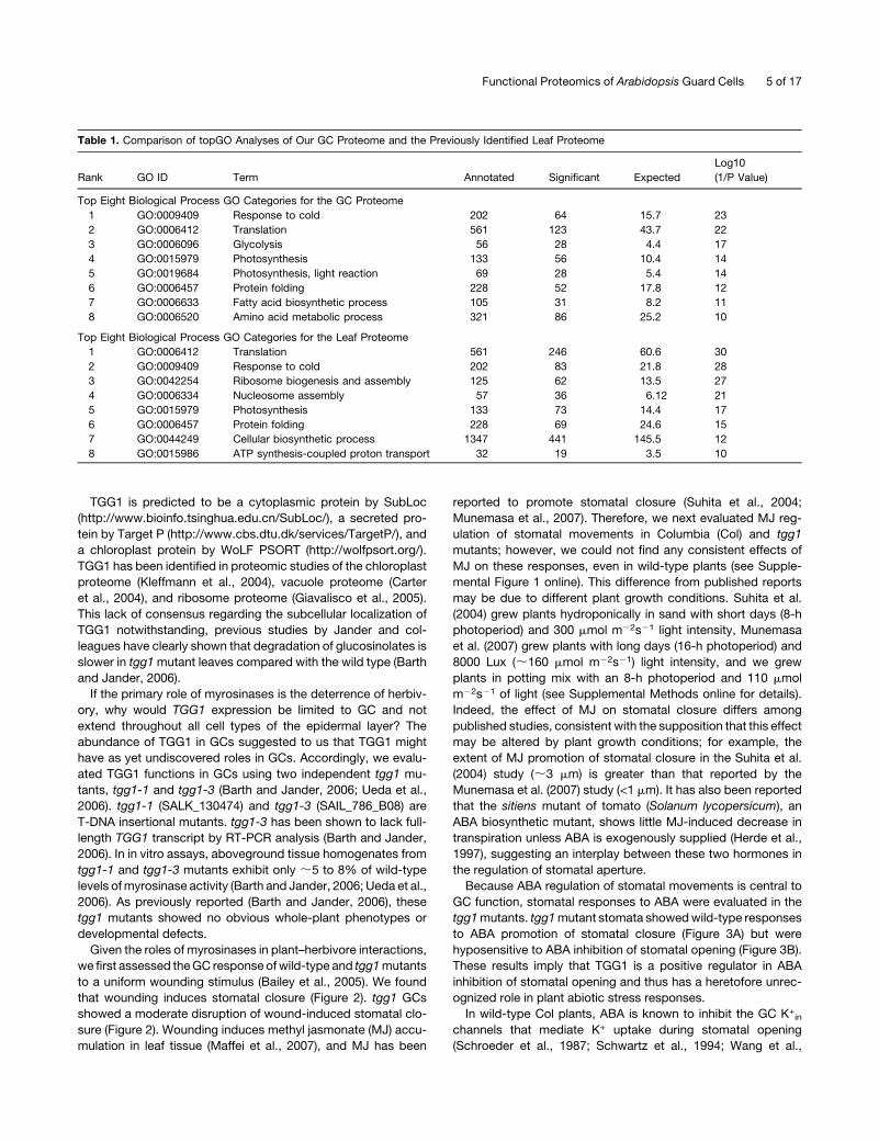
(Lee et al., 2007) for comparison to the GC proteome. Out of the
eight categories most significantly enriched in the GC proteome
compared with the entire (conceptual) proteome, four categories
(response to cold, translation, protein folding, andphotosynthesis–
general) were also among the eight most significantly enriched in
the leaf proteome (Table 1). However, the other four categories
(glycolysis, photosynthesis–light reactions, fatty acid biosyn-
thetic process, and amino acid metabolic process) were present
in the top GC hits but absent from the top leaf hits (Table 1),
suggestive of particular roles in GC (see Discussion).
To identify proteins in our GC proteome that may be specif-
ically expressed or enriched in GCs, we compared our GC
proteome to ;13,000 proteins in previously identified pro-
teomes of cell walls (Bayer et al., 2006), trichomes (Wienkoop
et al., 2004), epidermal cells (Wienkoop et al., 2004), and leaves
(Lee et al., 2007), as well as to the recently reported proteome
map of Arabidopsis organs (Baerenfaller et al., 2008). These
comparisons revealed 61 proteins that were identified in our GC
proteome and not in these other proteomes (Table 2). Of these
61 proteins, we found eight that were identified in other studies
in the literature: four of the eight (At3g16080, At3g18040,
At3g23840, and At5g54190) were identified in studies on specific
organelles (Friso et al., 2004; Heazlewood et al., 2004; Philippar
et al., 2007; Carroll et al., 2008), and the other four (At1g50010,
At5g08690, AtMg01190, and At5g08670) were identified by
proteomic analyses of certain mutants or certain treatments, or
in cultured plant cells (Rajjou et al., 2004; Dixon et al., 2005; Job
et al., 2005; Sorin et al., 2006). The remaining 53 proteins may
have unique roles in GC function, particularly since, to date, only
10 of these proteins have been shown to function in any other
tissues by mutant analysis (Table 2). Of the proteins identified by
Baerenfaller et al. (2008) as biomarkers of specific organ types,
nine of these proteins (three fromflowers, three from siliques, two
from roots, and one from seeds) were present in our GC
proteome (see Supplemental Table 7 online).
Twelve proteins previously shown to play a role in GC function
were present in our GCproteome (Table 3). In addition, functional
classification by GO analysis showed that 52 proteins out of our
identified GC proteome are predicted as signal transduction
proteins (Table 3). Of these, only two proteins, the phospholipase
PLDa1 (Mishra et al., 2006) and the calcium-dependent protein
kinase CPK3/CDPK6 (Mori et al., 2006), have been previously
studied in the context of GC function, where they have been
shown to participate in ABA signaling.
Functional Analysis ofOneof theMostAbundant Proteins in
GCs, the Myrosinase TGG1
The plant glucosinolate-myrosinase system is known as a de-
fense systemagainst bacteria, pathogens, and herbivores.When
tissue is damaged (e.g., by insect chewing), glucosinolates are
thought to be released from the vacuole and hydrolyzed by
myrosinases into a variety of toxic small molecules, including
thiocyanate, isothiocyanate, and nitrile, which are active against
biotic intruders (Wittstock and Halkier, 2002; Barth and Jander,
2006).
Although our gel-based analyses contributed only incremen-
tally to the total number of GC proteins identified, these analyses
revealed a remarkable abundance in GC of the myrosinase,
TGG1. TGG1 was by far the largest spot on either the BR (Figure
1C, spot 12) or the NR 2D gels (Figure 1D3, spot 3E12) and was
identified in multiple spots (see Supplemental Table 5 online; 12
spots on the BR and 37 spots on the NR gels), suggesting high
abundance and multiple posttranslational modifications.
TGG1 protein was not identified in proteomic analyses of
trichome and epidermal pavement cells (Wienkoop et al., 2004),
consistent with previous reporter gene analysis demonstrating
strong expression of the TGG1 gene in GC and no expression in
other epidermal cell types (Husebye et al., 2002; Barth and
Jander, 2006). However, the presence of the TGG1 protein in GC
has not been previously shown, and no function of TGG1 in GC
has been described previously. The other Arabidopsis myrosi-
nase, TGG2, is not expressed in GC by reporter gene analysis
(Barth and Jander, 2006). TGG1 and TGG2 are indistinguishable
by the probes used on Affymetrix microarrays, and the only other
demonstrated locus of expression of these two TGG genes is in
phloem idioblasts (Husebye et al., 2002; Barth and Jander,
2006). Different masses of trypsin-digested peptides and amino
acid sequences are generated from TGG1 versus the related
myrosinase, TGG2, and can be detected by mass spectrometry
and tandem mass spectrometry, respectively. Thus, TGG1 and
TGG2can be clearly distinguished by proteomicmethods. Unlike
TGG1, TGG2 was not found in any of the gel-based studies and
was identified only in one replicate of the LC-MALDI MudPIT
method using the Protein Pilot search algorithm. While 37 unique
peptides were identified from TGG1, only two unique peptides
were identified fromTGG2, plus one peptidewas identified that is
identical in TGG1 and TGG2. These results suggest a substan-
tially lower abundance of TGG2 in GC compared with TGG1.
Figure 1. (continued).
(A) Images showing high-purity GCP preparations (3100; inset magnification 3400).
(B) A total of 1712, 58, and 59 unique proteins were identified from 2D LC-MALDI MudPIT, BR, and NR methods, respectively; 19 proteins were
identified by all three methods. For each method, two independent biological samples were analyzed.
(C) A 2D gel image from the broad pH range method. The first dimension was run using a 24-cm, pH 3 to 10 IPG strip. In total, 138 protein spots were
detected via Coomassie blue staining. Twelve spots were identified as TGG1. Identifications of numbered spots can be found in Supplemental Table 5
online.
(D) 2D gel images from the narrow pH range method. Proteins were first fractionated into five fractions, and each protein fraction was separated on a
narrow pH range IPG strip. From B1 to B5, the pH ranges are 3 to 6, 4.5 to 5.5, 5.3 to 6.3, 6.1 to 7.1, and 6 to 10 respectively. Thirty-seven spots were
identified as TGG1. Identifications of numbered spots can be found in Supplemental Table 5 online.
4 of 17 The Plant Cell
TGG1 is predicted to be a cytoplasmic protein by SubLoc
(http://www.bioinfo.tsinghua.edu.cn/SubLoc/), a secreted pro-
tein by Target P (http://www.cbs.dtu.dk/services/TargetP/), and
a chloroplast protein by WoLF PSORT (http://wolfpsort.org/).
TGG1 has been identified in proteomic studies of the chloroplast
proteome (Kleffmann et al., 2004), vacuole proteome (Carter
et al., 2004), and ribosome proteome (Giavalisco et al., 2005).
This lack of consensus regarding the subcellular localization of
TGG1 notwithstanding, previous studies by Jander and col-
leagues have clearly shown that degradation of glucosinolates is
slower in tgg1mutant leaves compared with the wild type (Barth
and Jander, 2006).
If the primary role of myrosinases is the deterrence of herbiv-
ory, why would TGG1 expression be limited to GC and not
extend throughout all cell types of the epidermal layer? The
abundance of TGG1 in GCs suggested to us that TGG1 might
have as yet undiscovered roles in GCs. Accordingly, we evalu-
ated TGG1 functions in GCs using two independent tgg1 mu-
tants, tgg1-1 and tgg1-3 (Barth and Jander, 2006; Ueda et al.,
2006). tgg1-1 (SALK_130474) and tgg1-3 (SAIL_786_B08) are
T-DNA insertional mutants. tgg1-3 has been shown to lack full-
length TGG1 transcript by RT-PCR analysis (Barth and Jander,
2006). In in vitro assays, aboveground tissue homogenates from
tgg1-1 and tgg1-3 mutants exhibit only ;5 to 8% of wild-type
levels ofmyrosinase activity (Barth and Jander, 2006; Ueda et al.,
2006). As previously reported (Barth and Jander, 2006), these
tgg1 mutants showed no obvious whole-plant phenotypes or
developmental defects.
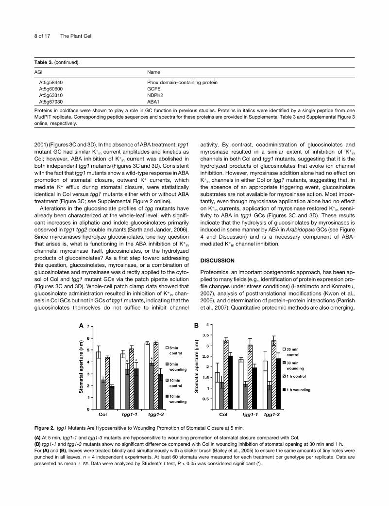
Given the roles of myrosinases in plant–herbivore interactions,
we first assessed theGC response ofwild-type and tgg1mutants
to a uniform wounding stimulus (Bailey et al., 2005). We found
that wounding induces stomatal closure (Figure 2). tgg1 GCs
showed a moderate disruption of wound-induced stomatal clo-
sure (Figure 2). Wounding induces methyl jasmonate (MJ) accu-
mulation in leaf tissue (Maffei et al., 2007), and MJ has been
reported to promote stomatal closure (Suhita et al., 2004;
Munemasa et al., 2007). Therefore, we next evaluated MJ reg-
ulation of stomatal movements in Columbia (Col) and tgg1
mutants; however, we could not find any consistent effects of
MJ on these responses, even in wild-type plants (see Supple-
mental Figure 1 online). This difference from published reports
may be due to different plant growth conditions. Suhita et al.
(2004) grew plants hydroponically in sand with short days (8-h
photoperiod) and 300 mmol m22s21 light intensity, Munemasa
et al. (2007) grew plants with long days (16-h photoperiod) and
8000 Lux (;160 mmol m22s21) light intensity, and we grew
plants in potting mix with an 8-h photoperiod and 110 mmol
m22s21 of light (see Supplemental Methods online for details).
Indeed, the effect of MJ on stomatal closure differs among
published studies, consistent with the supposition that this effect
may be altered by plant growth conditions; for example, the
extent of MJ promotion of stomatal closure in the Suhita et al.
(2004) study (;3 mm) is greater than that reported by the
Munemasa et al. (2007) study (<1 mm). It has also been reported
that the sitiens mutant of tomato (Solanum lycopersicum), an
ABA biosynthetic mutant, shows little MJ-induced decrease in
transpiration unless ABA is exogenously supplied (Herde et al.,
1997), suggesting an interplay between these two hormones in
the regulation of stomatal aperture.
Because ABA regulation of stomatal movements is central to
GC function, stomatal responses to ABA were evaluated in the
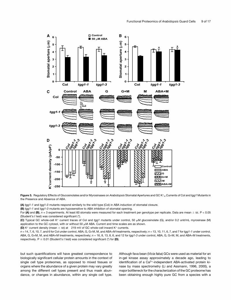
tgg1mutants. tgg1mutant stomata showedwild-type responses
to ABA promotion of stomatal closure (Figure 3A) but were
hyposensitive to ABA inhibition of stomatal opening (Figure 3B).
These results imply that TGG1 is a positive regulator in ABA
inhibition of stomatal opening and thus has a heretofore unrec-
ognized role in plant abiotic stress responses.
In wild-type Col plants, ABA is known to inhibit the GC K+in
channels that mediate K+ uptake during stomatal opening
(Schroeder et al., 1987; Schwartz et al., 1994; Wang et al.,
Table 1. Comparison of topGO Analyses of Our GC Proteome and the Previously Identified Leaf Proteome
Rank GO ID Term Annotated Significant Expected
Log10
(1/P Value)
Top Eight Biological Process GO Categories for the GC Proteome
1 GO:0009409 Response to cold 202 64 15.7 23
2 GO:0006412 Translation 561 123 43.7 22
3 GO:0006096 Glycolysis 56 28 4.4 17
4 GO:0015979 Photosynthesis 133 56 10.4 14
5 GO:0019684 Photosynthesis, light reaction 69 28 5.4 14
6 GO:0006457 Protein folding 228 52 17.8 12
7 GO:0006633 Fatty acid biosynthetic process 105 31 8.2 11
8 GO:0006520 Amino acid metabolic process 321 86 25.2 10
Top Eight Biological Process GO Categories for the Leaf Proteome
1 GO:0006412 Translation 561 246 60.6 30
2 GO:0009409 Response to cold 202 83 21.8 28
3 GO:0042254 Ribosome biogenesis and assembly 125 62 13.5 27
4 GO:0006334 Nucleosome assembly 57 36 6.12 21
5 GO:0015979 Photosynthesis 133 73 14.4 17
6 GO:0006457 Protein folding 228 69 24.6 15
7 GO:0044249 Cellular biosynthetic process 1347 441 145.5 12
8 GO:0015986 ATP synthesis-coupled proton transport 32 19 3.5 10
Functional Proteomics of Arabidopsis Guard Cells 5 of 17
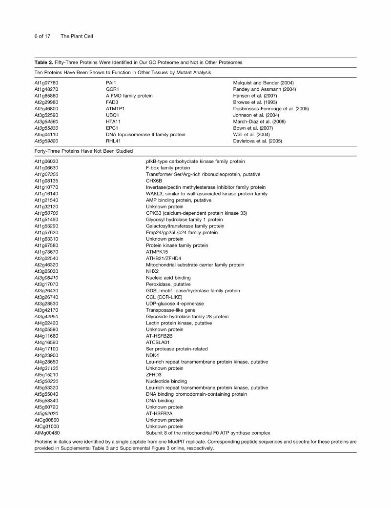
Table 2. Fifty-Three Proteins Were Identified in Our GC Proteome and Not in Other Proteomes
Ten Proteins Have Been Shown to Function in Other Tissues by Mutant Analysis
At1g07780 PAI1 Melquist and Bender (2004)
At1g48270 GCR1 Pandey and Assmann (2004)
At1g65860 A FMO family protein Hansen et al. (2007)
At2g29980 FAD3 Browse et al. (1993)
At2g46800 ATMTP1 Desbrosses-Fonrouge et al. (2005)
At3g52590 UBQ1 Johnson et al. (2004)
At3g54560 HTA11 March-Diaz et al. (2008)
At3g55830 EPC1 Bown et al. (2007)
At5g04110 DNA topoisomerase II family protein Wall et al. (2004)
At5g59820 RHL41 Davletova et al. (2005)
Forty-Three Proteins Have Not Been Studied
At1g06030 pfkB-type carbohydrate kinase family protein
At1g06630 F-box family protein
At1g07350 Transformer Ser/Arg-rich ribonucleoprotein, putative
At1g08135 CHX6B
At1g10770 Invertase/pectin methylesterase inhibitor family protein
At1g16140 WAKL3, similar to wall-associated kinase protein family
At1g21540 AMP binding protein, putative
At1g32120 Unknown protein
At1g50700 CPK33 (calcium-dependent protein kinase 33)
At1g51490 Glycosyl hydrolase family 1 protein
At1g53290 Galactosyltransferase family protein
At1g57620 Emp24/gp25L/p24 family protein
At1g63310 Unknown protein
At1g67580 Protein kinase family protein
At1g73670 ATMPK15
At2g02540 ATHB21/ZFHD4
At2g46320 Mitochondrial substrate carrier family protein
At3g05030 NHX2
At3g06410 Nucleic acid binding
At3g17070 Peroxidase, putative
At3g26430 GDSL-motif lipase/hydrolase family protein
At3g26740 CCL (CCR-LIKE)
At3g28530 UDP-glucose 4-epimerase
At3g42170 Transposase-like gene
At3g42950 Glycoside hydrolase family 28 protein
At4g02420 Lectin protein kinase, putative
At4g05590 Unknown protein
At4g11660 AT-HSFB2B
At4g16590 ATCSLA01
At4g17100 Ser protease protein-related
At4g23900 NDK4
At4g28650 Leu-rich repeat transmembrane protein kinase, putative
At4g31130 Unknown protein
At5g15210 ZFHD3
At5g50230 Nucleotide binding
At5g53320 Leu-rich repeat transmembrane protein kinase, putative
At5g55040 DNA binding bromodomain-containing protein
At5g58340 DNA binding
At5g60720 Unknown protein
At5g62020 AT-HSFB2A
AtCg00860 Unknown protein
AtCg01000 Unknown protein
AtMg00480 Subunit 8 of the mitochondrial F0 ATP synthase complex
Proteins in italics were identified by a single peptide from one MudPIT replicate. Corresponding peptide sequences and spectra for these proteins are
provided in Supplemental Table 3 and Supplemental Figure 3 online, respectively.
6 of 17 The Plant Cell
Table 3. Identified GC Signaling Proteins in Our GC Proteome
AGI Name AGI Name
Twelve Proteins Previously Shown to Have a Role in GC Function Were Identified in Our GC Proteome
At1g11260 STP1 At3g45780 PHOT1
At1g37130 NIA2 At3g53720 CHX20
At1g48270 GCR1 At4g18290 KAT2
At2g18960 OST2 At4g23650 CPK3 or CDPK6
At2g21660 GRP7 At5g23060 CAS
At3g15730 PLDa1 At5g58140 PHOT2
AGI Name
Fifty-Two Proteins in Our GC Proteome Are Predicted to Be Signaling Proteins by GO Software
At1g05810 ARA-1
At1g06840 Leu-rich repeat transmembrane protein kinase
At1g09100 26S protease regulatory subunit 6A
At1g35160 GF14/GRF4
At1g52280 Ras-related GTP binding protein
At1g71860 PTP1
At1g73670 At MPK15
At1g75640 Leu-rich repeat family protein
At1g76030 Vacuolar ATP synthase subunit B
At1g78580 At TPS1
At2g04880 ZAP1
At2g16600 ROC3
At2g19860 At HXK2
At2g20610 SUR1
At2g21880 At RABG2
At2g25170 PKL
At2g26730 Leu-rich repeat transmembrane protein kinase
At2g32410 AXL
At2g36830 GAMMA-TIP
At2g37970 SOUL heme-binding family protein
At2g44050 COS1
At2g46070 At MPK12
At3g02880 Leu-rich repeat transmembrane protein kinase
At3g08510 At PLC2
At3g08680 Leu-rich repeat transmembrane protein kinase
At3g15730 PLDa1
At3g18040 At MPK9
At3g18820 Ras-related GTP-binding protein
At3g46060 ARA3
At3g53020 STV1
At3g55020 RabGAP
At3g62030 ROC4
At4g01370 At MPK4
At4g02080 SAR1A
At4g03550 At GSL5
At4g15900 PRL1
At4g18430 Ras-related GTP binding protein
At4g23650 CPK3 or CDPK6
At4g28650 Leu-rich repeat transmembrane protein kinase
At4g29810 At MKK2
At4g33680 AGD2
At4g34870 ROC5
At4g38690 1-Phosphatidylinositol phosphodiesterase-related
At4g38740 ROC1
At5g16590 Leu-rich repeat transmembrane protein kinase
At5g19390 Similar to pleckstrin homology domain-containing protein
At5g39500 Pattern formation protein
At5g53320 Leu-rich repeat transmembrane protein kinase
(Continued)
Functional Proteomics of Arabidopsis Guard Cells 7 of 17
2001) (Figures 3C and 3D). In the absence of ABA treatment, tgg1
mutant GC had similar K+in current amplitudes and kinetics as
Col; however, ABA inhibition of K+in current was abolished in
both independent tgg1mutants (Figures 3C and 3D). Consistent
with the fact that tgg1mutants show awild-type response in ABA
promotion of stomatal closure, outward K+ currents, which
mediate K+ efflux during stomatal closure, were statistically
identical in Col versus tgg1 mutants either with or without ABA
treatment (Figure 3C; see Supplemental Figure 2 online).
Alterations in the glucosinolate profiles of tgg mutants have
already been characterized at the whole-leaf level, with signifi-
cant increases in aliphatic and indole glucosinolates primarily
observed in tgg1 tgg2 double mutants (Barth and Jander, 2006).
Since myrosinases hydrolyze glucosinolates, one key question
that arises is, what is functioning in the ABA inhibition of K+in
channels: myrosinase itself, glucosinolates, or the hydrolyzed
products of glucosinolates? As a first step toward addressing
this question, glucosinolates, myrosinase, or a combination of
glucosinolates and myrosinase was directly applied to the cyto-
sol of Col and tgg1 mutant GCs via the patch pipette solution
(Figures 3C and 3D). Whole-cell patch clamp data showed that
glucosinolate administration resulted in inhibition of K+in chan-
nels in Col GCs but not inGCs of tgg1mutants, indicating that the
glucosinolates themselves do not suffice to inhibit channel
activity. By contrast, coadministration of glucosinolates and
myrosinase resulted in a similar extent of inhibition of K+in
channels in both Col and tgg1 mutants, suggesting that it is the
hydrolyzed products of glucosinolates that evoke ion channel
inhibition. However, myrosinase addition alone had no effect on
K+in channels in either Col or tgg1 mutants, suggesting that, in
the absence of an appropriate triggering event, glucosinolate
substrates are not available for myrosinase action. Most impor-
tantly, even though myrosinase application alone had no effect
on K+in currents, application of myrosinase restored K+
in sensi-
tivity to ABA in tgg1 GCs (Figures 3C and 3D). These results
indicate that the hydrolysis of glucosinolates by myrosinases is
induced in some manner by ABA in Arabidopsis GCs (see Figure
4 and Discussion) and is a necessary component of ABA-
mediated K+in channel inhibition.
DISCUSSION
Proteomics, an important postgenomic approach, has been ap-
plied tomany fields (e.g., identification of protein expression pro-
file changes under stress conditions) (Hashimoto and Komatsu,
2007), analysis of posttranslational modifications (Kwon et al.,
2006), and determination of protein–protein interactions (Parrish
et al., 2007). Quantitative proteomic methods are also emerging,
Table 3. (continued).
AGI Name
At5g58440 Phox domain–containing protein
At5g60600 GCPE
At5g63310 NDPK2
At5g67030 ABA1
Proteins in boldface were shown to play a role in GC function in previous studies. Proteins in italics were identified by a single peptide from one
MudPIT replicate. Corresponding peptide sequences and spectra for these proteins are provided in Supplemental Table 3 and Supplemental Figure 3
online, respectively.
Figure 2. tgg1 Mutants Are Hyposensitive to Wounding Promotion of Stomatal Closure at 5 min.
(A) At 5 min, tgg1-1 and tgg1-3 mutants are hyposensitive to wounding promotion of stomatal closure compared with Col.
(B) tgg1-1 and tgg1-3 mutants show no significant difference compared with Col in wounding inhibition of stomatal opening at 30 min and 1 h.
For (A) and (B), leaves were treated blindly and simultaneously with a slicker brush (Bailey et al., 2005) to ensure the same amounts of tiny holes were
punched in all leaves. n = 4 independent experiments. At least 60 stomata were measured for each treatment per genotype per replicate. Data are
presented as mean 6 SE. Data were analyzed by Student’s t test, P < 0.05 was considered significant (*).
8 of 17 The Plant Cell
but such quantifications will have greatest correspondence to
biologically significant cellular protein amounts in the context of
single cell type proteomes, as opposed to mixed tissues or
organs where the abundance of a given protein may vary greatly
among the different cell types present and thus mask abun-
dance, or changes in abundance, within any single cell type.
Although fava bean (Vicia faba) GCs were used as material for an
in-gel kinase assay approximately a decade ago, leading to
identification of a Ca2+-independent ABA-activated protein ki-
nase by mass spectrometry (Li and Assmann, 1996, 2000), a
major bottleneck for the characterization of theGCproteome has
been obtaining enough highly pure GC from a species with a
Figure 3. Regulatory Effects of Glucosinolates and/or Myrosinases on Arabidopsis Stomatal Apertures and GC K+in Currents of Col and tgg1Mutants in
the Presence and Absence of ABA.
(A) tgg1-1 and tgg1-3 mutants respond similarly to the wild type (Col) in ABA induction of stomatal closure.
(B) tgg1-1 and tgg1-3 mutants are hyposensitive to ABA inhibition of stomatal opening.
For (A) and (B), n = 3 experiments. At least 60 stomata were measured for each treatment per genotype per replicate. Data are mean 6 SE. P < 0.05
(Student’s t test) was considered significant (*).
(C) Typical GC whole-cell K+ current traces of Col and tgg1 mutants under control, 50 mM glucosinolate (G), and/or 0.2 unit/mL myrosinase (M)
application to the GC cytosol, with or without 50 mM ABA. Current and time scales are as shown.
(D) K+ current density (mean 6 SE) at �219 mV of GC whole-cell inward K+ currents.
n = 14, 7, 6, 10, 7, and 6 for Col under control, ABA, G, G+M, M, and ABA+M treatments, respectively; n = 13, 10, 11, 6, 7, and 7 for tgg1-1 under control,
ABA, G, G+M, M, and ABA+M treatments, respectively; n = 16, 8, 13, 8, 6, and 12 for tgg1-3 under control, ABA, G, G+M, M, and ABA+M treatments,
respectively. P # 0.01 (Student’s t test) was considered significant (*) for (D).
Functional Proteomics of Arabidopsis Guard Cells 9 of 17
sequenced genome. Here, this challenge was overcome and
three proteomics methods were used to identify 1734 unique
proteins of the GC proteome.
Comparison of the Three Proteomic Methods
In our study, the gel-freemethod (2D LC-MALDI-MudPIT) yielded
by far the largest number of protein identifications: 1712 proteins
were identified by this method, and, of these, 1638 were not
identified by either of the gel-basedmethods. The two gel-based
methods (BR and NR) yielded similar numbers of identified
proteins (58 and 59 unique proteins, respectively), with an
identification rate ;30-fold lower than that of the gel-free
method. Twenty-one proteins (i.e., approximately one-third of
the proteins identified by BR or NR methods) were common to
both of these gel-based approaches. Eighty-eight percent of the
BR proteins (51 proteins) and 71% of the NR proteins (42
proteins) were also identified by 2D LC-MALDIMudPIT analyses.
Only 19 proteins (representing 33% of the BR proteins, 32% of
the NR proteins, and 1% of the 2D LC-MALDI MudPIT proteins)
were found from all three methods (Figure 1B; see Supplemental
Table 6 online). This low overlap is consistent with observations
from other organisms that have shown that multiple strategies
are required to obtain high coverage of the proteome (Kleffmann
et al., 2007). The limitations of gel-based methods regarding
identification of basic, high molecular mass, and membrane
proteins are well known (Jorrin et al., 2007). However, the gel-
based methods importantly allowed recognition of the high
abundance of TGG1 in GC. In addition, gel-based methods
provide more reliable inference of posttranslational modifica-
tions (see Supplemental Table 5 online). Moreover, 22 proteins of
the GC proteome were exclusively identified by gel-based
methods, indicating that gel-free methods cannot completely
replace traditional gel-based methods (Lambert et al., 2005).
Although we evaluated the 22 proteins for common character-
istics (e.g., molecular weight, pI, predicted subcellular localiza-
tion, and the predicted trypsin-digestion patterns of these
protein sequences) that might have enhanced their detection
by gel-based over gel-free methods, no such characteristics
could be identified.
Comparison of the GC Proteome with Other Proteomes
Although we used GCPs as starting material, 29 of the identified
GC proteins were identified by a previous cell wall proteomic
study (Bayer et al., 2006): these proteins may localize to multiple
subcellular compartments, including both apoplastic and sym-
plastic destinations, or be present in secretory vesicles that have
not yet fused with the plasma membrane (Lee et al., 2004).
Indeed, further GO analysis predicted that 23 of the 29 (79%)
proteins also localize to non–cell wall subcellular compartments.
Our topGO analysis revealed biological processes that were
enriched in the GC proteome (Table 1) relative to the entire
predicted Arabidopsis proteome and to the documented leaf
proteome (Lee et al., 2007). Four of the GC-enriched biological
processes were also enriched in the published leaf proteome
(Lee et al., 2007) and thus may typify leaf cell types in general but
not GC in particular. The GO category “amino acid metabolic
process” was also fairly highly ranked in leaves (rank = 10). More
interesting are the remaining GC-enriched biological processes:
glycolysis (rank 3 in GC but 14 in leaf), light reactions of photo-
synthesis (rank 5 in GC but 29 in leaf), and fatty acid biosynthesis
(rank 7 in GC but 142 in leaf).
Stomatal movement is estimated to have a high energetic
requirement (Assmann and Zeiger, 1987), consistent with the
topGO prediction of the importance of glycolysis and photo-
phosphorylation to this cell type. Indeed, biochemical assays
have shown disproportionately high rates of photophosphoryla-
tion in GC relative to their very low rates of carbon fixation
(Shimazaki et al., 2007), and red light–stimulated stomatal open-
ing is known to be inhibited by photosynthetic inhibitors, such as
DCMU (Schwartz and Zeiger, 1984). The enrichment in the GC
proteome of proteins involved in fatty acid biosynthesis may
reflect not only the importance of lipids to cuticle formation
(Samuels et al., 2008), but also the importance of lipids and lipid
metabolites as signaling entities in GC. For example, the guard
cell–specific HIGH CARBON DIOXIDE (HIC) gene encodes a
likely 3-keto acyl CoA synthase, involved in the synthesis of very-
long-chain fatty acids, yet hic mutants also show a dramatic
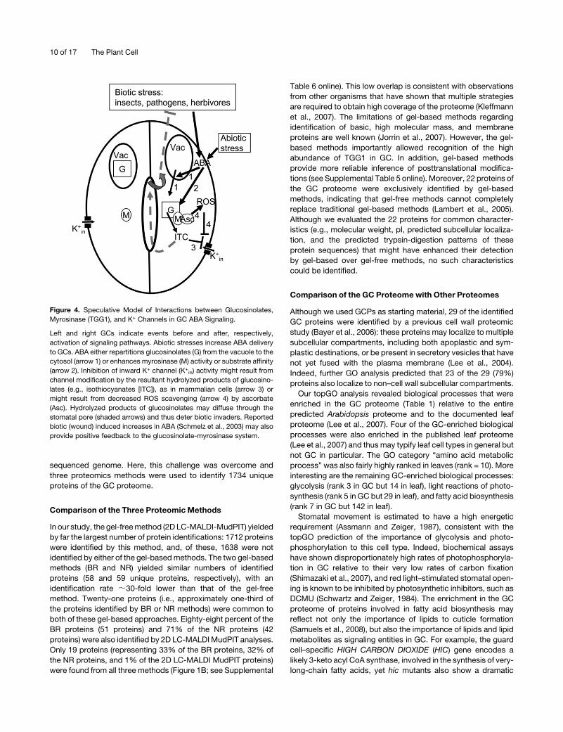
Figure 4. Speculative Model of Interactions between Glucosinolates,
Myrosinase (TGG1), and K+ Channels in GC ABA Signaling.
Left and right GCs indicate events before and after, respectively,
activation of signaling pathways. Abiotic stresses increase ABA delivery
to GCs. ABA either repartitions glucosinolates (G) from the vacuole to the
cytosol (arrow 1) or enhances myrosinase (M) activity or substrate affinity
(arrow 2). Inhibition of inward K+ channel (K+in) activity might result from
channel modification by the resultant hydrolyzed products of glucosino-
lates (e.g., isothiocyanates [ITC]), as in mammalian cells (arrow 3) or
might result from decreased ROS scavenging (arrow 4) by ascorbate
(Asc). Hydrolyzed products of glucosinolates may diffuse through the
stomatal pore (shaded arrows) and thus deter biotic invaders. Reported
biotic (wound) induced increases in ABA (Schmelz et al., 2003) may also
provide positive feedback to the glucosinolate-myrosinase system.
10 of 17 The Plant Cell
CO2-dependent alteration in GC production (Gray et al., 2000).
Other lipid-relatedmoleculeswith demonstrated roles inGC related
to ABA signaling include phosphatidic acid (Jacob et al., 1999;
Zhang et al., 2005), sphinogosine-1-phosphate (Ng et al., 2001;
Coursol et al., 2003), and inositol phosphates (Lee et al., 1996; Jung
et al., 2002; Hunt et al., 2003). Our topGO analysis of the GC
proteome suggests that additional study of mutants in enzymes
related to fatty acid synthesismayuncoverGC-relatedphenotypes.
Of the 53 proteins that were identified in our GC proteome
(Table 2) but not in other known proteomes, some may be
specific to GCs and thus can be considered as candidate GC
biomarkers. In this regard, it would be of particular interest to
characterize the seven proteins of unknown function in this set
(Table 2). Others may be more abundant in GCs than elsewhere;
thus, we succeeded in identifying these proteins as part of the
GC proteome while they were missed in other proteomic anal-
yses (e.g., the likely G-protein coupled receptor, GCR1, which
confers ABA hypersensitivity to both stomatal movements and
root growth) (Pandey andAssmann, 2004). Finally, someproteins
may be specifically present in GC plus a few other specialized
cell types and thus missed in proteome analyses of whole
organs. Conversely, our identification in the GC proteome of
proteins previously thought to be biomarkers for specific organs
(see Supplemental Table 7 online), including roots and seeds,
which lack GC, indicates the importance of single-cell-type
proteome analysis in determinations of protein distribution.
The GC Proteome and GC Signaling
We identified 67 proteins from the literature as previously shown
to function in mature Arabidopsis GCs/GCPs (see Supplemental
Table 8 online). Of these, 51 participate in GC responses to ABA,
light, and/or CO2, while six of the remaining 16 proteins function
in solute transport. Twelve of the 67 proteins were present in our
GC proteome (Table 3); the other 55 may be low abundance
proteins or induced under specific conditions. The 12 identified
proteins are involved in light (PHOT1, PHOT2, and CHX20) and
ABA (CPK3, GCR1, GRP7, OST2, NIA2, and PLDa1) signal-
ing and in solute transport (CHX20, STP1, KAT2, and OST2)
(Table 3).
GO analysis categorizes 52 proteins in our GC proteome as
signal transduction proteins by GO analysis (Table 3). Of these,
50 have yet to be studied in the context of GC function, high-
lighting the usefulness of proteome analysis in identifying targets
for further functional analyses. Thirteen are protein kinases,
including one CDPK (CPK3/CDPK6), seven LRR protein kinases,
and five MAP kinases; four of these kinases (At1g73670,
At3g18040, At4g28650, and At5g53320) are also in the list of
proteins only found to date in the GC proteome (Table 2). This
information strongly suggests that phosphorylation is one of the
main posttranslational modifications in GCs, consistent with
previous demonstrations of the importance of phosphorylation
to light and ABA signaling in GCs (Kinoshita et al., 1993; Li and
Assmann, 1996, 2000; Li et al., 2000; Mustilli et al., 2002;
Kinoshita et al., 2003; Sokolovski et al., 2005). Five of the 52
proteins are involved in auxin signaling, suggesting that auxin
may be more important in GC physiology than previously rec-
ognized (Acharya and Assmann, 2008).
TGG1 Function in GCs
While many interesting candidates for further downstream anal-
ysis were revealed in our GC proteome analysis, we chose to
perform an in-depth functional study of one protein. TGG1 was
chosen due to its superabundance in GC: TGG1 comprises 40 to
50% of the total protein identified on 2D gels and was identified
by >30 unique peptides (not shared with TGG2) in our MudPIT
analysis. However, no roles for TGG1 inGCs had been previously
demonstrated.
We also examined the effect of wounding on stomatal aper-
tures and found that wounding of the epidermis did stimulate a
stomatal response and that TGG1 seemed to participate in this
effect (Figure 2). However, TGG1 appeared to play a more
essential role in GC ABA signaling: tgg1 mutant plants lacking
this enzyme were unresponsive to ABA inhibition of stomatal
opening and K+in channel regulation. Intracellular application of
myrosinase alone did not restore K+in channel inhibition in tgg1
GCs or cause channel inhibition in wild-type GCs, yet application
of myrosinase in the presence of ABA restored channel inhibition
to the tgg1 mutants.
Our electrophysiological results are consistent with the fol-
lowing scenario (Figure 4): (1) myrosinase and its substrates, the
glucosinolates, are localized in distinct subcellular compart-
ments in GCs, as has been proposed for other cell types (Grubb
and Abel, 2006). (2) ABA induces the relocalization of glucosi-
nolates to the cytosol (arrow 1 in Figure 4). (3) The hydrolysis of
available glucosinolates by TGG1 leads to inhibition of K+in
channels in GCs, and this is one component of ABA inhibition of
stomatal opening. However, we note that there are also alterna-
tive interpretations that are consistent with our data (e.g., ABA
signaling might somehow increase the activity of myrosinase or
its affinity for its substrate) (arrow 2 in Figure 4).
ABA regulation of glucosinolate compartmentalization and
thus availability for hydrolysis by myrosinases, as hypothesized
here, could help to explain nondefensive developmental
changes in glucosinolate concentrations (Petersen et al., 2002;
Brown et al., 2003) that occur in the absence of the tissue
disruption that brings substrate and enzyme together during
herbivory. Such a phenomenon could also provide a mechanism
whereby exposure to abiotic stress could strengthen defenses
against subsequent biotic stressors. In addition, given themech-
anisms described here, biotic (wound)-induced increases in
ABA, as reported to occur in leaf tissue (Schmelz et al., 2003),
might also provide positive feedback to the glucosinolate-
myrosinase defense pathway (Figure 4), priming plant defense
mechanisms against abiotic and biotic (Beckers and Conrath,
2007) stressors.
Alteration in membrane potential is a rapid response to
wounding (Maffei et al., 2007), and our observations suggest
that K+ channel regulationmay contribute to these early electrical
events. Since application of glucosinolates to wild-type GCs, or
application of glucosinolates plus myrosinase to tgg1 mutant
GCs, also inhibits the K+in channels, we infer that it is the reaction
catalyzed by myrosinase (e.g., the hydrolyzed products of
glucosinolates) that evoke channel inhibition. Because the in-
ward K+ channels of GCs are, on a sequence homology basis,
most similar to metazoan Shaker channels (Pilot et al., 2003;
Functional Proteomics of Arabidopsis Guard Cells 11 of 17
Pandey et al., 2007), animal Shaker K+ channels may also be
targets for these plant secondary compounds, which are known
to have both toxic and anticarcinogenic effects in mammals
(Halkier and Gershenzon, 2006). Preharvest growth conditions,
harvesting processes, and storage conditions all can affect plant
glucosinolate concentrations (Johnson, 2002). The implication
from our data that ABAmay regulate glucosinolate sequestration
and stability may suggest modifications to extant agronomic
protocols, with implications for food quality.
Our results (Figures 3C and 3D) demonstrate an interconnec-
tion between the products of myrosinase activity and K+ channel
regulation, but further research will required to determine the
exact mechanistic basis of this response. It may be relevant that
recent studies have shown that isothiocyanates alter activity of
the mammalian pain-sensing TRP1A channels by covalent mod-
ification at Cys residues (Hinman et al., 2006). Alternatively, or in
addition, the fact that myrosinase binds the antioxidant ascorbic
acid and can catalyze formation of a condensation product of
ascorbic acid with methylindoles (Burmeister et al., 2000) may
suggest that myrosinase activity promotes ABA signaling via
decreasing the ability of theGC to scavenge ROS. ROS elevation
is a key signal transduction element in ABA signaling (Pei et al.,
2000; Kwak et al., 2003), and plants engineered for increased
ascorbate levels exhibit decreased levels of ROS and decreased
GC responsiveness to ABA (Chen and Gallie, 2004).
While it has recently been demonstrated that small mole-
cules, such as NO and ROS, are shared between ABA and
defense signaling pathways, including in GCs (Melotto et al.,
2006; Ali et al., 2007; Zhang et al., 2008), enzymes of secondary
metabolism, such as TGG1, have not previously been impli-
cated in ABA signaling. The interconnection discovered here
between ABA and the glucosinolate-based biotic defense
mechanism suggests a mechanism whereby exposure to abi-
otic stresses may enhance plant defense against subsequent
biotic invaders. One general property of the hydrolyzed prod-
ucts of glucosinolates is volatility (Yan and Chen, 2007). Spec-
ulatively, the localized and extremely high abundance of TGG1
in GC might facilitate evaporation of the hydrolyzed products
from stomatal pores and thus maximize both deterrence of
would-be herbivores and attraction of their parasites and
predators (Bradburne and Mithen, 2000) as well as possibly
initiate between- or within-plant defense signaling mechanisms
(Baldwin et al., 2006; Frost et al., 2007).
In conclusion, the GC is a model system in plant cell biology,
and the discovered GC proteome can be used to inform the
reconstruction of GC signaling networks in silico (Li et al., 2006)
and in planta. Assessment of candidate proteins identified by our
GC proteomic analysis also has the potential to enhance our
understanding of how plants interact with the local climate and
biotic environment. In particular, demonstration of TGG1 in-
volvement in ABA signaling demonstrates novel roles for this
known enzyme and highlights an interplay between biotic and
abiotic stress responses in plants. The strategy applied here,
beginning with global protein identification within a single cell
type and ending with discovery of novel signaling pathways by
functional analysis of protein candidates identified from proteo-
mic analyses, will be a powerful approach for future single cell
type studies in both plants and metazoans.
METHODS
Plant Material and GCP Isolation and Purity
Healthy rosette leaves harvested from 5-week-old plants were the tissue
source for GCP isolation. Fifty million GCPs were used per independent
replicate for the BR (pH 3 to 10) gel-based proteomics study, the NR gel-
based study, and the LC-MALDI MudPIT run. Two replicates were
performed for each method, and ;22,000 Wassilewskija Arabidopsis
thaliana plants were used, yielding;33 108GCPs. All plants were grown
in the same growth chamber (see Supplemental Methods online for
details).
GCPs were isolated using the same-day two-step enzyme digestion
method (Pandey et al., 2002). Epidermal peels are first obtained by
blending Arabidopsis leaves in buffer. Epidermal cell protoplasts are then
released from the epidermal peels by enzymatic digestion of their cell
walls in the first enzyme digestion step, while the thicker-walled GCs
remain attached to the cuticle. In the second enzyme digestion step,
GCPs are released by further cell wall digestion. The main contamination
for GCP isolation comes from mesophyll cell protoplasts, so we dis-
carded any GCP isolations with >1% mesophyll cell protoplast contam-
ination, as quantified by observing under the microscope and counting
numbers of GCPs versus mesophyll cell protoplasts. After obtaining the
GC proteome, we further evaluated the issue of contamination by
comparing the ribulose-1,5-bisphosphate carboxylase/oxygenase (Ru-
bisco) protein amount in leaves and GCP. Rubisco is a key enzyme in the
Calvin cycle and accounts for >50%of the total leaf protein (Evans, 1989).
However, in our proteomic studies, themost abundant protein spot on 2D
gels is TGG1 in both BR and NRmethods, and Rubisco protein spots are
hardly seen (Figures 1C and 1D; see Supplemental Table 4 online). This
result not only indicates that the protein contamination from chloroplasts
and chloroplast-rich mesophyll cells is low, but also is consistent with
previous indications that photosynthethic carbon fixation is not a major
function of GC chloroplasts (Shimazaki, 1989).
Protein Extraction and Separation for Mass Spectrometry Analysis
For the BR analysis, proteins were extracted (see Supplemental Methods
online) from ;50 million GCP. One milligram of total protein was cup-
loaded onto a pH 3 to 10, 24-cm IPG strip (Bio-Rad) that was rehydrated
overnight with 450mL rehydration solution (8Murea, 2%CHAPS, 2% IPG
buffer, 20 mM DTT, and 0.001% bromophenol blue). IPG strips were run
in a Multiphor II system (Pharmacia Biotech) at 500 V (gradient) for 1 min,
3500 V (gradient) for 1.5 h, and 3500 V for 10.5 h. The second dimension
was run in a Protean II Xi cell (Bio-Rad) at 45 mA for 4.5 h. Gels were
stained with Colloidal Coomassie Blue.
For the NR analysis, ;50 million GCPs per sample were ground into
fine powder under liquid nitrogen, and proteins were TCA precipitated.
One milligram of total protein per replicate was prefractioned into five
different pH ranges by six pH discs (3, 4.6, 5.4, 6.2, 7.0, and 10.0) using an
IEF fractionator (Invitrogen; see Supplemental Methods online for details).
Fractions were then separated on IPG strips of the corresponding pH
range. IPG strips were run at 175 V (gradient) for 15 min, 2000 V (gradient)
for 1 h, and 2000 V for 6 h. The second dimensionwas run in aminiprotean
cell system (Bio-Rad) at 100 V for 10 min and then 200 V for 45 min. Gels
were stained with Sypro-Ruby (Molecular Probes/Invitrogen).
For 2D LC-MALDI MudPIT analyses, total protein from ;50 million
GCP per sample was extracted as for IEF fractionation. Proteins were
in-solution digested according to Adachi et al. (2006), and then
trypsin-digested peptides were separated using two sequential sep-
aration methods, strong cation exchange and C18 nanoflow chroma-
tography. See Supplemental Methods online for detailed separation
methods.
12 of 17 The Plant Cell
Spot Cutting, Trypsin Digestion, and Spotting on MALDI Plates
All visible spots in both BR2Dgelswere cutmanually, and all spots in both
NR 2D gels were cut using a spot-cutter (Bio-Rad EXQuest spot cutter).
All spots were digested with trypsin (Promega Sequencing Grade) ac-
cording to http://www.hmc.psu.edu/core/proteins_MassSpec/MassSpec/
sampleprep.htm, desalted with SCX Ziptips (Millipore), and then spotted
on MALDI plates. After the samples were dried, each spot was overlaid
by 0.6 mL of matrix solution (5 mg/mL of a-cyano-4-hydroxycinnamic
acid, 2 mg/mL of ammonium phosphate, 0.1% trifluoroacetic acid, and
50% acetonitrile).
Mass Spectrometry and Data Analysis
All peptides were analyzed using a 4700 or 4800 proteomic analyzer
MALDI-TOF/TOF tandem system (Applied Biosysems). Two different
software packages were used: GPS Explorer (Applied Biosystems/MDS
Sciex), using as an underlying search algorithm a locally installed copy of
the Mascot software programs, version 2.1 (Matrix Science; http://www.
matrixscience.com), or Protein Pilot software version 2.0 (Applied Bio-
systems/MDS Sciex), using the Paragon algorithm (Shilov et al., 2007) for
searching and the ProFound algorithm for protein inference and grouping
from tandem mass spectrometry (MS/MS) spectral/peptide data.
All MS and MS/MS data obtained from gel-based methods were
analyzed using GPS Explorer (Applied Biosystems). Candidate protein
IDs from individual gel spots were accepted if they had a GPS Explorer
protein CI > 99.5% (equivalent to a Mascot Score of P < 0.005). MS/MS
data from 2D LC-MALDI MudPIT experiments were analyzed using both
Mascot and Protein Pilot software version 2.0. For both algorithms,
protein identification acceptance criteria were CI $ 98% (equal to a
Protein Pilot unused score of 1.7) for proteins identified with multiple
peptides andCI$ 99.9% for proteins detected froma single peptide, plus
acceptable estimated FDRs (see Supplemental Methods online for de-
tails). In addition to the much more stringent CI requirements for accep-
tance of protein identifications that were identified solely from single
peptide sequences fromonebiological replicate ofMudPIT (requirements
whose stringency guaranteed almost complete presence of appropriate
B- and Y-ions), all such identifications in Tables 2 and 3 were further
verified by manual analysis of spectra (see Supplemental Figure 3 online)
to verify the lack of significant unmatchedMS/MS peaks, the presence of
strong peaks representing fragmentation at expected favored residues
(e.g., after Asp and before Pro residues), and the presence of expected
immonium ions from sequences containing amino acid residues ex-
pected to give strong immonium ion signals (e.g., a 110 m/z peak from
His-containing peptides, an 86 m/z peak from Iso/Leu-containing pep-
tides, etc.).
Stomatal Aperture Measurement
tgg1 mutants were generously provided by Georg Jander, Cornell Uni-
versity. Stomatal aperture measurements were basically performed as
previously described (Fan et al., 2008). Leaves were harvested from
5-week-old healthy Arabidopsis plants just before initiation of the pho-
toperiod in the growth chambers for stomatal opening assays and after
the lights had been on for 5 min for stomatal closure measurements.
Excised leaves were placed abaxial side down in a 6-well Petri dish
containing 5 mL of solution in each well. The solution for assays of
stomatal openingwas 10mMKCl, 7.5mM IDA, and 10mMMES, pH 6.15,
with KOH. The solution for assays of stomatal closure was 20 mM KCl, 5
mM MES, and 1 mM CaCl2, pH 6.15, with KOH.
For wounding induction of stomatal closure, excised leaves in closure
solution were first put under light (200 mmol m22s21) for 3 h to open
stomata. For wounding inhibition of stomatal opening experiments,
excised leaves in opening solution were first put under darkness for 1 h
to close stomata. Next, leaves were treated with a slicker brush at the
same time to ensure that the same extent of wounding treatment was
administered to all the leaves (Bailey et al., 2005). Leaves were immedi-
ately returned to the dishes and put under light, and stomatal aperture
measurements were taken at time points as indicated in Figure 2.
For stomatal opening experiments with MJ or ABA, the dish containing
excised leaves was placed in darkness for 2 h to promote stomatal
closure. For assays of stomatal closure, the leaves were placed under
light (200 mmol m22s21) for 2 h to induce stomatal opening. Five
microliters of 50 mM MJ (Sigma-Aldrich), 50 mM ABA (A.G. Scientific)
(50 mM final concentration), DMSO (solvent control for MJ), or 100%
ethanol (solvent control for ABA) was then added in each well for
treatment or control, respectively, and leaves were further left under light
for 2 more h for both treatment and solvent controls.
Four independent replicates were performed for the wounding exper-
iment; eight and 10 independent replicates were performed for MJ
inhibition of opening and promotion of closure experiments, respectively,
and three replicates were performed for ABA regulation of stomatal
aperture experiments in tgg1 mutants. Epidermal peels were prepared
and 10 epidermal images were photographed per leaf. At least 50
stomatal apertures were measured per leaf. All stomatal apertures were
measured using free access Image J software, version 1.34s.
Electrophysiology
Arabidopsis GCP isolation and standard whole-cell K+ recording were as
previously described (Wang et al., 2001; Coursol et al., 2003). For ABA
treatment, 50 mM ABA was added in basic solution for $1.5 h pretreat-
ment of GCP, and the same concentration of ABA was also present in the
bath solution during patch clamping. For glucosinolate and myrosinase
treatments, final concentrations of 50 mM total glucosinolates (Sigma-
Aldrich) and/or 0.2 units/mL myrosinase (Sigma-Aldrich) were added
(from 50 mM and 50 units/mL stock solution for glucosinolates and
myrosinase, respectively) into the pipette solution immediately before the
start of the experiment. Glucosinolates were extracted according to the
protocol provided (Sigma-Aldrich). K+ current magnitudes were com-
pared with Student’s t test; results with P # 0.01 were considered
significantly different.
Accession Number
Sequence data from this article can be found in the Arabidopsis Genome
Initiative or GenBank/EMBL databases under the following accession
number: TGG1 (At5g26000, P37702).
Supplemental Data
The following materials are available in the online version of this article.
Supplemental Figure 1. MJ Does Not Affect Stomatal Movements.
Supplemental Figure 2. I/V Curves of Time-Activated Whole-Cell K+
Currents of Col and tgg1 Mutant Guard Cells.
Supplemental Figure 3. Spectra of Proteins Listed in Tables 2 and 3
That Were Identified by Single Peptides from One MudPIT Experi-
ment.
Supplemental Table 1. Proteins Identified in our GC Proteome by the
MudPIT Method.
Supplemental Table 2. Proteins Identified by Multiple Peptides and
Proteins Identified by Single Peptides but from Both Replicates of the
MudPIT Method.
Supplemental Table 3. Peptide Sequences for Proteins Identified by
a Single Peptide and in One Replicate of the MudPIT Method.
Functional Proteomics of Arabidopsis Guard Cells 13 of 17
Supplemental Table 4. Proteins Identified in the GC Proteome by
Gel-Based Methods.
Supplemental Table 5. Twenty-Eight Proteins Were Identified in
Multiple Spots from the Gel-Based Methods.
Supplemental Table 6. GC Proteins Identified by Any Two Proteomic
Methods.
Supplemental Table 7. Nine Previously Identified Organ Biomarker
Proteins Were Identified in Our GC Proteome.
Supplemental Table 8. Sixty-Seven Proteins Demonstrated to Func-
tion in Mature Arabidopsis Guard Cells Based on Published Literature.
Supplemental Methods.
ACKNOWLEDGMENTS
We acknowledge the College of Medicine Mass Spectromety and
Proteomics Facility and the Proteomics and Mass Spectrometry Core
Facility at Penn State University. We thank Georg Jander for tgg mutant
seeds and Hong Ma, Daniel Jones, and Jiaxu Li for comments on the
manuscript. This work was supported by National Science Foundation
Grants MCB-0209694 and 6-2066-01 and USDA Grant 2006-35100-
17254 to S.M.A.
Received September 13, 2008; revised November 26, 2008; accepted
December 15, 2008; published December 29, 2008.
REFERENCES
Acharya, B.R., and Assmann, S.M. (2008). Hormone interactions in
stomatal function. Plant Mol. Biol. 25: in press.
Adachi, J., Kumar, C., Zhang, Y., Olsen, J.V., and Mann, M. (2006).
The human urinary proteome contains more than 1500 proteins,
including a large proportion of membrane proteins. Genome Biol. 7:
R80.
Alexa, A., Rahnenfuhrer, J., and Lengauer, T. (2006). Improved
scoring of functional groups from gene expression data by decorre-
lating GO graph structure. Bioinformatics 22: 1600–1607.
Ali, R., Ma, W., Lemtiri-Chlieh, F., Tsaltas, D., Leng, Q., von Bodman,
S., and Berkowitz, G.A. (2007). Death don’t have no mercy and
neither does calcium: Arabidopsis CYCLIC NUCLEOTIDE GATED
CHANNEL2 and innate immunity. Plant Cell 19: 1081–1095.
Amme, S., Rutten, T., Melzer, M., Sonsmann, G., Vissers, J.P.,
Schlesier, B., and Mock, H.P. (2005). A proteome approach defines
protective functions of tobacco leaf trichomes. Proteomics 5: 2508–
2518.
Assmann, S.M., and Grantz, D.A. (1990). Stomatal response to hu-
midity in sugarcane and soybean: Effect of vapour pressure difference
on the kinetics of the blue light response. Plant Cell Environ. 13:
163–169.
Assmann, S.M., and Zeiger, E. (1987). Guard cell bioenergetics. In
Stomatal Function, E. Zeiger, G. Farquhar, and I. Cowan, eds
(Stanford, CA: Stanford University Press), pp. 163–194.
Baerenfaller, K., Grossmann, J., Grobei, M.A., Hull, R., Hirsch-
Hoffmann, M., Yalovsky, S., Zimmermann, P., Grossniklaus, U.,
Gruissem, W., and Baginsky, S. (2008). Genome-scale proteomics
reveals Arabidopsis thaliana gene models and proteome dynamics.
Science 320: 938–941.
Bailey, B.A., Strem, M.D., Bae, H., de Mayolo, G.A., and Guiltinan, M.
J. (2005). Gene expression in leaves of Theobroma cacao in response
to mechanical wounding, ethylene, and/or methyl jasmonate. Plant
Sci. 168: 1247–1258.
Baldwin, I.T., Halitschke, R., Paschold, A., von Dahl, C.C., and
Preston, C.A. (2006). Volatile signaling in plant-plant interactions:
“Talking trees” in the genomics era. Science 311: 812–815.
Barth, C., and Jander, G. (2006). Arabidopsis myrosinases TGG1 and
TGG2 have redundant function in glucosinolate breakdown and insect
defense. Plant J. 46: 549–562.
Bayer, E.M., Bottrill, A.R., Walshaw, J., Vigouroux, M., Naldrett, M.
J., Thomas, C.L., and Maule, A.J. (2006). Arabidopsis cell wall
proteome defined using multidimensional protein identification tech-
nology. Proteomics 6: 301–311.
Beckers, G.J., and Conrath, U. (2007). Priming for stress resistance:
from the lab to the field. Curr. Opin. Plant Biol. 10: 425–431.
Betts, R.A., Boucher, O., Collins, M., Cox, P.M., Falloon, P.D.,
Gedney, N., Hemming, D.L., Huntingford, C., Jones, C.D., Sexton,
D.M., and Webb, M.J. (2007). Projected increase in continental runoff
due to plant responses to increasing carbon dioxide. Nature 448:
1037–1041.
Birnbaum, K., Shasha, D.E., Wang, J.Y., Jung, J.W., Lambert, G.M.,
Galbraith, D.W., and Benfey, P.N. (2003). A gene expression map of
the Arabidopsis root. Science 302: 1956–1960.
Blatt, M.R. (2000). Ca2+ signalling and control of guard-cell volume in
stomatal movements. Curr. Opin. Plant Biol. 3: 196–204.
Bown, L., Kusaba, S., Goubet, F., Codrai, L., Dale, A.G., Zhang, Z.,
Yu, X., Morris, K., Ishii, T., Evered, C., Dupree, P., and Jackson, S.
(2007). The ectopically parting cells 1-2 (epc1-2) mutant exhibits an
exaggerated response to abscisic acid. J. Exp. Bot. 58: 1813–1823.
Bradburne, R.P., and Mithen, R. (2000). Glucosinolate genetics and
the attraction of the aphid parasitoid Diaeretiella rapae to Brassica.
Proc. Biol. Sci. 267: 89–95.
Breshears, D.D., et al. (2005). Regional vegetation die-off in response
to global-change-type drought. Proc. Natl. Acad. Sci. USA 102:
15144–15148.
Brown, P.D., Tokuhisa, J.G., Reichelt, M., and Gershenzon, J. (2003).
Variation of glucosinolate accumulation among different organs and
developmental stages of Arabidopsis thaliana. Phytochemistry 62:
471–481.
Browse, J., McConn, M., James, D., Jr., and Miquel, M. (1993).
Mutants of Arabidopsis deficient in the synthesis of a-linolenate.
Biochemical and genetic characterization of the endoplasmic reticu-
lum linoleoyl desaturase. J. Biol. Chem. 268: 16345–16351.
Burmeister, W.P., Cottaz, S., Rollin, P., Vasella, A., and Henrissat, B.
(2000). High resolution X-ray crystallography shows that ascorbate is
a cofactor for myrosinase and substitutes for the function of the
catalytic base. J. Biol. Chem. 275: 39385–39393.
Carroll, A.J., Heazlewood, J.L., Ito, J., and Millar, A.H. (2008).
Analysis of the Arabidopsis cytosolic ribosome proteome provides
detailed insights into its components and their post-translational
modification. Mol. Cell. Proteomics 7: 347–369.
Carter, C., Pan, S., Zouhar, J., Avila, E.L., Girke, T., and Raikhel, N.V.
(2004). The vegetative vacuole proteome of Arabidopsis thaliana
reveals predicted and unexpected proteins. Plant Cell 16: 3285–3303.
Chen, Z., and Gallie, D.R. (2004). The ascorbic acid redox state
controls guard cell signaling and stomatal movement. Plant Cell 16:
1143–1162.
Coursol, S., Fan, L.M., Le Stunff, H., Spiegel, S., Gilroy, S., and
Assmann, S.M. (2003). Sphingolipid signalling in Arabidopsis guard
cells involves heterotrimeric G proteins. Nature 423: 651–654.
Davletova, S., Schlauch, K., Coutu, J., and Mittler, R. (2005). The
zinc-finger protein Zat12 plays a central role in reactive oxygen and
abiotic stress signaling in Arabidopsis. Plant Physiol. 139: 847–856.
Desbrosses-Fonrouge, A.G., Voigt, K., Schroder, A., Arrivault, S.,
14 of 17 The Plant Cell
Thomine, S., and Kramer, U. (2005). Arabidopsis thaliana MTP1 is a
Zn transporter in the vacuolar membrane which mediates Zn detox-
ification and drives leaf Zn accumulation. FEBS Lett. 579: 4165–4174.
Diks, S.H., and Peppelenbosch, M.P. (2004). Single cell proteomics for
personalised medicine. Trends Mol. Med. 10: 574–577.
Dinneny, J.R., Long, T.A., Wang, J.Y., Jung, J.W., Mace, D., Pointer,
S., Barron, C., Brady, S.M., Schiefelbein, J., and Benfey, P.N.
(2008). Cell identity mediates the response of Arabidopsis roots to
abiotic stress. Science 320: 942–945.
Dixon, D.P., Skipsey, M., Grundy, N.M., and Edwards, R. (2005).
Stress-induced protein S-glutathionylation in Arabidopsis. Plant Physiol.
138: 2233–2244.
Evans, J.R. (1989). Photosynthesis and nitrogen relationships in leaves
of C3 plants. Oecologia 78: 9–19.
Fan, L.M., Zhang, W., Chen, J.G., Taylor, J.P., Jones, A.M., and
Assmann, S.M. (2008). Abscisic acid regulation of guard-cell K+ and
anion channels in Gb- and RGS-deficient Arabidopsis lines. Proc.
Natl. Acad. Sci. USA 105: 8476–8481.
Fan, L.M., Zhao, Z., and Assmann, S.M. (2004). Guard cells: A
dynamic signaling model. Curr. Opin. Plant Biol. 7: 537–546.
Friso, G., Giacomelli, L., Ytterberg, A.J., Peltier, J.B., Rudella, A.,
Sun, Q., and Wijk, K.J. (2004). In-depth analysis of the thylakoid
membrane proteome of Arabidopsis thaliana chloroplasts: New pro-
teins, new functions, and a plastid proteome database. Plant Cell 16:
478–499.
Frost, C.J., Appel, H.M., Carlson, J.E., De Moraes, C.M., Mescher,
M.C., and Schultz, J.C. (2007). Within-plant signalling via volatiles
overcomes vascular constraints on systemic signalling and primes
responses against herbivores. Ecol. Lett. 10: 490–498.
Giavalisco, P., Wilson, D., Kreitler, T., Lehrach, H., Klose, J., Gobom,
J., and Fucini, P. (2005). High heterogeneity within the ribosomal
proteins of the Arabidopsis thaliana 80S ribosome. Plant Mol. Biol. 57:
577–591.
Gray, J.E., Holroyd, G.H., van der Lee, F.M., Bahrami, A.R., Sijmons,
P.C., Woodward, F.I., Schuch, W., and Hetherington, A.M. (2000).
The HIC signalling pathway links CO2 perception to stomatal devel-
opment. Nature 408: 713–716.
Grubb, C.D., and Abel, S. (2006). Glucosinolate metabolism and its
control. Trends Plant Sci. 11: 89–100.
Halkier, B.A., and Gershenzon, J. (2006). Biology and biochemistry of
glucosinolates. Annu. Rev. Plant Biol. 57: 303–333.
Hansen, B.G., Kliebenstein, D.J., and Halkier, B.A. (2007). Identifi-
cation of a flavin-monooxygenase as the S-oxygenating enzyme
in aliphatic glucosinolate biosynthesis in Arabidopsis. Plant J. 50:
902–910.
Hashimoto, M., and Komatsu, S. (2007). Proteomic analysis of rice
seedlings during cold stress. Proteomics 7: 1293–1302.
Heazlewood, J.L., Tonti-Filippini, J.S., Gout, A.M., Day, D.A.,
Whelan, J., and Millar, A.H. (2004). Experimental analysis of the
Arabidopsis mitochondrial proteome highlights signaling and regula-
tory components, provides assessment of targeting prediction pro-
grams, and indicates plant-specific mitochondrial proteins. Plant Cell
16: 241–256.
Herde, O., Pena-Cortes, H., Willmitzer, L., and Eisahn, J. (1997).
Stomatal responses to jasmonic acid, linolenic acid and abscisic acid
in wild-type and ABA-deficient tomato plants. Plant Cell Environ. 20:
136–141.
Hetherington, A.M., and Woodward, F.I. (2003). The role of stomata in
sensing and driving environmental change. Nature 424: 901–908.
Hinman, A., Chuang, H.H., Bautista, D.M., and Julius, D. (2006). TRP
channel activation by reversible covalent modification. Proc. Natl.
Acad. Sci. USA 103: 19564–19568.
Holmes-Davis, R., Tanaka, C.K., Vensel, W.H., Hurkman, W.J., and
McCormick, S. (2005). Proteome mapping of mature pollen of
Arabidopsis thaliana. Proteomics 5: 4864–4884.
Hunt, L., Mills, L.N., Pical, C., Leckie, C.P., Aitken, F.L., Kopka, J.,
Mueller-Roeber, B., McAinsh, M.R., Hetherington, A.M., and Gray,
J.E. (2003). Phospholipase C is required for the control of stomatal
aperture by ABA. Plant J. 34: 47–55.
Husebye, H., Chadchawan, S., Winge, P., Thangstad, O.P., and
Bones, A.M. (2002). Guard cell- and phloem idioblast-specific ex-
pression of thioglucoside glucohydrolase 1 (myrosinase) in Arabidop-
sis. Plant Physiol. 128: 1180–1188.
Israelsson, M., Siegel, R.S., Young, J., Hashimoto, M., Iba, K., and
Schroeder, J.I. (2006). Guard cell ABA and CO2 signaling network updates
and Ca2+ sensor priming hypothesis. Curr. Opin. Plant Biol. 9: 654–663.
Jacob, T., Ritchie, S., Assmann, S.M., and Gilroy, S. (1999). Abscisic
acid signal transduction in guard cells is mediated by phospholipase
D activity. Proc. Natl. Acad. Sci. USA 96: 12192–12197.
Job, C., Rajjou, L., Lovigny, Y., Belghazi, M., and Job, D. (2005).
Patterns of protein oxidation in Arabidopsis seeds and during germi-
nation. Plant Physiol. 138: 790–802.
Johnson, I.T. (2002). Glucosinolates in the human diet. Bioavailability
and implications for health. Phytochem. Rev. 1: 183–188.
Johnson, M.A., von Besser, K., Zhou, Q., Smith, E., Aux, G., Patton,
D., Levin, J.Z., and Preuss, D. (2004). Arabidopsis hapless mutations
define essential gametophytic functions. Genetics 168: 971–982.
Jorrin, J.V., Maldonado, A.M., and Castillejo, M.A. (2007). Plant
proteome analysis: A 2006 update. Proteomics 7: 2947–2962.
Jung, J.Y., Kim, Y.W., Kwak, J.M., Hwang, J.U., Young, J., Schroeder,
J.I., Hwang, I., and Lee, Y. (2002). Phosphatidylinositol 3- and
4-phosphate are required for normal stomatal movements. Plant Cell
14: 2399–2412.
Kinoshita, T., Emi, T., Tominaga, M., Sakamoto, K., Shigenaga, A.,
Doi, M., and Shimazaki, K. (2003). Blue-light- and phosphorylation-
dependent binding of a 14-3-3 protein to phototropins in stomatal
guard cells of broad bean. Plant Physiol. 133: 1453–1463.
Kinoshita, T., Shimazaki, K., and Nishimura, M. (1993). Phosphoryla-
tion and dephosphorylation of guard-cell proteins from Vicia faba L. in
response to light and dark. Plant Physiol. 102: 917–923.
Kleffmann, T., Russenberger, D., von Zychlinski, A., Christopher,
W., Sjolander, K., Gruissem, W., and Baginsky, S. (2004). The
Arabidopsis thaliana chloroplast proteome reveals pathway abun-
dance and novel protein functions. Curr. Biol. 14: 354–362.
Kleffmann, T., von Zychlinski, A., Russenberger, D., Hirsch-
Hoffmann, M., Gehrig, P., Gruissem, W., and Baginsky, S.
(2007). Proteome dynamics during plastid differentiation in rice. Plant
Physiol. 143: 912–923.
Kwak, J.M., Mori, I.C., Pei, Z.M., Leonhardt, N., Torres, M.A., Dangl,
J.L., Bloom, R.E., Bodde, S., Jones, J.D., and Schroeder, J.I.
(2003). NADPH oxidase AtrbohD and AtrbohF genes function in ROS-
dependent ABA signaling in Arabidopsis. EMBO J. 22: 2623–2633.
Kwon, S.J., Choi, E.Y., Choi, Y.J., Ahn, J.H., and Park, O.K. (2006).
Proteomics studies of post-translational modifications in plants.
J. Exp. Bot. 57: 1547–1551.
Lambert, J.P., Ethier, M., Smith, J.C., and Figeys, D. (2005). Proteo-
mics: From gel based to gel free. Anal. Chem. 77: 3771–3788.
Lee, J., Garrett, W.M., and Cooper, B. (2007). Shotgun proteomic
analysis of Arabidopsis thaliana leaves. J. Sep. Sci. 30: 2225–2230.
Lee, S.-J., Saravanan, R.S., Damasceno, C.M.B., Yamane, H., Kim,
B.-D., and Rose, J.K.C. (2004). Digging deeper into the plant cell wall
proteome. Plant Physiol. Biochem. 42: 979–988.
Lee, Y., Choi, Y.B., Suh, S., Lee, J., Assmann, S.M., Joe, C.O.,
Kelleher, J.F., and Crain, R.C. (1996). Abscisic acid-induced phos-
phoinositide turnover in guard cell protoplasts of Vicia faba. Plant
Physiol. 110: 987–996.
Functional Proteomics of Arabidopsis Guard Cells 15 of 17
Leonhardt, N., Kwak, J.M., Robert, N., Waner, D., Leonhardt, G., and
Schroeder, J.I. (2004). Microarray expression analyses of Arabidop-
sis guard cells and isolation of a recessive abscisic acid hypersen-
sitive protein phosphatase 2C mutant. Plant Cell 16: 596–615.
Li, J., and Assmann, S.M. (1996). An abscisic acid-activated and
calcium-independent protein kinase from guard cells of fava bean.
Plant Cell 8: 2359–2368.
Li, J., and Assmann, S.M. (2000). Mass spectrometry. An essential tool
in proteome analysis. Plant Physiol. 123: 807–810.
Li, J., Wang, X.Q., Watson, M.B., and Assmann, S.M. (2000). Regu-
lation of abscisic acid-induced stomatal closure and anion channels
by guard cell AAPK kinase. Science 287: 300–303.
Li, S., Assmann, S.M., and Albert, R. (2006). Predicting essential
components of signal transduction networks: A dynamic model of
guard cell abscisic acid signaling. PLoS Biol. 4: e312.
Maffei, M.E., Mithofer, A., and Boland, W. (2007). Before gene
expression: Early events in plant-insect interaction. Trends Plant
Sci. 12: 310–316.
March-Diaz, R., Garcia-Dominguez, M., Lozano-Juste, J., Leon, J.,
Florencio, F.J., and Reyes, J.C. (2008). Histone H2A.Z and homo-
logues of components of the SWR1 complex are required to control
immunity in Arabidopsis. Plant J. 53: 475–487.
Melotto, M., Underwood, W., Koczan, J., Nomura, K., and He, S.Y.
(2006). Plant stomata function in innate immunity against bacterial
invasion. Cell 126: 969–980.
Melquist, S., and Bender, J. (2004). An internal rearrangement in an
Arabidopsis inverted repeat locus impairs DNA methylation triggered
by the locus. Genetics 166: 437–448.
Mishra, G., Zhang, W., Deng, F., Zhao, J., and Wang, X. (2006). A
bifurcating pathway directs abscisic acid effects on stomatal closure
and opening in Arabidopsis. Science 312: 264–266.
Mori, I.C., Murata, Y., Yang, Y., Munemasa, S., Wang, Y.F., Andreoli,
S., Tiriac, H., Alonso, J.M., Harper, J.F., Ecker, J.R., Kwak, J.M.,
and Schroeder, J.I. (2006). CDPKs CPK6 and CPK3 function in ABA
regulation of guard cell S-type anion- and Ca2+-permeable channels
and stomatal closure. PLoS Biol. 4: e327.
Munemasa, S., Oda, K., Watanabe-Sugimoto, M., Nakamura, Y.,
Shimoishi, Y., and Murata, Y. (2007). The coronatine-insensitive 1
mutation reveals the hormonal signaling interaction between abscisic
acid and methyl jasmonate in Arabidopsis guard cells. Specific
impairment of ion channel activation and second messenger produc-
tion. Plant Physiol. 143: 1398–1407.
Mustilli, A.C., Merlot, S., Vavasseur, A., Fenzi, F., and Giraudat, J.
(2002). Arabidopsis OST1 protein kinase mediates the regulation of
stomatal aperture by abscisic acid and acts upstream of reactive
oxygen species production. Plant Cell 14: 3089–3099.
Ng, C.K., Carr, K., McAinsh, M.R., Powell, B., and Hetherington, A.
M. (2001). Drought-induced guard cell signal transduction involves
sphingosine-1-phosphate. Nature 410: 596–599.
Noir, S., Brautigam, A., Colby, T., Schmidt, J., and Panstruga, R.
(2005). A reference map of the Arabidopsis thaliana mature pollen
proteome. Biochem. Biophys. Res. Commun. 337: 1257–1266.
Pandey, S., and Assmann, S.M. (2004). The Arabidopsis putative G
protein-coupled receptor GCR1 interacts with the G protein a subunit
GPA1 and regulates abscisic acid signaling. Plant Cell 16: 1616–1632.
Pandey, S., Wang, X.Q., Coursol, S.A., and Assmann, S.M. (2002).
Preparation and applications of Arabidopsis thaliana guard cell pro-
toplasts. New Phytol. 153: 517.
Pandey, S., Zhang, W., and Assmann, S.M. (2007). Roles of ion
channels and transporters in guard cell signal transduction. FEBS
Lett. 581: 2325–2336.
Parrish, J.R., et al. (2007). A proteome-wide protein interaction map for
Campylobacter jejuni. Genome Biol. 8: R130.
Pasini, E.M., Kirkegaard, M., Mortensen, P., Lutz, H.U., Thomas, A.
W., and Mann, M. (2006). In-depth analysis of the membrane and
cytosolic proteome of red blood cells. Blood 108: 791–801.
Pasini, E.M., Kirkegaard, M., Salerno, D., Mortensen, P., Mann, M.,
and Thomas, A.W. (2008). Deep coverage mouse red blood cell
proteome: A first comparison with the human red blood cell. Mol. Cell.
Proteomics 7: 1317–1330.
Pei, Z.M., Murata, Y., Benning, G., Thomine, S., Klusener, B., Allen,
G.J., Grill, E., and Schroeder, J.I. (2000). Calcium channels activated
by hydrogen peroxide mediate abscisic acid signalling in guard cells.
Nature 406: 731–734.
Petersen, B., Chen, S., Hansen, C., Olsen, C., and Halkier, B. (2002).
Composition and content of glucosinolates in developing Arabidopsis
thaliana. Planta 214: 562–571.
Philippar, K., Geis, T., Ilkavets, I., Oster, U., Schwenkert, S., Meurer,
J., and Soll, J. (2007). Chloroplast biogenesis: the use of mutants to
study the etioplast-chloroplast transition. Proc. Natl. Acad. Sci. USA
104: 678–683.
Pilot, G., Pratelli, R., Gaymard, F., Meyer, Y., and Sentenac, H.
(2003). Five-group distribution of the Shaker-like K+ channel family in
higher plants. J. Mol. Evol. 56: 418–434.
Rajjou, L., Gallardo, K., Debeaujon, I., Vandekerckhove, J., Job, C.,
and Job, D. (2004). The effect of a-amanitin on the Arabidopsis seed
proteome highlights the distinct roles of stored and neosynthesized
mRNAs during germination. Plant Physiol. 134: 1598–1613.
Roe, M.R., and Griffin, T.J. (2006). Gel-free mass spectrometry-based
high throughput proteomics: Tools for studying biological response of
proteins and proteomes. Proteomics 6: 4678–4687.
Samuels, L., Kunst, L., and Jetter, R. (2008). Sealing plant surfaces:
Cuticular wax formation by epidermal cells. Annu. Rev. Plant Biol. 59:
683–707.
Schirle, M., Heurtier, M.A., and Kuster, B. (2003). Profiling core
proteomes of human cell lines by one-dimensional PAGE and liquid
chromatography-tandem mass spectrometry. Mol. Cell. Proteomics
2: 1297–1305.
Schmelz, E.A., Engelberth, J., Alborn, H.T., O’Donnell, P., Sammons,
M., Toshima, H., and Tumlinson III, J.H. (2003). Simultaneous
analysis of phytohormones, phytotoxins, and volatile organic com-
pounds in plants. Proc. Natl. Acad. Sci. USA 100: 10552–10557.
Schroeder, J.I., Raschke, K., and Neher, E. (1987). Voltage depen-
dence of K+ channels in guard-cell protoplasts. Proc. Natl. Acad. Sci.
USA 84: 4108–4112.
Schroter, D., et al. (2005). Ecosystem service supply and vulnerability
to global change in Europe. Science 310: 1333–1337.
Schwartz, A., Wu, W.H., Tucker, E.B., and Assmann, S.M. (1994).
Inhibition of inward K+ channels and stomatal response by abscisic
acid: An intracellular locus of phytohormone action. Proc. Natl. Acad.
Sci. USA 91: 4019–4023.
Schwartz, A., and Zeiger, E. (1984). Metabolic energy for stomatal
opening. Roles of photophosphorylation and oxidative phosphoryla-
tion. Planta 161: 129–136.
Shilov, I.V., Seymour, S.L., Patel, A.A., Loboda, A., Tang, W.H.,
Keating, S.P., Hunter, C.L., Nuwaysir, L.M., and Schaeffer, D.A.
(2007). The Paragon algorithm, a next generation search engine that
uses sequence temperature values and feature probabilities to iden-
tify peptides from tandem mass spectra. Mol. Cell. Proteomics 6:
1638–1655.
Shimazaki, K., Doi, M., Assmann, S.M., and Kinoshita, T. (2007). Light
regulation of stomatal movement. Annu. Rev. Plant Biol. 58: 219–247.
Shimazaki, K.I. (1989). Ribulosebisphosphate carboxylase activity and
photosynthetic O2 evolution rate in Vicia guard-cell protoplasts. Plant
Physiol. 91: 459–463.
Sokolovski, S., Hills, A., Gay, R., Garcia-Mata, C., Lamattina, L., and
16 of 17 The Plant Cell

Blatt, M.R. (2005). Protein phosphorylation is a prerequisite for
intracellular Ca2+ release and ion channel control by nitric oxide and
abscisic acid in guard cells. Plant J. 43: 520–529.
Sorin, C., Negroni, L., Balliau, T., Corti, H., Jacquemot, M.P.,
Davanture, M., Sandberg, G., Zivy, M., and Bellini, C. (2006).
Proteomic analysis of different mutant genotypes of Arabidopsis led
to the identification of 11 proteins correlating with adventitious root
development. Plant Physiol. 140: 349–364.
Suhita, D., Raghavendra, A.S., Kwak, J.M., and Vavasseur, A. (2004).
Cytoplasmic alkalization precedes reactive oxygen species produc-
tion during methyl jasmonate- and abscisic acid-induced stomatal
closure. Plant Physiol. 134: 1536–1545.
Ueda, H., Nishiyama, C., Shimada, T., Koumoto, Y., Hayashi, Y.,
Kondo, M., Takahashi, T., Ohtomo, I., Nishimura, M., and Hara-
Nishimura, I. (2006). AtVAM3 is required for normal specification of
idioblasts, myrosin cells. Plant Cell Physiol. 47: 164–175.
Tyers, M., and Mann, M. (2003). From genomics to proteomics. Nature
422: 193–197.
Wall, M.K., Mitchenall, L.A., and Maxwell, A. (2004). Arabidopsis
thaliana DNA gyrase is targeted to chloroplasts and mitochondria.
Proc. Natl. Acad. Sci. USA 101: 7821–7826.
Wan, J., Torres, M., Ganapathy, A., Thelen, J., DaGue, B.B., Mooney,
B., Xu, D., and Stacey, G. (2005). Proteomic analysis of soybean root
hairs after infection by Bradyrhizobium japonicum. Mol. Plant Microbe
Interact. 18: 458–467.
Wang, X.Q., Ullah, H., Jones, A.M., and Assmann, S.M. (2001). G
protein regulation of ion channels and abscisic acid signaling in
Arabidopsis guard cells. Science 292: 2070–2072.
Wienkoop, S., Zoeller, D., Ebert, B., Simon-Rosin, U., Fisahn, J.,
Glinski, M., and Weckwerth, W. (2004). Cell-specific protein profiling
in Arabidopsis thaliana trichomes: Identification of trichome-located
proteins involved in sulfur metabolism and detoxification. Phytochem-
istry 65: 1641–1649.
Wittstock, U., and Halkier, B.A. (2002). Glucosinolate research in the
Arabidopsis era. Trends Plant Sci. 7: 263–270.
Yan, X., and Chen, S. (2007). Regulation of plant glucosinolate metab-
olism. Planta 226: 1343–1352.
Yang, Y., Costa, A., Leonhardt, N., Siegel, R.S., and Schroeder, J.I.
(2008). Isolation of a strong Arabidopsis guard cell promoter and its
potential as a research tool. Plant Methods 4: 6.
Zhang, W., He, S.-Y., and Assmann, S.M. (2008). The plant innate im-
munity response in stomatal guard cells invokes G-protein-dependent
ion channel regulation. Plant J. 16: in press.
Zhang, W., Yu, L., Zhang, Y., and Wang, X. (2005). Phospholipase D in
the signaling networks of plant response to abscisic acid and reactive
oxygen species. Biochim. Biophys. Acta 1736: 1–9.
Functional Proteomics of Arabidopsis Guard Cells 17 of 17
DOI 10.1105/tpc.108.063263; originally published online December 29, 2008;Plant Cell
Zhixin Zhao, Wei Zhang, Bruce A. Stanley and Sarah M. AssmannPathways
Guard Cells Uncovers New Stomatal SignalingArabidopsis thalianaFunctional Proteomics of
This information is current as of June 17, 2018
Supplemental Data /content/suppl/2008/12/23/tpc.108.063263.DC1.html
Permissions https://www.copyright.com/ccc/openurl.do?sid=pd_hw1532298X&issn=1532298X&WT.mc_id=pd_hw1532298X
eTOCs http://www.plantcell.org/cgi/alerts/ctmain
Sign up for eTOCs at:
CiteTrack Alerts http://www.plantcell.org/cgi/alerts/ctmain
Sign up for CiteTrack Alerts at:
Subscription Information http://www.aspb.org/publications/subscriptions.cfm
is available at:Plant Physiology and The Plant CellSubscription Information for
ADVANCING THE SCIENCE OF PLANT BIOLOGY © American Society of Plant Biologists