Embed Size (px)
Citation preview
Functional Neuroanatomy and the Rationale for Using EEGBiofeedback for Clients with Asperger’s Syndrome
Lynda Thompson Æ Michael Thompson ÆAndrea Reid
� Springer Science+Business Media, LLC 2009
Abstract This paper reviews the symptoms of Asperger’s
Syndrome (AS), a disorder along the autism continuum,
and highlights research findings with an emphasis on brain
differences. Existing theories concerning AS are described,
including theory of mind (Hill and Frith in Phil Trans
Royal Soc Lond, Bull 358:281–289, 2003), mirror neuron
system (Ramachandran and Oberman in Sci Am
295(5):62–69, 2006), and Porges’ (Ann N Y Acad Sci
1008:31–47, 2003, The neurobiology of autism, Johns
Hopkins University Press, Baltimore, 2004) polyvagal
theory. (A second paper, Outcomes using EEG Biofeed-
back Training in Clients with Asperger’s Syndrome, sum-
marizes clinical outcomes obtained with more than 150
clients.) Patterns seen with QEEG assessment are then
presented. Single channel assessment at the vertex (CZ)
reveals patterns similar to those found in Attention-Deficit/
Hyperactivity Disorder. Using 19-channel data, significant
differences (z-scores [ 2) were found in the amplitude of
both slow waves (excess theta and/or alpha) and fast waves
(beta) at various locations. Differences from the norm were
most often found in mirror neuron areas (frontal, temporal
and temporal-parietal). There were also differences in
coherence patterns, as compared to a normative database
(Neuroguide). Low Resolution Electromagnetic Tomogra-
phy Analysis (Pascual-Marqui et al. in Methods Find Exp
Clin Pharmacol 24C:91–95, 2002) suggested the source of
the abnormal activity was most often the anterior cingulate.
Other areas involved included the amygdala, uncus, insula,
hippocampal gyrus, parahippocampal gyrus, fusiform
gyrus, and the orbito-frontal and/or ventromedial areas of
the prefrontal cortex. Correspondence between symptoms
and the functions of the areas found to have abnormalities
is evident and those observations are used to develop a
rationale for using EEG biofeedback, called neurofeedback
(NFB), intervention. NFB training is targeted to improve
symptoms that include difficulty reading and mirroring
emotions, poor attention to the outside world, poor self-
regulation skills, and anxiety. Porges’ polyvagal theory is
used to emphasize the need to integrate NFB with bio-
feedback (BFB), particularly heart rate variability training.
We term this emerging understanding the Systems Theory
of Neural Synergy. The name underscores the fact that
NFB and BFB influence dynamic circuits and emphasizes
that, no matter where we enter the nervous system with
an intervention, it will seek its own new balance and
equilibrium.
Keywords Asperger’s � Neurofeedback � QEEG �EEG biofeedback � Anterior cingulate � Mirror neurons �Polyvagal theory � Systems theory of neural synergy
Introduction
Asperger’s Syndrome (AS) comprises a triad of qualitative
impairments in social interaction, repetitive and restricted
special interests, and differences in imagination (Wing
2001). Language proficiency constitutes a main feature of
those with Asperger’s, though there may be some differ-
ences in their speech, such as pedantic phrases or a voice
that is monotone and lacks prosody (intonation, loudness
variation, pitch, rhythm). AS is considered to be along the
spectrum of autistic disorders. As children, persons with
AS are often inappropriately friendly and open with
L. Thompson (&) � M. Thompson � A. Reid
ADD Centre, 50 Village Centre Place, Mississauga, ON L4Z
1V9, Canada
e-mail: [email protected];
123
Appl Psychophysiol Biofeedback
DOI 10.1007/s10484-009-9095-0

strangers, which is an example of problems with social
boundaries. As they progress into adolescence and adult-
hood they often withdraw socially, perhaps as a reaction to
rejection by peers. They are socially naıve, socially
immature and thus often the target of teasing or mistreat-
ment by bullies. Attwood (2007, p. 60) states that, in early
elementary school years, ‘‘their level of social maturity is
usually at least 2 years behind that of their age peers’’.
Recognition of AS came earlier in Europe than in North
America, in large part because nothing was available in
English until Lorna Wing, a British psychiatrist and autism
expert, wrote about the constellation of symptoms that was
first described by the Viennese pediatrician Hans Asperger
towards the end of WWII (Asperger 1944; Wing 1981;
Wing and Gould 1979). Asperger used the term ‘‘autistis-
chen Psychopathen’’ (autistic psychopathy), borrowing the
autism term from Bleuler (1911) and selecting the term
psychopathy to indicate it was a personality type. Asperger
described a group of boys who had excellent language
skills (albeit with pedantic use of language and unusual
prosody) and expert knowledge in areas of intense special
interests, yet revealed severe limitations in their social
relationships, abnormal eye contact, motor clumsiness,
behavioral problems (including both aggression and being
bullied) and limited facial or gestural expressiveness. His
paper was finally translated into English by Uta Frith of
University College, London in 1991.
Asperger’s Disorder was included in the Diagnostic and
Statistical Manual of the American Psychiatric Association
for the first time in 1994 (DSM-IV code number 299.80).
Those with Asperger’s Disorder show qualitative impair-
ments in social interaction and restricted, repetitive, and
stereotyped patterns of behavior, interests, and activities to
a degree that causes significant impairment in social,
occupational and other important areas of functioning.
There are exclusion criteria; namely, no significant delay in
language or cognitive development (DSM-IV, Text Revi-
sion, American Psychiatric Association 2000). This paper
deals primarily with Asperger’s Syndrome as delineated by
Asperger himself (1944) and with the symptoms articulated
by experienced clinicians including Wing (2001), Gillberg
(1991) and Attwood (2007). Asperger’s Syndrome is the
authors’ preference, both because it has always been used
at their ADD Centre, which pre-dated DSM-IV, and also
because of perceived problems with the DSM-IV criteria of
no significant language delay and at least average intel-
lectual functioning. The authors’ experience aligns with
Wing’s contention that a range of intellectual functioning
can be found in association with AS and with Attwood’s
view that language differences are important in AS and
having language delay as an exclusion criterion is not
clinically useful. The first author has seen rigid adherence
to the DSM-IV, for example, lead to a diagnosis of
Pervasive Developmental Disorder (PPD), Not Otherwise
Specified in a child who clearly had Asperger’s Syndrome.
The psychiatrist, limited by DSM-IV, made the PDD
diagnosis rather than Asperger’s Disorder because there
had been a speech delay. Language functioning at age
seven, when the diagnosis was made, was advanced, but
there was a history of delay, likely because the child grew
up in a tri-lingual household.
Prevalence is conservatively estimated at 2.5 per 10,000
in school-age children (Frombonne and Tidmarsh 2003). A
much higher rate of 36 per 10,000 and a male:female ratio
of 4:1 was found in a population study conducted in
Sweden (Ehlers and Gillberg 1993). The male predomi-
nance of 4:1 was also found in 1000 cases seen at a clinic
in Brisbane, Australia (Attwood 2007). Vocations which
require logical, sequential thinking without much emo-
tional content or social understanding, such as computer
specialists, appear to have higher rates of AS. A review on
autism in a TIME magazine article reported that Wired
magazine in December 2001 dubbed the ‘‘striking combi-
nation of intellectual ability and social cluelessness the
‘geek syndrome’ ‘‘and noted that rates of AS were rising in
Silicon Valley, California (Nash 2002). Of course, this
survey did not meet the standards of most epidemiological
studies, but it is suggestive. Increased awareness after
inclusion of Asperger’s Disorder in the DSM-IV may be
contributing to some of the increase in diagnosis. Writing
in 2007, Attwood noted that there were over 2000 articles
and 100 books on AS.
AS is much more frequent in boys and Asperger himself
suggested an extreme male analogy as a way of charac-
terizing this syndrome (cited in Wing 2001, p. 43). Males,
as contrasted with females, tend to be more interested in
how things work than in how people feel, and those diag-
nosed with AS are at the extreme end of that continuum.
One study that supported this idea compared responses of
three groups (males with AS, males without AS, and
females without AS) on empathizing and systemizing
tasks. On the tasks requiring empathy, females had higher
scores than males without AS and the latter had higher
scores than males with AS. On the task requiring figuring
out a logical system, the males with and without AS were
equal and both out-performed the females (Lawson et al.
2004).
AS comprises a heterogeneous group of individuals.
Alvarez (2004) noted the complex way in which a child’s
personality interacts with the symptomatology of the dis-
order. The majority of those diagnosed as having AS do not
have all of the traits, though they will have a sufficient
number for the diagnosis to be made. Most clients with AS
have very high IQ’s, as tested by the Wechsler Intelligence
Scales, possibly due to the fact that most of the subtests,
particularly on the latest version, the WISC-IV, can be
Appl Psychophysiol Biofeedback
123
completed using verbal mediation and logical left hemi-
sphere skills. The most common pattern is that Verbal IQ
exceeds Performance IQ. However, some cases show the
opposite pattern: strong Performance IQ (called Perceptual
Organization on the WISC-IV) and the ability to excel in
spatial reasoning and mathematics rather than in language
based areas. Attwood cites a review of the cases seen over
a 30 year period by Asperger and his colleagues showing
48 per cent had a higher Verbal IQ, 18 per cent had a
higher Performance IQ, and 38 per cent showed no sig-
nificant difference (2007, p. 229). A personal account of
the pattern of AS plus math genius is found in the auto-
biography, Born on a Blue Day by Tammet (2007) and it is
also portrayed in the novel, The Curious Incident of the
Dog in the Night-time (Haddon 2002). Film depictions of
AS include About a Boy, starring Hugh Grant (Weitz and
Weitz 2002), and Mozart and the Whale, a love story about
two young people with AS.
Emotional regulation is poor. Even in their teens, those
with AS may suddenly over-react emotionally, going from
placid to tears, or even extreme anger. Others observing the
behavior may feel the precipitating incident was quite
trivial. Anxiety may be most apparent with any transition
or change in routine. One client at the ADD Centre had a
morning routine of his brother being dropped off first
before he was driven to school. If this routine were not
maintained, even if the older brother did not have school
that day, the child with AS would be unable to get out of
the car. Behavior usually worsens if the family moves or if
the child has to change schools.
It is still quite rare for clients to come for assessment
and neurofeedback training complaining that they have
Asperger’s Syndrome. The most common presenting
symptom pictures at the ADD Centre, in order of fre-
quency, are: 1. ADHD symptoms, 2. Anxiety and Panic,
especially social anxiety, 3. Learning problems, 4. Emo-
tional Lability, 5. Depression, 6. Obsessive-Compulsive
Symptoms, and 7. Acquired Brain Injury (ABI). When any
of these are found in conjunction with Asperger’s Syn-
drome, there is a core symptom of anxiety. In line with
anxiety, the most common finding when LORETA analysis
is performed using QEEG data is involvement of the
anterior cingulate gyrus, as will be discussed later.
Sources of Social Difficulties in AS
Children who have Asperger’s syndrome are at risk of
being misunderstood and neglected because the syndrome
is not always obvious. Within the general population,
these people just ‘‘do not fit in’’ (Portway and Johnson
2005). Comorbidity with other disorders, such as
obsessive compulsive features, just makes their situation
more difficult.
There are many differences in how those with AS pro-
cess social-emotional information (Carothers and Taylor
2004). Nils Kaland and his colleagues have studied
how strange stories are understood. They found that the
Asperger’s group could not correctly understand inferences
in material that included pretence, joke, lie, white lie, fig-
ures of speech, misunderstanding, persuasion, irony, dou-
ble bluff, contrary emotions, appearance versus reality, and
forgetting (Kaland et al. 2005). Others have shown deficits
in those with AS when they were asked to perform com-
plex verbal tasks that involved cognitive switching and
initiation of efficient word retrieval strategies (Kleinhans
et al. 2005). Research by Emerich and colleagues found
that the ability of adolescents with Asperger’s syndrome to
comprehend humorous material, such as picking funny
endings for cartoons and jokes, was significantly impaired
(Emerich et al. 2003). There may be memory difficulties
with free recall, but not cued recall (Bowler et al. 2004),
though those with AS have prodigious memories for things
that interest them. Laurent and Rubin (2004) studied social
communication problems and showed difficulties in ver-
balizing emotions and interpreting intentions. Barton et al.
(2004) looked at facial recognition difficulties and
emphasized the heterogeneity in the perceptual processing
of faces rather than seeing them as a single pattern. Finally,
Deruelle and colleagues studied face processing strategies
and found all aspects, except for identity matching, were
deficient (Deruelle et al. 2004).
At our centre, a study was conducted to compare chil-
dren with AS to a group of children without any identified
problems, in terms of emotional reactions to stories. Sub-
jects completed an adjective check-list describing their
mood before and after reading a happy passage. Those with
Asperger’s responded differently in that they did not show
the shift towards positive emotion found in the control
group; indeed, some reported themselves as less happy.
After NFB training, the children with Asperger’s did
identify more adjectives that signified positive mood after
reading the story in the same way as matched, normal
controls (Martinez 2003).
Clients with AS display symptoms of both sensory and
motor aprosodia. Sensory aprosodia refers to an inability to
correctly interpret social innuendo, either verbal or non-
verbal. Sensory aprosodia resulting from neurological
damage has been reviewed by Ross (1981) who describes
how people who suffer an infarct to the right temporal-
parietal area often cannot understand emotional tones of
sadness or happiness in another person’s voice. Motor
aprosodia refers to lack of prosody; that is, an inability to
use emotionally appropriate vocal intonation and volume
control in conversation. When damage is right frontal,
Appl Psychophysiol Biofeedback
123

people will show motor aprosodia. Similarly, those with
AS often speak in a monotone voice or they may use a loud
voice, especially when feeling stressed.
An example of these difficulties is provided by Brian, a
19-year-old client diagnosed by his college as having
Learning Disabilities and at the ADD Centre as having
AS plus LD. In his early sessions Brian would watch
people and, if someone told a joke, he would see others
smiling and laughing and then he would produce a forced
laugh. After 40 neurofeedback sessions Brian not only
picked up on humor and laughed appropriately, but was
also telling truly funny stories and jokes. The main
training objective had been to decrease high frequency
beta activity, so-called beta spindling at frequencies above
20 Hz (Thompson and Thompson 2006a, b) whose
source, according to LORETA analysis performed on a
19-lead EEG, originated in the anterior cingulate, spe-
cifically Brodmann area (BA) 24. This inappropriate
activation was observed and successfully trained down,
with the active surface electrode for recording EEG
placed at a central location, FCz (half-way between CZ
and FZ). In younger children similar patterns are seen at
CZ.
Those with AS are often very honest (no social lies and
sometimes too open about personal topics) and one often
feels they would have a smoother time if the world were
a better place; that is, if people would say what they
mean, follow the rules, keep to routines, and be kind. It is
important with clients with AS to have clear communi-
cation without the use of confusing figures of speech,
pretence or sarcasm. Advise parents that yelling, anger
and impatience are all counter-productive when dealing
with someone with AS. If their child or adolescent with
AS is out of control and digging in their heels, parents
should understand that this usually is due to the child
trying to control the situation to reduce anxiety. Parents
and teachers must be flexible and model the calmness that
they want to see in the child and not escalate the
confrontation.
Children with AS typically interact well with those
younger than themselves and with adults, but are usu-
ally not successful in maintaining friendships with
peers. This is due to their difficulty in reading social
cues and responding appropriately, which often leads to
them being bullied, teased, or socially ostracized. There
are problems with boundaries, both physical (bumping
into others), and social (not understanding social
boundaries); for example, being seen as disrespectful
when they treat a teacher like an equal. They also may
have difficulty in taking part in normal peer group
activities. In team sports, motor clumsiness and spatial
awareness problems may interfere and make it hard for
them to obtain a sense of the game. In other peer
activities their superiority in verbal skills and impres-
sive vocabularies would superficially appear to be an
asset. However, people with AS tend to be literal and
have difficulty with figurative language and with cor-
rectly using terms to describe emotions in themselves
and others. They go on eloquently about their special
interest area when the listener is clearly bored. They
may have reading comprehension difficulties when the
stories involve emotional insight, innuendo, or infer-
ence. Their literal interpretations can be quite marked.
For example, a first grade child, told by his teacher that
she did not want to see him out of his seat, took his
chair with him when he got up. Another first grader,
given the same directive, went under the desks to the
pencil sharpener, presumably so the teacher would not
see him out of his seat. Both were genuinely confused
when sent to the principal’s office. They believed they
had done what they were told to do.
Children with AS are socially naıve: they lack ‘‘street
smarts’’. They are the ones ‘left holding the bag’ and other
children can set them up to do things that get them laughed
at or in trouble. They make easy victims for teasing, bul-
lying or extortion. They may copy behavior from books or
television, not realizing it is inappropriate outside of that
context. Their attire may set them apart because they wear
what is comfortable, rather than what is fashionable, due to
a combination of sensory sensitivity and not reading
fashion cues. Yet they possess great gifts in terms of
acquiring knowledge in an area of interest. One 5-year-old,
whose special interest was weather, not only explained
what a barometer was, he gave instructions for making one.
Instead of cartoons on television, he watched the weather
channel.
In the teenage years, social difficulties may lead to
withdrawal and even depression. In adulthood, fortunate
people with AS develop their special interests into careers
and may even become professors in a field where they
possess vast, arcane knowledge. There has been a retro-
spective diagnosis of AS in Jonathan Swift, author of
Gulliver’s Travels, and the eccentric Dean of St. Patrick’s
Cathedral in Dublin (Fitzgerald 2000).
The pattern of social difficulties persists through adult-
hood. The most common temperament configuration found
on the Temperament and Character Inventory for adults
with AS (Soderstrom et al. 2005) was the triad of obses-
sional, passive-dependent, and explosive features. Subjects
also scored high on measures of anxiety and the test indi-
cated they had difficulties with social interaction and self-
directedness.
Neurophysiologically, what many of the above behav-
iors have in common is a core symptom of anxiety. With
LORETA analysis, involvement of the cingulate gyrus is
the common finding.
Appl Psychophysiol Biofeedback
123
Overlap With Other Disorders
Clinical experience indicates overlap in symptoms with a
number of other diagnoses, especially attention-deficit/
hyperactivity disorder (ADHD), which is a frequent pre-
senting diagnosis in children (Klin et al. 2000). There may
also be co-morbidity with specific Learning Disabilities.
Often the LD problems will involve having difficulty with
organization, boundaries (physical ones and social ones),
and with many aspects of mathematics (geometry, concepts
relating to time and space). These problems are related to
right hemisphere dysfunction. There may also be white
matter damage in the brain (Rourke and Tsatsanis 2000)
and there is a smaller corpus callosum.
Autism
Autism is a disorder of neurodevelopment resulting in
pervasive abnormalities in social interaction and commu-
nication, repetitive behaviors and restricted interests. There
is evidence for functional abnormalities and metabolic
dysconnectivity in ‘‘social brain’’ circuitry in this condition
(McAlonan et al. 2005). The DSM-IV criteria for autism
and Asperger’s Disorder are very similar with the main
difference being that there are no significant delays in
language development or cognitive development in AS, as
discussed above. The differences are easily seen between
autism and AS in lower functioning children with autism
who have severe language limitations. High functioning
autism (HFA) can seem close to AS so the two terms are
often used almost interchangeably. There has been quite a
lot of debate over whether Asperger’s and high functioning
autism can be differentially diagnosed (Bregman 2005;
Ghaziuddin and Mountain-Kimchi 2004; Macintosh and
Dissanayake 2004; Mayes and Calhoun 2004; Rubin and
Lennon 2004; Simpson 2004). Attwood (2007) states that
there are no data that unequivocally confirm them as sep-
arate diagnoses. Yet some researchers have found funda-
mental differences between the two; for example, those
with Asperger’s tend to have less severe deficits in theory
of mind than HFA (Dissanayake and Macintosh 2003).
Those with HFA have more difficulties in comprehension
of humorous materials than Asperger’s, although both
groups perform more poorly than controls (Emerich et al.
2003). The age of diagnosis is usually several years older
for AS than for autism (Gillberg 1989), reflecting the fact
that the symptoms are less severe, especially at home
where the individual has their comfortable routines. Qual-
itatively, in comparison to autism, there is an increased
likelihood of seeking social interaction in those with AS
(Khouzam et al. 2004).
In the authors’ experience, clients with AS are quite
different from those with autism in terms of their emotional
responsiveness and interest in others. The term Pervasive
Developmental Disorder (PDD) is not appropriate for AS
because it should be reserved for those few children who
truly have a ‘‘pervasive’’ disorder in virtually all areas of
functioning. Such children are described well in older lit-
erature on childhood psychoses and autism (Thompson and
Havelkova 1983).
Attention-Deficit/Hyperactivity Disorder
The overlap with Attention Deficit/Hyperactivity Disorder
is discussed in a number of recent publications (Bara et al.
2001; Corbett and Constantine 2006; Fitzgerald and
Kewley 2005). Most clients presenting at the authors’
center come with a previous diagnosis of Attention-Deficit/
Hyperactivity Disorder. Like those with ADHD, clients
with AS are inattentive (more in their own world) and often
do not seem to listen well, but their inattention is in part
due to ego-centricity and not understanding social demands
and also, in part, due to anxiety and ruminating. Impul-
sivity could relate to behavior that appears inappropriate to
others, though the child may have his reasons; for example,
an unprovoked attack in the schoolyard in September
because the child with AS felt the other boy deserved it
because of something he had done the previous June. (That
example also illustrates the trait of exceptional memory.
Excellent memory coupled with being literal and honest
means these children will often correct their parents about
details of past events during history taking, something a
child with ADHD would not do.) Impulsive actions are
often related to their own special interest area or to their
anxiety. In younger children, bossy behavior, acting like
little policemen, and tattle-tale actions, are all attempts to
cope with social anxiety by being in charge and such
behavior also illustrates a lack of understanding of appro-
priate social interaction. Those with ADHD can also be
bossy and immature, but there is a different quality to it—
and they do not become upset when others do not follow
rules.
A British study found that children, on average, were
first diagnosed as having AS at age 11 and that they had
had three previous assessments, usually with a diagnosis of
ADHD, before they were diagnosed correctly (Fitzgerald
and Kewley 2005). They noted that, once medical treat-
ment of ADHD is undertaken, the Asperger-type symptoms
may also fade. They suggest that a diagnosis of Asperger’s
syndrome when features of ADHD are also present be
delayed until the ADHD has been effectively medically
treated. The authors have had the same experience treating
the ADHD symptoms with EEG biofeedback, namely, that
the AS features are less bothersome. This is perhaps partly
because there is less negative feedback from their envi-
ronment when they are less fidgety and inattentive so there
Appl Psychophysiol Biofeedback
123
is less to make them anxious. As will be discussed later,
successful outcomes may be based on the involvement of
the anterior cingulate in both disorders.
Nonverbal Learning Disorder
Some professionals will use the term non-verbal learning
disorder (NLD) almost interchangeably in persons with
Asperger’s Syndrome (Attwood 1997) whereas others
make a distinction (Klin and Miller 2004). Although it is
not unusual to see people with both types of difficulty, they
are not synonymous and a client can have either without
the other. Those with NLD typically have a much higher
verbal than performance IQ with related problems in
mathematics and written language. Some aspects of
mathematics can be difficult due to weak spatial reasoning
skills and problems seeing the relationships in number
patterns. Clients with NLD may have difficulties with
social interactions related to problems with boundaries, but
they have normal speech intonation and do not have such
severe social deficits as do people with AS. Nor do they
have narrow, special interest areas. In both NLD and AS
there is an interaction between the child’s personality and
the disorder so there is great heterogeneity in the popula-
tion for each diagnosis.
NLD is usually diagnosed by psychologists with an
interest in learning disabilities. It is based on a learning
profile and is not a psychiatric diagnosis in the way As-
perger’s Disorder is. Sometimes AS is the more appropriate
diagnosis but it is missed because the child interacts
appropriately with the psychologist during testing and they
do not ask about broader social functioning with peers.
Resources for those interested in NLD include Stewart’s
book (1998) Helping a Child with Non-verbal Learning
Disorder or Asperger’s Syndrome and Pamela Tanguay’s
Nonverbal Learning Disabilities at Home: A Parent’s
Guide (2001).
Pragmatic Language Disorder and Dyspraxia
The diagnosis of Pragmatic Language Disorder (Attwood
2007) may be made by a Speech and Language Pathologist
if they are the first person asked to assess the child with
AS. Those with AS do have the symptoms of Pragmatic
Language Disorder because their communication difficul-
ties lie partly in the practical applications of language, such
as in conversations with peers. Those with AS talk about
their interests too much and they fail to read the nonverbal
cues of the person(s) with whom they are talking. They
may not maintain appropriate eye contact. Their tone of
voice (loud or monotone) does not fit the subject matter.
They do not keep their audience’s viewpoint in mind when
explaining things. Certainly a speech and language
pathologist can do useful work with a child who has either
Pragmatic Language Disorder or AS if they focus on
training the practical, social applications of language, like
taking turns during a conversation.
An Occupational Therapist may be called upon for
consultation because the child is clumsy and has poor
printing skills. They may diagnose dyspraxia because of
poorly developed fine motor skills. Researchers who spent
3 years investigating autism in Lancashire, England,
meeting over 100 children with AS, note that those with
dyspraxia differ in terms of having a relatively intact
ability to form social relationships and being less rigid and
obsessional in their interests (Cumine et al. 1998). Those
with AS certainly have motor skills problems (just ask how
old the child was before he could tie his shoelaces) but
those children who just have dyspraxia do not have the
same problems in social communication.
As an aside with respect to fine-motor skills, a person
with AS can occasionally be artistic, but nearly all of those
assessed by the first author for this study showed reluctance
when asked to draw a person (d-a-p). Often they would
produce a drawing with facial features blank or hidden, or
they would draw a detailed object such as a train, bull-
dozer, or airplane with the person represented by a tiny
circle in the window of the vehicle. One young boy, who
will likely become an ornithologist, drew beautifully
detailed birds but declined to draw a person. We postulate
that the problem in drawing people could be related to
problems in reading people. Changes in the d-a-p task are
an interesting way to gauge clinical improvement. Though
they are not quantifiable with respect to emotional func-
tioning [drawings can be scored for IQ equivalence using
the Goodenough–Harris method (1963)] they provide
interesting hypotheses for clinical symptom correlation; for
example, the person without hands indicating the child is
not reaching out to others in his environment or the one
without feet suggesting the child does not feel grounded.
Approaches to Intervention
Psychotherapy, behavior therapy, social training, group
therapy and medications have been the most commonly
used interventions for children who present with the
symptoms of Asperger’s syndrome. These interventions,
plus speech therapy, are also commonly tried interventions
for Autistic Spectrum Disorders (ASD) (Green et al. 2006).
There is much less literature on intervention outcomes than
there is on diagnosis. Blandford (2005), Cumine et al.
(1998) and Loffler (2005) all offer information about As-
perger’s Syndrome and provide management advice to
teachers. Gattegno and De Fenoyl (2004) propose group
psychotherapy that involves learning social abilities. These
Appl Psychophysiol Biofeedback
123
writings are helpful and are based on clinical experience,
but they lack outcome data.
Diet should be discussed during the intake evaluation in
clients with AS because there is preliminary evidence that
ASD in some individuals may involve the digestive system
and the immune system. The group called Defeat Autism
Now! (DAN), co-founded by the late psychiatrist, Bernard
Rimland, who had a son who was autistic, encourages
practitioners to look at diet, often suggesting elimination
diets that avoid wheat (because of gluten) and dairy
products (because of casein). They focus on detoxification
and decreasing what they believe to be neuro-inflamma-
tion. The basic theory is that some individuals with autistic
spectrum disorders have a digestive problem such that their
bodies cannot handle the proteins found in gluten and
casein. The Online Asperger Syndrome Information and
Support (OASIS) website has conducted a Survey on
Alternative Treatments and concluded that, although some
individuals reported benefits, special diet regimens proba-
bly have higher success rates for persons with autism than
for AS (Bashe and Kirby 2005).
Although there is no specific pharmacological treatment
for AS or, for that matter, any of the autistic spectrum
disorders, psychotropic medications are frequently used to
treat symptoms (Sloman 2005). Many children with AS
show hyperactive behavior and are placed on medications
that range from stimulants and antidepressants to antipsy-
chotics. The stimulants target hyperactive behavior and the
commonly used ones are methylphenidate, either Ritalin or
the controlled release Concerta, and amphetamines, such as
Dexedrine and Adderall. Common side effects are appetite
suppression and insomnia. Stimulants reduce the seizure
threshold and rates of seizure disorders are higher among
people along the autistic spectrum, so that is one reason
they should be used with caution. Some children with
Asperger’s have angry outbursts and over-react to frustra-
tions that seem trivial to others. For symptoms of anger,
temper tantrums, and aggression psychiatrists may pre-
scribe Risperdal (risperidone), an anti-psychotic medica-
tion with calming properties. This medication, most
commonly used in nursing homes to help staff deal with
difficult elderly patients, is given to decrease agitation and
aggressive outbursts and increase social interaction.
Although it has fewer extrapyramidal (Parkinsonian) side
effects when compared to other commonly used neuro-
leptics, such as haloperidol and thioridazine, it can cause
significant weight gain (Committee on Children with Dis-
abilities 2001). If targeting symptoms of anxiety, panic,
obsessive-compulsive behavior and depression, the psy-
chiatrist usually starts with an anti-depressant from the
class of selective serotonin re-uptake inhibitors (SSRIs).
Sloman (2005) notes that most of the psychotropic medi-
cations used in children have not gone through the
evaluation necessary to establish their efficacy, tolerability,
and safety. There is also the limitation that, ‘‘Medication
does not ameliorate the basic deficits in social interaction
and communication.’’ Even in ADHD, stimulants are only
effective for the short-term management of behavior
(Swanson et al. 1993).
Drugs are prescribed for those with AS when their
symptoms bother other people and these difficulties arise
most often in school settings where the child feels over-
stimulated or confused. The list of psychotropic medica-
tions used includes antidepressants (SSRIs like Prozac,
Celexa, Zoloft and Paxil; atypical antidepressants like
Effexor and Wellbutrin; tricyclic antidepressants like
Elavil); stimulants like Adderall, Concerta and Ritalin; the
selective norepinephrine reuptake inhibitor, Strattera; an-
tipsychotics, like Mellaril, and atypical antipsychotics, like
Risperdal and Seroquel; mood stabilizers or anticonvul-
sants like Neurontin and Tegretol; anxiolytics like Ativan
and Valium; and antihypertensives like Catapres (cloni-
dine). Bashe and Kirby (2005) who run the OASIS website
have compiled helpful information into a book that
includes a comprehensive chapter on these medications
with discussion of the symptoms they target and cautions
concerning their use. The combination of clonidine and a
stimulant, for example, has been associated with sudden
death if one of the medications is stopped abruptly. The
heterogeneity of the medications underscores the individ-
ual differences in people with AS and the range of
comorbidity.
In the authors’ experience, the results of medications
used with children who have Asperger’s syndrome are
usually a lack of significant improvement and an array of
unfortunate side effects. Our experience may be biased
because some clients try neurofeedback when medications
have failed or have produced side effects. ‘‘Medications
when necessary but not necessarily medications’’ is a
conservative guideline for managing ADHD (Sears and
Thompson 1998) and this advice also applies to manage-
ment of AS. With stimulant medications, some children do
settle and produce more work with neater handwriting, just
as is found in those with ADHD, but there may be an
increase in anxiety and more of a tendency to become stuck
on things. Indeed, we have observed that, when beta
spindling is present, stimulants may make the patient’s
symptoms considerably worse. This is hypothesized to
occur because stimulant drugs increase narrow focus, and
that focus may be on an inner worry. Thus using stimulants
for dealing with symptoms of ADHD that present in
someone with AS may actually worsen behavior. Suffin
and Emory (1995) have reported on EEG patterns pre-
dicting drug response in those with attentional and affec-
tive problems and these observations may perhaps be
extended to those with AS; namely, frontal excess theta
Appl Psychophysiol Biofeedback
123
responds best to stimulants, frontal excess alpha responds
better to anti-depressants, and coherence problems (excess
frontal theta coherence) respond best to anticonvulsants
(seizure medications). Physicians interested in medication
approaches may be able to improve their hit rate by pre-
scribing based on QEEG analysis (Prichep et al. 1993; CNS
Response 2008; McCann 2006). Using neurometrics to
predict drug response was pioneered by psychologist
E. Roy John of New York University’s Psychiatry Depart-
ment in Manhatten, who has published extensively on this
subject, as well as Prichep. There now exists a publicly
traded company, CNS Response, which markets this ser-
vice to psychiatrists. Of course, QEEG and brain maps can
also be used to guide neurofeedback interventions.
Over the past dozen years, a few papers and presenta-
tions about intervention using neurofeedback for clients
with AS have appeared (Coben 2005, 2007; Jarusiewicz
2002; Reid 2005; Solnick 2005; Thompson and Thompson
1995, 2003a, 2007a; 2009). These papers all note favorable
clinical outcomes using neurofeedback based on case ser-
ies, some with large numbers of cases, such as the 150 cases
reported on at the Biofeedback Foundation of Europe
annual meeting in 2007 (Thompson and Thompson). More
well controlled studies appear warranted. Later in this
paper a rationale is developed for why neurofeedback could
be of value to people with AS manage their symptoms and
make changes in how they interact with the world.
Correlation of Symptoms, EEG Findings,
and Functional Neuroanatomy
Right Frontal and Right Parietal-Temporal Junction
Of particular interest with respect to neurofeedback are
studies that investigate how brain anatomy and neurologi-
cal functioning differs in those with Asperger’s. As noted
previously, an early review by Ross (1981) showed that
sensory and motor aprosodia may be acquired. In people
with Asperger’s they appear to be inborn. Motor aprosodia
refers to flat vocal tones and/or inappropriate vocal tone.
This can occur after an infarct in the right frontal lobe in an
area approximately corresponding to Broca’s area in the
left hemisphere. This is very close to the area now impli-
cated in mirror neuron functioning (Iacoboni and Dapretto
2006). Ross also noted that a stroke or infarct in the right
posterosuperior-temporal-lobe and posteroinferior-parietal-
lobe (an area in the right hemisphere that corresponds to
Wernicke’s area in the left hemisphere) may result in
sensory aprosodia. Sensory aprosodia refers to the inability
to correctly interpret social innuendo, either verbal or non-
verbal, with difficulty copying emotional tones that express
indifference, anger, sadness, or happiness. This area at the
junction of the parietal and temporal lobes includes part of
the angular gyrus and the right hemisphere site homolo-
gous to Wernicke’s area in the left hemisphere. It corre-
sponds in part to Brodmann area 39. The angular gyrus
merges with the supramarginal gyrus and is at the junction
of visual, auditory, and touch centers. It is known to con-
tain cells with mirror neuron properties (Ramachandran
and Oberman 2006). We have observed decreased activity
in these areas in the right hemisphere in clients with AS
who display these symptoms of motor and sensory apros-
odias (Thompson and Thompson 2003b).
Frontal and Prefrontal
Shamay-Tsoory et al. (2005) have hypothesized that pre-
frontal brain damage may result in impaired social
behavior, especially when the damage involves the orbito-
frontal and/or ventromedial areas of the prefrontal cortex
(but not dorsolateral areas). These authors note that pre-
frontal lesions resulted in significant impairment in the
understanding of irony and faux pas. In contrast to the
patient who has damage to the amygdala, who cannot
correctly understand the significance of another person’s
anger or aggressive behavior, the patient with orbital
frontal damage recognizes the significance of other peo-
ple’s emotions but may fail to modulate their behavior as
the social situation changes. This kind of impairment could
lead to difficulty in correctly recognizing the intentions of
others and thereby lead to inappropriate behavior (Bache-
valier and Loveland 2006). In their paper, Bachevalier and
Loveland posit that developmental dysfunction of the
orbitofrontal-amygdala circuit is a critical factor in the
development of autism and hypothesize further that
the degree of intellectual impairment is directly related to
the integrity of the dorsolateral prefrontal-hippocampal
circuit of the brain. Wing (2001) notes that, as early as
1966, a Positron Emission Tomography (PET) study
demonstrated that, unlike normal subjects, those with As-
perger’s syndrome did not show normal activation in the
left medial prefrontal cortex during tasks that required
them to consider what might be going on in another per-
son’s mind. Channon (2004) demonstrated that impair-
ments in real-life problem solving are associated with left
anterior frontal lobe lesions. Nikolaenko (2004) found that
problems in metaphorical thinking are associated with
decreased right hemisphere functioning.
Areas Related to Memory and Emotional Interpretation
Salmond et al. (2005) found that, in people with high
functioning autistic spectrum disorders (ASD) there can be
a profile of impaired episodic memory (hippocampus) with
relative preservation of semantic memory (temporal lobe
Appl Psychophysiol Biofeedback
123
cortex). Imaging studies have shown differences from
normal in the density of gray matter at the junction of the
amygdala, hippocampus and entorhinal cortex. These
findings are said to be consistent with a recovering
abnormality involving these areas. Structural abnormalities
were also seen in these studies in the medial temporal
lobes. These findings are of interest because, using LO-
RETA, we consistently find EEG differences from the data
base means (DBM) in the temporal lobe regions, including
the hippocampus, in our clients with AS and autistic
spectrum disorder (ASD). LORETA often shows EEG
activity in a particular frequency band being more than 2
standard deviations from the Neuroguide database mean in
these regions. Nacewicz and colleagues noted that: ‘‘Those
in the autism group who had a small amygdala were sig-
nificantly slower at identifying happy, angry, or sad facial
expressions and spent the least time looking at eyes relative
to other facial regions. Autistic subjects with the smallest
amygdalae took 40 percent longer than those with the
largest fear hubs to recognize such emotional facial
expressions’’. Their paper goes on to say that, ‘‘the autism
subjects with small amygdalae had the most non-verbal
social impairment as children’’ (Nacewicz et al. 2006).
Irrational social behavior and social disinhibition result
from amygdala damage (Adolphs 2003) and the human
amygdala is critical for the retrieval of socially relevant
knowledge on the basis of facial information (Adolphs
et al. 2005). Using LORETA we often see abnormal EEG
amplitudes in the right and/or left fusiform gyrus. The
fusiform gyrus has been implicated in face recognition.
Davidson’s research, performed at the Institute for the
Study of Emotions, has shown that persons with autistism
have reduced activation of this face-processing area on
both sides of their brains while performing a face-pro-
cessing task, whereas their well siblings showed reduced
activation only on the right side. He and his colleagues feel
that this suggests an ‘‘intermediate pattern’’ in the siblings
(Dalton et al. 2005).
Anterior Cingulate, Right Parietal Cortex, Attention,
and Proprioception
Of interest, considering the importance of the anterior
cingulate in fixation of attention and findings of abnor-
malities in the EEG in this area, are studies of attention.
Landry and Bryson (2004) have shown that, once
attention was first engaged on a central fixation stimulus,
persons with autistic spectrum disorder had a marked
difficulty in disengaging attention in order to shift
attention to a second stimulus as compared to normal
children and also compared to children with Down’s
Syndrome. Belmonte and Yurgelun-Todd (2003) note
that, in autism, physiological indices of selective
attention are abnormal even in situations where behavior
is intact. They used functional magnetic resonance
imaging (fMRI) while subjects performed a bilateral
visual spatial attention task. In normal subjects, the task
evoked activation in a network of cortical regions
including the superior parietal lobe. Weimer’s research
group has postulated that motor clumsiness associated
with Asperger’s Syndrome may be caused by a deficit in
proprioception due to an over-reliance on visual infor-
mation to maintain balance and position in space (Wei-
mer et al. 2001).
The ASD subjects’ differences from normal in the
studies noted above correspond to our EEG observations
where the clients with AS show EEG differences from the
normal data base in the AC and in the right superior tem-
poral lobe, hippocampus, and in the parietal cortex.
Theories for Grouping and Understanding
the Symptoms of AS
Mirror Neuron System
Recent research concerning the mirror neuron system
(MNS) is being applied to theories concerning what is
different in the brain functioning of people with autistic
spectrum disorders. The MNS is postulated to be involved
in the imitation of movements, and perhaps also to
copying appropriate social interactions, as well as being
critical to understanding and predicting the behavior of
others. The frontal MNS area may be responsible for
understanding the intention of others. The frontal cortex
mirror neurons are found in the pars opercularis: the
dorsal portion has a ‘mirror’ function while the ventral
portion may correlate with prediction of sensory conse-
quences of a motor action. The pars opercularis is located
in the posterior inferior frontal cortex (in the left hemi-
sphere this is posterior to Broca’s area near F5 in the 10–
20 electrode placement system) and the adjacent ventral
prefrontal cortex. Parietal mirror neurons (emphasis on
motoric description of action) are found in the rostral
portion of the inferior parietal lobule. The visual input to
the mirror neuron system (description of action, matching
of imitation plan to the description of the observed action)
comes from an area of the cortex in the posterior sector of
the superior temporal sulcus (Iacoboni and Dapretto
2006). An fMRI study demonstrated that activity of the
MNS is correlated with empathic concern and interper-
sonal competence (Pfeiffer et al. 2005). It has also been
shown that children with ASD have reduced activity in
MNS regions during tasks that require the child to mirror
facial expressions of different emotions (Dapretto et al.
2006).
Appl Psychophysiol Biofeedback
123
Mirror neurons have strong connections to the limbic
system including the anterior cingulate (AC) (Iacoboni
and Dapretto 2006). The cingulate and the insular cortices
both contain mirror neuron cells (Ramachandran and
Oberman 2006). The AC is well connected to the anterior
insula and the amygdala and other areas of the limbic
network and these areas are, in turn, connected to other
areas involved in the mirror neuron system (Carr et al.
2003). The importance of imitation in social learning has
been well described (Meltzoff and Prinz 2002). Imitation
can be directly linked to the MNS and, significant for
understanding ASD, structural abnormalities have been
found in the MNS in ASD (Hadjikhani et al. 2006). A
delayed conductivity in this MNS for imitation is also
found in people with ASD (Nishitani et al. 2004). It is not
surprising that deficiencies in this system are being
hypothesized to be a core deficit in ASD. At our center,
parietal and amygdala areas are both found to be outside
the Neuroguide data-base norms when 19-channel QEEG
and LORETA analyses are conducted.
Salience Landscape Theory
Although an inactive MNS could account for the lack of
appropriate imitation of social behaviors, poor under-
standing of the actions of others, and lack of empathy,
mirror neuron system deficiencies alone cannot account
for some of the other symptoms that may be seen in
children with disorders along the autistic spectrum, such
as repetitive movements, or the need to maintain same-
ness, and hypersensitivity to sounds or to touch. Rama-
chandran and colleagues have therefore put forth a theory
that they labeled the ‘‘salience landscape theory.’’ In the
typical child, sensory information is relayed to the
amygdala where it is compared to stored information and
an appropriate emotional response is selected. The sal-
ience of the input is compared to an environmental
landscape already in the child’s mind. They note the
importance of the amygdala in this process and suggest
that pathways from the sensory areas of the brain to the
amygdala may be altered, resulting in extreme emotional
responses to minimal stimuli. The amygdala may inap-
propriately trigger the autonomic nervous system so that
the child’s heart starts racing and distress is experienced.
Self-stimulation might actually dampen these responses
and be self-soothing for the child (Ramachandran and
Oberman 2006). For those with AS, engaging in activity
related to their special interest area could provide the
calming. The Mirror Neuron System theory, combined
with salience landscape theory, thus is able to cover two
groups of symptoms found in AS (and other ASDs) that
involve brain areas that are functionally distinct and
anatomically different.
Neuro-Cognitive Theories
Three older theories that attempt to explain ASD behaviors
are described briefly below. More extensive explanations
can be found in a publication by Hill and Frith (2003). The
three neuro-cognitive theories are called theory of mind,
central coherence (not to be confused with the term
coherence used in QEEG analysis), and executive
dysfunction.
Theory of Mind (ToM)
Theory of mind (which is sometimes more accurately
called theory of others’ minds) involves the ability to
‘‘mentalize about both the self and others’’ (Abu-Akel
2003). In other words, it is the ability to comprehend the
other person in order to make sense of their behavior and
predict what they are going to do next. Ahmed Abu-Akel
has created a neurobiological model to account for deficits
in AS regarding the ability to construct a theory about what
is going on in another person’s mind. This model impli-
cates the posterior brain (parietal and temporal) in repre-
sentational thinking and the prefrontal regions for the
application and execution of theory of mind. ToM proposes
that a fault in any component of the social brain can lead to
an inability to understand aspects of social communication.
Intuitive understanding of others, especially understanding
what they are feeling or thinking, has always been under-
stood to be a core deficit of the autistic spectrum disorders
(Thompson and Havelkova 1983). As noted above, in
neurological disorders resulting from infarcts, symptoms
that correspond to difficulties seen in Asperger’s were well
summarized under the terms sensory, motor, and global
aprosodias by Ross in 1981, so those with AS function in
some respects like people who have suffered brain damage
to the right hemisphere. These children do not ‘‘read’’ the
intentions of others and may be gullible, literal and con-
crete, symptoms described earlier in this paper. The
examples given in Hill and Frith’s paper (2003) are well
worth reading. These authors describe possible malfunction
in the medial prefrontal cortex (anterior paracingulate
cortex), the temporal-parietal junction, and the temporal
poles.
The reader will note that these are also areas referred to
in the above discussion of mirror neurons. The amygdala
may also be involved and the reader will see the overlap
here with the salience-landscape theory described above.
They mention findings of less connectivity between the
occipital and temporal regions and that is a finding that we
observe using coherence analysis in the EEG with these
subjects. Note that this theory does not account for the
difficulty in recognition of faces, a symptom linked to
dysfunction in the fusiform gyrus, which is another area
Appl Psychophysiol Biofeedback
123
often observed to be outside database norms in our clients
using surface EEG and LORETA.
Weak Central Coherence
The weak-central-coherence theory seeks to explain
behaviors subsumed under the term ‘preservation of
sameness’’ and also to the special interests and talents of
those with AS. The theory is that those with AS cannot
draw together information and make sense of it in the usual
way: they cannot come up with a coherent understanding of
what is going on because they fail to take note of (or
simply do not understand) how context changes the
meaning or appropriateness of what is said or done. The
child may only respond to part of what is said – the part
that refers to his special interest area. With respect to
context, think of the child who hugs you at the end of the
interview (has not figured out that visits to an office and
visits to a family friend’s home involve different etiquette),
or who hugs classmates the way he saw football players
hugging each other on television. Those behaviors were
engaged in by the same youngster who was sent to the
office for telling the supply teacher that she was not
allowed to yell at the class (no yelling was his regular
teacher’s rule). Other symptoms include rigidity, repetitive
movements, and obsessive or preservative behaviors. Weak
central coherence also relates to the observation that most
people with AS have an incredible ability to recall details
from past experiences that were important to them, even if
they do not get the whole context correct. Weak central-
coherence probably involves a lack of appropriate con-
nectivity between areas of the brain. Connectivity in this
discussion refers particularly to connections between the
posterior sensory processing areas of the brain and the
frontal areas that modulate responses to the sensory input
(‘‘top-down’’ modulation).
One result of this dysfunction may be piecemeal recall,
rather than recall that shows an understanding of the total
context, the Gestalt. Hill and Frith (2003) state that one
cause of this deficit could be a failure of normal develop-
mental ‘‘pruning’’ in early life that eliminates certain brain
connections and optimizes the coordination of neural
functioning. This could be one neural basis for the apparent
perceptual overload experienced by individuals with AS.
This overload may, in turn, be partly responsible for their
‘‘autistic’’ withdrawal. Withdrawal from social interaction
and a focus on a narrow area of interest results in a
reduction in the quantity of unpredictable sensory inputs.
Hill and Frith cite fMRI findings of right lingual gyrus
activation while processing local features of a visual pre-
sentation and suggest that this activation is associated with
left inferior occipital cortex activation. QEEG findings at
our clinic have similarly found the lingual gyrus to be
among the areas identified as the source of abnormal
activity in some children with AS. Perhaps these parietal-
occipital areas may be overly involved while, at the same
time, there may be a prefrontal failure of modulation of this
incoming sensory information resulting in the tendency to
focus on piecemeal and often inconsequential detail while
missing the big picture. One test they found to be difficult
for clients with autistic spectrum disorder uses homo-
graphs, words which can have more than one meaning but
which have the same spelling in each case. The meaning of
a word such as ‘‘tear,’’ for example (to tear a piece of paper
or to shed a tear), depends on the context of the sentence in
which it is used and can thus be a source of confusion.
Weak central coherence as a theory would be supported
by the work of Michael Greenberg, director of neurobiol-
ogy at Children’s Hospital in Boston. His animal research
investigates how experience shapes synaptic connections
and he suggests that, in those with ASD, the normal
pruning process goes awry. This would result in too much
information being relayed, which results in overload – too
much information to integrate efficiently—so just little bits
are processed and perceived.
Executive Dysfunction
The third cognitive theory that has been advanced to help
explain features that do not appear to be subsumed under
the former two theories is called the ‘‘executive dysfunc-
tion’’ theory. Executive functioning (including attention,
planning, inhibition and mental flexibility) appears to be
impaired in clients with autistic spectrum disorder. This
dysfunction is not unique to ASD but is also found in
clients with other frontal lobe problems including head
trauma, ADHD, obsessive compulsive disorder (OCD), and
Tourette’s syndrome.
One test that measures many of the functions subsumed
under the term executive-functioning is the Tower of
London test (sometimes called the Tower of Hanoi). This
test is difficult for clients with AS. The ToL requires
the subject to move colored rings that are placed over three
pegs of progressively shorter height until they match the
arrangement on the examiner’s pegs. The test requires the
subject to inhibit immediate response, plan, shift mental-
set, use working memory, initiate a response and then
monitor and evaluate the results of that response. The
required cognitive functions all depend on good prefrontal
functioning, an area also seen to be outside EEG database
norms in our clients with Asperger’s. Improvement in
performance on ToL has recently been reported in children
with AS who received neurofeedback training (Knezevic
2007).
Another test that seems to address several of these
parameters is the Wisconsin Card Sorting test that requires
Appl Psychophysiol Biofeedback
123
the subject to understand the whole context of the test in
addition to the detail and to be able, mid test, to evaluate
what is going on and make a decision to try sorting the
cards according to a new ‘rule’ (from sorting by, say,
shape, to sorting by color or number). The subject is
required to shift mental set (without being told to do so)
and sort the cards on a different theoretical understanding
of what is required. Clients with ASD perseverate and find
it very difficult to shift mental set and thus do poorly on
this test.
Polyvagal Theory
Porges (2004), director of the Brain-Body Center at the
University of Illinois, Chicago, has developed a compre-
hensive polyvagal theory that can be applied to help
explain the physiology underlying the social engagement
and attachment problems in ASD as well as account for
symptoms like tactile sensitivity and poor listening skills. It
involves three circuits that developed phylogenetically and
that regulate reactivity: the unmyelinated vagus, whose
behavioral function includes immobilization (as in death
feigning in animals and passive avoidance in humans); a
sympathetic-adrenal component that facilitates mobiliza-
tion (fight-flight); and the myelinated vagus that is involved
in the functions of social communication, self-soothing and
calming.
His theory has relevance to many psychiatric disorders
that involve emotional dysregulation and social interaction
problems. Of particular interest is Porges’ explication of
why a person has to feel safe in order to participate in
social behavior. Feeling safe involves evaluating the
environment and some of the neural structures involved
include the fusiform gyrus and the superior temporal sul-
cus. (Recall Iacoboni’s work, cited above, dealing with
visual input to the mirror neuron system coming from the
posterior portion of the superior temporal sulcus.) Because
these are not activated in those with ASD there is lack of
inhibition of the limbic defense system involving the
amygdala and the person remains vigilant and experiences
anxiety. Also present is difficulty regulating visceral states,
such as vagal regulation of the heart to slow it down. Of
particular interest for AS, and the symptoms of flat facial
expression (Fitzgerald 2004), poorly modulated tone of
voice, and poor listening skills, is his explanation of the
neural pathways that regulate the striated muscles of the
face and head. Reduced muscle tone in this circuit corre-
lates with less expressiveness in voice and face, less eye
contact (eyelids droop), and slack middle ear muscles
(distinguishing human voices from background noise
becomes more difficult). In addition, he discusses the
neurophysiological interactions between what he terms
the Social Engagement System and the hypothalamic-
pituitary-adrenal (HPA) axis, the neuropeptides of oxytocin
and vasopressin, and the immune system (Porges 2003).
His is the only theory that links head, heart and gut via bi-
directional vagal pathways, both myelinated vagus involved
in calming and unmyelinated vagus associated with
immobilization. It is thus a theory that supports using the
combination of neurofeedback and biofeedback. Heart rate
variability training, for example, which involves effortless
diaphragmatic breathing, can have a beneficial effect on
vagal tone (Gevirtz 2007; Gevirtz and Lehrer 2005).
EEG Findings Related to Core Symptoms
of Asperger’s
Attention Span
Four core symptoms found in Asperger’s clients are
attention span problems, difficulties with social interac-
tions, anxiety, and executive functions. Attention span and
executive functions are also compromised in ADHD so one
would expect overlap in EEG patterns given the overlap in
symptoms. Symptoms of ADHD are most often associated
with increased slow 4–8 Hz. (theta) activity in frontal and
central cortical regions (Jantzen et al. 1995; Lubar 1991;
Lubar et al. 1995; Mann et al. 1992; Monastra et al. 1999,
2002; Thompson and Thompson 1998) in conjunction with
low amplitude sensorimotor rhythm (SMR) 13–15 Hz and
reduced beta 13–21 Hz. Patterns seen in Asperger’s are
similar at the central location (Cz) and are often more
extreme than simple ADHD in terms of theta/beta power
ratios. (See Monastra et al. 1999, 2001, for norms for theta/
beta power ratios, discussion of their utility in diagnosing
ADHD, and validity and reliability information.) These
EEG differences provide the rationale for decreasing the
theta/SMR ratio at Cz and FCz using neurofeedback. The
goal of this form of NFB for the ADHD symptoms is to
train the subject to maintain a calm, relaxed, alert and
focused mental state while carrying out cognitive tasks.
These techniques have been developed over the last
30 years and have been described in previous publications
(Lubar 1991; Lubar and Lubar 1984; Monastra 2005;
Rossiter and LaVaque 1995; Shouse and Lubar 1979;
Sterman 2000b; Thompson and Thompson 1998, 2003b).
Social Interactions
Symptoms of Asperger’s that involve social interaction
include: sensory and motor aprosodia (neither reading or
expressing emotion appropriately), difficulty initiating and
maintaining close social relationships, and a pattern of
having an intense single interest area to the exclusion of
other activities. These areas of interest may be interpreted,
Appl Psychophysiol Biofeedback
123
in part, as a defensive withdrawal from reciprocal inter-
actions with others (Thompson and Havelkova 1983).
Social interaction difficulties suggest involvement of the
limbic system, including the anterior cingulate (AC), and
areas in the right hemisphere identified as important in the
aprosodias. These symptoms provide the rationale for
normalizing EEG differences at CZ (FCz in adults) to
influence AC functioning, decreasing the dominant high
amplitude slow wave activity (somewhere in the 3–10 Hz
range), decreasing high frequency beta (20–35 Hz) and
increasing beta (somewhere in the 13–18 Hz range). Note
that the particular frequency ranges would vary with each
client according to findings from the initial assessment, but
there would always be a slow frequency range to inhibit
and a faster frequency range to enhance. The amygdala is
important but, until LORETA NFB for direct feedback is
readily available, we have to assume that we may be
influencing it through its connections to the medial pre-
frontal area and the AC. Findings would also support
training to influence activation in the right hemisphere over
the parietal area (P4 and T6) and the right frontal area (F4).
There could also be training to normalize connectivity
between these areas. Connectivity is defined by coherence
in most databases (for example, Neuroguide), co-modula-
tion if using the SKIL analysis and database (Sterman
1999). (For a discussion of databases, see Thatcher et al.
2003.)
Anxiety and Executive Functions in AS
One of the most important factors affecting daily func-
tioning in people with AS is their underlying anxiety. In
part, anxiety may be related to difficulty in distinguishing
abstraction, innuendo and social meaning, which results in
defensive withdrawal from emotionally laden social situa-
tions. But, more importantly, there appears to be atypical
activation in areas of the brain related to anxiety. Attempts
to cope with anxiety may result in other presentations of
symptoms, such as those found in obsessive compulsive
disorder (OCD) and social anxiety disorder. The anterior
cingulate may be thought of as being the ‘‘hub’’ of the
emotional control system and thus central to affect regu-
lation and control. It also has executive functions. It has
connections to premotor areas, spinal cord, red nucleus,
locus coeruleus, and many connections with the thalamus.
It exerts control of sympathetic, parasympathetic, and
endocrine responses through its connections to all parts of
the limbic system including the amygdala, hypothalamus,
periaqueductal gray matter and autonomic brainstem motor
nuclei. It is engaged in both response selection and in
cognitively demanding information processing and in dis-
crimination tasks concerning the motivational content of
internal and external stimuli (Devinsky et al. 1995). It has
strong connections to the medial and orbital cortex of the
frontal lobes and, as noted above, there are connections
with the anterior insula and the amygdala and thus to the
mirror neuron system. All of the theories mentioned above,
from Ramachandran and Iacoboni’s work on MNS to
Porges’ polyvagal theory, would support training the AC
and its connections.
The surface location that best corresponds to AC find-
ings using LORETA analysis is between Cz and Fz
(Neuroguide 2007). This is the area that we have been
addressing in our work over the past 15 years, using the
research-validated neurofeedback (NFB) approach advo-
cated by Joel Lubar for dealing with symptoms of ADHD.
Perhaps our success with clients with Asperger’s has been
due to our (unwittingly in the early years) focusing our
NFB work at these sites after doing single channel
assessments at the vertex. Explaining to parents that part of
their child’s difficulties had to do with paying attention and
that we felt we could address those deficits, training typi-
cally involved reducing high amplitude theta (3–7 Hz) or
low frequency alpha (8–10 Hz) while increasing sensory
motor rhythm (12–15 or 13–15 Hz). All training was
individualized to the frequencies found to be either too
high or two low for each individual client; for examples,
inhibit frequency ranges might be 2–5, 3–7, 4–8, 6–10, or
whatever range differed for a particular individual. In
adolescents and adults this training might include
decreasing high frequency beta (20–35 Hz) (Thompson
and Thompson 2007b).
In older equipment, like the Autogen A620 program
(Stoelting Autogenics) designed with Lubar’s input, fre-
quencies went up to 32 Hz and so the frequency range
used to inhibit EMG artifact would also have inhibited
high frequency beta spindling and contributed to reduced
anxiety. Beta spindling (bursts of beta in a narrow, often
single Hertz, frequency band, of high amplitude and
synchronous morphology) has been found in people who
tend to ruminate (have trouble letting go of something
they are thinking or worrying about) and has been dubbed
a ‘‘busy-brain’’ pattern (Thompson and Thompson 2006a,
b). Beta spindling may correspond to an unstable, easily
kindled cortex so reducing this beta might be expected to
stabilize the cortex, and improve functioning (Johnstone
et al. 2005). However, it should be noted that high fre-
quency beta is not necessarily a negative finding. It may
be associated with a highly productive but very ‘‘busy’’
brain. Increasing SMR might also be expected to stabilize
the cortex as evidenced by research on epilepsy (Sterman
2000a).
Combining NFB with biofeedback, in particular training
to increase heart rate variability and skin temperature while
decreasing muscle tension, is an integral part of interven-
tion for these symptoms.
Appl Psychophysiol Biofeedback
123
Implications of 19 Channel QEEG and LORETA
Findings for NFB Training
By combining knowledge of functional neuroanatomy with
the foregoing theoretical discussions, a picture emerges
concerning the difficulties experienced by those with ASD.
Encouragingly for those who use neurofeedback, quanti-
tative electroencephalographic assessment (QEEG) can
pinpoint cortical areas with abnormal activation as com-
pared to database norms. These areas can then be addressed
using the neurofeedback plus biofeedback approach.
Based on the foregoing review, one might postulate
differences (an axis of disturbed functioning) in the right
temporal-parietal cortex, the posterior cingulate (Brod-
mann Area [BA] 31), anterior cingulate (BA 24, 25),
medial and orbital frontal cortex, prefrontal cortex, the
amygdala, hippocampus, and the fusiform gyrus. EEG and
LORETA findings may include very high or low amplitude
delta, theta, alpha or beta (13–18 Hz) activity and/or high
frequency spindling beta (a narrow frequency band within
the 19–35 Hz range) in these areas. Decreased activation
can be indicated by high amplitude slow wave activity and/
or low amplitude beta (13–18 Hz). In addition to differ-
ences found at the vertex (CZ), and between CZ and FZ,
abnormalities are frequently found at T6, or between T6
and P4 and, on occasion, at F4. Functions of the area near
T6 include spatial and emotional contextual comprehen-
sion, and non-verbal memory.
Figure 1 provides a picture of high amplitude, low fre-
quency alpha activity at T6 in the raw EEG with the tra-
ditional linked ears reference. In Fig. 2 the same pattern is
even more clearly seen with a sequential montage. Figure 3
is an example of frontal and central beta spindling in an
adult client with AS. Figure 4 is a LORETA image that
indicates that the probable source of the beta spindling is
the anterior cingulate (BA 24) in this client. These are
representative samples taken from two clients, but the same
pattern has been seen in dozens of clients evaluated at the
ADD Centre. The observations have been replicated by
others in the field: Linden (2006), Coben (2006) and the
authors took part in symposia concerning ASD at annual
meetings of the Association for Applied Psychophysiology
and Biofeedback (AAPB) and the International Society for
Neurofeedback and Research (ISNR) and EEG findings
were convergent from these three clinical settings.
Note that in the following figures the original Interna-
tional 10–20 System (Jasper 1858) designations for elec-
trode placements are used as they are throughout this paper
in order to be consistent with the nomenclature of the
Neuroguide database used in the illustrations. New termi-
nology renames T3 and T5 as T7 and P7. The sites T4
and T6 are now referred to as T8 and P8 (Fisch 1999,
Appendix II).
While these exemplars from two clients suggest a phe-
nomenon, many more cases are required to show that it is a
more generalized finding. Support for these patterns being
typical of people with AS came from a review of all 19-
channel assessments conducted with clients with diagnoses
of Asperger’s (n = 58) or autism (n = 11). These clients
were assessed at the ADD Centre but did not necessarily go
on to complete training; indeed, only 17 of these cases are
included in the case series of outcomes using neurofeed-
back reported elsewhere (Thompson et al. 2008). Others
were either consultations performed for people outside the
center’s geographic catchment area, families who wanted
diagnosis only, often in order to access special education
interventions at school, or children the first author deemed
inappropriate for training at the time they were seen.
(Criteria for the latter group included things like dysfunc-
tional families, inability to co-operate and/or attend during
the practice EEG session to the extent that little true
feedback could occur, the opinion that, given a family’s
time and financial resources, other interventions, such as
intensive speech therapy for an autistic child, should take
precedence. One wants to select clients where a cost-ben-
efit analysis justifies the neurofeedback intervention.)
In originally looking at the EEGs the emphasis was on
raw EEG observations or QEEG findings that really stood
out as being outside of expected norms and could therefore
be addressed in the NFB training program. We were not
searching for specific EEG findings, in large part because
we were not sure what to look for when the early EEGs
were completed. When Neuroguide plus LORETA were
eventually used, it became easier to distinguish levels as
outside the database mean (DBM) for children. Norms for
Cz theta/beta ratios (Monastra et al. 1999) were used for
the single channel EEG assessments. Note that when using
comparison to a database mean (DBM), the controls for age
and sex are already in the database. If 2sd is used as the cut
off, then findings are in the extreme 2% of the population
range and any records [3sd means the finding would be
expected in less than 1% of the general population.
In the 58 cases with Aspergers, 48 (83%) showed T6 to
be less active than T5. In half of these the difference
appeared to be very large. The slowing was usually indi-
cated by excess alpha 8–9 Hz. In a few cases it was excess
theta 3–5 Hz. In one case it was shown by very low
amplitude beta, 15–18 Hz, at T6. In a small number of
children, dyslexia may have accounted for slowing at T5
(and P3) that equaled the slowing observed at T6. Fifty of
the records (86%) demonstrated theta or low alpha excess at
Fz and Cz and theta/beta ratios at Cz above the Monastra
et al. (1999) norms. Of those records that did not show this
slow wave pattern (the remaining 14%), all showed beta
spindling. Some records showed both excess slow wave
activity and beta spindling, so the phenomena are not
Appl Psychophysiol Biofeedback
123
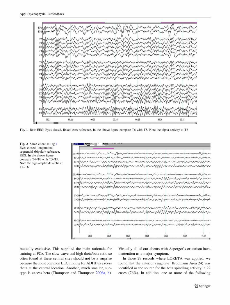
mutually exclusive. This supplied the main rationale for
training at FCz. The slow wave and high theta/beta ratio so
often found at these central sites should not be a surprise
because the most common EEG finding for ADHD is excess
theta at the central location. Another, much smaller, sub-
type is excess beta (Thompson and Thompson 2006a, b).
Virtually all of our clients with Asperger’s or autism have
inattention as a major symptom.
In those 29 records where LORETA was applied, we
found that the anterior cingulate (Brodmann Area 24) was
identified as the source for the beta spindling activity in 22
cases (76%). In addition, one or more of the following
Fig. 1 Raw EEG: Eyes closed, linked ears reference. In the above figure compare T6 with T5. Note the alpha activity at T6
Fig. 2 Same client as Fig 1.
Eyes closed, longitudinal
sequential (bipolar) reference,
EEG. In the above figure
compare T4–T6 with T3–T5.
Note the high amplitude alpha at
T4–T6
Appl Psychophysiol Biofeedback
123
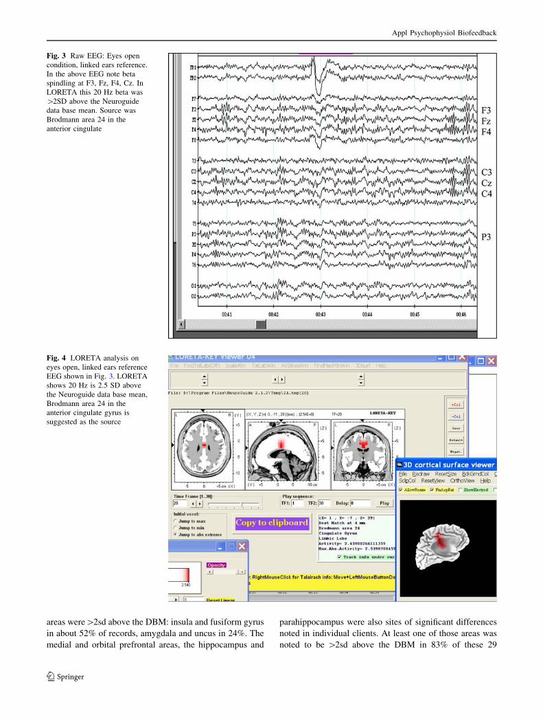
areas were[2sd above the DBM: insula and fusiform gyrus
in about 52% of records, amygdala and uncus in 24%. The
medial and orbital prefrontal areas, the hippocampus and
parahippocampus were also sites of significant differences
noted in individual clients. At least one of those areas was
noted to be [2sd above the DBM in 83% of these 29
Fig. 3 Raw EEG: Eyes open
condition, linked ears reference.
In the above EEG note beta
spindling at F3, Fz, F4, Cz. In
LORETA this 20 Hz beta was
[2SD above the Neuroguide
data base mean. Source was
Brodmann area 24 in the
anterior cingulate
Fig. 4 LORETA analysis on
eyes open, linked ears reference
EEG shown in Fig. 3. LORETA
shows 20 Hz is 2.5 SD above
the Neuroguide data base mean,
Brodmann area 24 in the
anterior cingulate gyrus is
suggested as the source
Appl Psychophysiol Biofeedback
123
records analyzed using LORETA. We reviewed whether
right or left side predominated for any one of these areas
and found it was, for the most part, both sides that were
affected with a slight trend for left amygdala, uncus and
fusiform gyrus and right insula (which influences sympa-
thetic system activity) to be implicated as the source of
abnormal activity.
Half of the children with autism demonstrated T6
slowing compared to T5. With this group generalized
parietal slow wave activity and coherence values well
outside the database for Neuroguide in the parietal region
were the common finding. In addition, compared to our
ADHD population who show high amplitude slow wave at
Fz and Cz, the group with autism showed high amplitude
theta at Pz and Cz.
In visual analysis of the raw EEG mu was noted to be
present in 7 cases (just 10%), as determined by morphology
of the waves and the lack of suppression during the eyes
open conditions. We feel this may be an underestimate as
we were not looking for this as a factor in the early years.
In addition, previously undetected seizure activity was seen
in 3 cases. The cases with seizure activity were referred to
a neurologist.
NFB Training
Training recommendations, based on the findings shown
in Figs. 1, 2, 3, 4, suggest that normalizing the EEG
could involve using two-channel NFB training. One
channel would have the active electrode placed at FCz
(to influence the AC) and the second channel would
have the active electrode at T6 (to decrease the sensory
aprosodia symptoms). Both would be referenced to
linked ears (or to a common reference point even less
influenced by surrounding electrical activity, such as the
nose) and have a common ground (Thompson and
Thompson 2007a, b). This training might reasonably be
expected to ameliorate some of the core symptoms of
AS (inattention, anxiety, not reading emotions/nonverbal
communication) (Thompson et al. 2008). Alternatively,
one could use single channel training and work first at
the central location and then later at T6 if the ability to
pick up on nonverbal communication continued to be a
problem for the client.
Increasing sensorimotor rhythm (SMR) using neuro-
feedback may have a stabilizing effect on a cortex that is
unstable and easily kindled (Sterman 2000a). As previously
noted, beta spindling is one indication of a cortex that may
be easily kindled, irritable, even unstable, in other words, a
cortex that may not be functioning properly. Beta spindles
are high amplitude, narrow band (often a single Hz),
synchronous beta (Johnstone et al. 2005; Thompson and
Thompson 2006a, b). Beta spindling is an EEG finding that
may be observed in a number of disorders where anxiety is
a symptom. In our experience using LORETA, spindling
beta has most often been associated with a source in the
cingulate gyrus. Perhaps our past success in these disor-
ders, when we emphasized normalizing theta, alpha, and
beta amplitudes and increasing SMR rhythm at CZ, was
only in part due to re-setting thalamic pacemakers. Perhaps
it was also due to normalizing anterior cingulate (AC)
activity with all its connections to various elements in the
mirror neuron system in addition to the hypothalamus and
the brainstem nuclei controlling the autonomic nervous
system.
In addition to the low activity observed at T6, another
factor that may, in the future, prove to be a helpful ‘mar-
ker’ for ASD could be the ‘mu’ rhythm response. In ASD
there is evidence of a reduction in mu rhythm suppression
during action observation (Oberman et al. 2005). However
we did not investigate this relatively new finding in our
analysis of these clients. In our experience mu, which is
considered a normal variant, is not observed in the majority
of clients. Therefore using this as a training parameter for
NFB, as suggested in an article in Scientific American Mind
(Ramachandran and Oberman 2006), would not be our
initial approach. Additionally, it can be difficult to distin-
guish between mu, alpha, and sensorimotor rhythm at the
C3 and C4 sites given that the frequency ranges overlap.
One must examine the raw EEG to check morphology of
the wave forms (mu being a wicket rhythm with one end
sharp and the other rounded, whereas alpha and SMR are
sinusoidal) and/or check for responsiveness (alpha is
attenuated by eye opening and SMR and mu respond to
movement of the hands).
With QEEG, Chan and colleagues at the University of
Hong Kong have recently attempted to find an EEG marker
for ASD. They found that children with ASD showed
significantly less relative alpha power. This was not spe-
cific to restricted location(s) but was a widespread pattern
(Chan et al. 2007). Less alpha may indicate a brain that
does not rest appropriately, which is perhaps the flip side of
the picture of spindling beta and low SMR frequently
found in clients with AS.
Both hyper and hypo coherence abnormalities are found
in clients with autistic spectrum disorders. Care must be
taken in evaluating theta and alpha hypercoherence
because sometimes it may be due to ear reference con-
tamination; for example, very high amplitude alpha at a site
such as T6 near the linked ear reference will make it appear
as though many other sites have alpha. Coherence often
changes after amplitude training is done to increase and
decrease frequencies found to differ from database norms.
Appl Psychophysiol Biofeedback
123
Coben (2007) has demonstrated good outcomes with nor-
malization of coherence after training that used sequential
(bipolar) placement at pairs of sites that showed hyperco-
herence on QEEG assessment.
Rationale for Neurofeedback Intervention
EEG differences observed in clients with Asperger’s syn-
drome provide a rationale for using neurofeedback. Spe-
cifically, these findings include
1. excess slow wave activity frontally corresponds to
‘‘being more in their own world’’;
2. excess slow wave and/or beta spindling at Fz, found
to originate using LORETA in the medial frontal
cortex with its connections to the amygdala and to the
AC, may correspond to difficulties modulating
emotions;
3. low SMR is consistent with fidgety, impulsive
behavior, tactile sensitivity, anxiety, and emotionally
labile behavior;
4. excess left prefrontal and frontal slow wave activity
is consistent with lack of appropriate inhibition and
failure to properly modulate sensory inputs;
5. excess right prefrontal beta activity is consistent with
lack of appropriate affect modulation and the inhi-
bition of impulsivity;
6. less activation, as evidenced by high slow wave
activity and/or low, low frequency beta activity (beta
frequencies \20 Hz) in the right parietal-temporal
area (homologous site to Wernicke’s area), is consis-
tent with difficulty interpreting social cues and emo-
tions (sensory aprosodia);
7. excess slow wave activity and/or reduced low
frequency beta activity in right frontal cortex (homol-
ogous site to Broca’s area), is consistent with under-
activation and inability to appropriately express
emotion through tone of voice (symptoms of motor
aprosodia);
8. deviations from a normal data base in frequencies
whose source was identified by LORETA to be in the
anterior cingulate (including beta spindling) may
correspond to anxiety related symptoms (including
obsessive-compulsive tendencies).
9. temporal lobe and, in particular, the superior tempo-
ral gyrus and fusiform gyrus activity outside of data
base norms may indicate difficulty inhibiting the
central nucleus of the amygdala (Porges 2007), which
can result in an adverse effect on vagal calming and
allow increased sympathetic drive
10. abnormalities in coherence suggest that training for
normalizing connectivity between the parietal lobes
and the temporal and frontal regions may prove to be
beneficial.
Rationale for Biofeedback Intervention
Changes in physiological variables with minor stressors and
the client’s inability to rapidly recover after stress, as may
be demonstrated with a stress test (Thompson and
Thompson 2007b), provide one rationale for using bio-
feedback. Learning comfortable, slow diaphragmatic
breathing at about six breaths per minute (faster in children)
gives those with AS a portable stress management tech-
nique. In addition, vagal afferents from the heart feed back
to the nucleus of the solitary tract in the medulla which is
connected to the parabrachial nucleus and the locus coe-
ruleus. These nuclei connect to the forebrain with links to
the hypothalamus, amygdala, thalamic connections to the
insula, orbitofrontal and prefrontal areas, all of which give
feedback to the anterior cingulate (Porges 2003, 2007).
Theoretically this could synergistically assist in normaliz-
ing the activity of the AC and its connections through both
the mirror neuron system and the limbic system. Using NFB
plus BFB and coaching in strategies exemplifies a holistic
approach that emphasizes skills not pills.
Summary and Discussion
Neuroanatomically, the common areas that are posited to
be influenced by neurofeedback plus biofeedback in clients
where FCz was the site for the NFB training and BFB
including HRV are first, the cingulate gyrus, usually the
anterior cingulate (AC) with all its connections through the
limbic system and the mirror neuron system and, second,
the autonomic nervous system with all its brainstem con-
nections to the amygdala and the forebrain. As previously
noted, the AC is central to affect regulation and control. It
has executive functions and it is critically involved in the
processes of attention and concentration. Additionally, the
AC is also well connected to the insula and the amygdala
and to the mirror neuron system (Carr et al. 2003).
As is reported in another paper (Thompson et al. 2008)
we have had success when we used a Cz or FCz site to train
down frequencies that were high amplitude compared to
the rest of the client’s EEG (theta 3–7 Hz or low alpha (8–
10 Hz), and/or high frequency beta (in the range 20–
35 Hz) and train up sensorimotor rhythm (13–15 Hz) based
on the findings of single channel EEG assessments. In
theoretical terms, given the clear relationship of the mirror
neuron system (MNS) to ASD, it seems reasonable to
hypothesize that influencing what we have termed the hub
Appl Psychophysiol Biofeedback
123
of the affective nervous system, the AC, may have been
responsible for improvement in reading and copying non-
verbal information (so-called social cues) when working
with clients with Asperger’s or autism. Perhaps the NFB
has had its positive effects by changing the responsiveness
of the MNS. We postulate that this may be why, in most
cases, we have not had to directly activate the T6 area
using NFB, even though it is an area that initially shows
less activation.
It would be interesting to conduct an experiment using
only BFB and, in particular, heart rate variability training
to see what effect this might have on clients with AS.
Performing another experiment with just NFB would also
be of interest to tease out specific effects. However, as a
clinician, one looks for the combination that best helps a
client realize their potential. In real life a brain-body sep-
aration is artificial. As Walter Hess said in his 1949 Nobel
prize acceptance speech, ‘‘Every living organism is not the
sum of a multitude of unitary processes, but is, by virtue of
interrelationships and of higher and lower levels of con-
trol, an unbroken unity’’. For our purpose of explaining
why neurofeedback plus biofeedback can re-balance and
thus improve mind-body functioning, we have developed a
Systems Theory of Neural Synergy. The name is used in
order to emphasize that, no matter where we enter the
nervous system with our interventions, the neural system
will adjust its own new balance and equilibrium (Von
Bertalanffy 1976). Whether we train the brain (NFB) or we
train the heart (BFB) the neural pathways do connect
across the forebrain, the midbrain and the hindbrain. In
particular, the anterior cingulate connects with the brain-
stem, as fits with Porges’ polyvagal theory. The AC con-
nects to the nucleus ambiguus giving it control over aspects
of the vagal parasympathetic efferents controlling such
important physiological functions as heart rate while it
receives vagal afferent feedback from such organs as the
heart via connections relayed through the nucleus solitarus
in the medulla of the brain stem. The vagal afferent sensory
information is conveyed from the medullary nucleus of the
solitary tract to the parabrachial nucleus and the locus
coeruleus. As noted under the Rationale for BFB Inter-
vention above, these nuclei connect to the forebrain with
links to the hypothalamus, amygdala, thalamic connections
to the insula, orbitofrontal and prefrontal areas, all of which
give feedback to the anterior cingulate. The central nucleus
of the amygdala also directly connects to the brainstem
autonomic nuclei. Our conclusion is that all of these con-
siderations support the importance of recognizing the
interconnectedness of the entire central nervous system
(CNS) and, in our work with the ASDs, the combined use
of NFB and BFB.
Studies have shown that neurofeedback is effective in
reducing the symptoms of ADHD and evidence is
mounting that EEG biofeedback can similarly play an
important role in helping those with Asperger’s Syndrome
learn self-regulation skills that address their unique chal-
lenges (Coben 2007; Jarusiewicz 2002; Knezevic 2007;
Linden et al. 1996; Reid 2005; Thompson and Thompson
1995, 2007a, b). This paper provides a theoretical basis,
derived from knowledge of functional neuroanatomy, for
the improvements being reported in clinical practice.
References
Abu-Akel, A. (2003). A neurobiological mapping of theory of mind.
Brain Research Reviews, 43, 39–40.
Adolphs, R. (2003). Is the human amygdala specialized for processing
social information? Annals of the New York Academy ofSciences, 985, 326.
Adolphs, R., Gosselin, F., Buchanan, T. W., Tranel, D., Schyns, P., &
Damasio, A. R. (2005). A mechanism for impaired fear
recognition after amygdala damage. Nature, 433, 68.
Alvarez, A. (2004). Issues in assessment: Asperger’s syndrome and
personality. In M. Rhode & T. Klauber (Eds.), The many faces ofAsperger’s syndrome. The Tavistock clinic series (pp. 113–128).
London: Karnac Books.
American Psychiatric Association. (2000). Diagnostic and statisticalmanual of mental disorders—4th edition—Text revision. Wash-
ington, DC: American Psychiatric Association.
Asperger, H. (1944). Die‘‘Autistischen Psychopathen’’ im Kindesalt-
er. Archiv fuer Psychiatrie und Nervenkrankheiten, 117, 76–136.
English translation (1991) Autistic psychopathy in childhood. In
U. Frith (Editor and Translator) Autism and Asperger’s syndrome(pp. 37–92). Cambridge, United Kingdom: Cambridge Univer-
sity Press.
Attwood, T. (1997). Asperger’s syndrome: A Guide for parents andprofessionals. London: Jessica Kingsley Publications.
Attwood, T. (2007). The complete guide to Asperger’s syndrome.
London: Jessica Kingsley Publishers.
Bachevalier, J., & Loveland, K. A. (2006). The orbitofrontal-
amygdala circuit and self-regulation of social-emotional behav-
ior in autism. Neuroscience and Behavioral Reviews, 30, 97–
117.
Bara, B. G., Bucciarelli, M., & Colle, L. (2001). Communicative
abilities in autism: Evidence for attentional deficits. Brain andLanguage, 77(2), 216–240.
Barton, J. S., Cherkasov, M. V., Hefter, R., Cox, T. A., O’Connor,
M., & Manoach, D. S. (2004). Are patients with social
developmental disorders prosopagnosic? Perceptual heterogene-
ity in the Asperger and socio-emotional processing disorders.
Brain, 127(8), 1706–1716.
Bashe, P. R., & Kirby, B. (2005). The OASIS guide to Aspergersyndrome. New York: Crown Publishers.
Belmonte, M. K., & Yurgelun-Todd, D. A. (2003). Functional
anatomy of impaired selective attention and compensatory
processing in autism. Cognitive Brain Research, 17(3), 651–664.
Blandford, S. (2005). Children can learn with their shoes off:
Supporting students with Asperger’s syndrome in mainstream
schools and colleges. Educational Psychology in Practice, 21(1),
76–78.
Bleuler, E. (1911). Dementia praecox oder gruppe der schizophre-nien. J. Zinken translation, 1950. New York: International
University Press.
Appl Psychophysiol Biofeedback
123
Bowler, D. M., Gardiner, J. M., & Berthollier, N. (2004). Source
memory in adolescents and adults with Asperger’s syndrome.
Journal of Autism and Developmental Disorders, 34(5), 533–
542.
Bregman, J. D. (2005). Definitions and characteristics of the spectrum.
In D. Zager (Ed.), Autism spectrum disorders: Identification,education, and treatment (3rd ed., pp. 3–46). Mahwah: Lawrence
Erlbaum Associates.
Carothers, D. E., & Taylor, R. L. (2004). Social cognitive processing
in elementary school children with Asperger syndrome. Educa-tion & Training in Developmental Disabilities, 39(2), 177–187.
Carr, L., Iacoboni, M., Dubeau, M. C., & Mazziotta, J. C. (2003).
Neural mechanisms of empathy in humans: A relay from neural
systems for imitation to limbic areas. Proceedings of theNational Academy of Sciences, USA, 100, 5497–5502.
Chan, A. S., Sze, S. L., & Cheung, M. (2007). Quantitative
electroencephalographic profiles for children with autistic spec-
trum disorder. Neuropsychology, 21(1), 74–81.
Channon, S. (2004). Frontal lobe dysfunction and everyday problem-
solving: Social and non-social contributions. Acta Psychologica,115(2–3), 235–254.
CNS Response. (2008). www.cnsresponse.com.
Coben, R. (2005). Assessment guided neurofeedback for autisticspectrum disorder. Presentation at the Society for Neuronal
Regulation 13th annual conference, Denver, Colorado.
Coben, R. (2006). Panel on autistic spectrum disorders at the 36thannual meeting of the Association for Applied Psychophysiologyand Biofeedback. Portland, OR.
Coben, R. (2007). Connectivity-guided neurofeedback for autistic
spectrum disorder. Biofeedback, 35(4), 131–135.
Committee on Children with Disabilities. (2001). Technical report:
The pediatrician’s role in the diagnosis and management of
autistic spectrum disorders in children. Pediatrics, 107(5), 1–35.
Corbett, B. A., & Constantine, L. J. (2006). Autism and attention
deficit hyperactivity disorder: Assessing attention and response
control with the integrated visual and auditory continuous
performance test. Child Neuropsychology, 12(4–5), 335–348.
Cumine, V., Leach, J., & Stevenson, G. (1998). Asperger syndrome: Apractical guide for teachers. London: David Fulton Publishers.
Dalton, K., Nacewicz, B., Johnstone, T., Schaefer, H., Gernsbacher,
M., Goldsmith, H., et al. (2005). Gaze fixation and the neural
circuitry of face processing in autism. Nature Neuroscience,8(4), 519–526.
Dapretto, M., Davies, M. S., Pfeifer, J. H., Scott, A. A., Sigman, M.,
Bookheimer, S. Y., et al. (2006). Understanding emotions in
others: Mirror neuron dysfunction in children with autism
spectrum disorders. Nature Neuroscience, 9(1), 28–30.
Deruelle, C., Rondan, C., Gepner, B., & Tardif, C. (2004). Spatial
frequency and face processing in children with autism and
Asperger syndrome. Journal of Autism and DevelopmentalDisorders, 34(2), 199–210.
Devinsky, O., Morrell, M., & Vogt, B. (1995). Contributions of
anterior cingulate cortex to behaviour. Brain, 118, 279–306.
Dissanayake, C., & Macintosh, K. (2003). Mind reading and social
functioning in children with autistic disorder and Asperger’s
disorder. In B. Repacholi & V. Slaughter (Eds.), Individualdifferences in theory of mind: Implications for typical andatypical development. Macquarie monographs in cognitivescience (pp. 213–239). New York: Psychology Press.
Ehlers, S., & Gillberg, C. (1993). The epidemiology of Asperger’s
syndrome: A total population study. Journal of Child Psychologyand Psychiatry, 34(8), 1327–1350.
Emerich, D. M., Creaghead, N. A., Grether, S. M., Murray,
D., & Grasha, C. (2003). The comprehension of humorous
materials by adolescents with high-functioning autism and
Asperger’s syndrome. Journal of Autism and DevelopmentalDisorders, 33(3), 253–257.
Fisch, B. (1999). Fisch and Spehlmann’s EEG primer: Basicprinciples of digital and analog EEG-third edition. New York:
Elsevier.
Fitzgerald, M. (2000). Jonathan swift: Victim of Asperger’s syn-
drome? The Canadian Journal of Diagnosis, 2000, 31–36.
Fitzgerald, M. (2004). Response to ‘‘Features of Alexithymia or
features of Asperger’s syndrome?’’ by M. Corcos. EuropeanChild & Adolescent Psychiatry, 13(2), 123.
Fitzgerald, M., & Kewley, G. (2005). Attention-deficit/hyperactivity
disorder and Asperger’s syndrome. Journal of the AmericanAcademy of Child and Adolescent Psychiatry, 44(3), 210.
Frith, U. (1991). (Editor and Translator) Autism and Asperger’ssyndrome. Cambridge, United Kingdom: Cambridge University
Press.
Frombonne, E., & Tidmarsh, L. (2003). Epidemiological data on
Asperger disorder. Child & Adolescent Psychiatric Clinics ofNorth America, 12(1), 15–21.
Gattegno, M. P., & De Fenoyl, C. (2004). Social abilities training in
people with Asperger syndrome/L’entraınement aux habiletes
sociales chez les personnes attients du syndrome d’Asperger.
Journal de Therapie Comportementale et Cognitive, 14(3), 109–
115.
Gevirtz, R. (2007). Biofeedback training to increase heart rate
variability. In P. M. Lehrer, R. L. Woolfolk, & W. E. Sime
(Eds.), Principles and practice of stress management (3rd ed.).
New York: Guilford Publications.
Gevirtz, R., & Lehrer, P. (2005). Resonant frequency heart rate
biofeedback. In M. S. Schwartz & F. Andrasik (Eds.), Biofeed-back a practitioner’s guide-third edition. New York: Guilford
Press.
Ghaziuddin, M., & Mountain-Kimchi, K. (2004). Defining the
intellectual profile of Asperger syndrome: Comparison with
high-functioning autism. Journal of Autism & DevelopmentalDisorders. 34(3), 279-284.
Gillberg, C. (1989). Asperger syndrome in 23 Swedish children.
Developmental Medicine and Child Neurology, 31, 520–531.
Gillberg, C. (1991). Clinical and neurobiological aspects of Asperger’s
syndrome in six families studied. In U. Frith (Ed.), Autism andAsperger’s syndrome (pp. 122–145). Cambridge, UK: Cambridge
University Press.
Goodenough-Harris Drawing Test. (1963). The psychological
corporation.
Green, V. A., Pituch, K. A., Itchon, J., Choi, A., O’Reilly, M., &
Sigafoos, J. (2006). Internet survey of treatments used by parents
of children with autism. Research in Developmental Disabilities,27, 70–84.
Haddon, M. (2002). The curious incident of the dog in the night-time.
Toronto: Doubleday Canada.
Hadjikhani, N., Joseph, R. M., Snyder, J., & Tager-Flusberg,
H. (2006). Anatomical differences in the mirror neuron system
and social cognition network in autism. Cerebral Cortex, 16,
1276–1282.
Hill, E. L., & Frith, U. (2003). Understanding Autism: Insights from
mind and brain. Theme Issue ‘Autism: mind and brain’. Philo-sophical Transactions of The Royal Society London, Bulletin 358,
281–289.
Iacoboni, M., & Dapretto, M. (2006). The mirror neuron system and
the consequences of its dysfunction. Nature Reviews andNeuroscience, December, 2006, 942–951.
Jantzen, T., Graap, K., Stephanson, S., Marshall, W., & Fitzsimmons,
G. (1995). Differences in baseline EEG measures for ADD and
normally achieving pre-adolescent males. Biofeedback and SelfRegulation, 20(1), 65–82.
Appl Psychophysiol Biofeedback
123
Jarusiewicz, E. (2002). Efficacy of neurofeedback for children in the
autistic spectrum: A pilot study. Journal of Neurotherapy, 6(4),
39–49.
Jasper, H. (1858). Report of the committee on methods of clinical
examination in electroencephalography. EEG and ClinicalNeurophysiology, 10, 374.
Johnstone, J., Gunkelman, J., & Lunt, J. (2005). Clinical database
development: Characterization of EEG phenotypes. ClinicalEEG and Neuroscience, 36(2), 99–107.
Kaland, N., Moller-Nielsen, A., Smith, L., Mortensen, E. L., Callesen,
K., & Gottlieb, D. (2005). The strange stories test: A replication
study of children and adolescents with Asperger syndrome.
European Child and Adolescent Psychiatry, 14(2), 73–82.
Khouzam, H. R., El-Gabalawi, F., Pirwani, N., & Priest, F. (2004).
Asperger’s disorder: A review of its diagnosis and treatment.
Comprehensive Psychiatry, 45(3), 184–191.
Kleinhans, N., Akshoomoff, N., & Delis, D. C. (2005). Executive
functions in autism and Asperger’s disorder: Flexibility, fluency,
and inhibition. Developmental Neuropsychology, 27(3), 379–401.
Klin, A., & Miller, K. (2004). When Asperger’s syndrome and a
nonverbal learning disability look alike. In M. T. Stein (Ed.),
Journal of Developmental & Behavioral Pediatrics. Special Issue:Challenging Cases in Developmental & Behavioral Pediatrics,
25(5S), S59–S64. (This reprinted article originally appeared in the
Journal of Developmental & Behavioral Pediatrics, 25(3), 190–
195).
Klin, A., Volkmar, & Sparrow, (2000). Asperger’s syndrome. New
York: Guilford Press.
Knezevic, B. (2007). Pilot project to ascertain utility of tower ofLondon test (ToL) to assess outcomes of neurofeedback in clientswith Asperger’s syndrome. Student Award paper presented at
International Society for Neurofeedback and Research, Annual
Meeting, San Diego, CA.
Landry, R., & Bryson, S. E. (2004). Impaired disengagement of attention
in young children with autism. Journal of Child Psychology andPsychiatry and Allied Disciplines, 45(6), 1115–1122.
Laurent, A. C., & Rubin, E. (2004). Challenges in emotional
regulation in Asperger syndrome and high-functioning autism.
Topics in Language Disorders, 24(4), 286–297.
Lawson, J., Baron-Cohen, S., & Wheelwright, S. (2004). Empathizing
and systemizing in adults with and without Asperger syndrome.
Journal of Autism and Developmental Disorders, 34(3), 301–310.
Linden, M. (2006). Panel on autistic spectrum disorders at the 36thannual meeting of the Association for Applied Psychophysiologyand Biofeedback. Portland, OR.
Linden, M., Habib, T., & Radojevic, V. (1996). A controlled study of
EEG biofeedback effects on cognitive and behavioral measures
with attention-deficit disorder and learning disabled children.
Biofeedback and Self-Reguation, 21(1), 35–49.
Loffler, D. (2005). Asperger syndrome: What teachers need to know.
Educational Psychology in Practice, 21(1), 80–81.
Lubar, J. F. (1991). Discourse on the development of EEG diagnostics
and biofeedback treatment for attention deficit/hyperactivity
disorders. Biofeedback and Self-Regulation, 16(3), 202–225.
Lubar, J. F., & Lubar, J. (1984). Electroencephalographic biofeedback
of SMR and beta for treatment of attention deficit disorder in a
clinical setting. Biofeedback and Self Regulation, 9(1), 1–23.
Lubar, J. F., Swartwood, M. O., Swartwood, J. N., & O’Donnell, P.
(1995). Evaluation of the effectiveness of EEG neurofeedback
training for ADHD in a clinical setting as measured by changes
in T.O.V.A. scores, behavioral ratings, and WISC-R perfor-
mance. Biofeedback and Self Regulation, 20(1), 83–99.
Macintosh, K. E., & Dissanayake, C. (2004). Annotation: The
similarities and differences between autistic disorder and
Asperger’s disorder: A review of the empirical evidence. Journalof Child Psychology and Psychiatry, 45(3), 421–434.
Mann, C. A., Lubar, J. F., Zimmerman, A. W., Miller, C. A., &
Muenchen, R. A. (1992). Quantitative analysis of EEG in
boys with attention-deficit/hyperactivity disorder: Controlled
study with clinical implications. Pediatric Neurology, 8(1),
30–36.
Martinez, Y. (2003). Unpublished thesis done as part of the
requirements for an Honours B.A. degree. Copy on file at the
ADD Centre, Toronto.
Mayes, S. D., & Calhoun, S. L. (2004). Influence of IQ and age in
childhood autism: Lack of support for DSM-IV Asperger’s
disorder. Journal of Developmental & Physical Disabilities,16(3), 257–272.
McAlonan, G. M., Cheung, V., Cheung, C., Suckling, J., Lam, G. Y.,
Tai, K. S., et al. (2005). Mapping the brain in autism. A voxel-
based MRI study of volumetric differences and intercorrelations
in autism. Brain: Journal of Neurology, 128(2), 268–276.
McCann, J. (2006). Reeg—the new image of diagnostic accuracy.
Neuropsychiatry May 7(8).
Meltzoff, A., & Prinz, W. (Eds.). (2002). The imitative mind:Development, evolution and brain bases. New York: Cambridge
University Press.
Monastra, V. J. (2005). Electroencephalographic biofeedback (neu-
rotherapy) as a treatment for attention deficit hyperactivity
disorder: Rationale and empirical foundation. Child and Ado-lescent Psychiatric Clinics of North America, 14(1), 55–82.
Monastra, V. J., Lubar, J. F., & Linden, M. (2001). The development
of a quantitative electroencephalographic scanning process for
attention deficit-hyperactivity disorder: Reliability and validity
studies. Neuropsychology, 15(1), 136–144.
Monastra, V. J., Lubar, J. F., Linden, M., VanDeusen, P., Green, G.,
Wing, W., et al. (1999). Assessing attention deficit hyperactivity
disorder via quantitative electroencephalography: An initial
validation study. Neuropsychology, 13(3), 424–433.
Monastra, V. J., Monastra, D., & George, S. (2002). The effects of
stimulant therapy, EEG biofeedback and parenting on primary
symptoms of ADHD. Applied Psychophysiology and Biofeed-back, 27(4), 250–272.
Nacewicz, B. M., Dalton, K. M., Johnstone, T., Long, M. T., McAuliff,
E. M., Oakes, T. R., et al. (2006). Amygdala volume and nonverbal
social impairment in adolescent and adult males with autism.
Archives of General Psychiatry, 63(12), 1417–1428.
Nash, J. M. (2002). The secrets of Autism. Time. May 6, 2002/159(18), 36–46.
Neuroguide Delux, 2.3.7. (2007). Robert Thatcher, applied neurosci-
ence Inc. (www.appliedneuroscience.com).
Nikolaenko, N. N. (2004). Metaphorical and associative thinking in
healthy children and in children with Asperger’s syndrome at
different ages. Human Physiology, 30(5), 532–536.
Nishitani, N., Avikainen, S., & Hari, R. (2004). Abnorml imitation-
related cortical activation sequences in Asperger’s syndrome.
Annals of Neurology, 55, 558–562.
Oberman, L. M., Hubbard, E. M., McCleery, J. P., Altschuler, E. L.,
Ramachandran, V. S., & Pineda, J. A. (2005). EEG evidence for
motor neuron dysfunction in autistic spectrum disorders. BrainResearch & Cognitive Brain Research, 24, 190–198.
Pascual-Marqui, R. D., Esslen, M., Kochi, K., & Lehmann, D. (2002).
Functional imaging with low resolution electromagnetic tomog-
raphy (LORETA): A review. Methods and Findings in Exper-imental and Clinical Pharmacology, 24C, 91–95.
Pfeiffer, H., Iacoboni, M., Mazziotta, C., & Dapretto, M. (2005).
Mirror neuron system activity in children and its relation to
empathy and interpersonal competence. In Abstract Viewer/
Itinerary Planner. Society of Neuroscience Abstract 660(24).Porges, S. W. (2003). Social engagement and attachment: A
phylogenetic perspective. Annals of the New York Academy ofSciences, 1008, 31–47.
Appl Psychophysiol Biofeedback
123

Porges, S. W. (2004). The Vagus: A mediator of behavioral and
physiologic features associated with autism. In M. L. Bauman &
T. L. Kemper (Eds.), The neurobiology of autism (pp. 65–78).
Baltimore: Johns Hopkins University Press.
Porges, S. W. (2007). The Polyvagal perspective. Biological Psychi-atry, 74, 116–143.
Portway, S. M., & Johnson, B. (2005). Do you know I have
Asperger’s syndrome? Risks of non-obvious disability. Health,Risk & Society, 7(1), 73–83.
Prichep, E. S., Mas, F., Hollander, E., Liebowitz, M., John, E. R.,
Almas, M., et al. (1993). Quantitative electroencephalographic
subtyping of obsessive-compulsive disorder. PsychiatryResearch: Neuroimaging, 50, 25–32.
Ramachandran, V. S., & Oberman, L. M. (2006). Broken mirrors.
Scientific American, 295(5), 62–69.
Reid, A. (2005). Autistic spectrum disorders: Assessment andintervention results after neurofeedback in 146 cases. Student
Award Presentation, International Society for Neuronal Regula-
tion annual meeting. Denver, Colorado.
Ross, E. D. (1981). The aprosodias: Functional-anatomic organization
of the affective components of language in the right hemisphere.
Archives of Neurology, 38, 561–569.
Rossiter, T. R., & LaVaque, T. J. (1995). A comparison of EEG
biofeedbaclk and psychostimulants in treating attention deficit
hyperactivity disorders. Journal of Neurotherapy, 1(1), 48–59.
Rourke, B., & Tsatsanis, K. (2000). Nonverbal learning disabilities
and Asperger’s syndrome. In A. Klin, F. Volkmar, & S. Sparrow
(Eds.), Asperger syndrome. New York: Guilford Press.
Rubin, E., & Lennon, L. (2004). Challenges in social communication
in Asperger Syndrome and High-Functioning Autism. Topics inLanguage Disorders, 24(4), 271–285.
Salmond, C. H., Ashburner, J., Connelly, A., Friston, K. J., Gadian, D.
G., & Vargha-Khadem, F. (2005). The role of the medial
temporal lobe in autistic spectrum disorders. European Journalof Neuroscienc, 22(3), 764–772.
Sears, W., & Thompson, L. (1998). The A.D.D. book: Newunderstandings, new approaches to parenting your child. New
York: Little, Brown and Co.
Shamay-Tsoory, S. G., Tomer, R., Berger, B. D., Goldsher, D., &
Aharon-Peretz, J. (2005). Impaired ‘‘Affective Theory of Mind’’
is associated with right ventromedial prefrontal damage. Cog-nitive & Behavioral Neurology, 18(1), 55–67.
Shouse, M. N., & Lubar, J. F. (1979). Sensorimotor rhythm (SMR)
operant conditioning and methylphenidate in the treatment of
hyperkinesis. Biofeedback & Self-Regulation, 4, 299–311.
Simpson, D. (2004). Asperger’s syndrome and autism: Distinct
syndromes with important similarities. In M. Rhode & T.
Klauber (Eds.), The many faces of Asperger’s syndrome. TheTavistock clinic series (Vol xviii, 302, pp. 25–38). London,
England: Karnac Books.
Sloman, L. (2005). Medication use in children with high functioning
pervasive developmental disorder and Asperger syndrome. In K.
Stoddart (Ed.), Children, youth and adults with Aspergersyndrome: Integrating multiple perspectives. London: Jessica
Kingsley Publishers.
Soderstrom, H., Nilsson, T., Sjodin, A., Carlstedt, A., & Forsman, A.
(2005). The childhood-onset neuropsychiatric background to
adulthood psychopathic traits and personality disorders. Com-prehensive Psychiatry, 46(2), 111–116.
Solnick, B. (2005). Effects of electroencephalogram biofeedback with
Asperger’s syndrome. International Journal of RehabilitationResearch, 28(2), 159–163.
Sterman, M. B. (1999). Atlas of Topometric clinical displays:Functional interpretations and neurofeedback strategied. New
Jersey: Sterman-Kaiser Imaging Laboratory.
Sterman, M. B. (2000a). Basic concepts and clinical findings in the
treatment of seizure disorders with EEG operant conditioning.
Clinical Electroencephalography, 31(1), 45–55.
Sterman, M. B. (2000b). EEG markers for attention deficit disorder:
Pharmacological and neurofeedback applications. Child StudyJournal, 30(1), 1–22.
Stewart, K. (1998). Helping a child with nonverbal learning disorderor Asperger’s syndrome. Place: Publisher.
Suffin, S. C., & Emory, W. H. (1995). Neurometric subgroups in
attentional and affective disorders and their association with
pharmacotherapeutic outcomes. Clinical Electroencephalogra-phy, 26(2), 76–83.
Swanson, J. M., McBurnett, K., Wigal, T., Pfiffner, L. J., Williams,
L., Christian, D. L., et al. (1993). The effect of stimulant
medication on children with attention deficit disorder: A
‘‘Review of Reviews’’. Exceptional Children, 60(2), 154–162.
Tammet, D. (2007). Born on a blue day. London: Free Press (Division
of Simon & Shuster).
Tanguay, P. B. (2001). Nonverbal learning disabilities at home: Aparent’s guide. London: Jessica Kingsley Publishers.
Thatcher, R. W., Walker, R. A., Biver, C. J., North, D. N., & Curtin,
R. (2003). Quantitative EEG normative databases: Validation
and clinical correlation. In J. F. Lubar (Ed.), Quantitativeelectroencephalographic analysis (QEEG) databases for neuro-therapy: Description, validation, and application. New York:
Haworth Press.
Thompson, M. G. G., & Havelkova, M. (1983). Childhood Psychosis.
In P. Steinhauer & Q. Rae-Grant (Eds.), Psychological Problemsof the Child in the Family. New York: Basic Books, Inc.
Thompson L., & Thompson M. (1995). Exceptional Results withExceptional Children. In Proceedings of the Society for the Study
of Neuronal Regulation. Annual Meeting: Scottsdale, Arizona.
Thompson, L., & Thompson, M. (1998). Neurofeedback combined
with training in metacognitive strategies: Effectiveness in
students with ADD. Applied Psychophysiology and Biofeedback,23(4), 243–263.
Thompson, L., & Thompson, M. (2003a). The neurofeedback book.
Wheatridge, CO: Association for Applied Psychophysiology and
Biofeedback.
Thompson, M. & Thompson, L. (2003b). Neurofeedback for Asper-
ger’s syndrome: Theoretical rationale and clinical results. TheNewsletter of the Biofeedback Society of California, 19(1).
Thompson, L., & Thompson, M. (2006a). Panel on Autistic SpectrumDisorders. 36th Annual meeting of the Association for Applied
Psychophysiology and Biofeedback, Portland, Oregon, April.
Thompson, M., & Thompson, L. (2006b). Improving attention in
adults and children: Differing electroencephalograhy profiles and
implications for training. Biofeedback Magazine, 34(3), 99–105.
Thompson, L., & Thompson, M. (2007a). Autistic spectrum disorders
including Asperger’s syndrome: EEG and QEEG findings,
results, and neurophysiological rationale for success using
neurofeedback training. Presented at the 11th Annual Meeting
of the Biofeedback Foundation of Europe, Berlin, Germany.
Abstract reprinted in Applied Psychophysiology and Biofeed-back, 32(3–4), 213–214.
Thompson, M., & Thompson, L. (2007b). Neurofeedback for stress
management. In P. Lehrer, R. Woolfolk, & W. Sime (Eds.),
Principles and practice of stress management (3rd ed.). New
York: Guilford Publications.
Thompson, M., Thompson, L., & Reid, A. (2008). Neurofeedback
outcomes in clients with Asperger’s syndrome. Submitted to
Journal of Applied Psychophysiology and Biofeedback.
Thompson, M., & Thompson, L. (2009). Asperger’s syndrome
intervention: Combining neurofeedback, biofeedback and
metacognition. In T. Budzinski, H. Budzinski, J. Evans, &
Appl Psychophysiol Biofeedback
123

A. Abarbanel (Eds.), Introduction to quantitative EEG andneurofeedback (2nd ed.). New York: Elsevier.
Tower of London Test. (1982). Colorado assessment tests.
www.catstests.com/prod03.htm.Von Bertalanffy, L. (1976). General systems theory: Foundations,
development, applications. New York: George Braziller, Inc.
Weimer, A. K., Schatz, A. M., Lincoln, A., Ballantyne, A. O., &
Truaner, D. A. (2001). ‘‘Motor’’ impairment in Asperger
syndrome: Evidence for a deficit in proprioception. Journal ofDevelopmental and Behavioral Pediatrics, 22(2), 92–101.
Weitz, P., & Weitz, C. (2002). ‘‘About a Boy’’. Movie starring Hugh
Grant, Universal pictures, Tribeca/working title production.
Wing, L. (1981). Asperger’s syndrome: A clinical account. Psycho-logical Medicine, 11, 115–129.
Wing, L. (2001). The autistic spectrum: A parent’s guide to under-standing and helping your child. Berkeley, CA: Ulysses Press.
Wing, L., & Gould, J. (1979). Severe impairments of social
interaction and associated abnormalities in children: Epidemiol-
ogy and classification. Journal of Autism and DevelopmentalDisorders, 9, 11–29.
Appl Psychophysiol Biofeedback
123