Embed Size (px)
Citation preview

JPET #239301
1
Title page
Functional, metabolic, and dynamic mitochondrial changes in the rat cirrhosis-
hepatocellular carcinoma model and the protective effect of IFC-305.
Enrique Chávez, María Guadalupe Lozano-Rosas, Mariana Domínguez-López, Gabriela Velasco-
Loyden, Jesús Rafael Rodríguez-Aguilera, Concepción José-Nuñez, Marietta Tuena de Gómez-
Puyou, Victoria Chagoya de Sánchez.
Departamento de Biología Celular y Desarrollo, Instituto de Fisiología Celular, Universidad
Nacional Autónoma de México, Mexico City, Mexico (E.C., M.G.L-R, M.D-L., G.V-L., J.R.R-
A., V.C.S.).
Departamento de Bioquímica y Biología Estructural, Instituto de Fisiología Celular, Universidad
Nacional Autónoma de México, Mexico City, Mexico (C.J-N., M.T.G-P.).
This article has not been copyedited and formatted. The final version may differ from this version.JPET Fast Forward. Published on February 16, 2017 as DOI: 10.1124/jpet.116.239301
at ASPE
T Journals on A
ugust 12, 2019jpet.aspetjournals.org
Dow
nloaded from
JPET #239301
2
Running Title: Mitochondrial changes and IFC-305 protection in an HCC model
Correspondence should be addressed to: Victoria Chagoya de Sánchez, Departamento de
Biología Celular y Desarrollo, Instituto de Fisiología Celular, Universidad Nacional Autónoma
de México. Ciudad de México, México. C.P. 04510. Tel. +52 (55) 5622-5614, Fax +52 (55)
5622-5611. E-mail: [email protected]
Number of text pages: 37
Number of tables: 2
Number of figures: 8
Number of references: 52
Number of words:
- Abstract - 186
- Introduction - 750
- Discussion- 1500
Abbreviations: diethylnitrosamine (DEN), dynamin- related protein 1 (DRP1), electron transport
chain (ETC), isocitrate dehydrogenase (IDH), malate dehydrogenase (MDH), mitofusin-2(MFN-
2), poly(ADP-ribose) polymerase 1 (PARP-1), peroxisome proliferator-activated receptor -
coactivator 1α (PGC-1α), sirtuin-1, -3 (Sirt-1, -3).
Recommended section assignment: Chemotherapy, Antibiotics, and Gene Therapy
This article has not been copyedited and formatted. The final version may differ from this version.JPET Fast Forward. Published on February 16, 2017 as DOI: 10.1124/jpet.116.239301
at ASPE
T Journals on A
ugust 12, 2019jpet.aspetjournals.org
Dow
nloaded from

JPET #239301
3
ABSTRACT
Background: Mitochondrion is an important metabolic and energetic organelle which regulates
several cellular processes. Mitochondrial dysfunction has been related with liver diseases
including hepatocellular carcinoma. As a result, the energetic demand is not properly supplied
and mitochondrial morphologic changes has been observed resulting in an altered metabolism.
We previously demonstrated the chemopreventive effect of the hepatoprotector IFC-305. Aim: In
this work we aimed to evaluate the functional, metabolic, and dynamic mitochondrial alterations
in the sequential model of cirrhosis-hepatocellular carcinoma induced by diethylnitrosamine in
rats and the possible beneficial effect of IFC-305. Methods: Experimental groups of rats were
formed to induce cirrhosis-hepatocellular carcinoma and to assess the IFC-305 effect during
cancer development and progression through the evaluation of functional, metabolic and dynamic
mitochondrial parameters. Results: In this experimental model, dysfunctional mitochondria were
observed and suspension of the diethylnitrosamine treatment was not enough to restore them.
Administration of IFC-305 maintained and restored the mitochondrial function and regulated
parameters implicated in metabolism as well as the mitochondrial dynamics modified by
diethylnitrosamine intoxication. Conclusion: This study supports IFC-305 as a potential
hepatocellular carcinoma treatment or as an adjuvant in chemotherapy.
This article has not been copyedited and formatted. The final version may differ from this version.JPET Fast Forward. Published on February 16, 2017 as DOI: 10.1124/jpet.116.239301
at ASPE
T Journals on A
ugust 12, 2019jpet.aspetjournals.org
Dow
nloaded from

JPET #239301
4
1. INTRODUCTION
Mitochondria are organelles responsible for most of the energetic metabolism in eukaryotic cells.
As an integral part of ATP production, through the oxidative phosphorylation (OXPHOS), the
tricarboxylic acid cycle (TCA) donates electrons to the electron transport chain (ETC), which
consists of five complexes (I-V), where complex I is the electron entry site for NADH and
generates NAD+ and complex V is in charge of ATP synthesis. The ability of mitochondria to
regulate the energetic, redox state and metabolism of the cells could result in the production of
epigenetic intermediates, point them out as a major therapeutic target because mitochondrial
dysfunction is involved in several diseases including cancer (Mughal et al., 2012; Boland et al.,
2013).
Studies have revealed several metabolic alterations in liver diseases including modifications in
energy supply (Hernandez-Munoz et al., 1991; Hernandez-Munoz and Chagoya de Sanchez,
1994). Otto Warburg suggests that mitochondria from tumor cells supply the energetic demand,
through the glycolytic flux because of the lack of oxygen or due to genetic-epigenetic alterations
that affect the oxidative metabolism (Wallace and Fan, 2010). In fact, metabolic alterations and
some anti-apoptotic proteins such as BCL-KL reduce the acetyl-CoA level (Ac-CoA) (Yi et al.,
2011); this intermediate also plays an important role as a signal transducer and gene expression
(Pietrocola et al., 2015). Oxidative stress diminishes oxidative metabolism flux, which includes
TCA enzymes such as isocitrate dehydrogenase (IDH) and malate dehydrogenase (MDH).
Indeed, studies have identified mutations in IDH producing an oncometabolite in different tumor
types (Dang et al., 2016). A good indicator of the mitochondrial redox state is the NAD+/NADH
ratio and if it is correctly regulated, an efficient ETC activity is possible; as shown in the
limitation of breast tumor growth (Santidrian et al., 2013). Previously our research group
This article has not been copyedited and formatted. The final version may differ from this version.JPET Fast Forward. Published on February 16, 2017 as DOI: 10.1124/jpet.116.239301
at ASPE
T Journals on A
ugust 12, 2019jpet.aspetjournals.org
Dow
nloaded from

JPET #239301
5
demonstrated the ability of adenosine to maintain the energetic and redox state of the cell
(Hernandez-Munoz et al., 1978; Hernandez-Munoz et al., 1987). In addition to the metabolic role
of NAD+, it participates in the activity of multiple enzymes. According to this, Sirtuin-1 (Sirt-1),
a NAD+-dependent protein and a member of class III histone deacetylase, targets several
transcription factors including the peroxisome proliferator-activated receptor -coactivator 1α
(PGC-1α). The latter coactivates major transcription factors involved in mitochondrial and
nuclear gene expression directing the complex program of mitochondrial biogenesis (Finley and
Haigis, 2009). However, Sirt-1 and PGC-1α have been found over-expressed in HCC and are
related to defective mitochondrial accumulation (Chen J, 2012; Boland et al., 2013). Another
NAD+-dependent protein is PARP-1 (Poly(ADP-ribose)) polymerase-1 whose activity modulates
transcription and DNA repair; nevertheless, over-expression and increased activity of PARP-1
have been found in HCC and has been considered as a cancer hallmark (Hanahan and Weinberg,
2011).
Mitochondria are not static organelles; their dynamism depends, at least in part, on the fission
and fusion phenomena that determine their shape. Mitochondrial fusion requires polarized
mitochondrial membrane and the activity of proteins such as mitofusin 1 and 2 (MFN 1, 2); this
process promotes cristae integrity and OXPHOS. Mitochondrial fission is induced by different
kinds of stress and requires proteins such as the dynamin-related protein 1 (DRP1); membrane
depolarization is observed during fission and, if the membrane potential is not recovered,
mitochondria are targeted to autophagy (Boland et al., 2013). Altered mitochondrial fission and
fusion have been observed in tumor cells mainly due to increased DRP1 and decreased MFN-2
expression (Rehman et al., 2012).
This article has not been copyedited and formatted. The final version may differ from this version.JPET Fast Forward. Published on February 16, 2017 as DOI: 10.1124/jpet.116.239301
at ASPE
T Journals on A
ugust 12, 2019jpet.aspetjournals.org
Dow
nloaded from

JPET #239301
6
Our research group demonstrated the hepatoprotective effects of IFC-305, an adenosine-derived
compound (Perez-Carreon et al., 2010; Velasco-Loyden et al., 2010; Chagoya de Sanchez et al.,
2012; Velasco-Loyden, 2016); this compound has been evaluated in a diethylnitrosamine (DEN)-
induced sequential rat model of cirrhosis-HCC, where it inhibited pre-neoplastic lesions
development in comparison to DEN-treated groups. The chemopreventive effect was associated
with the reduction of the expression of collagen, thymidylate synthase, the tumor marker –
glutamyl transferase, the hepatocyte growth factor, and the induction of the cell cycle inhibitor
p27 expression (Velasco-Loyden, 2016).
In this work we evaluated the mitochondrial alterations in the sequential cirrhosis-hepatocellular
carcinoma model, previously described by Schiffer et al. (Schiffer et al., 2005), and the possible
beneficial effects of the hepatoprotector IFC-305 during HCC development and progression on
mitochondrial function, metabolism, and dynamics. We measured the mitochondrial function
through the respiratory quotient, membrane potential, complex I activity, and ATP synthesis. The
activity of the malate-aspartate shuttle, IDH and MDH, and the level of Ac-CoA and lactate were
determined as metabolic parameters. Dynamic mitochondrial proteins were determined to
evaluate the morphology and biogenesis; electron microscopy was also used for morphology
studies.
This article has not been copyedited and formatted. The final version may differ from this version.JPET Fast Forward. Published on February 16, 2017 as DOI: 10.1124/jpet.116.239301
at ASPE
T Journals on A
ugust 12, 2019jpet.aspetjournals.org
Dow
nloaded from
JPET #239301
7
2. MATERIALS AND METHODS
2.1 Chemicals
IFC-305 is the aspartate salt of adenosine prepared with adenosine free base (MP Biomedicals,
LLC, Illkirch, France) and L-aspartic acid (MP Biomedicals, Inc, Eschwege, Germany) as
described (Patent No. MX220780; MX 207422; US 8,507,459 B2) (Chagoya de Sanchez, 2002;
Chagoya de Sanchez, 2004; Chagoya de Sanchez, 2013). Dietthylnitrosamine, sucrose, EDTA,
Trisma base, KCl, MgCl2, glutamate, ADP, rhodamine 123, HEPES, EGTA, cyclosporine A,
valinomycin, succinate, 2,6-dicholorindophenol, ATP, NAD. NADH, acetophenone were
purchased from the Sigma Chemical Company (St. Louis, MO, USA).
2.2 Animal treatment and experimental groups
Male Wistar rats (weighing 200 g) were obtained from and housed at the Animal Facility of the
National Autonomous University of Mexico (UNAM), and all procedures were conducted
according to our institutional guidelines for the care and use of laboratory animals. Groups of rats
(n = 6, each group) were divided in two different schemes: hepatocellular carcinoma (HCC) and
cancer progression (CP). These were treated as follows: a) the HCC groups were injected with
DEN (Sigma-Aldrich, St. Louis, MO) at 50 mg/kg body weight i.p., once a week and saline
solution or IFC-305 at 50 mg/kg body weight i.p., 3 times weekly (HCC+IFC-305) for 16 weeks
plus 2-weeks wash out; b) the CP groups were administered DEN at 50 mg/kg body weight i.p.,
once a week for 16 weeks and then received saline solution or IFC-305 (CP+IFC-305) at 50
mg/kg body weight i.p., 3 times weekly during 6 weeks (Table 1). To euthanize animals, a lethal
dose of sodium pentobarbital was used and the liver was removed.
2.3 Mitochondrial function
This article has not been copyedited and formatted. The final version may differ from this version.JPET Fast Forward. Published on February 16, 2017 as DOI: 10.1124/jpet.116.239301
at ASPE
T Journals on A
ugust 12, 2019jpet.aspetjournals.org
Dow
nloaded from
JPET #239301
8
2.3.1 Mitochondria isolation and oxygen uptake
Liver samples were homogenized (1:10 w/v) in a medium containing 250 mM sucrose, 1 mM
EDTA, 10 mM Trizma base, and 0.1% BSA, pH 7.3. The tissue homogenate was centrifuged at
755 g for 5 min to remove nuclei and plasma membrane fragments. The supernatant was filtered
through organza fabric and centrifuged at 8400 g for 10 min to obtain the mitochondrial pellet.
Mitochondria were resuspended in 250 mM sucrose, 1 mM EDTA, 10 mM Trizma base, pH 7.3.
Mitochondrial respiration was recorded polarographically with a Clarck-type oxygen electrode in
3 mL of a medium containing 250 mM sucrose, 10 mM KCl, 5 mM MgCl2, 10 mM potassium
phosphate, 10 mM Trizma base; 10 mM glutamate and malate were used as substrates for site I.
Mitochondrial state 3 was initiated by adding 226 µM ADP (final concentration).
2.3.2 Mitochondrial membrane potential
The mitochondrial membrane potential was determined according to Baracca et al. (Baracca et
al., 2003). Briefly, a calibration curve was done by plotting rhodamine 123 fluorescence against
the membrane potential calculated through the Nerst equation. To determine the mitochondrial
membrane potential 0.15 mg of mitochondria in a medium containing 250 mM sucrose, 10 mM
HEPES, 100 µM EGTA, 2 mM MgCl2, 4 mM KH2PO4, pH 7.4, and 10 mM sucrose and 2.5 U
hexokinase. Then, 1 µg/mL of 33 nM cyclosporine A and 0.1 mM ADP were added. Finally,
rhodamine (50 nM) fluorescence was measured in the presence of 20 mM succinate.
2.3.3 Complex I activity
The 2,6-dichloroindophenol (DCPIP) reduction was used to determine complex I activity in
mitochondria isolated from the liver. Briefly, 1 mg of mitochondrial protein was added to a
medium containing 0.25 M sucrose, 1 mM EDTA, 0.1% BSA, 100 µM KCN, and 10 mM
This article has not been copyedited and formatted. The final version may differ from this version.JPET Fast Forward. Published on February 16, 2017 as DOI: 10.1124/jpet.116.239301
at ASPE
T Journals on A
ugust 12, 2019jpet.aspetjournals.org
Dow
nloaded from

JPET #239301
9
glutamate as substrate, and 152 µM rotenone. The absorbance was followed at 600 nm and the
activity was calculated using the DCPIP extinction coefficient (21 mM-1cm-1).
2.3.4 Determination of the ATPase activity and the content of the αβ subunits
Rat liver mitochondria (2 mg) were resuspended in different volumes of the mitochondrial
preparation medium. Mitochondria were centrifuged at 180 g for 10 min at 4°C. The
mitochondrial pellet was resuspended in 50 µL of 250 mM sucrose and 1 mM MgCl2 and
centrifuged in an air-driven ultracentrifuge at 5 249 g at 10 psi. The pellet was resuspended in a
medium containing 50 mM NaCl, 50 mM imidazole, 1 mM EDTA, 2 mM aminocaproic acid,
0.66 mg digitonin /mg mitochondria. The suspension was centrifuged at 9 860 g at 22 psi for 5
min. The supernatant was used for protein determination with first dimension blue native PAGE
(BN-PAGE) and second dimension SDS PAGE.
First dimension. Protein extracted with digitonin (100 µg) was mixed with 10 µL buffer 3X (1.5
M 6-aminocaproic acid, 150 mM Bis-Tris) plus serva blue G (7 µg/µL stock serva blue G in 1.5
M 6-aminocaproic acid) (final concentration, 30 ng serva blue/1 µg protein). The sample was
charged to a 3.5-11% linear BN-PAGE and electrophoresis was performed at 70 V for 2 h and
100 V for 8 h at 4°C. ATPase activity was identified by incubating the first dimension gel in a
preincubation-solution containing 35 mM Tris, 250 mM glycine, pH 8.3. Then, the gel was
stirred and incubated for 1 h and 2 h at 37°C with 5 mM ATP, 5 mM MgCl2, 0.15 % (w/v) lead
acetate, and 150 mM glycine, pH 8.3. After that, the gel was stirred and incubated at room
temperature for 24 h. Monomer and dimer bands with dark background were scanned at 1, 2, and
24 h. Monomer bands were read at 1, 2, and 24 h, with Image J software.
2.4 Metabolic evaluation
This article has not been copyedited and formatted. The final version may differ from this version.JPET Fast Forward. Published on February 16, 2017 as DOI: 10.1124/jpet.116.239301
at ASPE
T Journals on A
ugust 12, 2019jpet.aspetjournals.org
Dow
nloaded from
JPET #239301
10
2.4.1 Malate-aspartate shuttle
The activity of the malate-aspartate shuttle was determined using the assay previously described
for Scholz et al. (Scholz et al., 1998). Briefly, 50 µL of mitochondrial suspension were mixed
with 2 mL of 300 mM mannitol, 10 mM potassium phosphate, 10 mM Tris, 10 mM KCl, 5 mM
MgCl2, 2 mM aspartate, 2 mM ADP, and 0.14 mM NADH, pH 7.4. Then, 2 IU/mL of AST was
added and basal oxidation of NADH was followed at 340 nm at 37°C for 4 min. The shuttle
activity was started with the addition of 4 mM malate and 4 mM glutamate. Oxidation of NADH
was followed at 340 nm for 4 min at 37°C.
2.4.2 Isocitrate dehydrogenase and malate dehydrogenase activities
Isocitrate dehydrogenase and malate dehydrogenase activities were estimated using the following
commercial kits: Isocitrate Dehydrogenase Activity Assay Kit (Cat. MAK062) and Malate
Dehydrogenase Assay Kit (Cat. MAK196) following the instructions provided by the
manufacturer (Sigma-Aldrich, Mexico).
2.4.4 Acetyl-CoA, 3-hydroxybutyric acid, and lactate determination
The levels of acetyl-CoA, 3-hydroxybutyric acid, acetoacetate and lactate were determined using
the following commercial kits: Acetyl-Coenzyme A Assay Kit (Cat. MAK039, Sigma-Aldrich,
Mexico); Acetoacetate Colorimetric Assay Kit (Cat. MAK199); EnzyChrom Ketone Body Assay
Kit (Cat. EKBD-100, Hayward, CA, USA). The instructions provided by the manufacturer were
followed.
2.4.5 PARP-1 enzymatic activity
The PARP-1 enzymatic activity was determined according to Karson et al. (Putt and
Hergenrother, 2004). Briefly, liver samples were homogenized in a buffer containing 50 mM
This article has not been copyedited and formatted. The final version may differ from this version.JPET Fast Forward. Published on February 16, 2017 as DOI: 10.1124/jpet.116.239301
at ASPE
T Journals on A
ugust 12, 2019jpet.aspetjournals.org
Dow
nloaded from

JPET #239301
11
Tris, 2 mM MgCl2, pH 8.0, and protease inhibitors cocktail (Roche Diagnosis, Indianapolis, IN,
USA). The tissue homogenate was centrifuged at 13 700 g for 10 min at 4°C. The supernatant
was separated and protein content was determined by the Bradford method (Bio-Rad,
Laboratories, Inc. USA). A DNA sample was obtained from the control liver tissue through a
modified salt extraction method described by Lopera-Barrero et al. (Lopera-Barrero, 2008).
Briefly, 20 µL of 100 µM NAD+ was added to the plate; subsequently, 30 µg of protein and DNA
(at 12.5 µg/mL), previously activated with UV irradiation, were added. The plate was incubated
at room temperature for 20 min and the amount of NAD+ present was determined by the addition
of 10 µL of 2 M KOH and 10 µL of 20% acetophenone (in ethanol). The plate was incubated at
4°C for 10 min. Finally, 45 µL of 88% formic acid was added and the plate incubated at 110°C
for 5 min. The plate was allowed to cool and was read at 378 nm. To quantitate the NAD+,
absorbances were interpolated in a NAD+ calibration curve, previously performed.
2.5 Evaluation of mitochondrial dynamics
2.5.1 Nuclear protein extraction
The isolation of intact and stable nuclei consisted in an iso-osmotic lysis procedure, as reported
by Dyer and Herzog (Dyer and Herzog, 1995). With this lysis procedure, the nuclear envelope
remained intact even during further manipulations of washing, freezing, and ultracentrifugation,
and provided nuclear protein extract. Briefly, 0.8 g of liver tissue was homogenized in 1.6 mL of
buffer containing 0.25 M sucrose, 0.05 M Tris-Cl, 0.005 M KCl2. The homogenate was filtered
and supplemented with 3 mL of a 2.3 M sucrose solution to increase the homogenate’s density.
Then, the sucrose gradient was created by adding 1.5 mL of a 2.3 M sucrose solution to the
bottom of the tube containing the homogenate. The nucleus was isolated by centrifugation for 30
min at 4°C at 255 000 g. After the isolation, nuclear lysis was performed by incubating and
This article has not been copyedited and formatted. The final version may differ from this version.JPET Fast Forward. Published on February 16, 2017 as DOI: 10.1124/jpet.116.239301
at ASPE
T Journals on A
ugust 12, 2019jpet.aspetjournals.org
Dow
nloaded from

JPET #239301
12
stirring with a hypotonic and hypertonic solution. The nuclear extract protein was determined by
the Bradford method and used for the corresponding assays.
2.5.2 Total protein extraction
Liver samples were homogenized with RIPA buffer (100 mg of tissue per milliliter of buffer)
containing protease and phosphatase inhibitors (Roche Diagnostics Corp.). The homogenate was
centrifuged at 16 000 g for 10 min at 4°C, the supernatant was utilized as total liver homogenate.
2.5.3 Western blot assays
Mitochondrial, nuclear, and total protein extracts were used as corresponding. Volumes
equivalent to 50 µg of protein were electrophoresed on 12% polyacrylamide gel (20%
polyacrylamide for histones); separated proteins were transferred onto PVDF (Immobilon P).
Next, blots were blocked with 5% skim milk and 0.05% Tween-20 for 30 min at room
temperature, and independently incubated overnight at 4°C with selective antibodies (dilution
1:1000) against PGC-1α (MAB 1032 from Chemicon Int. Inc.), Sirt-1, Drp-1, Sirt-3, Mfn-2
(Santa Cruz Biotechnology sc-15404, sc-32898, sc-99143, sc-50331, respectively), H4ac, Hsp60,
H3, H4 (06-946, MAB 3844, 06-755, 04-858 from Millipore, respectively), as appropriate. On
the following day, the membranes were washed and then exposed to a secondary peroxidase-
labeled antibody at dilution 1:10000 (Jackson ImmunoResearch) in the blocking solution for 1 h
at room temperature. Blots were washed and protein was developed using the ECL detection
system. Densitometric analyses of bands were performed with Quantity One software (Bio-Rad
Laboratories, Hercules, CA, USA).
2.5.4 Electron microscopy
This article has not been copyedited and formatted. The final version may differ from this version.JPET Fast Forward. Published on February 16, 2017 as DOI: 10.1124/jpet.116.239301
at ASPE
T Journals on A
ugust 12, 2019jpet.aspetjournals.org
Dow
nloaded from

JPET #239301
13
Liver samples for electron microscopy were fixed with glutaraldehyde (6%) and stained with
osmium tetroxide (1% phosphate buffered saline solution) according to Mascorro et al. (Mascorro
JA, 1976).
2.6 Statistical analysis
Data are expressed as mean values ± standard error of the mean (SEM). Comparisons were
carried out by analysis of variance, followed by Tukey's test, as appropriate, using Graph Pad
Prism 5.0 (Graph Pad Software Inc, La Jolla, CA, USA) for Windows. Differences were
considered statistically significant when p < 0.05.
This article has not been copyedited and formatted. The final version may differ from this version.JPET Fast Forward. Published on February 16, 2017 as DOI: 10.1124/jpet.116.239301
at ASPE
T Journals on A
ugust 12, 2019jpet.aspetjournals.org
Dow
nloaded from

JPET #239301
14
3. RESULTS
3.1 IFC-305 treatment was helpful to maintain and recover the mitochondrial function that was
altered in the sequential model of cirrhosis-hepatocellular carcinoma.
Mitochondria are the main source of energy and act as an energy modulator to orchestrate their
physiological responses. For this reason, we decided to evaluate some parameters that reflect the
mitochondrial function in the sequential model of cirrhosis-hepatocellular carcinoma in the rat
and the effect of IFC-305 treatment. The respiratory quotient was obtained from the state 3 and
state 4 ratio. Chronic treatment with DEN induced an increased oxidation of glutamate in the
absence of ADP (state 4) and a lower response for ADP (state 3) resulting in a diminished
respiratory quotient in the HCC and CP groups compared to the control group (Table 2); the IFC-
305 treatment prevented and reversed this alteration as seen in the HCC+IFC-305 and CP+IFC-
305 groups. Moreover, there was a significant decrease in ATP synthesis in the groups treated
with DEN that was not restored even in the CP group, and the beneficial effect of IFC-305 to
prevent and reverse the damage caused by DEN can be observed in the groups receiving IFC-305
(Table 2). It can be observed a significant reduction in the complex I activity in the HCC and CP
groups and the beneficial effect of the IFC-305 treatment which was able to reverse this alteration
to the normal level. Mitochondrial membrane potential provides an electrochemical gradient
required to regulate mitochondrial function. We found important changes in this parameter;
depolarization was observed in mitochondria from HCC and CP groups and a tendency to prevent
and reverse this effect can be observed in groups HCC+IFC-305 and CP+IFC-305 (Table 2).
These results clearly indicate the functional impairment due to the chronic intoxication with DEN
and the improvement effect exerted by the IFC-305 treatment.
This article has not been copyedited and formatted. The final version may differ from this version.JPET Fast Forward. Published on February 16, 2017 as DOI: 10.1124/jpet.116.239301
at ASPE
T Journals on A
ugust 12, 2019jpet.aspetjournals.org
Dow
nloaded from

JPET #239301
15
Considering the aforementioned findings, we evaluated complex V, which is in charge of ATP
synthesis as an integral part of the mitochondrial function. Through the isolation of the F1F0
complex from samples at week 22, we evaluated its ability to hydrolyze ATP. In this approach,
we observed that the ATPase activity remained lower in the CP group than in the control one.
The IFC-305 treatment restored this activity, which became higher at 24 h (Figure 1A).
Moreover, the absence of the dimeric form of the F1F0 complex was seen the DEN intoxication
even after it had been suspended (CP group); the control group and that treated with the IFC-305
compound (CP+IFC-305 group) revealed the presence of the dimeric form of the F1F0 complex
(Figure 1B). Similar results were obtained through the monomer quantification in blue gel and
Coomassie staining and, the αβ subunits content (Supplemental Figure 1). These findings support
the IFC-305 beneficial effect on the αβ ATPase subunits.
The mitochondrial function was damaged in the HCC and CP groups; as it was previously
mentioned, the suspension of DEN not recovered mitochondrial functionality but the IFC-305
compound helps to maintain mitochondrial integrity.
3.2 The hepatoprotector improved the metabolic alterations generated by dysfunctional
mitochondria.
Due to the previously described results, we decided to evaluate parameters that could evidence
metabolic modifications. As seen in Figure 2A, the malate-aspartate shuttle activity is decreased
in preparations obtained from HCC and CP groups; IFC-305 treatment inhibited the effect of
DEN and restored the activity of this shuttle, HCC+IFC-305 and CP+IFC-305, respectively.
From the levels of 3-hydroxybutyric acid and acetoacetate we calculated the NAD+/NADH ratio.
Mitochondria isolated from the HCC and CP groups had a marked diminution of the
This article has not been copyedited and formatted. The final version may differ from this version.JPET Fast Forward. Published on February 16, 2017 as DOI: 10.1124/jpet.116.239301
at ASPE
T Journals on A
ugust 12, 2019jpet.aspetjournals.org
Dow
nloaded from

JPET #239301
16
mitochondrial NAD+/NADH ratio. In the HCC+IFC-305 group small protection against this
damage was observed and the CP+IFC-305 group reached the control value (Figure 2B).
Because we observed alterations in the NAD+ mitochondrial regulation, we decided to evaluate
the activity of two NAD+-dependent enzymes that participate in the TCA cycle, IDH and MDH.
As shown in Figure 2C, IDH activity did not change significantly when it was compared to the
control group. Nevertheless, treatment with IFC-305 increased the activity of this enzyme
compared to the HCC and CP groups, respectively. MDH activity of HCC and HCC+IFC-305 is
similar to the control group. In the CP group, the MDH activity decreased but the treatment with
IFC-305 tended to reverse this effect (Figure 2D). The impaired IDH and MDH activity suggest
an altered TCA function due to DEN administration, IFC-305 treatment improved mainly the
IDH activity possibly related with the increase in redox potential.
As mentioned in the Introduction, the experimental model used in this study is associated with
HCC and because we found modifications in mitochondrial function, we determined the level of
hepatic lactate as an indicator of metabolic adaptation. Chronic administration of DEN increased
the amount of lactate in the liver and it did not return to the control level six weeks after the toxic
had been suspended as shown in the HCC and CP groups (Figure 3). IFC-305 treatment
significantly decreased the level of lactate (HCC+IFC-305) suggesting metabolic adaptation in
response to recuperated mitochondrial function.
Another intermediate that depends on mitochondrial energetics is Ac-CoA. DEN administration
had no significant effect on the amount of Ac-CoA in both groups, HCC and CP, but IFC-305-
treatment significantly increased Ac-CoA levels in the HCC+IFC-305 and CP+IFC-305 groups
(Figure 4A) consequently to the recuperation of mitochondrial function. Previous studies showed
the ability of IFC-305 to modulate some epigenetic modifications including histone acetylation
This article has not been copyedited and formatted. The final version may differ from this version.JPET Fast Forward. Published on February 16, 2017 as DOI: 10.1124/jpet.116.239301
at ASPE
T Journals on A
ugust 12, 2019jpet.aspetjournals.org
Dow
nloaded from

JPET #239301
17
(Rodríguez-Aguilera, 2015). Thanks to this and considering the increased level of this metabolic
intermediate, we decided to determine this post-translational modification because Ac-CoA is a
required cofactor for histone acetyltransferases. Diminution of H4ac was observed in the HCC
and CP groups; an increment trend was observed in HCC+IFC-305 and CP+IFC-305 groups
(Figure 4B and 4C), suggesting Ac-CoA availability. This result could be important because it
might establish a link between mitochondrial function and epigenetic control of gene expression.
Besides the mitochondrial alterations, other enzyme activities could be modified, dependent, at
least in part, on mitochondrial integrity and functionality. Specifically, PARP-1 over-expression
and over-activation have been shown to be a characteristic of HCC. The PARP-1 activity is
dependent on NAD+ and Figure 5 shows a significant increment in the CP group, whereas the
CP+IFC-305 group revealed the IFC-305 ability to decrease it. The over-activation of PARP-1
obtained in the DEN-intoxicated groups could be the cause of the diminution of NAD+ level
previously described in Figure 2B.
3.3 IFC-305 treatment was beneficial to avoid and recover the altered mitochondrial dynamics
induced by DEN intoxication.
The interplay between the mitochondrion and the nucleus is relevant in the regulation of the
mitochondrial response to stress and PGC-1α and Sirt-1 are proteins involved in this process. The
nuclear amount of both proteins was increased by DEN treatment, even in the CP group, as
compared to the control (Figure 6A and 6B). The pharmacological treatment with IFC-305 in,
HCC+IFC-305 and CP+IFC-305, reduced the nuclear content of both, Sirt-1 and PGC-1α
suggesting a diminution of mitochondrial stress induced by DEN.
This article has not been copyedited and formatted. The final version may differ from this version.JPET Fast Forward. Published on February 16, 2017 as DOI: 10.1124/jpet.116.239301
at ASPE
T Journals on A
ugust 12, 2019jpet.aspetjournals.org
Dow
nloaded from

JPET #239301
18
Mitochondrial networks determined by fission and fusion cycle were evaluated through the
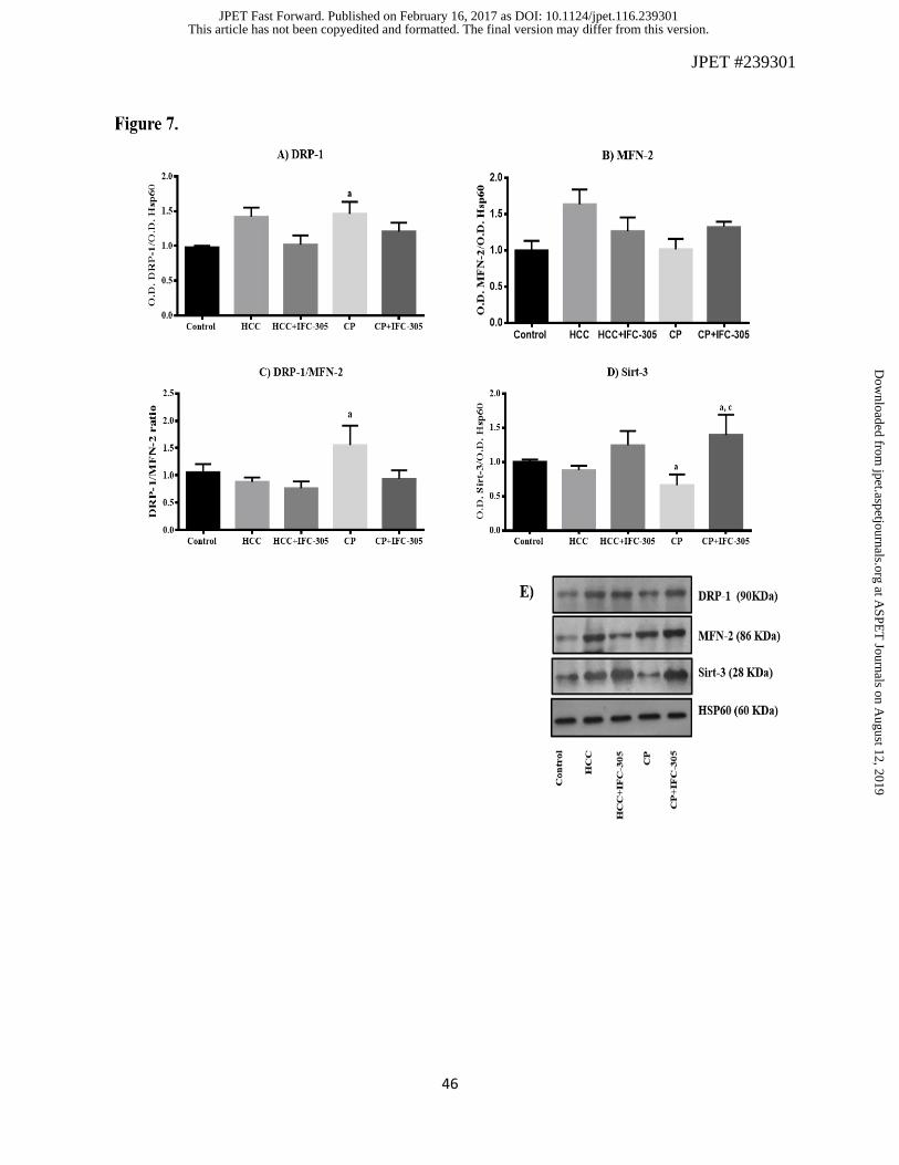
mitochondrial level of proteins regulating this process. Figure 7 shows the effect of DEN and
IFC-305 on mitochondrial localization of three important proteins related to fission and fusion
processes. DRP-1, required for mitochondrial fission by pinching them off into smaller
fragmented mitochondria, was induced in HCC and CP groups and it was prevented by the
hepatoprotector (Figure 7A). Figure 7B shows no significant differences in the MFN-2 content
between the experimental groups; however, a tendency to increase this protein in the HCC group
can be seen. However, when the DRP-1/MFN-2 ratio was determined (Figure 7C), an important
increase in this parameter was observed in the CP group reflecting a predominance of the fission
process. This alteration was not observed in the CP+IFC-305 group.
Until now important mitochondrial alterations were observed, but we wanted to know if there
were changes in mitochondrial content of Sirt-3, a NAD+-dependent deacetylase, that would be
indicative of a correlation between mitochondrial dynamics and metabolic function. The
progressive damage, observed when the toxicant was suspended, induced a decreased in
mitochondrial Sirt-3 (CP group) and the administration of IFC-305 increased Sirt-3 level
(CP+IFC-305), even more than the control group (Figure 7D). The up regulation in the presence
of the compound could be related to the improved mitochondrial redox state. Mitochondrial
modifications prompted us to obtain liver slices to observe the changes in mitochondrial
morphology through electron microscopy. The control group depicted mainly elongated
mitochondria. Circular shaped mitochondria were abundant in HCC and CP groups (Figures 8B
and 8D) and elongated shape was observed in groups receiving IFC-305 (Figures 8C and 8E). As
well as the altered mitochondrial shape, altered cristae were observed in the HCC and CP groups,
whereas the IFC-305-treated groups maintained and recovered cristae An important observation
This article has not been copyedited and formatted. The final version may differ from this version.JPET Fast Forward. Published on February 16, 2017 as DOI: 10.1124/jpet.116.239301
at ASPE
T Journals on A
ugust 12, 2019jpet.aspetjournals.org
Dow
nloaded from

JPET #239301
19
is the large amount of ribosomes observed in the slices from the HCC+IFC-305 and CP+IFC-305
groups marked with black arrows (Supplemental Figure 2). To appreciate better these effects, in
Supplemental Figure 2 mitochondria can be observed in an amplified size than in Figure 8. This
observations support the previous observations in mitochondrial functions, metabolism and
dynamic.
This article has not been copyedited and formatted. The final version may differ from this version.JPET Fast Forward. Published on February 16, 2017 as DOI: 10.1124/jpet.116.239301
at ASPE
T Journals on A
ugust 12, 2019jpet.aspetjournals.org
Dow
nloaded from

JPET #239301
20
4. DISCUSSION
This study shows that mitochondria from the liver of DEN-treated rats underwent alterations
resulting in decreased respiratory quotient which means that uncoupled mitochondria with low
phosphorylating capacity depicted lower ATP synthesis probably because of the altered m and
complex I activity (Table 2). Moreover, ATP synthase plays an essential role in energy
metabolism as it is in charge of ATP synthesis; the dimeric and oligomeric ATP synthase
complexes increase its stability and promote the formation of mitochondrial cristae (Garcia-Trejo
and Morales-Rios, 2008; Couoh-Cardel et al., 2010). The regeneration of the dimeric form of the
F1F0 ATPase is an indicator that the mitochondrial energetic machinery and morphology of
cristae were recovered with IFC-305 administration to the CP. Previously, the sensitivity of the
ETC complex I to the hepatotoxic action of CCl4, ethanol, and DEN had been demonstrated; for
DEN, this effect is attributed to the inhibition of NAD+-linked respiration at this site (Cederbaum
et al., 1974; Schilling and Reitz, 1980; Boitier et al., 1995). The malate-aspartate shuttle
maintains appropriate NADH equivalents, and its intermediates are coupled to the TCA cycle.
The activity of this system was diminished in mitochondria from DEN-treated rats (Figure 2A),
supporting the idea that mitochondria are the site for DEN-induced damaged. As mentioned
before, the treatment with IFC-305 improved the mitochondrial redox state damaged by DEN.
Mitochondrial function requires an optimal NAD+/NADH ratio to be efficient. The ability to
restore the mitochondrial NAD+/NADH ratio was achieved when the IFC-305 was given after the
discontinuation of DEN injection (CP+IFC-305 group) (Figure 2B). The maintenance of this ratio
allowed the correct supply of electrons to complex I and the right function of mitochondria. The
decreased m in the DEN-treated rats was expected because of the alterations previously
mentioned. These results demonstrate relevant mitochondrial function impairment induced by
This article has not been copyedited and formatted. The final version may differ from this version.JPET Fast Forward. Published on February 16, 2017 as DOI: 10.1124/jpet.116.239301
at ASPE
T Journals on A
ugust 12, 2019jpet.aspetjournals.org
Dow
nloaded from

JPET #239301
21
DEN and the beneficial effects of IFC-305 to maintain the mitochondrial function supporting the
previously mentioned adenosine findings which is the base molecule of the compound studied in
this work (Hernandez-Munoz et al., 1978).
IDH and MDH are mitochondrial TCA enzymes coupled to the mitochondrial redox potential.
IDH activity is decreased in patients with chronic liver injury after several years of alcohol abuse
and in rats treated with DEN (Suresh et al., 2013; Popov et al., 2014). We found a tendency to
diminish the IDH activity by DEN intoxication and its increased activity by IFC-305-treatment
(Figure 2C). This compound might promote the correct function of mitochondrial enzymes by
modulating the redox state as we mentioned above. A similar effect was seen in MDH activity in
the CP scheme.
The PARP-1 enzyme activity consists in the poly-adenosine diphosphate ribosylation
(Poly(ADP-ribosyl)ation) of different substrates and there are evidences of increased PARP-1
expression and auto-poly(ADP-ribosyl)ation in HCC and cirrhosis (Nomura et al., 2000;
Shiobara et al., 2001; Shimizu et al., 2004). Here, we demonstrated an increased PARP-1 activity
in the liver of rats treated with DEN and this result correlates with the diminution of the
NAD+/NADH ratio, suggesting depletion in NAD+ content. The IFC-305 compound prevented
PARP-1 activation by DEN as previously was showed in the presence of adenosine (Jagtap et al.,
2004). This result favor the NAD+ availability and contributing to the maintenance of the redox
state.
Mitochondrial changes affect the cytosolic redox state and, hence, metabolic alterations such as
lactate production. It has been suggested the aerobic glycolysis as the main source of energy in
cancer (Linehan and Rouault, 2013). In this context, glucose metabolism produces lactate. Here,
we found an induction of high levels of lactate with DEN even 6 weeks after intoxication was
This article has not been copyedited and formatted. The final version may differ from this version.JPET Fast Forward. Published on February 16, 2017 as DOI: 10.1124/jpet.116.239301
at ASPE
T Journals on A
ugust 12, 2019jpet.aspetjournals.org
Dow
nloaded from

JPET #239301
22
stopped. This metabolic adaptation was inhibited and reversed with the IFC-305 compound
(Figure 3). Probably the source of energy became glycolysis-dependent because of the altered
mitochondrial function observed in the DEN-treated rats; the beneficial effect of IFC-305
treatment could be explained by its ability to maintain mitochondrial integrity.
The synthesis of Ac-CoA, a carbon donor in anabolic reactions, is altered by mitochondrial
dysfunction (Wellen and Thompson, 2012). Moreover, it represents the link between pyruvate
and the mitochondrial TCA cycle and ETC; thus, it might reflect the mitochondrial energetic
supply. Besides its energetic role, Ac-CoA represents availability of the substrate for acetylation
reactions; it is important in the nucleus to modulate the gene expression through acetylation of
histone lysines in DNA-binding proteins reducing the protein-DNA interaction (Wallace and Fan,
2010). Even though DEN did not significantly change the Ac-CoA level respect to the control
group, IFC-305 treatment did change it (Figure 4A); moreover, we observed a tenuous and not
significant increased H4ac induced by IFC-305 suggesting that Ac-CoA levels regulated by IFC-
305 might affect the mark of histones acetylation (Figure 4B). In cancer cells, Ac-CoA is
ectopically synthesized in the nucleus and is involved in cell cycle progression and DNA
replication (Comerford et al., 2014). However, our results show Ac-CoA from mitochondria,
indicating that the source of this metabolite is from the normal glycolytic flux through pyruvate,
β-oxidation, and the catabolism of amino acids. These results confirm IFC-305’s ability to
increase the energy status of the liver and to improve the mitochondrial integrity. Possibly, H4ac
allowed the transactivation of genes involved in growth and replication, it must be considered
that the experimental model used implies cirrhosis, i.e., the whole liver was affected by the
administration of DEN. Further studies are necessary to test if replication of healthy hepatocytes
This article has not been copyedited and formatted. The final version may differ from this version.JPET Fast Forward. Published on February 16, 2017 as DOI: 10.1124/jpet.116.239301
at ASPE
T Journals on A
ugust 12, 2019jpet.aspetjournals.org
Dow
nloaded from

JPET #239301
23
is facilitated in this experimental model by IFC-305, as we previously reported in the CCl4 model
(Chagoya de Sanchez et al., 2012).
The diminished Sirt-3 mitochondrial content obtained with DEN reflects the inability to control
the redox state and the metabolic activity. Recently, Sultana et al. demonstrated the protective
effect of Sirt-3 on the mitochondrial function probably by the oxidative stress modulation
through the mitochondrial antioxidant enzymes such as Mn-SOD (Sultana et al., 2016).
Moreover, some authors have demonstrated downregulation of Sirt-3 in human HCC, suggesting
that this pattern is associated with differentiation and tumor multiplicity (Xiong et al., 2016). Our
results suggest a role of Sirt-3 in the mitochondrial effects of the IFC-305 compound possibly by
deacetylation of the ATPase and complex I subunits (Vassilopoulos et al., 2014). Further studies
are needed to demonstrate this. Overexpression of Sirt-3 induces apoptosis in HCC culture cells
through mitochondrial translocation of the Bcl-2-associated X protein (BAX) (Zhang and Zhou,
2012; Song et al., 2015). The transcriptional co-activator PGC-1α and Sirt-1 are considered
important inducers of mitochondrial biogenesis through nuclear-encoded mitochondrial genes.
Sirt-1 maintains PGC-1α in a deacetylated active state, promoting its transcriptional activity
(Gerhart-Hines et al., 2007). Our results suggest the ability of IFC-305 to inhibit the activation of
PGC-1α, at least in part, to maintain the Ac-CoA levels and the decreased nuclear amount of Sirt-
1. This effect allows us to explain the lower accumulation of dysfunctional mitochondria.
Additionally, Sirt-1 has been linked to tumorigenesis and its depletion inhibits proliferation of
HCC cells by cellular senescence or apoptosis (Chen J, 2012). The nuclear localization of both
proteins is increased in the rats intoxicated with DEN as compared to the control group (Figure
4). The IFC-305 compound was able to decrease the nuclear content of both, Sirt-1 and PGC-1 α.
This article has not been copyedited and formatted. The final version may differ from this version.JPET Fast Forward. Published on February 16, 2017 as DOI: 10.1124/jpet.116.239301
at ASPE
T Journals on A
ugust 12, 2019jpet.aspetjournals.org
Dow
nloaded from

JPET #239301
24
It is suggested that biogenesis induced by PGC-1α is tumor promoting, but production of new
healthy mitochondria could be tumor suppressive (Boland et al., 2013).
Mitochondria are dynamic organelles to maintain their correct functionality and morphology.
DRP1 regulates mitochondrial fission through scission of the outer mitochondrial membrane
(OMM). Mitochondrial fusion is regulated by MFN-1 and -2, which facilitates binding of the
OMM of different mitochondria (Dhingra and Kirshenbaum, 2014). According to this, small and
fragmented mitochondria have been found in mice with reduced levels of MFN-2, but when this
protein is restored, the normal mitochondria phenotype is obtained (Liesa et al., 2008). Here, we
show that DEN intoxication induces important changes in the DRP-1/MFN-2 ratio 6 weeks after
DEN administration was suspended. This alteration indicates a predominance fission over fusion.
The mitochondrial morphology observed by electronic microscopy corroborates these results,
since those isolated from HCC and CP groups depicted a circular shape instead of the elongated
phenotype shown by the control and those receiving IFC-305 treatment. These results support the
idea that mitochondrial shape regulates their function and integrity and that the hepatoprotector
IFC-305 favors the fusion process promoting the functional mitochondria.
We conclude that chronic DEN intoxication induces mitochondrial alterations favoring the
establishment of liver damage resulting in abnormal mitochondrial accumulation and cellular
behavior. The damage with DEN had been established for 16 weeks and, according to the results,
it continued even after the insult had been stopped. IFC-305 maintains and restores the
mitochondrial activity damaged by DEN and probably allows mitochondria to be repaired. The
beneficial effects of IFC-305 reveals an integrative effect of the compound between
mitochondrion and nucleus. This study demonstrated the mitochondrial impairment through
functional, metabolic and dynamic alterations in the sequential model of cirrhosis-hepatocellular
This article has not been copyedited and formatted. The final version may differ from this version.JPET Fast Forward. Published on February 16, 2017 as DOI: 10.1124/jpet.116.239301
at ASPE
T Journals on A
ugust 12, 2019jpet.aspetjournals.org
Dow
nloaded from

JPET #239301
25
carcinoma and the hepatoprotector IFC-305 helps to repair them, supporting its use as a potential
hepatocellular carcinoma treatment or as an adjuvant in chemotherapy.
This article has not been copyedited and formatted. The final version may differ from this version.JPET Fast Forward. Published on February 16, 2017 as DOI: 10.1124/jpet.116.239301
at ASPE
T Journals on A
ugust 12, 2019jpet.aspetjournals.org
Dow
nloaded from

JPET #239301
26
5. ACKNOWLEDGMENTS
The authors dedicate this work to the memory of Dr. Armando Gómez-Puyou one of initiator of
Bioenergetic in Mexico.
This article has not been copyedited and formatted. The final version may differ from this version.JPET Fast Forward. Published on February 16, 2017 as DOI: 10.1124/jpet.116.239301
at ASPE
T Journals on A
ugust 12, 2019jpet.aspetjournals.org
Dow
nloaded from

JPET #239301
27
6. AUTHORSHIP CONTRIBUTIONS
Participated in the research design: Enrique Chávez, Marietta Tuena de Gómez-Puyou and
Victoria Chagoya de Sánchez.
Conducted experiments: Enrique Chávez, María Guadalupe Lozano-Rosas, Jesús Rafael
Rodríguez-Aguilera, Mariana Domínguez-López, Gabriela Velasco-Loyden and Concepción
José-Nuñez.
Performed data analysis: Enrique Chávez, María Guadalupe Lozano-Rosas and Victoria
Chagoya de Sánchez.
Wrote or contributed to the writing of the manuscript: Enrique Chávez and Victoria Chagoya de
Sánchez.
This article has not been copyedited and formatted. The final version may differ from this version.JPET Fast Forward. Published on February 16, 2017 as DOI: 10.1124/jpet.116.239301
at ASPE
T Journals on A
ugust 12, 2019jpet.aspetjournals.org
Dow
nloaded from

JPET #239301
28
7. REFERENCES
Baracca A, Sgarbi G, Solaini G and Lenaz G (2003) Rhodamine 123 as a probe of mitochondrial
membrane potential: evaluation of proton flux through F(0) during ATP synthesis.
Biochimica et biophysica acta 1606:137-146.
Boitier E, Merad-Boudia M, Guguen-Guillouzo C, Defer N, Ceballos-Picot I, Leroux JP and
Marsac C (1995) Impairment of the mitochondrial respiratory chain activity in
diethylnitrosamine-induced rat hepatomas: possible involvement of oxygen free radicals.
Cancer Res 55:3028-3035.
Boland ML, Chourasia AH and Macleod KF (2013) Mitochondrial dysfunction in cancer.
Frontiers in oncology 3:292.
Cederbaum AI, Lieber CS and Rubin E (1974) Effects of chronic ethanol treatment of
mitochondrial functions damage to coupling site I. Archives of biochemistry and
biophysics 165:560-569.
Comerford SA, Huang Z, Du X, Wang Y, Cai L, Witkiewicz AK, Walters H, Tantawy MN, Fu
A, Manning HC, Horton JD, Hammer RE, McKnight SL and Tu BP (2014) Acetate
dependence of tumors. Cell 159:1591-1602.
Couoh-Cardel SJ, Uribe-Carvajal S, Wilkens S and Garcia-Trejo JJ (2010) Structure of dimeric
F1F0-ATP synthase. The Journal of biological chemistry 285:36447-36455.
Chagoya de Sanchez V, Martinez-Perez L, Hernandez-Munoz R and Velasco-Loyden G (2012)
Recovery of the Cell Cycle Inhibition in CCl(4)-Induced Cirrhosis by the Adenosine
Derivative IFC-305. International journal of hepatology 2012:212530.
This article has not been copyedited and formatted. The final version may differ from this version.JPET Fast Forward. Published on February 16, 2017 as DOI: 10.1124/jpet.116.239301
at ASPE
T Journals on A
ugust 12, 2019jpet.aspetjournals.org
Dow
nloaded from

JPET #239301
29
Chagoya de Sanchez VH-M, R. (2002) Método para preparar aspartato de adenosina a partir de la
adenosina, composición farmacéutica que las contiene y uso de las mismas en el
tratamiento de enfermedades hepáticas fibrosantes., in.
Chagoya de Sanchez VH-M, R. (2004) Uso de la adenosina para preparar medicamentos para el
tratamiento de enfermedades hepáticas fibrosantes., in.
Chagoya de Sanchez VH-M, R; Villa-Treviño, S. (2013) Use of adenosine Aspartate for the
preparation of pharmaceutical products for the treatment of liver cancer., in.
Chen J ZB, Wong N, Lo AW, To KF, Chan AW, Ng MH, Ho CY, Cheng SH, Lai PB, Yu J, Ng
HK, Ling MT, Huang AL, Cai XF, Ko BC. (2012) Sirtuin 1 is upregulated in a subset of
hepatocellular carcinomas where it is essential for telomere maintenance and tumor cell
growth. Cancer Res 72:4138-4149.
Dang L, Yen K and Attar EC (2016) IDH mutations in cancer and progress toward development
of targeted therapeutics. Annals of oncology : official journal of the European Society for
Medical Oncology / ESMO 27:599-608.
Dhingra R and Kirshenbaum LA (2014) Regulation of mitochondrial dynamics and cell fate.
Circulation journal : official journal of the Japanese Circulation Society 78:803-810.
Dyer RB and Herzog NK (1995) Isolation of intact nuclei for nuclear extract preparation from a
fragile B-lymphocyte cell line. BioTechniques 19:192-195.
Finley LW and Haigis MC (2009) The coordination of nuclear and mitochondrial communication
during aging and calorie restriction. Ageing research reviews 8:173-188.
Garcia-Trejo JJ and Morales-Rios E (2008) Regulation of the F1F0-ATP synthase rotary
nanomotor in its monomeric-bacterial and dimeric-mitochondrial forms. Journal of
biological physics 34:197-212.
This article has not been copyedited and formatted. The final version may differ from this version.JPET Fast Forward. Published on February 16, 2017 as DOI: 10.1124/jpet.116.239301
at ASPE
T Journals on A
ugust 12, 2019jpet.aspetjournals.org
Dow
nloaded from

JPET #239301
30
Gerhart-Hines Z, Rodgers JT, Bare O, Lerin C, Kim SH, Mostoslavsky R, Alt FW, Wu Z and
Puigserver P (2007) Metabolic control of muscle mitochondrial function and fatty acid
oxidation through SIRT1/PGC-1alpha. The EMBO journal 26:1913-1923.
Hanahan D and Weinberg RA (2011) Hallmarks of cancer: the next generation. Cell 144:646-
674.
Hernandez-Munoz R and Chagoya de Sanchez V (1994) In vivo correlation between liver and
blood energy status as evidenced by chronic treatment of carbon tetrachloride and
adenosine to rats. Canadian journal of physiology and pharmacology 72:1252-1256.
Hernandez-Munoz R, Diaz-Munoz M and Chagoya de Sanchez V (1987) In vivo and in vitro
adenosine stimulation of ethanol oxidation by hepatocytes, and the role of the malate-
aspartate shuttle. Biochimica et biophysica acta 930:254-263.
Hernandez-Munoz R, Glender W, Diaz-Munoz M, Suarez J, Lozano J and Chagoya de Sanchez
V (1991) Alterations of ATP levels and of energy parameters in the blood of alcoholic
and nonalcoholic patients with liver damage. Alcoholism, clinical and experimental
research 15:500-503.
Hernandez-Munoz R, Santamaria A, Garcia-Sainz JA, Pina E and Chagoya de Sanchez V (1978)
On the mechanism of ethanol-induced fatty liver and its reversibility by adenosine.
Archives of biochemistry and biophysics 190:155-162.
Jagtap PG, Southan GJ, Baloglu E, Ram S, Mabley JG, Marton A, Salzman A and Szabo C
(2004) The discovery and synthesis of novel adenosine substituted 2,3-dihydro-1H-
isoindol-1-ones: potent inhibitors of poly(ADP-ribose) polymerase-1 (PARP-1).
Bioorganic & medicinal chemistry letters 14:81-85.
This article has not been copyedited and formatted. The final version may differ from this version.JPET Fast Forward. Published on February 16, 2017 as DOI: 10.1124/jpet.116.239301
at ASPE
T Journals on A
ugust 12, 2019jpet.aspetjournals.org
Dow
nloaded from

JPET #239301
31
Liesa M, Borda-d'Agua B, Medina-Gomez G, Lelliott CJ, Paz JC, Rojo M, Palacin M, Vidal-Puig
A and Zorzano A (2008) Mitochondrial fusion is increased by the nuclear coactivator
PGC-1beta. PloS one 3:e3613.
Linehan WM and Rouault TA (2013) Molecular pathways: Fumarate hydratase-deficient kidney
cancer--targeting the Warburg effect in cancer. Clinical cancer research : an official
journal of the American Association for Cancer Research 19:3345-3352.
Lopera-Barrero NP, JA; Ribeiro, RP; Gomes, PC; Jacometo, CB; da Silva Lopes, T. (2008)
Comparación de protocolos de extracción de ADN con muestras de aleta y larva de peces:
extracción modificada con cloruro de sodio. Ciencia e Investigación Agraria 35:77-86.
Mascorro JA LM, Yates RD (1976) Rapid infiltration of biological tissues nucleoside. The latter
could be particularly true regarding utilizing h-hexenyl succinic anhydride (HXSA)/vinyl
ciclohexane dioxthe formation of the extracellular matrix,7 which is similar ide (VCD),
and ultra-low viscosity embedding medium. Proc Ann Meeting Soc Am 34:346-347.
Mughal W, Dhingra R and Kirshenbaum LA (2012) Striking a balance: autophagy, apoptosis,
and necrosis in a normal and failing heart. Current hypertension reports 14:540-547.
Nomura F, Yaguchi M, Togawa A, Miyazaki M, Isobe K, Miyake M, Noda M and Nakai T
(2000) Enhancement of poly-adenosine diphosphate-ribosylation in human hepatocellular
carcinoma. Journal of gastroenterology and hepatology 15:529-535.
Perez-Carreon JI, Martinez-Perez L, Loredo ML, Yanez-Maldonado L, Velasco-Loyden G,
Vidrio-Gomez S, Ramirez-Salcedo J, Hernandez-Luis F, Velazquez-Martinez I, Suarez-
Cuenca JA, Hernandez-Munoz R and de Sanchez VC (2010) An adenosine derivative
compound, IFC305, reverses fibrosis and alters gene expression in a pre-established
CCl(4)-induced rat cirrhosis. The international journal of biochemistry & cell biology
42:287-296.
This article has not been copyedited and formatted. The final version may differ from this version.JPET Fast Forward. Published on February 16, 2017 as DOI: 10.1124/jpet.116.239301
at ASPE
T Journals on A
ugust 12, 2019jpet.aspetjournals.org
Dow
nloaded from

JPET #239301
32
Pietrocola F, Galluzzi L, Bravo-San Pedro JM, Madeo F and Kroemer G (2015) Acetyl
coenzyme A: a central metabolite and second messenger. Cell metabolism 21:805-821.
Popov SS, Shulgin KK, Pashkov AN and Agarkov AA (2014) The effect of melaxen on the
activity of caspases and the glutathione antioxidant system in toxic liver injury. Acta
naturae 6:110-118.
Putt KS and Hergenrother PJ (2004) An enzymatic assay for poly(ADP-ribose) polymerase-1
(PARP-1) via the chemical quantitation of NAD(+): application to the high-throughput
screening of small molecules as potential inhibitors. Analytical biochemistry 326:78-86.
Rehman J, Zhang HJ, Toth PT, Zhang Y, Marsboom G, Hong Z, Salgia R, Husain AN, Wietholt
C and Archer SL (2012) Inhibition of mitochondrial fission prevents cell cycle
progression in lung cancer. FASEB journal : official publication of the Federation of
American Societies for Experimental Biology 26:2175-2186.
Rodríguez-Aguilera J (2015) Modificaciones de la regulación epigenética por el hepatoprotector
IFC-305, in.
Santidrian AF, Matsuno-Yagi A, Ritland M, Seo BB, LeBoeuf SE, Gay LJ, Yagi T and Felding-
Habermann B (2013) Mitochondrial complex I activity and NAD+/NADH balance
regulate breast cancer progression. The Journal of clinical investigation 123:1068-1081.
Schiffer E, Housset C, Cacheux W, Wendum D, Desbois-Mouthon C, Rey C, Clergue F, Poupon
R, Barbu V and Rosmorduc O (2005) Gefitinib, an EGFR inhibitor, prevents
hepatocellular carcinoma development in the rat liver with cirrhosis. Hepatology 41:307-
314.
Schilling RJ and Reitz RC (1980) A mechanism for ethanol-induced damage to liver
mitochondrial structure and function. Biochimica et biophysica acta 603:266-277.
This article has not been copyedited and formatted. The final version may differ from this version.JPET Fast Forward. Published on February 16, 2017 as DOI: 10.1124/jpet.116.239301
at ASPE
T Journals on A
ugust 12, 2019jpet.aspetjournals.org
Dow
nloaded from

JPET #239301
33
Scholz TD, Koppenhafer SL, tenEyck CJ and Schutte BC (1998) Ontogeny of malate-aspartate
shuttle capacity and gene expression in cardiac mitochondria. The American journal of
physiology 274:C780-788.
Shimizu S, Nomura F, Tomonaga T, Sunaga M, Noda M, Ebara M and Saisho H (2004)
Expression of poly(ADP-ribose) polymerase in human hepatocellular carcinoma and
analysis of biopsy specimens obtained under sonographic guidance. Oncology reports
12:821-825.
Shiobara M, Miyazaki M, Ito H, Togawa A, Nakajima N, Nomura F, Morinaga N and Noda M
(2001) Enhanced polyadenosine diphosphate-ribosylation in cirrhotic liver and carcinoma
tissues in patients with hepatocellular carcinoma. Journal of gastroenterology and
hepatology 16:338-344.
Song CL, Tang H, Ran LK, Ko BC, Zhang ZZ, Chen X, Ren JH, Tao NN, Li WY, Huang AL
and Chen J (2015) Sirtuin 3 inhibits hepatocellular carcinoma growth through the
glycogen synthase kinase-3beta/BCL2-associated X protein-dependent apoptotic
pathway. Oncogene.
Sultana MR, Bagul PK, Katare PB, Anwar Mohammed S, Padiya R and Banerjee SK (2016)
Garlic activates SIRT-3 to prevent cardiac oxidative stress and mitochondrial dysfunction
in diabetes. Life sciences 164:42-51.
Suresh V, Anbazhagan C, Thangam R, Senthilkumar D, Senthilkumar N, Kannan S, Rengasamy
R and Palani P (2013) Stabilization of mitochondrial and microsomal function of fucoidan
from Sargassum plagiophyllum in diethylnitrosamine induced hepatocarcinogenesis.
Carbohydrate polymers 92:1377-1385.
Vassilopoulos A, Pennington JD, Andresson T, Rees DM, Bosley AD, Fearnley IM, Ham A,
Flynn CR, Hill S, Rose KL, Kim HS, Deng CX, Walker JE and Gius D (2014) SIRT3
This article has not been copyedited and formatted. The final version may differ from this version.JPET Fast Forward. Published on February 16, 2017 as DOI: 10.1124/jpet.116.239301
at ASPE
T Journals on A
ugust 12, 2019jpet.aspetjournals.org
Dow
nloaded from

JPET #239301
34
deacetylates ATP synthase F1 complex proteins in response to nutrient- and exercise-
induced stress. Antioxidants & redox signaling 21:551-564.
Velasco-Loyden G, Perez-Carreon JI, Aguero JF, Romero PC, Vidrio-Gomez S, Martinez-Perez
L, Yanez-Maldonado L, Hernandez-Munoz R, Macias-Silva M and de Sanchez VC
(2010) Prevention of in vitro hepatic stellate cells activation by the adenosine derivative
compound IFC305. Biochemical pharmacology 80:1690-1699.
Velasco-Loyden G, Pérez-Martínez, L., Vidrio-Gómez S., Pérez-Carreón, JI and Chagoya de
Sánchez, V. (2016) Cancer chemoprevention by an adenosine derivative in a model of
cirrhosis-hepatocellular carcinoma induced by diethylnitrosamine in rats. Tumor Biology
In press.
Wallace DC and Fan W (2010) Energetics, epigenetics, mitochondrial genetics. Mitochondrion
10:12-31.
Wellen KE and Thompson CB (2012) A two-way street: reciprocal regulation of metabolism and
signalling. Nature reviews Molecular cell biology 13:270-276.
Xiong Y, Wang M, Zhao J, Han Y and Jia L (2016) Sirtuin 3: A Janus face in cancer (Review).
International journal of oncology 49:2227-2235.
Yi CH, Pan H, Seebacher J, Jang IH, Hyberts SG, Heffron GJ, Vander Heiden MG, Yang R, Li
F, Locasale JW, Sharfi H, Zhai B, Rodriguez-Mias R, Luithardt H, Cantley LC, Daley
GQ, Asara JM, Gygi SP, Wagner G, Liu CF and Yuan J (2011) Metabolic regulation of
protein N-alpha-acetylation by Bcl-xL promotes cell survival. Cell 146:607-620.
Zhang YY and Zhou LM (2012) Sirt3 inhibits hepatocellular carcinoma cell growth through
reducing Mdm2-mediated p53 degradation. Biochemical and biophysical research
communications 423:26-31.
This article has not been copyedited and formatted. The final version may differ from this version.JPET Fast Forward. Published on February 16, 2017 as DOI: 10.1124/jpet.116.239301
at ASPE
T Journals on A
ugust 12, 2019jpet.aspetjournals.org
Dow
nloaded from

JPET #239301
35
8. FOOTNOTES
This work was suppoted by Consejo Nacional de Ciencia y Tecnología (240315). E. Chávez was
a fellow of DGAPA-UNAM Mexico.
This article has not been copyedited and formatted. The final version may differ from this version.JPET Fast Forward. Published on February 16, 2017 as DOI: 10.1124/jpet.116.239301
at ASPE
T Journals on A
ugust 12, 2019jpet.aspetjournals.org
Dow
nloaded from

JPET #239301
36
LEGENDS FOR FIGURES
Figure 1. Changes in ATPase activity and determination of the monomeric and dimeric F1F0
ATPase complex in mitochondria. A) Monomer F1F0-ATPase activity quantification at 1, 2, and
24 h in digitonin mitochondrial extracts in gradient blue native gels. Optical density data were
normalized taking the control group as 100%. Each bar represents the mean of each group ± SE.
a, statistical significant difference vs. control; b, significant difference vs. CP group (p < 0.05). B)
Monomer and dimer F1F0-ATPase at 24 h in digitonin mitochondrial extracts from four different
samples in blue native gels.
Figure 2. Metabolic mitochondrial parameters evaluated in isolated mitochondria from the liver
in the different experimental groups. A) Malate-aspartate shuttle (*P<0.05); B) NAD+/NADH
ratio (***P<0.001); C) isocitrate dehydrogenase activity (**P<0.01), and D) malate
dehydrogenase activity (***P<0.001). Values are expressed as means ± SE. a, significant
difference vs. Control; b, statistical significant difference vs. HCC group; c, significant difference
vs CP group.
Figure 3. Lactate level in the liver as an indicator of metabolic adaptation. Perchloric extracts
from the liver were obtained to determine the levels of lactate. Values are expressed as means ±
SE. a, significant difference vs. Control; b, statistical significant difference vs. HCC group; c,
significant difference vs. CP group (**P<0.01).
Figure 4. Changes in the mitochondrial amount of Ac-CoA might reflect mitochondrial
dysfunction and epigenetic changes. A) Ac-CoA determined with a commercial kit in
mitochondria isolated following manufacturer’s instructions (***P<0.001). B) Densitometric
analysis of the acetylated H4, western blot normalized respect to H4 (*P<0.05). C)
This article has not been copyedited and formatted. The final version may differ from this version.JPET Fast Forward. Published on February 16, 2017 as DOI: 10.1124/jpet.116.239301
at ASPE
T Journals on A
ugust 12, 2019jpet.aspetjournals.org
Dow
nloaded from

JPET #239301
37
Representative western blot of B). a, significant difference vs. Control; c, statistically significant
difference vs. CP group (p < 0.05).
Figure 5. Enzymatic activity of PARP-1, a NAD+-dependent enzyme. This parameter was
determined in liver slices through the consumption of NAD+ in presence of DNA previously
activated. Values are expressed as means ± SE. a, statistically significant difference vs, Control;
c, significant difference vs. CP group (**P<0.01).
Figure 6. Changes in the nuclear amount of proteins implicated in mitochondrial biogenesis. A)
PGC-1α and B) Sirt-1; the bars correspond to the densitometric analysis. Values are expressed as
means ± SE. a, significant difference vs. Control; b, significant difference vs. HCC group; c,
significant difference vs. CP group (***P<0.001). C) Representative western blot of A) and B).
Figure 7. Level of proteins related to the mitochondrial dynamics determined by western blot. A)
DRP-1 (*P<0.05), B) MFN-2, C) DRP-1/MFN-2 ratio (*P<0.05), and D) Sirt-3 (***P<0.001)
(*P<0.05). The bars correspond to the densitometric analysis. Values are expressed as means ±
SE. a, significant difference vs. Control; b, means significant difference vs. HCC group; c, means
significant difference vs. CP group. E) Representative western blot of each protein.
Figure 8. Morphologic changes evaluated through electron microscopy. Each panel is
representative of each group. Liver slices were fixed with glutaraldehyde and stained with
osmium tetroxide. The square in the right inferior border represents a magnification of a
mitochondrion. A) Control; B) HCC; C) HCC+IFC-305; D) CP; E) CP+IFC-305.
This article has not been copyedited and formatted. The final version may differ from this version.JPET Fast Forward. Published on February 16, 2017 as DOI: 10.1124/jpet.116.239301
at ASPE
T Journals on A
ugust 12, 2019jpet.aspetjournals.org
Dow
nloaded from

JPET #239301
38
Table 1. Experimental groups design.
Group Treatment
16 weeks 6 weeks
Control Saline solution
HCC DEN + saline solution
HCC+IFC-305 DEN + IFC-305
CP DEN Saline solution
CP+IFC-305 DEN IFC-305
Five experimental groups (n=6) were formed as indicated. HCC (hepatocellular carcinoma), CP
(cancer progression). The control group received saline solution only. DEN was administered at
50 mg/kg i.p., once a week for 16 weeks and the IFC-305 compound at 50 mg/kg i.p. 3 times
weekly during the DEN intoxication (HCC+IFC-305) or 6 weeks after DEN administration was
stopped (CP+IFC-305).
This article has not been copyedited and formatted. The final version may differ from this version.JPET Fast Forward. Published on February 16, 2017 as DOI: 10.1124/jpet.116.239301
at ASPE
T Journals on A
ugust 12, 2019jpet.aspetjournals.org
Dow
nloaded from

JPET #239301
39
Table 2. Effect of DEN and IFC-305 administration on the mitochondrial respiratory
quotient, ATP synthesis, complex I activity, and membrane potential.
Group State III/State IV ATP synthesis
(nmol/minmg)
Complex I
activity
(µmol/minmg)
m
(mV)
Control 8.39 ± 0.62 171.60 ± 22.01 20.16 ± 3.03 163.77 ± 11.17
HCC 2.98 ± 1.43a 100.27 ± 24.61a 1.62 ± 0.76a 58.29 ± 7.19a
HCC+IFC-305 6.13 ± 0.55b 173.46 ± 17.27b 4.46 ± 1.30a 100.55 ± 13.94a,b
CP 4.86 ± 1.39a 125.02 ± 4.18a 6.96 ± 2.17a 67.01 ± 8.46a
CP+IFC-305 7.80 ± 1.25 174.94 ± 12.81c 17.57 ± 3.70c 102.19 ± 15.65a,c
Isolated mitochondria were used to determine the respiratory quotient with glutamate plus malate
as substrates (*P<0.05), complex I activity (**P<0.01), and mitochondrial membrane potential
(m) (***P<0.001). ATP synthesis was calculated from data obtained in the oximetry analysis
in the presence of glutamate plus malate (state III X ADP/O) (*P<0.05). Values are expressed as
mean ± SE
a, significant difference vs. control; b, significant difference vs. HCC; c, significant difference
vs. CP .
This article has not been copyedited and formatted. The final version may differ from this version.JPET Fast Forward. Published on February 16, 2017 as DOI: 10.1124/jpet.116.239301
at ASPE
T Journals on A
ugust 12, 2019jpet.aspetjournals.org
Dow
nloaded from

JPET #239301
40
Figures
This article has not been copyedited and formatted. The final version may differ from this version.JPET Fast Forward. Published on February 16, 2017 as DOI: 10.1124/jpet.116.239301
at ASPE
T Journals on A
ugust 12, 2019jpet.aspetjournals.org
Dow
nloaded from

JPET #239301
41
This article has not been copyedited and formatted. The final version may differ from this version.JPET Fast Forward. Published on February 16, 2017 as DOI: 10.1124/jpet.116.239301
at ASPE
T Journals on A
ugust 12, 2019jpet.aspetjournals.org
Dow
nloaded from

JPET #239301
42
This article has not been copyedited and formatted. The final version may differ from this version.JPET Fast Forward. Published on February 16, 2017 as DOI: 10.1124/jpet.116.239301
at ASPE
T Journals on A
ugust 12, 2019jpet.aspetjournals.org
Dow
nloaded from

JPET #239301
43
This article has not been copyedited and formatted. The final version may differ from this version.JPET Fast Forward. Published on February 16, 2017 as DOI: 10.1124/jpet.116.239301
at ASPE
T Journals on A
ugust 12, 2019jpet.aspetjournals.org
Dow
nloaded from

JPET #239301
44
This article has not been copyedited and formatted. The final version may differ from this version.JPET Fast Forward. Published on February 16, 2017 as DOI: 10.1124/jpet.116.239301
at ASPE
T Journals on A
ugust 12, 2019jpet.aspetjournals.org
Dow
nloaded from

JPET #239301
45
This article has not been copyedited and formatted. The final version may differ from this version.JPET Fast Forward. Published on February 16, 2017 as DOI: 10.1124/jpet.116.239301
at ASPE
T Journals on A
ugust 12, 2019jpet.aspetjournals.org
Dow
nloaded from
JPET #239301
46
This article has not been copyedited and formatted. The final version may differ from this version.JPET Fast Forward. Published on February 16, 2017 as DOI: 10.1124/jpet.116.239301
at ASPE
T Journals on A
ugust 12, 2019jpet.aspetjournals.org
Dow
nloaded from

JPET #239301
47
This article has not been copyedited and formatted. The final version may differ from this version.JPET Fast Forward. Published on February 16, 2017 as DOI: 10.1124/jpet.116.239301
at ASPE
T Journals on A
ugust 12, 2019jpet.aspetjournals.org
Dow
nloaded from