Embed Size (px)
Citation preview
1241
Functional diversity of resilin in ArthropodaJan Michels*, Esther Appel and Stanislav N. Gorb
Review Open Access
Address:Department of Functional Morphology and Biomechanics, Institute ofZoology, Christian-Albrechts-Universität zu Kiel, Am BotanischenGarten 1–9, D-24118 Kiel, Germany
Email:Jan Michels* - [email protected]
* Corresponding author
Keywords:biological materials; biomechanics; composites; elastomeric proteins;functional morphology
Beilstein J. Nanotechnol. 2016, 7, 1241–1259.doi:10.3762/bjnano.7.115
Received: 18 April 2016Accepted: 15 July 2016Published: 01 September 2016
This article is part of the Thematic Series "Biological and biomimeticmaterials and surfaces".
Associate Editor: K. Koch
© 2016 Michels et al.; licensee Beilstein-Institut.License and terms: see end of document.
AbstractResilin is an elastomeric protein typically occurring in exoskeletons of arthropods. It is composed of randomly orientated coiled
polypeptide chains that are covalently cross-linked together at regular intervals by the two unusual amino acids dityrosine and
trityrosine forming a stable network with a high degree of flexibility and mobility. As a result of its molecular prerequisites, resilin
features exceptional rubber-like properties including a relatively low stiffness, a rather pronounced long-range deformability and a
nearly perfect elastic recovery. Within the exoskeleton structures, resilin commonly forms composites together with other proteins
and/or chitin fibres. In the last decades, numerous exoskeleton structures with large proportions of resilin and various resilin func-
tions have been described. Today, resilin is known to be responsible for the generation of deformability and flexibility in mem-
brane and joint systems, the storage of elastic energy in jumping and catapulting systems, the enhancement of adaptability to
uneven surfaces in attachment and prey catching systems, the reduction of fatigue and damage in reproductive, folding and feeding
systems and the sealing of wounds in a traumatic reproductive system. In addition, resilin is present in many compound eye lenses
and is suggested to be a very suitable material for optical elements because of its transparency and amorphousness. The evolution of
this remarkable functional diversity can be assumed to have only been possible because resilin exhibits a unique combination of dif-
ferent outstanding properties.
1241
ReviewResilin – the pliant proteinElastomeric proteins occur in a large range of organisms and bi-
ological structures, and the spectrum of their biological func-
tions is very broad [1]. They feature a great diversity including
well-known examples such as elastin, titin and fibrillin present
in vertebrate muscles and connective tissues, byssus and
abductin of bivalve molluscs and gluten of wheat [1]. Besides
spider silk proteins, resilin is certainly the best-known among
the elastomeric proteins existing in arthropods. The first de-
scription of resilin, which has often been called rubber-like pro-
tein, was based on analyses of three different insect exoskel-
Beilstein J. Nanotechnol. 2016, 7, 1241–1259.
1242
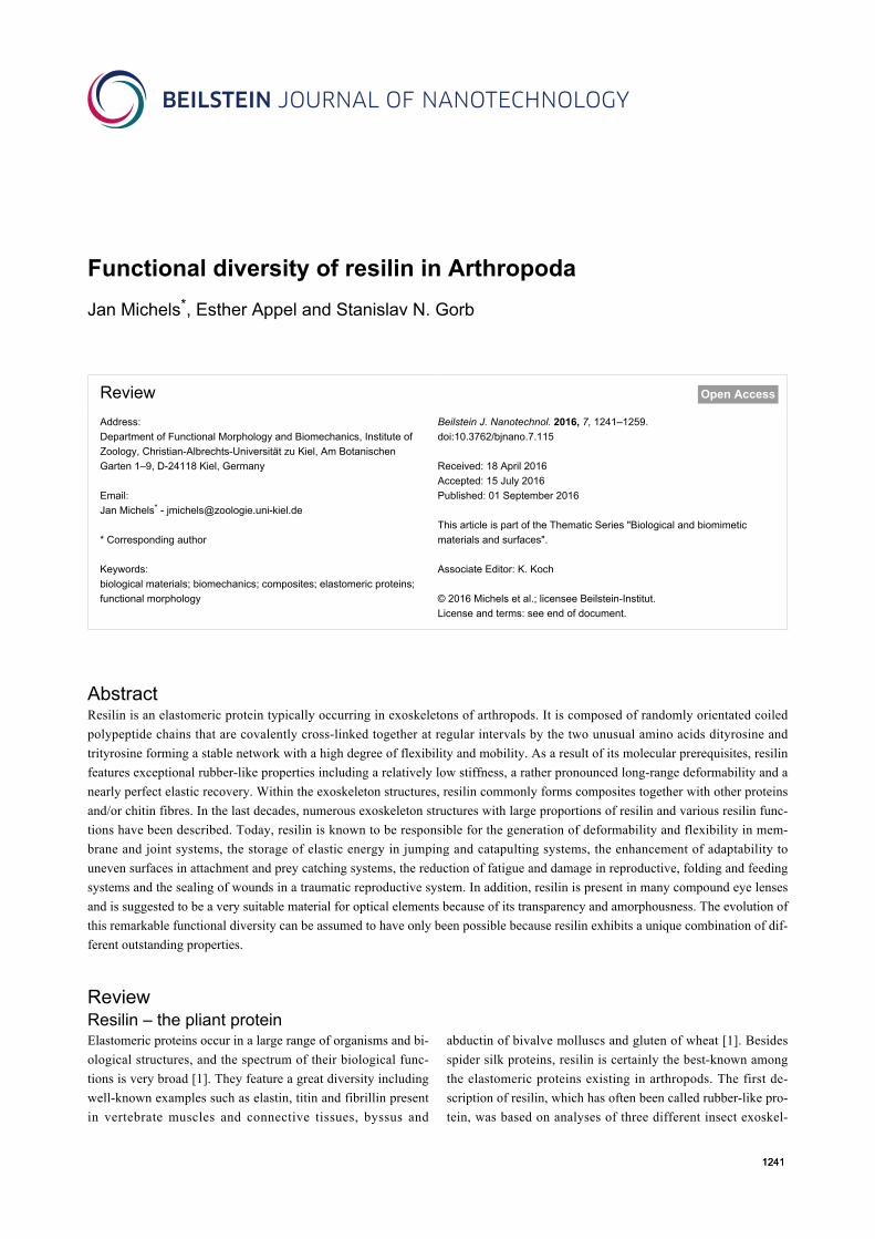
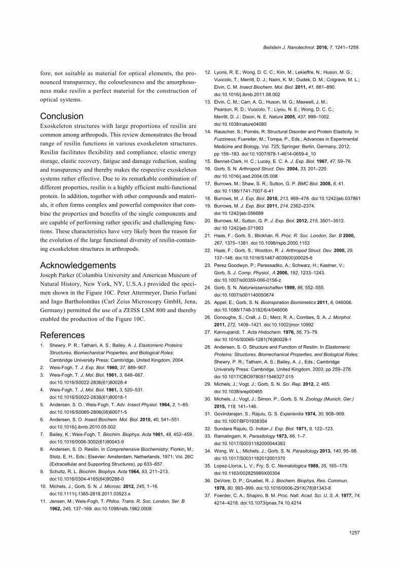
Figure 1: Occurrence of resilin in insects and crustaceans. Confocal laser scanning micrographs showing a wing hinge (A) and a prealar arm (B) ofthe migratory locust (Locusta migratoria), a wing vein joint of the common darter (Sympetrum striolatum) (C) and the ventral side of a female copepodof the species Temora longicornis (D). (A–C) Overlays of four different autofluorescences exhibited by the exoskeleton. Blue colours indicate largeproportions of resilin, while green structures consist mainly of non- or weakly-sclerotised chitinous material, and red structures are composed of rela-tively strongly sclerotised chitinous structures. (D) Blue = autofluorescence of resilin, red = Congo red fluorescence of stained chitinous exoskeletonparts, green = mixture of autofluorescence and Congo red fluorescence of stained chitinous exoskeleton parts. (A, C, D) Maximum intensity projec-tions. (B) Optical section. Scale bars = 100 µm (A, B), 20 µm (C), 200 µm (D). (A–D) Adapted with permission from [10], copyright 2011 John Wileyand Sons.
eton elements: the wing hinge and the prealar arm of the desert
locust (Schistocerca gregaria) (also described for the migratory
locust (Locusta migratoria), Figure 1A,B) and the so-called
elastic tendon of the pleuro-subalar muscles in dragonflies of
the genus Aeshna [2]. Additional insights into the character-
istics of resilin that had been gained shortly after this descrip-
tion [3,4] resulted in a comprehensive compilation of the then
existing knowledge of resilin properties [5]. Resilin consists of
a network of randomly orientated coiled polypeptide chains that
have a high degree of flexibility and mobility and are linked
together at regular intervals by stable covalent cross-links. Only
the fully cross-linked protein is called resilin, whereas the not
yet cross-linked or not fully cross-linked protein is called pro-
resilin [6]. Within hydrolysates of resilin, glycine constitutes the
largest proportion (30–40%) of the total residues [7,8]. Such
hydrolysates also feature the two unusual amino acids dityr-
osine and trityrosine, which were identified to form the cross-
links between the polypeptide chains [9].
The mechanical properties of resilin strongly depend on the
degree of hydration because resilin is plasticised by water [5].
When resilin is completely hydrated, it behaves close to a
Beilstein J. Nanotechnol. 2016, 7, 1241–1259.
1243
perfect rubber [3,4,11]. Due to its molecular prerequisites,
resilin then exhibits a near-perfect resilience of up to 92–97%
and a fatigue limit of over 300 million cycles [12]. With respect
to resilience, resilin is unmatched by any other elastomeric pro-
tein and the best synthetic rubbers such as unfilled poly-
butadiene [13,14]. Fully hydrated resilin has a rather low stiff-
ness. In the elastic tendons of dragonflies and locust ligaments
mentioned above, it was found to have a Young’s modulus of
0.6–0.7 MPa and 0.9 MPa, respectively [11]. In addition, fully
hydrated resilin can be stretched to more than three times its
original length and compressed to one third of its original
length, and when the tensile and compressive forces are re-
leased, resilin goes back to its initial state without having any
residual deformations [3,4,13].
Until today, resilin has been found to exist mainly in insect
exoskeleton structures where this protein has a number of
different functions, which include (1) the storage of elastic
energy in jumping systems [15-20], (2) the reduction of fatigue
in folding wings of beetles and dermapterans [21,22], (3)
the enhancement of the adaptability of attachment pads to
uneven surfaces [23] and (4) the generation of flexibility of
wing vein joints in dragonflies and damselflies [24-26]. Resilin
has also been reported to be present in the exoskeletons of other
arthropod taxa such as crustaceans [10,27-30] (Figure 1D),
scorpions [31] and centipedes [32]. In addition, resilin-like pro-
teins that contain dityrosine and trityrosine are known to exist in
several non-arthropod taxa including monogeneans [33,34],
nematodes [35], mussels [36] and sea urchins [37] indicating
that resilin likely originated much earlier in the evolution of
invertebrates than previously assumed.
The properties of resilin-containing exoskeletons can strongly
differ between structures and organisms. The reason is that in
biological structures resilin seems to be rarely present in pure or
nearly pure form but is known to commonly exist together with
other proteins and/or chitin fibres in resilin-containing compos-
ites, which exhibit a mixture of the properties of the single com-
ponents. In such composites, the resilin properties can even be
‘overlain’ by the properties of the other components making an
identification of the presence of resilin in the respective struc-
tures with the criteria of Andersen and Weis-Fogh [5] very
difficult. In addition, certain exoskeleton structures feature only
some of the typical characteristics of resilin-containing material
but lack the others. It is then often not possible to determine
whether these structures contain resilin or other proteins resem-
bling resilin. In such cases, it is conceivable that the respective
exoskeleton material consists either of a protein with properties
that are similar to those of resilin or of a mixture of resilin and
other proteins. For exoskeleton structures with such properties,
the term ‘transitional cuticle’ was established [5].
In order to allow a classification of exoskeleton structures as
resilin-containing exoskeleton according to the definition of
Andersen and Weis-Fogh [5], these structures must conform to
the wing hinge, the prealar arm and the elastic tendon
mentioned above with respect to their properties, which can be
tested with a number of methods [5]. Resilin is colourless,
transparent and amorphous. Accordingly, structures with very
large resilin proportions can be easily distinguished from struc-
tures with relatively large proportions of chitin that are typical-
ly pigmented and only slightly transparent or sometimes, when
the sclerotisation is very pronounced, not transparent at all.
When immersed in aqueous media and in many anhydrous
hydrophilic liquids, resilin exhibits an isotropic swelling, which
is reversible and depends on the pH. (It is least pronounced at
pH values of about 4.) In its hydrated state, resilin is swollen
and features its typical rubbery nature, long-range deformab-
ility and complete elastic recovery. Furthermore, if hydrated
resilin is tensioned, it will become birefringent, and the birefrin-
gence will be positive in the direction of the extension. When
resilin is completely dried, it loses its rubber-like character-
istics and becomes relatively hard and brittle. Proteolytic en-
zymes such as pepsin or trypsin can be applied to test for the
presence and distribution of resilin, because resilin is known to
be digested by such enzymes. Resilin has been shown to be
stained by single conventional dyes. Chemical reactions with
the Masson and Mallory dyes were mentioned to stain resilin
red. Staining of resilin with aqueous solutions of methylene
blue and toluidine blue is a common method and can provide
good information about the presence and distribution of resilin.
When resilin is stained with one of these two dyes, it does not
show metachromasia. Among the amino acids that form resilin,
dityrosine and trityrosine exhibit a relatively pronounced auto-
fluorescence. This autofluorescence is present in natural resilin-
containing structures and in isolated resilin (both before and
after boiling in water) and in resilin hydrolysates. In neutral and
alkaline solutions, its excitation and emission maxima are at
about 320 nm and 415 nm, respectively [38,39]. The excitation
spectrum of the resilin autofluorescence differs with changing
pH conditions. In acid solutions, the excitation maximum is
shifted considerably to about 285 nm, and the upper edge of the
excitation peak is at about 330–340 nm [38,39]. The pH-in-
duced changes of the excitation properties are reversible and
take place rapidly [40].
The described resilin identification and visualization methods
are not absolutely specific. Therefore, it is strongly advisable to
apply not only one single method but a combination of several
different ones to increase the reliability of the identification and
detection of resilin. In recent years, an antibody to a recom-
binant Drosophila melanogaster pro-resilin (rec1-resilin) was
developed and has been shown to be cross-reactive and to label
Beilstein J. Nanotechnol. 2016, 7, 1241–1259.
1244
resilin in different insects [12,13,41,42]. Until today, this
immunohistochemical method has been tested for only a small
number of insects and only within the studies mentioned above.
If it proves efficient in tests with a larger number of arthropod
species, it will represent the first reliable method that specific-
ally identifies resilin.
The development and improvement of methods applying tech-
niques such as micromechanical testing, atomic force microsco-
py and confocal laser scanning microscopy (CLSM) have facil-
itated detailed studies of the distribution, composition and me-
chanical properties of resilin-containing exoskeleton structures
in diverse organisms and at different levels of their organisa-
tion. One of these methods utilises a combination of different
autofluorescences. In addition to resilin, other arthropod
exoskeleton materials also exhibit autofluorescences, which can
be efficiently visualized with fluorescence microscopy. This
allows the production of overlays consisting of different micro-
graphs that show different autofluorescences. Such overlays
nicely exhibit differences in the autofluorescence composition,
which are good indications for differences in the material com-
position and clearly reveal structures with relatively large resilin
proportions within the analysed specimens [21,23,43]. Howev-
er, when analysing arthropod exoskeleton structures for the
presence of the autofluorescence of resilin, one has to bear in
mind that some other compounds present in organisms exhibit
autofluorescences whose properties are similar to those of the
resilin autofluorescence, with excitation maxima in the UV
range and emission maxima in the violet and blue ranges of the
light spectrum [44-47].
If the autofluorescences are visualized by means of CLSM, the
results will be very detailed and precise [10]. With a recently
described method, four different autofluorescences are excited
and detected separately, and certain colours are allocated to
each of the four visualized fluorescence signals. On the result-
ing overlays, exoskeleton structures with large proportions of
resilin are blue, while green structures consist mainly of non- or
weakly-sclerotised chitinous material, and red structures are
composed of relatively strongly sclerotised chitinous structures
(for details see [10]; Figure 1A–C). Many of the confocal laser
scanning micrographs shown in this review were created using
this method.
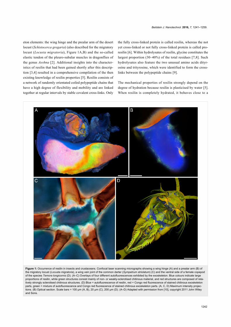
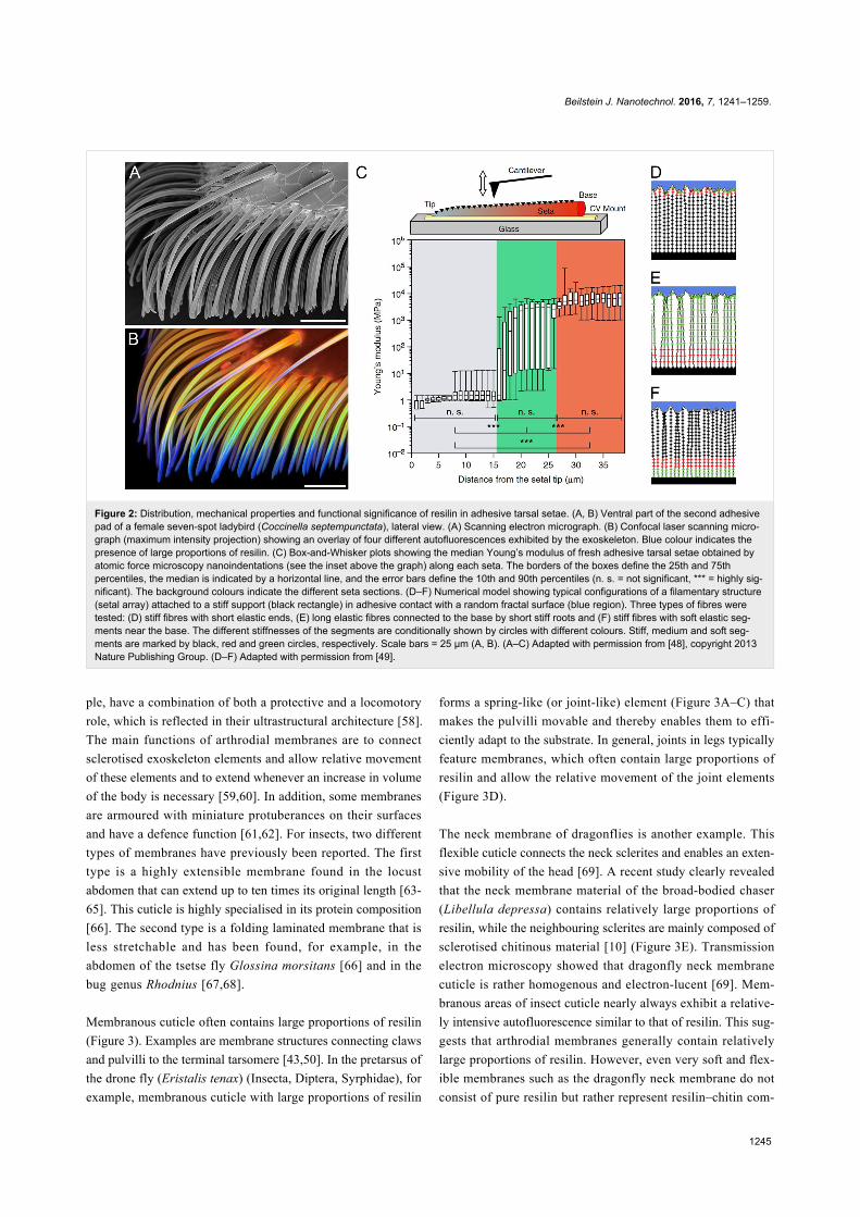
Very often, gradients of the material composition with a consid-
erably changing proportion of resilin are present in arthropod
exoskeleton structures. Such resilin proportion gradients must
also be reflected by gradients of the mechanical properties of
the respective resilin-containing composites. The material com-
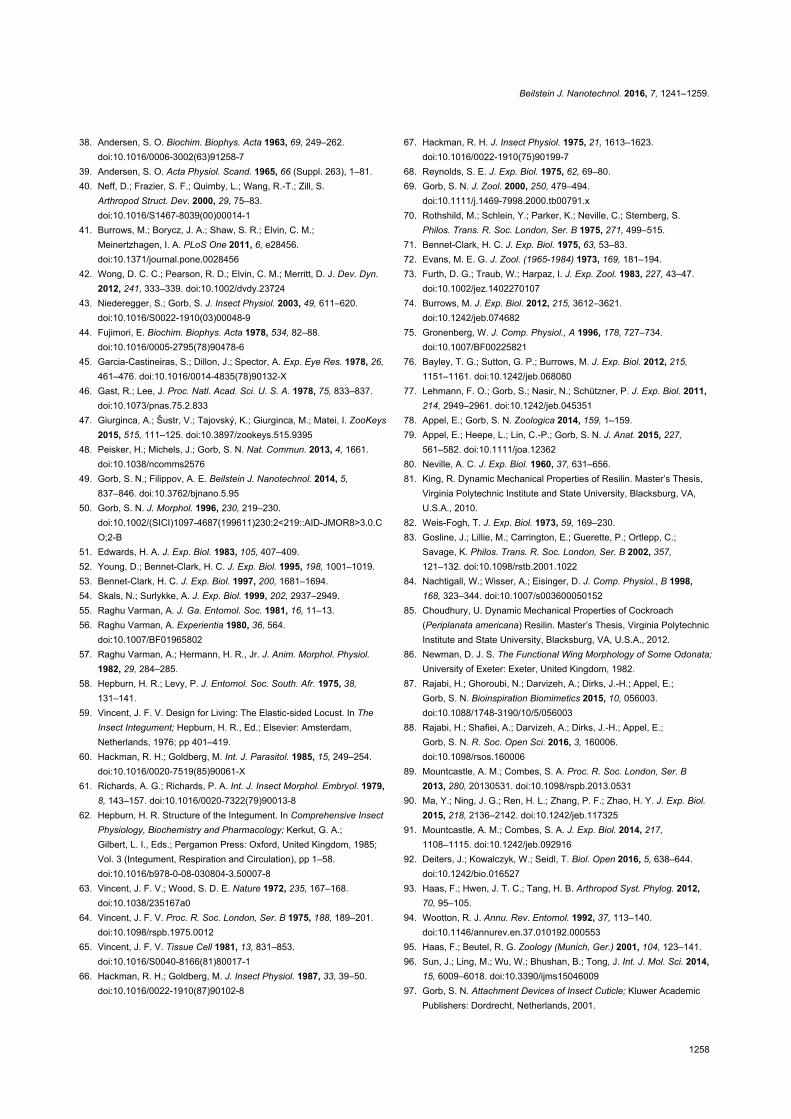
position of adhesive tarsal setae of beetles (Figure 2B) repres-
ents a good example for such gradients. Recently, the Young’s
modulus of such setae was measured along the longitudinal axis
of the setae (Figure 2C). The measurements revealed that the
Young’s modulus of the material in the most distal section of
each seta is relatively low (1.2 ± 0.3 MPa), whereas it is consid-
erably higher at the setal base (6.8 ± 1.2 GPa). The differences
in the Young’s modulus between different regions correlate
with the resilin proportion observed in the seta material [48].
When the setae are dehydrated, the Young’s modulus of the
setal tip material strongly increases from 1.2 to 7.2 GPa, and it
exhibits no statistically significant differences along the com-
plete setae [48], which is in accordance with the relationship be-
tween the material properties and the hydration status of resilin
mentioned above. Besides the differences in the Young’s
modulus, the mechanical behaviour of the respective materials
shows the pronounced differences in the material composition
between the tips and the bases of fresh adhesive tarsal setae.
While the material of the tip features only elastic deformation,
both elastic and, to some extent, plastic deformation are ob-
served in the material of the base [48]. This means that the
purely elastic response of the tip is due to the presence of
resilin, whereas the partially plastic deformation at the base is
mainly due to the presence of stiffer tanned exoskeleton. It is
very likely that effects similar to those observed in beetle
adhesive tarsal setae exist in other exoskeleton structures with
comparable gradients of the resilin proportion.
Occurrence and functions of resilin in differ-ent arthropod exoskeleton systemsResilin is known from numerous arthropod exoskeletons where
it is present in diverse structures and allows manifold functions,
which in most cases are based on its very pronounced elasticity
and its ability to completely recover after deformation. For ex-
ample, resilin plays an important role in flight systems of
insects, in particular in insects that use a wing beat with a low
frequency (10–50 Hz) (see below). Resilin-containing exoskel-
eton structures have been described for various mechanical
systems including leg joints [40,50], vein joints and mem-
branous areas of insect wings [21,22,24], the food-pump of
reduviid bugs [51], tymbal sound production organs of cicadas
[52,53] and moths [54], abdominal cuticle of honey ant workers
[55] and termite queens [56], the fulcral arms of the poison
apparatus of ants [57] and the tendons of dragonfly flight
muscles and basal wing joints of locusts (as already mentioned
above) [5]. In the following, some selected representative struc-
tures and systems with large proportions of resilin are high-
lighted, and their functions are described.
Arthrodial membranesArthrodial membranes are cuticle areas that are typically thin,
non-sclerotised and very flexible. Such membranes often are
multifunctional units. The soft cuticles of caterpillars, for exam-
Beilstein J. Nanotechnol. 2016, 7, 1241–1259.
1245
Figure 2: Distribution, mechanical properties and functional significance of resilin in adhesive tarsal setae. (A, B) Ventral part of the second adhesivepad of a female seven-spot ladybird (Coccinella septempunctata), lateral view. (A) Scanning electron micrograph. (B) Confocal laser scanning micro-graph (maximum intensity projection) showing an overlay of four different autofluorescences exhibited by the exoskeleton. Blue colour indicates thepresence of large proportions of resilin. (C) Box-and-Whisker plots showing the median Young’s modulus of fresh adhesive tarsal setae obtained byatomic force microscopy nanoindentations (see the inset above the graph) along each seta. The borders of the boxes define the 25th and 75thpercentiles, the median is indicated by a horizontal line, and the error bars define the 10th and 90th percentiles (n. s. = not significant, *** = highly sig-nificant). The background colours indicate the different seta sections. (D–F) Numerical model showing typical configurations of a filamentary structure(setal array) attached to a stiff support (black rectangle) in adhesive contact with a random fractal surface (blue region). Three types of fibres weretested: (D) stiff fibres with short elastic ends, (E) long elastic fibres connected to the base by short stiff roots and (F) stiff fibres with soft elastic seg-ments near the base. The different stiffnesses of the segments are conditionally shown by circles with different colours. Stiff, medium and soft seg-ments are marked by black, red and green circles, respectively. Scale bars = 25 µm (A, B). (A–C) Adapted with permission from [48], copyright 2013Nature Publishing Group. (D–F) Adapted with permission from [49].
ple, have a combination of both a protective and a locomotory
role, which is reflected in their ultrastructural architecture [58].
The main functions of arthrodial membranes are to connect
sclerotised exoskeleton elements and allow relative movement
of these elements and to extend whenever an increase in volume
of the body is necessary [59,60]. In addition, some membranes
are armoured with miniature protuberances on their surfaces
and have a defence function [61,62]. For insects, two different
types of membranes have previously been reported. The first
type is a highly extensible membrane found in the locust
abdomen that can extend up to ten times its original length [63-
65]. This cuticle is highly specialised in its protein composition
[66]. The second type is a folding laminated membrane that is
less stretchable and has been found, for example, in the
abdomen of the tsetse fly Glossina morsitans [66] and in the
bug genus Rhodnius [67,68].
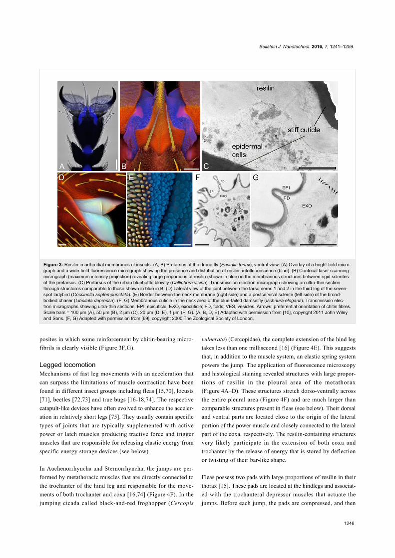
Membranous cuticle often contains large proportions of resilin
(Figure 3). Examples are membrane structures connecting claws
and pulvilli to the terminal tarsomere [43,50]. In the pretarsus of
the drone fly (Eristalis tenax) (Insecta, Diptera, Syrphidae), for
example, membranous cuticle with large proportions of resilin
forms a spring-like (or joint-like) element (Figure 3A–C) that
makes the pulvilli movable and thereby enables them to effi-
ciently adapt to the substrate. In general, joints in legs typically
feature membranes, which often contain large proportions of
resilin and allow the relative movement of the joint elements
(Figure 3D).
The neck membrane of dragonflies is another example. This
flexible cuticle connects the neck sclerites and enables an exten-
sive mobility of the head [69]. A recent study clearly revealed
that the neck membrane material of the broad-bodied chaser
(Libellula depressa) contains relatively large proportions of
resilin, while the neighbouring sclerites are mainly composed of
sclerotised chitinous material [10] (Figure 3E). Transmission
electron microscopy showed that dragonfly neck membrane
cuticle is rather homogenous and electron-lucent [69]. Mem-
branous areas of insect cuticle nearly always exhibit a relative-
ly intensive autofluorescence similar to that of resilin. This sug-
gests that arthrodial membranes generally contain relatively
large proportions of resilin. However, even very soft and flex-
ible membranes such as the dragonfly neck membrane do not
consist of pure resilin but rather represent resilin–chitin com-
Beilstein J. Nanotechnol. 2016, 7, 1241–1259.
1246
Figure 3: Resilin in arthrodial membranes of insects. (A, B) Pretarsus of the drone fly (Eristalis tenax), ventral view. (A) Overlay of a bright-field micro-graph and a wide-field fluorescence micrograph showing the presence and distribution of resilin autofluorescence (blue). (B) Confocal laser scanningmicrograph (maximum intensity projection) revealing large proportions of resilin (shown in blue) in the membranous structures between rigid scleritesof the pretarsus. (C) Pretarsus of the urban bluebottle blowfly (Calliphora vicina). Transmission electron micrograph showing an ultra-thin sectionthrough structures comparable to those shown in blue in B. (D) Lateral view of the joint between the tarsomeres 1 and 2 in the third leg of the seven-spot ladybird (Coccinella septempunctata). (E) Border between the neck membrane (right side) and a postcervical sclerite (left side) of the broad-bodied chaser (Libellula depressa). (F, G) Membranous cuticle in the neck area of the blue-tailed damselfly (Ischnura elegans). Transmission elec-tron micrographs showing ultra-thin sections. EPI, epicuticle; EXO, exocuticle; FD, folds; VES, vesicles. Arrows: preferential orientation of chitin fibres.Scale bars = 100 µm (A), 50 µm (B), 2 µm (C), 20 µm (D, E), 1 µm (F, G). (A, B, D, E) Adapted with permission from [10], copyright 2011 John Wileyand Sons. (F, G) Adapted with permission from [69], copyright 2000 The Zoological Society of London.
posites in which some reinforcement by chitin-bearing micro-
fibrils is clearly visible (Figure 3F,G).
Legged locomotionMechanisms of fast leg movements with an acceleration that
can surpass the limitations of muscle contraction have been
found in different insect groups including fleas [15,70], locusts
[71], beetles [72,73] and true bugs [16-18,74]. The respective
catapult-like devices have often evolved to enhance the acceler-
ation in relatively short legs [75]. They usually contain specific
types of joints that are typically supplemented with active
power or latch muscles producing tractive force and trigger
muscles that are responsible for releasing elastic energy from
specific energy storage devices (see below).
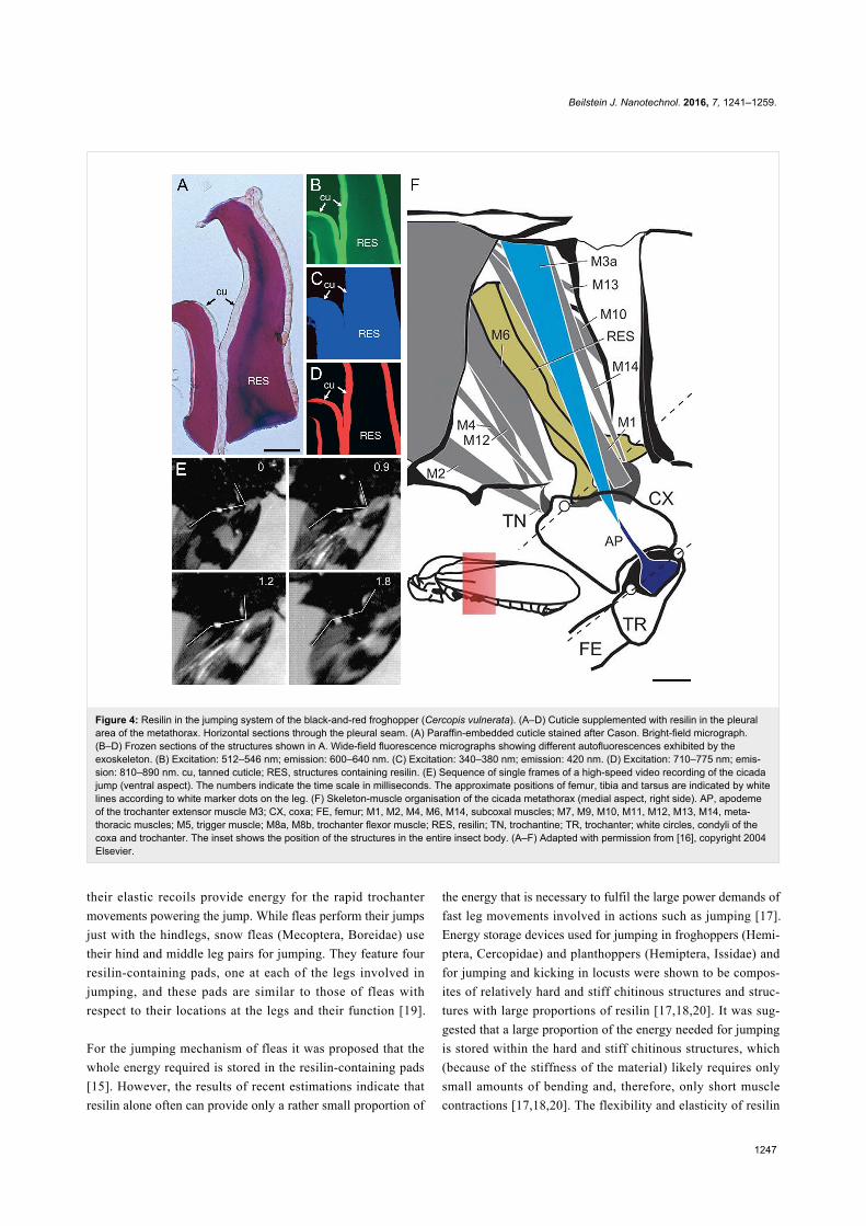
In Auchenorrhyncha and Sternorrhyncha, the jumps are per-
formed by metathoracic muscles that are directly connected to
the trochanter of the hind leg and responsible for the move-
ments of both trochanter and coxa [16,74] (Figure 4F). In the
jumping cicada called black-and-red froghopper (Cercopis
vulnerata) (Cercopidae), the complete extension of the hind leg
takes less than one millisecond [16] (Figure 4E). This suggests
that, in addition to the muscle system, an elastic spring system
powers the jump. The application of fluorescence microscopy
and histological staining revealed structures with large propor-
tions of resilin in the pleural area of the metathorax
(Figure 4A–D). These structures stretch dorso-ventrally across
the entire pleural area (Figure 4F) and are much larger than
comparable structures present in fleas (see below). Their dorsal
and ventral parts are located close to the origin of the lateral
portion of the power muscle and closely connected to the lateral
part of the coxa, respectively. The resilin-containing structures
very likely participate in the extension of both coxa and
trochanter by the release of energy that is stored by deflection
or twisting of their bar-like shape.
Fleas possess two pads with large proportions of resilin in their
thorax [15]. These pads are located at the hindlegs and associat-
ed with the trochanteral depressor muscles that actuate the
jumps. Before each jump, the pads are compressed, and then
Beilstein J. Nanotechnol. 2016, 7, 1241–1259.
1247
Figure 4: Resilin in the jumping system of the black-and-red froghopper (Cercopis vulnerata). (A–D) Cuticle supplemented with resilin in the pleuralarea of the metathorax. Horizontal sections through the pleural seam. (A) Paraffin-embedded cuticle stained after Cason. Bright-field micrograph.(B–D) Frozen sections of the structures shown in A. Wide-field fluorescence micrographs showing different autofluorescences exhibited by theexoskeleton. (B) Excitation: 512–546 nm; emission: 600–640 nm. (C) Excitation: 340–380 nm; emission: 420 nm. (D) Excitation: 710–775 nm; emis-sion: 810–890 nm. cu, tanned cuticle; RES, structures containing resilin. (E) Sequence of single frames of a high-speed video recording of the cicadajump (ventral aspect). The numbers indicate the time scale in milliseconds. The approximate positions of femur, tibia and tarsus are indicated by whitelines according to white marker dots on the leg. (F) Skeleton-muscle organisation of the cicada metathorax (medial aspect, right side). AP, apodemeof the trochanter extensor muscle M3; CX, coxa; FE, femur; M1, M2, M4, M6, M14, subcoxal muscles; M7, M9, M10, M11, M12, M13, M14, meta-thoracic muscles; M5, trigger muscle; M8a, M8b, trochanter flexor muscle; RES, resilin; TN, trochantine; TR, trochanter; white circles, condyli of thecoxa and trochanter. The inset shows the position of the structures in the entire insect body. (A–F) Adapted with permission from [16], copyright 2004Elsevier.
their elastic recoils provide energy for the rapid trochanter
movements powering the jump. While fleas perform their jumps
just with the hindlegs, snow fleas (Mecoptera, Boreidae) use
their hind and middle leg pairs for jumping. They feature four
resilin-containing pads, one at each of the legs involved in
jumping, and these pads are similar to those of fleas with
respect to their locations at the legs and their function [19].
For the jumping mechanism of fleas it was proposed that the
whole energy required is stored in the resilin-containing pads
[15]. However, the results of recent estimations indicate that
resilin alone often can provide only a rather small proportion of
the energy that is necessary to fulfil the large power demands of
fast leg movements involved in actions such as jumping [17].
Energy storage devices used for jumping in froghoppers (Hemi-
ptera, Cercopidae) and planthoppers (Hemiptera, Issidae) and
for jumping and kicking in locusts were shown to be compos-
ites of relatively hard and stiff chitinous structures and struc-
tures with large proportions of resilin [17,18,20]. It was sug-
gested that a large proportion of the energy needed for jumping
is stored within the hard and stiff chitinous structures, which
(because of the stiffness of the material) likely requires only
small amounts of bending and, therefore, only short muscle
contractions [17,18,20]. The flexibility and elasticity of resilin
Beilstein J. Nanotechnol. 2016, 7, 1241–1259.
1248
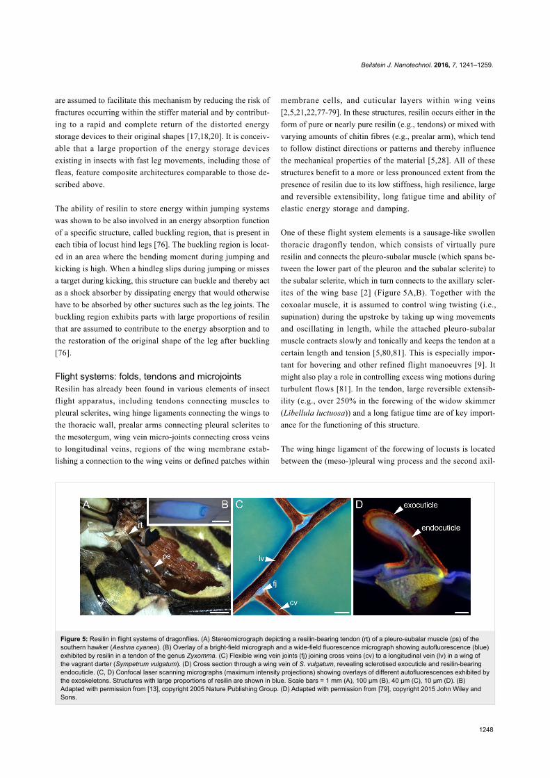
Figure 5: Resilin in flight systems of dragonflies. (A) Stereomicrograph depicting a resilin-bearing tendon (rt) of a pleuro-subalar muscle (ps) of thesouthern hawker (Aeshna cyanea). (B) Overlay of a bright-field micrograph and a wide-field fluorescence micrograph showing autofluorescence (blue)exhibited by resilin in a tendon of the genus Zyxomma. (C) Flexible wing vein joints (fj) joining cross veins (cv) to a longitudinal vein (lv) in a wing ofthe vagrant darter (Sympetrum vulgatum). (D) Cross section through a wing vein of S. vulgatum, revealing sclerotised exocuticle and resilin-bearingendocuticle. (C, D) Confocal laser scanning micrographs (maximum intensity projections) showing overlays of different autofluorescences exhibited bythe exoskeletons. Structures with large proportions of resilin are shown in blue. Scale bars = 1 mm (A), 100 µm (B), 40 µm (C), 10 µm (D). (B)Adapted with permission from [13], copyright 2005 Nature Publishing Group. (D) Adapted with permission from [79], copyright 2015 John Wiley andSons.
are assumed to facilitate this mechanism by reducing the risk of
fractures occurring within the stiffer material and by contribut-
ing to a rapid and complete return of the distorted energy
storage devices to their original shapes [17,18,20]. It is conceiv-
able that a large proportion of the energy storage devices
existing in insects with fast leg movements, including those of
fleas, feature composite architectures comparable to those de-
scribed above.
The ability of resilin to store energy within jumping systems
was shown to be also involved in an energy absorption function
of a specific structure, called buckling region, that is present in
each tibia of locust hind legs [76]. The buckling region is locat-
ed in an area where the bending moment during jumping and
kicking is high. When a hindleg slips during jumping or misses
a target during kicking, this structure can buckle and thereby act
as a shock absorber by dissipating energy that would otherwise
have to be absorbed by other suctures such as the leg joints. The
buckling region exhibits parts with large proportions of resilin
that are assumed to contribute to the energy absorption and to
the restoration of the original shape of the leg after buckling
[76].
Flight systems: folds, tendons and microjointsResilin has already been found in various elements of insect
flight apparatus, including tendons connecting muscles to
pleural sclerites, wing hinge ligaments connecting the wings to
the thoracic wall, prealar arms connecting pleural sclerites to
the mesotergum, wing vein micro-joints connecting cross veins
to longitudinal veins, regions of the wing membrane estab-
lishing a connection to the wing veins or defined patches within
membrane cells, and cuticular layers within wing veins
[2,5,21,22,77-79]. In these structures, resilin occurs either in the
form of pure or nearly pure resilin (e.g., tendons) or mixed with
varying amounts of chitin fibres (e.g., prealar arm), which tend
to follow distinct directions or patterns and thereby influence
the mechanical properties of the material [5,28]. All of these
structures benefit to a more or less pronounced extent from the
presence of resilin due to its low stiffness, high resilience, large
and reversible extensibility, long fatigue time and ability of
elastic energy storage and damping.
One of these flight system elements is a sausage-like swollen
thoracic dragonfly tendon, which consists of virtually pure
resilin and connects the pleuro-subalar muscle (which spans be-
tween the lower part of the pleuron and the subalar sclerite) to
the subalar sclerite, which in turn connects to the axillary scler-
ites of the wing base [2] (Figure 5A,B). Together with the
coxoalar muscle, it is assumed to control wing twisting (i.e.,
supination) during the upstroke by taking up wing movements
and oscillating in length, while the attached pleuro-subalar
muscle contracts slowly and tonically and keeps the tendon at a
certain length and tension [5,80,81]. This is especially impor-
tant for hovering and other refined flight manoeuvres [9]. It
might also play a role in controlling excess wing motions during
turbulent flows [81]. In the tendon, large reversible extensib-
ility (e.g., over 250% in the forewing of the widow skimmer
(Libellula luctuosa)) and a long fatigue time are of key import-
ance for the functioning of this structure.
The wing hinge ligament of the forewing of locusts is located
between the (meso-)pleural wing process and the second axil-
Beilstein J. Nanotechnol. 2016, 7, 1241–1259.
1249
lary wing sclerite [5]. In Odonata, Dictyoptera and Orthoptera,
wing hinge ligaments exist in the form of thick, rubber-like
pads (Figure 1A) and, as reported for locust structures, consist
of a rather tough, mainly chitinous ventral part and a soft dorsal
part, which can be divided in a region of pure resilin and a
region containing both chitin lamellae and resilin [5]. The
results of several studies suggest that wing hinge ligaments take
up compressive as well as tensile forces and can contribute to
(1) the storage of kinetic energy at maximum wing deflection,
for example during the upstroke when the wing hinge ligament
is stretched, and (2) wing acceleration during the downstroke by
elastic recoil [5,28,82]. In other insects, such as Lepidoptera,
some Coleoptera and some Hymenoptera, these ligaments are
tough and inextensible, and elastic energy storage is likely pro-
vided by the rigid thoracic cuticle and the flight muscle itself
[5,28]. Due to the fact that resilin has mainly been found in the
flight apparatus of insects flying with synchronous flight
muscles at low wing beat frequencies of less than 50 Hz and
with inertial forces being larger than aerodynamic forces, it is
assumed that its resilience might be too small at high frequen-
cies [5,11,28,83]. However, there is still some controversy
about the frequency-dependent behaviour of resilin and
chitin–resilin composites and its function in the wing hinge
ligaments of insects with high wing beat frequencies [5]. For
example, some small wing hinge ligaments have been found be-
tween different sclerites in the genera Calliphora, Bombus, Apis
and Oryctes [5,84]. So far, only a few studies have investigated
the decrease in resilience with increasing frequencies in the
dragonfly tendon, locust prealar arm and cockroach tibia-tarsal
joint resilin [5,11,81,83,85]. Whether the partly pronounced
differences in the decrease rate of resilience between different
frequency ranges are due to different measurement techniques
or are actually due to differences in the material composition,
still needs to be elucidated.
The prealar arm is located at the front edge of the mesotergum
and establishes a connection to the first basalar sclerite of the
pleural thoracic wall via a tough, flexible ligament [5]. The
basalar sclerite in turn is connected to the humeral angle of the
anterior part of the wing base. The prealar arm consists of
around 23% chitin and 77% resilin and is structured by altern-
ating layers of resilin and chitin fibrils, with the fibrils
continuing into the dark, sclerotised cuticle at its base [5]
(Figure 1B). Due to the directional arrangement of chitin fibrils,
the mechanical behaviour of the prealar arm is assumed to be
dominated by the mechanical properties of the chitin fibrils
during stretching and by the properties of resilin during bending
and compression [5,11]. In contrast to the subalar muscle,
which is involved in wing supination, the contraction of the
basalar muscle causes wing pronation through the connection to
the humeral angle via the basalar sclerite. During muscle
contraction, the prealar arm is deformed and can be assumed to
play a role in elastic energy storage.
Cross veins in wings of dragonflies and damselflies were shown
to form either stiff, inflexibly fused joints or flexible, resilin-
bearing joints to the adjacent longitudinal veins [24-26,78,86]
(Figure 5C). The distribution pattern of different wing vein joint
types on the dorsal and ventral wing sides in various species is
quite diverse, but was found to follow phylogenetic trends prob-
ably related to wing morphology and flight behaviour [26,78].
In general, flexible wing vein joints, together with the overall
corrugated design of odonate wings, are assumed to feature a
larger angular displacement than fused vein joints and, as a
result, to provide the wing with increased chord-wise flexibility,
which promotes passive wing deformations such as camber-for-
mation during the downstroke, and, thereby, to improve the
aerodynamic and mechanical performance of the wing
[24,26,78,86,87]. Moreover, resilin is important for reducing
stress concentrations in vein joints [87]. Resilin is not only
present in wing vein joints but also in the wing membrane
directly abutting on wing veins and internal cuticle layers of
wing veins (i.e., the endocuticle) [78,79] (Figure 5D). A flex-
ible suspension of the wing membrane is suggested to allow
larger strain and thereby to help preventing its tear-off from the
wing veins [79]. Furthermore, the stiffness gradient in wing
veins, generated by a stiff, sclerotised outer layer (exocuticle)
and a soft, compliant, resilin-bearing inner layer (endocuticle) is
assumed to reduce the overall vein stiffness and to improve the
damping properties of the vein as well as to delay Brazier oval-
isation and to enhance the load-bearing capacity under large de-
formations [79,88].
By artificially stiffening single flexible, resilin-bearing vein
joints in bumblebee wings through the application of micro-
splints (extra-fine polyester glitter glued with cyanoacrylate), it
was experimentally shown that even a single resilin-bearing
joint plays an important role in overall wing flexibility and
vertical aerodynamic force production [89]. Ma et al. [90] found
comparable resilin joints (e.g., the 1 m-cu joint) in wings of
western honey bees (Apis mellifera) and assumed that they
might play an analogous role in increasing the chordwise wing
flexibility. Based on the distribution of resilin patches, wing
veins, the occurrence of a f lexible hook-mediated
forewing–hindwing connection and observed wing deforma-
tions, they further suggested the existence of five flexion lines
in one forewing–hindwing entity and assumed that these proba-
bly increase the cordwise flexibility and support camber forma-
tion. In addition, Mountcastle and Combes [91] demonstrated
that a resilin-bearing joint at the leading edge (the costal break)
in the wings of wasps plays a major role in mitigating wing
wear by flexion along this joint when the wings hit an obstacle.
Beilstein J. Nanotechnol. 2016, 7, 1241–1259.
1250
This mechanism is especially important for wings with wing
veins extending all the way to the tip because such a design
endows a wing with more spanwise rigidity than, for example,
bumblebee wings that lack veins at the wing tip [91].
The occurrence of resilin in several broadened vein patches as
well as in membranous folding lines was described for fan-like
dermapteran hind wings [22,92]. These structures help folding
the wing into a wing package being ten times smaller than the
unfolded wing. This package can then be hidden under the short
sclerotised forewings. The four-fold wing folding can be
achieved without musculature activity and is assumed to be
driven by elastic recoil of the anisotropically distributed resilin
on either the ventral or the dorsal sides of broadened vein
patches in intercalary and radiating veins, supported by the
resilin-bearing radiating folds that influence the folding direc-
tion [22]. Unfolding of the hind wings is achieved either by
wiping movements of the cerci (e.g., in the European earwig
(Forficula auricularia) and the lesser earwig (Labia minor)) or
by wing flapping (e.g., in the earwigs Timomenus lugens, which
has very long cerci, and Auchenomus sp.) [22,93]. Both
unfolding mechanisms are supported by several wing stiffening
mechanisms such as the mid-wing mechanism and the claval
flexion line, which keep the wing unfolded in all species exam-
ined [22,93]. These mechanisms were found to play an impor-
tant role both in the static unfolded state of the wing and during
flapping flight, in which they help to inhibit an unfavorable
folding of the wing [92]. Furthermore, the flexible resilin-bear-
ing folding lines were found to not only serve wing folding but
also act as flexion lines at which the wing flexes during flight,
thereby supporting the generation of an aerodynamically
favourable cambered wing profile [92,94].
In beetle wings, resilin was found to occur at the marginal joint,
between veins that separate during folding, and along flexion
lines in membranous areas, leading to the hypothesis that elastic
energy storage by resilin can support wing unfolding also in
beetle wings [21]. However, this can, if at all, only be a
supportive role because wing unfolding in beetles was stated to
be mainly achieved by scissor-like movements of the RA and
MP1+2 veins via contraction of the Musculus pleura alaris and
the basalar muscles, which is possibly supported by hydraulic
hemolymph pressure [95,96]. Like in dermapteran wings, in
beetle wings resilin most probably delays material fatigue in
highly stressed wing regions and might further play a role in
wing deformation during flight [22].
In wings of the urban bluebottle blowfly (Calliphora vicina),
resilin is mainly present in the proximal part of the wing, pre-
dominantly in the form of resilin-bearing patches between veins
[77]. The occurence of resilin coincides with the proximal dis-
tribution of the maximum spanwise bending stress at the begin-
ning of each stroke cycle and suggests that the resilin patches
reduce the risk of breaking near the wing hinge due to a de-
crease in peak stress in the rigid wing parts [77].
Attachment systemsThe contact formation of insect adhesive pads on substrates
depends on the ability of the pads to adapt to the surface topog-
raphy. In this context, specific micro- and nanostructures can
enhance the quality of the contact [97-101]. In the case of
attachment on rough substrates, multiple contacts, being formed
by some adhesive systems, provide great advantages [102]. The
formation of multiple contacts, which contribute to an increase
of the overall length of the total peeling line, is facilitated by a
hierarchical organisation of the attachment structures [103]. It
was shown that the combination of thin tape-like contact tips of
hairs (setae) and applied shear force lead to the formation of a
maximal real contact area without slippage within the contact
[104]. This indicates that material flexibility is very important
for the contact formation of adhesive pads. With a minimal
normal load, flexible materials can create a large contact area
between the attachment structures and the substrate. However,
elongated structures that are too flexible have a low mechanical
stability [105]. For example, if insect setae are too soft, they can
buckle and collapse, and so-called clusterisation (or condensa-
tion) can take place [106,107]. As a result of this, the func-
tional advantages achieved through multiple adhesive contacts
can be strongly reduced [103]. Accordingly, the composition
and the properties of the material of insect adhesive setae
represent an optimisation problem. There is evidence that
during the evolution gradients of the thickness and the mechani-
cal properties of the setae have developed as a solution of this
problem. The presence of thickness gradients, revealed by scan-
ning electron microscopy, is well-known for various insect
adhesive setae [97]. Recently, a gradient of the material compo-
sition, present on the level of each single adhesive tarsal seta,
was shown to exist in the seven-spot ladybird (Coccinella
septempunctata) [48] (Figure 2A–C). The material of the setal
tip contains large proportions of resilin, while the base of the
seta consists mainly of sclerotised chitinous material. Between
the tip and the base, a pronounced material composition
gradient was revealed by CLSM. This gradient is reflected by a
pronounced gradient of the material properties: the setal tip is
rather soft, whereas the setal base is relatively stiff. Both gradi-
ents were hypothesised to represent an evolutionary optimisa-
tion that increases the attachment performance of the adhesive
pads when they attach to rough surfaces due to an efficient
adaptation of the soft and flexible setal tips to the substrate and
a simultaneous prevention of setal clusterisation by means of
the stiffer setal bases [48]. Since this hypothesis is difficult to
test experimentally using biological specimens, it was tested
Beilstein J. Nanotechnol. 2016, 7, 1241–1259.
1251
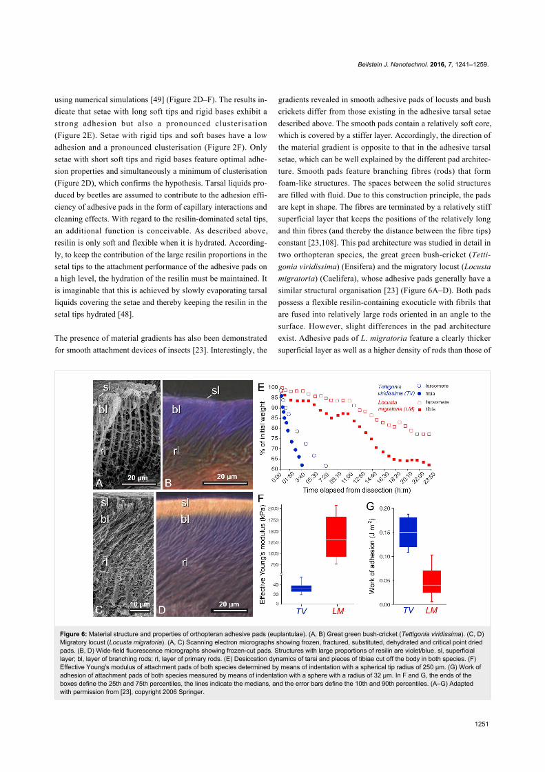
Figure 6: Material structure and properties of orthopteran adhesive pads (euplantulae). (A, B) Great green bush-cricket (Tettigonia viridissima). (C, D)Migratory locust (Locusta migratoria). (A, C) Scanning electron micrographs showing frozen, fractured, substituted, dehydrated and critical point driedpads. (B, D) Wide-field fluorescence micrographs showing frozen-cut pads. Structures with large proportions of resilin are violet/blue. sl, superficiallayer; bl, layer of branching rods; rl, layer of primary rods. (E) Desiccation dynamics of tarsi and pieces of tibiae cut off the body in both species. (F)Effective Young's modulus of attachment pads of both species determined by means of indentation with a spherical tip radius of 250 µm. (G) Work ofadhesion of attachment pads of both species measured by means of indentation with a sphere with a radius of 32 µm. In F and G, the ends of theboxes define the 25th and 75th percentiles, the lines indicate the medians, and the error bars define the 10th and 90th percentiles. (A–G) Adaptedwith permission from [23], copyright 2006 Springer.
using numerical simulations [49] (Figure 2D–F). The results in-
dicate that setae with long soft tips and rigid bases exhibit a
strong adhesion but also a pronounced clusterisation
(Figure 2E). Setae with rigid tips and soft bases have a low
adhesion and a pronounced clusterisation (Figure 2F). Only
setae with short soft tips and rigid bases feature optimal adhe-
sion properties and simultaneously a minimum of clusterisation
(Figure 2D), which confirms the hypothesis. Tarsal liquids pro-
duced by beetles are assumed to contribute to the adhesion effi-
ciency of adhesive pads in the form of capillary interactions and
cleaning effects. With regard to the resilin-dominated setal tips,
an additional function is conceivable. As described above,
resilin is only soft and flexible when it is hydrated. According-
ly, to keep the contribution of the large resilin proportions in the
setal tips to the attachment performance of the adhesive pads on
a high level, the hydration of the resilin must be maintained. It
is imaginable that this is achieved by slowly evaporating tarsal
liquids covering the setae and thereby keeping the resilin in the
setal tips hydrated [48].
The presence of material gradients has also been demonstrated
for smooth attachment devices of insects [23]. Interestingly, the
gradients revealed in smooth adhesive pads of locusts and bush
crickets differ from those existing in the adhesive tarsal setae
described above. The smooth pads contain a relatively soft core,
which is covered by a stiffer layer. Accordingly, the direction of
the material gradient is opposite to that in the adhesive tarsal
setae, which can be well explained by the different pad architec-
ture. Smooth pads feature branching fibres (rods) that form
foam-like structures. The spaces between the solid structures
are filled with fluid. Due to this construction principle, the pads
are kept in shape. The fibres are terminated by a relatively stiff
superficial layer that keeps the positions of the relatively long
and thin fibres (and thereby the distance between the fibre tips)
constant [23,108]. This pad architecture was studied in detail in
two orthopteran species, the great green bush-cricket (Tetti-
gonia viridissima) (Ensifera) and the migratory locust (Locusta
migratoria) (Caelifera), whose adhesive pads generally have a
similar structural organisation [23] (Figure 6A–D). Both pads
possess a flexible resilin-containing exocuticle with fibrils that
are fused into relatively large rods oriented in an angle to the
surface. However, slight differences in the pad architecture
exist. Adhesive pads of L. migratoria feature a clearly thicker
superficial layer as well as a higher density of rods than those of
Beilstein J. Nanotechnol. 2016, 7, 1241–1259.
1252
T. viridissima (Figure 6A–D). In addition, indentation experi-
ments revealed a higher effective Young’s modulus and a lower
work of adhesion for L. migratoria pads (Figure 6F,G). The
lower adhesive properties of L. migratoria pads can be ex-
plained by the larger thickness of the relatively stiff superficial
layer, which likely reduces the adaptability of the pad to the
substrate much more than the relatively thin superficial layer of
the T. viridissima adhesive pad. The superficial layer is
assumed to also protect the pad from desiccation as indicated by
experiments showing that cut-off adhesive pads of T.
viridissima (with the relatively thin superficial layer) lose water
much faster than those of L. migratoria (Figure 6E).
Consequently, the material gradient provides a combination of
conformability to the surface roughness of the substrate (The
compliant material of the pad contributes to the efficient con-
tact formation with the substrate.) and resistance to the dry
environment. Such pad architectures likely depend on the
preferred environment of each species and are the result of
trade-offs between different factors such as evaporation rate,
stiffness, stability and adhesion.
MouthpartsThe first mouthpart-related structures containing resilin were
already mentioned shortly after the description of resilin. In the
respective studies, resilin was found in the salivary and feeding
pumps of assassin bugs [109] (cited in [110]), [111]. Later, the
findings were confirmed and complemented by additional
information about the resilin distribution [51]. In these pumps,
which enable the bugs to suck relatively large amounts of blood
in a short time period and to inject proteolytic enzymes into
prey or assaulters or to spit on the latter, the resilin-containing
structures function as elastic spring antagonists to muscles. A
similar function was described for resilin-containing structures
present in the maxillipeds of decapod crustaceans [112]. The
movements of the flagella of these mouthparts influence the
water flow through the gills as well as over chemoreceptors lo-
cated on the head, and thereby they importantly contribute to
active chemoreception and to signalling by distributing urine
odours. Each of the flagella is abducted by the contraction of a
single muscle. Due to this abduction, a structure that contains
relatively large resilin proportions and is located in the joint be-
tween the flagellum und the exopodite of the maxilliped is bent.
After relaxation of the muscle, this elastic structure recovers its
original shape and moves the flagellum back to its resting posi-
tion.
In general, due to its very pronounced elasticity and fatigue
resistance, resilin appears to be a very suitable material for
exoskeleton structures that are typically intensively deformed
for a rather large number of times during the lifetime of the
organisms. A butterfly proboscis, for example, is tightly and
spirally coiled when it is in its resting position [113]. For the
uptake of food, hemolymph is pumped into the proboscis result-
ing in the generation of hydrostatic pressure that completely
uncoils the proboscis [113-115] and strongly changes the shape
of certain proboscis elements. During this process, dorsal parts
of the proboscis are compressed. These parts contain relatively
large proportions of resilin and act as springs that cause the
recoiling of the proboscis when the hydrostatic pressure is
removed [115].
A remarkable resilin-containing adhesive prey-capture device,
which is formed by the elongated labium, exists in rove beetles
of the genus Stenus (Staphylinidae). This prey-capture appar-
atus can be protruded towards a prey within a few milliseconds.
When sticky pads (modified paraglossae), which are located at
the distal end of the prementum, adhere to the prey, the labium
is withdrawn immediately, and thereby the prey is transported
to the mouth region of the beetle where it can be seized with the
mandibles [116-118]. The sticky pads feature a surface that is
subdivided into numerous terminally branched outgrowths.
During the prey capture, these surface structures are completely
covered by an adhesive secretion that is produced in special
glands located in the head capsule and makes the sticky pads a
hairy, hierarchically structured and wet adhesive system. Simi-
lar to the insect tarsal adhesive pads mentioned above, softness
and compliance of the pad cuticle contribute to the generation
of strong adhesive forces by the pads. The cuticle material of
certain parts of the sticky pads contains large proportions of
resilin providing flexibility and elasticity and enabling the pads
to efficiently adapt to the surface of the prey items [118].
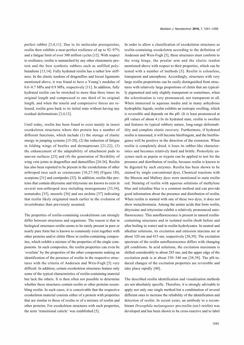
Copepods are tiny crustaceans that inhabit nearly all aquatic
habitats worldwide and are particularly abundant in the marine
water column where they contribute large proportions of the
zooplankton [119,120]. The diet of many of the marine plank-
tonic species comprises relatively large fractions of diatoms
(i.e., unicellular algae with silica-containing shells called frust-
ules). Copepods use the gnathobases of their mandibles to grab
and mince food particles. To be able to efficiently digest the
diatom cells, the copepods must crack the frustules before the
ingestion of the cells. The gnathobases possess tooth-like struc-
tures (called teeth in the following) at their distal ends [121]. In
copepod species feeding on large amounts of diatoms, these
teeth are rather compact and consist of complex composites that
combine diverse structures and materials with a wide range of
properties. Recently, the morphology and material composition
of the gnathobases of two copepod species have been analysed
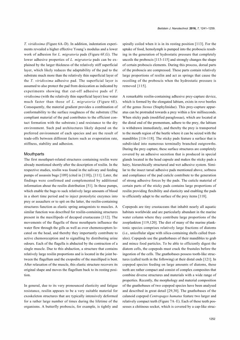
and described in great detail [29,30]. The gnathobases of the
calanoid copepod Centropages hamatus feature two larger and
relatively compact teeth (Figure 7A–E). Each of these teeth pos-
sesses a chitinous socket, which is covered by a cap-like struc-
Beilstein J. Nanotechnol. 2016, 7, 1241–1259.
1253
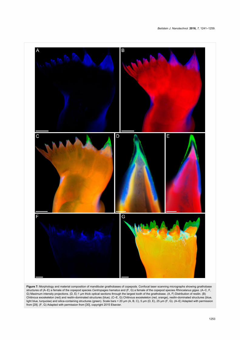
Figure 7: Morphology and material composition of mandibular gnathobases of copepods. Confocal laser scanning micrographs showing gnathobasestructures of (A–E) a female of the copepod species Centropages hamatus and (F, G) a female of the copepod species Rhincalanus gigas. (A–C, F,G) Maximum intensity projections. (D, E) 1 µm thick optical sections through the largest tooth of the gnathobase. (A, F) Distribution of resilin. (B)Chitinous exoskeleton (red) and resilin-dominated structures (blue). (C–E, G) Chitinous exoskeleton (red, orange), resilin-dominated structures (blue,light blue, turquoise) and silica-containing structures (green). Scale bars = 20 µm (A, B, C), 5 µm (D, E), 25 µm (F, G). (A–E) Adapted with permissionfrom [29]. (F, G) Adapted with permission from [30], copyright 2015 Elsevier.
Beilstein J. Nanotechnol. 2016, 7, 1241–1259.
1254
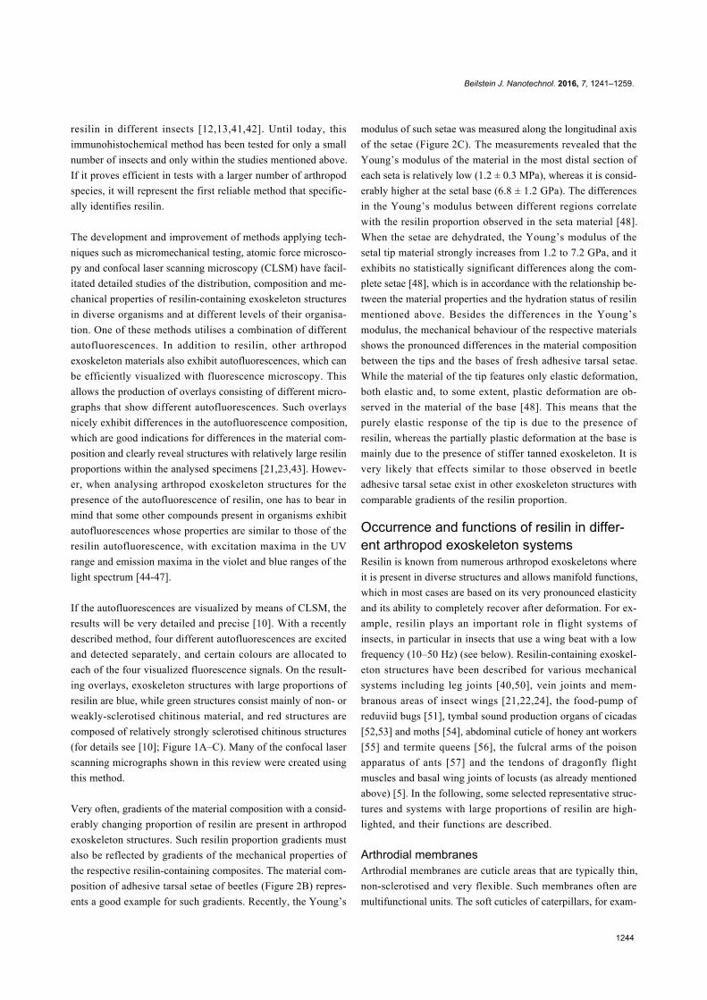
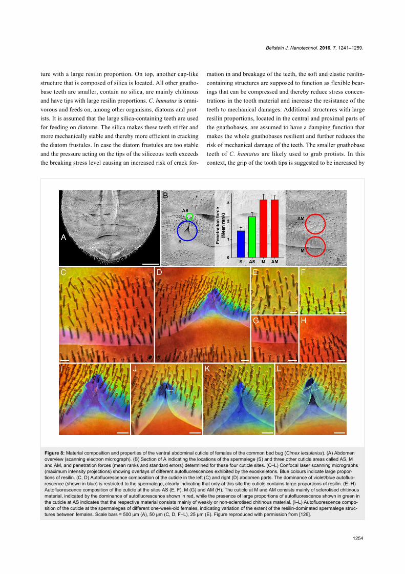
Figure 8: Material composition and properties of the ventral abdominal cuticle of females of the common bed bug (Cimex lectularius). (A) Abdomenoverview (scanning electron micrograph). (B) Section of A indicating the locations of the spermalege (S) and three other cuticle areas called AS, Mand AM, and penetration forces (mean ranks and standard errors) determined for these four cuticle sites. (C–L) Confocal laser scanning micrographs(maximum intensity projections) showing overlays of different autofluorescences exhibited by the exoskeletons. Blue colours indicate large propor-tions of resilin. (C, D) Autofluorescence composition of the cuticle in the left (C) and right (D) abdomen parts. The dominance of violet/blue autofluo-rescence (shown in blue) is restricted to the spermalege, clearly indicating that only at this site the cuticle contains large proportions of resilin. (E–H)Autofluorescence composition of the cuticle at the sites AS (E, F), M (G) and AM (H). The cuticle at M and AM consists mainly of sclerotised chitinousmaterial, indicated by the dominance of autofluorescence shown in red, while the presence of large proportions of autofluorescence shown in green inthe cuticle at AS indicates that the respective material consists mainly of weakly or non-sclerotised chitinous material. (I–L) Autofluorescence compo-sition of the cuticle at the spermaleges of different one-week-old females, indicating variation of the extent of the resilin-dominated spermalege struc-tures between females. Scale bars = 500 µm (A), 50 µm (C, D, F–L), 25 µm (E). Figure reproduced with permission from [126].
ture with a large resilin proportion. On top, another cap-like
structure that is composed of silica is located. All other gnatho-
base teeth are smaller, contain no silica, are mainly chitinous
and have tips with large resilin proportions. C. hamatus is omni-
vorous and feeds on, among other organisms, diatoms and prot-
ists. It is assumed that the large silica-containing teeth are used
for feeding on diatoms. The silica makes these teeth stiffer and
more mechanically stable and thereby more efficient in cracking
the diatom frustules. In case the diatom frustules are too stable
and the pressure acting on the tips of the siliceous teeth exceeds
the breaking stress level causing an increased risk of crack for-
mation in and breakage of the teeth, the soft and elastic resilin-
containing structures are supposed to function as flexible bear-
ings that can be compressed and thereby reduce stress concen-
trations in the tooth material and increase the resistance of the
teeth to mechanical damages. Additional structures with large
resilin proportions, located in the central and proximal parts of
the gnathobases, are assumed to have a damping function that
makes the whole gnathobases resilient and further reduces the
risk of mechanical damage of the teeth. The smaller gnathobase
teeth of C. hamatus are likely used to grab protists. In this
context, the grip of the tooth tips is suggested to be increased by
Beilstein J. Nanotechnol. 2016, 7, 1241–1259.
1255
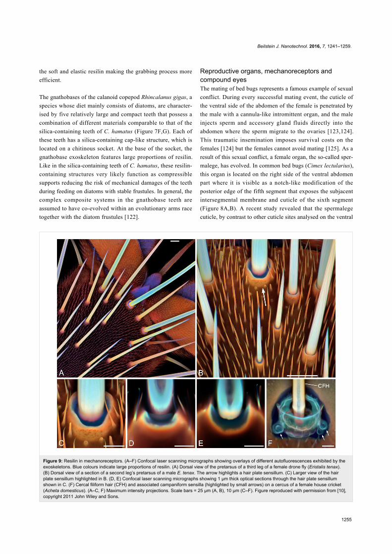
Figure 9: Resilin in mechanoreceptors. (A–F) Confocal laser scanning micrographs showing overlays of different autofluorescences exhibited by theexoskeletons. Blue colours indicate large proportions of resilin. (A) Dorsal view of the pretarsus of a third leg of a female drone fly (Eristalis tenax).(B) Dorsal view of a section of a second leg’s pretarsus of a male E. tenax. The arrow highlights a hair plate sensillum. (C) Larger view of the hairplate sensillum highlighted in B. (D, E) Confocal laser scanning micrographs showing 1 µm thick optical sections through the hair plate sensillumshown in C. (F) Cercal filiform hair (CFH) and associated campaniform sensilla (highlighted by small arrows) on a cercus of a female house cricket(Acheta domesticus). (A–C, F) Maximum intensity projections. Scale bars = 25 µm (A, B), 10 µm (C–F). Figure reproduced with permission from [10],copyright 2011 John Wiley and Sons.
the soft and elastic resilin making the grabbing process more
efficient.
The gnathobases of the calanoid copepod Rhincalanus gigas, a
species whose diet mainly consists of diatoms, are character-
ised by five relatively large and compact teeth that possess a
combination of different materials comparable to that of the
silica-containing teeth of C. hamatus (Figure 7F,G). Each of
these teeth has a silica-containing cap-like structure, which is
located on a chitinous socket. At the base of the socket, the
gnathobase exoskeleton features large proportions of resilin.
Like in the silica-containing teeth of C. hamatus, these resilin-
containing structures very likely function as compressible
supports reducing the risk of mechanical damages of the teeth
during feeding on diatoms with stable frustules. In general, the
complex composite systems in the gnathobase teeth are
assumed to have co-evolved within an evolutionary arms race
together with the diatom frustules [122].
Reproductive organs, mechanoreceptors andcompound eyesThe mating of bed bugs represents a famous example of sexual
conflict. During every successful mating event, the cuticle of
the ventral side of the abdomen of the female is penetrated by
the male with a cannula-like intromittent organ, and the male
injects sperm and accessory gland fluids directly into the
abdomen where the sperm migrate to the ovaries [123,124].
This traumatic insemination imposes survival costs on the
females [124] but the females cannot avoid mating [125]. As a
result of this sexual conflict, a female organ, the so-called sper-
malege, has evolved. In common bed bugs (Cimex lectularius),
this organ is located on the right side of the ventral abdomen
part where it is visible as a notch-like modification of the
posterior edge of the fifth segment that exposes the subjacent
intersegmental membrane and cuticle of the sixth segment
(Figure 8A,B). A recent study revealed that the spermalege
cuticle, by contrast to other cuticle sites analysed on the ventral
Beilstein J. Nanotechnol. 2016, 7, 1241–1259.
1256
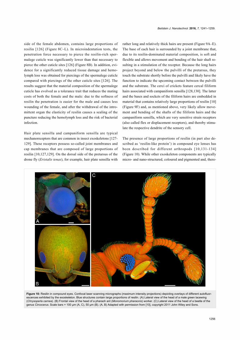
Figure 10: Resilin in compound eyes. Confocal laser scanning micrographs (maximum intensity projections) depicting overlays of different autofluor-escences exhibited by the exoskeleton. Blue structures contain large proportions of resilin. (A) Lateral view of the head of a male green lacewing(Chrysoperla carnea). (B) Frontal view of the head of a pharaoh ant (Monomorium pharaonis) worker. (C) Lateral view of the head of a beetle of thegenus Circocerus. Scale bars = 100 µm (A, C), 50 µm (B). (A, B) Adapted with permission from [10], copyright 2011 John Wiley and Sons.
side of the female abdomen, contains large proportions of
resilin [126] (Figure 8C–L). In microindentation tests, the
penetration force necessary to pierce the resilin-rich sper-
malege cuticle was significantly lower than that necessary to
pierce the other cuticle sites [126] (Figure 8B). In addition, evi-
dence for a significantly reduced tissue damage and hemo-
lymph loss was obtained for piercings of the spermalege cuticle
compared with piercings of the other cuticle sites [126]. The
results suggest that the material composition of the spermalege
cuticle has evolved as a tolerance trait that reduces the mating
costs of both the female and the male: due to the softness of
resilin the penetration is easier for the male and causes less
wounding of the female, and after the withdrawal of the intro-
mittent organ the elasticity of resilin causes a sealing of the
puncture reducing the hemolymph loss and the risk of bacterial
infection.
Hair plate sensilla and campaniform sensilla are typical
mechanoreceptors that are common in insect exoskeletons [127-
129]. These receptors possess so-called joint membranes and
cap membranes that are composed of large proportions of
resilin [10,127,129]. On the dorsal side of the pretarsus of the
drone fly (Eristalis tenax), for example, hair plate sensilla with
rather long and relatively thick hairs are present (Figure 9A–E).
The base of each hair is surrounded by a joint membrane that,
due to its resilin-dominated material composition, is soft and
flexible and allows movement and bending of the hair shaft re-
sulting in a stimulation of the receptor. Because the long hairs
project beyond and below the pulvilli of the pretarsus, they
touch the substrate shortly before the pulvilli and likely have the
function to indicate the upcoming contact between the pulvilli
and the substrate. The cerci of crickets feature cercal filiform
hairs associated with campaniform sensilla [128,130]. The latter
and the bases and sockets of the filiform hairs are embedded in
material that contains relatively large proportions of resilin [10]
(Figure 9F) and, as mentioned above, very likely allow move-
ment and bending of the shafts of the filiform hairs and the
campaniform sensilla, which are very sensitive strain receptors
(also called flex or displacement receptors), and thereby stimu-
late the respective dendrite of the sensory cell.
The presence of large proportions of resilin (in part also de-
scribed as ‘resilin-like protein’) in compound eye lenses has
been described for different arthropods [10,131-134]
(Figure 10). While other exoskeleton components are typically
micro- and nano-structured, coloured and pigmented and, there-
Beilstein J. Nanotechnol. 2016, 7, 1241–1259.
1257
fore, not suitable as material for optical elements, the pro-
nounced transparency, the colourlessness and the amorphous-
ness make resilin a perfect material for the construction of
optical systems.
ConclusionExoskeleton structures with large proportions of resilin are
common among arthropods. This review demonstrates the broad
range of resilin functions in various exoskeleton structures.
Resilin facilitates flexibility and compliance, elastic energy
storage, elastic recovery, fatigue and damage reduction, sealing
and transparency and thereby makes the respective exoskeleton
systems rather effective. Due to its remarkable combination of
different properties, resilin is a highly efficient multi-functional
protein. In addition, together with other compounds and materi-
als, it often forms complex and powerful composites that com-
bine the properties and benefits of the single components and
are capable of performing rather specific and challenging func-
tions. These characteristics have very likely been the reason for
the evolution of the large functional diversity of resilin-contain-
ing exoskeleton structures in arthropods.
AcknowledgementsJoseph Parker (Columbia University and American Museum of
Natural History, New York, NY, U.S.A.) provided the speci-
men shown in the Figure 10C. Peter Attermeyer, Dario Furlani
and Ingo Bartholomäus (Carl Zeiss Microscopy GmbH, Jena,
Germany) permitted the use of a ZEISS LSM 800 and thereby
enabled the production of the Figure 10C.
References1. Shewry, P. R.; Tatham, A. S.; Bailey, A. J. Elastomeric Proteins:
Structures, Biomechanical Properties, and Biological Roles;Cambridge University Press: Cambridge, United Kingdom, 2004.
2. Weis-Fogh, T. J. Exp. Biol. 1960, 37, 889–907.3. Weis-Fogh, T. J. Mol. Biol. 1961, 3, 648–667.
doi:10.1016/S0022-2836(61)80028-44. Weis-Fogh, T. J. Mol. Biol. 1961, 3, 520–531.
doi:10.1016/S0022-2836(61)80018-15. Andersen, S. O.; Weis-Fogh, T. Adv. Insect Physiol. 1964, 2, 1–65.
doi:10.1016/S0065-2806(08)60071-56. Andersen, S. O. Insect Biochem. Mol. Biol. 2010, 40, 541–551.
doi:10.1016/j.ibmb.2010.05.0027. Bailey, K.; Weis-Fogh, T. Biochim. Biophys. Acta 1961, 48, 452–459.
doi:10.1016/0006-3002(61)90043-98. Andersen, S. O. Resilin. In Comprehensive Biochemistry; Florkin, M.;
Stotz, E. H., Eds.; Elsevier: Amsterdam, Netherlands, 1971; Vol. 26C(Extracellular and Supporting Structures), pp 633–657.
9. Schultz, R. L. Biochim. Biophys. Acta 1964, 93, 211–213.doi:10.1016/0304-4165(64)90288-0
10. Michels, J.; Gorb, S. N. J. Microsc. 2012, 245, 1–16.doi:10.1111/j.1365-2818.2011.03523.x
11. Jensen, M.; Weis-Fogh, T. Philos. Trans. R. Soc. London, Ser. B1962, 245, 137–169. doi:10.1098/rstb.1962.0008
12. Lyons, R. E.; Wong, D. C. C.; Kim, M.; Lekieffre, N.; Huson, M. G.;Vuocolo, T.; Merritt, D. J.; Nairn, K. M.; Dudek, D. M.; Colgrave, M. L.;Elvin, C. M. Insect Biochem. Mol. Biol. 2011, 41, 881–890.doi:10.1016/j.ibmb.2011.08.002
13. Elvin, C. M.; Carr, A. G.; Huson, M. G.; Maxwell, J. M.;Pearson, R. D.; Vuocolo, T.; Liyou, N. E.; Wong, D. C. C.;Merritt, D. J.; Dixon, N. E. Nature 2005, 437, 999–1002.doi:10.1038/nature04085
14. Rauscher, S.; Pomès, R. Structural Disorder and Protein Elasticity. InFuzziness; Fuxreiter, M.; Tompa, P., Eds.; Advances in ExperimentalMedicine and Biology, Vol. 725; Springer: Berlin, Germany, 2012;pp 159–183. doi:10.1007/978-1-4614-0659-4_10
15. Bennet-Clark, H. C.; Lucey, E. C. A. J. Exp. Biol. 1967, 47, 59–76.16. Gorb, S. N. Arthropod Struct. Dev. 2004, 33, 201–220.
doi:10.1016/j.asd.2004.05.00817. Burrows, M.; Shaw, S. R.; Sutton, G. P. BMC Biol. 2008, 6, 41.
doi:10.1186/1741-7007-6-4118. Burrows, M. J. Exp. Biol. 2010, 213, 469–478. doi:10.1242/jeb.03786119. Burrows, M. J. Exp. Biol. 2011, 214, 2362–2374.
doi:10.1242/jeb.05668920. Burrows, M.; Sutton, G. P. J. Exp. Biol. 2012, 215, 3501–3512.
doi:10.1242/jeb.07199321. Haas, F.; Gorb, S.; Blickhan, R. Proc. R. Soc. London, Ser. B 2000,
267, 1375–1381. doi:10.1098/rspb.2000.115322. Haas, F.; Gorb, S.; Wootton, R. J. Arthropod Struct. Dev. 2000, 29,
137–146. doi:10.1016/S1467-8039(00)00025-623. Perez Goodwyn, P.; Peressadko, A.; Schwarz, H.; Kastner, V.;
Gorb, S. J. Comp. Physiol., A 2006, 192, 1233–1243.doi:10.1007/s00359-006-0156-z
24. Gorb, S. N. Naturwissenschaften 1999, 86, 552–555.doi:10.1007/s001140050674
25. Appel, E.; Gorb, S. N. Bioinspiration Biomimetics 2011, 6, 046006.doi:10.1088/1748-3182/6/4/046006
26. Donoughe, S.; Crall, J. D.; Merz, R. A.; Combes, S. A. J. Morphol.2011, 272, 1409–1421. doi:10.1002/jmor.10992
27. Kannupandi, T. Acta Histochem. 1976, 56, 73–79.doi:10.1016/S0065-1281(76)80028-1
28. Andersen, S. O. Structure and Function of Resilin. In ElastomericProteins: Structures, Biomechanical Properties, and Biological Roles;Shewry, P. R.; Tatham, A. S.; Bailey, A. J., Eds.; CambridgeUniversity Press: Cambridge, United Kingdom, 2003; pp 259–278.doi:10.1017/CBO9780511546327.015
29. Michels, J.; Vogt, J.; Gorb, S. N. Sci. Rep. 2012, 2, 465.doi:10.1038/srep00465
30. Michels, J.; Vogt, J.; Simon, P.; Gorb, S. N. Zoology (Munich, Ger.)2015, 118, 141–146.
31. Govindarajan, S.; Rajulu, G. S. Experientia 1974, 30, 908–909.doi:10.1007/BF01938354
32. Sundara Rajulu, G. Indian J. Exp. Biol. 1971, 9, 122–123.33. Ramalingam, K. Parasitology 1973, 66, 1–7.
doi:10.1017/S003118200004438334. Wong, W. L.; Michels, J.; Gorb, S. N. Parasitology 2013, 140, 95–98.
doi:10.1017/S003118201200137035. Lopez-Llorca, L. V.; Fry, S. C. Nematologica 1989, 35, 165–179.
doi:10.1163/002825989X0030436. DeVore, D. P.; Gruebel, R. J. Biochem. Biophys. Res. Commun.
1978, 80, 993–999. doi:10.1016/0006-291X(78)91343-837. Foerder, C. A.; Shapiro, B. M. Proc. Natl. Acad. Sci. U. S. A. 1977, 74,
4214–4218. doi:10.1073/pnas.74.10.4214
Beilstein J. Nanotechnol. 2016, 7, 1241–1259.
1258
38. Andersen, S. O. Biochim. Biophys. Acta 1963, 69, 249–262.doi:10.1016/0006-3002(63)91258-7
39. Andersen, S. O. Acta Physiol. Scand. 1965, 66 (Suppl. 263), 1–81.40. Neff, D.; Frazier, S. F.; Quimby, L.; Wang, R.-T.; Zill, S.
Arthropod Struct. Dev. 2000, 29, 75–83.doi:10.1016/S1467-8039(00)00014-1
41. Burrows, M.; Borycz, J. A.; Shaw, S. R.; Elvin, C. M.;Meinertzhagen, I. A. PLoS One 2011, 6, e28456.doi:10.1371/journal.pone.0028456
42. Wong, D. C. C.; Pearson, R. D.; Elvin, C. M.; Merritt, D. J. Dev. Dyn.2012, 241, 333–339. doi:10.1002/dvdy.23724
43. Niederegger, S.; Gorb, S. J. Insect Physiol. 2003, 49, 611–620.doi:10.1016/S0022-1910(03)00048-9
44. Fujimori, E. Biochim. Biophys. Acta 1978, 534, 82–88.doi:10.1016/0005-2795(78)90478-6
45. Garcia-Castineiras, S.; Dillon, J.; Spector, A. Exp. Eye Res. 1978, 26,461–476. doi:10.1016/0014-4835(78)90132-X
46. Gast, R.; Lee, J. Proc. Natl. Acad. Sci. U. S. A. 1978, 75, 833–837.doi:10.1073/pnas.75.2.833
47. Giurginca, A.; Šustr, V.; Tajovský, K.; Giurginca, M.; Matei, I. ZooKeys2015, 515, 111–125. doi:10.3897/zookeys.515.9395
48. Peisker, H.; Michels, J.; Gorb, S. N. Nat. Commun. 2013, 4, 1661.doi:10.1038/ncomms2576
49. Gorb, S. N.; Filippov, A. E. Beilstein J. Nanotechnol. 2014, 5,837–846. doi:10.3762/bjnano.5.95
50. Gorb, S. N. J. Morphol. 1996, 230, 219–230.doi:10.1002/(SICI)1097-4687(199611)230:2<219::AID-JMOR8>3.0.CO;2-B
51. Edwards, H. A. J. Exp. Biol. 1983, 105, 407–409.52. Young, D.; Bennet-Clark, H. C. J. Exp. Biol. 1995, 198, 1001–1019.53. Bennet-Clark, H. C. J. Exp. Biol. 1997, 200, 1681–1694.54. Skals, N.; Surlykke, A. J. Exp. Biol. 1999, 202, 2937–2949.55. Raghu Varman, A. J. Ga. Entomol. Soc. 1981, 16, 11–13.56. Raghu Varman, A. Experientia 1980, 36, 564.
doi:10.1007/BF0196580257. Raghu Varman, A.; Hermann, H. R., Jr. J. Anim. Morphol. Physiol.
1982, 29, 284–285.58. Hepburn, H. R.; Levy, P. J. Entomol. Soc. South. Afr. 1975, 38,
131–141.59. Vincent, J. F. V. Design for Living: The Elastic-sided Locust. In The
Insect Integument; Hepburn, H. R., Ed.; Elsevier: Amsterdam,Netherlands, 1976; pp 401–419.
60. Hackman, R. H.; Goldberg, M. Int. J. Parasitol. 1985, 15, 249–254.doi:10.1016/0020-7519(85)90061-X
61. Richards, A. G.; Richards, P. A. Int. J. Insect Morphol. Embryol. 1979,8, 143–157. doi:10.1016/0020-7322(79)90013-8
62. Hepburn, H. R. Structure of the Integument. In Comprehensive InsectPhysiology, Biochemistry and Pharmacology; Kerkut, G. A.;Gilbert, L. I., Eds.; Pergamon Press: Oxford, United Kingdom, 1985;Vol. 3 (Integument, Respiration and Circulation), pp 1–58.doi:10.1016/b978-0-08-030804-3.50007-8
63. Vincent, J. F. V.; Wood, S. D. E. Nature 1972, 235, 167–168.doi:10.1038/235167a0
64. Vincent, J. F. V. Proc. R. Soc. London, Ser. B 1975, 188, 189–201.doi:10.1098/rspb.1975.0012
65. Vincent, J. F. V. Tissue Cell 1981, 13, 831–853.doi:10.1016/S0040-8166(81)80017-1
66. Hackman, R. H.; Goldberg, M. J. Insect Physiol. 1987, 33, 39–50.doi:10.1016/0022-1910(87)90102-8
67. Hackman, R. H. J. Insect Physiol. 1975, 21, 1613–1623.doi:10.1016/0022-1910(75)90199-7
68. Reynolds, S. E. J. Exp. Biol. 1975, 62, 69–80.69. Gorb, S. N. J. Zool. 2000, 250, 479–494.
doi:10.1111/j.1469-7998.2000.tb00791.x70. Rothshild, M.; Schlein, Y.; Parker, K.; Neville, C.; Sternberg, S.
Philos. Trans. R. Soc. London, Ser. B 1975, 271, 499–515.71. Bennet-Clark, H. C. J. Exp. Biol. 1975, 63, 53–83.72. Evans, M. E. G. J. Zool. (1965-1984) 1973, 169, 181–194.73. Furth, D. G.; Traub, W.; Harpaz, I. J. Exp. Zool. 1983, 227, 43–47.
doi:10.1002/jez.140227010774. Burrows, M. J. Exp. Biol. 2012, 215, 3612–3621.
doi:10.1242/jeb.07468275. Gronenberg, W. J. Comp. Physiol., A 1996, 178, 727–734.
doi:10.1007/BF0022582176. Bayley, T. G.; Sutton, G. P.; Burrows, M. J. Exp. Biol. 2012, 215,
1151–1161. doi:10.1242/jeb.06808077. Lehmann, F. O.; Gorb, S.; Nasir, N.; Schützner, P. J. Exp. Biol. 2011,
214, 2949–2961. doi:10.1242/jeb.04535178. Appel, E.; Gorb, S. N. Zoologica 2014, 159, 1–159.79. Appel, E.; Heepe, L.; Lin, C.-P.; Gorb, S. N. J. Anat. 2015, 227,
561–582. doi:10.1111/joa.1236280. Neville, A. C. J. Exp. Biol. 1960, 37, 631–656.81. King, R. Dynamic Mechanical Properties of Resilin. Master’s Thesis,
Virginia Polytechnic Institute and State University, Blacksburg, VA,U.S.A., 2010.
82. Weis-Fogh, T. J. Exp. Biol. 1973, 59, 169–230.83. Gosline, J.; Lillie, M.; Carrington, E.; Guerette, P.; Ortlepp, C.;
Savage, K. Philos. Trans. R. Soc. London, Ser. B 2002, 357,121–132. doi:10.1098/rstb.2001.1022
84. Nachtigall, W.; Wisser, A.; Eisinger, D. J. Comp. Physiol., B 1998,168, 323–344. doi:10.1007/s003600050152
85. Choudhury, U. Dynamic Mechanical Properties of Cockroach(Periplanata americana) Resilin. Master’s Thesis, Virginia PolytechnicInstitute and State University, Blacksburg, VA, U.S.A., 2012.
86. Newman, D. J. S. The Functional Wing Morphology of Some Odonata;University of Exeter: Exeter, United Kingdom, 1982.
87. Rajabi, H.; Ghoroubi, N.; Darvizeh, A.; Dirks, J.-H.; Appel, E.;Gorb, S. N. Bioinspiration Biomimetics 2015, 10, 056003.doi:10.1088/1748-3190/10/5/056003
88. Rajabi, H.; Shafiei, A.; Darvizeh, A.; Dirks, J.-H.; Appel, E.;Gorb, S. N. R. Soc. Open Sci. 2016, 3, 160006.doi:10.1098/rsos.160006
89. Mountcastle, A. M.; Combes, S. A. Proc. R. Soc. London, Ser. B2013, 280, 20130531. doi:10.1098/rspb.2013.0531
90. Ma, Y.; Ning, J. G.; Ren, H. L.; Zhang, P. F.; Zhao, H. Y. J. Exp. Biol.2015, 218, 2136–2142. doi:10.1242/jeb.117325
91. Mountcastle, A. M.; Combes, S. A. J. Exp. Biol. 2014, 217,1108–1115. doi:10.1242/jeb.092916
92. Deiters, J.; Kowalczyk, W.; Seidl, T. Biol. Open 2016, 5, 638–644.doi:10.1242/bio.016527
93. Haas, F.; Hwen, J. T. C.; Tang, H. B. Arthropod Syst. Phylog. 2012,70, 95–105.
94. Wootton, R. J. Annu. Rev. Entomol. 1992, 37, 113–140.doi:10.1146/annurev.en.37.010192.000553
95. Haas, F.; Beutel, R. G. Zoology (Munich, Ger.) 2001, 104, 123–141.96. Sun, J.; Ling, M.; Wu, W.; Bhushan, B.; Tong, J. Int. J. Mol. Sci. 2014,
15, 6009–6018. doi:10.3390/ijms1504600997. Gorb, S. N. Attachment Devices of Insect Cuticle; Kluwer Academic
Publishers: Dordrecht, Netherlands, 2001.
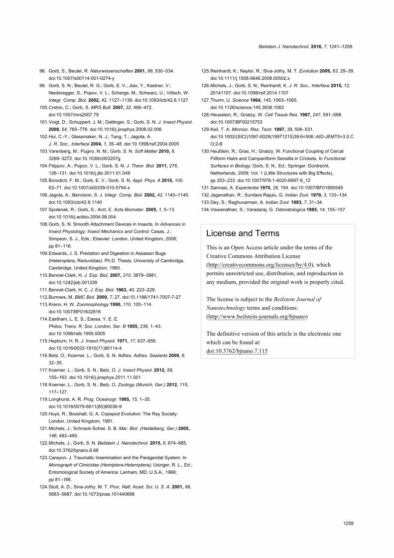
Beilstein J. Nanotechnol. 2016, 7, 1241–1259.
1259
98. Gorb, S.; Beutel, R. Naturwissenschaften 2001, 88, 530–534.doi:10.1007/s00114-001-0274-y
99. Gorb, S. N.; Beutel, R. G.; Gorb, E. V.; Jiao, Y.; Kastner, V.;Niederegger, S.; Popov, V. L.; Scherge, M.; Schwarz, U.; Vötsch, W.Integr. Comp. Biol. 2002, 42, 1127–1139. doi:10.1093/icb/42.6.1127
100.Creton, C.; Gorb, S. MRS Bull. 2007, 32, 466–472.doi:10.1557/mrs2007.79
101.Voigt, D.; Schuppert, J. M.; Dattinger, S.; Gorb, S. N. J. Insect Physiol.2008, 54, 765–776. doi:10.1016/j.jinsphys.2008.02.006
102.Hui, C.-Y.; Glassmaker, N. J.; Tang, T.; Jagota, A.J. R. Soc., Interface 2004, 1, 35–48. doi:10.1098/rsif.2004.0005
103.Varenberg, M.; Pugno, N. M.; Gorb, S. N. Soft Matter 2010, 6,3269–3272. doi:10.1039/c003207g
104.Filippov, A.; Popov, V. L.; Gorb, S. N. J. Theor. Biol. 2011, 276,126–131. doi:10.1016/j.jtbi.2011.01.049
105.Borodich, F. M.; Gorb, E. V.; Gorb, S. N. Appl. Phys. A 2010, 100,63–71. doi:10.1007/s00339-010-5794-x
106.Jagota, A.; Bennison, S. J. Integr. Comp. Biol. 2002, 42, 1140–1145.doi:10.1093/icb/42.6.1140
107.Spolenak, R.; Gorb, S.; Arzt, E. Acta Biomater. 2005, 1, 5–13.doi:10.1016/j.actbio.2004.08.004
108.Gorb, S. N. Smooth Attachment Devices in Insects. In Advances inInsect Physiology: Insect Mechanics and Control; Casas, J.;Simpson, S. J., Eds.; Elsevier: London, United Kingdom, 2008;pp 81–116.
109.Edwards, J. S. Predation and Digestion in Assassin Bugs(Heteroptera, Reduviidae). Ph.D. Thesis, University of Cambridge,Cambridge, United Kingdom, 1960.
110.Bennet-Clark, H. J. Exp. Biol. 2007, 210, 3879–3881.doi:10.1242/jeb.001339
111.Bennet-Clark, H. C. J. Exp. Biol. 1963, 40, 223–229.112.Burrows, M. BMC Biol. 2009, 7, 27. doi:10.1186/1741-7007-7-27113.Krenn, H. W. Zoomorphology 1990, 110, 105–114.
doi:10.1007/BF01632816114.Eastham, L. E. S.; Eassa, Y. E. E.
Philos. Trans. R. Soc. London, Ser. B 1955, 239, 1–43.doi:10.1098/rstb.1955.0005
115.Hepburn, H. R. J. Insect Physiol. 1971, 17, 637–656.doi:10.1016/0022-1910(71)90114-4
116.Betz, O.; Koerner, L.; Gorb, S. N. Adhes. Adhes. Sealants 2009, 6,32–35.
117.Koerner, L.; Gorb, S. N.; Betz, O. J. Insect Physiol. 2012, 58,155–163. doi:10.1016/j.jinsphys.2011.11.001
118.Koerner, L.; Gorb, S. N.; Betz, O. Zoology (Munich, Ger.) 2012, 115,117–127.
119.Longhurst, A. R. Prog. Oceanogr. 1985, 15, 1–35.doi:10.1016/0079-6611(85)90036-9
120.Huys, R.; Boxshall, G. A. Copepod Evolution; The Ray Society:London, United Kingdom, 1991.
121.Michels, J.; Schnack-Schiel, S. B. Mar. Biol. (Heidelberg, Ger.) 2005,146, 483–495.
122.Michels, J.; Gorb, S. N. Beilstein J. Nanotechnol. 2015, 6, 674–685.doi:10.3762/bjnano.6.68
123.Carayon, J. Traumatic Insemination and the Paragenital System. InMonograph of Cimicidae (Hemiptera-Heteroptera); Usinger, R. L., Ed.;Entomological Society of America: Lanham, MD, U.S.A., 1966;pp 81–166.
124.Stutt, A. D.; Siva-Jothy, M. T. Proc. Natl. Acad. Sci. U. S. A. 2001, 98,5683–5687. doi:10.1073/pnas.101440698
125.Reinhardt, K.; Naylor, R.; Siva-Jothy, M. T. Evolution 2009, 63, 29–39.doi:10.1111/j.1558-5646.2008.00502.x
126.Michels, J.; Gorb, S. N.; Reinhardt, K. J. R. Soc., Interface 2015, 12,20141107. doi:10.1098/rsif.2014.1107
127.Thurm, U. Science 1964, 145, 1063–1065.doi:10.1126/science.145.3636.1063
128.Heusslein, R.; Gnatzy, W. Cell Tissue Res. 1987, 247, 591–598.doi:10.1007/BF00215753
129.Keil, T. A. Microsc. Res. Tech. 1997, 39, 506–531.doi:10.1002/(SICI)1097-0029(19971215)39:6<506::AID-JEMT5>3.0.CO;2-B
130.Heußlein, R.; Gras, H.; Gnatzy, W. Functional Coupling of CercalFiliform Hairs and Campaniform Sensilla in Crickets. In FunctionalSurfaces in Biology; Gorb, S. N., Ed.; Springer: Dordrecht,Netherlands, 2009; Vol. 1 (Little Structures with Big Effects),pp 203–233. doi:10.1007/978-1-4020-6697-9_12
131.Sannasi, A. Experientia 1970, 26, 154. doi:10.1007/BF01895549132.Jaganathan, R.; Sundara Rajulu, G. Indian Zool. 1979, 3, 133–134.133.Dey, S.; Raghuvarman, A. Indian Zool. 1983, 7, 31–34.134.Viswanathan, S.; Varadaraj, G. Odonatologica 1985, 14, 155–157.
License and TermsThis is an Open Access article under the terms of the
Creative Commons Attribution License
(http://creativecommons.org/licenses/by/4.0), which
permits unrestricted use, distribution, and reproduction in
any medium, provided the original work is properly cited.
The license is subject to the Beilstein Journal of
Nanotechnology terms and conditions:
(http://www.beilstein-journals.org/bjnano)
The definitive version of this article is the electronic one
which can be found at:
doi:10.3762/bjnano.7.115