Embed Size (px)
Citation preview
59Copyright © 2014 The Korean Society of Cardiology
Korean Circulation Journal
Introduction
Advances in diagnosis, catheter and surgical interventions, and intensive care have significantly improved the survival of children with congenital cardiac malformations. In the present era, 85% or more of children born with congenital heart diseases are expected to survive into adulthood.1) The significantly improved survival has resulted in an increasing population of adolescent and adult pa-tients with congenital heart disease who are confronted with unique cardiac and non-cardiac issues.1)2) Of the long-term cardiac problems, ventricular dysfunction remains an important issue of concern. De-spite corrective or palliative repair of congenital heart lesions, the right ventricle, which may be the subpulmonary or systemic ventricular
Review
http://dx.doi.org/10.4070/kcj.2014.44.2.59Print ISSN 1738-5520 • On-line ISSN 1738-5555
Functional Assessment for Congenital Heart DiseaseYiu-fai Cheung, MDDivision of Paediatric Cardiology, Department of Paediatrics and Adolescent Medicine, Queen Mary Hospital, The University of Hong Kong, Hong Kong, China
Significant improvement in survival of children with congenital cardiac malformations has resulted in an increasing population of adoles-cent and adult patients with congenital heart disease. Of the long-term cardiac problems, ventricular dysfunction remains an important issue of concern. Despite corrective or palliative repair of congenital heart lesions, the right ventricle, which may be the subpulmonary or systemic ventricular chamber, and the functional single ventricle are particularly vulnerable to functional impairment. Regular assessment of cardiac function constitutes an important aspect in the long-term follow up of patients with congenital heart disease. Echocardiography remains the most useful imaging modality for longitudinal monitoring of cardiac function. Conventional echocardiographic assessment has focused primarily on quantification of changes in ventricular size and blood flow velocities during the cardiac cycles. Advances in echo-cardiographic technologies including tissue Doppler imaging and speckle tracking echocardiography have enabled direct interrogation of myocardial deformation. In this review, the issues of ventricular dysfunction in congenital heart disease, conventional echocardiographic and novel myocardial deformation imaging techniques, and clinical applications of these techniques in the functional assessment of con-genital heart disease are discussed. (Korean Circ J 2014;44(2):59-73)
KEY WORDS: Congenital heart disease; Ventricular function; Tissue Doppler imaging; Speckle tracking echocardiography; Echocardiography, three-dimensional.
Correspondence: Yiu-fai Cheung, MD, Division of Paediatric Cardiology, Department of Paediatrics and Adolescent Medicine, Queen Mary Hospital, The University of Hong Kong, 102 Pokfulam Road, Hong Kong, ChinaTel: 852-22554090, Fax: 852-25539491E-mail: [email protected]
• The author has no financial conflicts of interest.
This is an Open Access article distributed under the terms of the Creative Commons Attribution Non-Commercial License (http://creativecommons.org/licenses/by-nc/3.0) which permits unrestricted non-commercial use, distribution, and reproduction in any medium, provided the original work is properly cited.
chamber, and the functional single ventricle are particularly vulner-able to functional impairment.
Regular assessment of cardiac function constitutes an important aspect in the long-term follow up of patients with congenital heart disease. Given its non-invasiveness and widespread availability, echocardiography remains the most useful imaging modality for longitudinal monitoring of cardiac function. While conventional echocardiographic assessment of cardiac function has focused on quantification of changes in ventricular size and blood flow veloci-ties during the cardiac cycles, advances in echocardiographic tech-nologies have enabled direct evaluation of myocardial deformation. In the recent few years, novel myocardial deformation imaging has found increasing applications in functional assessment of congen-ital heart disease. In this review, the issues of ventricular dysfunc-tion in congenital heart disease, conventional echocardiographic and novel myocardial deformation imaging techniques, and clinical applications of these techniques in the functional assessment of congenital heart disease are discussed.
Ventricular Dysfunction in Congenital Heart Disease
Subpulmonary right ventricleTransannular patch repair is commonly required during surgical

60 Functional Assessment for CHD
http://dx.doi.org/10.4070/kcj.2014.44.2.59 www.e-kcj.org
repair of tetralogy of Fallot (TOF), as the pulmonary valve annulus is usually small. While initially thought to be a relatively benign con-dition, chronic pulmonary regurgitation with consequential volume overloading of the subpulmonary right ventricle is now recognized as one of the most important determinants of late outcome.3-5) De-velopment of right ventricular (RV) systolic and diastolic dysfunc-tion in association with chronic severe pulmonary regurgitation is well documented.6)7) Aneurysmal dilation and akinesia of the RV outflow contribute further to RV dilation and detrimentally affect RV ejection fraction.8) Development of right bundle branch block and QRS prolongation after surgery may potentially lead to RV me-chanical dyssynchrony.9) All of these factors may act synergistically and contribute to progressive RV dysfunction late after TOF repair, with the clinical consequences of exercise intolerance, development of cardiac arrhythmias, and congestive heart failure.10-12)
The phenomenon of restrictive RV physiology occurs both early and late after surgical repair of TOF, which is a manifestation of ab-normal relaxation and probably reduced compliance of the subpul-monary right ventricle. During early postoperative period, restric-tive RV physiology may account for low cardiac output syndrome.13) On the other hand, persistence of restrictive RV physiology in the long-term has been associated with less progressive dilation of the right ventricle and better exercise capacity.14) Restrictive RV physiol-ogy has also been described after biventricular repair of pulmonary atresia with intact ventricular septum (PAIVS) due probably to the relatively small RV cavity and intrinsic myocardial and endocardial abnormalities that reduce RV compliance.15) Limited data also sug-gest that restrictive RV physiology also predicts better exercise ca-pacity in these patients.15)
Systemic right ventricleIn congenitally corrected transposition of the great arteries (cc-
TGA) and in patients with complete transposition of the great arter-ies (TGA) after atrial repair, the right ventricle acts as the systemic pumping chamber.
Systemic RV dysfunction occurs with increasing prevalence with advancing age in cc-TGA. It is estimated that about half of the pa-tients may develop RV dysfunction and congestive heart failure by the fifth decade, even in the absence of significant associated cardi-ac lesions.16) Occurrence of systemic RV dysfunction in cc-TGA is usu-ally accompanied by progressive RV dilation and worsening of tri-cuspid regurgitation.17) Notwithstanding the ‘chicken and egg’ con-troversy, it appears that the intrinsic architectural design of the tricuspid valve and geometry of the right ventricle may render them more vulnerable to progressive failure.18) Dilation of the systemic right ventricle may set up a vicious cycle by shifting the ventricular septum to the left side, pulling the tricuspid septal leaflet for from
the other tricuspid leaflets, which aggravates preexisting tricuspid regurgitation. This hypothesis is supported by the observation of amelioration of tricuspid regurgitation with placement of a pulmo-nary arterial band,19)20) which increases subpulmonary left ventric-ular (LV) pressure and restores, albeit partially, the septal geometry. Single-chamber ventricular pacing in patients with cc-TGA and com-plete heart block may further aggravate systemic RV failure possi-bly by inducing mechanical dyssynchrony.21)
Substrates other than tricuspid valve design and RV geometry may predispose to development of systemic RV dysfunction in pa-tients after the Senning or Mustard procedure. Myocardial perfusion defects and impaired flow reserve involving the systemic right ven-tricle have been found in survivors of the Mustard operation,22)23) which may suggest inadequate coronary arterial supply to the hy-pertrophic RV myocardium to cope with the systemic arterial load. Studies using late gadolinium enhancement (LGE) and equilibrium contrast cardiac magnetic resonance (CMR) have shown areas of fibrosis,24)25) the extent of which has been found to correlate with RV dilation and impairment of systolic function. Reduction of systemic RV ejection fraction and long-axis function has been demonstrat-ed in patients with TGA after atrial repair and may suggest systolic dysfunction.26-28) Nonetheless, whether this represents intrinsic RV failure or an adaptive response to increased systemic afterload is contentious. On the other hand, accumulating data suggest that im-pairment of atrioventricular coupling due to restrictive atrial baf-fling pathways may limit the augmentation of stroke volume during exercise and other forms of stress, and constitute a unique form of diastolic dysfunction of the systemic right ventricle.29)
Right ventricular-left ventricular interactionThe function of the right and left ventricles is closely linked given
the enclosure of the two ventricles within the same pericardial cavi-ty, sharing of ventricular septum, and coursing of superficial epi-cardial myocardial fibres through two ventricles. These anatomic substrates provide the basis of adverse ventricular-ventricular inter-action in patients with congenital heart disease with abnormal RV geometry and function.
In repaired TOF patients, impaired LV function has been related to abnormal RV function and shown to be a significant independent risk factor for long-term adverse outcomes. Suboptimal RV-LV dia-stolic interaction secondary to RV dilation and septal shift with con-sequential reduction of LV compliance and diastolic filling has been described.30)31) With interrogation of LV mechanics by deformation imaging techniques, abnormal systolic ventricular-ventricular inter-action after TOF repair is also being increasingly recognized. Addi-tionally, limited data suggest the presence of adverse systemic RV-subpulmonary LV interaction in patients after atrial repair. These
61Yiu-fai Cheung
http://dx.doi.org/10.4070/kcj.2014.44.2.59www.e-kcj.org
unfavourable ventricular-ventricular interactions are discussed be-low.
Functional single ventricleVentricular dysfunction remains one of the most important long-
term complications in patients with functionally univentricular hearts palliated by Fontan-type procedures.32) Intrinsic abnormalities of myocardial architecture are found in functional single ventri-cles.33)34) Pressure and volume loading of the systemic ventricle oc-curs as a result of the parallel supply of the systemic and pulmo-nary circulation prior to univentricular repair. The volume overload may be further aggravated by atrioventricular valvar insufficiency, which may be related to dysplasia of the common atrioventricular valve or annular dilation secondary to ventricular dilation. Conver-sion from parallel systemic and pulmonary circulation to the in-se-ries Fontan circulation is associated with changes in ventricular ge-ometry, acquired ventricular hypertrophy, and preload reduction. The consequences of incoordinate ventricular relaxation, diastolic ven-tricular dysfunction, and probably progressive reduction in ventric-ular compliance have been recognized.35)36) Furthermore, increased systemic afterload with abnormal ventriculo-arterial interaction found in post Fontan patients may have further negative impact on diastolic function of the functional single ventricle.37)
Conventional Echocardiographic Assessment of Cardiac Function
Conventional echocardiographic assessment of cardiac function in patients with congenital heart disease has focused on quantifica-tion of changes in ventricular dimensions and chamber volumes for assessment of systolic function, and Doppler-derived blood flow velocities across atrioventricular valves for evaluation of dia-stolic function.
Two-dimensional and M-mode assessmentsM-mode has conventionally been used to derive LV shortening
fraction for assessing global LV systolic function. Shortening frac-tion is affected by loading conditions, regional myocardial abnor-malities, and alteration of LV geometry. Existence of paradoxical sep-tal motion in RV volume- and pressure-loaded conditions invalidates the interpretation of this LV functional parameter. Furthermore, this geometric parameter is of little use in the assessment of RV and single ventricular systolic function.
Fractional area change of the right ventricle determined by two-dimensional (2D) planimetry has also been used as an index of RV systolic function in adults,38) although this has not been validated in children. To assess RV longitudinal shortening as a measure of RV
systolic function, placement of the M-mode curser through the tri-cuspid annulus in the four-chamber view enables the derivation of the so-called tricuspid annular plane systolic excursion (TAPSE). TAPSE has been used to evaluate subpulmonary and systemic RV systolic function, although conclusions on its usefulness are mixed.39-42) Nor-mal reference values of TAPSE in children are available to facilitate the interpretation of patient data.43)
Volumetric measurementsCardiac magnetic resonance is regarded as the gold standard in
the measurement of ventricular volumes and quantification of ejec-tion fraction. Avoidance of the need for geometric assumption is of particular importance in the evaluation of right ventricles, func-tional single ventricles, and distorted left ventricles due to septal shift in patients with congenital heart disease.
Different 2D echocardiographic methods including the Simpson algorithms, area-length method, and bullet method have been used to estimate LV volumes.44) Nonetheless, violation of geometric as-sumptions is not uncommon in patients with congenital heart dis-ease, which limits their accuracy and usefulness in the clinical setting. Although different formulae have also been proposed to estimate RV volume by 2D echocardiography, there are little or no data re-garding their utility and accuracy in children.44)
The introduction of real-time three-dimensional (3D) echocar-diography has shown great promise in evaluating ventricular vol-umes regardless of ventricular geometry (Fig. 1). Its initial applica-tions in children are encouraging. Importantly, LV and RV volumes and volumes of functional single ventricles obtained by real-time 3D echocardiography compare favorably with those obtained using CMR.45)46) Hence, real-time 3D echocardiography has an important advantage in evaluating volume-based functional parameters in pa-tients with congenital heart disease having ventricles of different geometries.
Doppler interrogationPulsed-wave Doppler analysis of mitral early (E) and late (A) dia-
stolic inflow velocities and E-deceleration time is commonly used to assess LV diastolic function. In children with fast heart rates, the utility of these indices may however by limited due to frequent fu-sion of the E and A waves. Isovolumic relaxation time, another pa-rameter of LV diastolic function, can be measured by simultaneous continuous-wave Doppler interrogation of LV inflow and outflow.
Respiratory variations of tricuspid valve inflow velocities render their measurement difficult in children. Reduction of RV compliance in certain congenital heart lesions further confounds the interpreta-tion of the tricuspid inflow velocities. A good surrogate pulsed-Dop-pler measure of impaired RV diastolic compliance is the antegrade

62 Functional Assessment for CHD
http://dx.doi.org/10.4070/kcj.2014.44.2.59 www.e-kcj.org
diastolic flow in the pulmonary artery coincident with atrial systole (Fig. 2).13)
The spectral Doppler-derived myocardial performance index (MPI) can be used to assess combined systolic and diastolic function of the left and right ventricles.47) It is calculated as the sum of isovolu-mic relaxation time and isovolumic contraction time divided by ejec-tion time. The MPI has increasingly been utilized in the assessment of global RV function in patients with different types of congenital heart lesions.44)
Tissue Doppler Imaging of Myocardial Motion
Velocities of myocardial tissue motion can be determined using tissue Doppler imaging.48) Nonetheless, tissue Doppler imaging al-lows only one-dimensional assessment of the tissue regional veloci-ty vector along the beam of the ultrasound. Hence, commonly mea-
sured myocardial velocities are those at the base and mid-levels of the LV lateral wall, ventricular septum, and RV free wall determined from the four-chamber view. Increasingly, tissue Doppler evaluation of the mitral and tricuspid annular junction is utilized to assess re-spective LV and RV function in children with congenital heart diseases.
From the spectral or colour tissue Doppler tracings, the peak sys-tolic and two diastolic peak velocities can be measured (Fig. 3). The positive systolic peak represents annular motion towards the apex and reflects ventricular systolic function. The early negative diastolic annular velocity (e’) reflects ventricular recoil from the contracted state. The late negative diastolic peak represents annular motion during atrial contraction and is affected by ventricular diastolic and atrial systolic function.
An additional peak velocity is seen during isovolumic contrac-tion. A relatively-load independent index, isovolumic acceleration
Fig. 1. Real-time three-dimensional echocardiographic analysis of left ventricular (LV) and right ventricular (RV) volumes. The regional time-volume curves of the 16 LV segments enable the assessment of LV mechanical dyssynchrony.
Fig. 2. Pulsed-Doppler echocardiography demonstrating antegrade dia-stolic flow in the main pulmonary artery during atrial systole.
Fig. 3. Tissue Doppler imaging with the curser at the right ventricular free wall-tricuspid annular junction. a: late diastolic myocardial velocity, e: early diastolic myocardial velocity, IVA: isovolumic acceleration, s: systolic myo-cardial velocity, Δt: time difference, Δv: velocity change.
63Yiu-fai Cheung
http://dx.doi.org/10.4070/kcj.2014.44.2.59www.e-kcj.org
during myocardial acceleration (IVA), can be calculated as the differ-ence between peak and baseline myocardial velocities divided by the time interval from onset of the wave during isovolumic contrac-tion at zero-crossing to the time at peak velocity.49)
The E/e’ ratios on the respective sides may provide an estimation of LV and RV filling pressures,50)51) although pediatric validation is lacking. The MPI can also be determined by measuring the time in-tervals on spectral tissue Doppler tracings, which correlates well with that derived from conventional Doppler interrogation.52)
Novel Evaluation of Myocardial Function
Conventional echocardiographic assessment of cardiac function based on evaluation of changes in cardiac dimensions and blood flow can perhaps be regarded only as an indirect assessment of car-diac function consequential upon shortening and lengthening of myocardium throughout the cardiac cycle. Techniques that allow direct interrogation of myocardial mechanics may be more sensitive in the detection of subtle preclinical functional impairment.
Myocardial deformationThe heart undergoes complex systolic and diastolic deformation
in three dimensions in relation to the architectural design of the myocardium. The different aspects of myocardial deformation can broadly be described in terms of linear deformation, torsional defor-mation, and synchronicity of deformation.
Linear deformation of the myocardium can be quantified by strain. Strain is defined as the percentage change in the length of myocar-dial segment relative to the reference length taken usually at end-diastole. Linear deformation of the left ventricle is usually described in terms of strain along its three principle geometrical coordinates: longitudinal, circumferential, and radial. As the RV myocardial fibres are arranged longitudinally, longitudinal strain is usually used to describe RV deformation.
The speed of myocardial deformation, on the other hand is quan-tified by strain rate. Peak systolic strain rate, compared with sys-tolic strain, might better reflect the rate of myocardial force gener-ation and hence cardiac contractility.53)54) Diastolic strain rates have been used to assess ventricular diastolic function.55)
The helical arrangement of myocardial fibres accounts for the tor-sional deformation of the left ventricle.56) Anti-clockwise rotation of the left heart, as viewed from the cardiac apex, occurs during iso-volumic contraction. During ventricular ejection, the apex continues its anti-clockwise rotation while the base rotates in the clockwise direction.57) Untwisting with clockwise rotation of the apex occurs during relaxation to generate a suction force that draws blood into the LV from the left atrium.58) Torsional deformation of the left ven-
tricle contributes significantly to LV diastolic recoil and function.59) Recent data further suggest the possibility of ventricular torsion as an aid to ejection.60) But, the phenomenon of RV torsion is poorly understood.
Dyssynchrony of myocardial deformation refers to the incoordi-nate contraction of regional myocardial segments. This may be re-lated to electrical conduction delay, abnormalities in excitation-con-traction coupling, or abnormal myocardial tissue leading to regional delay in the onset of myocardial shortening.61) Incoordinate contrac-tion of the early- and late-activated segments leads to energy loss, delays the rise of intra-ventricular cavity pressure, and reduces car-diac output. In congenital heart diseases, pathological and electrical substrates for development of dyssynchrony may potentially be present. Although data in congenital heart disease literature on the aetiology and prevalence are lacking, there is increasing interest in the application of cardiac resynchronization therapy in the man-agement of patients with congenital heart disease complicated by ventricular failure.62)63)
Deformation imaging
Tissue Doppler imagingWhereas spectral Doppler acquisition enables only direct mea-
surement of peak velocities and time intervals as aforementioned, color Doppler data acquired at a frame rate of 100 frames per sec-ond or more can be used to derive regional myocardial deformation parameters.64)65) Strain rate is numerically identical to the spatial gradient tissue velocity, while strain can be derived as the temporal integral of strain rate.
While a major strength of tissue Doppler strain imaging is its abil-ity to quantify regional myocardial mechanics, this technique suf-fers from several major limitations. These include angle dependency, noisy signals with resultant poor quality strain and strain rate curves, and assessment of limited number of myocardial segments that are parallel to the direction of ultrasound beam. These limita-tions negate the global assessment of cardiac function.
Two-dimensional speckle tracking echocardiographyTwo-dimensional speckle tracking echocardiography is a relative-
ly new and largely angle-independent technique for imaging myo-cardial deformation66)67) and has increasingly been applied to assess cardiac function in patients with congenital heart disease. The prin-ciple of this technique is utilization of a template matching algo-rithm to track the motion of clusters of speckles in 2D grey scale images frame by frame. Speckles are natural acoustic markers gener-ated from constructive and destructive interference of ultrasound backscattered from tissues. Acquisition of grey scale images at a
64 Functional Assessment for CHD
http://dx.doi.org/10.4070/kcj.2014.44.2.59 www.e-kcj.org
frame rate of 40 to 80 per second provides spatial and temporal in-formation for quantification of strain, strain rate, and ventricular twist and torsion.
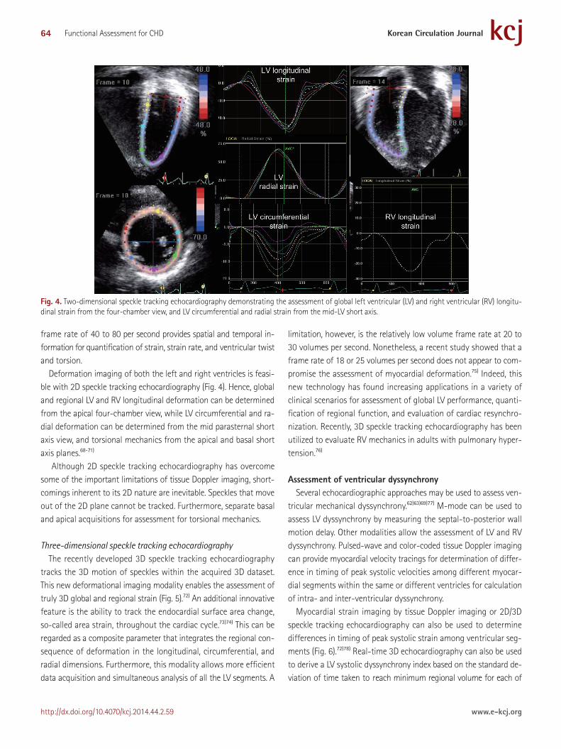
Deformation imaging of both the left and right ventricles is feasi-ble with 2D speckle tracking echocardiography (Fig. 4). Hence, global and regional LV and RV longitudinal deformation can be determined from the apical four-chamber view, while LV circumferential and ra-dial deformation can be determined from the mid parasternal short axis view, and torsional mechanics from the apical and basal short axis planes.68-71)
Although 2D speckle tracking echocardiography has overcome some of the important limitations of tissue Doppler imaging, short-comings inherent to its 2D nature are inevitable. Speckles that move out of the 2D plane cannot be tracked. Furthermore, separate basal and apical acquisitions for assessment for torsional mechanics.
Three-dimensional speckle tracking echocardiographyThe recently developed 3D speckle tracking echocardiography
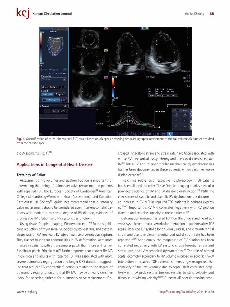
tracks the 3D motion of speckles within the acquired 3D dataset. This new deformational imaging modality enables the assessment of truly 3D global and regional strain (Fig. 5).72) An additional innovative feature is the ability to track the endocardial surface area change, so-called area strain, throughout the cardiac cycle.73)74) This can be regarded as a composite parameter that integrates the regional con-sequence of deformation in the longitudinal, circumferential, and radial dimensions. Furthermore, this modality allows more efficient data acquisition and simultaneous analysis of all the LV segments. A
limitation, however, is the relatively low volume frame rate at 20 to 30 volumes per second. Nonetheless, a recent study showed that a frame rate of 18 or 25 volumes per second does not appear to com-promise the assessment of myocardial deformation.75) Indeed, this new technology has found increasing applications in a variety of clinical scenarios for assessment of global LV performance, quanti-fication of regional function, and evaluation of cardiac resynchro-nization. Recently, 3D speckle tracking echocardiography has been utilized to evaluate RV mechanics in adults with pulmonary hyper-tension.76)
Assessment of ventricular dyssynchronySeveral echocardiographic approaches may be used to assess ven-
tricular mechanical dyssynchrony.62)63)69)77) M-mode can be used to assess LV dyssynchrony by measuring the septal-to-posterior wall motion delay. Other modalities allow the assessment of LV and RV dyssynchrony. Pulsed-wave and color-coded tissue Doppler imaging can provide myocardial velocity tracings for determination of differ-ence in timing of peak systolic velocities among different myocar-dial segments within the same or different ventricles for calculation of intra- and inter-ventricular dyssynchrony.
Myocardial strain imaging by tissue Doppler imaging or 2D/3D speckle tracking echocardiography can also be used to determine differences in timing of peak systolic strain among ventricular seg-ments (Fig. 6).72)78) Real-time 3D echocardiography can also be used to derive a LV systolic dyssynchrony index based on the standard de-viation of time taken to reach minimum regional volume for each of
Fig. 4. Two-dimensional speckle tracking echocardiography demonstrating the assessment of global left ventricular (LV) and right ventricular (RV) longitu-dinal strain from the four-chamber view, and LV circumferential and radial strain from the mid-LV short axis.
65Yiu-fai Cheung
http://dx.doi.org/10.4070/kcj.2014.44.2.59www.e-kcj.org
the LV segments (Fig. 1).79)
Applications in Congenital Heart Disease
Tetralogy of FallotAssessment of RV volumes and ejection fraction is important for
determining the timing of pulmonary valve replacement in patients with repaired TOF. The European Society of Cardiology,2) American College of Cardiology/American Heart Association,1) and Canadian Cardiovascular Society80) guidelines recommend that pulmonary valve replacement should be considered even in asymptomatic pa-tients with moderate-to-severe degree of RV dilation, evidence of progressive RV dilation, and RV systolic dysfunction.
Using tissue Doppler imaging, Weidemann et al.81) found signifi-cant reduction of myocardial velocities, systolic strain, and systolic strain rate of RV free wall, LV lateral wall, and ventricular septum. They further found that abnormalities in RV deformation were more marked in patients with a transannular patch than those with an in-fundibular patch. Frigiola et al.6) further reported that a lower RV IVA in children and adults with repaired TOF was associated with more severe pulmonary regurgitation and longer QRS duration, suggest-ing that reduced RV contractile function is related to the degree of pulmonary regurgitation and that RV IVA may be an early sensitive index for selecting patients for pulmonary valve replacement. De-
creased RV systolic strain and strain rate have been associated with worse RV mechanical dyssynchrony and decreased exercise capac-ity.82) Intra-RV and interventricular mechanical dyssynchrony has further been documented in these patients, which becomes worse during exercise.83)
The clinical relevance of restrictive RV physiology in TOF patients has been alluded to earlier. Tissue Doppler imaging studies have also provided evidence of RV and LV diastolic dysfunction.84) With the coexistence of systolic and diastolic RV dysfunction, the document-ed increase in RV MPI in repaired TOF patients is perhaps expect-ed.85-87) Importantly, RV MPI correlates negatively with RV ejection fraction and exercise capacity in these patients.86)
Deformation imaging has shed light on the understanding of ad-verse systolic ventricular-ventricular interaction in patients after TOF repair. Reduced LV systolic longitudinal, radial, and circumferential strain and diastolic circumferential and radial strain rate has been reported.70)84) Additionally, the magnitude of RV dilation has been correlated negatively with LV systolic circumferential strain and strain rate, and LV mechanical dyssynchrony.70) The role of altered septal geometry secondary to RV volume overload in adverse RV-LV interaction in repaired TOF patients is increasingly recognized. Ec-centricity of the left ventricle due to septal shift correlates nega-tively with LV peak systolic torsion, systolic twisting velocity, and diastolic untwisting velocity.88)89) A recent 3D speckle tracking echo-
Fig. 5. Quantification of three-dimensional (3D) strain based on 3D speckle tracking echocardiographic assessment of the full volume 3D dataset acquired from the cardiac apex.
66 Functional Assessment for CHD
http://dx.doi.org/10.4070/kcj.2014.44.2.59 www.e-kcj.org
cardiographic study showed that reduced septal curvature is relat-ed to impairment of LV 3D systolic strain, mechanical dyssynchro-ny, and reduced ventricular torsion in repaired TOF patients with and without pulmonary valve replacement.90) The new 3D technique has enabled comprehensive on stop-shop evaluation of LV global per-
formance by taking simultaneously into account the different as-pects of deformation.74)90)
Limited data exist on the impact of pulmonary valve replacement on ventricular performance. Acutely after transcatheter pulmonary valve implantation in a cohort of children including those with re-
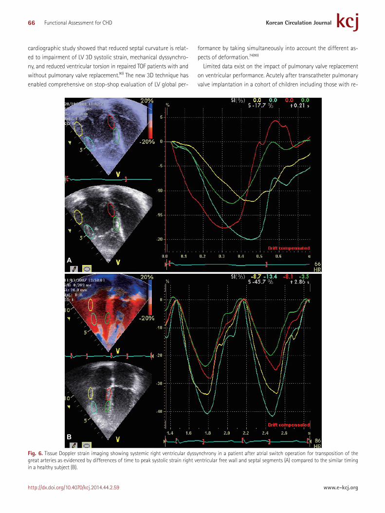
Fig. 6. Tissue Doppler strain imaging showing systemic right ventricular dyssynchrony in a patient after atrial switch operation for transposition of the great arteries as evidenced by differences of time to peak systolic strain right ventricular free wall and septal segments (A) compared to the similar timing in a healthy subject (B).
A
B
67Yiu-fai Cheung
http://dx.doi.org/10.4070/kcj.2014.44.2.59www.e-kcj.org
paired TOF, improvement in RV free wall strain has been noted.91) Transcatheter pulmonary valve replacement in patients with repaired TOF and other congenital heart diseases has also resulted in im-provement of LV circumferential and longitudinal strain and longi-tudinal dyssynchrony.92)
Recently, Diller et al.93) described the potential prognostic value of deformation imaging in repaired TOF patients. In 413 adults patients studied at a mean of 36 years and followed up for a median of 2.9 years, mitral annular plane systolic excursion and LV global longitu-dinal 2D strain were related to the composite endpoint of sudden cardiac death or life-threatening ventricular arrhythmias indepen-dent of QRS duration.
Pulmonary atresia with intact ventricular septumThe understanding of RV mechanics after biventricular repair of
PAIVS is limited. A tissue Doppler echocardiographic study has shown regional systolic and diastolic longitudinal myocardial dys-function involving both the right and left ventricles in adolescent pa-tients after biventricular repair in infancy.94) A subsequent study us-ing 2D speckle tracking imaging confirmed the impairment of RV systolic and diastolic deformation.15) LGE CMR showed evidence of RV myocardial fibrosis. About 80% of these patients have a restric-tive RV physiology, which is related to more severe RV fibrosis and better exercise capacity in the long-term.15)
Transposition of the great arteries post atrial repairThe consistently reported reduction of systemic RV ejection frac-
tion in patients with TGA after atrial repair and the controversy re-garding its interpretation has been discussed previously. Nonethe-less, accumulating functional data based on deformation imaging suggest the possibility of impaired systemic RV mechanics, which may have prognostic implications in these patients.
Tissue Doppler imaging revealed significantly lower regional sys-tolic strain and strain rate of RV free wall, septum, and LV lateral wall in patients after the Senning procedure.95)96) Significant correla-tion was found between regional longitudinal RV systolic strain pa-rameters and RV ejection fraction. Vogel et al.97) validated the use of IVA in assessing systemic RV contractile function. Abnormal wall motion in these patients after atrial repair was associated with re-duced IVA. Furthermore, reduced contractile reserve of the systemic right ventricle as assessed by changes in IVA with dobutamine in-fusion was associated with increased brain natriuretic peptide lev-els.98) Indeed, plasma brain natriuretic peptide has been positively correlated with systemic RV MPI and negatively correlated with sys-temic RV free wall and septal annular myocardial velocities.99)
Chow et al.69) demonstrated the reproducibility and potential use-fulness of 2D speckle tracking echocardiography in assessing sys-
temic RV function. Global systemic RV longitudinal strain and strain rate has been correlated with CMR-derived systemic RV ejection fraction. These strain parameters were also found to correlate with systemic RV IVA and MPI. Altered systemic RV contraction and de-formation pattern has further been unveiled by strain imaging. Us-ing CMR, Pettersen et al.100) found predominant circumferential over longitudinal free wall shortening in the systemic right ventricle as in the normal left ventricle, which is opposite from the findings in the normal subpulmonary right ventricle. However, unlike the nor-mal LV, the systemic RV did not exhibit torsional deformation.
Cardiac resynchronization therapy improves systemic RV function in patients after atrial repair.62)63) Strain imaging has helped define intra-systemic RV and inter-ventricular mechanical delay in these patients. Using tissue Doppler strain imaging, Chow et al.101) reported a prevalence of 32% and 57% for intra-systemic RV dyssynchrony and inter-ventricular dyssynchrony, respectively. Importantly, the in-tra- and inter-ventricular mechanical delays negatively affect the systemic RV ejection fraction and exercise capacity. Although the exact cause is unknown, non-uniform alteration of RV myocardium due to ischaemia and fibrotic process might contribute to asynchro-nous myocardial deformation.22-25)
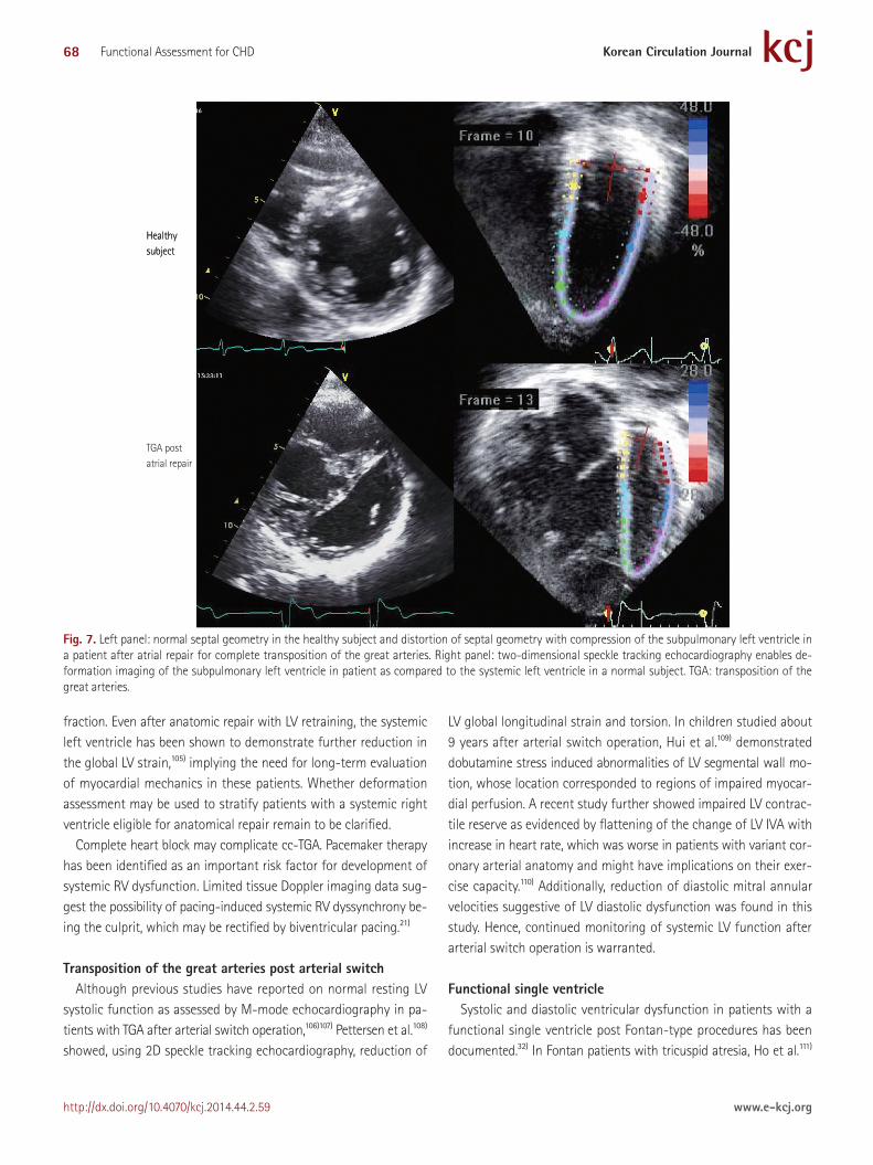
Studies on ventricular-ventricular interaction in patients after atrial repair are limited. Unfavorable diastolic ventricular interaction was suggested by a 2D specking tracking study. In both the systemic right ventricle and subpulmonary left ventricle, early and late dia-stolic strain rates were significantly reduced. Subpulmonary LV ec-centricity index, a reflection of septal shift (Fig. 7), correlated nega-tively with LV early and late diastolic strain rates.102) For systolic interaction, Diller et al.103) reported correlations between systemic RV and subpulmonary LV 2D longitudinal strain and CMR-derived ejection fraction in patients after atrial repair and those with cc-TGA. Furthermore, they103) and others41) showed that systemic RV global longitudinal strain predicts adverse clinical outcomes in these pa-tients in terms of symptomatic progression towards worse func-tional class, development of cardiac arrhythmias, and death.
Congenitally corrected transposition of the great arteriesSimilar to patients after atrial switch operation, patients with cc-
TGA have reduced systemic RV peak systolic strain and strain rate.104) A direct piece of evidence of ventricular-ventricular interaction in this setting can be appreciated in these patients and in those with TGA post atrial switch with failing systemic right ventricle undergo-ing pulmonary arterial banding.19)20) The consequent increase in sub-pulmonary LV load may partially reverse the septal shift and improve systemic RV geometry with reduction in tricuspid regurgitation.
The placement of the pulmonary arterial band may, however, cause acute reduction in global subpulmonary LV strain and ejection
68 Functional Assessment for CHD
http://dx.doi.org/10.4070/kcj.2014.44.2.59 www.e-kcj.org
fraction. Even after anatomic repair with LV retraining, the systemic left ventricle has been shown to demonstrate further reduction in the global LV strain,105) implying the need for long-term evaluation of myocardial mechanics in these patients. Whether deformation assessment may be used to stratify patients with a systemic right ventricle eligible for anatomical repair remain to be clarified.
Complete heart block may complicate cc-TGA. Pacemaker therapy has been identified as an important risk factor for development of systemic RV dysfunction. Limited tissue Doppler imaging data sug-gest the possibility of pacing-induced systemic RV dyssynchrony be-ing the culprit, which may be rectified by biventricular pacing.21)
Transposition of the great arteries post arterial switchAlthough previous studies have reported on normal resting LV
systolic function as assessed by M-mode echocardiography in pa-tients with TGA after arterial switch operation,106)107) Pettersen et al.108) showed, using 2D speckle tracking echocardiography, reduction of
LV global longitudinal strain and torsion. In children studied about 9 years after arterial switch operation, Hui et al.109) demonstrated dobutamine stress induced abnormalities of LV segmental wall mo-tion, whose location corresponded to regions of impaired myocar-dial perfusion. A recent study further showed impaired LV contrac-tile reserve as evidenced by flattening of the change of LV IVA with increase in heart rate, which was worse in patients with variant cor-onary arterial anatomy and might have implications on their exer-cise capacity.110) Additionally, reduction of diastolic mitral annular velocities suggestive of LV diastolic dysfunction was found in this study. Hence, continued monitoring of systemic LV function after arterial switch operation is warranted.
Functional single ventricleSystolic and diastolic ventricular dysfunction in patients with a
functional single ventricle post Fontan-type procedures has been documented.32) In Fontan patients with tricuspid atresia, Ho et al.111)
Fig. 7. Left panel: normal septal geometry in the healthy subject and distortion of septal geometry with compression of the subpulmonary left ventricle in a patient after atrial repair for complete transposition of the great arteries. Right panel: two-dimensional speckle tracking echocardiography enables de-formation imaging of the subpulmonary left ventricle in patient as compared to the systemic left ventricle in a normal subject. TGA: transposition of the great arteries.
HealthysubjectHealthysubject
TGA postatrial repair

69Yiu-fai Cheung
http://dx.doi.org/10.4070/kcj.2014.44.2.59www.e-kcj.org
demonstrated using 2D speckle tracking echocardiography reduc-tion of global systemic LV longitudinal, circumferential, and radial strain. Furthermore, the findings of reduced systolic and diastolic strain rates reflect subclinical systolic and diastolic ventricular dys-function, which can be attributed to chronic volume and pressure overload before the Fontan procedure, and reduced preload, ac-quired wall thickening, uncoordinated ventricular relaxation, and in-crease afterload after the operation. The speckle tracking algorithm as applied to CMR has revealed altered torsional mechanics in pa-tients with functional single ventricles of LV or indeterminate mor-phologies after cavopulmonary connection.112)
Potential differences in single RV and LV function have also been investigated at different stages of univentricular repair by deforma-tion imaging. Kaneko et al.113) showed that single right ventricle had the worst systolic performance in before cavopulmonary anastomo-sis, although parity with single LV function was regained after Fon-tan-type procedures. Indeed, Cheung et al.114) have shown that while functional single ventricles exhibit reduced ventricular contractile reserve as assessed by tissue Doppler-derived myocardial force-fre-quency relationship, no differences in contractile function was found between functional single ventricles with RV and those with LV morphology. Adaption of RV deformation in hypoplastic left heart syndrome with reduction of RV longitudinal to circumferential strain ratio before cavopulmonary anastomosis is associated with better contractile dyssychrony.115)
Limited data suggest dyssynchronous myocardial deformation of functional single ventricles. In children with hypoplastic left heart syndrome, Friedberg et al.116) demonstrated mechanical dyssyn-chrony of the systemic right ventricle using velocity vector imaging based on the principle of 2D speckle tracking. Earlier CMR studies showed regional LV wall motion abnormalities in patients with tri-cuspid atresia after the Fontan procedure.117)118) Based on 3D seg-mental volume assessment of the systemic left ventricle, Ho et al.111) documented a 55% prevalence of mechanical dyssynchrony in Fon-tan patients with tricuspid atresia. This study also demonstrated a positive correlation between severity of mechanical dyssynchrony and calibrated integrated backscatter. This finding together with the reported association between LGE and regional wall motion abnor-mality and dyskinesia in Fontan patients119) suggest that myocardial fibrosis may play a role in the origin of asynchronous myocardial de-formation in function single ventricles.
Caveats
Limitations of the various techniques have to be taken into ac-count when translating these modalities into clinical use. The angle dependency of Doppler-based techniques, the varying load depen-
dency of different parameters, and the relatively low temporal res-olution of current 3D techniques are of particular relevance when assessing cardiac function in congenital heart disease. To facilitate the clinical applications of the novel techniques, establishment of normal references for different parameters in different age groups is necessary.120-122) Discordance in deformation measurements among manufactures has to be resolved.123-125)
Conclusions
Functional assessment of cardiac function in congenital heart dis-eases, in particular RV function and function of a single ventricle, has been facilitated by the introduction of novel echocardiograph-ic techniques that enable the quantification of ventricular volumes independent of ventricular geometry and direct evaluation of myo-cardial global and regional motion and deformation. These new tech-niques show great potential in the comprehensive functional evalu-ation of patients with congenital heart disease at risk of late ventricular dysfunction and cardiac failure. Furthermore, the functional impact of medical, device, and surgical interventions can be ascertained more objectively. The ability to directly interrogate myocardial func-tion may allow early sensitive detection of subclinical myocardial dysfunction, better risk stratification, and timely institution of in-terventions. Large scale longitudinal studies are nonetheless required to substantiate the clinical values of these novel functional param-eters in the management, risk stratification, and prognostication of different congenital heart diseases.
References1. Warnes CA, Williams RG, Bashore TM, et al. ACC/AHA 2008 guidelines
for the management of adults with congenital heart disease: a report of the American College of Cardiology/American Heart Association Task Force on Practice Guidelines (Writing Committee to Develop Guidelines on the Management of Adults With Congenital Heart Dis-ease). Developed in Collaboration With the American Society of Echo-cardiography, Heart Rhythm Society, International Society for Adult Congenital Heart Disease, Society for Cardiovascular Angiography and Interventions, and Society of Thoracic Surgeons. J Am Coll Cardiol 2008;52:e143-263.
2. Baumgartner H, Bonhoeffer P, De Groot NM, et al. ESC Guidelines for the management of grown-up congenital heart disease (new version 2010). Eur Heart J 2010;31:2915-57.
3. Bouzas B, Kilner PJ, Gatzoulis MA. Pulmonary regurgitation: not a be-nign lesion. Eur Heart J 2005;26:433-9.
4. Knauth AL, Gauvreau K, Powell AJ, et al. Ventricular size and function assessed by cardiac MRI predict major adverse clinical outcomes late after tetralogy of Fallot repair. Heart 2008;94:211-6.
5. Redington AN. Determinants and assessment of pulmonary regurgi-tation in tetralogy of Fallot: practice and pitfalls. Cardiol Clin 2006;24:

70 Functional Assessment for CHD
http://dx.doi.org/10.4070/kcj.2014.44.2.59 www.e-kcj.org
631-9, vii.6. Frigiola A, Redington AN, Cullen S, Vogel M. Pulmonary regurgitation
is an important determinant of right ventricular contractile dysfunc-tion in patients with surgically repaired tetralogy of Fallot. Circulation 2004;110(11 Suppl 1):II153-7.
7. Helbing WA, Niezen RA, Le Cessie S, et al. Right ventricular diastolic function in children with pulmonary regurgitation after repair of te-tralogy of Fallot: volumetric evaluation by magnetic resonance veloci-ty mapping. J Am Coll Cardiol 1996;28:1827-35.
8. Davlouros PA, Kilner PJ, Hornung TS, et al. Right ventricular function in adults with repaired tetralogy of Fallot assessed with cardiovascular magnetic resonance imaging: detrimental role of right ventricular outflow aneurysms or akinesia and adverse right-to-left ventricular interaction. J Am Coll Cardiol 2002;40:2044-52.
9. Mueller M, Rentzsch A, Hoetzer K, et al. Assessment of interventricular and right-intraventricular dyssynchrony in patients with surgically re-paired tetralogy of Fallot by two-dimensional speckle tracking. Eur J Echocardiogr 2010;11:786-92.
10. Geva T, Sandweiss BM, Gauvreau K, Lock JE, Powell AJ. Factors associ-ated with impaired clinical status in long-term survivors of tetralogy of Fallot repair evaluated by magnetic resonance imaging. J Am Coll Cardiol 2004;43:1068-74.
11. Wessel HU, Cunningham WJ, Paul MH, Bastanier CK, Muster AJ, Idriss FS. Exercise performance in tetralogy of Fallot after intracardiac repair. J Thorac Cardiovasc Surg 1980;80:582-93.
12. Gatzoulis MA, Balaji S, Webber SA, et al. Risk factors for arrhythmia and sudden cardiac death late after repair of tetralogy of Fallot: a mul-ticentre study. Lancet 2000;356:975-81.
13. Cullen S, Shore D, Redington A. Characterization of right ventricular diastolic performance after complete repair of tetralogy of Fallot. Re-strictive physiology predicts slow postoperative recovery. Circulation 1995;91:1782-9.
14. Gatzoulis MA, Clark AL, Cullen S, Newman CG, Redington AN. Right ventricular diastolic function 15 to 35 years after repair of tetralogy of Fallot. Restrictive physiology predicts superior exercise performance. Circulation 1995;91:1775-81.
15. Liang XC, Lam WW, Cheung EW, Wu AK, Wong SJ, Cheung YF. Restric-tive right ventricular physiology and right ventricular fibrosis as as-sessed by cardiac magnetic resonance and exercise capacity after bi-ventricular repair of pulmonary atresia and intact ventricular septum. Clin Cardiol 2010;33:104-10.
16. Graham TP Jr, Bernard YD, Mellen BG, et al. Long-term outcome in congenitally corrected transposition of the great arteries: a multi-in-stitutional study. J Am Coll Cardiol 2000;36:255-61.
17. Prieto LR, Hordof AJ, Secic M, Rosenbaum MS, Gersony WM. Progres-sive tricuspid valve disease in patients with congenitally corrected transposition of the great arteries. Circulation 1998;98:997-1005.
18. Warnes CA. Transposition of the great arteries. Circulation 2006;114: 2699-709.
19. Metton O, Gaudin R, Ou P, et al. Early prophylactic pulmonary artery banding in isolated congenitally corrected transposition of the great arteries. Eur J Cardiothorac Surg 2010;38:728-34.
20. Winlaw DS, McGuirk SP, Balmer C, et al. Intention-to-treat analysis
of pulmonary artery banding in conditions with a morphological right ventricle in the systemic circulation with a view to anatomic biven-tricular repair. Circulation 2005;111:405-11.
21. Van de Veire NR, Yu CM, Ajmone-Marsan N, et al. Triplane tissue Dop-pler imaging: a novel three-dimensional imaging modality that pre-dicts reverse left ventricular remodelling after cardiac resynchronisa-tion therapy. Heart 2008;94:e9.
22. Lubiszewska B, Gosiewska E, Hoffman P, et al. Myocardial perfusion and function of the systemic right ventricle in patients after atrial switch procedure for complete transposition: long-term follow-up. J Am Coll Cardiol 2000;36:1365-70.
23. Singh TP, Humes RA, Muzik O, et al. Myocardial flow reserve in patients with a systemic right ventricle after atrial switch repair. J Am Coll Cardiol 2001;37:2120-5.
24. Babu-Narayan SV, Goktekin O, Moon JC, et al. Late gadolinium en-hancement cardiovascular magnetic resonance of the systemic right ventricle in adults with previous atrial redirection surgery for trans-position of the great arteries. Circulation 2005;111:2091-8.
25. Plymen CM, Sado DM, Taylor AM, et al. Diffuse myocardial fibrosis in the systemic right ventricle of patients late after Mustard or Senning surgery: an equilibrium contrast cardiovascular magnetic resonance study. Eur Heart J Cardiovasc Imaging 2013;14:963-8.
26. Dos L, Teruel L, Ferreira IJ, et al. Late outcome of Senning and Mustard procedures for correction of transposition of the great arteries. Heart 2005;91:652-6.
27. Wilson NJ, Clarkson PM, Barratt-Boyes BG, et al. Long-term outcome after the mustard repair for simple transposition of the great arteries. 28-year follow-up. J Am Coll Cardiol 1998;32:758-65.
28. Roos-Hesselink JW, Meijboom FJ, Spitaels SE, et al. Decline in ventricu-lar function and clinical condition after Mustard repair for transposi-tion of the great arteries (a prospective study of 22-29 years). Eur Heart J 2004;25:1264-70.
29. Derrick GP, Narang I, White PA, et al. Failure of stroke volume aug-mentation during exercise and dobutamine stress is unrelated to load-independent indexes of right ventricular performance after the Mus-tard operation. Circulation 2000;102(19 Suppl 3):III154-9.
30. Colan SD. Ventricular function in volume overload in ventricular func-tion and blood flow. In: Fogel MA, editor. Congenital Heart Disease. New York: Blackwell Futura;2005. p.217-20.
31. Kelly DT, Spotnitz HM, Beiser GD, Pierce JE, Epstein SE. Effects of chronic right ventricular volume and pressure loading on left ventric-ular performance. Circulation 1971;44:403-12.
32. Gewillig M. The Fontan circulation. Heart 2005;91:839-46.33. Sanchez-Quintana D, Climent V, Ho SY, Anderson RH. Myoarchitecture
and connective tissue in hearts with tricuspid atresia. Heart 1999;81: 182-91.
34. Ho SY, Jackson M, Kilpatrick L, Smith A, Gerlis LM. Fibrous matrix of ventricular myocardium in tricuspid atresia compared with normal heart. A quantitative analysis. Circulation 1996;94:1642-6.
35. Penny DJ, Redington AN. Diastolic ventricular function after the Fon-tan operation. Am J Cardiol 1992;69:974-5.
36. Cheung YF, Penny DJ, Redington AN. Serial assessment of left ven-tricular diastolic function after Fontan procedure. Heart 2000;83:
71Yiu-fai Cheung
http://dx.doi.org/10.4070/kcj.2014.44.2.59www.e-kcj.org
420-4.37. Senzaki H, Masutani S, Kobayashi J, et al. Ventricular afterload and
ventricular work in fontan circulation: comparison with normal two-ventricle circulation and single-ventricle circulation with blalock-taussig shunts. Circulation 2002;105:2885-92.
38. Jiang L, Wiegers SE, Weyman AE. Right ventricle. In: Weyman AE, edi-tor. Principles and Practice of Echocardiography. 2nd ed. Philadelphia: Lea & Febiger;1994. p.901-21.
39. Koestenberger M, Nagel B, Ravekes W, et al. Systolic right ventricular function in pediatric and adolescent patients with tetralogy of Fallot: echocardiography versus magnetic resonance imaging. J Am Soc Echo-cardiogr 2011;24:45-52.
40. Mercer-Rosa L, Parnell A, Forfia PR, Yang W, Goldmuntz E, Kawut SM. Tricuspid annular plane systolic excursion in the assessment of right ventricular function in children and adolescents after repair of tetral-ogy of Fallot. J Am Soc Echocardiogr 2013;26:1322-9.
41. Kalogeropoulos AP, Deka A, Border W, et al. Right ventricular function with standard and speckle-tracking echocardiography and clinical events in adults with D-transposition of the great arteries post atrial switch. J Am Soc Echocardiogr 2012;25:304-12.
42. De Caro E, Bondanza S, Calevo MG, et al. Tricuspid Annular Plane Systolic Excursion for the Assessment of Ventricular Function in Adults Operated on with Mustard Procedure for Complete Transposition of the Great Arteries. Congenit Heart Dis 2013. [Epub ahead of print]
43. Koestenberger M, Ravekes W, Everett AD, et al. Right ventricular func-tion in infants, children and adolescents: reference values of the tri-cuspid annular plane systolic excursion (TAPSE) in 640 healthy patients and calculation of z score values. J Am Soc Echocardiogr 2009;22: 715-9.
44. Lopez L, Colan SD, Frommelt PC, et al. Recommendations for quanti-fication methods during the performance of a pediatric echocardio-gram: a report from the Pediatric Measurements Writing Group of the American Society of Echocardiography Pediatric and Congenital Heart Disease Council. J Am Soc Echocardiogr 2010;23:465-95; quiz 576-7.
45. Hung J, Lang R, Flachskampf F, et al. 3D echocardiography: a review of the current status and future directions. J Am Soc Echocardiogr 2007; 20:213-33.
46. Soriano BD, Hoch M, Ithuralde A, et al. Matrix-array 3-dimensional echocardiographic assessment of volumes, mass, and ejection fraction in young pediatric patients with a functional single ventricle: a com-parison study with cardiac magnetic resonance. Circulation 2008;117: 1842-8.
47. Tei C, Ling LH, Hodge DO, et al. New index of combined systolic and di-astolic myocardial performance: a simple and reproducible measure of cardiac function--a study in normals and dilated cardiomyopathy. J Cardiol 1995;26:357-66.
48. Sutherland GR, Stewart MJ, Groundstroem KW, et al. Color Doppler myocardial imaging: a new technique for the assessment of myocar-dial function. J Am Soc Echocardiogr 1994;7:441-58.
49. Vogel M, Schmidt MR, Kristiansen SB, et al. Validation of myocardial acceleration during isovolumic contraction as a novel noninvasive in-dex of right ventricular contractility: comparison with ventricular pressure-volume relations in an animal model. Circulation 2002;105:
1693-9.50. Hillis GS, Møller JE, Pellikka PA, et al. Noninvasive estimation of left
ventricular filling pressure by E/e’ is a powerful predictor of survival after acute myocardial infarction. J Am Coll Cardiol 2004;43:360-7.
51. Abbas A, Lester S, Moreno FC, Srivathsan K, Fortuin D, Appleton C. Noninvasive assessment of right atrial pressure using Doppler tissue imaging. J Am Soc Echocardiogr 2004;17:1155-60.
52. Tekten T, Onbasili AO, Ceyhan C, Unal S, Discigil B. Novel approach to measure myocardial performance index: pulsed-wave tissue Doppler echocardiography. Echocardiography 2003;20:503-10.
53. Abraham TP, Nishimura RA. Myocardial strain: can we finally measure contractility? J Am Coll Cardiol 2001;37:731-4.
54. Greenberg NL, Firstenberg MS, Castro PL, et al. Doppler-derived myo-cardial systolic strain rate is a strong index of left ventricular contrac-tility. Circulation 2002;105:99-105.
55. Nagueh SF, Appleton CP, Gillebert TC, et al. Recommendations for the evaluation of left ventricular diastolic function by echocardiography. J Am Soc Echocardiogr 2009;22:107-33.
56. Vendelin M, Bovendeerd PHM, Engelbrecht J, Arts T. Optimizing ven-tricular fibers: uniform strain or stress, but not ATP consumption, leads to high efficiency. Am J Physiol Heart Circ Physiol 2002;283: H1072-81.
57. Lorenz CH, Pastorek JS, Bundy JM. Delineation of normal human left ventricular twist throughout systole by tagged cine magnetic reso-nance imaging. J Cardiovasc Magn Reson 2000;2:97-108.
58. Burns AT, La Gerche A, Prior DL, Macisaac AI. Left ventricular untwist-ing is an important determinant of early diastolic function. JACC Car-diovasc Imaging 2009;2:709-16.
59. Rüssel IK, Götte MJ, Bronzwaer JG, Knaapen P, Paulus WJ, van Rossum AC. Left ventricular torsion: an expanding role in the analysis of myo-cardial dysfunction. JACC Cardiovasc Imaging 2009;2:648-55.
60. Young AA. Ventricular torsion: an aid to ejection? JACC Cardiovasc Im-aging 2012;5:282-4.
61. Cheng A, Helm RH, Abraham TP. Pathophysiological mechanisms un-derlying ventricular dyssynchrony. Europace 2009;11 Suppl 5:v10-4.
62. van der Hulst AE, Delgado V, Blom NA, et al. Cardiac resynchronization therapy in paediatric and congenital heart disease patients. Eur Heart J 2011;32:2236-46.
63. Janousek J. Cardiac resynchronisation in congenital heart disease. Heart 2009;95:940-7.
64. Heimdal A, Støylen A, Torp H, Skjaerpe T. Real-time strain rate imaging of the left ventricle by ultrasound. J Am Soc Echocardiogr 1998;11: 1013-9.
65. Urheim S, Edvardsen T, Torp H, Angelsen B, Smiseth OA. Myocardial strain by Doppler echocardiography. Validation of a new method to quantify regional myocardial function. Circulation 20005;102:1158-64.
66. Leitman M, Lysyansky P, Sidenko S, et al. Two-dimensional strain-a novel software for real-time quantitative echocardiographic assess-ment of myocardial function. J Am Soc Echocardiogr 2004;17:1021-9.
67. Amundsen BH, Helle-Valle T, Edvardsen T, et al. Noninvasive myocardial strain measurement by speckle tracking echocardiography: validation against sonomicrometry and tagged magnetic resonance imaging. J Am Coll Cardiol 2006;47:789-93.
72 Functional Assessment for CHD
http://dx.doi.org/10.4070/kcj.2014.44.2.59 www.e-kcj.org
68. Reisner SA, Lysyansky P, Agmon Y, Mutlak D, Lessick J, Friedman Z. Global longitudinal strain: a novel index of left ventricular systolic function. J Am Soc Echocardiogr 2004;17:630-3.
69. Chow PC, Liang XC, Cheung EW, Lam WW, Cheung YF. New two-di-mensional global longitudinal strain and strain rate imaging for as-sessment of systemic right ventricular function. Heart 2008;94:855-9.
70. Cheung EW, Liang XC, Lam WW, Cheung YF. Impact of right ventricular dilation on left ventricular myocardial deformation in patients after surgical repair of tetralogy of fallot. Am J Cardiol 2009;104:1264-70.
71. Notomi Y, Lysyansky P, Setser RM, et al. Measurement of ventricular torsion by two-dimensional ultrasound speckle tracking imaging. J Am Coll Cardiol 2005;45:2034-41.
72. Cheung YF. The role of 3D wall motion tracking in heart failure. Nat Rev Cardiol 2012;9:644-57.
73. Seo Y, Ishizu T, Enomoto Y, Sugimori H, Aonuma K. Endocardial surface area tracking for assessment of regional LV wall deformation with 3D speckle tracking imaging. JACC Cardiovasc Imaging 2011;4:358-65.
74. Li SN, Wong SJ, Cheung YF. Novel area strain based on three-dimen-sional wall motion analysis for assessment of global left ventricular performance after repair of tetralogy of Fallot. J Am Soc Echocardiogr 2011;24:819-25.
75. Yodwut C, Weinert L, Klas B, Lang RM, Mor-Avi V. Effects of frame rate on three-dimensional speckle-tracking-based measurements of myo-cardial deformation. J Am Soc Echocardiogr 2012;25:978-85.
76. Onishi T, Onishi T, Tanaka T, Haberman SC, Champion H, Gorcsan J. Three dimensional speckle tracking strain evaluation of right heart function and hemodynamics in patients with pulmonary hyperten-sion. Circulation 2012;126:A14407.
77. Smiseth OA, Russell K, Skulstad H. The role of echocardiography in quantification of left ventricular dyssynchrony: state of the art and fu-ture directions. Eur Heart J Cardiovasc Imaging 2012;13:61-8.
78. Nesser HJ, Winter S. Speckle tracking in the evaluation of left ventricu-lar dyssynchrony. Echocardiography 2009;26:324-36.
79. Kapetanakis S, Kearney MT, Siva A, Gall N, Cooklin M, Monaghan MJ. Real-time three-dimensional echocardiography: a novel technique to quantify global left ventricular mechanical dyssynchrony. Circulation 2005;112:992-1000.
80. Silversides CK, Salehian O, Oechslin E, et al. Canadian Cardiovascular Society 2009 Consensus Conference on the management of adults with congenital heart disease: complex congenital cardiac lesions. Can J Cardiol 2010;26:e98-117.
81. Weidemann F, Eyskens B, Mertens L, et al. Quantification of regional right and left ventricular function by ultrasonic strain rate and strain indexes after surgical repair of tetralogy of Fallot. Am J Cardiol 2002; 90:133-8.
82. Friedberg MK, Fernandes FP, Roche SL, et al. Relation of right ventricu-lar mechanics to exercise tolerance in children after tetralogy of Fallot repair. Am Heart J 2013;165:551-7.
83. Roche SL, Grosse-Wortmann L, Redington AN, et al. Exercise induces biventricular mechanical dyssynchrony in children with repaired te-tralogy of Fallot. Heart 2010;96:2010-5.
84. Friedberg MK, Fernandes FP, Roche SL, et al. Impaired right and left ventricular diastolic myocardial mechanics and filling in asymptomatic
children and adolescents after repair of tetralogy of Fallot. Eur Heart J Cardiovasc Imaging 2012;13:905-13.
85. Abd El Rahman MY, Abdul-Khaliq H, Vogel M, et al. Value of the new Doppler-derived myocardial performance index for the evaluation of right and left ventricular function following repair of tetralogy of fal-lot. Pediatr Cardiol 2002;23:502-7.
86. Cheung EW, Lam WW, Cheung SC, Cheung YF. Functional implications of the right ventricular myocardial performance index in patients af-ter surgical repair of tetralogy of Fallot. Heart Vessels 2008;23:112-7.
87. Mercer-Rosa L, Yang W, Kutty S, Rychik J, Fogel M, Goldmuntz E. Quantifying pulmonary regurgitation and right ventricular function in surgically repaired tetralogy of Fallot: a comparative analysis of echo-cardiography and magnetic resonance imaging. Circ Cardiovasc Imag-ing 2012;5:637-43.
88. Takayasu H, Takahashi K, Takigiku K, et al. Left ventricular torsion and strain in patients with repaired tetralogy of Fallot assessed by speckle tracking imaging. Echocardiography 2011;28:720-9.
89. Cheung YF, Wong SJ, Liang XC, Cheung EW. Torsional mechanics of the left ventricle in patients after surgical repair of tetralogy of Fallot. Circ J 2011;75:1735-41.
90. Li SN, Yu W, Lai CT, Wong SJ, Cheung YF. Left ventricular mechanics in repaired tetralogy of Fallot with and without pulmonary valve replace-ment: analysis by three-dimensional speckle tracking echocardiogra-phy. PLoS One 2013;8:e78826.
91. Moiduddin N, Asoh K, Slorach C, Benson LN, Friedberg MK. Effect of transcatheter pulmonary valve implantation on short-term right ven-tricular function as determined by two-dimensional speckle tracking strain and strain rate imaging. Am J Cardiol 2009;104:862-7.
92. Harrild DM, Marcus E, Hasan B, et al. Impact of transcatheter pulmo-nary valve replacement on biventricular strain and synchrony assessed by cardiac magnetic resonance feature tracking. Circ Cardiovasc Interv 2013;6:680-7.
93. Diller GP, Kempny A, Liodakis E, et al. Left ventricular longitudinal function predicts life-threatening ventricular arrhythmia and death in adults with repaired tetralogy of fallot. Circulation 2012;125:2440-6.
94. Mi YP, Cheung YF. Assessment of right and left ventricular function by tissue Doppler echocardiography in patients after biventricular repair of pulmonary atresia with intact ventricular septum. Int J Cardiol 2006; 109:329-34.
95. Eyskens B, Weidemann F, Kowalski M, et al. Regional right and left ventricular function after the Senning operation: an ultrasonic study of strain rate and strain. Cardiol Young 2004;14:255-64.
96. Rentzsch A, Abd El Rahman MY, Hui W, et al. Assessment of myocardi-al function of the systemic right ventricle in patients with D-transpo-sition of the great arteries after atrial switch operation by tissue Dop-pler echocardiography. Z Kardiol 2005;94:524-31.
97. Vogel M, Derrick G, White PA, et al. Systemic ventricular function in patients with transposition of the great arteries after atrial repair: a tissue Doppler and conductance catheter study. J Am Coll Cardiol 2004; 43:100-6.
98. Vogt M, Kühn A, Wiese J, Eicken A, Hess J, Vogel M. Reduced contrac-tile reserve of the systemic right ventricle under Dobutamine stress is associated with increased brain natriuretic peptide levels in patients
73Yiu-fai Cheung
http://dx.doi.org/10.4070/kcj.2014.44.2.59www.e-kcj.org
with complete transposition after atrial repair. Eur J Echocardiogr 2009; 10:691-4.
99. Chow PC, Cheung EW, Chong CY, et al. Brain natriuretic peptide as a biomarker of systemic right ventricular function in patients with transposition of great arteries after atrial switch operation. Int J Cardi-ol 2008;127:192-7.
100. Pettersen E, Helle-Valle T, Edvardsen T, et al. Contraction pattern of the systemic right ventricle shift from longitudinal to circumferential shortening and absent global ventricular torsion. J Am Coll Cardiol 2007;49:2450-6.
101. Chow PC, Liang XC, Lam WW, Cheung EW, Wong KT, Cheung YF. Me-chanical right ventricular dyssynchrony in patients after atrial switch operation for transposition of the great arteries. Am J Cardiol 2008; 101:874-81.
102. Chow PC, Liang XC, Cheung YF. Diastolic ventricular interaction in pa-tients after atrial switch for transposition of the great arteries: a speck-le tracking echocardiographic study. Int J Cardiol 2011;152:28-34.
103. Diller GP, Radojevic J, Kempny A, et al. Systemic right ventricular lon-gitudinal strain is reduced in adults with transposition of the great ar-teries, relates to subpulmonary ventricular function, and predicts ad-verse clinical outcome. Am Heart J 2012;163:859-66.
104. Bos JM, Hagler DJ, Silvilairat S, et al. Right ventricular function in as-ymptomatic individuals with a systemic right ventricle. J Am Soc Echo-cardiogr 2006;19:1033-7.
105. Sun HY, Behzadian F, Punn R, Tacy TA. Decremental left ventricular de-formation after pulmonary artery band training and subsequent repair in ventriculoarterial discordance. J Am Soc Echocardiogr 2013;26: 765-74.
106. Losay J, Touchot A, Serraf A, et al. Late outcome after arterial switch operation for transposition of the great arteries. Circulation 2001;104 (12 Suppl 1):I121-6.
107. Wernovsky G, Hougen TJ, Walsh EP, et al. Midterm results after the ar-terial switch operation for transposition of the great arteries with in-tact ventricular septum: clinical, hemodynamic, echocardiographic, and electrophysiologic data. Circulation 1988;77:1333-44.
108. Pettersen E, Fredriksen PM, Urheim S, et al. Ventricular function in pa-tients with transposition of the great arteries operated with arterial switch. Am J Cardiol 2009;104:583-9.
109. Hui L, Chau AK, Leung MP, Chiu CS, Cheung YF. Assessment of left ventricular function long term after arterial switch operation for trans-position of the great arteries by dobutamine stress echocardiography. Heart 2005;91:68-72.
110. Chen RH, Wong SJ, Wong WH, Cheung YF. Left ventricular contractile reserve after arterial switch operation for complete transposition of the great arteries: an exercise echocardiographic study. Eur Heart J Car-diovasc Imaging 2013;14:480-6.
111. Ho PK, Lai CT, Wong SJ, Cheung YF. Three-dimensional mechanical dyssynchrony and myocardial deformation of the left ventricle in pa-tients with tricuspid atresia after Fontan procedure. J Am Soc Echocar-diogr 2012;25:393-400.
112. Truong UT, Li X, Broberg CS, et al. Significance of mechanical altera-
tions in single ventricle patients on twisting and circumferential strain as determined by analysis of strain from gradient cine magnetic reso-nance imaging sequences. Am J Cardiol 2010;105:1465-9.
113. Kaneko S, Khoo NS, Smallhorn JF, Tham EB. Single right ventricles have impaired systolic and diastolic function compared to those of left ventricular morphology. J Am Soc Echocardiogr 2012;25:1222-30.
114. Cheung MM, Smallhorn JF, McCrindle BW, Van Arsdell GS, Redington AN. Non-invasive assessment of ventricular force-frequency relations in the univentricular circulation by tissue Doppler echocardiography: a novel method of assessing myocardial performance in congenital heart disease. Heart 2005;91:1338-42.
115. Khoo NS, Smallhorn JF, Kaneko S, Myers K, Kutty S, Tham EB. Novel in-sights into RV adaptation and function in hypoplastic left heart syn-drome between the first 2 stages of surgical palliation. JACC Cardio-vasc Imaging 2011;4:128-37.
116. Friedberg MK, Silverman NH, Dubin AM, Rosenthal DN. Right ventricu-lar mechanical dyssynchrony in children with hypoplastic left heart syndrome. J Am Soc Echocardiogr 2007;20:1073-9.
117. Akagi T, Benson LN, Williams WG, Freedom RM. Regional ventricular wall motion abnormalities in tricuspid atresia after the Fontan proce-dure. J Am Coll Cardiol 1993;22:1182-8.
118. Fogel MA, Gupta KB, Weinberg PM, Hoffman EA. Regional wall motion and strain analysis across stages of Fontan reconstruction by mag-netic resonance tagging. Am J Physiol 1995;269(3 Pt 2):H1132-52.
119. Rathod RH, Prakash A, Powell AJ, Geva T. Myocardial fibrosis identi-fied by cardiac magnetic resonance late gadolinium enhancement is associated with adverse ventricular mechanics and ventricular tachy-cardia late after Fontan operation. J Am Coll Cardiol 2010;55:1721-8.
120. Lorch SM, Ludomirsky A, Singh GK. Maturational and growth-related changes in left ventricular longitudinal strain and strain rate measured by two-dimensional speckle tracking echocardiography in healthy pe-diatric population. J Am Soc Echocardiogr 2008;21:1207-15.
121. Marcus KA, Mavinkurve-Groothuis AM, Barends M, et al. Reference values for myocardial two-dimensional strain echocardiography in a healthy pediatric and young adult cohort. J Am Soc Echocardiogr 2011; 24:625-36.
122. Marcus KA, Janousek J, Barends ME, Weijers G, de Korte CL, Kapusta L. Synchronicity of systolic deformation in healthy pediatric and young adult subjects: a two-dimensional strain echocardiography study. Am J Physiol Heart Circ Physiol 2012;302:H196-205.
123. Manovel A, Dawson D, Smith B, Nihoyannopoulos P. Assessment of left ventricular function by different speckle-tracking software. Eur J Echocardiogr 2010;11:417-21.
124. Gayat E, Ahmad H, Weinert L, Lang RM, Mor-Avi V. Reproducibility and inter-vendor variability of left ventricular deformation measurements by three-dimensional speckle-tracking echocardiography. J Am Soc Echocardiogr 2011;24:878-85.
125. Yuda S, Sato Y, Mina M. Inter-vendor variability of left ventricular vol-umes and strains determined by three-dimensional speckle tracking echocardiography. Echocardiography 2013. [Epub ahead of print]