Embed Size (px)
Citation preview

LUND UNIVERSITY
PO Box 117221 00 Lund+46 46-222 00 00
Functional and pharmacological characteristics of permeability transition in isolatedhuman heart mitochondria.
Morota, Saori; Manolopoulos, Theodor; Eyjolfsson, Atli; Kimblad, Per Ola; Wierup, Per;Metzsch, Carsten; Blomquist, Sten; Hansson, MagnusPublished in:PLoS ONE
DOI:10.1371/journal.pone.0067747
2013
Link to publication
Citation for published version (APA):Morota, S., Manolopoulos, T., Eyjolfsson, A., Kimblad, P. O., Wierup, P., Metzsch, C., Blomquist, S., & Hansson,M. (2013). Functional and pharmacological characteristics of permeability transition in isolated human heartmitochondria. PLoS ONE, 8(6), [e67747]. https://doi.org/10.1371/journal.pone.0067747
Total number of authors:8
General rightsUnless other specific re-use rights are stated the following general rights apply:Copyright and moral rights for the publications made accessible in the public portal are retained by the authorsand/or other copyright owners and it is a condition of accessing publications that users recognise and abide by thelegal requirements associated with these rights. • Users may download and print one copy of any publication from the public portal for the purpose of private studyor research. • You may not further distribute the material or use it for any profit-making activity or commercial gain • You may freely distribute the URL identifying the publication in the public portal
Read more about Creative commons licenses: https://creativecommons.org/licenses/Take down policyIf you believe that this document breaches copyright please contact us providing details, and we will removeaccess to the work immediately and investigate your claim.

Functional and Pharmacological Characteristics ofPermeability Transition in Isolated Human HeartMitochondriaSaori Morota1, Theodor Manolopoulos2, Atli Eyjolfsson3, Per-Ola Kimblad3, Per Wierup3,
Carsten Metzsch2, Sten Blomquist2, Magnus J. Hansson1,4*
1 Mitochondrial Pathophysiology Unit, Skane University Hospital & Lund University, Lund, Sweden, 2 Department of Cardiothoracic Anesthesiology and Intensive Care,
Skane University Hospital & Lund University, Lund, Sweden, 3 Department of Cardiothoracic Surgery, Skane University Hospital & Lund University, Lund, Sweden,
4 Department of Clinical Physiology, Skane University Hospital & Lund University, Lund, Sweden
Abstract
The objective of the present study was to validate the presence and explore the characteristics of mitochondrialpermeability transition (mPT) in isolated mitochondria from human heart tissue in order to investigate if previous findings inanimal models of cardiac disorders are translatable to human disease. Mitochondria were rapidly isolated from fresh atrialtissue samples obtained from 14 patients undergoing Maze surgery due to atrial fibrillation. Human heart mitochondriaexhibited typical mPT characteristics upon calcium overload such as swelling, evaluated by changes in light scattering,inhibition of respiration and loss of respiratory coupling. Swelling was a morphologically reversible event following transientcalcium challenge. Calcium retention capacity (CRC), a quantitative measure of mPT sensitivity assayed by followingextramitochondrial [Ca2+] and changes in respiration during a continuous calcium infusion, was significantly increased bycyclophilin D (CypD) inhibitors. The thiol-reactive oxidant phenylarsine oxide sensitized mitochondria to calcium-inducedmPT. Release of the pro-apoptotic intermembrane protein cytochrome c was increased after, but not before, calciumdischarge and respiratory inhibition in the CRC assay. From the present study, we conclude that adult viable heartmitochondria have a CypD- and oxidant-regulated mPT. The findings support that inhibition of mPT may be a relevantpharmacological target in human cardiac disease and may underlie the beneficial effect of cyclosporin A in reperfusioninjury.
Citation: Morota S, Manolopoulos T, Eyjolfsson A, Kimblad P-O, Wierup P, et al. (2013) Functional and Pharmacological Characteristics of Permeability Transitionin Isolated Human Heart Mitochondria. PLoS ONE 8(6): e67747. doi:10.1371/journal.pone.0067747
Editor: Mika Jekabsons, University of Mississippi, United States of America
Received February 28, 2013; Accepted May 22, 2013; Published June 28, 2013
Copyright: � 2013 Morota et al. This is an open-access article distributed under the terms of the Creative Commons Attribution License, which permitsunrestricted use, distribution, and reproduction in any medium, provided the original author and source are credited.
Funding: This work was supported by the Swedish Research Council (Reference number 2011-3470), the Royal Physiographic Society, the Foundation of theSwedish National Board of Health and Welfare and the Swedish Society of Medicine. The funders had no role in study design, data collection and analysis, decisionto publish, or preparation of the manuscript.
Competing Interests: M. J. Hansson is a shareholder and receives consultancy fees, and S. Morota is a part time employee of NeuroVive Pharmaceutical ABwhich is active in the field of mitochondrial medicine including development of cyclophilin D inhibitors. This does not alter the authors’ adherence to all the PLOSONE policies on sharing data and materials.
* E-mail: [email protected]
Introduction
The mitochondrial permeability transition (mPT) is considered
to be a major cause of cell death in ischemia-reperfusion injury of
the heart. Opening of the mPT pore is characterized by
uncoupling of oxidative phosphorylation, in vitro swelling of
mitochondria and release of proapoptotic factors such as
cytochrome c (CytC) [1,2]. Pharmacological inhibition or genetic
ablation of the mitochondrial matrix protein cyclophilin D (CypD)
prevents mPT and cardiac injury in animal models of ischemia-
reperfusion injury and heart failure [3–7]. Ischemic precondition-
ing has been proposed to exert its beneficial effect through reduced
mPT activation, although the signaling pathways remain to be
fully elucidated [8–11]. The immunosuppressive agent and CypD
inhibitor cyclosporin A (CsA) has also been shown to limit
myocardial injury in a Phase II clinical trial of patients with acute
myocardial infarction [12,13]. CsA and other cyclophilin inhib-
itors are however not specific to CypD. Cyclophilins are found
widely distributed in eukaryotes in all the major compartments of
the cell, and the majority of the 17 identified human cyclophilins
have cytoplasmic or nuclear localization [14]. The complex of
cytoplasmic cyclophilin A and CsA inhibits the phosphatase
calcineurin, which mediates the immunosuppressive activity of
CsA [15].
An important step in translating experimental findings to
clinical use and to increase the strength of the biologic rationale for
treatment is to verify the pharmacological target in human tissue.
Previously, mPT has been implicated indirectly in human atrial
heart tissue by demonstrations of improved atrial trabeculae and
myocyte viability following simulated ischemia in vitro and by
prolonged time to depolarization following tetramethylrhodamine
methyl ester (TMRM)-induced oxidative stress by cyclophilin
inhibitors [16,17]. Repetitive calcium loads has also been shown to
cause respiratory inhibition in permeabilized human atrial
myofibres [18]. Even though cellular assays posses several
strengths, the specificity may be lower compared to studies in
isolated mitochondria with increased risk of confounding variables
both in regard to the studied phenomena and the pharmacological
PLOS ONE | www.plosone.org 1 June 2013 | Volume 8 | Issue 6 | e67747
effects. There is no previous study exploring the specific
characteristics of permeability transition or the direct effect and
potencies of cyclophilin inhibitors in isolated human heart
mitochondria.
The objective of the present study was to confirm the presence
of mPT in the human heart by assessing characteristics of mPT in
freshly isolated human heart mitochondria. Further, the aim was
to explore the pharmacological modulation of mPT by CypD
inhibitors in order to evaluate whether mPT constitutes a relevant
target for cardioprotection in pathologies of the heart where this
disease mechanism has been implicated in animal models. The
study demonstrates that viable mitochondria from human cardiac
tissue undergo calcium- and oxidant-sensitive mPT similar to what
has previously been described in non-human mitochondria and
human brain and liver mitochondria [19,20], and that its
activation is dose-dependently inhibited by CypD ligands.
Materials and Methods
Material To obtain fresh human heart tissue for functional mitochondrial
analyses, left atrial appendage tissue samples were collected from
14 patients undergoing Maze surgery due to atrial fibrillation at
the Skane University Hospital, Lund, Sweden. For further patient
characteristics, see Table 1. In Maze surgery, incisions are
performed in the atria to disrupt abnormal electrical impulses
and the left atrial appendage is removed. Tissue samples which
would otherwise have been discarded, 0.3–4.3 g, were transferred
into ice-cold Buffer A (100 mM KCl, 50 mM MOPS, 5 mM
MgCl2, 1 mM EGTA, 1 mM ATP(K), pH 7.4).
The study procedures were approved by the regional ethical
review board of Lund, Sweden (permit number 2009/507) and
comply with the World Medical Association Declaration of
Helsinki - Ethical Principles for Medical Research Involving
Human Subjects. Samples were obtained after written informed
consent was acquired.
Isolation of Heart Mitochondria Heart tissue samples were rapidly prepared for mitochondrial
isolation. Non-muscle tissue was removed and remaining muscle
was finely chopped in ice-cold Buffer A with BSA (2 mg/ml)
[21,22]. After rinsing off BSA by adding excess ice-cold Buffer A,
tissues were transferred to a Potter-Elvehjem homogenizer and
trypsinized (10 mg trypsin/6 ml ice-cold Buffer A) for 30 minutes
on ice. BSA, 12 mg, was added to stop the trypsinization, and the
tissue was homogenized gently with a Teflon pestle. Following 10
minutes centrifugation at 600 g, supernatant was collected and
centrifuged for 5 minutes at 3000 g. The pellet was suspended in
8 ml of 26% Percoll solution in Buffer B (100 mM KCl, 50 mM
MOPS, 0.5 mM EGTA, pH 7.4) and centrifuged for 7 minutes at
30000 g to remove contaminating membranes [23]. The pellet was
resuspended in 8 ml of Buffer B with BSA (0.2 mg/ml Buffer B)
and centrifuged for 3 minutes at 7000 g to wash away Percoll. A
second washing step in Buffer B without BSA was performed with
centrifugation for 3 minutes at 3000 g. The mitochondrial pellet
was finally resuspended in Buffer B. Protein content was measured
using Bradford analysis after which 1 mg/ml BSA was added. All
centrifugations were performed at 4uC.
Mitochondrial RespirationOxygen consumption of mitochondria was analyzed using an
Oxygraph-2k with a Titration-Injection microPump TIP-2k
(Oroboros instruments, Innsbruck Austria). Experiments were
performed at 37uC. Mitochondria, 40 mg, were suspended in 2 ml
respiration medium (MIR05) containing 110 mM sucrose, 20 mM
HEPES, 20 mM taurine, 60 mM K -lactobionate, 3 mM MgCl2,
10 mM KH2PO4, 0.5 mM EGTA, 1 g/l BSA and 5 mM of the
NADH-linked respiratory substrates malate and glutamate,
pH 7.1. Mitochondrial suspensions were supplemented with
0.25 mM ADP to induce state 3 respiration and to evaluate
respiratory coupling of the isolated mitochondrial preparations.
State 4 respiration was measured after the ADP was consumed. In
another experimental group, mitochondria were exposed to 1 mM
CaCl2 to evaluate the effect of calcium-induced mPT on
respiratory function. Then, 1 mM ADP was added in both
experimental groups followed by the ATP synthase inhibitor
oligomycin, 1 mg/ml, to induce State 4oligo. A stepwise titration of
the protonophore CCCP (500 nM/addition) was performed to
evaluate maximal capacity of the electron transport system (ETS)
independent of the phosphorylation system.
De-energized SwellingA Perkin-Elmer Luminescence Spectrometer LS-50B (Emery-
ville, CA, USA) with a temperature controlled cuvette holder was
used for all fluorescence and light scattering experiments. De-
energized swelling experiments were performed at 28uC in a
150 mM KCl-based buffer containing 0.5 mM rotenone, 0.2 mg/
ml antimycin A, 2 mM calcium ionophore A23187, 0.5 mM PPi.
Mitochondria were pre-treated for two minutes with 10, 100,
1000 nM CsA or 10, 100, 1000 nM of the non-immunosuppres-
sive cyclosporin analog MeAla3EtVal4-cyclosporin (NI-Cs, also
known as alisporivir, UNIL025, Debio-025 or DEB025). The
mitochondrial suspensions were then exposed to 300 mM CaCl2 to
induce swelling. The extent of swelling was calculated by dividing
the calcium-induced decrease in light scattering during the first
minute following CaCl2 addition with that induced by the
ionophore alamethicin (10 mg/ml) [24].
Reversible SwellingThe reversibility of calcium-induced swelling was evaluated in
respiring mitochondria at 37uC. Experiments were performed in
buffer containing 125 mM KCl, 20 mM Trizma base, 2 mM Pi
(K), 1 mM MgCl2, 1 mM EGTA and 5 mM of the NADH-linked
respiratory substrates malate and glutamate, pH 7.1. Following 2
minutes exposure of mitochondria to 300 mM CaCl2 to induce
Table 1. Patient characteristics.
Age, median (range) 71 (55–81) years
Sex Male 11 (79%)
Female 3 (21%)
Previous AMIa 6 (43%)
Diabetes mellitus 3 (21%)
Medication Nitroglycerin 1 (7%)
ACEb inhibitors 10 (71%)
Aspirin 5 (36%)
Beta-blocker 10 (71%)
Statin 12 (86%)
Calcium channel blocker 6 (43%)
Digoxin 2 (14%)
aAMI = acute myocardial infarction, bACE = Angiotensin-converting enzyme.doi:10.1371/journal.pone.0067747.t001
Permeability Transition in the Human Heart
PLOS ONE | www.plosone.org 2 June 2013 | Volume 8 | Issue 6 | e67747
Ethics Statement
swelling, 0.5 mM EGTA was added to chelate the CaCl2. A
second exposure of CaCl2, using 400 mM, was performed
following 11 minutes of recovery. Experiments were terminated
by adding 10 mg/ml alamethicin, and carried out with or without
1 mM CsA.
Calcium Retention Capacity (CRC)Mitochondrial CRC was evaluated using both measurements of
calcium fluxes and changes in respiration during a continuous
CaCl2 infusion using the luminescence spectrometer and oxygraph
described above. Mitochondrial Ca2+ uptake and release were
monitored by following the excitation ratio of the extramitochon-
drial calcium-sensitive fluorescent probe Fura 6F (250 nM, Ex.
340/380 nm, Em. 509 nm). Release of sequestered calcium or
initiation of respiratory inhibition were attributed to activation of
mPT [25]. Mitochondria, 40 mg, were suspended in 2 ml buffer
containing 125 mM KCl, 20 mM Trizma base, 2 mM Pi (K),
1 mM MgCl2, 1 mM EGTA, 200 mM ATP, 10 mM BSA, 5 mM of
malate and glutamate, pH 7.1. At start of experiment, 1 mg/ml
oligomycin, 50 mM ADP and then 1 mM CsA, 1 mM NI-Cs, 1 mM
of the vicinal thiol reagent phenylarsine oxide (PhArs) or vehicle
(ethanol) was added. The suspensions were infused with 0.2 mmol
CaCl2Nmin–1Nmg–1. CRC was calculated as the amount of infused
calcium from the start of infusion until start of maximal calcium
release or start of the rapid phase of respiratory inhibition.
Cytochrome C ReleaseThe extent of cytochrome c (CytC) release was evaluated in the
CRC experimental setup described above. Samples were prepared
before and during calcium infusion as well as after induction of
respiratory inhibition. Samples were also collected following
incubation of mitochondria with 10 mg/ml alamethicin. An
ELISA kit for detection of human CytC (QuantikineH, R&D
Systems) was employed to measure the extent of its release as
described previously [26].
Electron MicrographsMitochondrial morphology was evaluated in the CRC and
reversible swelling experimental setups as described above.
Samples were prepared before or during calcium infusion and
after induction of respiratory inhibition in the CRC assay. In the
reversible swelling experiments, samples were collected before and
after the first CaCl2 addition, following the light scattering
recovery after EGTA chelation of CaCl2, after second CaCl2exposure and following addition of 10 mg/ml alamethicin. The
suspensions were rapidly chilled and centrifuged in an Eppendorf
microcentrifuge, 12000 g, for 2 minutes. Samples were fixed in a
solution containing 0.1 M Sorensen buffer, 1.5% Paraformalde-
hyde and 1.5% Glutaraldehyde over night, and further processed
as described previously [27].
Statistical AnalysesAll average results are presented as mean 6 SD and were,
unless otherwise noted, evaluated using student’s t-tests or for
multiple groups one-way ANOVA followed by Dunnett’s multiple
comparison post hoc test using GraphPad Prism v5.0 software.
Differences were considered significant where P,0.05.
Results
Functional Integrity of Isolated MitochondriaThe mitochondria isolated from fresh atrial heart tissue
(isolation yield 376.16205.2 mg mitochondria/g tissue) displayed
good coupling of oxidation to ATP production. The state 3 and
state 4 respiratory rates were 9157.162430.4 and
994.96249.3 pmol O2Ns–1Nmg–1, respectively, with a respiratory
control ratio (RCR) of 9.2261.15 (Fig. 1). State 4 respiration
following addition of the ATP synthase inhibitor oligomycin (State
4oligo) was not different from state 4 respiration without oligomycin
(1054.5654.5 pmol O2Ns–1Nmg–1, p = 0.66), indicating that there
was no contaminating ATPase activity, e.g. disrupted mitochon-
dria, in the preparations.
Calcium–induced Alterations of MitochondrialRespiration
The coupling of oxidative phosphorylation was virtually lost in
mitochondria following a calcium exposure. There was no
respiratory stimulation upon ADP addition and the RCRO (State
3/State 4oligo) decreased from 8.6462.65 in control samples to
1.1360.28 in mitochondria exposed to calcium (Fig. 1). A
permeable inner membrane in mitochondria oxidizing complex
1-linked substrates will besides loss of proton motive force also lead
to respiratory inhibition due to dilution of the NAD(H) pool
[25,26], and a disrupted outer membrane following mitochondrial
swelling may cause respiratory inhibition due to loss of CytC [28].
Upon calcium addition, there was a transient increase in
respiration followed by a decrease in all respiratory rates. Titration
of the protonophore CCCP to induce maximal non-phosphory-
lating respiration likewise was without stimulatory effect in
mitochondria exposed to calcium (Fig. 1A–B).
Mitochondrial Calcium RetentionHeart mitochondria exposed to a continuous calcium challenge
buffered the infused calcium resulting in a steady state extrami-
tochondrial Ca2+ concentration. The latter lasted until a threshold
where retained calcium was released and extramitochondrial Ca2+
concentration was increased (Fig. 2A). The same type of
experiment performed during measurement of oxygen consump-
tion demonstrated a slight increase in respiration during calcium
infusion followed by a rapid phase of respiratory inhibition, which
defined the limit of mitochondrial CRC (Fig. 2B). The cyclophilin
inhibitor NI-Cs (alisporivir) significantly increased CRC whereas
the oxidant PhArs significantly decreased CRC (Fig. 2C). CsA was
evaluated in the CRC assay using measurement of extramito-
chondrial Ca2+ concentration (Fig. 2A). Due to a high degree of
variation in this set of experiments, the effect of CsA was only
significant using paired analysis, i.e. when the experiments with
CsA were compared to their respective controls (p = 0.034 using
paired t-test, n = 4). CytC release from mitochondria was
increased following the rapid phase of respiratory inhibition but
not during the calcium infusion before (Fig. 2D). Electron
micrographs indicated a dramatic alteration of morphological
appearance following calcium-induced respiratory inhibition
showing decreased cristae and expanded matrices (Fig. 2E–G).
Reversible SwellingRespiring mitochondria exposed to a short-lasting bolus load of
calcium demonstrated a decrease in light scattering, which was
reversed following chelation of calcium by EGTA (Fig. 3A).
Electron micrographs prepared during different time points in the
experiment showed a transition from condensed to less condensed
cristae following calcium exposure, consistent with the light
scattering changes (Fig. 3B–C). Corresponding to the increase in
light scattering following EGTA chelation of calcium, the
mitochondrial cristae appeared hypercondensed (Fig. 3D). A
second calcium exposure as well as subsequent addition of
Permeability Transition in the Human Heart
PLOS ONE | www.plosone.org 3 June 2013 | Volume 8 | Issue 6 | e67747

alamethicin induced decreased light scattering and appearance of
disrupted cristae (Fig. 3E–F).
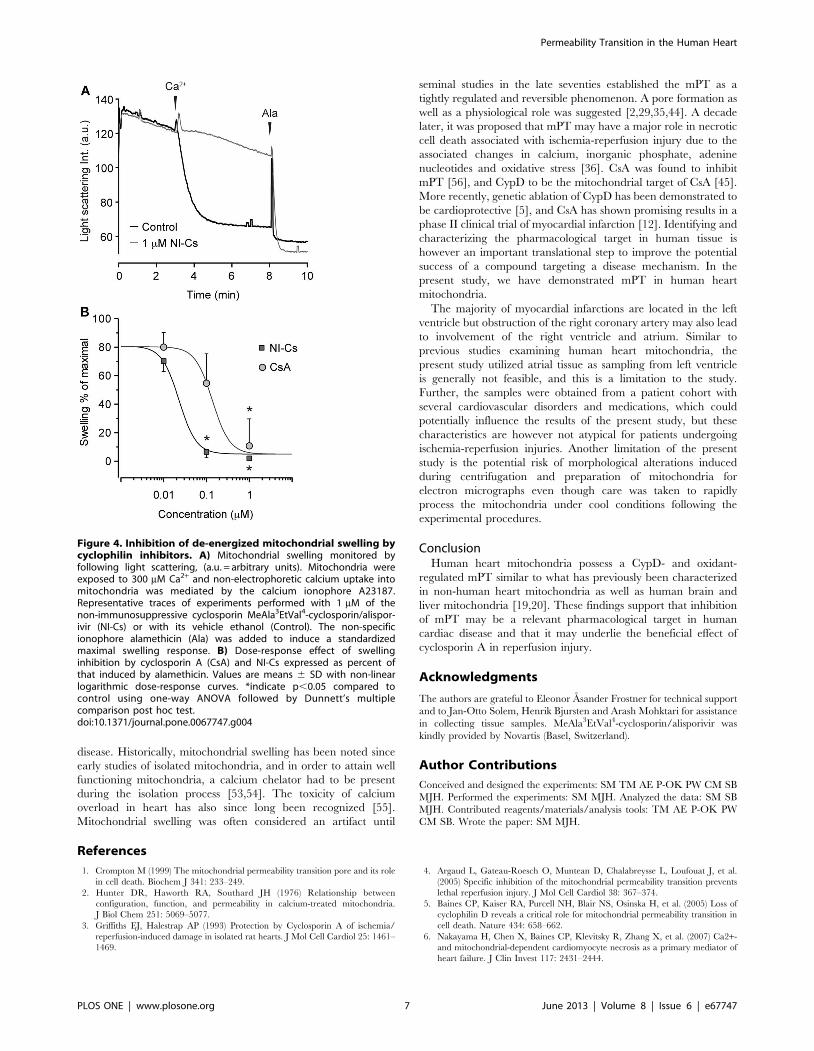
Potency of Cyclophilin InhibitorsThe potencies of the cyclophilin inhibitors CsA and NI-Cs to
reduce calcium-induced swelling were compared under de-
energized conditions (Fig. 4). Both compounds significantly
inhibited swelling at 1 mM and NI-Cs also at 100 nM. The half
maximal effective concentration (EC50) values were 138 nM for
CsA and 23 nM for NI-Cs under the conditions used.
Discussion
In the present study, we demonstrate that viable mitochondria
from human cardiac tissue undergo calcium- and oxidant-sensitive
mPT, which is morphologically reversible following a transient
calcium insult. We also demonstrate that mPT activation in
human heart mitochondria is inhibited by CypD ligands.
Indications of mPT have previously been shown in human atrial
myocytes and permeabilized muscle fibres [16–18]. Here, we used
isolated mitochondria in order to more specifically evaluate mPT
characteristics in the human heart.
Opening of the mPT pore allows passage of solutes with
molecular weight below approximately 1500 Da [29,30]. Respi-
ratory uncoupling due to loss of proton motive force and
respiratory inhibition due to loss of e.g. NAD(H) are more specific
indications for mPT compared to e.g. changes in membrane
potential probes in cell cultures. Although depolarization is a
direct consequence of mPT pore opening, depolarization may not
necessarily be caused by mPT. Further, a potential confounder
when evaluating CsA effects and mitochondrial membrane
potential is that membrane potential dyes such as TMRM as well
as CsA are both substrates of the multidrug-resistance pump
(MDR)/P-glycoprotein which may affect cellular loading and
extrusion of the dyes [31]. The CRC assay is a sensitive and
quantitative technique for assessing mitochondrial susceptibility to
mPT [32]. The mPT pore is regulated by several endogenous
factors such as redox status, adenine nucleotides, membrane
potential and pH. Whether these factors will increase or decrease
mPT activation in mitochondria actively buffering calcium will
however also depend on their effects on intramitochondrial Ca2+
concentration e.g. by affecting calcium phosphate complex
formation [25,32,33]. A potential pitfall when evaluating putative
inhibitors of mPT using swelling or CRC assays is if the compound
rather induces respiratory inhibition with reduced driving force for
calcium uptake rather than direct modulation of mPT [20,34]. By
including simultaneous measurement of oxygen consumption and
evaluation of the permeability of NAD(H), the specificity of e.g. the
CRC assay for mPT is increased [25]. The findings of the present
study with respiratory uncoupling and inhibition following calcium
overload in combination with a PhArs-induced reduction and a
CsA- and NI-Cs-induced increase in CRC demonstrate a specific
activation of mPT in human heart mitochondria that is oxidant-
and CypD-modulated. Whereas there are several cyclophilins
present in the cell, there is no other target than CypD for the
cyclosphilin inhibitors in mitochondria [14].
The increased permeability of mitochondria in vitro is revers-
ible when mPT-inducing factors are removed [35,36]. It has also
been demonstrated that swelling is a morphologically reversible
event if calcium is chelated following mPT activation [24,37]. In
the present study, we find evidence that a transient mPT can be
induced in human heart mitochondria, as assayed by reversible
light scattering decrease and reversible alteration of cristae
appearance on electron micrographs, following a short-term
calcium exposure. However, compared to the alteration following
the transient calcium insult, the disruption of matrix cristae
appeared more severe after a second calcium exposure (Fig. 3) or
Figure 1. Respiration of isolated heart mitochondria with andwithout exposure to calcium. A) Representative traces of changesin respiration rates and B) decrease of oxygen concentration in theclosed chambers. In black traces (1), 0.25 mM ADP was added to induceState 3 respiration. After the initial ADP was consumed (State 4respiration) a second addition of ADP, 1 mM, was added followed bythe ATP synthase inhibitor oligomycin (Oligo) to induce State 4oligo
respiration. Finally, the protonophore CCCP was titrated, 0.5 mM peraddition, to induce maximal non-phosphorylation-dependent respira-tion. In gray traces (2), the initial ADP addition was replaced by 1 mMCaCl2. C) Calculated respiratory control ratios (RCR) of mitochondriawith or without exposure to CaCl2. RCR (State 3/State 4) is depicted forcontrol mitochondria and RCRO (State 3/State 4oligo) for bothexperimental groups. Values are means 6 SD, n = 4. *indicate p,0.05using student’s t-test.doi:10.1371/journal.pone.0067747.g001
Permeability Transition in the Human Heart
PLOS ONE | www.plosone.org 4 June 2013 | Volume 8 | Issue 6 | e67747
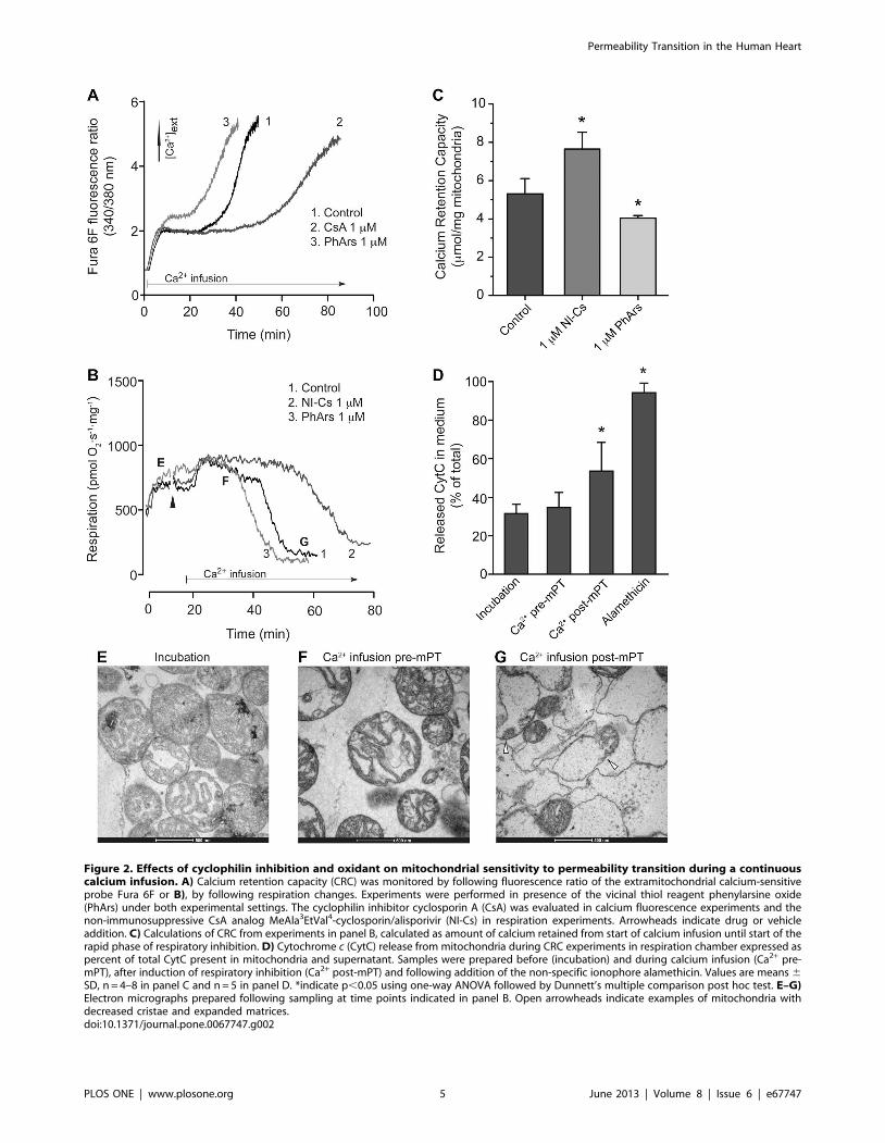
Figure 2. Effects of cyclophilin inhibition and oxidant on mitochondrial sensitivity to permeability transition during a continuouscalcium infusion. A) Calcium retention capacity (CRC) was monitored by following fluorescence ratio of the extramitochondrial calcium-sensitiveprobe Fura 6F or B), by following respiration changes. Experiments were performed in presence of the vicinal thiol reagent phenylarsine oxide(PhArs) under both experimental settings. The cyclophilin inhibitor cyclosporin A (CsA) was evaluated in calcium fluorescence experiments and thenon-immunosuppressive CsA analog MeAla3EtVal4-cyclosporin/alisporivir (NI-Cs) in respiration experiments. Arrowheads indicate drug or vehicleaddition. C) Calculations of CRC from experiments in panel B, calculated as amount of calcium retained from start of calcium infusion until start of therapid phase of respiratory inhibition. D) Cytochrome c (CytC) release from mitochondria during CRC experiments in respiration chamber expressed aspercent of total CytC present in mitochondria and supernatant. Samples were prepared before (incubation) and during calcium infusion (Ca2+ pre-mPT), after induction of respiratory inhibition (Ca2+ post-mPT) and following addition of the non-specific ionophore alamethicin. Values are means 6SD, n = 4–8 in panel C and n = 5 in panel D. *indicate p,0.05 using one-way ANOVA followed by Dunnett’s multiple comparison post hoc test. E–G)Electron micrographs prepared following sampling at time points indicated in panel B. Open arrowheads indicate examples of mitochondria withdecreased cristae and expanded matrices.doi:10.1371/journal.pone.0067747.g002
Permeability Transition in the Human Heart
PLOS ONE | www.plosone.org 5 June 2013 | Volume 8 | Issue 6 | e67747

following mPT activation in the CRC experiments (Fig. 2),
possibly due to the severity and duration of the calcium insult. A
transient and reversible mPT has both been suggested to represent
a physiological calcium release mechanism of mitochondria [38],
and to underlie mitochondrial remodeling and a trigger of cell
death following excitotoxicity [39,40]. Transient mPT has also
been proposed to mediate preconditioning in cardiac ischemia
since cyclophilin inhibitors and genetic ablation of CypD were
found to inhibit the protective effects of preconditioning [41,42].
On the other hand, entrapment of 2-deoxyglucose was not
detected following preconditioning only, whereas preconditioning
inhibited entrapment of 2-deoxyglucose and improved mPT pore
closure following ischemia [43]. Transient mPT thus seems to be
activated during ischemia-reperfusion injury but it is more unclear
whether it also plays a role in mediating ischemic preconditioning.
In order to further evaluate the pharmacological modulation of
mPT in human heart mitochondria, the potencies of the CypD
inhibitors CsA and NI-Cs were compared in a de-energized model
of mitochondrial swelling. The absence of mitochondrial sub-
strates and inhibition of the respiratory chain prevent the
formation of a membrane potential and hence calcium uptake
into the matrix, and by addition of a calcium ionophore,
equilibration of calcium ions across the inner mitochondrial
membrane is facilitated. Although less physiological and quanti-
tative compared to the CRC assay, the de-energized model offers a
specific and dose-dependent evaluation of regulation and phar-
macological inhibition of the mPT pore components without
interference from possible confounding effects on mitochondrial
respiration, membrane potential and electrophoretic calcium
uptake as well as calcium-phosphate complex formation [44–48].
The EC50 values of CsA and NI-Cs inhibition of mPT in de-
energized mitochondria were somewhat higher in the present
study (138 and 23 nM, respectively) compared to what we have
previously demonstrated in rodent brain mitochondria using
similar experimental conditions (49 and 5.5 nM, respectively) [49].
One possible explanation for the difference is the relative severity
of the calcium insult which was higher in the present study. The
relative difference in potency between CsA and NI-Cs was
however similar.
The amount of calcium sequestered before induction of mPT in
the CRC assay was somewhat higher in human heart mitochon-
dria compared to what we have previously demonstrated in
human brain and liver mitochondria using similar experimental
conditions, 5.360.8 mmol/mg in heart versus 2.3460.5 and
1.060.5 in human brain and liver mitochondria, respectively
[19]. Rodent mitochondria display comparable tissue differences
with a somewhat higher CRC in heart mitochondria compared to
brain mitochondria and substantially higher CRC compared to
liver mitochondria (unpublished observations and [20,25], respec-
tively). This indicates that under the conditions used, human heart
mitochondria are more resistant to calcium-induced mPT than
human brain and liver mitochondria. In contrast, studies exposing
isolated mitochondria to bolus loads of calcium without exoge-
nously added adenine nucleotides have demonstrated rodent heart
mitochondria to be more sensitive to calcium-induced mPT
compared to liver and brain mitochondria [50]. Such differences
may be caused by different endogenous content of inhibitory
factors of the mPT pore such as adenine nucleotides [51].
However, the comparison of mitochondria from different tissues
has to be taken with some care as isolation of heart, liver and brain
mitochondria require separate procedures, with possible different
influence on both protective and sensitizing factors on mPT.
Several promising drugs in preclinical models have failed to
translate into effective clinical use, which emphasizes the need to
better validate the molecular targets chosen for drug discovery and
development [52]. The mPT has been extensively characterized in
animal models and may prove to be an important pathophysio-
logical factor and pharmacological target in human cardiac
Figure 3. Light scattering and electron micrographs of mitochondria exposed to transient calcium insult. A) Light scattering ofrespiring mitochondria exposed to a transient calcium insult. Experiments were performed with 1 mM cyclosporin A (CsA, n = 2) or its vehicle ethanol(EtOH, Control, n = 5). A decrease in light scattering is attributed to swelling of mitochondria whereas an increase reflects reversal of swelling andaccumulation of calcium-phosphate complexes [32], (a.u. = arbitrary units). Where indicated by arrowheads, mitochondria were exposed to 300 mMCaCl2 to induce swelling, 0.5 mM EGTA to chelate the CaCl2, a second addition of 400 mM CaCl2, and finally 10 mg/ml alamethicin. Electronmicrographs were prepared at indicated time-points for control experiments as follows; B) during incubation, C) after initial CaCl2 addition, D)subsequent to EGTA, E) following second CaCl2 addition, and F) after alamethicin exposure. Arrows indicate mitochondria with condensed cristae.Open arrowheads indicate mitochondria with less condensed or disrupted cristae.doi:10.1371/journal.pone.0067747.g003
Permeability Transition in the Human Heart
PLOS ONE | www.plosone.org 6 June 2013 | Volume 8 | Issue 6 | e67747
disease. Historically, mitochondrial swelling has been noted since
early studies of isolated mitochondria, and in order to attain well
functioning mitochondria, a calcium chelator had to be present
during the isolation process [53,54]. The toxicity of calcium
overload in heart has also since long been recognized [55].
Mitochondrial swelling was often considered an artifact until
seminal studies in the late seventies established the mPT as a
tightly regulated and reversible phenomenon. A pore formation as
well as a physiological role was suggested [2,29,35,44]. A decade
later, it was proposed that mPT may have a major role in necrotic
cell death associated with ischemia-reperfusion injury due to the
associated changes in calcium, inorganic phosphate, adenine
nucleotides and oxidative stress [36]. CsA was found to inhibit
mPT [56], and CypD to be the mitochondrial target of CsA [45].
More recently, genetic ablation of CypD has been demonstrated to
be cardioprotective [5], and CsA has shown promising results in a
phase II clinical trial of myocardial infarction [12]. Identifying and
characterizing the pharmacological target in human tissue is
however an important translational step to improve the potential
success of a compound targeting a disease mechanism. In the
present study, we have demonstrated mPT in human heart
mitochondria.
The majority of myocardial infarctions are located in the left
ventricle but obstruction of the right coronary artery may also lead
to involvement of the right ventricle and atrium. Similar to
previous studies examining human heart mitochondria, the
present study utilized atrial tissue as sampling from left ventricle
is generally not feasible, and this is a limitation to the study.
Further, the samples were obtained from a patient cohort with
several cardiovascular disorders and medications, which could
potentially influence the results of the present study, but these
characteristics are however not atypical for patients undergoing
ischemia-reperfusion injuries. Another limitation of the present
study is the potential risk of morphological alterations induced
during centrifugation and preparation of mitochondria for
electron micrographs even though care was taken to rapidly
process the mitochondria under cool conditions following the
experimental procedures.
ConclusionHuman heart mitochondria possess a CypD- and oxidant-
regulated mPT similar to what has previously been characterized
in non-human heart mitochondria as well as human brain and
liver mitochondria [19,20]. These findings support that inhibition
of mPT may be a relevant pharmacological target in human
cardiac disease and that it may underlie the beneficial effect of
cyclosporin A in reperfusion injury.
Acknowledgments
The authors are grateful to Eleonor Asander Frostner for technical support
and to Jan-Otto Solem, Henrik Bjursten and Arash Mohktari for assistance
in collecting tissue samples. MeAla3EtVal4-cyclosporin/alisporivir was
kindly provided by Novartis (Basel, Switzerland).
Author Contributions
Conceived and designed the experiments: SM TM AE P-OK PW CM SB
MJH. Performed the experiments: SM MJH. Analyzed the data: SM SB
MJH. Contributed reagents/materials/analysis tools: TM AE P-OK PW
CM SB. Wrote the paper: SM MJH.
References
1. Crompton M (1999) The mitochondrial permeability transition pore and its role
in cell death. Biochem J 341: 233–249.
2. Hunter DR, Haworth RA, Southard JH (1976) Relationship between
configuration, function, and permeability in calcium-treated mitochondria.
J Biol Chem 251: 5069–5077.
3. Griffiths EJ, Halestrap AP (1993) Protection by Cyclosporin A of ischemia/
reperfusion-induced damage in isolated rat hearts. J Mol Cell Cardiol 25: 1461–
1469.
4. Argaud L, Gateau-Roesch O, Muntean D, Chalabreysse L, Loufouat J, et al.
(2005) Specific inhibition of the mitochondrial permeability transition prevents
lethal reperfusion injury. J Mol Cell Cardiol 38: 367–374.
5. Baines CP, Kaiser RA, Purcell NH, Blair NS, Osinska H, et al. (2005) Loss of
cyclophilin D reveals a critical role for mitochondrial permeability transition in
cell death. Nature 434: 658–662.
6. Nakayama H, Chen X, Baines CP, Klevitsky R, Zhang X, et al. (2007) Ca2+-
and mitochondrial-dependent cardiomyocyte necrosis as a primary mediator of
heart failure. J Clin Invest 117: 2431–2444.
Figure 4. Inhibition of de-energized mitochondrial swelling bycyclophilin inhibitors. A) Mitochondrial swelling monitored byfollowing light scattering, (a.u. = arbitrary units). Mitochondria wereexposed to 300 mM Ca2+ and non-electrophoretic calcium uptake intomitochondria was mediated by the calcium ionophore A23187.Representative traces of experiments performed with 1 mM of thenon-immunosuppressive cyclosporin MeAla3EtVal4-cyclosporin/alispor-ivir (NI-Cs) or with its vehicle ethanol (Control). The non-specificionophore alamethicin (Ala) was added to induce a standardizedmaximal swelling response. B) Dose-response effect of swellinginhibition by cyclosporin A (CsA) and NI-Cs expressed as percent ofthat induced by alamethicin. Values are means 6 SD with non-linearlogarithmic dose-response curves. *indicate p,0.05 compared tocontrol using one-way ANOVA followed by Dunnett’s multiplecomparison post hoc test.doi:10.1371/journal.pone.0067747.g004
Permeability Transition in the Human Heart
PLOS ONE | www.plosone.org 7 June 2013 | Volume 8 | Issue 6 | e67747
7. Lim SY, Hausenloy DJ, Arjun S, Price AN, Davidson SM, et al. (2011)
Mitochondrial cyclophilin-D as a potential therapeutic target for post-myocardial infarction heart failure. J Cell Mol Med 15: 2443–2451.
8. Hausenloy DJ, Maddock HL, Baxter GF, Yellon DM (2002) Inhibiting
mitochondrial permeability transition pore opening: a new paradigm formyocardial preconditioning? Cardiovasc Res 55: 534–543.
9. Liu Y, Sato T, O’Rourke B, Marban E (1998) Mitochondrial ATP-dependentpotassium channels: novel effectors of cardioprotection? Circulation 97: 2463–
2469.
10. Ardehali H, O’Rourke B (2005) Mitochondrial K(ATP) channels in cell survivaland death. J Mol Cell Cardiol 39: 7–16.
11. Halestrap AP, Clarke SJ, Khaliulin I (2007) The role of mitochondria inprotection of the heart by preconditioning. Biochim Biophys Acta 1767: 1007–
1031.12. Piot C, Croisille P, Staat P, Thibault H, Rioufol G, et al. (2008) Effect of
cyclosporine on reperfusion injury in acute myocardial infarction. N Engl J Med
359: 473–481.13. Mewton N, Croisille P, Gahide G, Rioufol G, Bonnefoy E, et al. (2010) Effect of
cyclosporine on left ventricular remodeling after reperfused myocardialinfarction. J Am Coll Cardiol 55: 1200–1205.
14. Pemberton TJ, Kay JE (2005) Identification and comparative analysis of the
peptidyl-prolyl cis/trans isomerase repertoires of H. sapiens, D. melanogaster, C.elegans, S. cerevisiae and Sz. pombe. Comp Funct Genomics 6: 277–300.
15. Liu J, Farmer JD, Jr., Lane WS, Friedman J, Weissman I, et al. (1991)Calcineurin is a common target of cyclophilin-cyclosporin A and FKBP-FK506
complexes. Cell 66: 807–815.16. Shanmuganathan S, Hausenloy DJ, Duchen MR, Yellon DM (2005)
Mitochondrial permeability transition pore as a target for cardioprotection in
the human heart. Am J Physiol Heart Circ Physiol 289: H237–242.17. Schneider A, Ad N, Izhar U, Khaliulin I, Borman JB, et al. (2003) Protection of
myocardium by cyclosporin A and insulin: in vitro simulated ischemia study inhuman myocardium. Ann Thorac Surg 76: 1240–1245.
18. Anderson EJ, Rodriguez E, Anderson CA, Thayne K, Chitwood WR, et al.
(2011) Increased propensity for cell death in diabetic human heart is mediatedby mitochondrial-dependent pathways. Am J Physiol Heart Circ Physiol 300:
H118–124.19. Hansson MJ, Morota S, Chen L, Matsuyama N, Suzuki Y, et al. (2011)
Cyclophilin D-sensitive mitochondrial permeability transition in adult humanbrain and liver mitochondria. J Neurotrauma 28: 143–153.
20. Mansson R, Morota S, Hansson MJ, Sonoda I, Yasuda Y, et al. (2010)
Minocycline sensitizes rodent and human liver mitochondria to the permeabilitytransition: implications for toxicity in liver transplantation. Hepatology 51: 347–
348; author reply 349–350.21. Pande SV, Blanchaer MC (1971) Reversible inhibition of mitochondrial
adenosine diphosphate phosphorylation by long chain acyl coenzyme A esters.
J Biol Chem 246: 402–411.22. Rosca MG, Vazquez EJ, Kerner J, Parland W, Chandler MP, et al. (2008)
Cardiac mitochondria in heart failure: decrease in respirasomes and oxidativephosphorylation. Cardiovasc Res 80: 30–39.
23. Halestrap AP (1987) The regulation of the oxidation of fatty acids and othersubstrates in rat heart mitochondria by changes in the matrix volume induced by
osmotic strength, valinomycin and Ca2+. Biochem J 244: 159–164.
24. Hansson MJ, Mansson R, Mattiasson G, Ohlsson J, Karlsson J, et al. (2004)Brain-derived respiring mitochondria exhibit homogeneous, complete and
cyclosporin-sensitive permeability transition. J Neurochem 89: 715–729.25. Hansson MJ, Morota S, Teilum M, Mattiasson G, Uchino H, et al. (2010)
Increased potassium conductance of brain mitochondria induces resistance to
permeability transition by enhancing matrix volume. J Biol Chem 285: 741–750.26. Hansson MJ, Mansson R, Morota S, Uchino H, Kallur T, et al. (2008) Calcium-
induced generation of reactive oxygen species in brain mitochondria is mediatedby permeability transition. Free Radic Biol Med 45: 284–294.
27. Morota S, Hansson MJ, Ishii N, Kudo Y, Elmer E, et al. (2007) Spinal cord
mitochondria display lower calcium retention capacity compared with brainmitochondria without inherent differences in sensitivity to cyclophilin D
inhibition. J Neurochem 103: 2066–2076.28. Pasdois P, Parker JE, Griffiths EJ, Halestrap AP (2011) The role of oxidized
cytochrome c in regulating mitochondrial reactive oxygen species productionand its perturbation in ischaemia. Biochem J 436: 493–505.
29. Haworth RA, Hunter DR (1979) The Ca2+-induced membrane transition in
mitochondria. II. Nature of the Ca2+ trigger site. Arch Biochem Biophys 195:460–467.
30. Zoratti M, Szabo I (1995) The mitochondrial permeability transition. BiochimBiophys Acta 1241: 139–176.
31. Bernardi P, Scorrano L, Colonna R, Petronilli V, Di Lisa F (1999) Mitochondria
and cell death. Mechanistic aspects and methodological issues. Eur J Biochem264: 687–701.
32. Chalmers S, Nicholls DG (2003) The relationship between free and total calcium
concentrations in the matrix of liver and brain mitochondria. J Biol Chem 278:19062–19070.
33. Kristian T, Bernardi P, Siesjo BK (2001) Acidosis promotes the permeability
transition in energized mitochondria: implications for reperfusion injury.J Neurotrauma 18: 1059–1074.
34. Mansson R, Hansson MJ, Morota S, Uchino H, Ekdahl CT, et al. (2007) Re-evaluation of mitochondrial permeability transition as a primary neuroprotective
target of minocycline. Neurobiol Dis 25: 198–205.
35. Hunter DR, Haworth RA (1979) The Ca2+-induced membrane transition inmitochondria. III. Transitional Ca2+ release. Arch Biochem Biophys 195: 468–
477.36. Crompton M, Costi A, Hayat L (1987) Evidence for the presence of a reversible
Ca2+-dependent pore activated by oxidative stress in heart mitochondria.Biochem J 245: 915–918.
37. Petronilli V, Nicolli A, Costantini P, Colonna R, Bernardi P (1994) Regulation
of the permeability transition pore, a voltage-dependent mitochondrial channelinhibited by cyclosporin A. Biochim Biophys Acta 1187: 255–259.
38. Bernardi P, von Stockum S (2012) The permeability transition pore as a Ca(2+)release channel: new answers to an old question. Cell Calcium 52: 22–27.
39. Shalbuyeva N, Brustovetsky T, Bolshakov A, Brustovetsky N (2006) Calcium-
dependent spontaneously reversible remodeling of brain mitochondria. J BiolChem 281: 37547–37558.
40. Liu RR, Murphy TH (2009) Reversible cyclosporin A-sensitive mitochondrialdepolarization occurs within minutes of stroke onset in mouse somatosensory
cortex in vivo: a two-photon imaging study. J Biol Chem 284: 36109–36117.41. Hausenloy DJ, Yellon DM, Mani-Babu S, Duchen MR (2004) Preconditioning
protects by inhibiting the mitochondrial permeability transition. Am J Physiol
Heart Circ Physiol 287: H841–849.42. Hausenloy DJ, Lim SY, Ong SG, Davidson SM, Yellon DM (2010)
Mitochondrial cyclophilin-D as a critical mediator of ischaemic preconditioning.Cardiovasc Res 88: 67–74.
43. Javadov SA, Clarke S, Das M, Griffiths EJ, Lim KH, et al. (2003) Ischaemic
preconditioning inhibits opening of mitochondrial permeability transition poresin the reperfused rat heart. J Physiol 549: 513–524.
44. Hunter DR, Haworth RA (1979) The Ca2+-induced membrane transition inmitochondria. I. The protective mechanisms. Arch Biochem Biophys 195: 453–
459.45. Halestrap AP, Davidson AM (1990) Inhibition of Ca2(+)-induced large-
amplitude swelling of liver and heart mitochondria by cyclosporin is probably
caused by the inhibitor binding to mitochondrial-matrix peptidyl-prolyl cis-transisomerase and preventing it interacting with the adenine nucleotide translocase.
Biochem J 268: 153–160.46. Bernardi P, Vassanelli S, Veronese P, Colonna R, Szabo I, et al. (1992)
Modulation of the mitochondrial permeability transition pore. Effect of protons
and divalent cations. J Biol Chem 267: 2934–2939.47. Hansson MJ, Persson T, Friberg H, Keep MF, Rees A, et al. (2003) Powerful
cyclosporin inhibition of calcium-induced permeability transition in brainmitochondria. Brain Res 960: 99–111.
48. Morota S, Mansson R, Hansson MJ, Kasuya K, Shimazu M, et al. (2009)Evaluation of putative inhibitors of mitochondrial permeability transition for
brain disorders - Specificity vs. toxicity. Exp Neurol 218: 353–362.
49. Hansson MJ, Mattiasson G, Mansson R, Karlsson J, Keep MF, et al. (2004) Thenonimmunosuppressive cyclosporin analogs NIM811 and UNIL025 display
nanomolar potencies on permeability transition in brain-derived mitochondria.J Bioenerg Biomembr 36: 407–413.
50. Eliseev RA, Filippov G, Velos J, VanWinkle B, Goldman A, et al. (2007) Role of
cyclophilin D in the resistance of brain mitochondria to the permeabilitytransition. Neurobiol Aging 28: 1532–1542.
51. Friberg H, Connern C, Halestrap AP, Wieloch T (1999) Differences in theactivation of the mitochondrial permeability transition among brain regions in
the rat correlate with selective vulnerability. J Neurochem 72: 2488–2497.
52. Feuerstein GZ, Chavez J (2009) Translational medicine for stroke drugdiscovery: the pharmaceutical industry perspective. Stroke 40: S121–125.
53. Lehninger AL (1949) Esterification of inorganic phosphate coupled to electrontransport between dihydrodiphosphopyridine nucleotide and oxygen. J Biol
Chem 178: 625–644.54. Hunter FE, Jr., Ford L (1955) Inactivation of oxidative and phosphorylative
systems in mitochondria by preincubation with phosphate and other ions. J Biol
Chem 216: 357–369.55. Leder O, Doring HJ, Reindell A, Fleckenstein A (1969) [Protective effect of
organic Ca antagonists (iproveratril, D 600, prenylamine) against isoproterenol-induced myocardial necrosis]. Pflugers Arch 312: R9–10.
56. Crompton M, Ellinger H, Costi A (1988) Inhibition by cyclosporin A of a Ca2+-
dependent pore in heart mitochondria activated by inorganic phosphate andoxidative stress. Biochem J 255: 357–360.
Permeability Transition in the Human Heart
PLOS ONE | www.plosone.org 8 June 2013 | Volume 8 | Issue 6 | e67747