Embed Size (px)
Citation preview

Functional Anatomy in Low Back Rehabilitation
Balance in the Biopsychosocial Model
Drukwerk: Gildeprint Drukkerijen, Enschede ISBN 978-94-90122-66-9 © 2009, J.P. van Wingerden, Rotterdam All rights reserved. No part of this book may be reproduced or transmitted in any form or by any means, electronic or mechanical, including photocopying, recording or any information storage and retrieval system, without written permission of the author.
Functional Anatomy in Low Back Rehabilitation
Balance in the Biopsychosocial Model
Functionele anatomie bij revalidatie van lage rugklachten
Balans in het biopsychosociaal model
Proefschrift
ter verkrijging van de graad van doctor aan de Erasmus Universiteit Rotterdam
op gezag van rector magnificus Prof.dr. H.G. Schmidt
en volgens besluit van het College voor Promoties.
De openbare verdediging zal plaatsvinden op woensdag 11 november 2009 om 15.30 uur
door
Jan-Paul van Wingerden
geboren te Rotterdam
Promotiecommissie
Promotor Prof.dr. C.I. De Zeeuw Leden Prof.dr. B.W. Koes Prof.dr. J.F. Lange Dr.ir. R.H.M. Goossens Copromotoren Dr. G-J. Kleinrensink Dr. R. Stoeckart

Personal isn’t the same as important Dat iets persoonlijk is, maakt het nog niet belangrijk
Corporal Carrot Ironfoundersson in
Men at Arms by Terry Pratchett
The following parts of this thesis have been published or submitted
for publication
van Wingerden JP, Vleeming A, Snijders CJ, Stoeckart R. A functional-anatomical approach to the spine-pelvis mechanism: interaction between the biceps femoris muscle and the sacrotuberous ligament. Eur Spine J 1993 ; 2:140-144. Reprinted with permission. Copyright (1993), European Spine journal. van Wingerden JP, Vleeming A, Buyruk HM, Raissadat K. Stabilization of the sacroiliac joint in vivo: verification of muscular contribution to force closure of the pelvis. Eur Spine J 2004;13:199-205. Reprinted with permission. Copyright (2004), European Spine journal. van Wingerden JP, Vleeming A. Ronchetti I. Differences in standing and forward bending in women with chronic low back or pelvic girdle pain; indications for physical compensation strategies. Spine 2008;33:E334-E341. Reprinted with permission. Copyright (2008), Spine. van Wingerden JP, Stoeckart R, Ronchetti I, Burdorf A, Kleinrensink GJ. Kinesiophobia in woman with chronic pelvic pain; fear of motion in physical perspective. Submitted to Spine. van Wingerden JP, Ronchetti I, Vleeming A, Stoeckart R, Burdorf A, Kleinrensink GJ. Balancing the biopsychosocial model for multidisciplinary treatment of non-specific chronic low back pain: merging motor control education and behavioural principles. Submitted to Spine.
Contents 1. Introduction
1.1. Historical development of disease models 1.2. New developments in the biological domain 1.3. Reconsideration of the physical aspect in the BPS model 1.4. Aim of this thesis
2. A functional-anatomical approach to the spine-
pelvis mechanism: interaction between the biceps
femoris muscle and the sacrotuberous ligament
3. Stabilization of the sacroiliac joint in vivo:
verification of muscular contribution to force
closure of the pelvis 4. Differences in standing and forward bending in
women with chronic low back or pelvic girdle pain;
indications for physical compensation strategies
5. Kinesiophobia in woman with chronic pelvic pain; fear of motion in physical perspective
6. Balancing the biopsychosocial model for
multidisciplinary treatment of non-specific chronic low back pain: merging motor control education and
behavioural principles
7. Discussion and conclusion
7.1. Should the role of functional anatomy within the BPS
model be reconsidered? 7.1.1. Development of functional anatomical knowledge,
an example.
9 9
12
14
14
21
37
55
75
93
113 113
113
7.1.2. Functional anatomy in healthy subjects and patients; clinical relevance
7.1.3. Application of functional anatomy in other areas
7.2. Better diagnosis with functional anatomy 7.2.1. Difficulties in integration of information from
domains within the BPS model 7.2.2. Integrating information from the biological and
psychological domains
7.3. Can functional anatomy improve therapy? 7.3.1. Acquisition and evaluation of therapy data 7.3.2. Comparison of therapy protocols 7.3.3. The surplus value of functional anatomy
7.4. Considerations 7.4.1. The contribution of functional anatomy in
behavioural programs 7.4.2. The BPS model and medical disciplines 7.4.3. The social domain in the BPS model
7.5. Conclusions
Summary
Samenvatting
Dankwoord
List of publications
Curriculum vitae
Glossary
114
115
116 116
117
118 118 119 119
120 120
121 123
124
129
135
141
145
147
149
9
Chapter 1
Introduction
Low back pain, especially non-specific chronic low back pain (NCLBP) is an
ever-increasing problem for society, despite enormous investments in
terms of money and time for scientific research 62. Finding an adequate
solution to this problem remains a challenge. The present thesis accepts
this challenge by exploring whether more emphasis on physical aspects
within the contemporary biopsychosocial (BPS) disease model may improve
the diagnostic process and, consequently, therapeutic results.
For a better understanding of the outline of this thesis it is essential to
provide, with respect to treatment of NCLBP, a brief overview of the
historical development of disease models and their clinical consequences.
1.1. Historical development of disease models
In the past hundred years, at least until 1977, the traditional disease
model was widely accepted as an adequate model to explain and to treat
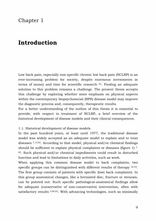
diseases 7,13,60. According to that model, physical and/or chemical findings
should be sufficient to explain physical complaints or diseases (figure 1) 7,
60. Such physical and/or chemical impediments could result in disturbed
function and lead to limitations in daily activities, such as work.
When applying this common disease model to back complaints, two
specific groups can be distinguished with different results of therapy 35,44.
The first group consists of patients with specific (low) back complaints. In
this group anatomical changes, like a herniated disc, fracture or stenosis,
can be pointed out. Such specific pathological-anatomical findings allow
for adequate (conservative of non-conservative) intervention, often with
satisfactory results 3,60,62. With advancing technologies, such as minimally

10
invasive techniques, the results of interventions are still improving for
these patients.
In the second group of low back pain patients no specific pathologic
substrate can be found; they have so called non-specific low back
complaints. Lack of a clear relation between lesions in anatomical
structures and complaints limits or hampers the therapeutic options. In
this group of patients there is no anatomical structure or tissue that can
be operated upon, and the results of conservative treatment, like physical
therapy, are often disappointing 4,9,46-51. The limited therapeutic
possibilities and results in the NCLBP patient group are also recognized in
patients with other certain complaints or diseases 7, 8. At that time it was
argued that the traditional biomedical model failed to provide an adequate
explanation for those diseases for which no chemical or physical cause
could be found. Apparently, there was a need for another disease model
7,8.
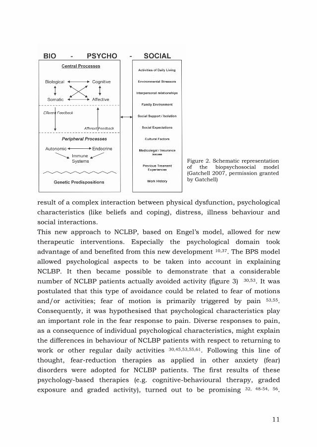
In 1977 an alternative was suggested by a psychiatrist, George Engel: the
biopsychosocial model 7,60,62. A key factor in the BPS model is that it
describes disease not only as a purely physical process but also as a
complex interaction between biological, psychological and social factors
(figure 2). The model was also applied to chronic complaints such as
NCLBP 31,60. Subsequently, the medical world gradually became aware of
the fact that NCLBP is not merely the result of tissue damage but the
Figure 1. Illustration of classical view on relationship between structural damage
and pain (Descartes 1664)
11
result of a complex interaction between physical dysfunction, psychological
characteristics (like beliefs and coping), distress, illness behaviour and
social interactions.
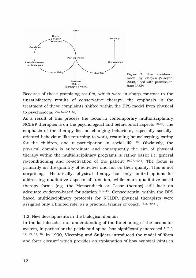
This new approach to NCLBP, based on Engel’s model, allowed for new
therapeutic interventions. Especially the psychological domain took
advantage of and benefited from this new development 10,37. The BPS model
allowed psychological aspects to be taken into account in explaining
NCLBP. It then became possible to demonstrate that a considerable
number of NCLBP patients actually avoided activity (figure 3) 30,53. It was
postulated that this type of avoidance could be related to fear of motions
and/or activities; fear of motion is primarily triggered by pain 53,55.
Consequently, it was hypothesised that psychological characteristics play
an important role in the fear response to pain. Diverse responses to pain,
as a consequence of individual psychological characteristics, might explain
the differences in behaviour of NCLBP patients with respect to returning to
work or other regular daily activities 30,45,53,55,61. Following this line of
thought, fear-reduction therapies as applied in other anxiety (fear)
disorders were adopted for NCLBP patients. The first results of these
psychology-based therapies (e.g. cognitive-behavioural therapy, graded
exposure and graded activity), turned out to be promising 32, 48-54, 56.
Figure 2. Schematic representation of the biopsychosocial model (Gatchell 2007, permission granted
by Gatchell)
12
Because of these promising results, which were in sharp contrast to the
unsatisfactory results of conservative therapy, the emphasis in the
treatment of these complaints shifted within the BPS model from physical
to psychosocial 20,28,29,48-52.
As a result of this process the focus in contemporary multidisciplinary
NCLBP therapies is on the psychological and behavioural aspects 60,62. The
emphasis of the therapy lies on changing behaviour, especially socially-
oriented behaviour like returning to work, resuming housekeeping, caring
for the children, and re-participation in social life 29. Obviously, the
physical domain is subordinate and consequently the aim of physical
therapy within the multidisciplinary programs is rather basic: i.e. general
re-conditioning and re-activation of the patient 16,27,40,41. The focus is
primarily on the quantity of activities and not on their quality. This is not
surprising. Historically, physical therapy had only limited options for
addressing qualitative aspects of function, while more qualitative-based
therapy forms (e.g. the Mensendieck or Cesar therapy) still lack an
adequate evidence-based foundation 6,14,43. Consequently, within the BPS
based multidisciplinary protocols for NCLBP, physical therapists were
assigned only a limited role, as a practical trainer or coach 16,27,40,41.
1.2. New developments in the biological domain
In the last decades our understanding of the functioning of the locomotor
system, in particular the pelvis and spine, has significantly increased 1, 2, 5,
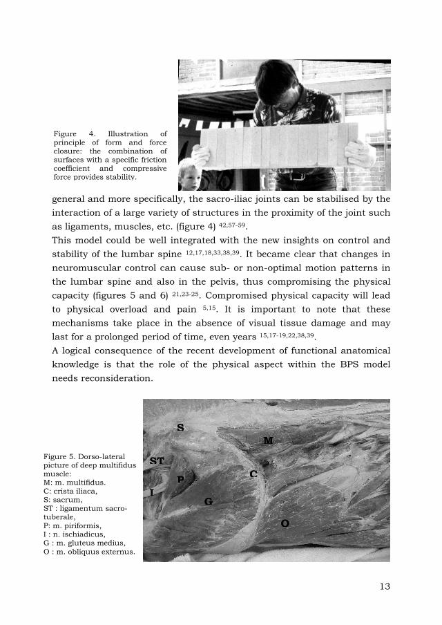
12, 15, 17, 38. In 1990, Vleeming and Snijders introduced the model of ‘form
and force closure’ which provides an explanation of how synovial joints in
Figure 3. Fear avoidance model by Vlaeyen (Vlaeyen 2000, used with permission from IASP)
13
general and more specifically, the sacro-iliac joints can be stabilised by the
interaction of a large variety of structures in the proximity of the joint such
as ligaments, muscles, etc. (figure 4) 42,57-59.
This model could be well integrated with the new insights on control and
stability of the lumbar spine 12,17,18,33,38,39. It became clear that changes in
neuromuscular control can cause sub- or non-optimal motion patterns in
the lumbar spine and also in the pelvis, thus compromising the physical
capacity (figures 5 and 6) 21,23-25. Compromised physical capacity will lead
to physical overload and pain 5,15. It is important to note that these
mechanisms take place in the absence of visual tissue damage and may
last for a prolonged period of time, even years 15,17-19,22,38,39.
A logical consequence of the recent development of functional anatomical
knowledge is that the role of the physical aspect within the BPS model
needs reconsideration.
Figure 4. Illustration of principle of form and force closure: the combination of surfaces with a specific friction coefficient and compressive force provides stability.
C
M
P
G
O
I
S
STFigure 5. Dorso-lateral picture of deep multifidus muscle: M: m. multifidus. C: crista iliaca, S: sacrum, ST : ligamentum sacro-tuberale, P: m. piriformis, I : n. ischiadicus, G : m. gluteus medius,
O : m. obliquus externus.
14
1.3. Reconsideration of the physical aspect in the BPS model
In contemporary multidisciplinary treatments for NCLBP the main focus is
on the psychological, behavioural aspects 11,26,34,36,40,41,52,56. At first glance
the results of these behavioural-based therapy forms appear to be better
than traditional conservative methods. Closer observation shows that the
results of these interventions often reflect their original purpose: the
patients return to work and take up their social life. However, when
parameters such as experienced pain or improved physical performance
are taken into account, the results are far less positive 11,26,34,36,40,41,48,52.
The assumption that the purpose of therapy is to provide a cure and not
just to change behaviour leaves the behavioural-based therapies with
significant room for improvement.
One option to improve LBP therapy lies in revaluation of the physical
domain within the BPS model. New scientific data within the physical
domain, especially those based on functional anatomy, may provide
possibilities to improve BPS-based interventions, especially by addressing
the quality of behaviour. It must be determined whether it is possible to
implement functional anatomical principles within existing behavioural
therapy leading to better therapy results.
1.4. Aim of this thesis
In the context of multidisciplinary treatment of NCLBP patients, the aim of
this thesis is to answer the following three questions:
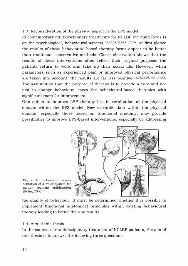
Figure 6. Schematic repre-sentation of a reflex system for motion segment stabilization
(Holm, 2002).
15
1. Taking into account the available recent data on functional
anatomy, is there a need to reconsider the role of the physical
domain within the BPS model?
2. Will a more pronounced role of functional anatomy in the BPS
model contribute to better diagnosis?
3. Will functional anatomy applied in the BPS model contribute to
improved therapy?
In answer to the first question, Chapters 2, 3 and 4 present a specific
sample of functional anatomy and elaborate on their clinical implications.
Chapter 5 deals with the contribution of functional anatomy to the
diagnostic process (the second question); this chapter explores the surplus
value of combining the results of a physical test (the Active Straight Leg
Raise, or ASLR test) with a psychological questionnaire (Tampa Scale of
Kinesiophobia Dutch language version (TSK-DV)). Chapter 6 presents an
answer to the third question; this chapter describes a multidisciplinary
therapy, characterized by a better balance between the physical and
psychological domains. The results of this therapy are presented and
compared with other behavioural therapies.
Finally, in Chapter 7 the main issues addressed in this thesis are
discussed. An answer to the question whether more appreciation for
functional anatomy in the BPS model improves diagnosis and therapy of
patients with NCLBP is formulated. The results of the studies are
discussed in a larger perspective and suggestions for future research are
provided.
16
References
1. Adams M.A. The Biomechanics of Back Pain, Churchill-Livingstone,
2002.
2. Bogduk N. The anatomical basis for spinal pain syndromes. J Manipul
Physiol Ther 1995;18:603–5.
3. Carreon LY, Glassman SD, Howard J. Fusion and nonsurgical treatment
for symptomatic lumbar degenerative disease: a systematic review of
Oswestry Disability Index and MOS Short Form-36 outcomes. Spine J
2007:21.
4. Chavannes A.W. Oefenen bij lage rugpijn. PhD thesis, Utrecht
University, The Netherlands, 1992.
5. van Dieen JH, Cholewicki J, Radebold A. Trunk muscle recruitment
patterns in patients with low back pain enhance the stability of the lumbar
spine. Spine 2003;28:834-41.
6. van Eijsden-Besseling MD, Staal JB, van Attekum A, de Bie RA, van den
Heuvel WJ. No difference between postural exercises and strength and
fitness exercises for early, non-specific, work-related upper limb disorders
in visual display unit workers: a randomised trial. Aust J Physiother
2008;54:95-101.
7. Engel GL. The need for a new medical model: a challenge for bio-
medicine. Science 1977;196:129-36.
8. Engel GL. The clinical application of the biopsychosocial model. Am J
Psychiatry 1980;137:535-544.
9. Faas A. Oefentherapie bij acute lage rugpijn. PhD thesis, Enschede
University, The Netherlands, 1993.
10. Gatchel RJ, Peng YB, Peters ML, Fuchs PN, Turk DC. The
biopsychosocial approach to chronic pain: scientific advances and future
directions. Psychological Bulletin 2007;133:581-624.
11. van Geen JW, Edelaar MJ, Janssen M, van Eijk JT. The long-term
effect of multidisciplinary back training: a systematic review. Spine
2007;32:249-55.
12. Gracovetsky S. The Spinal Engine, Spinger, 1989.
13. Hadler NM. The disabling backache. An international perspective.
Spine 1995;20:640-9.
14. Haugstad GK, Haugstad TS, Kirste UM, Leganger S, Klemmetsen I,
Malt UF. Mensendieck somatocognitive therapy as treatment approach to
17
chronic pelvic pain: results of a randomized controlled intervention study.
Am J Obstet Gynecol 2006;194:1303-10.
15. Hides JA, Richardson CA, Jull GA. Multifidus muscle recovery is not
automatic after resolution of acute, first-episode low back pain. Spine
1996;21:2763-9.
16. Hildebrandt J, Pfingsten M, Saur P, Jansen J. Prediction of success
from a multidisciplinary treatment program for chronic low back pain.
Spine 1997;22:990-1001.
17. Hodges PW, Richardson CA. Inefficient muscular stabilization of the
lumbar spine associated with low back pain. A motor control evaluation of
transversus abdominis. Spine 1996;21:2640-50.
18. Hodges PW, Richardson CA. Delayed postural contraction of
transversus abdominis associated with movement of the lower limb in
people with low back pain. J Spin Disord 1998;11:46-56.
19. Hodges PW, Richardson CA. Altered trunk muscle recruitment in
people with low back pain with upper limb movement at different speeds.
Arch Phys Med Rehabil 1999;80:1005-12.
20. Hoffman BM, Papas RK, Chatkoff DK, Kerns RD. Meta-analysis of
psychological interventions for chronic low back pain. Health Psychol
2007;26:1-9.
21. Holm S, Indahl A, Solomonow M. Sensorimotor control of the spine. J
Electromyography Kinesiol 2002;12:219-34.
22. Hungerford B, Gilleard W, Hodges P. Evidence of altered lumbopelvic
muscle recruitment in the presence of sacroiliac joint pain. Spine
2003;28:1593-600.
23. Indahl A, Kaigle A, Reikeras O, Holm S. Electromyographic response of
the porcine multifidus musculature after nerve stimulation. Spine
1995;20:2652-8.
24. Indahl A, Kaigle A, Reikeras O, Holm S. Interaction between the
porcine lumbar intervertebral disc, zygapophysial joints, and paraspinal
muscles. Spine 1997;22:2834-40.
25. Indahl A, Kaigle A, Reikeras O, Holm S. Sacroiliac joint involvement in
activation of the porcine spinal and gluteal musculature. J Spinal Disord
1999;12:325-30.
26. Johnson RE, Jones GT, Wiles NJ, Chaddock C, Potter RG, Roberts C,
Symmons DP, Watson PJ, Torgerson DJ, Macfarlane GJ. Active exercise,
18
education, and cognitive behavioral therapy for persistent disabling low
back pain: a randomized controlled trial. Spine 2007;32:1578-85.
27. Kääpä EH, Frantsi K, Sarna S, Malmivaara A. Multidisciplinary group
rehabilitation versus individual physiotherapy for chronic nonspecific low
back pain: a randomized trial. Spine 2006;31:371-6.
28. Keller A, Hayden J, Bombardier C, van Tulder M. Effect sizes of non-
surgical treatments of non-specific low-back pain. Eur Spine J
2007;16:1776-88.
29. Klaber Moffett J, Mannion AF. What is the value of physical therapies
for back pain? Best Pract Res Clin Rheumatol 2005;19:623-38.
30. Kori SH, Miller RP, Todd, DD. Kinisiophobia: a new view of chronic
pain behaviour. Pain Manag 1990;3:35-43.
31. Langendoen J. The patient-centredness of evidence-based practice. A
case example to discuss the clinical application of the bio-psychosocial
model. Man Therap 2004;9:228-33.
32. McCracken LM, Gross RT, Eccleston C. Multimethod assessment of
treatment process in chronic low back pain: comparison of reported pain-
related anxiety with directly measured physical capacity. Behav Res Ther
2002;40:585-94.
33. McGill SM, Grenier S, Kavcic N, Cholewicki J. Coordination of muscle
activity to assure stability of the lumbar spine. J Electromyograph Kinesiol
2003;13:353-9.
34. Nicholas MK, Wilson PH, Goyen J. Comparison of cognitive-behavioral
group treatment and an alternative non-psychological treatment for
chronic low back pain. Pain 1992;48:339-47.
35. Njoo KH. Non specific low back pain in general practice: a delicate
point. PhD thesis, Erasmus University Rotterdam, the Netherlands, 1996.
36. Ostelo RW, van Tulder MW, Vlaeyen JW, Linton SJ, Morley SJ,
Assendelft WJ. Behavioural treatment for chronic low-back pain. Cochrane
Database Syst Rev 2005;25, CD002014.
37. Pfingsten M. [Functional restoration-it depends on an adequate
mixture of treatment] Schmerz 2001;15:492-8.
38. Richardson CA, Jull GA, Muscle control-pain control. What exercises
would you prescribe? Man Ther 1995;1:2-10.
19
39. Richardson CA, Jull GA, Hodges PW, Hides JA. Therapeutic exercise
for spinal segmental stabilisation in low back pain: scientific basis and
clinical approach. Edinburgh, Churchill Livingstone, 1999.
40. Smeets RJ, Vlaeyen JW, Hidding A, Kester AD, van der Heijden GJ,
van Geel AC, Knottnerus JA. Active rehabilitation for chronic low back
pain: Cognitive-behavioral, physical, or both? First direct post-treatment
results from a randomized controlled trial. BMC Musculoskelet Disord
2006a;7:5.
41. Smeets RJ, Vlaeyen JW, Hidding A, Kester AD, van der Heijden GJ,
Knottnerus JA. Chronic low back pain: physical training, graded activity
with problem solving training, or both? Pain. 2008;134:263-76.
42. Snijders CJ, Vleeming A, Stoeckart R. Transfer of lumbosacral load to
iliac bones and legs. Part 1 and 2. Clin Biomech 1993;8:285-294.
43. Soukup MG, Glomsröd B, Lönn JH, Bö K, Larsen S. The effect of a
Mensendieck exercise program as secondary prophylaxis for recurrent low
back pain. A randomized, controlled trial with 12-month follow-up. Spine
1999 24:1585-91.
44. Spitzer WO, LeBlanc FE, Dupuis M. Scientific approach to the
assessment and management of activity-related spinal disorders. A
monograph for clinicians. Report of the Quebec Task Force on spinal
disorders. Spine 1987;12:1-59.
45. Thomas JS, France CR. Pain-related fear is associated with avoidance
of spinal motion during recovery from low back pain. Spine 2007;32:E460-
6.
46. Van Tulder MW. Koes BW. Assendelft WJJ. Bouter LM. The
effectiveness of conservative treatment of acute and chronic low back pain.
Amsterdam: Fac. Der geneeskunde VU, EMGO instituut, 1999.
47. Van Tulder MW, Ostelo R, Vlaeyen JW, Linton SJ, Morley SJ,
Assendelft WJ. Behavioral treatment for chronic low back pain: a
systematic review within the framework of the Cochrane Back Review
Group. Spine 2001;26:270-81.
48. Van Tulder WM , Waddell G. Evidence-based medicine for non-specific
low back pain. Best Pract Res Clin Rheumatol 2005;19:7-9.
49. Van Tulder M, Koes B. Chronic low back pain. Am Fam Phys
2006;74:1577-9.
20
50. Van Tulder M, Koes B. Low back pain (chronic). Clin Evid
2006;15:1634-53.
51. Van Tulder MW, Koes B, Malmivaara A. Outcome of non-invasive
treatment modalities on back pain: an evidence-based review. Eur Spine J
2006;15:S64-81.
52. Vendrig AA, van Akkerveeken PF, Sanders RJ. [Good results from a
multidisciplinary and behavioral program for chronic back pain] Ned
Tijdschr Geneeskd 2000;144:560-3.
53. Vlaeyen JW, Kole-Snijders AM, Boeren RG, van Eek H. Fear of
movement/(re)injury in chronic low back pain and its relation to
behavioral performance. Pain 1995;62:363-72.
54. Vlaeyen JW, Haazen IW, Schuerman JA, Kole-Snijders AM, van Eek H.
Behavioural rehabilitation of chronic low back pain: comparison of an
operant treatment, an operant-cognitive treatment and an operant-
respondent treatment. Br J Clin Psychol 1995;34:95-118.
55. Vlaeyen JW, Linton SJ. Fear-avoidance and its consequences in
chronic musculoskeletal pain: a state of the art. Pain 2000;85:317-32.
56. Vlaeyen JW, De Jong JR, Onghena P, Kerckhoffs-Hanssen M, Kole-
Snijders AM. Can pain-related fear be reduced? The application of
cognitive-behavioural exposure in vivo. Pain Res Manag 2002;7:144-53.
57. Vleeming A, Stoeckart R, Volkers ACW, Snijders CJ. Relation between
form and function in the sacro-iliac joint, Part 1: Clinical anatomical
aspects. Spine 1990a;15:130-2.
58. Vleeming A, Volkers ACW, Snijders CJ, Stoeckart R. Relation between
form and function in the sacro-iliac joint, Part 2: Biomechanical aspects.
Spine 1990b;15:133-6.
59. Vleeming A. The sacroiliac joint, a clinical-anatomical, biomechanical
and radiological study. PhD thesis, Erasmus University Rotterdam, the
Netherlands, 1990.
60. Waddell G. 1987 Volvo award in clinical sciences. A new clinical model
for the treatment of low-back pain. Spine 1987;12:632–44.
61. Waddell G, Newton M, Henderson I, Somerville D, Main CJ. A Fear-
Avoidance Beliefs Questionnaire (FABQ) and the role of fear-avoidance
beliefs in chronic low back pain and disability. Pain 1993;52:157-68.
62. Waddell G. The back pain revolution. Second edition. Churchill
Livingstone 2004.
Chapter 2
A functional-anatomical approach to the spine-pelvis mechanism: interaction between the biceps femoris muscle and the sacrotuberous liga-ment
J.P. van Wingerden*,I, A. Vleeming, PhD*, C.J. Snijders, PhDI, R. Stoeckart,
PhD*
From the Faculty of Medicine, Departments of * Anatomy and I Biomedical
Physics and Technology, Research Group of Clinical Anatomy and Medical
Technology, Erasmus University, Rotterdam, The Netherlands
22
Abstract
Sacroiliac joint dysfunction is often overlooked as a possible cause of "low
back" pain. This is due to the use of reductionistic anatomical models.
From a kinematic point of view, topographic anatomical models are
generally not sufficient since they categorize pelvis, lower vertebral column
and legs as distinct entities. This functional-anatomical study focuses on
the question whether anatomical connections between the biceps femoris
muscle and the sacrotuberous ligament are kinematically useful. Forces
applied to the tendon of the biceps femoris muscle, simulating biceps
femoris muscle force, were shown to influence sacrotuberous ligament
tension. Since sacrotuberous ligament tension influences sacroiliac joint
kinematics, hamstring training could influence the sacroiliac joint and as
such low back kinematics. The clinical implications with respect to "short
hamstrings", pelvic instability and walking are discussed.
Acknowledgement
The authors appreciate mrs. C. Langendoen for her dedicated assistance
and mr. J.V. de Bakker for his technical support throughout this study.
23
Introduction
For a successful treatment of pelvic and spinal disorders, it is essential to
have a clear insight into the morphology and function of the connections
between spine and pelvis, i.e., the sacrum and its joints. As a rule
discussions on "low back" pain are based on classifications used in
topographical-anatomical models. In these models spine, pelvis and lower
extremities are considered as separate entities. However, from a
neurophysiological, biomechanical and functional-anatomical point of view
these structures are fully coupled. The topographical-anatomical approach
is shown by reductionistic terminology as in the word "back" muscles. After
all, these muscles are not only connected to head and ribs but also to
"pelvic" structures such as the iliac crests, sacrum and sacroiliac liga-
ments 2,10,11,12,17. Obviously, parts of the backmuscles act directly and
indirectly at the sacroiliac (SI) joints. Consequently, neglecting SI joint
dysfunction as a cause of "low back" pain may well be the result of the use
of reductionistic anatomical models leading to an artificial classification.
Preceding studies 18-21,24 were dealing with the intertwined relation between
pelvis and spine. Specific symmetrical roughening patterns on the surface
of the SI joints, already commencing in the fetal period, were considered as
functional adaptations, increasing stability 3. As shown in a biomechanical
study, the specific roughening of the SI joint surfaces goes with a higher
friction coefficient. Furthermore, it was shown that the stability of the SI
joint was increased by a larger wedge-angle of the joint. As a result, less
ligament force is required for bearing the upper part of the body. Vleeming
et al. 21 described this as the selfbracing effect of the SI joint. This refers to
the dynamic mechanism by which the internal friction in the SI joint can
be enlarged.
Since the sacrotuberous ligament influences the selfbracing mechanism,
muscles connected to the ligament could play an important role in
obtaining SI joint stability 18,19,24. Connections between the gluteus
maximus muscle and the sacrotuberous ligament were found 18. In the
same study the sacrotuberous ligament was shown to be fused with the
tendon of the long head of the biceps muscle in six out of twelve cadavers,
in four cases even bilaterally.
The anatomical findings were substantiated by a biomechanical study:
24
when minor loads in the direction of gluteus maximus and biceps femoris
muscle were bilaterally applied to the sacrotuberous ligament, ventral
rotation (nutation) of the sacrum, as a result of simulated bodyweight,
diminished significantly. Since in some cases the long head of the biceps
femoris muscle is connected to the sacrotuberous ligament, it is
hypothesized that force from this muscle can influence sacrotuberous
ligament tension, and in doing so dynamically influence stability of the SI
joints 19.
This article deals with the question whether biceps femoris muscle force
indeed influences sacrotuberous ligament tension.
25
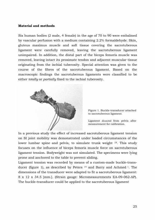
Material and methods
Six human bodies (2 male, 4 female) in the age of 70 to 90 were embalmed
by vascular perfusion with a medium containing 2.2% formaldehyde. Skin,
gluteus maximus muscle and soft tissue covering the sacrotuberous
ligament were carefully removed, leaving the sacrotuberous ligament
unimpaired. In addition, the distal part of the biceps femoris muscle was
removed, leaving intact its proximate tendon and adjacent muscular tissue
originating from the ischial tuberosity. Special attention was given to the
course of the fibres of the sacrotuberous ligament. Based on the
macroscopic findings the sacrotuberous ligaments were classified to be
either totally or partially fixed to the ischial tuberosity.
In a previous study the effect of increased sacrotuberous ligament tension
on SI joint mobility was demonstrated under loaded circumstances of the
lower lumbar spine and pelvis, to simulate trunk weight 19. This study
focuses on the influence of biceps femoris muscle force on sacrotuberous
ligament tension. Bodyweight was not simulated. The specimens were lying
prone and anchored to the table to prevent sliding.
Ligament tension was recorded by means of a custom-made buckle-trans-
ducer (figure 1), as described by Peters 14 and Barry and Achmed 1. The
dimensions of the transducer were adapted to fit a sacrotuberous ligament:
8 x 12 x 34.5 [mm.]. (Strain gauge: Micromeasurements EA-09-062-AP).
The buckle-transducer could be applied to the sacrotuberous ligament
Figure 1. Buckle-transducer attached
to sacrotuberous ligament.
Ligament dissected from pelvis after
measurement for calibration.
26
without affecting its anatomical integrity.
Biceps femoris muscle forces from 0 to 100 N with a 10 N increment were
simulated with weights. As site of impact, the biceps femoris muscle
tendon was chosen five centimeters caudal from the tuber ischiadicum.
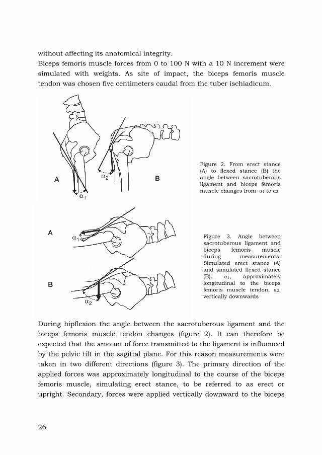
During hipflexion the angle between the sacrotuberous ligament and the
biceps femoris muscle tendon changes (figure 2). It can therefore be
expected that the amount of force transmitted to the ligament is influenced
by the pelvic tilt in the sagittal plane. For this reason measurements were
taken in two different directions (figure 3). The primary direction of the
applied forces was approximately longitudinal to the course of the biceps
femoris muscle, simulating erect stance, to be referred to as erect or
upright. Secondary, forces were applied vertically downward to the biceps
Figure 2. From erect stance
(A) to flexed stance (B) the
angle between sacrotuberous
ligament and biceps femoris
muscle changes from F1 to F2
Figure 3. Angle between
sacrotuberous ligament and
biceps femoris muscle
during measurements.
Simulated erect stance (A)
and simulated flexed stance
(B). F1, approximately
longitudinal to the biceps
femoris muscle tendon, F2,
vertically downwards
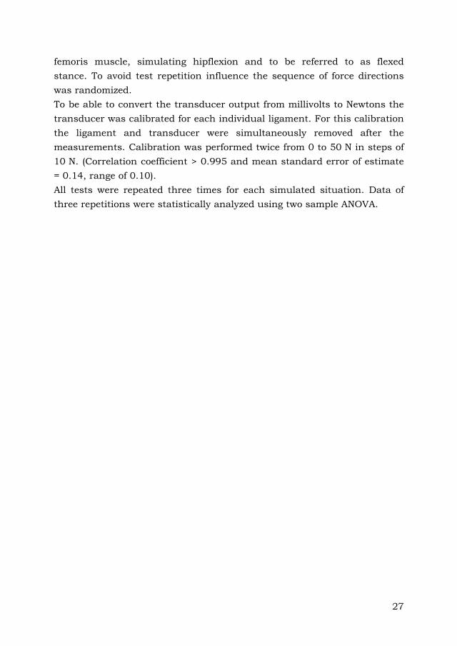
27
femoris muscle, simulating hipflexion and to be referred to as flexed
stance. To avoid test repetition influence the sequence of force directions
was randomized.
To be able to convert the transducer output from millivolts to Newtons the
transducer was calibrated for each individual ligament. For this calibration
the ligament and transducer were simultaneously removed after the
measurements. Calibration was performed twice from 0 to 50 N in steps of
10 N. (Correlation coefficient > 0.995 and mean standard error of estimate
= 0.14, range of 0.10).
All tests were repeated three times for each simulated situation. Data of
three repetitions were statistically analyzed using two sample ANOVA.
28
Results
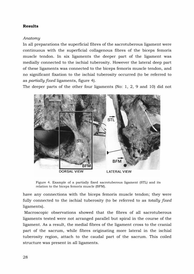
Anatomy
In all preparations the superficial fibres of the sacrotuberous ligament were
continuous with the superficial collagenous fibres of the biceps femoris
muscle tendon. In six ligaments the deeper part of the ligament was
medially connected to the ischial tuberosity. However the lateral deep part
of these ligaments was connected to the biceps femoris muscle tendon, and
no significant fixation to the ischial tuberosity occurred (to be referred to
as partially fixed ligaments, figure 4).
The deeper parts of the other four ligaments (No: 1, 2, 9 and 10) did not
have any connections with the biceps femoris muscle tendon; they were
fully connected to the ischial tuberosity (to be referred to as totally fixed
ligaments).
Macroscopic observations showed that the fibres of all sacrotuberous
ligaments tested were not arranged parallel but spiral in the course of the
ligament. As a result, the medial fibres of the ligament cross to the cranial
part of the sacrum, while fibres originating more lateral in the ischial
tuberosity region, attach to the caudal part of the sacrum. This coiled
structure was present in all ligaments.
Figure 4. Example of a partially fixed sacrotuberous ligament (STL) and its
relation to the biceps femoris muscle (BFM).
29
In the partially, as well as in the totally fixed ligaments the long head of
the biceps femoris muscle has the shape of a firm oval tendon on the level
of the ischial tuberosity.
Biomechanics
The results are presented as the ratio of the force applied to the biceps
femoris muscle tendon and the force measured on the sacrotuberous
ligament (table 1). In table 1 every two sequential ligaments belong to one
body, except for ligaments 3 and 4 which belong to different bodies.
Table 1. Collected ligament data and applied force/measured force ratio's for simulation of
biceps femoris muscle force in erect (upright) stance and flexed stance. Ratio's averaged over
three repetitions. Correlation coefficient of all ratio's > 0.98
Ligament Side Gender Fixation Upright Flexed
1 L F Total 0.09 0.13
2 R F Total 0.08 0.13
3 L F Partial 0.20 0.43
4 L F Partial 0.54 0.33
5 L M Partial 0.08 0.42
6 R M Partial 0.16 0.52
7 L M Partial 0.69 0.19
8 R M Partial 0.19 0.31
9 L F Total 0.07 0.15
10 R F Total 0.07 0.17
Statistical analysis showed that part of the force applied to the biceps
femoris muscle tendon was transferred to the sacrotuberous ligament, in
all preparations and in all situations. However, interindividual differences
were large (table 1). Transferred forces tended to be higher during the
simulated flexed stance than during simulated erect stance (table 1), but
30
differences were not significant. Between genders no significant differences
in force transfer could be demonstrated, nor between left and right (table
1).
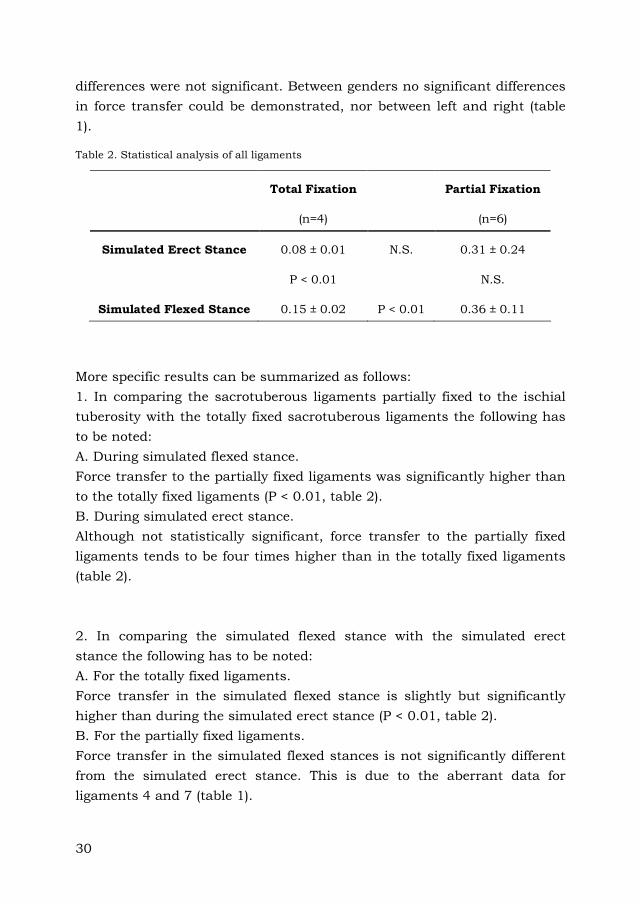
Table 2. Statistical analysis of all ligaments
Total Fixation
(n=4)
Partial Fixation
(n=6)
Simulated Erect Stance 0.08 ± 0.01 N.S. 0.31 ± 0.24
P < 0.01 N.S.
Simulated Flexed Stance 0.15 ± 0.02 P < 0.01 0.36 ± 0.11
More specific results can be summarized as follows:
1. In comparing the sacrotuberous ligaments partially fixed to the ischial
tuberosity with the totally fixed sacrotuberous ligaments the following has
to be noted:
A. During simulated flexed stance.
Force transfer to the partially fixed ligaments was significantly higher than
to the totally fixed ligaments (P < 0.01, table 2).
B. During simulated erect stance.
Although not statistically significant, force transfer to the partially fixed
ligaments tends to be four times higher than in the totally fixed ligaments
(table 2).
2. In comparing the simulated flexed stance with the simulated erect
stance the following has to be noted:
A. For the totally fixed ligaments.
Force transfer in the simulated flexed stance is slightly but significantly
higher than during the simulated erect stance (P < 0.01, table 2).
B. For the partially fixed ligaments.
Force transfer in the simulated flexed stances is not significantly different
from the simulated erect stance. This is due to the aberrant data for
ligaments 4 and 7 (table 1).
31
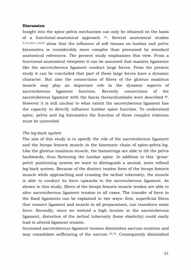
Discussion
Insight into the spine-pelvis mechanism can only be obtained on the basis
of a functional-anatomical approach 23. Several anatomical studies
2,7,8,10,11,18,23 show that the influence of soft tissues on lumbar and pelvic
kinematics is considerably more complex than presumed by standard
anatomical references. The present study emphasizes this view. From a
functional-anatomical viewpoint it can be assumed that massive ligaments
like the sacrotuberous ligament conduct large forces. From the present
study it can be concluded that part of these large forces have a dynamic
character. But also the connections of fibres of the gluteus maximus
muscle may play an important role in the dynamic aspects of
sacrotuberous ligament function. Recently connections of the
sacrotuberous ligament with the fascia thoracolumbalis were described 26.
However it is still unclear to what extent the sacrotuberous ligament has
the capacity to directly influence lumbar spine function. To understand
spine, pelvis and leg kinematics the function of these complex relations
must be unraveled.
The leg-back system
The aim of this study is to specify the role of the sacrotuberous ligament
and the biceps femoris muscle in the kinematic chain of spine-pelvis-leg.
Like the gluteus maximus muscle, the hamstrings are able to tilt the pelvis
backwards, thus flattening the lumbar spine. In addition to this "gross"
pelvic positioning system we want to distinguish a second, more refined
leg-back system. Because of the distinct tendon form of the biceps femoris
muscle while approaching and crossing the ischial tuberosity, the muscle
is able to conduct its force upwards to the sacrotuberous ligament. As
shown in this study, fibres of the biceps femoris muscle tendon are able to
alter sacrotuberous ligament tension in all cases. The transfer of force in
the fixed ligaments can be explained in two ways: first, superficial fibres
that connect ligament and muscle in all preparations, can transduce some
force. Secondly, since we noticed a high tension in the sacrotuberous
ligament, distortion of the ischial tuberosity (bone elasticity) could easily
lead to altered ligament tension.
Increased sacrotuberous ligament tension diminishes sacrum nutation and
may consolidate selfbracing of the sacrum 18,19. Consequently diminished
32
sacrotuberous ligament tension may increase SI joint mobility. This
mechanism may even be more subtle: in eight of all ten ligaments tested a
relatively higher percentage of force was transferred from the biceps
femoris muscle to the sacrotuberous ligament during the flexed situation if
compared with the erect situation. From a biomechanical point of view this
could be expected, since the flexion torque on the lumbar spine increases
when changing from erect stance to flexed stance 9,22. Therefore, in the
flexed position larger contranutating forces are needed to prevent the
sacrum from tilting forward. As emphasized by the present findings in
most individuals part of this force can be derived from the biceps femoris
muscle.
The specific role of the described coiled structure of the sacrotuberous
ligament is still unclear however, some speculations can be made. As a
result of the coiled structure of the sacrotuberous ligament, the lateral part
of the biceps femoris tendon creates a force which is directed to the
sacrum horizontally. This force has the same direction as the resultant of
ligament forces (Fl), which compress the SI joint and are essential for the
selfbracing mechanism as described by Vleeming 21. It can be noted that
the coiled structure of the sacrotuberous ligament resembles the structure
of the cruciate ligaments 4,16. This could imply that different parts of the
sacrotuberous ligament, like the cruciate ligaments, are loaded during
different stages of motion of the SI joint.
SI joint stabilization during walking
Stabilization of the SI joints during daily activities like walking must be
considered a dynamic process. During walking the leg as well as the
homolateral SI joint become weight-bearing at heel-strike. On this very
moment or better, just before, its selfbracing system must be activated to
stabilize the SI joint. Gait analysis shows the hamstrings to become active
just before heel-strike 27. This action increases sacrotuberous ligament
tension and presumably selfbracing of the SI joint in addition to limiting
knee extension. On heel-strike the homolateral SI joint and the spine will
benefit from an optimal stabilization induced by muscular activity of the
lower extremity. However, small physical changes, like functional short
hamstrings can disturb this leg-spine mechanism.
33
"Short hamstrings" phenomenon
The phenomenon of "tight-" or "short-" hamstrings is often considered as a
secondary effect or residual sign of low back trouble 5,6,12,13,15. According to
the data presented here, shortened hamstrings can affect the selfbracing
mechanism of the pelvis. An altered selfbracing mechanism might change
the pattern of forces in spine and pelvis. Consequently, short hamstrings
may prolong or even initiate low back problems. Whether stretching the
hamstrings influences "low back" pain is unclear, since scientific data are
lacking 12. However, it might well be that stretching the hamstrings
restores pelvic and lumbar kinematics and breaks the vicious circle of "low
back" pain and shortened hamstrings.
Pelvic instability and leg-muscle training
Exercise of muscles, which influence the pelvis directly, or indirectly via
the sacrotuberous ligament can be of special importance for women
suffering from hypermobility of the pelvis 25. Pelvic instability is often
regarded as exclusively a failure of the pelvic ligaments, the passive struc-
tures stabilizing the pelvis. As emphasized here, leg and pelvic muscles can
actively influence the mobility of the SI joint and thus influence pelvic
stability. By leg-muscle training the selfbracing mechanism can be
influenced. Specific muscle training is therefore recommended for women
with complaints of pelvic hypermobility 25.
Conclusion
Sacrotuberous ligament tension can be influenced by biceps femoris
muscle force. Consequently a leg muscle like the biceps femoris can affect
the SI joint and hence pelvic and lumbar stability. In solving complex low
back problems, it is essential to see the spine, pelvis and lower extremities
as integrated and mutual influencing entities.
34
References
1. Barry D, and Ahmed AM (1986) Design and performance of a modified
buckle transducer for the measurement of ligament tension. J of Biomech
Eng 108:149-52.
2. Bogduk N, and Macintosh JE (1984) The applied anatomy of the
thoracolumbar fascia. Spine 9:164-70.
3. Bowen V. Cassidy JD (1981) Macroscopic anatomy of the sacroiliac joint
from embryonic life until the eight decade. Spine 6:620-8.
4. Butler DL, Guan Y, Kay MD, Cummings JF, Feder SM, Levy MS (1992)
Location-dependent variations in the material properties of the anterior
cruciate ligament. J. Biomechanics 25:511-18.
5. Gajdosik RL, Giuliani CA, and Bohannon RW (1990) Passive compliance
and length of the hamstring muscles of healthy men and woman. Clin
Biomech 5:23-9.
6. Göeken LNH (1988) Straight-leg raising in "short hamstrings"
[Dissertation]. Groningen (NL):Univ of Groningen.
7. Gracovetsky S, Kary M, Pitchen I, Levy B, and Ben Said R (1989) The
importance of pelvic tilt in reducing compressive stress in the spine during
flexion-extension exercises. Spine 14:412-6.
8. Gracovetsky S, Kary M, Levy S, Ben Said R, Pitchen I, and Helie J (1990)
Analysis of spinal and muscular activity during flexion/extension and free
lifts. Spine 15:1333-9.
9. Lindh M (1984) Biomechanica van het skeletsysteem. In: Frankel VH,
Nordin M, and Snijders CJ, editors. Biomechanica van het skeletsysteem.
Lochem: De Tijdstroom BV, pp 265-303.
10. Macintosh JE, and Bogduk N (1986) The Biomechanics of the Lumbar
Multifidus. Clin Biomech 1:205-13.
11. Macintosh JE, and Bogduk N (1991) The Attachments of the Lumbar
Erector Spinae. Spine 16:783-92.
12. Mellin G (1988) Correlations of hip mobility with degree of back pain
and lumbar spinal mobility in chronic low-back pain patients. Spine
13:668-70.
13. Mierau D, Cassidy JD, and Yong-Hing K (1984) Low back pain and
straigth leg raising in children and adolescents. Spine 14:526-8.
14. Peters G (1987) Tools for the measurement of stress and strain fields in
35
soft tissue [Dissertation]. Eindhoven (NL): Univ of Eindhoven.
15. Stokes IA, and Abery JM (1980) Influence of the hamstring muscles on
lumbar spine curvature in sitting. Spine 5:525-8.
16. Strocchi R, De Pasquale V, Gubellini P, Facchini A, Marcacci M, Buda
R, Zaffagnini S, Ruggeri A (1992) The human anterior cruciate
ligament:histological and ultrastructural observations. J. Anat. 180:515-
519.
17. Vink P (1989) Functions of the lumbar back muscles [Dissertation].
Leiden(NL):Univ of Leiden.
18. Vleeming A, Stoeckart R, and Snijders CJ (1989) The sacrotuberous
ligament: a conceptual approach to its dynamic role in stabilizing the SI-
joint. Clin Biomech 4:201-3.
19. Vleeming A, Van Wingerden JP, Snijders CJ, Stoeckart R, and Stijnen
T (1989) Load application to the sacrotuberous ligament; influences on SI-
joint mechanics. Clin Biomech 4:204-9.
20. Vleeming A, Stoeckart R, Volkers ACW, and Snijders CJ (1990) Relation
between form and function in the sacro-iliac joint, Part 1: Clinical
anatomical aspects. Spine 15:130-2.
21. Vleeming A, Volkers ACW, Snijders CJ, and Stoeckart R (1990)
Relation between form and function in the sacro-iliac joint, Part 2:
Biomechanical aspects. Spine 15:133-6.
22. Vleeming A, Snijders CJ and Stoeckart R, editors (1991) A new and
functional approach to the biomechanics of the human pelvis. Proceedings
of the First international symposium on the SI-joint: Its role in posture and
locomotion; Maastricht(NL):98-109.
23. Vleeming A, Snijders CJ, and Stoeckart R, editors (1991) Progress in
vertebral column research; Proceedings of the First international
symposium on the SI- joint: Its role in posture and locomotion;
Maastricht(NL).
24. Vleeming A, Van Wingerden JP, Dijkstra PF, Stoeckart R, Snijders CJ
and Stijnen T (1992) Mobility in the sacroiliac joints in the elderly: A
kinematic and radiological study. Clin Biomech 7:170-6.
25. Vleeming A, H.M. Buyruk, Stoeckart R, Karamursel S, Snijders CJ
(1991) An integrated therapy for peripartum pelvic instability: A study of
the biomechanical effects of pelvic belts. The Am J Obstet Gynacol
166:1243-7.
36
26. Vleeming A, Pool A, Snijders CJ, Wingerden JP, Stoeckart R. A study of
the posterior layer of the lumbo-dorsal fascia. In preparation.
27. Weil S, Weil UH (1966) Mechanik des Gehens. Stuttgart, Georg Thieme
Verlag.
Chapter 3
Stabilization of the sacroiliac joint in vivo: verification of muscular contribution to force closure of the pelvis
Wingerden JP vana, Vleeming Aa, Buyruk HM.b, Raissadat K.c
a Spine & Joint Centre, The Netherlands, b Institute of Rehabilitation,
University Hospital Dijkzigt, Rotterdam, The Netherlands, c St. Antonius
Hospital dept. Surgery, The Netherlands
38
Abstract
Objectives
To study in vivo whether muscles contribute to force closure of the
sacroiliac joint (SIJ).
Summary of background data
A model on SIJ function postulates that SIJ shear is prevented by friction,
dynamically influenced by muscle force and ligament tension. Thus, SIJ
stability can be accommodated to specific loading situations.
The amount of SIJ friction can be measured as stiffness using a verified
method combining Color Doppler Imaging and induced oscillation of the
ilium relative to the sacrum.
Study design and methods
SIJ stiffness was measured using Color Doppler Imaging combined with
pelvic oscillation in six healthy women. SIJ stiffness was measured both in
a relaxed situation and during isometric voluntary contractions
(electromyographically recorded). The biceps femoris, gluteus maximus,
erector spinae, and contralateral latissimus dorsi were included in this
study. Results were statistically analyzed.
Results
SIJ stiffness significantly increased when the individual muscles were
activated. This held especially for activation of the erector spinae, the
biceps femoris and the gluteus maximus muscles. During some tests
significant cocontraction of other muscles occurred.
Conclusions
SIJ stiffness increased even with slight muscle activity, supporting the
notion that effectiveness of load transfer from spine to legs is improved
when muscle forces actively compress the SIJ preventing shear. When
joints are manually tested, the influence of muscle activation patterns
must be considered since both inter-and intra tester reliability of the test
can be affected by muscle activity. In this respect the relation between

39
emotional states, muscle activity and joint stiffness, deserves further
exploration.
Acknowledgment
The authors wish to acknowledge the department of Biomedical Physics
and Technology of the Erasmus University Rotterdam, The Netherlands,
for their technical support.

40
Introduction
This study was initiated to demonstrate in vivo that muscles contribute to
force closure of the sacroiliac joint (SIJ). According to the model of form
and force closure, shear in the SIJs is prevented by increased friction due
to a combination of two factors: 1) specific anatomic features increased the
friction coefficient (form closure) and 2) tension of muscles and ligaments
crossing the SIJ led to higher friction and hence stiffness (force closure)
16,22,23,26. Thus, stabilization of the SIJs can be dynamically accommodated
to the specific loading situation 16,17,20,21,23,27,28,29,30,31. Stability of the SIJs
is partly realized by tension of ligaments due to SIJ motion 16,20,22,23,24,28.
The model assumed that for effective transfer of load from the spine
through the pelvis to the legs, muscles acting on the pelvis must be
activated to increase force closure of the SIJ 17,29,30. Research on joint
stability in general and SIJ stability specifically, is mainly focussed on
quantitative measurements including recording of the range of motion
10,12,15,18,19,25,26. No studies were found on qualitative measurements like
establishing the stiffness of the SIJ, or to determine the ability of the SIJ to
resist shear forces. The need for a reliable and non-invasive method to
quantify SIJ stability in vivo resulted in the development of a measuring
technique, combining Color Doppler Imaging (CDI) with excitation of the
pelvis by means of an oscillation device 1,2,3. With this method force closure
of the SIJ can be measured in vivo as a function of the amount of SIJ
friction.
Experimental application of this method on an artificial mechanical model
of the pelvis showed reproducible results 1,2,3. Further validation of this
method was performed in three different studies: on embalmed specimen,
on healthy subjects, and a comparative clinical study demonstrating this
technique to be objective and reproducible in determining SIJ stiffness
(Reliability coefficients: left SIJ 0.97 and right SIJ 0.94) 1,2,3.
Former anatomical in vitro studies identified specific muscles that could
contribute to SIJ stabilization. Biceps femoris and gluteus maximus
muscles could increase force closure of the SIJ, through their specific and
massive attachments to the sacrotuberous ligament 20,21,30. Gluteus
maximus and latissimus dorsi were found to be partially coupled by the
41
posterior layer of the thoracolumbar fascia, creating a compressive force
acting perpendicular to the SIJ. This was confirmed by a study of Mooney
et al 13. Finally, it was shown that the tendinous aponeurose of the erector
muscle was closely linked to the sacrum and posterior superficial SIJ
ligaments 24.
The present study attempts to determine whether muscles contribute to
force closure in vivo. This study combines CDI and artificially generated
oscillation of the SIJ with controlled activation electromyography (EMG) of
specific muscles, applied to a group of healthy volunteers. Because of their
assumed role in force closure of the SIJ, this study focused on the effect of
unilateral activation of the biceps femoris, gluteus maximus and erector
spinae, and contralateral activation of the latissimus dorsi muscle
13,20,21,29,30. It was expected to reject the null hypothesis that muscles
cannot stabilize the SIJs.
42
Material and Methods
Volunteers
Fifteen female volunteers (aged 15 to 30 years) participated in this study.
They were all in good physical health with no recent complaints of spine,
pelvis or hipjoints. To increase sensitivity of the CDI method only pelves
that exhibited considerable motion were included. Joint stiffness was
initially measured three times with CDI during application of oscillation to
the pelvis. Only in six volunteers (average age 22 sd 2.6 years) threshold
values of the CDI were high enough to be included in the study (see
results). Average height and weight of the subjects were respectively 170
(sd 4.1) cm and 62 (sd 4.9) kg. Preliminary tests showed the protocol to be
fairly straining to the subjects. Because testing both sides may have led to
unreliable results due to fatigue 11, during the experiment, tests were
performed unilaterally (4 right, 2 left side).
Testing procedure
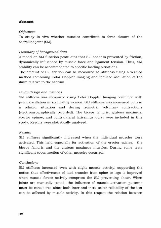
Volunteers were lying prone with the anterior superior iliac spine in
contact with the oscillator plate (figure 1). Before the measurements a
maximal voluntary contraction (MVC) of each separate muscle was
recorded, using isometric muscle test procedures with manual resistance
Figure 1. Outline of test position for combined CDI and EMG measurements. A indicates CDI probe location over both sacrum and ilium on one side of the pelvis. B the location where the oscillator plate is positionned against the anterior
superior iliac spine.
43
as described by Kendall et al. 9.
Each measurement started with determination of SIJ stiffness without any
muscle activation using CDI. Then the volunteers were asked to activate
only one particular muscle for the period of the measurement using the
technique as for the MVC test. However, in contrast to the MVC test, no
maximal voluntary contraction but only slight effort of the tested muscle
was pursued (>10% of MVC), with no or only minimal coactivation of other
muscles (<10% of MVC) and minimal disturbance of the initial posture.
Since only minimal exertion was required no manual resistance (in
contrast with the MVC test) was applied during the tests.
During each test, EMGs of all four muscles were recorded simultaneously
to test for cocontractions. Sustained muscle contractions with an average
duration of 10 seconds were required to analyze SIJ stiffness by means of
the CDI method.
The test sequence was repeated three times with biceps femoris, gluteus
maximus, latissimus dorsi and erector muscles tested in randomized order
for each subject.
Finally, to verify that EMG signal quality did not change during the
measurements, a second maximal voluntary contraction test, similar to the
initial MVC test was performed for each muscle.
EMG recording
Electrode location was determined as described by Delagi et al. 4,6,11.
Volunteers were instrumented with surface EMG electrodes (Meditrace
pallet electrodes) after the skin was scrubbed and cleaned with alcohol.
EMG signals were amplified and 10 - 2 kHz filtered (bipolar EMG amplifier
PS-800, Twente Medical System). The signals were rectified, low-pass
filtered (10 Hz) and simultaneously fed to a computer with a sample
frequency of 50 Hz. Preliminary studies showed no interference of the
vibration device with the EMG recordings.
Color Echo Doppler imaging (CDI)
The application of CDI in combination with generated oscillation and the
subsequent validation of this method, is described in detail in previous
studies on SIJ stiffness 1,2,3. Vibrations with a frequency of 200 Hz (using a
44
Derritron VP3 oscillator) were unilaterally applied to the anterior superior
iliac spine. The vibrations from ilium and sacrum were measured by a
Philips Quantum AD1 CDI transducer covering both sides of one SIJ (see
figure 1).
The threshold indicates the necessary signal power to display perceived
vibration in color. The height of the threshold is set by the operator by
means of the threshold button on the control panel of the CDI apparatus.
During a measurement the threshold is precisely set to the level were no
vibrations are visible on the CDI screen. A large difference between the
thresholds (threshold difference; THD) set at the sacrum and ilium
indicates little stiffness of the SIJ. A small or absent THD indicates a stiff
joint 1,2,3. In this study differences between THD in the relaxed position
and the THD during a muscle test were used as a measure for change in
SIJ stiffness. A decreased THD during the muscle test indicates that the
joint has become more stiff.
Analysis
To determine changes in SIJ stiffness during muscle activity, THD’s found
during muscle tests were subtracted from THD’s found during relaxed
postures for each individual. The muscle tests were: 1) the biceps femoris
test, 2) the gluteus maximus test, 3) the erector spinae test and 4) the
latissimus dorsi test. From the three repetitions of each muscle test the
mean THD was calculated. The statistical significance of mean differences
between THD during relaxed postures and the THD during each muscle
test was determined using a paired two sample t-test.
To quantify the activity level of each muscle during the tests, the recorded
EMG signals were averaged. From the three repetitions of each muscle test
the mean activity level was calculated. To compare between subjects, the
muscle activity levels are presented as percentages of the MVC for each
muscle.
Muscle activity (in percentage of MVC) during relaxed position and the
muscle tests was compared using a paired t-test. A muscle was considered
active when the activity level during the tests was more than 10% of MVC.
P-values less then 0.05 were considered significant.
45
Results
Mean results of all subjects are presented in Table 1. Individual results are
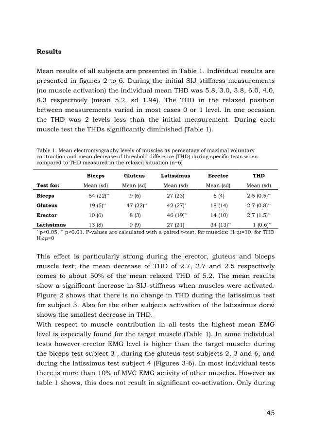
presented in figures 2 to 6. During the initial SIJ stiffness measurements
(no muscle activation) the individual mean THD was 5.8, 3.0, 3.8, 6.0, 4.0,
8.3 respectively (mean 5.2, sd 1.94). The THD in the relaxed position
between measurements varied in most cases 0 or 1 level. In one occasion
the THD was 2 levels less than the initial measurement. During each
muscle test the THDs significantly diminished (Table 1).
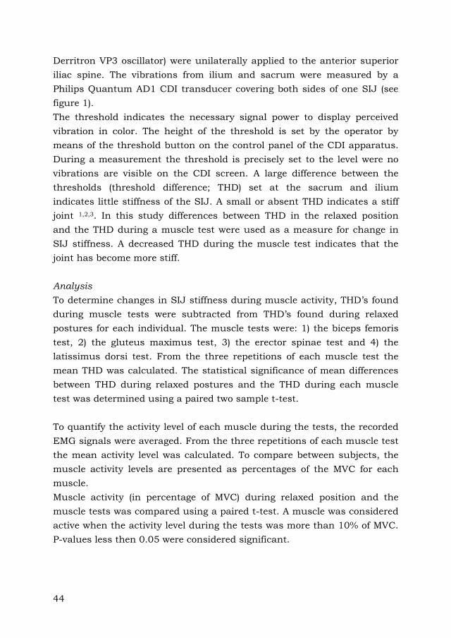
Table 1. Mean electromyography levels of muscles as percentage of maximal voluntary contraction and mean decrease of threshold difference (THD) during specific tests when compared to THD measured in the relaxed situation (n=6)
Test for:
Biceps
Mean (sd)
Gluteus
Mean (sd)
Latissimus
Mean (sd)
Erector
Mean (sd)
THD
Mean (sd)
Biceps 54 (22)** 9 (6) 27 (23) 6 (4) 2.5 (0.5)**
Gluteus 19 (5)** 47 (22)** 42 (27)* 18 (14) 2.7 (0.8)**
Erector 10 (6) 8 (3) 46 (19)** 14 (10) 2.7 (1.5)**
Latissimus 13 (8) 9 (9) 27 (21) 34 (13)** 1 (0.6)** * p<0.05, ** p<0.01. P-values are calculated with a paired t-test, for muscles: H0:µ=10, for THD H0:µ=0
This effect is particularly strong during the erector, gluteus and biceps
muscle test; the mean decrease of THD of 2.7, 2.7 and 2.5 respectively
comes to about 50% of the mean relaxed THD of 5.2. The mean results
show a significant increase in SIJ stiffness when muscles were activated.
Figure 2 shows that there is no change in THD during the latissimus test
for subject 3. Also for the other subjects activation of the latissimus dorsi
shows the smallest decrease in THD.
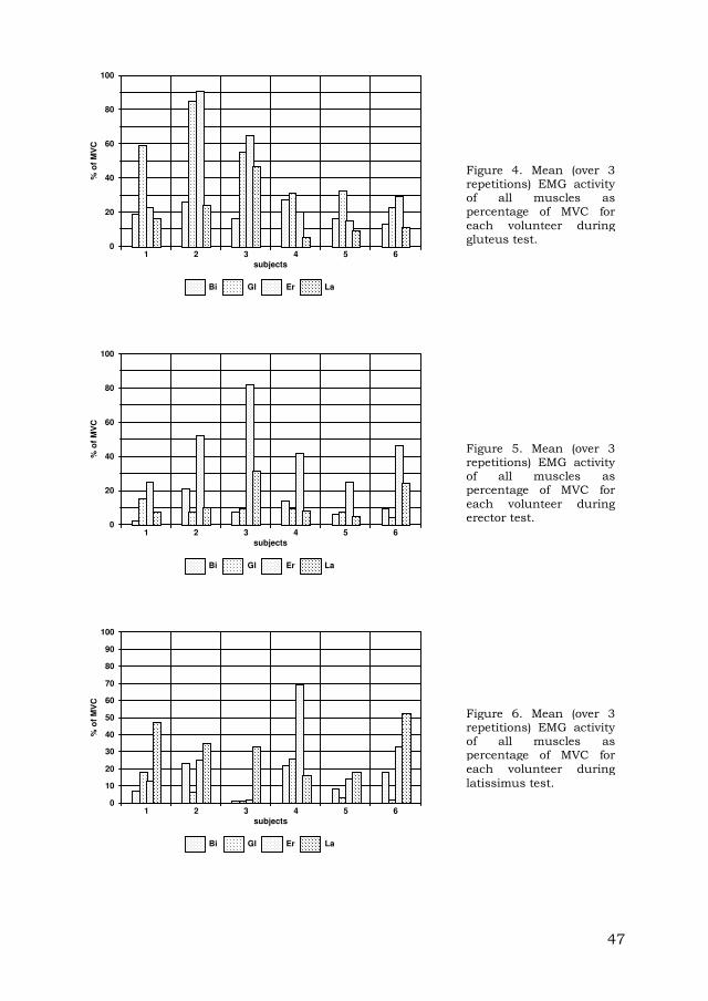
With respect to muscle contribution in all tests the highest mean EMG
level is especially found for the target muscle (Table 1). In some individual
tests however erector EMG level is higher than the target muscle: during
the biceps test subject 3 , during the gluteus test subjects 2, 3 and 6, and
during the latissimus test subject 4 (Figures 3-6). In most individual tests
there is more than 10% of MVC EMG activity of other muscles. However as
table 1 shows, this does not result in significant co-activation. Only during
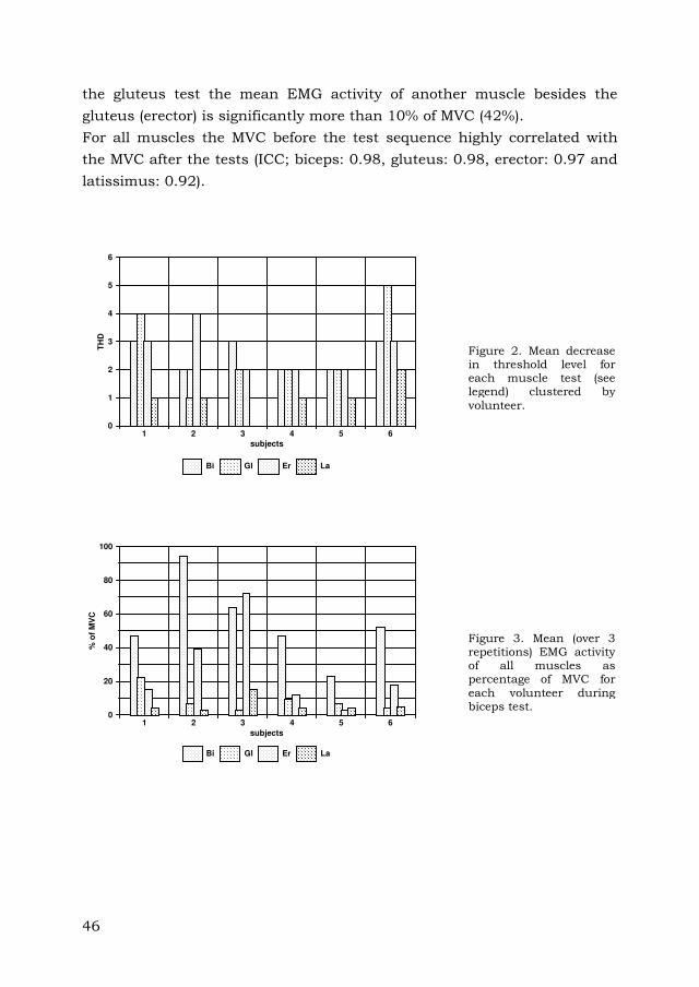
46
the gluteus test the mean EMG activity of another muscle besides the
gluteus (erector) is significantly more than 10% of MVC (42%).
For all muscles the MVC before the test sequence highly correlated with
the MVC after the tests (ICC; biceps: 0.98, gluteus: 0.98, erector: 0.97 and
latissimus: 0.92).
Figure 2. Mean decrease in threshold level for each muscle test (see legend) clustered by volunteer.
Figure 3. Mean (over 3 repetitions) EMG activity of all muscles as percentage of MVC for each volunteer during biceps test.
0
1
2
3
4
5
6
TH
D
1 2 3 4 5 6
subjects
Bi Gl Er La
0
20
40
60
80
100
% o
f M
VC
1 2 3 4 5 6
subjects
Bi Gl Er La
47
0
20
40
60
80
100
% o
f M
VC
1 2 3 4 5 6
subjects
Bi Gl Er La
0
20
40
60
80
100
% o
f M
VC
1 2 3 4 5 6
subjects
Bi Gl Er La
Figure 4. Mean (over 3 repetitions) EMG activity of all muscles as percentage of MVC for each volunteer during gluteus test.
0
10
20
30
40
50
60
70
80
90
100
% o
f M
VC
1 2 3 4 5 6
subjects
Bi Gl Er La
Figure 5. Mean (over 3 repetitions) EMG activity of all muscles as percentage of MVC for each volunteer during erector test.
Figure 6. Mean (over 3 repetitions) EMG activity of all muscles as percentage of MVC for each volunteer during
latissimus test.
48
Discussion
SIJ motion is characterized by minute movements 18,19,20. Color doppler
imaging in combination with pelvic oscillation can be applied to study
sacroiliac stiffness in vivo 1,2,3. This method was used to analyze the
influence of muscle activity on SIJ stiffness. It showed that contraction of
the selected muscles increased SIJ stiffness. The null hypothesis that SIJ
stiffness cannot be influenced by muscle activation must therefore be
rejected. The erector spinae, the biceps femoris and the gluteus maximus
muscles were shown to have the greatest effect on SIJ stiffness. The
latissimus dorsi muscle was shown to have a small effect on SIJ stiffness.
Subject three was able to activate the latissimus dorsi nearly in isolation
(figures 2 and 6), with no change in SIJ stiffness. It can be argued that the
increased SIJ stiffness during the latissimus test in other subjects was due
to action of other muscles than the latissimus dorsi. Besides statistical
significance of the results some intriguing inter-individual differences
occurred in both muscle activation and diminishing of THD (figures 2-6).
These differences may be partly due to individual initial threshold values,
but also to individual muscle activation patterns. Therefore the relative
contribution of specific muscles to SIJ stiffness needs further study.
Although the activated muscle was the most electromyo-graphically active
muscle during all tests (Table 1), the coactivation of other muscles
occurred. The significant cocontraction of biceps femoris and erector
spinae muscles during the gluteus maximus test can be expected, since
effective movement requires orchestrated contractions of multiple muscles
to evoke tailored joint reaction forces [23]. Cocontractions could have been
precluded by using electric muscle stimulation instead of intentional
voluntary isometric muscle activation. A reason for not opting for this
latter solution is that optimal recording of CDI threshold values and thus
establishing realistic values for SIJ stiffening, requires maximal relaxation
of the volunteers. Electric stimulation can be painful with possible
involuntary increase of muscle tone, directly affecting the measurements.
The considerable coactivation of the erector muscle during the biceps,
latissimus and gluteus maximus tests, could be expected since it has been
shown that the aponeurosis and muscle strains of the erector spinae insert
49
on the sacrum, the ilium (PSIS) and partially the long dorsal sacroiliac
ligament and sacrotuberous ligament 24,28. These anatomical connections
explain how the muscle can contribute to stability of the SIJ. This
coactivated function of the erector as described here, is also in agreement
with the stabilizing function of the multifidus part of the muscle as
described by Hides et al. 5. Their study shows that the multifidus is
coactive with the transverse abdominals and possibly oblique abdominals
as primary stabilizers of spine and pelvis 5,6,7. Since in the present study
surface electrodes were used, the abdominal muscles could not be
included.
During the gluteus maximus test the activity of erector spinae is
particularly high. An additional reason for this activity could be that the
subjects were asked to ‘take the weight of their upper leg from the table’,
thus activating the erector in the process of stabilizing pelvis and spine.
The influence of muscles on SIJ stiffness as demonstrated in this study
could have clinical consequences. In the clinic, joint stiffness is commonly
determined by means of the manual skills of the clinician. However, it was
shown that the intra and inter tester reliability of manual tests is low 14. To
our knowledge no studies have been performed to reveal to what extent
poor reproducibility of manual tests, could be related to variance of muscle
tension and hence joint stiffness between tests (in fact intra-joint or
patient reliability). The present study showed that SIJ stiffness is
influenced by muscle activity and thus by motor patterns. It can be
expected that this also holds for joint stiffness in general. Small variations
in the excitation pattern of muscles can lead to differences in joint
stiffness. Consequently, during retesting of joints in patients, relatively
small postural changes can result in altered muscle contraction patterns
and subsequently influence the inter and intra tester reliability of manual
joint play tests.
The use of CDI in combination with bone oscillation gives valid results;
however, the method is not easy to use in daily practice 1,2,3. To ascertain
valid results in this study only subjects with a relatively high (more than
2.5) THD during the relaxed posture were chosen. The particular aim of
the study was only to demonstrate the effect of muscle contraction on SIJ
stiffness. Therefore the small number of included subjects 6 as a
50
consequence of the high THD criterion, was considered acceptable for this
study. New studies on specific muscles like the transverse and oblique
abdominous, using selective electro-stimulation, are necessary 5, 6, 7,17.
This study wanted to show that joint stiffness is not only influenced by
structural quality and integrity of the joint but is also influenced by the
dynamics of muscle activity. It therefore can be assumed that even when
no muscle activity is detected on EMG, basic muscle tone already
influences joint stiffness. Emotional states are known to influence basic
muscle tone and patterning 8. The effect of emotional states on specific
muscle patterns needs to be taken into account when analyzing SIJ
function.
In conclusion, this in vivo study showed that stiffness of the SIJ was
increased by certain muscle activity. This supported the model proposed
that load transfer from spine to legs is enhanced when muscles actively
compress the SIJ thus preventing shear 16, 17,21,22,23. This agrees with a
recent study by Sturesson et al. who demonstrated that in postures with
long lever arms, as in stooped positions, SIJ motion became restricted 18,
19.
This in vivo study enhanced our understanding on how muscles
dynamically influence SIJ stiffness. The results however, could have
implications for joints in general. When joints are manually tested, the
influence of muscle activation patterns must be taken into consideration to
recognize how both inter and intra tester reliability can be influenced. In
this respect the relation between emotional states, muscle activities, SIJ
stiffness and joint stiffness in general deserves further exploration.
51
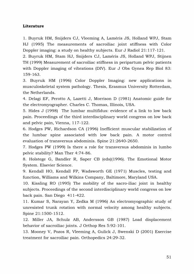
Literature
1. Buyruk HM, Snijders CJ, Vleeming A, Laméris JS, Holland WPJ, Stam
HJ (1995) The measurements of sacroiliac joint stiffness with Color
Doppler imaging: a study on healthy subjects. Eur J Radiol 21:117-121.
2. Buyruk HM, Stam HJ, Snijders CJ, Laméris JS, Holland WPJ, Stijnen
TH (1999) Measurement of sacroiliac stiffness in peripartum pelvic patients
with Doppler imaging of vibrations (DIV). Eur J Obs Gynea Rep Biol 83:
159-163.
3. Buyruk HM (1996) Color Doppler Imaging: new applications in
musculoskeletal system pathology. Thesis, Erasmus University Rotterdam,
the Netherlands.
4. Delagi EF, Perotto A, Lazetti J, Morrison D (1981) Anatomic guide for
the electromyographer. Charles C. Thomas, Illinois, USA.
5. Hides J (1998) The lumbar multifidus: evidence of a link to low back
pain. Proceedings of the third interdisciplinary world congress on low back
and pelvic pain, Vienna, 117-122.
6. Hodges PW, Richardson CA (1996) Inefficient muscular stabilization of
the lumbar spine associated with low back pain. A motor control
evaluation of transversus abdominis. Spine 21:2640-2650.
7. Hodges PW (1999) Is there a role for transversus abdominis in lumbo
pelvic stability? Man Ther 4:74-86.
8. Holstege G, Bandler R, Saper CB (eds)(1996). The Emotional Motor
System. Elsevier Science.
9. Kendall HO, Kendall FP, Wadsworth GE (1971) Muscles, testing and
function, Williams and Wilkins Company, Baltimore, Maryland USA.
10. Kissling RO (1995) The mobility of the sacro-iliac joint in healthy
subjects. Proceedings of the second interdisciplinary world congress on low
back pain. San Diego 411-422.
11. Kumar S, Narayan Y, Zedka M (1996) An electromyographic study of
unresisted trunk rotation with normal velocity among healthy subjects.
Spine 21:1500-1512.
12. Miller JA, Schulz AB, Andersson GB (1987) Load displacement
behavior of sacroiliac joints. J Orthop Res 5:92-101.
13. Mooney V, Pozos R, Vleeming A, Gulick J, Swenski D (2001) Exercise
treatment for sacroiliac pain. Orthopedics 24:29-32.
52
14. Potter NA, Rothstein JM (1989) Intertester reliability for selected
clinical tests of the sacroiliac joint. Phys Ther 65:1671-1675.
15. Smidt GL, Wei S-H, McQuade K, Barakatt E, Tiansheng S, Stanford W
(1997) Sacroiliac motion for extreme hip positions. Spine 22:2073-2082.
16. Snijders CJ, Vleeming A, Stoeckart R (1993) Transfer of lumbosacral
load to iliac bones and legs. Part 1 and 2. Clin Biomech 8: 285-294.
17. Snijders CJ, Slagter AHE, Strik R van, Vleeming A, Stoeckart R, Stam
HJ (1995) Why leg-crossing? The influence of common postures on
abdominal muscle activity. Spine 20:1989-1993.
18. Sturesson B, Selvik GA, Udén U (1989) Movements of the sacroiliac
joints. Spine 14:162-165.
19. Sturesson B, Udén U, Vleeming A (2000) A radiostereometric analysis
of movement of the sacroiliac joints during the standing hip flexion test.
Spine 25:364-368.
20. Vleeming A, Stoeckart R, Snijders CJ (1989a) The sacrotuberous
ligament: a conceptual approach to its dynamic role in stabilizing the
sacroiliac joint. Clin Biomech 4:201-203.
21. Vleeming A, Wingerden JP van, Snijders CJ, Stoeckart R, Stijnen T
(1989b) Load application to the sacrotuberous ligament; influences on
sacroiliac joint mechanics. Clin Biomech 4:204-209.
22. Vleeming A, Stoeckart R, Volkers ACW, Snijders CJ (1990a) Relation
between form and function in the sacro-iliac joint, Part 1: Clinical
anatomical aspects. Spine 15:130-132.
23. Vleeming A, Volkers ACW, Snijders CJ, Stoeckart R (1990) Relation
between form and function in the sacro-iliac joint, Part 2: Biomechanical
aspects. Spine 15:133-136.
24. Vleeming A, Pool-Goudzwaard AL, Hammudoghlu D, Stoeckart R,
Snijders CJ, Mens JMA (1996) The function of the long dorsal sacroiliac
ligament; its implication for understanding low back pain. Spine 21:556-
562.
25. Vleeming A, Wingerden JP van, Dijkstra PF, Stoeckart R, Snijders CJ,
Stijnen T(1992) Mobility in the sacroiliac joints in the elderly: A kinematic
and radiological study. Clin Biomech 7:170-176.
26. Vleeming A (1990) The sacroiliac joint, a clinical-anatomical,
biomechanical and radiological study. Thesis, Erasmus University
Rotterdam, the Netherlands.
53
27. Vleeming A, Buyruk HM, Stoeckart R, Karamürsel S, Snijders CJ
(1992) An integrated therapy for peripartum pelvic instability: A study of
the biomechanical effects of pelvic belts. Am J Obstet Gynaecol 166:1243-
1247.
28. Vleeming A, Pool-Goudzwaard AL, Stoeckart R, Wingerden JP van,
Snijders CJ (1995) The posterior layer of the thoracolumbar fascia; its
function in load transfer from spine to legs. Spine 20:753-758.
29. Vleeming A, Snijders CJ, Stoeckart R, Mens JMA (1997) The role of the
sacroiliac joints in coupling between spine, pelvis, legs, and arms. In:
Vleeming A, Mooney V, Dorman T, Snijders CJ, Stoeckart R (eds.)
Movement, Stability & Low Back Pain. Churchill Livingstone, 53-71.
30. Wingerden JP van, Vleeming A, Snijders CJ, Stoeckart R (1993) A
functional-anatomical approach to the spine-pelvis mechanism: interaction
between the biceps femoris muscle and the sacrotuberous ligament. Eur
Spine J 2:140-144.
31. Wingerden JP van, Vleeming A, Stam HJ , Stoeckart R (1995)
Interaction of spine and legs: influence of hamstring tension on lumbo-
pelvic rhythm. Proceedings of second interdisciplinary world congress on
low back pain.

54
Chapter 4
Differences in standing and forward bending in women with chronic low back or pelvic girdle pain; indications for physical compensation strategies
J.P. van Wingerden BSc, PT, A. Vleeming PhD, I. Ronchetti MSc.
Spine & Joint Centre, The Netherlands

56
Abstract
Study design
This cohort study compares motion characteristics during forward bending
of a group of chronic female patients either with low back pain (LBP) or
pelvic girdle pain (PGP) and healthy subjects using computer-video
analysis.
Objective
This study determines whether subcategories of back pain patients could
be distinguished by motion characteristics of the pelvis and lumbar spine.
Summary of background data
Compared with healthy subjects, patients with low back pain bend forward
in distinct manners. Clustering these motion patterns into specific patient
subgroups has been challenging since a basis for subcategorizing was
lacking.
Chronic LBP can be distinguished from PGP using specific evidence based
diagnostic tests. This allows comparing the motion characteristics of
subgroups of chronic patients with either LBP or PGP.
Methods
Forward bending was recorded in both female patients groups and healthy
female individuals, using a computer video analysis system.
Trunk motion, pelvic tilt and lumbar lordosis are represented as sagittal
plane angles. From these angles the relative contribution of the lumbar
spine and hip joint to forward bending can be derived.
Results
Specific and discriminating motion characteristics were found between
groups. During erect stance in the PGP group the pelvis is significantly
tilted backwards. At maximally forward bending the ROM of the trunk is
limited in all patient groups, but only the PGP group has significantly
limited hip motion. During the initial part of forward bending lumbar
motion is increased in PGP patients and decreased in LBP patients. In the

57
final part of forward bending contribution of the lumbar spine is increased
in both patient groups.
Conclusions
LBP and PGP patients show specific, consistent and distinct motion
patterns. These motion patterns are assumed to be functional
compensation strategies, following altered neuromuscular coordination.
58
Introduction
One of the main problems in non-specific low back pain is the lack of
adequate sub- categories allowing specific fine-tuning of therapeutic
interventions. Since in the past sub-categorizing on a structural basis was
found to be fruitless, present research predominantly aims at sub
categorisation based on functional analysis 1-11. For successful functional
sub categorisation both an adequate parameter and a preliminary sub
classification preferably based on a “gold standard” are essential. This
study aims at subcategorizing two back pain groups using forward bending
as a discriminating tool.
In healthy subjects forward bending consists of trunk flexion, which is
flexion of the (lumbar) spine combined with pelvic tilt (hip flexion). The
coordination of the lumbar spine and pelvis during this motion is not
arbitrary, but specifically and consistently coupled 12-19.
In the 1960’s, Cailliet described the specific motion pattern of spine and
pelvis, coined the lumbar-pelvic rhythm, similar to the scapulo-thoracal
rhythm 20. Compared with healthy subjects, patients with low back pain
(LBP) usually bend forward in a distinct manner 5,7,15,20-28.
Because of the assumed relation between low back pain and the specific
motion patterns during forward bending, this subject has been well
studied 4,5,7,10,15,21,22,26,27,29. Most studies found differences in the motion
patterns between healthy individuals and LBP patients. However attempts
to cluster motion patterns of specific subgroups of “non-specific” low back
pain patients remains difficult especially because of the considerable
variation in the motion patterns found and also the lacking of evidence
based diagnostic tests to discriminate subgroups 1,29,30,31.
With respect to the latter problem, in a recent European Guideline on
Pelvic Girdle Pain (PGP) a definition was constructed for pelvic
musculoskeletal pain as follows:
“Pelvic girdle pain (PGP) generally arises in relation to pregnancy, trauma or
reactive arthritis. Pain is experienced between the posterior iliac crest and
the gluteal fold, particularly in the vicinity of the sacroiliac joints (SIJ). The
pain may radiate in the posterior thigh and can also occur in conjunction
with/or separately in the symphysis. The endurance capacity for standing,
walking, and sitting is diminished. The diagnosis of PGP can be reached

59
after exclusion of lumbar causes. The pain or functional disturbances in
relation to PGP must be reproducible by specific clinical tests” 32.
This European PGP guideline considers, among valid tests like the the
Gaenslen and Patricks Faber test, specific tests such as the Active Straight
Leg Raise (ASLR), the Long Dorsal Ligament (LDL) and the Posterior Pelvic
Pain Provocation (PPPP) test valuable in discriminating PGP patients from
healthy subjects and low back pain patients 31-35. Functionally, PGP
patients can be distinguished from regular LBP patients by certain motion
characteristics like in walking 3,11,36. As shown in the study by Wu et al.,
PGP patients do not only walk at lower speed, their coordination during
walking is also distinct from LBP patients and healthy subjects 11. This
leads to the assumption in the present study that also the coupled motion
of lumbar spine and pelvis could differ between LBP and PGP patients
Comparison and analysis of the motion patterns of LBP and PGP patients
may provide new insight in the aetiology of chronic of low back and pelvic
pain.
The specific patient population of a Dutch rehabilitation centre specialized
in the treatment of severe low back pain and pelvic girdle pain allowed to
compare the motion patterns of LBP and PGP patients.
The aim of the present study was to demonstrate that consistent and
discriminating motion patterns exist for the mentioned subgroups. When
this assumption is proven correct, analysis of coordination may provide
useful information for therapy of LBP and PGP.
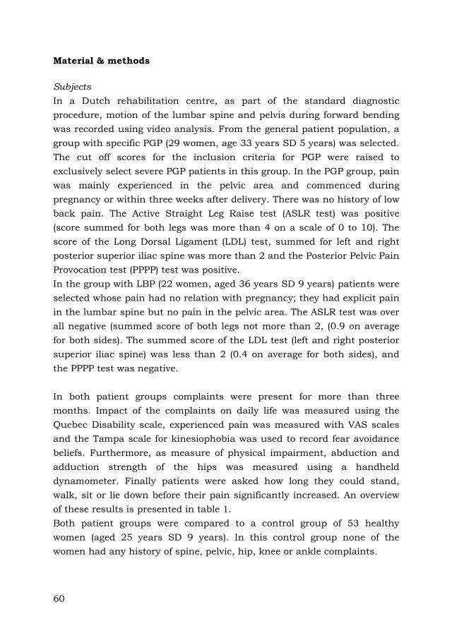
60
Material & methods
Subjects
In a Dutch rehabilitation centre, as part of the standard diagnostic
procedure, motion of the lumbar spine and pelvis during forward bending
was recorded using video analysis. From the general patient population, a
group with specific PGP (29 women, age 33 years SD 5 years) was selected.
The cut off scores for the inclusion criteria for PGP were raised to
exclusively select severe PGP patients in this group. In the PGP group, pain
was mainly experienced in the pelvic area and commenced during
pregnancy or within three weeks after delivery. There was no history of low
back pain. The Active Straight Leg Raise test (ASLR test) was positive
(score summed for both legs was more than 4 on a scale of 0 to 10). The
score of the Long Dorsal Ligament (LDL) test, summed for left and right
posterior superior iliac spine was more than 2 and the Posterior Pelvic Pain
Provocation test (PPPP) test was positive.
In the group with LBP (22 women, aged 36 years SD 9 years) patients were
selected whose pain had no relation with pregnancy; they had explicit pain
in the lumbar spine but no pain in the pelvic area. The ASLR test was over
all negative (summed score of both legs not more than 2, (0.9 on average
for both sides). The summed score of the LDL test (left and right posterior
superior iliac spine) was less than 2 (0.4 on average for both sides), and
the PPPP test was negative.
In both patient groups complaints were present for more than three
months. Impact of the complaints on daily life was measured using the
Quebec Disability scale, experienced pain was measured with VAS scales
and the Tampa scale for kinesiophobia was used to record fear avoidance
beliefs. Furthermore, as measure of physical impairment, abduction and
adduction strength of the hips was measured using a handheld
dynamometer. Finally patients were asked how long they could stand,
walk, sit or lie down before their pain significantly increased. An overview
of these results is presented in table 1.
Both patient groups were compared to a control group of 53 healthy
women (aged 25 years SD 9 years). In this control group none of the
women had any history of spine, pelvic, hip, knee or ankle complaints.
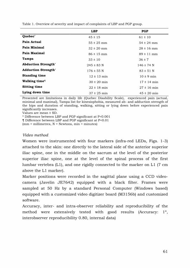
61
Table 1. Overview of severity and impact of complaints of LBP and PGP group.
LBP PGP
Quebec* 45 ± 15 61 ± 10
Pain Actual 55 ± 25 mm 54 ± 24 mm
Pain Minimal 32 ± 20 mm 28 ± 16 mm
Pain Maximal 86 ± 15 mm 89 ± 11 mm
Tampa 33 ± 10 36 ± 7
Abduction Strength* 245 ± 83 N 146 ± 74 N
Adduction Strength* 176 ± 55 N 83 ± 51 N
Standing time 12 ± 13 min 10 ± 9 min
Walking time¶ 30 ± 20 min 17 ± 14 min
Sitting time 22 ± 18 min 27 ± 16 min
Lying down time 37 ± 25 min 45 ± 20 min
Presented are limitations in daily life (Quebec Disability Scale), experienced pain (actual, minimal and maximal), Tampa list for kinesiophobia, measured ab- and adduction strength of the hips and duration of standing, walking, sitting or lying down before experienced pain significantly increases. Values are mean ± SD. * Difference between LBP and PGP significant at P<0.001 ¶ Difference between LBP and PGP significant at P<0.01 (mm = millimetres, N = Newtons, min = minutes)
Video method
Women were instrumented with four markers (infra-red LEDs, Figs. 1-3)
attached to the skin: one directly to the lateral side of the anterior superior
iliac spine, one in the middle on the sacrum at the level of the posterior
superior iliac spine, one at the level of the spinal process of the first
lumbar vertebra (L1), and one rigidly connected to the marker on L1 (7 cm
above the L1 marker).
Marker positions were recorded in the sagittal plane using a CCD video-
camera (Javelin JE7642) equipped with a black filter. Frames were
sampled at 50 Hz by a standard Personal Computer (Windows based)
equipped with a customized video digitizer board (M3156b) and customized
software.
Accuracy, inter- and intra-observer reliability and reproducibility of the
method were extensively tested with good results (Accuracy: 1º,
interobserver reproducibility 0.80, internal data)
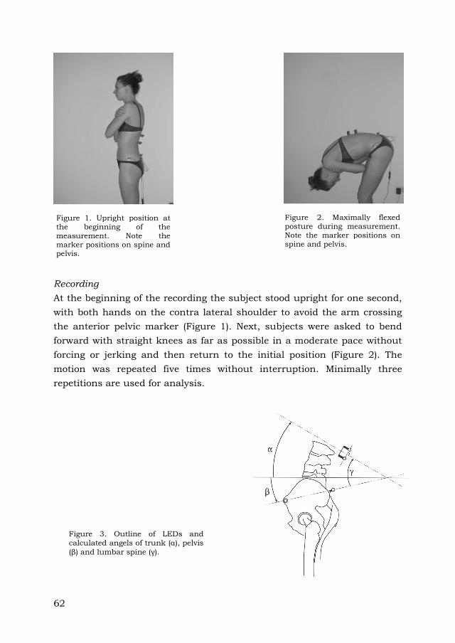
62
Recording
At the beginning of the recording the subject stood upright for one second,
with both hands on the contra lateral shoulder to avoid the arm crossing
the anterior pelvic marker (Figure 1). Next, subjects were asked to bend
forward with straight knees as far as possible in a moderate pace without
forcing or jerking and then return to the initial position (Figure 2). The
motion was repeated five times without interruption. Minimally three
repetitions are used for analysis.
Figure 1. Upright position at the beginning of the measurement. Note the marker positions on spine and pelvis.
Figure 2. Maximally flexed posture during measurement. Note the marker positions on spine and pelvis.
Figure 3. Outline of LEDs and calculated angels of trunk (α), pelvis
(β) and lumbar spine (γ).
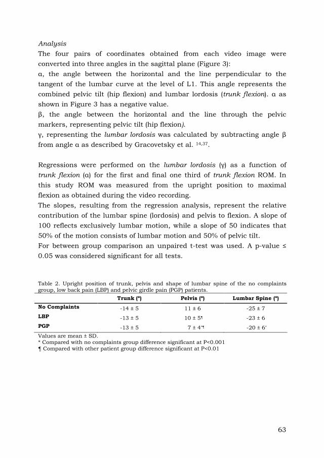
63
Analysis
The four pairs of coordinates obtained from each video image were
converted into three angles in the sagittal plane (Figure 3):
α, the angle between the horizontal and the line perpendicular to the
tangent of the lumbar curve at the level of L1. This angle represents the
combined pelvic tilt (hip flexion) and lumbar lordosis (trunk flexion). α as
shown in Figure 3 has a negative value.
β, the angle between the horizontal and the line through the pelvic
markers, representing pelvic tilt (hip flexion).
γ, representing the lumbar lordosis was calculated by subtracting angle β
from angle α as described by Gracovetsky et al. 14,37.
Regressions were performed on the lumbar lordosis (γ) as a function of
trunk flexion (α) for the first and final one third of trunk flexion ROM. In
this study ROM was measured from the upright position to maximal
flexion as obtained during the video recording.
The slopes, resulting from the regression analysis, represent the relative
contribution of the lumbar spine (lordosis) and pelvis to flexion. A slope of
100 reflects exclusively lumbar motion, while a slope of 50 indicates that
50% of the motion consists of lumbar motion and 50% of pelvic tilt.
For between group comparison an unpaired t-test was used. A p-value ≤
0.05 was considered significant for all tests.
Table 2. Upright position of trunk, pelvis and shape of lumbar spine of the no complaints group, low back pain (LBP) and pelvic girdle pain (PGP) patients.
Trunk (º) Pelvis (º) Lumbar Spine (º)
No Complaints -14 ± 5 11 ± 6 -25 ± 7
LBP -13 ± 5 10 ± 5¶ -23 ± 6
PGP -13 ± 5 7 ± 4*¶ -20 ± 6*
Values are mean ± SD. * Compared with no complaints group difference significant at P<0.001 ¶ Compared with other patient group difference significant at P<0.01
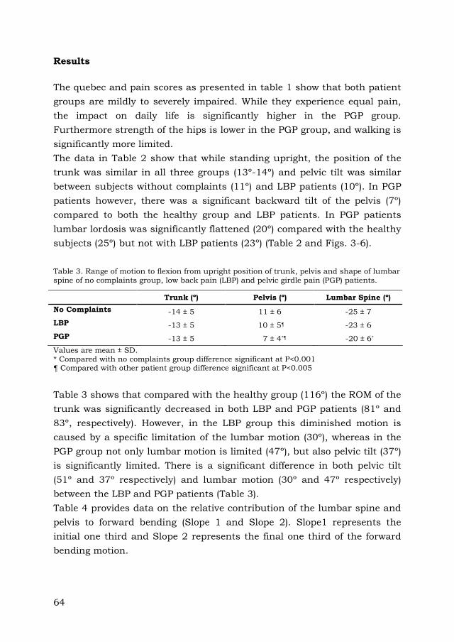
64
Results
The quebec and pain scores as presented in table 1 show that both patient
groups are mildly to severely impaired. While they experience equal pain,
the impact on daily life is significantly higher in the PGP group.
Furthermore strength of the hips is lower in the PGP group, and walking is
significantly more limited.
The data in Table 2 show that while standing upright, the position of the
trunk was similar in all three groups (13º-14º) and pelvic tilt was similar
between subjects without complaints (11º) and LBP patients (10º). In PGP
patients however, there was a significant backward tilt of the pelvis (7º)
compared to both the healthy group and LBP patients. In PGP patients
lumbar lordosis was significantly flattened (20º) compared with the healthy
subjects (25º) but not with LBP patients (23º) (Table 2 and Figs. 3-6).
Table 3. Range of motion to flexion from upright position of trunk, pelvis and shape of lumbar spine of no complaints group, low back pain (LBP) and pelvic girdle pain (PGP) patients.
Trunk (º) Pelvis (º) Lumbar Spine (º)
No Complaints -14 ± 5 11 ± 6 -25 ± 7
LBP -13 ± 5 10 ± 5¶ -23 ± 6
PGP -13 ± 5 7 ± 4*¶ -20 ± 6*
Values are mean ± SD. * Compared with no complaints group difference significant at P<0.001 ¶ Compared with other patient group difference significant at P<0.005
Table 3 shows that compared with the healthy group (116º) the ROM of the
trunk was significantly decreased in both LBP and PGP patients (81º and
83º, respectively). However, in the LBP group this diminished motion is
caused by a specific limitation of the lumbar motion (30º), whereas in the
PGP group not only lumbar motion is limited (47º), but also pelvic tilt (37º)
is significantly limited. There is a significant difference in both pelvic tilt
(51º and 37º respectively) and lumbar motion (30º and 47º respectively)
between the LBP and PGP patients (Table 3).
Table 4 provides data on the relative contribution of the lumbar spine and
pelvis to forward bending (Slope 1 and Slope 2). Slope1 represents the
initial one third and Slope 2 represents the final one third of the forward
bending motion.
65
Figure 4. Example of motion pattern typical for healthy subjects, with trunk flexion (α) on the x-axis and lumbar lordosis (γ) on the y-axis. Slope1 and Slope2 represent the relative contribution of the lumbar spine (lordosis) to the first and final one third of flexion respectively.
Figure 5. Example of motion pattern typical for LBP patients subjects, with trunk flexion (α) on the x-axis and lumbar lordosis (γ) on the y-axis. Slope1 and Slope2 represent the relative contribution of the lumbar spine (lordosis) to the first and final one third of flexion respectively.
Figure 6. Example of motion pattern typical for PGP patients, with trunk flexion (α) on the x-axis and lumbar lordosis (γ) on the y-axis. Slope1 and Slope2 represent the relative contribution of the lumbar spine (lordosis) to the first and final one third of flexion respectively.
66
For the LBP patients the Slope 1 is significantly smaller compared to
healthy controls (57.7% and 66.9%, respectively), indicating that LBP
patients maintain lordosis in the initial flexion. In contrast, the Slope 1 of
PGP patients (71.2%) is significantly increased compared to healthy
controls (Table 4 and Figs. 4-6). This result shows that in contrast to both
healthy subjects and LBP patients, PGP patients emphasise lumbar motion
in the initial phase of forward bending.
In the final phase of forward bending the Slope 2 is significantly increased
in both LBP and PGP patients compared to healthy controls,
demonstrating that both patient groups have more lumbar motion in the
final stage of flexion.
Table 4. Range of motion to flexion from upright position of trunk, pelvis and shape of lumbar spine of no complaints group, low back pain (LBP) and pelvic girdle pain (PGP) patients.
Trunk (º) Pelvis (º) Lumbar Spine (º)
No Complaints 116 ± 14 56 ± 13 60 ± 9
LBP 81 ± 23* 51 ± 18¶ 30 ± 16*¶
PGP 83 ± 28* 37 ± 19*¶ 47 ± 14*¶
Values are mean ± SD. * Compared with no complaints group difference significant at P<0.001 ¶ Compared with other patient group difference significant at P<0.005
Table 5. Relative contribution (RC) of lumbar spine to forward bending of no complaints group, low back pain (LBP) and pelvic girdle pain (PGP) patients.
Slope 1 (%) Slope 2 (%)
No Complaints 66.9 ± 7.4 29.6 ± 12.0
LBP 57.7 ± 14.7*¶ 49.3 ± 17.5*
PGP 71.2 ± 12.7*¶ 47.0 ± 17.3*
Values are mean ± SD. * Compared with no complaints group difference significant at P<0.05 ¶ Compared with other patient group difference significant at P<0.001 Slope1 = the relative contribution of the lumbar spine (lordosis) to the first one third of flexion. Slope2 = the relative contribution of the lumbar spine (lordosis) to the final one third of flexion.
67
Discussion
This study investigated motion strategies in female patients with female
chronic LBP and chronic PGP compared to healthy female controls. Firstly,
before the initiation of movement PGP patients stand with especially more
backward pelvic tilt but also with a slight flattened lordosis compared to
both healthy subjects and LBP patients. Secondly, during forward bending
the coupled motion of lumbar spine and pelvis during the initial phase of
the motion differed significantly between both patient groups. Especially
during the first one third of forward bending LBP patients tend to maintain
lordosis, whereas PGP pain patients emphasise lumbar flexion. Although
the coupled motion of lumbar spine and pelvis has been well investigated,
this distinct motion pattern between two groups of “non-specific” back
pain patients has not been reported previously. In two earlier studies, LBP
patients could be divided in two subgroups: one with normal coupled
motion of lumbar spine and pelvis and one with altered coupled motion 7,31, however, no satisfactory reason for these differences was provided. In
the study by Paquet et al., it is unclear whether male or female subjects (or
both) were included 7, so it is likely that LPB and PGP patients were mixed.
In the study by Porter et al. 26 a subgroup was found with reduced hip
flexion (e.g. limited pelvic tilt during forward bending). Although this
motion pattern is similar to that found in the PGP group in this study
there are no clear indications in the Porter study that their subgroup had
PGP rather than LBP. This also applies to the study by Esola et al. which
compared the coupled motion of spine and pelvis during forward bending
of 14 males and 6 females 9. In their study, the spine/hip ratios (as a
measure of relative contribution of spine and pelvis to forward bending)
have large standard deviations, especially for the first part of the flexion,
indicating a substantial variation in the spine/hip ratios. Such variation
can occur when LBP and PGP patients, with distinct motion patterns as
shown in the present study, are mixed in the same study population.
Possible explanations why LBP patients maintain lordosis
In contrast to the motion strategies of healthy subjects, LBP patients tend
to maintain lordosis during forward bending. Many authors consider this
specific motion pattern as a natural protection response of the body during
68
a back problem 14,22,28,35,37-42. Consequently this motion pattern is often
advised to patients as the “squat” lifting technique 43. However, it can be
argued that maintaining lordosis is not a solution for a back problem, but
a direct consequence of the back problem. In LBP patients the recruitment
pattern of the m. multifidus frequently changes, diminishing its
anticipatory, stabilizing effect 15,39,44,45. To guarantee stability, despite this
altered activity of multifidus, other muscles (especially the m. erector
spinae) become more active 2,39. Due to its anatomic orientation the m.
erector spinae does not stabilize the lumbar spine on a segmental level,
but merely increases compression, pulling the lumbar spine into lordosis.
Consequently, coordination of segmental motion during forward bending is
disturbed when using m. erector spinae predominantly. Therefore, it could
be speculated that when m. erector spinae activity is increased to
compensate for diminished m. multifidus activity this results in
maintained lordosis during forward bending, as shown in the present
study. Since many other factors could lead to the described patterning this
is still an incomplete analysis which requires further study.
Possible explanations why PGP patients emphasise lumbar flexion
In contrast to LBP patients, PGP patients emphasise lumbar flexion in the
initial phase of forward bending. Like in the LBP group the motion pattern
found could be a consequence of the specific pelvic problem.
To comprehend stability of the pelvis, a joint model of form and force
closure has been introduced 46. According to this model several structures
surrounding the SIJ can stabilize the joint by increasing joint compression 19,34,46-49. The sacrotuberous ligament is one such structure that stabilizes
the SIJ 32,49,50. Since the sacrotuberous ligament is connected to the long
head of the m. biceps femoris and the m. gluteus maximus, by increasing
tension of the sacrotuberous ligament these muscles can dynamically
stabilize the SIJ 13,19,32,48,49. Indahl et al. showed that, in analogy to
zygapophysial joints, the capsule of the SIJ plays an important role in the
neuromuscular control of its stabilizing muscles 51. When neuromuscular
control of the SIJ is disturbed compensatory means of stabilization could
be addressed, such as increased activation of the biceps femoris muscle,
which was elegantly explained in a study by Hungerford et al, but also
indicated by other studies 3,5,6,32,49,50. Because increased tension of the

69
biceps femoris or m. gluteus maximus also resists the pelvic rotation in the
hip joint this limits the contribution of the pelvis to forward bending.
Consequently, lumbar motion will be emphasised, as was shown in the
present study. However, other explanations for the presented phenomenon
can not be excluded.
The present study compared two specifically selected groups of patients
with low back problems. However, it can be expected that when groups
with less outspoken differences are compared, the motion patterns will be
less distinct: the lumbar spine and pelvis are not separate entities but are,
from a functional perspective, mutually dependent systems 19,46,34. Low
back problems and their compensatory strategies will have an impact on
pelvic function, and vice versa. The explicit distinction between groups in
this study was made for methodological reasons but LBP and PGP can
occur in mixed variations. Therefore in a clinical setting pelvic function
should also be examined in LBP patients, and consequently, lumbar
function should be analysed in PGP patients.
In this study no men were included because it was assumed that specific
differences in motion patterns could occur between sexes. Motion patterns
in men, as the differences in motion patterns between men and women,
needs further study.
Conclusion
This study shows that coupled motion of the low back and pelvis, studied
in strictly classified subgroups of LBP and PGP patients is specific and
discriminating between groups. It is postulated that the specific motion
patterns in patients could be functional compensation strategies of the
body possibly following adjusted neuromuscular coordination [45, 51].
The distinct coupled motion of lumbar spine and pelvis, combined with
more impaired walking and lower hip strength in PGP patients emphasises
the notion that LBP and PGP patients belong to distinct patients groups.
Analysis of their specific compensatory patterns may elucidate how our
body attempts to compensate for functional disturbances. In the clinical
setting this may enable more specific exercise programmes to be developed
for both LBP and PGP patients.
70
References
1. Dankaerts W, O’Sullivan P, Burnett A. Straker L. Altered patterns of
superficial trunk muscle activation during sitting in non-specific chronic
low back pain patients: importance of subclassification. Spine 2006 Aug
1;31(17):2017-2023.
2. Danneels LA, Coorevits PL, Cools AM, Vanderstraeten GG, Cambier DC,
Witvrouw EE. Differences in electromyographic activity in the multifidus
muscle and the iliocostalis lumborum between healthy subjects and
patients with sub-acute and chronic low back pain. Eur Spine J
2002;11:13-9.
3. Hungerford B, Gilleard W, Hodges P. Evidence of altered lumbopelvic
muscle recruitment in the presence of sacroiliac joint pain. Spine 2003
28:1593-600.
4. Leinonen V, Kankaanpaa M, Airaksinen O, Hanninen O. Back and hip
extensor activities during trunk flexion/extension: effects of low back pain
and rehabilitation. Arch Phys Med Rehabil. 2000 Jan;81(1):32-37.
5. Neblett R, Mayer TG, Gatchel RJ, Keeley J, Procter T, Anagnostis C.
Quantifying the lumbar flexion-relaxation phenomenon: theory, normative
data, and clinical applications. Spine 2003 Jul 1;28(13): 1435-1446.
6. O'Sullivan PB, Beales DJ, Beetham JA, Cripps J, Graf F, Lin IB, Tucker
B, Avery A. Altered motor control strategies in subjects with sacroiliac
joint pain during the active straight-leg-raise test. Spine 2002;27:E1-8.
7. Paquet N, Malouin F, Richards CL. Hip-spine movement interaction and
muscle activation patterns during sagittal movement in low back pain
patients. Spine 1994;19:596-603.
8. Pirouzi S, Hides J, Richardson C, Darnell R, Toppenberg R. Low back
pain patients demonstrate increased hip extensor muscle activity during
standardized submaximal rotation efforts. Spine 2006 Dec 15;31(26):E999-
E1005.
9. Pool-Goudswaard AL, Vleeming A, Stoeckart R, Snijders CJ, Mens JMA.
Insufficient lumbopelvic stability: a clinical and biomechanical approach to
‘specific’ low back pain. Man Ther 1998;3:2-20.
10. Silfies SP, Squillante D, Maurer P, Westcott S, Karduna AR. Trunk
muscle recruitment patterns in specific chronic low back pain populations.
Clin Biomech 2005 Jun; 20(5):456-473.
71
11. Wu W, Meijer OG, Jutte PC, Uegaki K, Lamoth CJ, de Wolf SG., van
Dieen JH, Wuisman PI, Kwakkel G, de Vries JI, Beek PJ. Gait in patients
with pregnancy-related pain in the pelvis: an emphasis on the coordination
of transverse pelvic and thoracic rotations. Clin Biomech 2002;17:678-86.
12. Adams MA, Dolan P, Marx C, Hutton WC. An electronic inclinometer
for measuring lumbar curvature. Clin Biomech 1986;1:130-34.
13. Gajdosik RL, Hatcher CK, Whitsell S. Influence of short hamstring
muscles on the pelvis and lumbar spine in standing and during the
toe-touch test. Clin Biomech 1992;7:38-42.
14. Gracovetsky S, Kary M, Levy S, Ben Said R, Pitchen I, Helie J. Analysis
of spinal and muscular activity during flexion/extension and free lifts.
Spine 1990;15:1333-9.
15. Sihvonen T, Partanen J, Häninnen O, Soimakallio S. Electric behaviour
of low back muscles during lumbar pelvic rhythm in low back pain
patients and healthy controls. Arch Phys Med Rehabil 1991;72:1080-7.
16. Stokes IA, Abery JM. Influence of the hamstring muscles on lumbar
spine curvature in sitting. Spine 1980;6:525-8.
17. Toppenberg R, Bullock M. The interrelation of spinal curves, pelvic tilt
and muscle length in the adolescent female. Aust J Physiother 1986;32:6-
12.
18. Tully EA, Wagh P, Galea MP. Lumbofemoral rhythm during hip flexion
in young adults and children. Spine 2002;27:432-40 .
19. van Wingerden JP, Vleeming A, Stam HJ, Stoeckart R. Interaction of
spine and legs: influence of hamstring tension on lumbo-pelvic rhythm.
Proceedings of second interdisciplinary world congress on low back pain,
San Diego, 1995.
20. Cailliet R. Low back pain syndrome. Second edition, Philadelphia: F.A.
Davis Company. 1968:23-26.
21. Esola MA, McClure PW, Fitzgerald GK, Siegler S. Analysis of lumbar
spine and hip motion during forward bending in subjects with and without
a history of low back pain. Spine 1996;21:71-8.
22. Kaigle AM, Wessberg P, Hansson TH. Muscular and kinematic
behaviour of the lumbar spine during flexion-extension. J. Spinal disorders
1998;11:163-74.
72
23. Mayer TG, Tencer AF, Kristoferson S, Mooney V. Use of non-invasive
techniques for quantification of spinal range-of-motion in normal subjects
and chronic low-back dysfunction patients. Spine 1984;9:588-95.
24. Mellin G. Correlations of hip mobility with degree of back pain and
lumbar spinal mobility in chronic low-back pain patients. Spine
1988;13:668-70.
25. Nelson JM, Walmsley RP, Stevenson JM. Relative lumbar and pelvic
motion during loaded spinal flexion/exension. Spine 1995;20:199-204.
26. Porter JL, Wilkinson A. Lumbar-hip flexion motion. A comparative
study between asymptomatic and chronic low back pain in 18- to 36-year-
old men. Spine 1997;22:1508-13.
27. Shirado O, Ito T, Kaneda K, Strax TE. Flexion-relaxation in the back
muscles. A comparative study between healthy subjects and patients with
chronic low back pain. Am J Phys Med Rehabil 1995;74:139-44.
28. Toussaint HM, de Winter AF, de Haas Y, de Looze MP, van Dieen JH,
Kingma I. Flexion-relaxation during lifting: implications for torque
production by muscle activity and tissue strain at the lumbo-sacral joint. J
Biomech 1995;28:199-210.
29. McClure PW, Esola M, Schreier R, Siegler S. Kinematic analysis of
lumbar and hip motion while rising from a forward, flexed position in
patients with and without a history of low back pain. Spine 1997;22:552-
8.
30. Östgaard HC, Andersson GBJ, Karlsson K. Prevalence of back pain in
pregnancy. Spine 1991;16:549-52.
31. Östgaard HC, Zetherstrom GBJ, Roos-Hansson E. The posterior pelvic
pain provocation test in pregnant woman. Eur Spine J 1994;3:258-60.
32. Vleeming A, Albert H, Östgaard HC, Stuge B, Sturesson B. COST B14,
working group 4 on pelvic girdle.
33. Mens JMA. Pregnancy related low back pain. PhD thesis, Erasmus
University, Rotterdam, the Netherlands, 2000.
34. Vleeming A, Pool-Goudzwaard AL, Hammudoghlu D, Stoeckart R,
Snijders CJ, Mens JM. The function of the long dorsal sacroiliac ligament:
its implication for understanding low back pain. Spine 1996;21:556-62.
35. Vleeming A. de Vries HJ, Mens JMA, van Wingerden JP. Possible role
of the long dorsal sacroiliac ligament in women with peripartum pelvic
pain. Acta Obstet Gynecol Scand 2002;81:430-6.
73
36. Lamoth CJ, Meijer OG, Daffertshofer A, Wuisman PI, BeekPJ. Effects of
chronic low back pain on trunk coordination and back muscle activity
during walking: changes in motor control. Eur Spine J 2006 Jan(1):23-40.
37. Gracovetsky S, Kary M, Pitchen I, Levy S, Ben Said R. 1989 The
importance of pelvic tilt in reducing compressive stress in the spine during
flexion-extension exercises. Spine 1989;14:412-6.
38. Adams MA, Bogduk N., Burton K., Dolan P. The Biomechanics of back
pain. Chapter 7, Forces acting on the lumbar spine, 95-105, Churchill
Livingston, 2002.
39. van Dieen JH, Cholewicki J, Radebold A. Trunk muscle recruitment
patterns in patients with low back pain enhance the stability of the lumbar
spine. Spine 2003;28:834-41.
40. Dolan P, Mannion AF, Adams MA. Passive tissues help the back
muscles to generate extensor moments during lifting. J Biomech
1994;27:1077-85.
41. Macintosh JE, Bogduk N, Gracovetsky S. The biomechanics of the
thoracolumbar fascia. Clin Biomech 1987;2:78-83.
42. McGill SM, Kippers V. Transfer of loads between lumbar tissues during
the flexion-relaxation phenomenon. Spine 1994;19:2190-6.
43. van Dieen JH, Hoozemans MJ, Toussaint HM. Stoop or squat: a review
of biomechanical studies on lifting technique. Clin Biomech 1999 14: 685-
96.
44. Hides JA, Richardson CA, Jull GA. Multifidus muscle recovery is not
automatic after resolution of acute, first-episode low back pain. Spine
1996 21:2763-9.
45. Indahl A, Kaigle AM , Reikeras O, Holm SH. Interaction between the
porcine lumbar intervertebral disc, zygapophysial joints, and paraspinal
muscles. Spine 1997 22:2834-40.
46. Snijders CJ, Vleeming A, Stoeckart R. Transfer of lumbosacral load to
iliac bones and legs. Part 1 and 2. Clin Biomech 1993;8:285-94.
47. Vleeming A, Pool-Goudzwaard AL, Stoeckart R, Wingerden JP van,
Snijders CJ. The posterior layer of the thoracolumbar fascia; its function in
load transfer from spine to legs. Spine 1995;20:753-8.
48. van Wingerden JP, Vleeming A, Snijders CJ, Stoeckart R. A functional-
anatomical approach to the spine-pelvis mechanism: interaction between
74
the biceps femoris muscle and the sacrotuberous ligament. Eur Spine J
1993;2:140-4.
49. van Wingerden JP, Vleeming A, Buyruk HM, Raissadat K. Stabilization
of the sacroiliac joint in vivo: verification of muscular contribution to force
closure of the pelvis. Eur Spine J 2004;13:199-205.
50. Vleeming A, Stoeckart R, Snijders CJ. The sacrotuberous ligament: a
conceptual approach to its dynamic role in stabilizing the sacroiliac joint.
Clin Biomech 1989a;4:201-3.
51. Indahl A, Kaigle A, Reikeras O, Holm S. Sacroiliac joint involvement in
activation of the porcine spinal and gluteal musculature. J Spinal Disord
1999;12:325-30.
Chapter 5
Kinesiophobia in women with chronic pelvic girdle pain: fear of motion in a physical perspective
J.P. van Wingerden1,2, R. Stoeckart2, I. Ronchetti1, A. Burdorf3, G-J.
Kleinrensink2
1Spine & Joint Centre, the Netherlands, 2Erasmus MC, Department of
Neurosciences, 3Erasmus MC, Department of Public Health
76
Abstract
Study Design
A cohort study investigating female patients with chronic pelvic girdle pain
(PGP).
Objective
The study was designed to determine the interrelationship between fear of
motion and physical impairment in chronic PGP patients.
Summary of Background Data
Since the introduction of the biopsychosocial model, fear of motion has
become an unequivocal factor in the aetiology of chronic back pain.
Presuming that injured tissue regenerates in about 12 weeks, avoidance
behaviour persisting for more than 12 weeks is considered to be
predominantly based on psychological factors and not on pain stimuli from
damaged tissue.
The present study challenges this assumption. In chronic PGP patients, a
subgroup of non-specific chronic low back pain patients, significant
impairment of the mechanical function of the pelvis is demonstrated. This
patient group also displays fear avoidance behaviour. In this specific
patient group, the interrelationship between fear of motion and physical
impairment was determined.
Methods
In a group of 582 chronic PGP patients, Pearson correlation coefficients
were determined between fear of motion and physical impairment. Fear of
motion was measured with the Tampa Scale for Kinesiophobia Dutch
language Version (TSK-DV), and physical impairment with the Active
Straight Leg Raise (ASLR) test, the Quebec Back pain Disability
Questionnaire (QBDS) and pain VAS, as well as standing, walking, sitting
and lying down. To evaluate agreement between outcome measures simple
linear regression analyses were conducted.
77
Results
Correlation coefficients between scores on the TSK-DV (as measure of fear
of motion) and the ASLR, QBDS, VAS, standing, walking, sitting and lying
down were 0.04, 0.13, 0.12, -0.03, -0.05, -0.07 and -0.05, respectively.
Conclusions
No interrelationship was found between fear of motion and physical
impairment in chronic PGP patients. However, based on specific
combinations of TSK-DV and ASLR scores subgroups of patients can be
recognized that require a different therapeutic approach. Especially
patients with low TSK-DV scores and a high level of physical impairment
need more attention.
78
Introduction
Fear of motion
The introduction of the biopsychosocial (BPS) model in the medical world
allowed psychosocial factors to be considered as potential aetiological
factors for non-specific chronic low back pain (NCLBP) 1-3. Due to limited
evidence for structural damage in NCLBP, there is even a tendency
towards accepting psychosocial factors as the primary explanation for
NCLBP 3. In this context, fear of motion (kinesiophobia) has received
specific attention with respect to NCLBP 4-8. Some authors consider
kinesiophobia to be one of the most important factors responsible for the
chronicity of back pain 5,7,8.
Avoidance behaviour is a normal psycho-physiologic response to pain that
prevents the organism from further injury 4. Injured tissue is expected to
recover within 12 weeks. Consequently, avoidance behaviour persisting for
more than 12 weeks is assumed to be predominantly based on
psychological and no longer on physical factors. Prolonged avoidance
behaviour may lead to detrimental physical deconditioning and is
considered counterproductive for recovery 7,8. An important flaw in this
reasoning is that it is difficult to determine the actual state of (internal)
tissue regeneration. Therefore, the assumed absence of pain stimuli after
12 weeks is generally based on common physiological rules and not on
actual physical assessment.
Functional anatomy and pelvic pain
Chronic pelvic girdle pain (PGP) is a subgroup of NCLBP 9,10. The
symptoms of this sub-group of NCLBP patients are described in the
European COST guideline as follows:
“Pelvic girdle pain (PGP) generally arises in relation to pregnancy, trauma or
reactive arthritis. Pain is experienced between the posterior iliac crest and
the gluteal fold, particularly in the vicinity of the sacroiliac joints (SIJ). The
pain may radiate in the posterior thigh and can also occur in conjunction
with/or separately in the symphysis. The endurance capacity for standing,
walking, and sitting is diminished. The diagnosis of PGP can be reached
after exclusion of lumbar causes. The pain or functional disturbances in
relation to PGP must be reproducible by specific clinical tests” 11.
79
Chronic PGP patients, like other NCLBP patients, often display fear
avoidance behaviour even after 12 weeks 9,12. In chronic PGP patients,
however, the mechanical function of the pelvis is in fact impaired 10,13-15.
To determine the level of physical impairment of the pelvis, Mens et al.
introduced the Active Straight Leg Raise (ASLR) test 16,17. The ASLR test
establishes the mechanical load capacity of the pelvis. Impaired capacity of
the pelvis can lead to limitations in daily activities, re-injury and pain.
These symptoms are present in the absence of visual tissue damage and
can last for a prolonged period of time, even years 10,12,13. Pain resulting
from impaired pelvic function can induce fear of motion. In this particular
situation, despite the ever present psychological mechanisms, fear of
motion may primarily have a biological cause.
The present study was designed to determine the interrelationship between
fear of motion and the level of physical impairment in patients with
chronic PGP.
80
Material and Methods
Study population
From 2002 to 2007, 582 females with chronic pelvic pain visited a Dutch
outpatient-rehabilitation centre for treatment. As part of the standard
diagnostic procedure, all patients completed the Tampa Scale for
Kinesiophobia-Dutch Version (TSK-DV), and all were seen by a physician
who also performed the tests.
Variables
Kinesiophobia was measured using the TSK-DV 7,8. This 17-item
questionnaire, based on a 4- point Likert scale (range 17-68), determines
the level of fear of motion 7,8,18. This questionnaire has been extensively
studied and found adequate to determine the fear of motion in patients
with low back pain 18-21.
As a measure of physical impairment of the pelvis the ASLR test is
routinely used. The test establishes the level of compromised load capacity
of the pelvis and is designed for patients with pelvic pain. In the present
study, the ASLR was performed as described by Mens et al., asking the
(supine) patient to lift one straight leg 20 cm from the table 16,17. Patients
were asked to estimate the effort needed to perform this task (as measure
of impairment and not the pain involved), on a 6-point scale: not difficult at
all = 0, minimally difficult =1, somewhat difficult = 2, fairly difficult = 3,
very difficult = 4, unable to do = 5. The scores of both sides were summed,
giving a final score ranging from 0 to 10. In addition to the patient’s
interpretation (ASLR-P), the patient’s effort was also estimated (using the
same scale) by the physician performing the test (ASLR-A).
As a second measure of physical impairment the Quebec Back Pain
Disability Scale (QBDS) was used. The QBDS is a 20-item self-
administered instrument designed to assess the level of functional
disability (range 0-100) in individuals with back pain 22, 23.
As a third measure of compromised physical performance, patients
reported how long they could stand, walk, sit or lay down before their pain
significantly increased. For this measure a scale from 5 to 60 minutes
(with a 5-minute interval) was used. Five minutes indicated an immediate
81
increase in pain and 60 minutes or longer indicated that the activity could
be performed without an increase of pain (range 5-60).
A Visual Analogue Scale for pain (Pain VAS) is used as a measure for
experienced subjective pain. Patients were asked to mark their current
level of pain on a 100 mm long horizontal line ranging from 'no pain' to
'unbearable pain' (range 0-100 mm) 24.
Statistical analysis
The interrelationships between fear of motion (TSK-DV), physical
impairment (ASLR), perceived pain (pain VAS), QBDS, and standing,
walking, sitting and lying down were determined by means of Pearson
correlation coefficients. To evaluate agreement between outcome measures
as independent continuous variables and the TSK-DV as dependent
continuous variable, simple linear regression analyses were conducted. In
this analysis the intercept represents the systematic difference between the
TSK-DV and another outcome measure and the regression coefficient
expresses the agreement between two measures with a value of 1
indicating a perfect agreement and a value of 0 a complete lack of
agreement.
The explained variance (R2) represents how much of the variance in the
TSK-DV across all subjects can be explained by another outcome measure
25.
82
Results
Table 1 shows that the patients in the present study were relatively young
(mean 34 years), experiencing their complaints for an average of 4.0 years.
Over time their ability to work had decreased from 27.2 to 7.8 hours per
week.
Table 2 shows that estimation of the ASLR by the patient (ASLR-P) closely
matched the interpretation made by the physician (ASLR-A) (r=0.77).
Furthermore the ASLR-P and ASLR-A show a similar trend in correlations
regarding the other parameters (Table 3).
There is no correlation between TSK-DV and ASLR-P (r=0.04) and only a
slight correlation between TSK-DV and QBDS (r=0.13) and between TSK-
DV and pain VAS (r=0.12) (Table 3). In addition, no relationship was found
between TSK-DV and standing, walking, sitting or lying down (r = -0.03, -
0.05, -0.07 and -0.05, respectively). Strong correlations were found
between ASLR-P and QBDS, pain VAS, standing and walking (r= 0.50,
0.30, -0.28 and -0.26, respectively). The correlation between ASLR-P and
sitting and lying down was weaker (-0.13 and -0.15, respectively). QBDS
was strongly correlated with all other parameters, except for TSK-DV
(r=0.13). The strongest correlation was found between QBDS and pain VAS
(r=0.54). Pain VAS was also strongly correlated with standing, walking,
sitting and lying down (r=-0.31, -0.26, -0.30, -0.28, respectively).
The scores for duration of standing and walking are strongly correlated
(0.39), while the correlations between standing and sitting or lying down
are somewhat weaker (both 0.20). The correlation between walking and
sitting or lying down were only 0.16, whereas the correlation between
sitting and lying was 0.28 (Table 3).
Table 4 again indicates the poor agreement between the TSK-DV and the
other outcome measures of interest. The regression coefficients and
explained variance are close to zero for each comparison. For those
measures with a significant correlation with the TSK-DV, the linear
regression coefficients were very low and the intercept were very high, the
latter indicating large systematic differences and a lack of agreement.
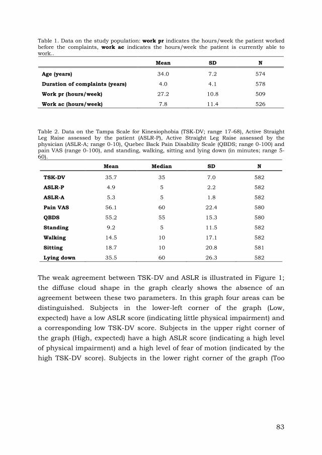
83
Table 1. Data on the study population: work pr indicates the hours/week the patient worked before the complaints, work ac indicates the hours/week the patient is currently able to work..
Mean SD N
Age (years) 34.0 7.2 574
Duration of complaints (years) 4.0 4.1 578
Work pr (hours/week) 27.2 10.8 509
Work ac (hours/week) 7.8 11.4 526
Table 2. Data on the Tampa Scale for Kinesiophobia (TSK-DV; range 17-68), Active Straight Leg Raise assessed by the patient (ASLR-P), Active Straight Leg Raise assessed by the physician (ASLR-A; range 0-10), Quebec Back Pain Disability Scale (QBDS; range 0-100) and pain VAS (range 0-100), and standing, walking, sitting and lying down (in minutes; range 5-60).
Mean Median SD N
TSK-DV 35.7 35 7.0 582
ASLR-P 4.9 5 2.2 582
ASLR-A 5.3 5 1.8 582
Pain VAS 56.1 60 22.4 580
QBDS 55.2 55 15.3 580
Standing 9.2 5 11.5 582
Walking 14.5 10 17.1 582
Sitting 18.7 10 20.8 581
Lying down 35.5 60 26.3 582
The weak agreement between TSK-DV and ASLR is illustrated in Figure 1;
the diffuse cloud shape in the graph clearly shows the absence of an
agreement between these two parameters. In this graph four areas can be
distinguished. Subjects in the lower-left corner of the graph (Low,
expected) have a low ASLR score (indicating little physical impairment) and
a corresponding low TSK-DV score. Subjects in the upper right corner of
the graph (High, expected) have a high ASLR score (indicating a high level
of physical impairment) and a high level of fear of motion (indicated by the
high TSK-DV score). Subjects in the lower right corner of the graph (Too
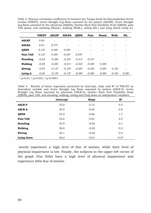
84
Table 3. Pearson correlation coefficients (r) between the Tampa Scale for Kinesiophobia-Dutch version (TSKDV), Active Straight Leg Raise assessed by the patient (ASLRP), Active Straight Leg Raise assessed by the physician (ASLRA), Quebec Back Pain Disability Scale (QBDS), pain VAS (pain), and standing (Stand.), walking (Walk.), sitting (Sit.) and lying down( Lying d.).
TSKDV ASLRP ASLRA QBDS Pain Stand. Walk. Sit.
ASLRP 0.04 - - - - - - -
ASLRA 0.01 0.77§ - - - - - -
QBDS 0.13¶ 0.50§ 0.49§ - - - - -
Pain VAS 0.12¶ 0.30§ 0.24§ 0.54§ - - - -
Standing -0.03 -0.28§ -0.22§ -0.41§ -0.31§ - - -
Walking -0.05 -0.26§ -0.21§ -0.42§ -0.26§ 0.39§ - -
Sitting -0.07 -0.13¶ -0.10§ -0.28* -0.30§ 0.20§ 0.16§ -
Lying d. -0.05 -0.15§ -0.12¶ -0.29§ -0.28§ 0.20§ 0.16§ 0.28§
* p<0.01, ¶ p<0.001, § p<0.0001
Table 4. Results of linear regression presented by Intercept, slope and R2 of TSK-DV as dependent variable and Active Straight Leg Raise assessed by patient (ASLR-P), Active Straight Leg Raise assessed by physician (ASLR-A), Quebec Back Pain Disability Scale (QBDS), pain VAS, and standing, walking, sitting and lying down as independent variables.
Intercept Slope R2
ASLR-P 35.0 0.13 0.2
ASLR-A 35.5 0.04 0.0
QBDS 32.3 0.06 1.7
Pain VAS 33.6 0.04 0.4
Standing 35.9 -0.02 0.1
Walking 36.0 -0.02 0.3
Sitting 36.1 -0.02 0.4
Lying down 36.2 -0.01 0.27
much) experience a high level of fear of motion, while their level of
physical impairment is low. Finally, the subjects in the upper left corner of
the graph (Too little) have a high level of physical impairment and
experience little fear of motion.
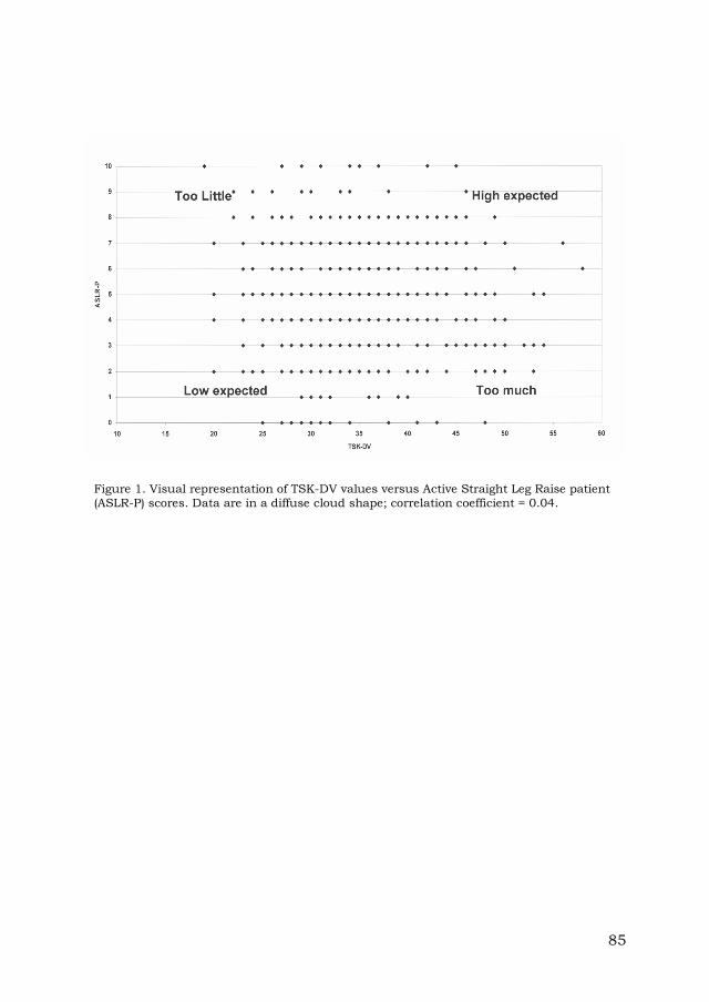
85
Figure 1. Visual representation of TSK-DV values versus Active Straight Leg Raise patient
(ASLR-P) scores. Data are in a diffuse cloud shape; correlation coefficient = 0.04.
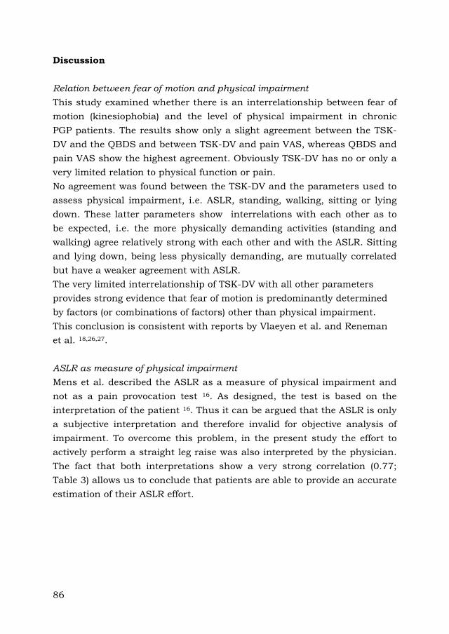
86
Discussion
Relation between fear of motion and physical impairment
This study examined whether there is an interrelationship between fear of
motion (kinesiophobia) and the level of physical impairment in chronic
PGP patients. The results show only a slight agreement between the TSK-
DV and the QBDS and between TSK-DV and pain VAS, whereas QBDS and
pain VAS show the highest agreement. Obviously TSK-DV has no or only a
very limited relation to physical function or pain.
No agreement was found between the TSK-DV and the parameters used to
assess physical impairment, i.e. ASLR, standing, walking, sitting or lying
down. These latter parameters show interrelations with each other as to
be expected, i.e. the more physically demanding activities (standing and
walking) agree relatively strong with each other and with the ASLR. Sitting
and lying down, being less physically demanding, are mutually correlated
but have a weaker agreement with ASLR.
The very limited interrelationship of TSK-DV with all other parameters
provides strong evidence that fear of motion is predominantly determined
by factors (or combinations of factors) other than physical impairment.
This conclusion is consistent with reports by Vlaeyen et al. and Reneman
et al. 18,26,27.
ASLR as measure of physical impairment
Mens et al. described the ASLR as a measure of physical impairment and
not as a pain provocation test 16. As designed, the test is based on the
interpretation of the patient 16. Thus it can be argued that the ASLR is only
a subjective interpretation and therefore invalid for objective analysis of
impairment. To overcome this problem, in the present study the effort to
actively perform a straight leg raise was also interpreted by the physician.
The fact that both interpretations show a very strong correlation (0.77;
Table 3) allows us to conclude that patients are able to provide an accurate
estimation of their ASLR effort.

87
Physical impairment in patients with fear of motion
Fear of motion discourages patients in being physically active. This is both
a problem and a challenge for clinicians who aim to increase the level of
physical activity in chronic pelvic or low back pain patients.
Questionnaires such as the TSK-DV help to estimate the level of fear of
motion. Combined with an understanding of the patient’s underlying
psychological mechanisms, this can help to customise behavioural-based
therapy to suit the individual’s specific needs. However, in the present
study no correlation was found between the TSK-DV and the ASLR.
Consequently, chronic PGP patients with high levels of fear of motion show
different levels of physical impairment. Fear of motion in chronic patients
is usually considered in the context of little or no physical impairment 3-6,8.
Figure 1 demonstrates that this situation is more complex. Subjects in
‘Low expected’ and ‘High expected’ areas experience a fear of motion level
that corresponds with their actual physical impairment, i.e. with little
impairment there is little fear, and vice versa. Subjects in the ‘Too much’
demonstrate too much fear in relation to their relatively slight impairment.
These latter patients can be considered as classic kinesiophobic patients,
i.e. their (high level of) fear does not match their minor physical
impairment. Finally, subjects in the ‘Too low’ area show too little fear in
relation to their substantial physical impairment.
This clinically relevant, specific subgroup of chronic PGP patients with
relatively little fear of motion has not been described in literature. For
these patients, their daily activities may lead to physical overload thus
contributing to the persistence and/or exacerbation of complaints.
Perceiving too little fear of motion might cause these patients to have
problems regarding the appropriate boundaries of their physical capacity.
Consequently, it is difficult to motivate this type of patient to diminish
their daily activities in order to make a proper recovery.
ASLR and fear of motion
Some comment is required regarding the set-up of the present study. To
assess the level of physical impairment a physical performance test (ASLR)
was used. Since fear of motion is known to affect physical performance,
the question arises to what extent fear of motion might have affected the
ASLR test 26-30. To answer this question the ASLR procedure needs to be
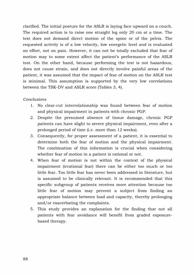
88
clarified. The initial posture for the ASLR is laying face upward on a couch.
The required action is to raise one straight leg only 20 cm at a time. The
test does not demand direct motion of the spine or of the pelvis. The
requested activity is of a low velocity, low energetic level and is evaluated
on effort, not on pain. However, it can not be totally excluded that fear of
motion may to some extent affect the patient’s performance of the ASLR
test. On the other hand, because performing the test is not hazardous,
does not cause strain, and does not directly involve painful areas of the
patient, it was assumed that the impact of fear of motion on the ASLR test
is minimal. This assumption is supported by the very low correlations
between the TSK-DV and ASLR score (Tables 3, 4).
Conclusions
1. No clear-cut interrelationship was found between fear of motion
and physical impairment in patients with chronic PGP.
2. Despite the presumed absence of tissue damage, chronic PGP
patients can have slight to severe physical impairment, even after a
prolonged period of time (i.e. more than 12 weeks).
3. Consequently, for proper assessment of a patient, it is essential to
determine both the fear of motion and the physical impairment.
The combination of this information is crucial when considering
whether fear of motion in a patient is rational or not.
4. When fear of motion is not within the context of the physical
impairment (irrational fear) there can be either too much or too
little fear. Too little fear has never been addressed in literature, but
is assumed to be clinically relevant. It is recommended that this
specific subgroup of patients receives more attention because too
little fear of motion may prevent a subject from finding an
appropriate balance between load and capacity, thereby prolonging
and/or exacerbating the complaints.
5. This study provides an explanation for the finding that not all
patients with fear avoidance will benefit from graded exposure-
based therapy.
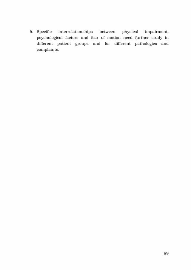
89
6. Specific interrelationships between physical impairment,
psychological factors and fear of motion need further study in
different patient groups and for different pathologies and
complaints.
90
References
1. Engel GL. The need for a new medical model: a challenge for bio-
medicine. Science 1977;196:129-36.
2. Engel GL. The clinical application of the biopsychosocial model. Am
J Psychiatry 137:5 535-44.
3. Waddel G. The back pain revolution. Churchill Livingstone 1998.
4. Asmundson GJ, Norton PJ, Norton GR. Beyond pain: the role of fear
and avoidance in chronicity. Clin Psychol Rev 1999;19:97-119.
5. Crombez G, Vlaeyen JW, Heuts PH et al. Pain-related fear is more
disabling than pain itself: evidence on the role of pain-related fear in
chronic back pain disability. Pain 1999; 80:329-39.
6. Grotle M, Vollestad NK, Brox JI. Clinical course and impact of fear-
avoidance beliefs in low back pain: prospective cohort study of acute
and chronic low back pain: II. Spine 2006;31:1038-46.
7. Kori SH, Miller RP, Todd, DD. Kinesiophobia: a new view of chronic
pain behaviour. Pain Manage 1990;3:35-43.
8. Vlaeyen JW, Linton SJ. Fear-avoidance and its consequences in
chronic musculoskeletal pain: a state of the art. Pain 2000;85:317-32.
9. O'Sullivan PB, Beales DJ. Diagnosis and classification of pelvic
girdle pain disorders--Part 1: a mechanism based approach within a
biopsychosocial framework. Man Ther 2007;12:86-97.
10. Ronchetti I, Vleeming A, van Wingerden JP. Physical characteristics
of women with severe pelvic girdle pain after pregnancy: a descriptive
cohort study. Spine 2008;33: 145-51.
11. Vleeming A, Albert HB, Ostgaard HC et al. European guidelines for
the diagnosis and treatment of pelvic girdle pain. Eur Spine J
2008;17:794-819.
12. O'Sullivan PB, Beales DJ. Diagnosis and classification of pelvic
girdle pain disorders, Part 2: illustration of the utility of a classification
system via case studies. Man Ther 2007;12:e1-12.
13. O'Sullivan PB, Beales DJ, Beetham JA et al. Altered motor control
strategies in subjects with sacroiliac joint pain during the active
straight-leg-raise test. Spine 2002; 27:E1-8.
91
14. O'Sullivan P. Diagnosis and classification of chronic low back pain
disorders: maladaptive movement and motor control impairments as
underlying mechanism. Man Ther 2005;10:242-55.
15. Wu WH, Meijer OG, Bruijn SM et al. Gait in Pregnancy-related
Pelvic girdle Pain: amplitudes, timing, and coordination of horizontal
trunk rotations. Eur Spine J 2008;17:1160-9.
16. Mens JM, Vleeming A, Snijders CJ et al. Validity of the active
straight leg raise test for measuring disease severity in patients with
posterior pelvic pain after pregnancy. Spine 2002;27:196-200.
17. Mens JM, Vleeming A, Snijders CJ et al. Reliability and validity of
the active straight leg raise test in posterior pelvic pain since
pregnancy. Spine 2001;26:1167-71.
18. Vlaeyen JW, Kole-Snijders AM, Boeren RG et al. Fear of
movement/(re)injury in chronic low back pain and its relation to
behavioral performance. Pain 1995; 62:363-72.
19. Houben RM, Leeuw M, Vlaeyen JW et al. Fear of movement/injury
in the general population: factor structure and psychometric properties
of an adapted version of the Tampa Scale for Kinesiophobia. J Behav
Med 2005;28:415-24.
20. French DJ, France CR, Vigneau F et al. Fear of
movement/(re)injury in chronic pain: a psychometric assessment of the
original English version of the Tampa scale for kinesiophobia (TSK).
Pain 2007;127:42-51.
21. Leeuw M, Houben RM, Severeijns R et al. Pain-related fear in low
back pain: a prospective study in the general population. Eur J Pain
2007;11:256-66.
22. Kopec JA, Esdaile JM, Abrahamowicz M et al. The Quebec Back
Pain Disability Scale. Measurement properties. Spine 1995;20:341-52.
23. Davidson M, Keating JL. A comparison of five low back disability
questionnaires: reliability and responsiveness. Phys Ther 2002;82:8-
24. Dyer, J.S. and Sarin, R.K. Measurable multiattribute utility
functions. Operations Res 1979;810-22.
25. Bland JM, Altman DG. Correlation, regression, and repeated data.
BMJ 1994;308:896.
92
26. Reneman MF, Jorritsma W, Dijkstra SJ et al. Relationship between
kinesiophobia and performance in a functional capacity evaluation. J.
Occup Rehabil 2003;13:277-85.
27. Reneman MF, Schiphorts HR, Geertzen M et al. Are pain intensity
and Pain related fear related to capacity evaluation performances of
patients with back pain? J Occup Rehabil 2007;17:247-58.
28. Geisser ME, Haig AJ, Wallbom AS et al. Pain-related fear, lumbar
flexion, and dynamic EMG among persons with chronic
musculoskeletal low back pain. Clin J Pain 2004;20:61-9.
29. Koho P, Aho S, Watson P et al. Assessment of chronic pain
behaviour: reliability of the method and its relationship with perceived
disability, physical impairment and function. J Rehabil Med 2001;
33:128-32.
30. Thomas JS, France CR. The relationship between pain-related fear
and lumbar flexion during natural recovery from low back pain. Eur
Spine J 2008;17:97-103.
Chapter 6
Balancing the biopsychosocial model for multidisciplinary treatment of non-specific chronic low back pain: merging motor control education and behavioural principles
J.P. van Wingerden1,2, I. Ronchetti1, A. Vleeming1, R. Stoeckart2, L.
Burdorf3, G-J. Kleinrensink2
1Spine & Joint Centre, the Netherlands, 2Erasmus MC, Department of
Neurosciences, 3Erasmus MC, Department of Public Health
94
Abstract
Study Design
An uncontrolled open label trial investigating 245 patients with non-
specific chronic low back pain (NCLBP).
Objective
The aim was to describe from a biopsychosocial (BPS) perspective the
integration of physical exercise and behavioural principles within a
therapeutic protocol for NCLBP and to demonstrate its therapeutic
potential.
Summary of Background Data
Since the introduction of the BPS model, psychosocial aspects have
dominated the multidisciplinary treatment of NCLBP. However, there are
new and promising developments with respect to physical exercise for
recovery from low back pain. It is assumed that integration of these new
exercises with cognitive behavioural treatment protocols may be beneficial
for therapy outcome.
Methods
A treatment protocol was developed with specific physical exercises
integrated with cognitive behavioural principles. A group of 245 severe,
seriously impaired and therapy-resistant NCLBP patients with average
duration of complaints more that 9 years was treated according to this 8-
week protocol. The protocol is described and the impact of the therapeutic
approach is evaluated using the Quebec Back Pain Disability Scale
(QBDS), a visual analogue scale for pain (VAS) basic function scores,
medical consumption, and the patient’s global impression of change.
Results
Medical consumption and use of medication decreased significantly after 8
weeks of therapy. Based on the patient’s global impression of change, 73%
of all patients experienced improvement. Based on the criteria of the
minimal clinically important change, 62% of the patients significantly
95
improved on the QBDS and 53% on the pain VAS; in specific subgroups
these beneficial results were even better (70% and 81%, respectively).
Conclusions
This study demonstrates that physical exercise can be successfully merged
with cognitive behavioural principles in a multidisciplinary treatment
protocol for NCLBP. Our findings support the assumption that therapy
outcome improves when more attention is paid to the physical aspects of a
multidisciplinary program. Creating and maintaining the balance between
the BPS domains within a therapeutic protocol is a challenge for
multidisciplinary teams.
96
Introduction
Multidisciplinary therapy based on the biopsychosocial (BPS) model has
gained ground in the search for the most effective treatment of non-specific
chronic low back pain (NCLBP) 1-5. A major advantage of the BPS model is
that besides biological factors it incorporates psychological and social
aspects in the aetiology of chronic back pain thus broadening the
spectrum of factors that may contribute to chronic back pain 6,7. It was
subsequently demonstrated that specific behaviours, such as fear
avoidance, play a role in chronicity of low back pain 8-10. Following this line
of thought, behaviour modification therapies, developed for other
behaviour-oriented disorders, were adopted for NCLBP patients 1,4,11-13. The
first results of these predominantly psychology-oriented therapies (e.g.
cognitive-behavioural therapy, graded exposure and graded activity) were
promising and in sharp contrast to the unsatisfactory results of
traditional, monodisciplinary physical therapy 14-16. Consequently, within
the BPS model, in the treatment of these complaints the emphasis shifted
from the physical to the psychosocial elements 3,16-21. Presently, in many
multidisciplinary NCLBP therapies, psychological aspects are predominant
1,4,13. In these therapies changing the patient´s behaviour (especially
socially-oriented behaviour such as returning to work, resuming
housekeeping, child care, and re-participation in social life) is emphasised
14,22. The physical domain is subordinate and the goals of physical therapy
within the multidisciplinary programs are relatively shallow, i.e. general re-
conditioning and physical re-activation of the patient 1,15,23-25. The focus is
on the quantity of activity, not the quality. This is not surprising since
historically physical therapy has only limited options for addressing the
qualitative aspects of function (flexibility, strength), while more quality-
based therapies (e.g. the Mensendieck or Caesar therapy) still lack an
adequate evidence-based foundation 26-28. As a result, the role of a physical
therapist within multidisciplinary protocols for NCLBP has diminished to
that of a practical trainer or coach 15, 23-25.
Behavioural-based therapies appear to produce better results than regular
exercise programs. Closer inspection of earlier studies reveals that the
parameters used to evaluate the results of these therapies often specifically
reflect their main objective, i.e. to have patients return to work and regain

97
their social life. However, when parameters such as experienced pain or
physical performance are taken into account, the results are far less
positive 14,19,22,24,25,29-31. Behavioural-based therapies may excel in changing
behaviour, but the question remains whether they provide a cure for the
patient’s actual complaints, i.e. pain and functional limitations. In the
process of learning how to change behaviour from the psycho-social
perspective, the biological aspects may have been neglected.
In the last decades our understanding of the functioning of the locomotor
system, in particular the pelvis and spine, has increased substantially32-38.
In 1990, Vleeming et al. and Snijders et al. introduced the model of ‘form
and force closure’ which provides an explanation of how the joints of the
pelvis (sacro-iliac joints) specifically, but also synovial joints in general,
can be controlled by the interaction of a large number of different
structures (like muscles, ligaments and capsules) in the proximity of the
joint 39-42. This model could be well integrated with new insights on control
and stability of the lumbar spine 35,38,43-46. It became clear that changes in
neuromuscular control can cause non-optimal motion patterns in both the
lumbar spine and in the pelvis, thus compromising physical capacity 47,48-
51. In turn, compromised physical capacity can lead to physical overload
and pain 34,36,52. It is important to note that these mechanisms operate in
the absence of perceptible tissue damage and may last for a long period of
time, even years 36,38-43,45,52,53 Based on this knowledge new forms of
physical training have been developed and were found to be effective in
patients with low back pain 45,53,54.
It is proposed that more focus on physical aspects within multidisciplinary
programs will improve therapy outcome in NCLBP. To test this assumption
the multidisciplinary team of a Dutch outpatient rehabilitation centre
merged cognitive behavioural principles with specific physical training. The
goal of the present study was to outline this integration of physical
exercise and behavioural principles within the therapeutic protocol, and to
provide preliminary results to endorse the therapeutic potential of this
balanced multidisciplinary approach to NCLBP.
98
Patients and methods
Population
Between 2000 and 2007 regional pain clinics referred 402 patients
diagnosed with chronic back and neck pain to a Dutch outpatient
rehabilitation clinic (Rotterdam). From this latter group, 245 met the
following inclusion criteria: pain predominantly in the region of the lower
back; no significant structural deformations; complaints persisting for
longer than 6 months; no acute radicular symptoms; received extensive
treatment from a physical therapist; evaluated by at least one medical
specialist (e.g. a rheumatologist, neurologist or orthopaedic surgeon); and
adequate command of the Dutch language. Patients with multiple back
surgery were excluded from the study.
Although 33% of the study population reported having experienced
radicular symptoms in the past no acute radicular symptoms were present
at the start of therapy. Since none of the patients had acute radicular
symptoms, reported disc herniations (30%) were considered to be
unrelated to the present complaints.
Of the original 402 selected patients, 148 were excluded because they did
not meet the inclusion criteria and another 9 patients were excluded
because of insufficient/unreliable data or because they stopped treatment.
Therefore, the final evaluation is based on data of 245 (150 females, 95
males) NCLBP patients.
Treatment protocol
In the present therapy protocol, cognitive behavioural principles are
applied to stimulate patients to adopt adequate behaviour aimed at
physical recovery. The program, which was no isolated study design, but
part of regular care, consists of 16 sessions of 3 hours each, over an 8-
week period (total of 48 hours). Patients are divided into groups of 6
patients accompanied by three therapists. Each session includes training
time (1 hour), a group lesson (1 hour) and individual coaching of the
patient (1 hour). The objective of the lessons is to modify the patient’s
cognitions with respect to their complaints thus reinforcing proper
behaviour. The lessons include information on functional anatomy of the
spine, principles of chronic pain, the role and impact of emotions,
99
communication, and finding the balance between the load of daily life and
physical capacity.
The training is performed in a progressive sequence adjusted to the
patient’s specific situation and progress. Initially the patient’s awareness of
excessive or aberrant muscle tension is enhanced by means of relaxation
and breathing exercises. Subsequently, patients learn to normalise muscle
tension in unloaded situations using breathing and relaxation exercises,
e.g. lying down, sitting and standing upright. Basic lumbar and pelvic
stability is restored by motor control education using various stabilisation
exercises for m. multifidus, m. transversus abdominis, the diaphragm and
pelvic floor as suggested in literature 37,38,44-46+2. When the patient is able to
maintain basic lumbar stability in unloaded postures and without
compensatory muscle activity, intensity of the exercises is increased in
time, load and impact44. Control of proper muscle activation patterns is
emphasised throughout the entire therapeutic process. Behavioural
aspects that are specifically addressed include balancing physical capacity
with the load of daily activities, and performing common activities like
walking, running, bending forward and lifting.
In addition to the training and learning program, the patient is coached on
an individual basis to cope with physical or mental impediments in
performing the desired behaviour. Additional assistance can be provided
by a manual therapist, psychologist or therapist specialised in body
awareness (in Dutch called hapto-therapist).
Evaluation
Results of the therapy were evaluated on six parameters:
1. Pain, determined using the Visual Analogue Scale (Pain VAS).
2. Limitations in daily life, determined using the Quebec Back Pain
Disability Scale (QBDS). 55,56
3. Basic function, based on duration of walking, standing, sitting, lying
down.
4. Isometric trunk strength, extension and rotation, using isometric
strength testing equipment.
5. Medical consumption, obtained from questionnaires.
6. Patient’s global impression of change (PGIC). 57
100
Data were obtained during the intake procedure, after 8 weeks of therapy,
and at the follow-up 3 months after the end of therapy.
All continuous variables were tested with the Student's t-test and in case
of non-normally distributions with the Mann-Whitney U test. All
categorical variables were tested with the Chi-Square test. Linear mixed
models for analysis of variance with repeated measures was used to
evaluate the change in the outcome measures limitations in daily life, pain,
basic functions, and isometric strength directly after treatment and 3
months after the end of treatment. These models take account of the
correlation between repeated measures on the same subject and allow for
incomplete outcome data. In each model, the potential determinants of
outcome measures were included as fixed (categorical) effects and the
variances between and within subjects were regarded as random effects.
The random variance component was pooled across all determinants of
exposure and assumed to be equal across all fixed determinants
(compound symmetry covariance structure). In all linear mixed models sex
and education were included as determinant, since both variables had a
significant effect on some outcomes measures and, thus, for reason of
comparability were included in all models (table 3). SPSS version 10.01
and SAS version 8.02 were used for the statistical analyses of the data.
To demonstrate clinical relevance, for the QBDS and the pain VAS the
minimal clinically important change (MCIC) was calculated, as proposed by
Ostelo et al 58,59. The MCIC was calculated as the percentage of patients
with a more than 30% change compared with baseline.
Since baseline values may affect the MCIC outcome, the results were also
analysed in subgroups based on three ranges of initial values. For the
QBDS, patients were divided into groups with baseline values less than 45
(<45), from 45 to 65 (≥45 to <65), and 65 or higher (≥65). For the Pain VAS,
patients were divided into groups with baseline values less than 50 (<50),
from 50 to 70 (≥50 to <70), and 70 or higher (≥70).
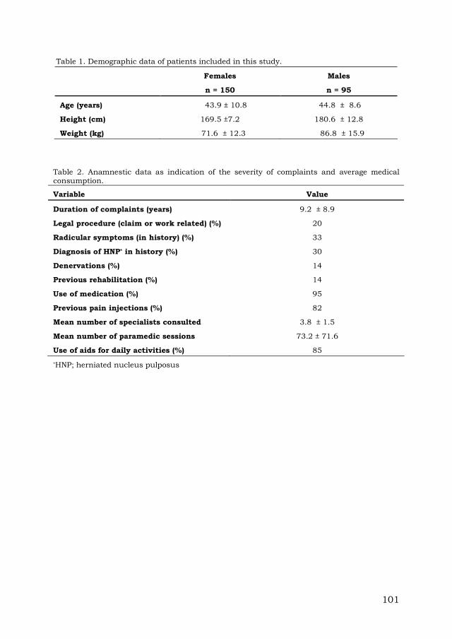
101
Table 1. Demographic data of patients included in this study.
Females
n = 150
Males
n = 95
Age (years) 43.9 ± 10.8 44.8 ± 8.6
Height (cm) 169.5 ±7.2 180.6 ± 12.8
Weight (kg) 71.6 ± 12.3 86.8 ± 15.9
Table 2. Anamnestic data as indication of the severity of complaints and average medical consumption.
Variable Value
Duration of complaints (years) 9.2 ± 8.9
Legal procedure (claim or work related) (%) 20
Radicular symptoms (in history) (%) 33
Diagnosis of HNP* in history (%) 30
Denervations (%) 14
Previous rehabilitation (%) 14
Use of medication (%) 95
Previous pain injections (%) 82
Mean number of specialists consulted 3.8 ± 1.5
Mean number of paramedic sessions 73.2 ± 71.6
Use of aids for daily activities (%) 85
*HNP; herniated nucleus pulposus
102
Results
The subjects included in this study (Table 1) are NCLBP patients with an
average duration of complaints of 9.2 years (SD 8.9) (Table 2). There is a
significant, but small difference in intake values between men and woman
for QBDS, standing and walking, and isometric strength (Table 3). At the
start of therapy medical consumption was high: 95% of the patients used
medication on a frequent basis which diminished to 40% after therapy.
After 8 weeks of therapy all outcome parameters significantly changed
towards improvement compared to the level at intake (Table 3). This result
was found for the whole study population, but also when men or woman
were analysed separately. The Cohen’s D for QBDS and Pain VAS (mean
difference / baseline sd) at 8 weeks is 0.9 and 1.38, respectively, indicating
a large improvement on physical limitations and pain. This change was
maintained at 3-months follow-up. There was no significant change in data
between the end of therapy and follow-up. The use of aids decreased from
85% to 9%.
Scores on the PGIC indicate that 73% of the patients experienced
improvement (30% much better and 34% better); 20% of patients reported
no change and in 7% the outcome was negative.
Based on the MCIC criteria, 62% of all patients significantly changed
towards improvement on the QBDS and 53% on the pain VAS. Table 4
gives the MCIC results for subgroups of patients grouped by baseline
scores on the QBDS and pain VAS. The best results were obtained by the
subgroup with initial scores of 45 to 65 on the QBDS (70%) and scores
from 50 to 70 on the pain VAS (81%).
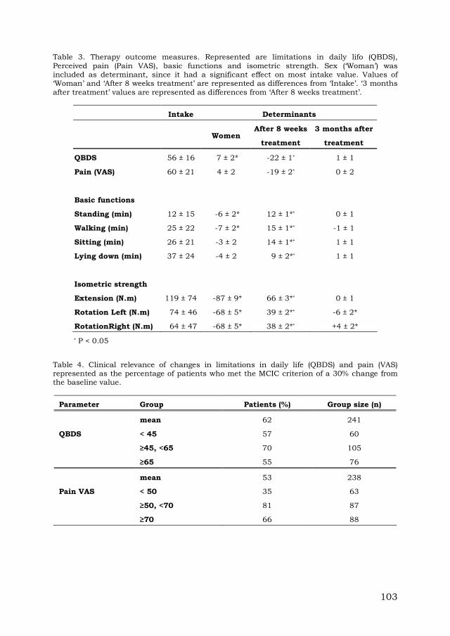
103
Table 3. Therapy outcome measures. Represented are limitations in daily lifo (QBDS), Perceived pain (Pain VAS), basic functions and isometric strength. Sex (‘Woman’) was included as determinant, since it had a significant effect on most intake value. Values of ‘Woman’ and ‘After 8 weeks treatment’ are represented as differences from ‘Intake’. ‘3 months after treatment’ values are represented as differences from ‘After 8 weeks treatment’.
Intake Determinants
Women
After 8 weeks
treatment
3 months after
treatment
QBDS 56 ± 16 7 ± 2* -22 ± 1* 1 ± 1
Pain (VAS) 60 ± 21 4 ± 2 -19 ± 2* 0 ± 2
Basic functions
Standing (min) 12 ± 15 -6 ± 2* 12 ± 1** 0 ± 1
Walking (min) 25 ± 22 -7 ± 2* 15 ± 1** -1 ± 1
Sitting (min) 26 ± 21 -3 ± 2 14 ± 1** 1 ± 1
Lying down (min) 37 ± 24 -4 ± 2 9 ± 2** 1 ± 1
Isometric strength
Extension (N.m) 119 ± 74 -87 ± 9* 66 ± 3** 0 ± 1
Rotation Left (N.m) 74 ± 46 -68 ± 5* 39 ± 2** -6 ± 2*
RotationRight (N.m) 64 ± 47 -68 ± 5* 38 ± 2** +4 ± 2*
* P < 0.05
Table 4. Clinical relevance of changes in limitations in daily life (QBDS) and pain (VAS) represented as the percentage of patients who met the MCIC criterion of a 30% change from the baseline value.
Parameter Group Patients (%) Group size (n)
mean 62 241
QBDS < 45 57 60
≥45, <65 70 105
≥65 55 76
mean 53 238
Pain VAS < 50 35 63
≥50, <70 81 87
≥70 66 88

104
Discussion
Impact of combining physical exercise and behavioural principles
The goal of this study was to demonstrate the possibility and therapeutic
potential of emphasising specific physical exercises within a
multidisciplinary rehabilitation programme for NCLBP. The results of this
therapy programme must be considered in perspective of the specific
patient group. The patients in this group had complaints that persisted for
more than 9 years on average. There were severe limitations in daily life
(QBDS) and extensive consumption of medication. All patients had
received extensive paramedical and medical treatment without adequate
relief. Therefore, it can be concluded that the subjects in the present study
were severe, seriously impaired and therapy-resistant NCLBP patients.
Thus it is encouraging that, in contrast to their history of unsuccessful
treatments, most of the patients in the present study experienced
improvement (PGIC: 73% much better or better). This subjective experience
is in line with the significant decrease in experienced pain (Pain VAS) and
in diminished limitations in daily activity (QBDS). Isometric strength and
function scores (duration of walking, standing, sitting and lying down)
improved significantly. In the light of the long history of treatment and
recurrence of complaints, the finding that the therapeutic effects remained
after three months is also encouraging. Furthermore, medication use
dropped from 95% to 40%, and in those patients still using medication the
daily dosage was also reduced. The number of patients using aids for
support reduced from 85% to only 9% indicating functional improvement
in the majority of patients.
Results using the MCIC criterion
Evaluation of the QBDS and pain VAS using the MCIC criteria shows
promising results. Limitations in daily life showed a significant decrease in
62% of all patients (QBDS) and 55% had significantly less pain (Pain VAS).
For some subgroups this outcome was even more impressive: in patients
with a baseline QBDS score of 45-65 and in patients with a baseline pain
VAS of 50-70 almost 70% and 80%, respectively, showed a clinically
significant improvement. Results are slightly less positive in patients with
lower or higher baseline scores on the QBDS and pain VAS. Although it is
105
tempting to conclude that therapy is less effective in these latter groups
this result may be more complex than it seems. For the more ‘severe’
patient group it is logical to assume that they need more time or care to
obtain the results achieved in the ‘average’ group. However, this does not
hold for the patients with lower baseline scores. It is more likely that the
low baseline scores thwart a significant decrease in the outcome value,
despite the use of a relative change (30% of the baseline value) instead of
an absolute value (20 or 15 points on the QBDS or pain VAS, respectively)
58,59.
Additional value of specific physical training in a behavioural program
This study shows that specific physical training can be successfully
emphasised within a behavioural program. Since the emphasise on
physical training is the only adaptation in the behavioural programme this
study also provides strong indications for the additional value of this
specific combination. This latter result supports the assumption that
physical functioning may be impaired in CLBP patients, despite the
absence of recognisable tissue damage 38,52. With impaired functioning,
graded exposure or graded activity programs may not succeed without
specific physical training. When physically impaired patients increase their
level of activity without concern for the quality of the motion,
overburdening may occur and consequently inflammation, leading to an
increase of pain 52. This undesired response to the increased level of
activity leads to a negative experience and discourages the patient from
continuing therapy.
Balance not dominance
This study advocates more appreciation for the role of physical aspects in
multidisciplinary programs for non-specific CLBP; it does not claim that
the physical aspect is the most important factor. Basically, the BPS model
does not consider any single factor to be the most prominent. Important
questions are how, and to what extent, should these factors be
implemented in therapy. It seems logical to assume that these
considerations largely depend on the individual’s needs. In daily practice,
however, it is not uncommon that the most dominant discipline in the
multidisciplinary team will determine the focus of attention 15,24. This often

106
results in cognitive behavioral therapy, with physical training being
subordinate to the behavioural aspects of the treatment; this is not in line
with the essence of the BPS model 6,7.
Conclusions
The present study allows to draw the following conclusions:
1. It is possible to have a cognitive behavioural therapy for NCLBP with
more focus on specific exercises, contributing to quality of behaviour.
2. There are strong indications that merging physical exercises with
cognitive behavioural principles can improve the results of
multidisciplinary treatment for NCLBP.
3. Finding and maintaining the balance between the BPS domains in a
therapeutic protocol is a challenge for multidisciplinary teams.
107
References
1. Gatchel RJ, Peng YB, Peters ML, Fuchs PN, Turk DC. The
biopsychosocial approach to chronic pain: scientific advances and future
directions. Psychol Bull 2007;133:581-624.
2. Guzmán J, Esmail R, Karjalainen K, Malmivaara A, Irvin E, Bombardier
C. Multidisciplinary bio-psycho-social rehabilitation for chronic low back
pain. Cochrane Database Syst Rev. 2002;(1):CD000963.
3. Van Tulder MW, Koes B, Malmivaara A. Outcome of non-invasive
treatment modalities on back pain: an evidence-based review. Eur Spine J
2006;15:S64-81.
4. Waddell G. 1987 Volvo award in clinical sciences. A new clinical model
for the treatment of low-back pain. Spine 1987;12:632–44.
5. Waddell G, Burton AK. Concepts of rehabilitation for the management of
low back pain. Best Pract Res Clin Rheumatol 2005;9:655–70.
6. Engel GL. The need for a new medical model: a challenge for
biomedicine. Science 1977;196:129-36.
7. Engel GL. The clinical application of the biopsychosocial model. Am J
Psychiatry 137:5 535-44.
8. Kori SH, Miller RP, Todd, DD. Kinisiophobia: a new view of chronic pain
behaviour. Pain Manag 1990;3:35-43
9. Vlaeyen JW, Kole-Snijders AM, Boeren RG, van Eek H. Fear of
movement/(re)injury in chronic low back pain and its relation to
behavioral performance. Pain 1995;62:363-72.
10. Asmundson GJ, Norton PJ, Norton GR. Beyond pain: the role of fear
and avoidance in chronicity. Clin Psychol Rev 1999;19:97-119.
11. Vlaeyen JW, Haazen IW, Schuerman JA, Kole-Snijders AM, van Eek H.
Behavioural rehabilitation of chronic low back pain: comparison of an
operant treatment, an operant-cognitive treatment and an operant-
respondent treatment. Br J Clin Psychol 1995;34:95-118.
12. Vlaeyen JW, De Jong JR, Onghena P, Kerckhoffs-Hanssen M, Kole-
Snijders AM. Can pain-related fear be reduced? The application of
cognitive-behavioural exposure in vivo. Pain Res Manag 2002;7:144-53.
13. Waddell G. The back pain revolution. Second edition. Churchill
Livingstone 2004.
108
14. Vendrig AA, van Akkerveeken PF, Sanders RJ. [Good results from a
multidisciplinary and behavioral program for chronic back pain] Ned
Tijdschr Geneeskd 2000;144:560-3.
15. Hildebrandt J, Pfingsten M, Saur P, Jansen J. Prediction of success
from a multidisciplinary treatment program for chronic low back pain.
Spine 1997;22:990-1001.
16. Hoffman BM, Papas RK, Chatkoff DK, Kerns RD. Meta-analysis of
psychological interventions for chronic low back pain. Health Psychol
2007;26:1-9.
17. Keller A, Hayden J, Bombardier C, van Tulder M. Effect sizes of non-
surgical treatments of non-specific low-back pain. Eur Spine J
2007;16:1776-88.
18. Klaber Moffett J, Mannion AF. What is the value of physical therapies
for back pain? Best Pract Res Clin Rheumatol 2005;19:623-38.
19. Van Tulder WM , Waddell G. Evidence-based medicine for non-specific
low back pain. Best Pract Res Clin Rheumatol 2005;19:7-9.
20. Van Tulder M, Koes B. Chronic low back pain. Am Fam Phys
2006;74:1577-9.
21. Van Tulder M, Koes B. Low back pain (chronic). Clin Evid
2006;15:1634-53.
22. van Geen JW, Edelaar MJ, Janssen M, van Eijk JT. The long-term
effect of multidisciplinary back training: a systematic review. Spine
2007;32:249-55.
23. Kääpä EH, Frantsi K, Sarna S, Malmivaara A. Multidisciplinary group
rehabilitation versus individual physiotherapy for chronic nonspecific low
back pain: a randomized trial. Spine 2006;31:371-6.
24. Smeets RJ, Vlaeyen JW, Hidding A, Kester AD, van der Heijden GJ,
van Geel AC, Knottnerus JA. Active rehabilitation for chronic low back
pain: Cognitive-behavioral, physical, or both? First direct post-treatment
results from a randomized controlled trial. BMC Musculoskelet Disord
2006;20; 7:5 doi:10.1186/1471-2474-7-5.
25. Smeets RJ, Vlaeyen JW, Hidding A, Kester AD, van der Heijden GJ,
Knottnerus JA. Chronic low back pain: physical training, graded activity
with problem solving training, or both? Pain 2008;134:263-76.
26. van Eijsden-Besseling MD, Staal JB, van Attekum A, de Bie RA, van
den Heuvel WJ. No difference between postural exercises and strength and
109
fitness exercises for early, non-specific, work-related upper limb disorders
in visual display unit workers: a randomised trial. Aust J Physiother
2008;54:95-101.
27. Haugstad GK, Haugstad TS, Kirste UM, Leganger S, Klemmetsen I,
Malt UF. Mensendieck somatocognitive therapy as treatment approach to
chronic pelvic pain: results of a randomized controlled intervention study.
Am J Obstet Gynecol 2006;194:1303-10.
28. Soukup MG, Glomsröd B, Lönn JH, Bö K, Larsen S. The effect of a
Mensendieck exercise program as secondary prophylaxis for recurrent low
back pain. A randomized, controlled trial with 12-month follow-up. Spine
1999;24:1585-91.
29. Johnson RE, Jones GT, Wiles NJ, Chaddock C, Potter RG, Roberts C,
Symmons DP, Watson PJ, Torgerson DJ, Macfarlane GJ. Active exercise,
education, and cognitive behavioral therapy for persistent disabling low
back pain: a randomized controlled trial. Spine 2007;32:1578-85.
30. Nicholas MK, Wilson PH, Goyen J. Comparison of cognitive-behavioral
group treatment and an alternative non-psychological treatment for
chronic low back pain. Pain 1992;48:339-47.
31. Ostelo RW, van Tulder MW, Vlaeyen JW, Linton SJ, Morley SJ,
Assendelft WJ. Behavioural treatment for chronic low-back pain.
2005;25:Cochrane Database Syst Rev CD002014.
32. Adams MA. The Biomechanics of Back Pain, Churchill-Livingstone,
2002.
33. Bogduk N. The anatomical basis for spinal pain syndromes. J Manipul
Physiol Ther 1995;18:603–5.
34. van Dieen JH, Cholewicki J, Radebold A. Trunk muscle recruitment
patterns in patients with low back pain enhance the stability of the lumbar
spine. Spine 2003;28:834-41.
35. Gracovetsky S. The Spinal Engine, Spinger, 1989.
36. Hides JA, Richardson CA, Jull GA. Multifidus muscle recovery is not
automatic after resolution of acute, first-episode low back pain. Spine
1996;21:2763-9.
37. Hungerford B, Gilleard W, Hodges P. Evidence of altered lumbopelvic
muscle recruitment in the presence of sacroiliac joint pain. Spine
2003;28:1593-600.
110
38. Hodges PW, Richardson CA. Inefficient muscular stabilization of the
lumbar spine associated with low back pain. A motor control evaluation of
transversus abdominis. Spine 1996;21:2640-50.
39. Snijders CJ, Vleeming A, Stoeckart R. Transfer of lumbosacral load to
iliac bones and legs. Part 1 and 2. Clin Biomech 1993;8:285-94.
40. Vleeming A, Stoeckart R, Volkers ACW, Snijders CJ. Relation between
form and function in the sacro-iliac joint, Part 1: Clinical anatomical
aspects. Spine 1990a;15:130-2.
41. Vleeming A, Volkers ACW, Snijders CJ, Stoeckart R. Relation between
form and function in the sacro-iliac joint, Part 2: Biomechanical aspects.
Spine 1990b;15:133-6.
42. Vleeming A. The sacroiliac joint, a clinical-anatomical, biomechanical
and radiological study. PhD Thesis, Erasmus University Rotterdam, the
Netherlands, 1990.
43. Hodges PW, Richardson CA. Delayed postural contraction of
transversus abdominis associated with movement of the lower limb in
people with low back pain. J Spin Disord 1998;11:46-56.
44. McGill SM, Grenier S, Kavcic N, Cholewicki J. Coordination of muscle
activity to assure stability of the lumbar spine. J Electromyograph Kinesiol
2003;13:353-9.
45. Richardson CA, Jull GA, Muscle control-pain control. What exercises
would you prescribe? Man Ther 1995;1:2-10.
46. Richardson CA, Jull GA, Hodges PW, Hides JA. Therapeutic exercise
for spinal segmental stabilisation in low back pain: scientific basis and
clinical approach. Edinburgh, Churchill Livingstone, 1999.
47. Holm S, Indahl A, Solomonow M. Sensorimotor control of the spine. J
Electromyography Kinesiol 2002;12:219-34.
48. Indahl A, Kaigle A, Reikeras O, Holm S. Electromyographic response of
the porcine multifidus musculature after nerve stimulation. Spine
1995;20:2652-8.
49. Indahl A, Kaigle A, Reikeras O, Holm S. Interaction between the
porcine lumbar intervertebral disc, zygapophysial joints, and paraspinal
muscles. Spine 1997;22:2834-40.
50. Indahl A, Kaigle A, Reikeras O, Holm S. Sacroiliac joint involvement in
activation of the porcine spinal and gluteal musculature. J Spinal Disord
1999;12:325-30.
111
51. van Wingerden JP, Vleeming A, Ronchetti I. Differences in standing
and forward bending in women with chronic low back or pelvic girdle pain:
indications for physical compensation strategies. Spine 2008;15;33:E334-
41.
52. MacDonald D, Moseley GL, Hodges PW. Why some patients keep
hurting their back? Evidence of ongoing back muscle dysfunction during
remission from recurrent back pain. Pain (2009),
doi:10.1016/j.pain.2008.12.002.
53. Taimela S., Negrini, S, Paroli C. Functional rehabilitation of low back
disorders. Eur Med Phys 2004; 40:29-36.
54. Rasmussen-Barr E, Ang B, Arvidsson I, Nilsson-Wikmar L. Graded
exercise for recurrent low-back pain: a randomized, controlled trial with 6-,
12-, and 36-month follow-ups. Spine 2009;34:221-8.
55. Hodges PW, Eriksson M.A.E., Shirley D et al. Intra-abdominal pressure
increases stiffness of the lumbar spine. Journal of Biomechanics
2005;38:1873–1880.
56. Pool-Goudzwaard AL, Slieker ten Hove MC, Vierhout ME et al.
Relations between pregnancy-related low back pain, pelvic floor activity
and pelvic floor dysfunction. Int Urogynecol J Pelvic Floor Dysfunct.
2005;16:468-74.
57. Kopec JA, Esdaile JM, Abrahamowicz M et al. The Quebec Back Pain
Disability Scale. Measurement properties. Spine 1995;20:341-52.
58. Davidson M, Keating JL. A comparison of five low back disability
questionnaires: reliability and responsiveness. Phys Ther 2002;82:8-24.
59. Jaeschke R, Singer J, Guyatt GH. Measurement of health status.
Ascertaining the minimal clinically important difference. Controlled Clin
Trials 1989;10:407-15.
60. Ostelo RW, Deyo RA, Stratford P, Waddell G, Croft P, Von Korff M,
Bouter LM, de Vet HC. Interpreting change scores for pain and functional
status in low back pain: towards international consensus regarding
minimal important change. Spine 2008;33:90-4.
61. van der Roer N, Ostelo RW, Bekkering GE et al. Minimal clinically
important change for pain intensity, functional status, and general health
status in patients with nonspecific low back pain. Spine 2006;31:578-82.

112
113
Chapter 7
Discussion
The main goal of this thesis is to explore whether a more prominent role of
functional anatomy, as part of the physical domain within the
biopsychosocial (BPS) model, improves multidisciplinary NCLBP therapy.
This goal is pursued by defining the following research questions in
perspective of multidisciplinary treatment of NCLBP:
1. Taking into account recent data on functional anatomy, is there a
need to reconsider the role of the physical domain within the BPS
model?
2. Will a more pronounced role of functional anatomy in the BPS
model contribute to better diagnosis?
3. Will functional anatomy applied in the BPS model contribute to
improved therapy?
7.1. Should the role of functional anatomy within the BPS model be
reconsidered?
The question whether the role of functional anatomy within the BPS model
should be reconsidered is subdivided in two other questions: First, is there
actually new knowledge on functional anatomy and second, if so, does this
knowledge have clinical significance?
7.1.1. Development of functional anatomical knowledge, an example
The description of the connection and interaction of the long head of the
biceps femoris muscle with the sacrotuberous ligament (chapter 2) is an
example of new knowledge in functional anatomy. The described dynamic
114
connection has a clinical consequence since it implies that intrinsic
stability of the sacroiliac (SI) joints is determined not only by local
muscles. More distant muscles, in this case the m. biceps femoris, may
contribute to SI stiffness as well. The study in chapter 3 confirms the
clinical implication of the anatomical finding in vivo. It is demonstrated in
healthy subjects that activity of the m. biceps femoris indeed increases SI
joint stiffness. The study also shows the influence of other muscles (m.
gluteus maximus, m. erector spinae and m. latissimus dorsi) on SI joint
stiffness. The study was designed to demonstrate the stabilizing effect of
m. biceps femoris on the SI joint; it was not intended to determine the
relative contribution of m. biceps femoris or other muscles to SI joint
stability. However, this would be a logical next step in understanding the
complex mechanism of SI joint stability.
The first two studies focus on the SI joint and the influence of a limited
number of muscles was observed. Notwithstanding the limited scope, this
information clearly demonstrates the importance and potential of the study
of functional anatomy.
The next question concerns the clinical relevance of this functional
anatomical information.
7.1.2. Functional anatomy in healthy subjects and patients; clinical
relevance
The third study as described in chapter 4 focuses on the clinical relevance
of functional anatomical knowledge as presented in the previous studies.
This third study is performed on healthy female subjects and female
patients with NCLBP. In none of the patients an anatomical substrate
explaining their complaints could be pointed out. And on gross observation
they appear to be able to perform normal daily activities like bending
forward. However, objective analysis of the relative contribution of the
lumbar spine and pelvis (hip flexion) to forward bending shows specific
and different motion patterns. Furthermore, in the patient group, two
subgroups were identified with distinct altered motion patterns: the low
back pain (LBP) and pelvic girdle pain (PGP) patients. Based on the first
two studies and literature a functional anatomical explanation for the
distinct patterns is proposed 2,11,18,25. When joint stability is compromised,
daily activities can only be performed with altered muscle function.
115
Consequently, altered muscle function changes motion patterns. It is to be
expected that depending on the specific joint, part of a joint or combination
of joints, other motion patterns will occur. Moreover it must be taken into
account that such compensation patterns also can depend on the subjects
preferred motion strategies.
As expected the human body is capable of using various motion strategies
to perform daily activities. But not all possible strategies will be equally
economic 6,7,32. This leads to the assumption that motion strategies which
compensate compromised joint stability are not economically optimal.
Thus, abnormal motion patterns may lead to physical overload and
consequently induce or prolong physical complaints such as pain. From
this perspective functional anatomical analysis helps us to understand
that physical factors can play a role in chronic back pain, even without
detectable structural disorders 17. This is in contrast with the present
common clinical attitude towards chronic pain. Nowadays, many clinicians
consider physical aspects, besides maybe deconditioning, irrelevant in
chronic pain patients 30,31. The present studies and recent literature
suggest that this situation is more complex 17. Based on physiological rules
damaged tissue recovers within 12 weeks. However, this does not imply
that proper function has restored also. Actually it is demonstrated by
Richardson et al that functional disturbances can persist, sometimes even
for years 9,17,23. Consequently, physical function of a NCLBP patient should
not be considered irrelevant. Functional anatomical analysis is crucial to
provide information on physical (dys-) function of a patient.
7.1.3. Application of functional anatomy in other areas
At present many physical complaints have obscure aetiology, or worse, are
primarily attributed to psychosocial factors 5,13,30. More knowledge on
functional anatomy will enhance our understanding of the biological
aspects of these complaints. In this thesis the connection between the m.
biceps femoris and the sacrotuberous ligament (chapter 2) is described in
detail as an example of the complexity of functional anatomical
mechanisms. Although this is only one relatively straightforward example,
other functional anatomical systems have been studied, or are waiting to
be unravelled. Examples of such systems are the function of m. transverse
abdominis and m. multifidus as stabilizing muscles for the lumbar spine
116
16,23. In this perspective the discussion on the function of the
thoracolumbar fascia is intriguing 1,7,8,29. Several studies point towards a
function of the pelvic floor and diaphragm in lumbar and pelvic support
19,21,22. These new insights do not only provide guidelines for therapy, they
may also provide explanations for numerous symptoms that could not be
explained from the traditional descriptive anatomical perspective.
Examples of these symptoms are the piriformis syndrome, pelvic floor
incontinence, hyperventilation syndrome, short hamstrings and hypertone
erector spinae.
The number of studies on functional anatomy still increases and it is clear
that we are only at the beginning of the functional anatomical exploration
of the human body. More research will be necessary to unravel and fully
understand the functional anatomical mechanisms of the human body and
its clinical consequences. Based on the first three studies in this thesis,
and supported by literature, it can be concluded that the role of the
physical domain within the BPS model with respect to NCLBP definitely
needs reconsideration.
7.2. Better diagnosis with functional anatomy
The results of the study on coordination in chapter 4 imply that functional
anatomy contributes to quality of diagnosis. Obviously, more improved
functional anatomical knowledge will make the diagnostic process more
thorough and will provide valuable information for therapy; evidence based
medicine includes evidence based diagnostics. The strength of the BPS
model, however, lies especially in the interaction between the domains.
7.2.1. Difficulties in integration of information from domains within the
BPS model
Combining information from different domains is difficult. One important
factor is that traditionally disciplines work in one specific domain. Physical
therapists work in the physical domain, whereas the psychologist works in
the psychological domain and the social worker in the social domain. This
creates a problem since therapists working within a specific domain are
inclined to interpret clinical findings from their own perspective. Raised
shoulders in a patient can be interpreted by the physical therapist as
compensating behaviour due to shoulder or neck problems, while the
117
psychologist may interpret this behaviour as a sign of defence or
psychological discomfort.
Another problem is the different terminology between disciplines. For
example a ‘functional complaint’ has a completely different meaning in the
biological or psychological domain (disturbance of physical function or
change in physical function related to psychological distress, respectively).
Consequently, combining information from the domains is difficult and
therefore, often omitted.
The study in chapter 5 emphasises the importance of combining
information from the different domains in achieving a balanced diagnosis.
7.2.2. Integrating information from the biological and psychological
domains
The study in chapter 5 focuses on combining a physical parameter (Active
Straight Leg Raise, ASLR) with a psychological parameter (Tampa Scale for
Kinesiophobia Dutch language Version, TSK-DV). The study shows that
the information of the physical test alters the interpretation of the
kinesiophobia scale. The TSK-DV provides information on fear of motion.
According to physiological principles, damaged tissue recovers within 12
weeks. This principle easily leads to the assumption that in chronic pain
(lasting longer than 12 weeks) a high score on the TSK scale is irrational
since it can not be related to tissue damage. However, the ASLR test, as
measure of physical impairment, shows that several patients have physical
limitations, despite the presumed absence of tissue damage. Since
impaired function is a potential threat for injury it can be expected that
patients with such limitations are reluctant to move 17. In these patients a
high TSK-DV score is rational. In patients with only limited impairment, a
low TSK-DV score is also consistent. Only in patients with a high TSK-DV
score and limited impairment and in patients with a low TSK-DV score and
vast impairment, the fear of motion could be considered disproportional or
irrational. The first combination (High TSK-DV, Low ASLR) is the classical
irrational fear of motion patient and frequently referred to in literature 13,28,
the second group (Low TSK-DV, High ASLR = ‘too little fear’) is
underexposed 15. This latter group, however, is clinically relevant and
difficult to treat. It consists of patients who according to their TSK-DV
scores do not experience enough fear to move, causing them to frequently
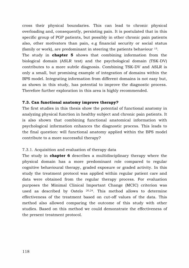
118
cross their physical boundaries. This can lead to chronic physical
overloading and, consequently, persisting pain. It is postulated that in this
specific group of PGP patients, but possibly in other chronic pain patients
also, other motivators than pain, e.g financial security or social status
(family or work), are predominant in steering the patients behaviour 15.
The study in chapter 5 shows that combining information from the
biological domain (ASLR test) and the psychological domain (TSK-DV)
contributes to a more subtle diagnosis. Combining TSK-DV and ASLR is
only a small, but promising example of integration of domains within the
BPS model. Integrating information from different domains is not easy but,
as shown in this study, has potential to improve the diagnostic process.
Therefore further exploration in this area is highly recommended.
7.3. Can functional anatomy improve therapy?
The first studies in this thesis show the potential of functional anatomy in
analyzing physical function in healthy subject and chronic pain patients. It
is also shown that combining functional anatomical information with
psychological information enhances the diagnostic process. This leads to
the final question: will functional anatomy applied within the BPS model
contribute to a more successful therapy?
7.3.1. Acquisition and evaluation of therapy data
The study in chapter 6 describes a multidisciplinary therapy where the
physical domain has a more predominant role compared to regular
cognitive behavioural therapy, graded exposure or graded activity. In this
study the treatment protocol was applied within regular patient care and
data were obtained from the regular therapy process. For evaluation
purposes the Minimal Clinical Important Change (MCIC) criterion was
used as described by Ostelo 20,24. This method allows to determine
effectiveness of the treatment based on cut-off values of the data. This
method also allowed comparing the outcome of this study with other
studies. Based on this method we could demonstrate the effectiveness of
the present treatment protocol.
119
7.3.2. Comparison of therapy protocols
A problem related to the study in chapter 6 was how to distinguish the
content of the therapy protocol from other protocols. Most
multidisciplinary approaches to back pain use combinations of physical
treatment, lessons, group conversations and individual coaching 5,10,26,27.
On this level there are no outspoken differences between the protocols.
Only when looking at the detailed content of the programme, differences
can be pointed out. For that reason it was attempted to describe the
treatment protocol as detailed as possible. Regrettably this is not done in
most other study descriptions. Despite extensive protocol descriptions,
minute but important details are often left out. It is these details that
distinguish the protocols and contribute to the effectiveness of the method.
Also in the present study it was difficult to describe the protocol in
sufficient detail. A major reason for this problem is the limited space
available in scientific articles. To overcome this problem we strongly
recommend to supply such information by referring to an internet address
or website where elaborate information is provided.
7.3.3. The surplus value of functional anatomy
The results of the multidisciplinary protocol as described in chapter 6 are
very promising. As far as could be compared, they are even better than the
results of other multidisciplinary treatments 10,26,27. However, the question
remains whether it is especially the functional anatomy that makes the
difference. First of all there is the limited description of therapy protocols
which obstructs proper comparison of therapy content. Secondly the BPS
model dictates an interaction of all contributing factors, not only in
aetiology, but also in the healing process. This interaction makes it
difficult to identify and rate the specific contributing factors.
Communication between the patient and therapist will not be similar
between the different multidisciplinary therapies. Social, environmental
and even cultural circumstances will not be alike between populations.
And finally behavioural principles will not be implemented in all protocols
in the same manner. Therefore it can not be concluded that the main
difference in the study in chapter 6 is the attention for functional
anatomy. However what we do know is that a vast majority of patients in
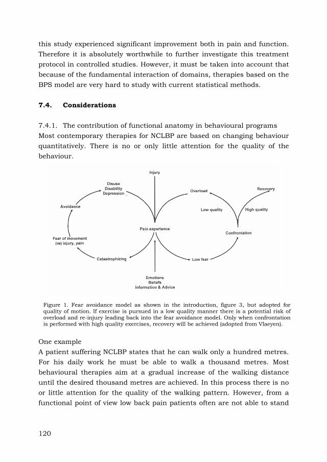
120
this study experienced significant improvement both in pain and function.
Therefore it is absolutely worthwhile to further investigate this treatment
protocol in controlled studies. However, it must be taken into account that
because of the fundamental interaction of domains, therapies based on the
BPS model are very hard to study with current statistical methods.
7.4. Considerations
7.4.1. The contribution of functional anatomy in behavioural programs
Most contemporary therapies for NCLBP are based on changing behaviour
quantitatively. There is no or only little attention for the quality of the
behaviour.
One example
A patient suffering NCLBP states that he can walk only a hundred metres.
For his daily work he must be able to walk a thousand metres. Most
behavioural therapies aim at a gradual increase of the walking distance
until the desired thousand metres are achieved. In this process there is no
or little attention for the quality of the walking pattern. However, from a
functional point of view low back pain patients often are not able to stand
Figure 1. Fear avoidance model as shown in the introduction, figure 3, but adopted for quality of motion. If exercise is pursued in a low quality manner there is a potential risk of overload and re-injury leading back into the fear avoidance model. Only when confrontation is performed with high quality exercises, recovery will be achieved (adopted from Vlaeyen).
121
in a neutral lordosis, but adopt some lumbar flexion. In doing so, the
patient can not efficiently transfer torsional energy from his legs to his
trunk and vice versa. This energy transfer is crucial for effective, energy-
economical walking 7,8,14.
Increasing the walking distance of the patient without improving the
quality of walking will disproportionably load the spine. As a result this
can induce overload, pain and in the end structural change of or even
damage to the spine. The possibility that the patient straightens his spine
and corrects his posture by coincidence is small. Therefore it should be
considered a professional mistake not to focus on improving the quality of
posture and the patient’s walking pattern. It is to be expected that
incorporation of qualitative behavioural modification based on functional
anatomy significantly improves therapy results as illustrated in the
modified model of Vlaeyen in figure 1.
7.4.2. The BPS model and medical disciplines
This thesis showed how functional anatomy can play a role in improving
quality of a multidisciplinary treatment for NCLBP. It can be taken for
granted that there will be more functional anatomical relations that play a
role in low back pain aetiology. A vast amount of fundamental research on
this specific topic is necessary to unravel these functional relations and
validate their clinical impact. However, this research does not guarantee
optimal balance of the BPS model. Finding a balance in the BPS model
also includes more research with respect to the psychological and social
domains. And finally balance can only be achieved when communication
between medical disciplines is optimized 12. This aspect of the BPS model
is especially challenging because of the traditional separation of disciplines
in the (para-)medical world However, if this succeeds their potentials can
be merged to truly optimize the BPS model, as proposed by Engel 3,4.
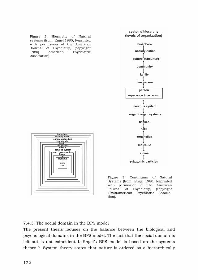
122
7.4.3. The social domain in the BPS model
The present thesis focuses on the balance between the biological and
psychological domains in the BPS model. The fact that the social domain is
left out is not coincidental. Engel’s BPS model is based on the systems
theory 3. System theory states that nature is ordered as a hierarchically
Figure 2. Hierarchy of Natural systems (from: Engel 1980, Reprinted with permission of the American Journal of Psychiatry, (copyright 1980) American Psychiatric Association).
Figure 3. Continuum of Natural Systems (from: Engel 1980, Reprinted with permission of the American Journal of Psychiatry, (copyright 1980)American Psychiatric Associa-tion).
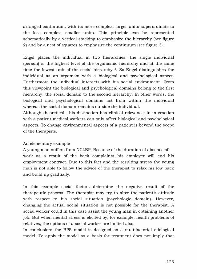
123
arranged continuum, with its more complex, larger units superordinate to
the less complex, smaller units. This principle can be represented
schematically by a vertical stacking to emphasize the hierarchy (see figure
2) and by a nest of squares to emphasize the continuum (see figure 3).
Engel places the individual in two hierarchies: the single individual
(person) is the highest level of the organismic hierarchy and at the same
time the lowest unit of the social hierarchy 4. So Engel distinguishes the
individual as an organism with a biological and psychological aspect.
Furthermore the individual interacts with his social environment. From
this viewpoint the biological and psychological domains belong to the first
hierarchy, the social domain to the second hierarchy. In other words, the
biological and psychological domains act from within the individual
whereas the social domain remains outside the individual.
Although theoretical, this distinction has clinical relevance: in interaction
with a patient medical workers can only affect biological and psychological
aspects. To change environmental aspects of a patient is beyond the scope
of the therapists.
An elementary example
A young man suffers from NCLBP. Because of the duration of absence of
work as a result of the back complaints his employer will end his
employment contract. Due to this fact and the resulting stress the young
man is not able to follow the advice of the therapist to relax his low back
and build up gradually.
In this example social factors determine the negative result of the
therapeutic process. The therapist may try to alter the patient’s attitude
with respect to his social situation (psychologic domain). However,
changing the actual social situation is not possible for the therapist. A
social worker could in this case assist the young man in obtaining another
job. But when mental stress is elicited by, for example, health problems of
relatives, the options of a social worker are limited also.
In conclusion: the BPS model is designed as a multifactorial etiological
model. To apply the model as a basis for treatment does not imply that
124
every contributing factor can or should be addressed. Especially social
factors are difficult to incorporate in the treatment.
7.5. Conclusions
This thesis provides strong arguments for the presumption that more
emphasise on physical aspects within the contemporary BPS disease
model improves the diagnostic process and therapeutic results. Of course
this thesis does not reject that other (psycho-social) factors contribute to
low back pain aetiology. It only propagates more appreciation for the
biological domain, especially functional anatomy, within the BPS model.
It is to be expected that further study of human functional anatomy
contributes to the revaluation of the bio-domain within the BPS model.
This not only holds for the low back and pelvis, but for every other part of
the human body, including the neck and extremities. New functional
anatomical knowledge may contribute to comprehension of the aetiology of
until now obscure syndromes like fibromyalgia, the post whiplash
syndrome, and chronic fatigue syndrome.
The BPS model provides a profound and fertile basis for contemporary
therapy. Extensive application of the model is not limited by the amount of
research in this area. The prime restraint is found in the troublesome and
often disturbed communication between disciplines isolated in their
domain. To fully benefit from the principles of the BPS model it is strongly
suggested to improve interdisciplinary communication and reconsider the
traditional boundaries between medical, psychological and social
disciplines.
Non-specific low back pain does not exist: it is only our lack of insight in
aetiological mechanisms that leaves back pain unexplained. Balancing the
factors within the BPS model will help to improve this insight.
125
References
1. Barker PJ, Briggs CA, Bogeski G. Tensile transmission across the
lumbar fasciae in unembalmed cadavers: effects of tension to various
muscular attachments. Spine 2004;29:129-38.
2. Dankaerts W, O’Sullivan PB, Straker LM, A.F. Burnett AF, Skouen JS.
The inter-examiner reliability of a classification method for non-specific
chronic low back pain patients with motor control impairment. Man Ther
2006;11:28–39.
3. Engel GL. The need for a new medical model: a challenge for bio-
medicine. Science 1977;196:129-36.
4. Engel GL. The clinical application of the biopsychosocial model. Am J
Psychiatry 1980;137:5 535-44.
5. Gatchel RJ, Peng YB, Peters ML, Fuchs PN, Turk DC. The
biopsychosocial approach to chronic pain: scientific advances and future
directions. Psychol Bull 2007;133:581-624.
6. Gracovetsky S, Kary M, Pitchen I, Levy S, Ben Said R. 1989 The
importance of pelvic tilt in reducing compressive stress in the spine during
flexion-extension exercises. Spine 1989;14:412-6.
7. Gracovetsky S. The Spinal Engine, Spinger, 1989.
8. Gracovetsky S. Is the lumbodorsal fascia necessary? J Bodyw Mov Ther.
2008;12:194-7.
9. Hides JA, Richardson CA, Jull GA. Multifidus muscle recovery is not
automatic after resolution of acute, first-episode low back pain. Spine
1996; 21:2763-9.
10. Hildebrandt J, Pfingsten M, Saur P, Jansen J. Prediction of success
from a multidisciplinary treatment program for chronic low back pain.
Spine 1997;22:990-1001.
11. Hungerford B, Gilleard W, Hodges P. Evidence of altered lumbopelvic
muscle recruitment in the presence of sacroiliac joint pain. Spine
2003;28:1593-600.
12. Jones M,Edwards I, Gifford L. Conceptual models for implementing
biopsychosocial theory in clinical practice. Man Ther 2002;7:2–9.
13. Kori SH, Miller RP, Todd, DD. Kinisiophobia: a new view of chronic
pain behaviour. Pain Manag 1990;3:35-43.
126
14. Lamoth CJ, Meijer OG, Wuisman PI, van Dieën JH, Levin MF, Beek PJ.
Pelvis-thorax coordination in the transverse plane during walking in
persons with nonspecific low back pain. Spine 2002;27:E92-9.
15. Lundberg M, Styf J. Kinesiophobia among physiological overusers with
musculoskeletal pain. Eur.J. Pain:10.1016/j.ejpain.2008.08.004.
16. MacDonald DA, Moseley GL,Hodges PW. The lumbar multifidus: Does
the evidence support clinical beliefs? Man Ther 2006;11:254–263.
17. MacDonald D, Moseley GL, Hodges PW. Why some patients keep
hurting their back? Evidence of ongoing back muscle dysfunction during
remission from recurrent back pain. Pain doi:10.1016/j.pain.2008.12.002.
18. McGill SM, Grenier S, Kavcic N, Cholewicki J. Coordination of muscle
activity to assure stability of the lumbar spine. J Electromyograph Kinesiol
2003;13:353-9.
19. Mens J, Hoek van Dijke G, Pool-Goudzwaard A, van der Hulst V, Stam
H.J. Possible harmful effects of high intra-abdominal pressure on the
pelvic girdle. Biomech 2006;39:627-35.
20. Ostelo RW, Deyo RA, Stratford P, Waddell G, Croft P, Von Korff M,
Bouter LM, de Vet HC. Interpreting change scores for pain and functional
status in low back pain: towards international consensus regarding
minimal important change. Spine 2008;33:90-4.
21. Pool-Goudzwaard A, van Dijke GH, van Gurp M, Mulder P, Snijders C,
Stoeckart R. Contribution of pelvic floor muscles to stiffness of the pelvic
ring. Clin Biomech 2004;19:564-71.
22. Pool-Goudzwaard AL, Slieker ten Hove MC, Vierhout ME, Mulder PH,
Pool JJ, Snijders CJ, Stoeckart R. Relations between pregnancy-related low
back pain, pelvic floor activity and pelvic floor dysfunction. Int Urogynecol
J Pelvic Floor Dysfunct 2005;16:468-74.
23. Richardson CA, Jull GA, Muscle control-pain control. What exercises
would you prescribe? Man Ther 1995;1:2-10.
24. van der Roer N, Ostelo RW, Bekkering GE, van Tulder MW, de Vet HC.
Minimal clinically important change for pain intensity, functional status,
and general health status in patients with nonspecific low back pain.
Spine 2006;31:578-82.
25. Slaboda JC, Boston JR, Rudy TE, Lieber SJ. Classifying subgroups of
chronic low back pain patients based on lifting patterns. Arch Phys Med
Rehabil 2008;89:1542-1549.

127
26. Smeets RJ, Vlaeyen JW, Hidding A, Kester AD, van der Heijden GJ,
van Geel AC, Knottnerus JA. Active rehabilitation for chronic low back
pain: Cognitive-behavioral, physical, or both? First direct post-treatment
results from a randomized controlled trial. BMC Musculoskelet Disord
2006;20;7:5.
27. Smeets RJ, Vlaeyen JW, Hidding A, Kester AD, van der Heijden GJ,
Knottnerus JA. Chronic low back pain: physical training, graded activity
with problem solving training, or both? Pain 2008;134:263-76.
28. Vlaeyen JW, Kole-Snijders AM, Boeren RG, van Eek H. Fear of
movement/(re)injury in chronic low back pain and its relation to
behavioral performance. Pain 1995;62:363-72.
29. Vleeming A, Pool-Goudzwaard AL, Stoeckart R, van Wingerden JP,
Snijders CJ. The posterior layer of the thoracolumbar fascia. Its function in
load transfer from spine to legs. Spine 1995;20:753-8.
30. Waddell G. The back pain revolution. Second edition. Churchill
Livingstone, 2004.
31. Waddell G, Burton AK. Concepts of rehabilitation for the management
of low back pain. Best Pract Res Clin Rheumatol 2005;9:655–70.
32. Wu WH, Meijer OG, Bruijn SM, Hu H, van Dieën JH, Lamoth CJ, van
Royen BJ, Beek PJ. Gait in Pregnancy-related Pelvic girdle Pain:
amplitudes, timing, and coordination of horizontal trunk rotations. Eur
Spine J 2008;17:1160-9.

128
129
Summary
The work presented in this thesis examines whether a more
prominent role of functional anatomy within multidisciplinary
treatment of non-specific chronic low back pain (NCLBP) will increase
its therapeutic effect. The multidisciplinary treatment of NCLBP is
based on the biopsychosocial (BPS) model. This model is derived from
systems theory and was introduced by Engel in 1977 to replace the
traditional biomedical model.
Fundamental to the BPS model is that not only biological but also
psychological and social aspects are included in the aetiology of
diseases, such as chronic back pain. However, in the ongoing
development of new diagnostic and therapeutic techniques based on
this BPS model, the behavioural aspects prevailed whilst the physical
aspects (especially physical exercises) lagged behind. Consequently, in
contemporary multidisciplinary treatment protocols, physical
training is subordinate and mainly in service of the desired
modification of behaviour.
Recent studies of multidisciplinary programmes for NCLBP show that
the results of these predominantly psychological, behaviour-
orientated treatments are far from optimal. Therefore, the question
arose as to how multidisciplinary treatment can be improved. One
option for improvement is to intensify the application of functional
anatomical knowledge and incorporate corresponding specific training
within existing multidisciplinary programmes.
Functional anatomical research has made significant progress in the
last decade; this has led to new knowledge on spine function and,

130
consequently, to the development of new physical exercises. In the
context of multidisciplinary treatment of NCLBP patients, and based
on the new functional anatomical knowledge, the aim of this thesis
was to address the following questions:
1. Taking into account the available recent data on functional
anatomy, is there a need to reconsider the role of the physical
domain within the BPS model?
2. Will a more pronounced role of functional anatomy in the BPS
model contribute to better diagnosis?
3. Will functional anatomy applied in the BPS model contribute to
improved therapy?
Study 1
In the first study, which used human dissection material, an
anatomical connection was demonstrated between a pelvic ligament
(the sacrotuberous ligament) and a leg muscle (the m. biceps femoris).
The study shows that - in every case - a part of the muscle’s pulling
force tenses the ligament (7%-69% of the pulling force). From this
study we concluded that it is theoretically possible that a leg muscle,
due to its connection with the pelvic ligament, plays a dynamic role
in controlling sacro-iliac (SI) joint stiffness. This implies that SI joint
control is more complex than formerly presumed and that anatomical
structures, even when not in direct proximity of the joint (e.g. the
arm or leg muscles), can be involved in this control. This conclusion
holds not only for the SI joints, but for other synovial joints in the
body as well.
Study 2
Since the first study was performed on embalmed human material, it
was considered essential to confirm the assumed influence of muscles
on SJ joint stability on living subjects. Therefore the second study
was performed on living, healthy subjects. In 6 young women the
effect of muscle tension on SI joint stiffness was recorded using echo-
Doppler in combination with vibration and EMG measurements. First,
SI joint stiffness without muscle activation was determined using
echo-Doppler. Next, the subjects were asked to activate specific
131
muscles in isolation (homolateral: m.biceps femoris, m. gluteus
maximus, m. erector spinae; and heterolateral: m. latissimus dorsi).
During this muscle activation (controlled with EMG measurement), SI
joint stiffness was determined again with echo-Doppler. For many
subjects it was difficult to activate the muscles in isolation.
Nevertheless, it was demonstrated that the muscles included in this
study, even those not in the proximity of the SI joint, were able to
increase SI joint stiffness. Consequently, a relationship was assumed
between disturbed muscle activation patterns and diminished control
of the SI joint. In turn, diminished muscle control may induce
overload, even under conditions of normal load. Such relative
overload can cause physical complaints. In the context of this thesis,
this is the first evidence that functional anatomical information can
be of importance in explaining the onset or continuation of
complaints of the low back or pelvis.
Study 3
The results of the first and second study led to the conclusion that
different muscle activations lead to distinct motion patterns. In
patients with low back and pelvic problems, it is presumed that those
changes in motion will appear during activities that involve the lower
back and pelvis, such as bending forward. This third study explored
the relative contribution of the lower back and the pelvis (hip motion)
during forward bending. The action of forward bending was
investigated and determined in healthy women, in women with pelvic
girdle pain (PGP), and in women with low back pain (LBP). The
coordination pattern of the women with complaints is distinctly
different from that of the healthy subjects. Moreover, the
coordination pattern of the women with PGP is opposite to that of the
women suffering from LBP. Whereas women with LBP fix their spine
during the bending motion, women with PGP emphasise lumbar
motion and vice versa. This is a clear indication of a specific
complaint and its relation to the motion pattern. Therefore, it seems
feasible to assume that more understanding and appropriate
therapeutic handling of these specific motion patterns will be of value
for the recovery process. This is the second argument in favour of
132
increasing physical information and applications in multidisciplinary
therapy for NCLBP.
Study 4
The fourth study emphasises the importance of integration of
biological (physical) and psychological factors in the diagnostic
process. This study was performed among women with chronic PGP.
The physical impairment, as measured with the Active Straight Leg
Raise (ASLR) test, is compared with the fear of motion, as measured
with the Tampa Scale for Kinesiophobia Dutch language Version (TSK-
DV). It was shown that ASLR and TSK-DV measure distinct aspects of
the patient and that there is no interrelationship between the
physical limitations and the fear of motion. In line with this result, it
is suggested that two specific subgroups can be distinguished:
patients with a logical relation between the recorded impairment and
fear of motion, and patients without a logical relation between
physical impairment and fear of motion. It is important to note that
this latter group can be further subdivided in two groups. Namely, one
group which has little physical impairment, but has considerable fear
of motion; this group can be considered as the ‘classical’ group with
too much fear of motion. The other group has, curiously enough, too
little fear of motion with respect to the considerable amount of
physical impairment. This combination is clinically recognized in
patients that ignore their functional limitations and attempt to
persist functioning on a relatively high level. Although this behaviour
contributes to persistence of the complaints and is, as such, clinically
relevant, this specific group has not yet been described in the
literature in this particular context.
Study 5
The fifth study describes a multidisciplinary treatment protocol for
patients with NCLBP. In this protocol a specific exercise programme
is merged with cognitive behavioural principles. The study is
performed as part of regular patient care and not as an isolated,
randomized study. The population included 245 severe therapy-
resistant NCLBP patients. It is concluded that, in contrast to other
133
programmes where physical training is subordinate, physical exercise
can be leading within a behaviour-oriented approach. Moreover, this
specific combination yielded remarkably positive results in the
majority of patients that received this treatment (55%-80%
significant improvement, depending on the parameters and group
selected).
Based on the studies presented in this thesis the research questions
originally posed can be positively answered as follows:
1. Functional anatomy indeed deserves more emphasis in the
multidisciplinary treatment of NCLBP.
2. Functional anatomy within the BPS model can improve the
diagnostic process in NCLBP.
3. There is sufficient evidence to suggest that functional anatomy
can help improve the multidisciplinary treatment of NCLBP.

134
135
Samenvatting
Dit proefschrift richt zich op de vraag of een meer prominente rol van
functionele anatomie in een multidisciplinaire behandeling voor a-
specifieke chronische lage rugklachten (ACLR) het effect van deze
behandeling verhoogt. De multidisciplinaire behandeling is gebaseerd
op het biopsychosociale (BPS) model. Dit van de systeemtheorie
afgeleide model is in 1977 geïntroduceerd door Engel ter vervanging
van het traditionele biomedische model. Fundamenteel in het BPS
model is dat niet alleen biologische maar ook psychologische en
sociale aspecten worden betrokken in de etiologie van aandoeningen
zoals chronische rug pijn. Bij de ontwikkeling van nieuwe
diagnostische en therapeutische technieken, gebaseerd op het BPS
model, traden de gedragsmatige aspecten op de voorgrond terwijl
fysieke aspecten, en met name oefenvormen achterbleven. Dit had tot
gevolg dat in hedendaagse multidisciplinaire behandelprotocollen de
lichamelijke training ondergeschikt is en ten dienste staat van de
gewenste gedragssturing.
Uit recente studies van multidisciplinaire behandeling van ACLR
blijkt dat de resultaten van deze overwegend psychologische,
gedragsgeoriënteerde behandelvormen nog verre van optimaal zijn.
Dit is aanleiding om na te gaan of de multidisciplinaire behandeling
nog verbeterd kan worden. Een mogelijkheid tot verbetering ligt in
het intensiever toepassen van functioneel anatomische kennis en
daaraan gekoppelde specifieke training binnen de bestaande
multidisciplinaire programma’s. Het onderzoek naar functioneel
anatomische kennis van het bewegingsapparaat heeft de laatste jaren
136
een sterke ontwikkeling doorgemaakt die heeft geleid tot nieuwe
inzichten in de werking van de wervelkolom en het bekken . Daarop
aansluitend zijn nieuwe vormen van fysieke training ontwikkeld. Met
deze nieuwe functioneel anatomische kennis als uitgangspunt is het
doel van dit proefschrift om, in de context van multidisciplinaire
behandeling van patiënten met ACLR, een antwoord te formuleren op
de volgende drie vragen:
1. Dient de rol van het fysieke domein binnen het BPS model
heroverwogen te worden, de recente ontwikkelingen van
functioneel anatomische kennis in aanmerking nemende?
2. Kan een meer uitgesproken rol voor functionele anatomie in
het BPS model bijdragen aan een betere diagnose van lage
rugklachten?
3. Kan meer aandacht voor functionele anatomie toegepast
binnen een multidisciplinaire behandeling bijdragen aan een
betere therapie voor ACLR?
Eerste studie
In de eerste studie verricht op menselijke preparaten wordt een
anatomische verbinding aangetoond tussen een van de banden van
het bekken, de sacrotuberale band, en een van de beenspieren, de m.
biceps femoris. De studie laat zien dat in alle gevallen een deel van de
kracht waarmee de spier aan de band trekt deze op spanning brengt (7
tot 69% van de trekkracht). Uit deze studie wordt geconcludeerd dat
het theoretisch mogelijk is dat de bewuste beenspier, door de
beschreven verbinding met de bekkenband, een dynamische rol kan
spelen bij het controleren van de stijfheid van de sacro-iliacale (SI)
gewrichten. Dit impliceert dat controle over het SI gewicht complexer
is dan voorheen werd verondersteld en dat anatomische structuren
die verder van het gewricht verwijderd zijn, zoals been- of zelfs
armspieren, hierbij betrokken kunnen zijn. De bevinding dat verder
weggelegen spieren een rol kunnen spelen bij gewrichtssturing geldt
niet alleen voor de SI gewrichten, maar ook voor andere synoviale
gewrichten in het lichaam.
137
Tweede studie
De eerste studie heeft plaats gevonden op gefixeerde en geprepareerde
lichamen. Het was daarom noodzakelijk de in de eerste studie
veronderstelde werking van spieren op de SI gewrichten ook bij
levende proefpersonen (in vivo) aan te tonen. Bij 6 proefpersonen is
met Echo-Doppler in combinatie met vibratie, en EMG-metingen de
invloed van spierspanning op de stijfheid van het SI gewricht
gemeten. Eerst is met de Echo-Doppler de stijfheid van de SI
gewrichten in rust bepaald. Vervolgens is aan de proefpersonen
gevraagd bepaalde spieren ( ipsi lateraal: m.biceps femoris, m. gluteus
maximus, m. erector spinae en contralateraal: m. latissimus dorsi)
aan te spannen. Tijdens deze spieractivatie die werd gecontroleerd
met de EMG metingen, is met de Echo-doppler weer de stijfheid van
de SI gewrichten bepaald. Voor veel proefpersonen bleek het lastig de
verschillende spier(groepen) werkelijk geïsoleerd aan te spannen.
Desondanks kon aangetoond worden dat de in deze studie betrokken
spieren de stijfheid van het SJ gewricht konden vergroten. Ook
wanneer deze spieren niet direct rond het SI gewricht liggen. Op basis
van deze bevindingen wordt een samenhang verondersteld tussen een
verstoring in activatiepatronen van deze spieren en verminderde
controle over het SI gewricht. Een verminderde controle over de SI
gewrichten kan, ook bij gelijk blijvende belasting, een relatieve
overbelasting veroorzaken. Dergelijke overbelasting kan aanleiding
zijn tot klachten. In het kader van dit proefschrift is dit de eerste
aanwijzing dat functioneel-anatomische informatie van belang is bij
het verklaren van het ontstaan of voortduren van rug- en
bekkenklachten.
Derde studie
Op basis van literatuur en de eerste en tweede studie wordt
aangenomen dat veranderingen in spieractivatiepatronen kunnen
leiden tot veranderingen in bewegingspatronen. Bij mensen met rug-
of bekkenklachten ligt het voor de hand te veronderstellen dat dit
onder meer tot uiting zal komen bij bewegingen waarbij rug en bekken
betrokken zijn, zoals bij voorover buigen. In deze studie wordt de
relatieve bijdrage van de lage rug en het bekken (heupbeweging)
138
tijdens het voorover buigen beoordeeld. Deze beweging werd
vastgelegd bij gezonde vrouwen, vrouwen met bekkenklachten en
vrouwen met rugklachten. De coördinatie van de vrouwen met
klachten wijkt af van de coördinatie van de gezonde vrouwen. Tussen
de vrouwen met bekkenklachten en de vrouwen met rugklachten is de
coördinatie echter precies omgekeerd. Waar de vrouwen met
rugklachten de lage rug tijdens de beweging fixeren, leggen de
vrouwen met bekkenklachten juist de nadruk op de beweging van de
lage rug, en omgekeerd. Dit is duidelijke aanwijzing voor een verband
tussen de specifieke klacht (bekken of rug) en de wijze van bewegen.
Het ligt daarbij voor de hand dat inzicht in en therapeutisch
aangrijpen op dit specifieke bewegingspatroon van meerwaarde kan
zijn voor herstel. Dit is een tweede argument voor het intensiever
betrekken van informatie over het fysiek functioneren in
multidisciplinaire programma’s.
Vierde studie
De vierde studie benadrukt het belang van het integreren van
biologische (fysieke) en psychische factoren in het diagnostische
proces. Deze studie is uitgevoerd bij vrouwen met chronische
bekkenklachten. De fysieke beperking, gemeten met de Active
Straight Leg Raise (ASLR) test, wordt vergeleken met de mate van
bewegingsangst, gemeten met de Tampa Scale voor Kinesiophobia
Dutch language Version (TSK-DV). Uit dit onderzoek blijkt dat de
ASLR en de TSK-DV geheel verschillende aspecten van een patiënt
meten. Er blijkt dan ook geen enkel verband tussen de gemeten
fysieke beperking en de angst voor bewegen. Juist daarom is het van
belang dat een aantal subgroepen wordt onderscheiden, te weten: a.
patiënten waarbij er een logisch verband is tussen de gemeten
beperking en de bewegingsangst en b. patiënten zonder een logisch
verband tussen de fysieke beperking en de bewegingsangst. Het is van
belang op te merken dat deze laatste groep weer in twee geheel
verschillende groepen is op te splitsen. Eén deel van de patiënten in
deze groep heeft weinig fysieke beperking maar wel veel
bewegingsangst. Deze subgroep kan gezien worden als de klassieke
139
groep met teveel angst voor bewegen. Daarnaast is ook een groep die
in verhouding tot de gemeten beperking juist merkwaardig weinig
bewegingsangst heeft. Dit beeld wordt klinisch herkend als een groep
patiënten die de klachten negeert en tracht te blijven functioneren op
een verhoudingsgewijs te hoog niveau. Hoewel dit gedrag bijdraagt bij
aan het in stand houden van de klachten als zodanig dus klinisch
relevant is, is deze laatste groep in literatuur echter nog niet eerder
op deze wijze beschreven.
Vijfde studie
In de vijfde studie wordt een multidisciplinaire behandeling voor
patiënten met ACLR beschreven waarin een specifiek fysiek
oefenprogramma wordt geïntegreerd met cognitief gedragsmatige
principes. Deze studie is uitgevoerd als onderdeel van reguliere
patiëntenzorg en niet als geïsoleerde, gerandomiseerde studie. De
populatie die is gebruikt in deze studie bestond uit 245 ernstige,
therapie resistente ACLR patiënten. Het wordt vastgesteld dat het
mogelijk is om, in tegenstelling tot andere programma’s waar fysieke
training ondergeschikt is, meer nadruk te leggen op een fysiek
oefenprogramma binnen een gedragsgeoriënteerde benadering.
Bovendien blijkt dat deze specifieke combinatie bijzonder positieve
resultaten geeft bij een groot deel van de behandelde patiënten (55 tot
80% significante verbetering afhankelijk van de gekozen parameter en
groep).
Op basis van de studies in dit proefschrift kunnen de eerder gestelde
vragen als volgt positief worden beantwoord:
1. Functionele anatomie dient meer betrokken te worden in
multidisciplinaire behandelingen van ACLR.
2. Functionele anatomie kan binnen het BPS model bijdragen aan
een betere diagnose bij ACLR.
3. Er zijn duidelijke aanwijzingen dat functionele anatomie kan
bijdragen aan een betere therapie voor ACLR.

140

141
Dankwoord
Van dit proefschrift heb ik het schrijven van het dankwoord als een van de
moeilijkste stukken ervaren. Voor een rechtgeaarde doe-het-zelver is het al
niet gemakkelijk hulp te vragen. Maar als je meer dan 10 jaar over het
schrijven van je proefschrift doet dan ontkom je er niet aan onderweg hulp
te moeten vragen. En hulp heb ik gekregen van veel verschillende mensen
op veel verschillende manieren. Graag wil ik al die mensen bedanken voor
hun bijdrage aan dit boekje en mijn pad. Iedereen persoonlijk bedanken
gaat niet lukken, maar een aantal mensen wil ik zeker noemen.
Beste Chris, onze samenwerking was kort en vooral krachtig. Ik heb je
doortastende, no-nonsense begeleiding in het laatste en meest hectische
stuk van dit proefschrift zeer gewaardeerd. Jouw rotsvaste vertrouwen in
mijn stukken (of was dat meer in Gert-Jan en Rob?), heeft mij zeer
geholpen dit proefschrift ook echt af te ronden.
Beste Rob, dank je voor je geduldige, nooit aflatende, liefdevolle, maar
meedogenloze begeleiding. Op alle vlakken weet je steeds weer
scherpzinnig de juiste snaar te raken, of je rust niet voor hij geraakt wordt.
Bovendien heb je me, samen met Corrie, figuurlijk, maar ook letterlijk de
ruimte gegeven om dit proefschrift te laten groeien. Wil je me nu leren hoe
je mooie teksten schrijft?
Beste Gert-Jan, jouw benadering van wetenschap blijft onnavolgbaar:
bodemloze kennis verpakt in razendsnelle humor op zen-boeddistische
basis. En dat gecombineerd met ongeëvenaarde sociale vaardigheden. Ik
hoop in de toekomst nog veel met je samen te mogen beleven, te leren….
en te jammen?

142
Lieve Ria, hoe waardeer je het opkomen van de zon in de ochtend? Zo
vanzelfsprekend maar tegelijk zo onmisbaar en onbeschrijflijk. Op jou is
stelling 11 absoluut van toepassing. Dank je voor alle dingen die je voor
me gedaan en geregeld hebt maar bovenal omdat je een van mijn vrienden
bent.
Beste Astrid, je hebt me geholpen tijd vrij te maken voor dit proefschrift.
Zonder die ruimte was dit werk nooit afgerond.
Andry, het helpen afronden van jouw proefschrift was het begin van dit
proefschrift. De jaren die ik voor jou gewerkt heb zijn voor mij een kostbaar
leerproces geweest.
Beste Chris, op jouw afdeling aan het ErasmusMC (afdeling Biomedische
Natuurkunde en Technologie) heb ik mogen leren hoe onderzoek doen
werkt. Ik ben je dankbaar voor de plaats die je me daar gegund hebt.
Professor Voogd, op uw afdeling (Anatomie) mocht ik mij verder
ontwikkelen. Dank u dat u mij deze mogelijkheid geboden heeft.
Marcel, bij jou heb ik mijn eerste stappen in de wetenschappelijke praktijk
mogen maken. Dank je voor je geduld bij het steeds weer uitleggen hoe
bepaalde zaken eigenlijk in elkaar staken. “wacht even, is dat echt wel zo”
zit er, tot frustratie van mijn collega’s na al die jaren bij mij nog steeds
ingebakken.
Muzaffer, dank je voor je onmisbare hulp bij de Doppler metingen.
Alle andere medewerkers van de afdeling Biomedische Natuurkunde en
Technologie, en de afdeling Anatomie , waar ik in het verleden en heden
mee heb samengewerkt.
Jan, John, en Annelies dank jullie voor het sparren over de SJC
protocollen. Alle discussies over testen, oefeningen, (chronische) pijn.
Jullie kennis en visie heeft bijgedragen aan de werkwijze van het Spine &
Joint Centre en uiteindelijk aan mijn proefschrift.
Lex, dank je voor je efficiënte en nuchtere statistische ondersteuning
gebaseerd op je ongeëvenaarde statistische kennis die je glashelder weet
over te brengen.
Laraine, dank je voor je superefficiënte taalcorrecties en bemoedigende
woorden.
Inge en Vincent, het wetenschappelijke SJC team. Jullie kunnen het ook,
echt!

143
Alle SJC collega’s uit heden en verleden die met elkaar steeds weer het
onmogelijk geachte mogelijk maken voor mensen met chronische pijn:
An, Andrea, Annelies, Annemarie, Annemarieke, Annemiek, Anton,
Barbara, Britta, Carolien, Cees, Cindy, Colinda, Crispijn, Danielle, Derek,
Emmy, Esther, Evelien, Ferry, Gaby, Haitze, Helen, Hubert, Inge, Ingrid,
Irene, Irma, Jacqueline, Jan, Jeanet, Jeannette, Jeroen, JohnM, JohnV,
Jolanda, Jolan, Joosje, Jorien, Karin, Katarina, Leanne, Liesbeth,
Maarten, Maarten, Madelon, Marika, Marja, Merit, Natasja, Nora, Noor,
Oliver, Rieneke, Rob, Rosaline, Samantha, Servaas, Suzan, Suzanne,
Timothy, Tineke, Twan, Victor, Vincent, Wies, Wil
Alle stagiaires die over de jaren stukken informatie hebben vergaard die
mede geleid hebben tot dit proefschrift: Carola, Roland, Renee, Anton,
Nesrin, Karim, Bart, Yvonne, Arnold, Bertil, Marijke, Renate, Igor, Aaike,
Andries, Karline, Marieke, Danny, Ludo.
De praktijken die het eerste video onderzoek op patienten hebben mogelijk
gemaakt Dick Zaanen, Liem Lim, Spanjersberg, Bergweg, Walenburgerweg,
Bergpolder.
Irenea, voor het essentiële duwtje in de goede richting.
En omdat ik het beste stukje altijd voor het laatst bewaar: lieve Wilma,
dank je dat je me laat zien en ervaren dat het wel kan.

144
145
List of publications
van Wingerden JP, Vleeming A, Snijders CJ, Stoeckart R. A functional-
anatomical approach to the spine-pelvis mechanism: interaction between
the biceps femoris muscle and the sacrotuberous ligament. Eur Spine J
1993 ; 2:140-144.
van Wingerden JP, Vleeming A, Buyruk HM, Raissadat K. Stabilization of
the sacroiliac joint in vivo: verification of muscular contribution to force
closure of the pelvis. Eur Spine J 2004;13:199-205.
van Wingerden JP, Vleeming A. Ronchetti I. Differences in standing and
forward bending in women with chronic low back or pelvic girdle pain;
indications for physical compensation strategies. Spine 2008;33:E334-
E341.
van Wingerden JP, Stoeckart R, Ronchetti I, Burdorf A, Kleinrensink G-J.
Kinesiophobia in woman with chronic pelvic pain; fear of motion in
physical perspective.
Submitted to Spine
van Wingerden JP, Ronchetti I, Vleeming A, Stoeckart R, Burdorf A,
Kleinrensink G-J. Balancing the biopsychosocial model for
multidisciplinary treatment of non-specific chronic low back pain: merging
motor control education and behavioural principles. Submitted to Spine
146
Vleeming A, van Wingerden JP, Snijders CJ, Stoeckart R, and Stijnen T.
Load application to the sacrotuberous ligament; influences on SI-joint
mechanics. Clin Biomech 1989;4:204-9.
de Wall M, van Riel MP, Snijders CJ, van Wingerden JP. The effect on
sitting posture of a desk with a 10 degree inclination for reading and
writing. Ergonomics 1991;34:575-84.
Vleeming A, van Wingerden JP, Dijkstra PF, Stoeckart R, Snijders CJ and
Stijnen T. Mobility in the sacroiliac joints in the elderly: A kinematic and
radiological study. Clin Biomech 1992;7:170-6.
Derksen JC, Van Riel MP, van Wingerden JP, Snijders CJ. A comparison of
working postures of parcel sorters using three different working methods.
Ergonomics 1994;37:299-309.
Vleeming A, Pool-Goudzwaard AL, Stoeckart R, van Wingerden JP, Snijders
CJ. The posterior layer of the thoracolumbar fascia. Its function in load
transfer from spine to legs. Spine. 1995 1;20:753-8.
Burdorf A, van Riel M, van Wingerden JP, van Wingerden S, Snijders C.
Isodynamic evaluation of trunk muscles and low-back pain among workers
in a steel factory. Ergonomics 1995;38:2107-17.
Kleinrensink G-J, Stoeckart R, Vleeming A, Snijders CJ, Mulder PG, van
Wingerden JP. Peripheral nerve tension due to joint motion. A comparison
between embalmed and unembalmed human bodies. Clin Biomech 1995
;10:235-239.
Vleeming A, de Vries HJ, Mens JM, van Wingerden JP. Possible role of the
long dorsal sacroiliac ligament in women with peripartum pelvic pain. Acta
Obstet Gynecol Scand. 2002;81:430-6.
Ronchetti I, Vleeming A, van Wingerden JP. Physical Characteristics of
Women With Severe Pelvic Girdle Pain After Pregnancy, A Descriptive
Cohort Study. Spine 2008;33:145–51.
147
Curriculum Vitae
The author of this thesis was born in 1962 in Rotterdam as the son of
Jan van Wingerden and Thea van Wingerden-Willebrand. After
receiving his VWO diploma (Atheneum) in 1982, he joined the Royal
Netherlands Naval College (Koninklijk Instituut voor de Marine, KIM) in
Den Helder as marine cadet (‘adelborst’) until October 1983.
In 1988 he graduated at the Academy of Physical Therapy Rotterdam
as physical therapist with a cum laude paper on the application of
artificial intelligence for clinical assessment protocols.
From 1988 untill 1996 he worked at the departments of Biomedical
Technology (Prof.dr.ir. C.J. Snijders) and Anatomy (Prof.dr. J. Voogd)
of the Erasmus University Rotterdam. During this period as a
research assistant he became acquainted with anatomical dissection,
biomechanics, measurement techniques, study design, data analysis,
statistics and programming.
Teaching is one of the author’s passions. At both graduate and
postgraduate level he has taught courses on computer science,
biomechanics, ergonomics, dissection-room anatomy, anatomy in vivo
and functional anatomy.
In 1996 he joined the newly-founded Dutch rehabilitation centre - the
Spine & Joint Centre – first as head of therapy and since 2001 as
managing director.
The author is the delighted father of Thomas (14) and Roos (10).

148
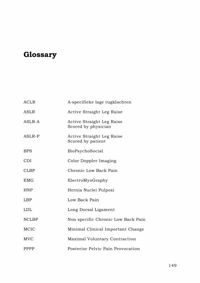
149
Glossary
ACLR A-specifieke lage rugklachten
ASLR Active Straight Leg Raise
ASLR-A Active Straight Leg Raise
Scored by physician
ASLR-P Active Straight Leg Raise
Scored by patient
BPS BioPsychoSocial
CDI Color Doppler Imaging
CLBP Chronic Low Back Pain
EMG ElectroMyoGraphy
HNP Hernia Nuclei Pulposi
LBP Low Back Pain
LDL Long Dorsal Ligament
NCLBP Non specific Chronic Low Back Pain
MCIC Minimal Clinical Important Change
MVC Maximal Voluntary Contraction
PPPP Posterior Pelvic Pain Provocation

150
PGIC Patients Global Impression of Change
PGP Pelvic Girdle Pain
QBDS Quebec Back Pain Disability Scale
ROM Range Of Motion
SI SacroIliac
SIJ SacroIliac Joint
THD ThresHold Difference
TSK-DV Tampa Scale of Kinesiophobia –
Dutch language Version
VAS Visual Analogue Scale