Embed Size (px)
Citation preview

ADHM201500298.xml Generated by PXE using XMLPublishSM June 16, 2015 17:28 APT: WF JID: ADHM
Full Paper
xxxx
J. H. Ahire, M. Behray, C. A. Web-ster, Q. Wang, V. Sherwood, N.Saengkrit, U. Ruktanonchai, N. Wora-mongkolchai, Y. Chao* .........x–xxSynthesis of Carbohydrate Capped Sil-icon Nanoparticles and their ReducedCytotoxicity, In Vivo Toxicity, and Cel-lular Uptake
Carbohydrate capped silicon nanoparti-cles including galactose, glucose, man-nose, and lactose have been successfullydeveloped as targeting and monitoringagent for drug delivery. They show re-duced toxicity both in vitro and in vivocompared to amine terminated siliconnanoparticles, and can be taken up morereadily by cancerous cells than noncancer-ous cells.

ADHM201500298.xml Generated by PXE using XMLPublishSM June 16, 2015 17:28 APT: WF JID: ADHMF
ullP
aper
ADHM201500298(201500298)
Author Proofwww.MaterialsViews.com www.advhealthmat.de
Synthesis of Carbohydrate Capped Silicon Nanoparticlesand their Reduced Cytotoxicity, In Vivo Toxicity, andCellular Uptake
Jayshree H. Ahire, Mehrnaz Behray, Carl A. Webster, Qi Wang, Victoria Sherwood,Nattika Saengkrit, Uracha Ruktanonchai, Noppawan Woramongkolchai,and Yimin Chao*
Keywords: cancer cells, carbohydrates, nanotoxicity, silicon nanoparticles, uptake
ABSTRACT: The development of smart targeted nanoparticles (NPs) that can identify
and deliver drugs at a sustained rate directly to cancer cells may provide better effi-
cacy and lower toxicity for treating primary and advanced metastatic tumors. Obtain-
ing knowledge of the diseases at the molecular level can facilitate the identification
of biological targets. In particular, carbohydrate-mediated molecular recognitions us-
ing nano-vehicles are likely to increasingly affect cancer treatment methods, opening a
new area in biomedical applications. Here, silicon NPs (SiNPs) capped with carbohy-
drates including galactose, glucose, mannose, and lactose have been successfully syn-
thesized from amine terminated SiNPs. The MTT analysis shows an extensive reduc-
tion in toxicity of SiNPs by functionalizing with carbohydrate moiety both in vitro and
in vivo. Cellular uptake has been investigated with flow cytometry and confocal fluo-
rescence microscope. The results show the carbohydrate capped SiNPs can be inter-
nalized in the cells within 24 h of incubation, and can be taken up more readily by
cancer cells than noncancerous cells. Moreover, these results reinforce the use of car-
bohydrates for the internalization of a variety of similar compounds into cancer cells. Q1
1. Introduction
Every year, millions of people’s lives worldwide are affectedby a complex group of diseases known as cancer. Even thoughtraditional chemotherapies that indiscriminately act against ac-tively dividing cells have been identified as one of the mosteffective methods of cancer therapy, chemotherapeutic agentsexhibit poor specificity in reaching tumor tissue and are of-ten restricted by dose-limiting toxicity. To overcome these ma-jor drawback of chemotherapy, targeted drug delivery, andcontrolled drug release technology may provide a more effi-
Dr. J. H. Ahire, M. Behray, Dr. Q. Wang, Dr. Y. Chao, School ofChemistry, University of East Anglia, Norwich NR4 7TJ, UKC. A. Webster, Dr. V. Sherwood, School of Pharmacy,University of East Anglia, Norwich NR4 7TJ, UKDr. N. Saengkrit, Dr. U. Ruktanonchai, Dr. N.Woramongkolchai, National Nanotechnology Center(NANOTEC), National Science and Technology DevelopmentAgency (NSTDA), Pathumthani 12120, ThailandCorrespondence to: Dr. Y. Chao (E-mail: [email protected])Q2
10.1002/adhm.201500298
cient and less harmful solution, as targeted therapies specifi-cally interfere with those molecules essential for tumor growthand progression.[1] Targeted therapies include monoclonal an-tibodies and small molecule inhibitors targeting tumor spe-cific markers. It is well known that tumors express particularmolecular signatures that discriminate them from healthy tis-sues, providing a rich source of potential targets. However,there are some methodological weakness to this general ap-proach. For example, the identification of tumor specific mark-ers is a time consuming process. validating any applicationrequires that patients must be stratified prior to treatment toensure that the appropriate targets are present in each case.As such these treatments are expensive, for example colorectalcancer targeted therapies can cost several thousand dollars permonth.[2] Therefore, as people live longer, with accompanyingimprovements in early detection rates, the financial burdenof providing targeted cancer therapies is expected to rise sub-stantially. Furthermore, tumor cells have high tendencies tomutate, which changes their antigenic modifications leadingto negative results. A good example of this is the multiple re-sistance mechanisms observed in targeting the constitutively
Adv. Healthcare Mater. 2015, 00, 1–10 c© 2015 WILEY-VCH Verlag GmbH & Co. KGaA, Weinheim wileyonlinelibrary.com 1

ADHM201500298.xml Generated by PXE using XMLPublishSM June 16, 2015 17:28 APT: WF JID: ADHMF
ull
Pap
er
Author Proofwww.MaterialsViews.com www.advhealthmat.de
active, pro-proliferative BrafV600E mutation in melanoma.[3–9]
A possible alternative to tumor-specific targeting using over-expressed oncogenes is to take advantage of other moleculespresent on the surface of cells, such as cell surface carbohy-drates.
Carbohydrates are attractive targets for receptor–mediatedinteraction and in particular glycol-conjugates, which play im-portant roles in cancer development metastasis. Many typesof mammalian cells are covered with a dense layer of carbo-hydrates known as the glycol-calyx, in which carbohydratesare bound to proteins and lipids to form glycoproteins, pro-teoglycans, and glycolipids. These naturally occurring glycol-conjugates play an important role in the process of cell–cell interactions and cell–cell communication that is vital forphysiological and pathological processes.[10, 11] As one of themost common cell–surface ligands, carbohydrates can directthe initiation of many medicinally important physiologicalprocesses where they are involved in a wide variety of cel-lular events,[12–14] including inflammatory and immunologi-cal responses,[15–17] tumor metastasis,[18] cell–cell signaling,[19]
apoptosis, adhesion,[20] bacterial and viral recognition,[20, 21] andanticoagulation.[22]
The biological roles of carbohydrates as signaling effectorsand recognition markers are associated with specific molecularrecognition in which proteins[23] or other carbohydrates[24] areinvolved. This characteristic led to the identification of tumor-associated carbohydrate molecules,[19, 25] which has greatly im-proved the development of carbohydrate-based anticancer vac-cine studies.[26] However, the understanding of carbohydrate-binding properties of tumors is not well defined. Cancer cellscan interact with the extracellular matrix in their microenvi-ronment through endogenous receptors binding with cognatecarbohydrates.[26, 27] These interactions vary, depending on thephysiological state of the cells, as supported by histological stud-ies of tumor tissues.[28, 29] Therefore, the ability to characterizeand distinguish carbohydrate binding profiles of a variety ofcells can expedite both the mechanistic understanding of theirrole in disease development and the expansion of diagnosticand therapeutic tools.[30–32]
When nanoparticles (NPs) are functionalized with ligandssuch as antibodies, proteins or peptides, oligonucleotides orcarbohydrates, they become excellent vehicles for biologicalapplications at the cellular and molecular level.[33] However,several features need to be fine-tuned including ligand den-sity, particle diameter, surface charges, magnetic, electronic oroptical properties, stability, and targeting specificity. Nanoma-terials can serve as promising platforms for displaying carbo-hydrates for biological recognition. Due to the smaller sizesof NPs compared to their micrometer sized counterparts, NPshave much larger surface areas, which can enable higher ca-pacity in receptor binding. In addition, multiple carbohydrateligands can be immobilized onto one NP, which can poten-tially enhance the weak affinities of individual ligands to theirbinding partners.
Silicon NPs (SiNPs) hold prominent interest in variousfields of biomedical research including imaging, detection,sensing to drug delivery and new therapeutic uses for a vari-ety of diseases including cancer.[34, 35] This is in addition to the
electronic, magnetic, and optical properties exhibited by theseNPs. SiNPs or quantum dots have size dependent tuneablelight emission, bright luminescence and stability against pho-tobleaching compared to organic fluorescent dye molecules,which makes them ideal tools for fluorescence imaging. Allthese properties have enabled SiNPs to be used as fluorescentcellular markers in a number of diagnostic and assay roles.[36, 37]
Moreover, when comparing with heavy metal and other typesof semiconductor quantum dots, SiNPs exhibit low inherenttoxicity.[37–40]
Herein, we explore the possibility of using glyco-conjugatedSiNPs to detect various cancer cell types on the basis of the morephysiologically related carbohydrate–receptor interactions. Wehave focused on SiNPs functionalized with carbohydrates thatplay key roles in molecular recognition processes, rather thanthose where carbohydrates mainly function as NP stabilizingagents.[41–43] The information obtained on the physiologicallyrelevant carbohydrate–receptor interaction will not only en-hance our understanding of the role carbohydrates play in can-cer, but also guide the development of potential therapeuticssuch as novel contrast agents for tumor detection.
2. Results and Discussions
2.1. Stability of Carbohydrate Capped SiNPs in BiologicalMedia
A significant challenge in the use of NPs for biomedical ap-plications is to retain their stability in biologically associatedenvironments. In order to achieve this, NPs have to be hy-drophilic and maintain superior stability in biological media.For advanced biomedical applications of NPs (e.g., in vivo di-agnostics and therapy), additional requirements such as mini-mization of nonspecific uptake by reticuloendothelial systemsmust be imposed in order to achieve long blood circulationtime and high diagnostic or therapeutic efficiency.[44] To in-vestigate the in vivo effects of NPs in the circulation, and tomeasure the effects of NPs on different cell types in vitro, NPshave to be dispersed in physiological solutions. However, parti-cles which are dispersed in solutions containing physiologicalsalt concentrations and pH values can form micrometer-sizedcoarse agglomerates.[45–47] Coarse agglomerates of NPs havebeen shown to exert different biological effects compared towell-dispersed nanoparticles.[48–50] Moreover, aggregation caninduce toxicity both in vitro and in vivo models.[51] Therefore,it is important to investigate the effect of biological media oncarbohydrate capped SiNPs.
Dynamic light scattering (DLS) was used to assess the stabil-ity of carbohydrate capped SiNPs in biological media by mon-itoring the variation of the average hydrodynamic size of theNPs. For long-term stability testing, 0.1 mL solution of carbohy-drate capped SiNPs was incubated in 1 mL of Dulbecco’s mod-ified eagle’s medium (DMEM) with 10% FBS, Roswell ParkMemorial Institute (RPMI) with 10% FBS and 0.1 X Marc’sModified Ringers (MMR) as a function of time. The averagehydrodynamic diameter was measured at various time points.Figure S1, Supporting Information, and Table 1 show the sta-bility of carbohydrate capped SiNPs by DLS in DMEM, RPMI,
2 wileyonlinelibrary.com c© 2015 WILEY-VCH Verlag GmbH & Co. KGaA, Weinheim Adv. Healthcare Mater. 2015, 00, 2–10
ADHM201500298.xml Generated by PXE using XMLPublishSM June 16, 2015 17:28 APT: WF JID: ADHMF
ullP
aper
Author Proofwww.MaterialsViews.com www.advhealthmat.de
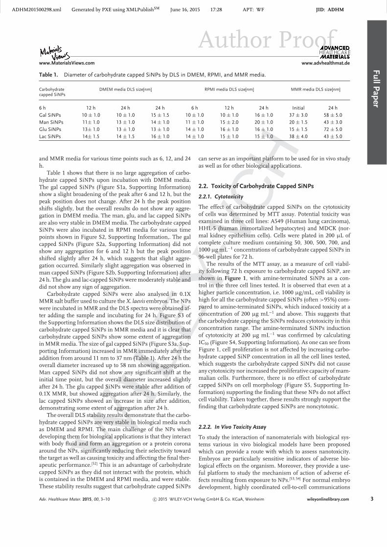
Table 1. Diameter of carbohydrate capped SiNPs by DLS in DMEM, RPMI, and MMR media.
Carbohydratecapped SiNPs
DMEM media DLS size[nm] RPMI media DLS size[nm] MMR media DLS size[nm]
6 h 12 h 24 h 24 h 6 h 12 h 24 h Initial 24 h
Gal SiNPs 10 ± 1.0 10 ± 1.0 15 ± 1.5 10 ± 1.0 10 ± 1.0 16 ± 1.0 37 ± 3.0 58 ± 5.0
Man SiNPs 11± 1.0 13 ± 1.0 14 ± 1.0 11 ± 1.0 15 ± 2.0 20 ± 1.0 20 ± 1.5 43 ± 3.0
Glu SiNPs 13± 1.0 13 ± 1.0 13 ± 1.0 14 ± 1.0 16 ± 1.0 16 ± 1.0 15 ± 1.5 72 ± 5.0
Lac SiNPs 14± 1.5 14 ± 1.5 16 ± 1.0 14 ± 1.0 15 ± 1.0 15 ± 1.0 38 ± 4.0 43 ± 5.0
and MMR media for various time points such as 6, 12, and 24h.
Table 1 shows that there is no large aggregation of carbo-hydrate capped SiNPs upon incubation with DMEM media.The gal capped SiNPs (Figure S1a, Supporting Information)show a slight broadening of the peak after 6 and 12 h, but thepeak position does not change. After 24 h the peak positionshifts slightly, but the overall results do not show any aggre-gation in DMEM media. The man, glu, and lac capped SiNPsare also very stable in DMEM media. The carbohydrate cappedSiNPs were also incubated in RPMI media for various timepoints shown in Figure S2, Supporting Information,. The galcapped SiNPs (Figure S2a, Supporting Information) did notshow any aggregation for 6 and 12 h but the peak positionshifted slightly after 24 h, which suggests that slight aggre-gation occurred. Similarly slight aggregation was observed inman capped SiNPs (Figure S2b, Supporting Information) after24 h. The glu and lac-capped SiNPs were moderately stable anddid not show any sign of aggregation.
Carbohydrate capped SiNPs were also analysed in 0.1XMMR salt buffer used to culture the X. laevis embryos. The NPswere incubated in MMR and the DLS spectra were obtained af-ter adding the sample and incubating for 24 h. Figure S3 ofthe Supporting Information shows the DLS size distribution ofcarbohydrate capped SiNPs in MMR media and it is clear thatcarbohydrate capped SiNPs show some extent of aggregationin MMR media. The size of gal capped SiNPs (Figure S3a, Sup-porting Information) increased in MMR immediately after theaddition from around 11 nm to 37 nm (Table 1). After 24 h theoverall diameter increased up to 58 nm showing aggregation.Man capped SiNPs did not show any significant shift at theinitial time point, but the overall diameter increased slightlyafter 24 h. The glu capped SiNPs were stable after addition of0.1X MMR, but showed aggregation after 24 h. Similarly, thelac capped SiNPs showed an increase in size after addition,demonstrating some extent of aggregation after 24 h.
The overall DLS stability results demonstrate that the carbo-hydrate capped SiNPs are very stable in biological media suchas DMEM and RPMI. The main challenge of the NPs whendeveloping them for biological applications is that they interactwith body fluid and form an aggregation or a protein coronaaround the NPs, significantly reducing their selectivity towardthe target as well as causing toxicity and affecting the final ther-apeutic performance.[52] This is an advantage of carbohydratecapped SiNPs as they did not interact with the protein, whichis contained in the DMEM and RPMI media, and were stable.These stability results suggest that carbohydrate capped SiNPs
can serve as an important platform to be used for in vivo studyas well as for other biological applications.
2.2. Toxicity of Carbohydrate Capped SiNPs
2.2.1. Cytotoxicity
The effect of carbohydrate capped SiNPs on the cytotoxicityof cells was determined by MTT assay. Potential toxicity wasexamined in three cell lines: A549 (Human lung carcinoma),HHL-5 (human immortalized hepatocytes) and MDCK (nor-mal kidney epithelium cells). Cells were plated in 200 µL ofcomplete culture medium containing 50, 300, 500, 700, and1000 µg mL−1 concentrations of carbohydrate capped SiNPs in96-well plates for 72 h.
The results of the MTT assay, as a measure of cell viabil-ity following 72 h exposure to carbohydrate capped SiNP, areshown in Figure 1, with amine-terminated SiNPs as a con-trol in the three cell lines tested. It is observed that even at ahigher particle concentration, i.e. 1000 µg/mL, cell viability ishigh for all the carbohydrate capped SiNPs (often >95%) com-pared to amine-terminated SiNPs, which induced toxicity at aconcentration of 200 µg mL−1 and above. This suggests thatthe carbohydrate capping the SiNPs reduces cytotoxicity in thisconcentration range. The amine-terminated SiNPs inductionof cytotoxicity at 200 µg mL−1 was confirmed by calculatingIC50 (Figure S4, Supporting Information). As one can see fromFigure 1, cell proliferation is not affected by increasing carbo-hydrate capped SiNP concentration in all the cell lines tested,which suggests the carbohydrate capped SiNPs did not causeany cytotoxicity nor increased the proliferative capacity of mam-malian cells. Furthermore, there is no effect of carbohydratecapped SiNPs on cell morphology (Figure S5, Supporting In-formation) supporting the finding that these NPs do not affectcell viability. Taken together, these results strongly support thefinding that carbohydrate capped SiNPs are noncytotoxic.
2.2.2. In Vivo Toxicity Assay
To study the interaction of nanomaterials with biological sys-tems various in vivo biological models have been proposedwhich can provide a route with which to assess nanotoxicity.Embryos are particularly sensitive indicators of adverse bio-logical effects on the organism. Moreover, they provide a use-ful platform to study the mechanism of action of adverse ef-fects resulting from exposure to NPs.[53, 54] For normal embryodevelopment, highly coordinated cell-to-cell communications
Adv. Healthcare Mater. 2015, 00, 3–10 c© 2015 WILEY-VCH Verlag GmbH & Co. KGaA, Weinheim wileyonlinelibrary.com 3

ADHM201500298.xml Generated by PXE using XMLPublishSM June 16, 2015 17:28 APT: WF JID: ADHMF
ull
Pap
er
Author Proofwww.MaterialsViews.com www.advhealthmat.de
Figure 1. MTT analysis of carbohydrate capped SiNPs in a)A549, b) MDCK, and c) HHL5 cell lines at various concentrationsas indicated. Error bars show standard deviation from threeindependent experiments.
and molecular signaling are required; any perturbations bynanomaterials will disrupt orderly embryogenesis leading toabnormal development, which can manifest as morphologicalmalformations and embryonic death.[55]
To validate our findings that carbohydrate capped SiNPsproduce low to no cytotoxicity (Figure 1), we used X. laevisembryos to test SiNP toxicity in vivo, X. laevis offers severaladvantages as a toxicity assessment tool: large numbers of em-bryos with each fecundation (thousands) with a very short earlydevelopment time (3 d to reach tadpole stages), external devel-opment, close homology with human genes, and less expensivehusbandry/housing compared to small mammalian models.[56]
In this work we have used X. laevis embryos as nanotoxicity as-sessment models for the carbohydrate capped SiNPs. Figure
Figure 2. Representative range of X. laevis embryos exposed tocarbohydrate capped SiNPs at a concentration of 200 µg mL−1
a) control, b) gal capped SiNPs, c) man capped SiNPs, d) Glucapped SiNPs, e) Lac capped SiNPs, and f) amine-terminatedSiNPs. Embryos were exposed to the SiNPs at NF stage 15 andscored at NF stage 38.
2 shows the images of treated X. laevis embryos. The X. lae-vis embryos were exposed to carbohydrate capped SiNPs andamine-terminated SiNPs at Nieuwkoop & Faber (NF) stage 15and fixed at NF stage 38.[57] The toxicity was compared to con-trol embryos (without NPs) shown in Figure 2a. In total 30embryos were assessed with each NP, at three concentrationlevels (50, 100, and 200 µg mL−1). They were then classified asto what effect the SiNP treatment had: dead, presence or not ofabnormalities, common malformations (such as stunted devel-opment, bent spine and tail, eye deformities, gut abnormalities,edema, and blistering).
From Figure 2 it is clearly observed that carbohydratecapped SiNPs do not induce severe toxicity in the X. laevis em-bryos at the highest concentration of NPs tested. Lac cappedSiNPs shown to be slightly toxic by looking at tail deformi-ties compared to that of mannose (c) and glucose (d) cappedSiNPs. Similarly, a few gal capped SiNPs exposed embryosshowed bent spine suggesting some limited toxicity. Such ob-servations were made only at the highest concentrations tested.In contrast, the lower concentrations did not show any mor-phological abnormalities in X. laevis embryos for carbohydratecapped SiNPs (Figure S6, Supporting Information). Comparedto carbohydrate capped SiNPs, amine terminated SiNPs wereshown to be highly toxic and resulted in the death of the em-bryos as shown in Figure 2 and Figure S6, Supporting Infor-mation. By studying both in vitro and in vivo toxicity it is clearthat the carbohydrate capped SiNPs are nontoxic in the modelstested, which highlights their strong potential for biomedicalapplications.
4 wileyonlinelibrary.com c© 2015 WILEY-VCH Verlag GmbH & Co. KGaA, Weinheim Adv. Healthcare Mater. 2015, 00, 4–10
ADHM201500298.xml Generated by PXE using XMLPublishSM June 16, 2015 17:28 APT: WF JID: ADHMF
ullP
aper
Author Proofwww.MaterialsViews.com www.advhealthmat.de
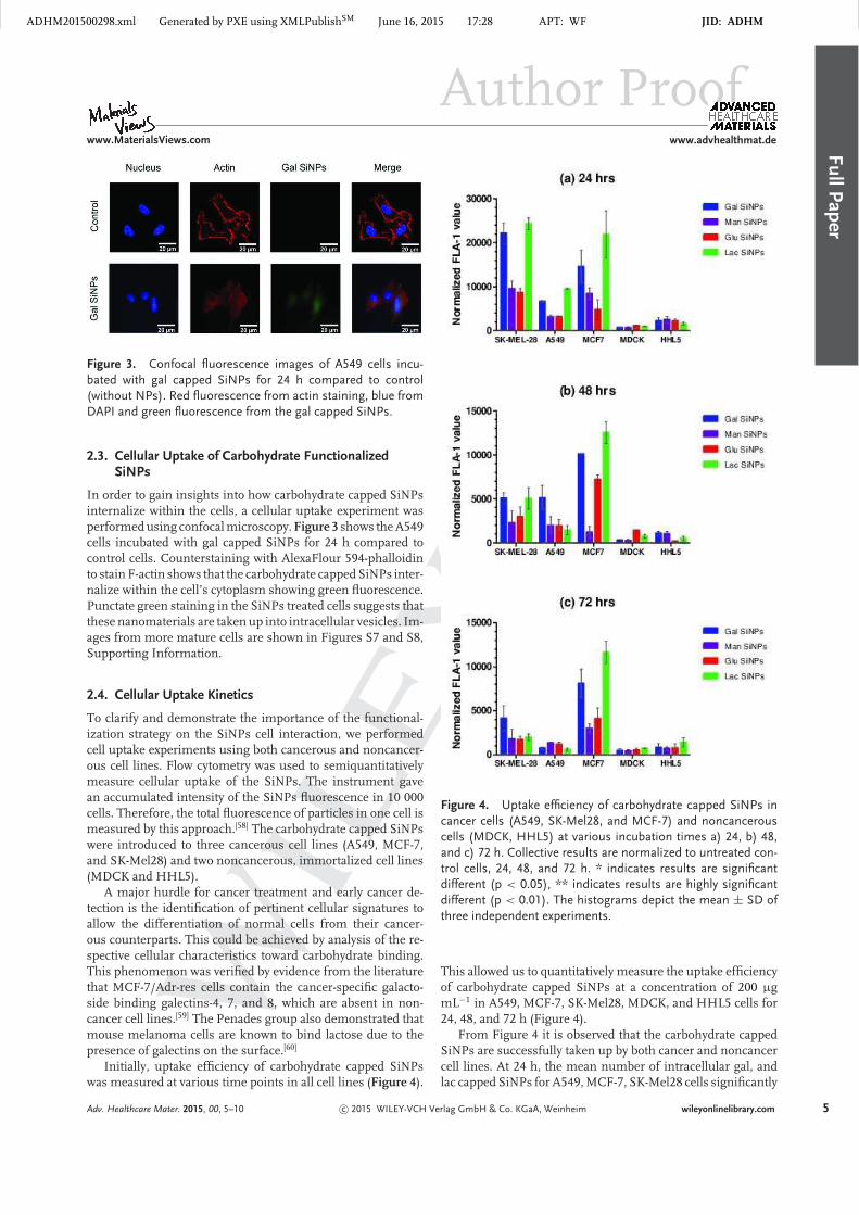
Figure 3. Confocal fluorescence images of A549 cells incu-bated with gal capped SiNPs for 24 h compared to control(without NPs). Red fluorescence from actin staining, blue fromDAPI and green fluorescence from the gal capped SiNPs.
2.3. Cellular Uptake of Carbohydrate FunctionalizedSiNPs
In order to gain insights into how carbohydrate capped SiNPsinternalize within the cells, a cellular uptake experiment wasperformed using confocal microscopy. Figure 3 shows the A549cells incubated with gal capped SiNPs for 24 h compared tocontrol cells. Counterstaining with AlexaFlour 594-phalloidinto stain F-actin shows that the carbohydrate capped SiNPs inter-nalize within the cell’s cytoplasm showing green fluorescence.Punctate green staining in the SiNPs treated cells suggests thatthese nanomaterials are taken up into intracellular vesicles. Im-ages from more mature cells are shown in Figures S7 and S8,Supporting Information.
2.4. Cellular Uptake Kinetics
To clarify and demonstrate the importance of the functional-ization strategy on the SiNPs cell interaction, we performedcell uptake experiments using both cancerous and noncancer-ous cell lines. Flow cytometry was used to semiquantitativelymeasure cellular uptake of the SiNPs. The instrument gavean accumulated intensity of the SiNPs fluorescence in 10 000cells. Therefore, the total fluorescence of particles in one cell ismeasured by this approach.[58] The carbohydrate capped SiNPswere introduced to three cancerous cell lines (A549, MCF-7,and SK-Mel28) and two noncancerous, immortalized cell lines(MDCK and HHL5).
A major hurdle for cancer treatment and early cancer de-tection is the identification of pertinent cellular signatures toallow the differentiation of normal cells from their cancer-ous counterparts. This could be achieved by analysis of the re-spective cellular characteristics toward carbohydrate binding.This phenomenon was verified by evidence from the literaturethat MCF-7/Adr-res cells contain the cancer-specific galacto-side binding galectins-4, 7, and 8, which are absent in non-cancer cell lines.[59] The Penades group also demonstrated thatmouse melanoma cells are known to bind lactose due to thepresence of galectins on the surface.[60]
Initially, uptake efficiency of carbohydrate capped SiNPswas measured at various time points in all cell lines (Figure 4).
Figure 4. Uptake efficiency of carbohydrate capped SiNPs incancer cells (A549, SK-Mel28, and MCF-7) and noncancerouscells (MDCK, HHL5) at various incubation times a) 24, b) 48,and c) 72 h. Collective results are normalized to untreated con-trol cells, 24, 48, and 72 h. * indicates results are significantdifferent (p < 0.05), ** indicates results are highly significantdifferent (p < 0.01). The histograms depict the mean ± SD ofthree independent experiments.
This allowed us to quantitatively measure the uptake efficiencyof carbohydrate capped SiNPs at a concentration of 200 µgmL−1 in A549, MCF-7, SK-Mel28, MDCK, and HHL5 cells for24, 48, and 72 h (Figure 4).
From Figure 4 it is observed that the carbohydrate cappedSiNPs are successfully taken up by both cancer and noncancercell lines. At 24 h, the mean number of intracellular gal, andlac capped SiNPs for A549, MCF-7, SK-Mel28 cells significantly
Adv. Healthcare Mater. 2015, 00, 5–10 c© 2015 WILEY-VCH Verlag GmbH & Co. KGaA, Weinheim wileyonlinelibrary.com 5

ADHM201500298.xml Generated by PXE using XMLPublishSM June 16, 2015 17:28 APT: WF JID: ADHMF
ull
Pap
er
Author Proofwww.MaterialsViews.com www.advhealthmat.de
Figure 5. Time dependent uptake efficiency of carbohydratecapped SiNPs in SK-Mel28 cells at various incubation timesof 1, 3, 6, 24, 48, and 72 h. Collective results are normalized tountreated control cells, ** indicates results are highly significantdifferent (p < 0.01). Values are mean ± SD of the results fromthree independent experiments.
exceeds the amount of NPs inside MDCK and HHL5 cells. Itis clear that the uptake efficiency of cancer cells is greater thanthat of the normal cells. Notably, uptake of the four carbo-hydrate capped SiNPs are significantly different in all threecancer cell lines at all-time point (Figure 4 and Figure S9, Sup-porting Information). This indicates there is some degree ofcarbohydrates uptake selectivity among these cell lines. Basedon flow cytometry response, it is observed that the binding ofgal capped SiNPs and lac capped SiNPs in cancerous cells ishigher, suggesting that these cell lines have active galactoseand lactose receptors. SK-Mel28 cells were found to interactwith gal and lac SiNPs more efficiently. This is of special inter-est since it is reported that melanoma cells bind to lactose, dueto the presence of galactin on the surface.[60]
Furthermore, the time dependent uptake study of the car-bohydrate capped SiNPs was performed in SK-Mel28 cells atvarious time points. The cells were exposed to the NPs at vari-ous time points from a few hours to several days and the resultsquantified by flow cytometry as shown in Figure 5.
Figure 5 shows the carbohydrate capped SiNPs were inter-nalized within 24 h in SK-Mel28 cells, but the internalizationof NPs was found to be decreasing at 48 and 72 h. The optimaluptake efficiency was at 24 h, the speed of which indicates re-ceptor mediate endocytosis as a possible uptake mechanism.After 24 h lower uptake could be due to exocytosis process,metabolising Si to silicate, and cell division.
To further investigate the internalization mechanism of car-bohydrate capped SiNPs we incubated the cells with the car-bohydrate capped SiNPs at 4 and 37 ◦C. Traditionally, it hasbeen proposed that active transport of molecules across thecell membrane is temperature dependent.[61] At low temper-atures transport activity is strongly reduced, thus uptake ofmolecules could be attributed to a nonspecific diffusional en-try into the cells.[62] Effects from low temperature may affectthe binding of the ligand to specific cell receptors, the lateralmobility of the ligand-receptor complex,[63] the formation ofnecks in the clathrin coated pits,[64] and/or the transport of
Figure 6. Uptake efficiency of carbohydrate capped SiNPs inSK-Mel28 cell line at a) 4 ◦C and b) 37 ◦C: Control – black, gal– red, man – orange, glu – purple, lac – green at concentrationof 200 µg mL−1; c) Uptake efficiency of carbohydrate cappedSiNPs in SK-Mel cells at 4 ◦C (blue) and 37 ◦C (red), presentedas normalized to untreated control cells. Values are mean ± SDof the results from three independent experiments.
endocytosed material from endosomes to lysosomes.[65] Endo-cytosis of ligands such as transferrin, cholera toxin or someviruses has been shown to be temperature dependent,[66–68] asthe ligands are able to attach to cell membranes at low tem-peratures, but not internalized. Recent work has shown uptakeis energy dependant.[69–71] Based on this concept, carbohydratecapped SiNPs were incubated with the SK-Mel28 cells at lowand normal incubation temperatures, and SiNP uptake ana-lyzed using flow cytometry analysis (Figure 6). From Figure 6it is clearly observed that the SiNPs incubated with cells at 37◦C were internalized, while the flow cytometry signals were ata significantly lower level for those incubated at 4 ◦C. There-fore, the obtained results suggest that the cellular uptake ofcarbohydrate capped SiNPs is likely to be energy dependent,suggesting it could be receptor-mediated.[72]
3. Conclusion
Our findings suggest that carbohydrate capped SiNPs could beuseful tumor detection agents. These NPs proved to be verystable in biological media, shown by DLS measurements. Theminimal toxicity of carbohydrate capped SiNPs was confirmedusing both in vitro and in vivo models. The in vitro toxicity wasverified by MTT assay in three types of mammalian cell lines.The carbohydrate capped SiNPs were found to have no signifi-cant toxicity at the highest concentration of 1000 µg mL−1. Thein vivo toxicity of carbohydrate capped SiNPs was examined
6 wileyonlinelibrary.com c© 2015 WILEY-VCH Verlag GmbH & Co. KGaA, Weinheim Adv. Healthcare Mater. 2015, 00, 6–10

ADHM201500298.xml Generated by PXE using XMLPublishSM June 16, 2015 17:28 APT: WF JID: ADHMF
ullP
aper
Author Proofwww.MaterialsViews.com www.advhealthmat.de
in X. laevis embryos; the SiNPs were shown to be nontoxic forembryos up to 200 µg mL−1 and no death or morphologicaldamage was observed. The results were compared with amine-terminated SiNPs, which were as expected highly toxic andresulted in the death of embryos. The obtained results sug-gest that the SiNPs are uniformly capped with carbohydratemolecules, which make them stable as well as nontoxic in bothin vivo and in vitro models.
The uptake efficiency of carbohydrate capped SiNPs wasquantified by flow cytometry. The obtained results indicatedthat carbohydrate capped SiNPs internalize in the cell within24 h. The fluorescence uptake of carbohydrate capped SiNPswas quantified in both cancer and noncancerous cell lines andthe cancerous cells more readily took up carbohydrate cappedSiNPs than noncancerous lines, which is important in termsof developing future diagnostic tools or even drug delivery sys-tems to tumors. The uptake of carbohydrate capped SiNPs wasvisualized by fluorescence and confocal microscopy. The NPsshowed quick accumulation inside the cancer cells.
Our understanding of cancer cell functions, such as endocy-tosis, cell–matrix and cell–cell communications, can be greatlyenhanced by studying carbohydrate–receptor functions as a re-sult of carbohydrate capped SiNP utilization. In addition, suchstudies can help further understanding of specificity and ligandoptimization, and exploitation of carbohydrate capped SiNPsfor in vivo cancer detection.
4. Experimental Section
Synthesis of Carbohydrate capped SiNPs: Carbohydrate cappedSiNPs were synthesized from amine-terminated SiNPs usinga previously described method.[73, 74] The acid functionalizedcarbohydrate derivatives (gal, man, glu, and lac) were syn-thesized according to the literature, followed by the isolationof pure anomers (detailed synthesis is presented in the sup-porting document). The gal, man, glu, and lac capped SiNPswere synthesized using their corresponding pyranosyl acid. Apyranosyl acid galactose, mannose, glucose, and lactose (30mg, 0.078 mmol) and 1-ethyl-3-(3-dimethylaminopropyl) car-bodiimide (EDC 14 mg, 1 equiv, 0.085 mmol) were dissolvedin dichloromethane (5 mL) and left with stirring for 2 h atroom temperature. After 2 h N-hydroxysuccinimide (NHS) andfreshly prepared amine-terminated SiNPs were dissolved inmethanol and added into the reaction mixture. The reactionwas stirred for 24 h at room temperature (Scheme 1).
In order to remove the unreacted amine-terminated SiNPs,the crude reaction mixture was washed three times with water(3 × 10 mL) and extracted into CH2Cl2. The mixture was driedwith Na2SO4, and solvent was removed under vacuum. Afterwashing, the reaction mixture was further ensued for deacety-lation. Deacetylation of carbohydrate capped SiNPs was per-formed with sodium methoxide in methanol and then stirredfor 30 min with Dowex 50W-X8 [H+]c resin. The byproduct ureaand impurities were difficult to remove at this stage becauseboth the byproduct and OAc-carbohydrate-capped SiNPs (gal,man, glu, and lact) are soluble in dichloromethane. Deacetyla-tion of the reaction mixture removes the acetate group of the
Scheme 1. Schematic representation of carbohydrate cappedSiNPs synthesis.
carbohydrate capped SiNPs and offers hydroxyl carbohydratecapped SiNPs, which are soluble in water. In order to obtain apure product, the mixture was washed with dichloromethane toremove impurities and urea. A pinkish solid product of the purecarbohydrate capped SiNPs was obtained after deacetylation ofeach crude reaction mixture. This sticky solid was redissolvedin water for further characterization. Physical characterisationresults of synthesized SiNPs, such as size, size distribution,and Zeta-potential, are listed in Figures S10 and S11, TablesS1 and S2 of the Supporting Information.
Dynamic Light Scattering (DLS): DLS measurement wasrecorded with a Zetasizer Nano ZS (Malvern InstrumentsLtd., U.K.). Carbohydrate capped SiNPs were dissolved inDMEMwith 10% FBS, RPMI medium with 10% FBS and 0.1XMarc’s modified ringers pH7.4 (MMR; 100 × 10−3 M NaCl, 2 ×
10−3 M KCl, 2 × 10⁻³ M CaCl2, 1 × 10⁻³ M MgCl2, and 5 × 10⁻³
M HEPES) as a function of time. The hydrodynamic diameterswere obtained at room temperature.
Cell Culture: The human-derived hepatocyte HHL5 andbreast cancer MCF7 cell lines were a kind gift from Y. Bao,and the immortalized kidney MDCK cell line was a kind giftfrom M. Mogensen, University of East Anglia, Norwich. The
Adv. Healthcare Mater. 2015, 00, 7–10 c© 2015 WILEY-VCH Verlag GmbH & Co. KGaA, Weinheim wileyonlinelibrary.com 7

ADHM201500298.xml Generated by PXE using XMLPublishSM June 16, 2015 17:28 APT: WF JID: ADHMF
ull
Pap
er
Author Proofwww.MaterialsViews.com www.advhealthmat.de
human melanoma SK-mel28 was a kind gift from A. Chienand R. T. Moon, University of Washington, Seattle. The lungcarcinoma A549 cell line was a kind gift from D. Sexton, Liv-erpool John Moores University, Liverpool. All lines used wereregularly confirmed as mycoplasma free using a PCR-basedassay.[75] Cells were sub-cultured as previously described.[76]
Briefly, cells were incubated at 37 ◦C, 5% CO2/95% air and cul-tured in RPMI (MCF7, A549) and DMEM (SK-Mel28, MDCK,HHL5) supplemented with 10% heat-inactivated foetal bovineserum, 2 × 10⁻³ M L-glutamine, 100 µg mL−1 penicillin, and100 µg mL−1 streptomycin (all purchased from Life Technolo-gies, Carlsbad, CA). Cells were never cultured beyond passage30.
Cell Proliferation Assay: Cell proliferation in the presenceof carbohydrate capped SiNPs was evaluated by MTT [3-(4,5-dimethylthiazol-2-yl)-2,5-diphenyltetrazolium bromide] assay.Different cell lines were used in order to achieve a more com-plete study: HHL5 (immortalized human hepatocytes), A549(lung carcinoma cells), and MDCK (Madin Darby canine kid-ney cells) cells were seeded in a 96-well plate for 24 h. Thecells were then treated with carbohydrate capped SiNPs (gal,glu, man, and lac) at various concentrations (50, 300, 500, 700,and 1000 µg mL−1) for 72 h. Afterward, normal procedure wasapplied as described in the previous publication.[38] All experi-ments were repeated on at least three different occasions.
Culturing Embryos: These experiments were performed incompliance with institutional guidelines at the University ofEast Anglia. The local ethical review committee (according toUK Home Office regulations) has approved the research. X.laevis embryos were obtained as previously described.[77] Em-bryos were incubated at 18 ◦C until the required stage wasachieved according to the Nieuwkoop and Faber (NF) devel-opmental staging system.[57] Any dead embryos were removedthroughout the culturing process. Embryos were cultured in0.1× MMR supplemented with 25 µg mL−1 gentamycin.
Exposure to Carbohydrate Capped SiNPs: Live embryos werecollected for exposure to SiNPs at NF stage 15 to 38, as describedabove. These stages of Xenopus were selected to assess NPtoxicity as they represent a key stage in development of theembryos, through neuralation (NF stage 15), to tadpole stages(NF stage 38). Concentrations of NPs were made up usingserial dilutions in 0.1× MMR. In a 24-well plate, 5 embryos perwell were exposed to the varying concentrations of SiNPs ina total volume of 1000 µL per well. Embryos were incubated,exposed to SiNPs, at 18 ◦C until the required NF stage.
Fixing Embryos: Embryos exposed to SiNPs were fixed at NFstage 38. Fixing was carried out as previously described.[77]Oncethe embryos reached the required NF stage they were washedin 0.1× MMR and fixed in MEMFA (0.1% MOPS [pH7.4], 2 ×
10−3 M EGTA, 1 × 10⁻³ M MgSO4, and 3.7% formaldehyde) for1 h at room temperature. After two washes in PBS all embryoswere scored for gross phenotypic abnormalities.
Confocal Laser Scanning Microscopy (CLSM): HHL5 andA549 cells were seeded on 12-well plates with cover slips ata density of 3 × 10⁴ cells per well and exposed to 150 µgmL−1 of carbohydrate capped SiNPs for 24 h. The cell werethen washed twice by PBS and fixed by Paraformaldehyde so-lution for 10 min. To visualize the cells, they were stained
with Lysotracker-Red (Invitrogen) or Texas Red-X Phalloidin(Invitrogen) according to manufacturer’s protocols. Then, adrop (approximately 2 µL) of fluorescent mounting medium(VECTASHIELD hard, Vector Labs) was added on top of themicroscope slide. The cover slip in which cells were grown wasturned upside down on top of the mounting medium. The slidewas then dried in the fridge for ≈30 min before use. The im-ages were taken under a confocal microscope (Zeiss LSM510META system) using a 40X oil immersion objective lens.
Flow Cytometery: HHL5, A549, MDCK, MCF-7, and SK-mel28 cells were seeded on 24-well plates at a density of 3× 10⁴ cells per well and incubated at 37 ◦C overnight. The cellswere washed with PBS and treated with 300 µg mL−1 of car-bohydrate capped SiNPs at various time points from 1 to 72h. Afterward, cells were harvested by trypsinization and sus-pended in the medium (300 µL). The cellular uptake of SiNPswas examined by Flow cytometry (Accuri C6 Flow CytometerSystem) using 380 nm excitation wavelength. A total of 10 000events were recorded for each sample. The samples were ana-lyzed with FlowJo Software and an average of the mean of atleast three different experiments (with different sets of NPs)was calculated.
Supporting Information
Supporting Information is available from the Wiley OnlineLibrary or from the author.
Acknowledgements
J.H.A is grateful to a Tyndall studentship and an ORS award.V.S. would like to thank the Royal Society and the British SkinFoundation for funding to support the cell culture work. V.S.was currently supported by a CRUK programme grant awardedto the CRUK-Skin Tumor Laboratory, Medical Research Insti-tute, University of Dundee.
Received: April 21, 2015Revised: June 3, 2015
Published Online: MM DD, YYYY
[1] D. E. Gerber, Am. Fam. Physician 2008, 77, 311.
[2] D. Schrag, N. Engl. J. Med. 2004, 351, 317.
[3] J. Villanueva, A. Vultur, J. T. Lee, R. Somasundaram, M.
Fukunaga-Kalabis, A. K. Cipolla, B. Wubbenhorst, X. Xu, P. A.
Gimotty, D. Kee, A. E. Santiago-Walker, R. Letrero, K. D’Andrea,
A. Pushparajan, J. E. Hayden, K. D. Brown, S. Laquerre, G. A.
McArthur, J. A. Sosman, K. L. Nathanson, M. Herlyn, Cancer
Cell 2010, 18, 683.
[4] R. Nazarian, H. Shi, Q. Wang, X. Kong, R. C. Koya, H. Lee, Z.
Chen, M. K. Lee, N. Attar, H. Sazegar, T. Chodon, S. F. Nelson,
G. McArthur, J. A. Sosman, A. Ribas, R. S. Lo, Nature 2010,
468, 973.
[5] M. Lidsky, G. Antoun, P. Speicher, B. Adams, R. Turley, C.
Augustine, D. Tyler, F. Ali-Osman, J. Biol. Chem. 2014, 289,
27714.
8 wileyonlinelibrary.com c© 2015 WILEY-VCH Verlag GmbH & Co. KGaA, Weinheim Adv. Healthcare Mater. 2015, 00, 8–10

ADHM201500298.xml Generated by PXE using XMLPublishSM June 16, 2015 17:28 APT: WF JID: ADHMF
ullP
aper
Author Proofwww.MaterialsViews.com www.advhealthmat.de
[6] F. Su, W. D. Bradley, Q. Q. Wang, H. Yang, L. Z. Xu, B.
Higgins, K. Kolinsky, K. Packman, M. J. Kim, K. Trunzer, R. J.
Lee, K. Schostack, J. Carter, T. Albert, S. Germer, J. Rosinski,
M. Martin, M. E. Simcox, B. Lestini, D. Heimbrook, G. Bollag,
Cancer Res. 2012, 72, 969.
[7] C. M. Johannessen, J. S. Boehm, S. Y. Kim, S. R. Thomas, L.
Wardwell, L. A. Johnson, C. M. Emery, N. Stransky, A. P. Cogdill,
J. Barretina, G. Caponigro, H. Hieronymus, R. R. Murray, K.
Salehi-Ashtiani, D. E. Hill, M. Vidal, J. J. Zhao, X. P. Yang, O.
Alkan, S. Kim, J. L. Harris, C. J. Wilson, V. E. Myer, P. M. Finan,
D. E. Root, T. M. Roberts, T. Golub, K. T. Flaherty, R. Dummer,
B. L. Weber, W. R. Sellers, R. Schlegel, J. A. Wargo, W. C. Hahn,
L. A. Garraway, Nature 2010, 468, 968.
[8] R. Straussman, T. Morikawa, K. Shee, M. Barzily-Rokni, Z.
R. Qian, J. Y. Du, A. Davis, M. M. Mongare, J. Gould, D. T.
Frederick, Z. A. Cooper, P. B. Chapman, D. B. Solit, A. Ribas, R.
S. Lo, K. T. Flaherty, S. Ogino, J. A. Wargo, T. R. Golub, Nature
2012, 487, 500.
[9] T. R. Wilson, J. Fridlyand, Y. B. Yan, E. Penuel, L. Burton, E.
Chan, J. Peng, E. Lin, Y. L. Wang, J. Sosman, A. Ribas, J. Li, J.
Moffat, D. P. Sutherlin, H. Koeppen, M. Merchant, R. Neve, J.
Settleman, Nature 2012, 487, 505.
[10] R. A. Dwek, Chem. Rev. 1996, 96, 683.
[11] A. Varki, Glycobiology 1993, 3, 97.
[12] D. B. Werz, P. H. Seeberger, Chem. Eur. J. 2005, 11, 3194.
[13] R. Raman, V. Sasisekharan, R. Sasisekharan, Chem. Biol. 2005,
12, 267.
[14] P. H. Seeberger, D. B. Werz, Nature 2007, 446, 1046.
[15] E. E. Simanek, G. J. McGarvey, J. A. Jablonowski, C.-H. Wong,
Chem. Rev. 1998, 98, 833.
[16] B. A. Macher, U. Galili, Biochim. Biophys. Acta, Gen. Subj. 2008,
1780, 75.
[17] G. A. Rabinovich, M. A. Toscano, Nat. Rev. Immunol. 2009, 9,
338.
[18] P. Nangia-Makker, J. Conklin, V. Hogan, A. Raz, Trends Mol.
Med. 2002, 8, 187.
[19] S.-I. Hakomori, Y. Zhang, Chem. Biol. 1997, 4, 97.
[20] R. J. Pieters, Org. Biomol. Chem. 2009, 7, 2013.
[21] C. A. Lingwood, Curr. Opin. Chem. Biol. 1998, 2, 695.
[22] M. Petitou, C. A. A. van Boeckel, Angew. Chem. Int. Ed. 2004,
43, 3118.
[23] Y. C. Lee, R. T. Lee, Acc. Chem. Res. 1995, 28, 321.
[24] S. I. Hakomori, Pure Appl. Chem. 1991, 63, 473.
[25] S. J. Danishefsky, J. R. Allen, Angew. Chem. Int. Ed. 2000, 39,
836.
[26] Z. Dai, J. Zhou, S.-J. Qiu, Y.-K. Liu, J. Fan, Electrophoresis 2009,
30, 2957.
[27] S. R. Barthel, J. D. Gavino, L. Descheny, C. J. Dimitroff, Expert
Opin. Ther. Targets 2007, 11, 1473.
[28] K. Kayser, M. Heil, H. J. Gabius, Pathol.,– Res. Pract. 1989, 184,
621.
[29] K. Kayser, N. Bovin, T. Zemlyanukhina, S. Donaldo-Jacinto, J.
Koopmann, H.-J. Gabius, Glycoconjugate J. 1994, 11, 339.
[30] P. D. Rye, N. V. Bovin, Glycobiology 1997, 7, 179.
[31] H.-G. Lerchen, J. Baumgarten, N. Piel, V. Kolb-Bachofen, Angew.
Chem. Int. Ed. 1999, 38, 3680.
[32] E. Y. L. Kim, C. Gronewold, A. Chatterjee, C.-W. von der Lieth,
C. Kliem, B. Schmauser, M. Wiessler, E. Frei, ChemBioChem
2005, 6, 422.
[33] M. Moros, B. Hernaez, E. Garet, J. T. Dias, B. Saez, V. Grazu,
A. Gonzalez-Fernandez, C. Alonso, J. M. de la Fuente, ACS Nano
2012, 6, 1565.
[34] Q. Wang, H. Ni, A. Pietzsch, F. Hennies, Y. Bao, Y. Chao, J.
Nanopart. Res. 2011, 13, 405.
[35] N. O’Farrell, A. Houlton, B. R. Horrocks, Int. J. Nanomed. 2006,
1, 451.
[36] F. Erogbogbo, K.-T. Yong, I. Roy, G. Xu, P. N. Prasad, M. T.
Swihart, ACS Nano 2008, 2, 873.
[37] A. H. Mayne, S. C. Bayliss, P. Barr, M. Tobin, L. D. Buckberry,
Phys. Status Solidi A 2000, 182, 505.
[38] Q. Wang, Y. Bao, J. Ahire, Y. Chao, Adv. Healthcare Mater. 2013,
2, 459.
[39] C. Kirchner, T. Liedl, S. Kudera, T. Pellegrino, A. Munoz Javier,
H. E. Gaub, S. Stölzle, N. Fertig, W. J. Parak, Nano Lett. 2004,
5, 331.
[40] J. R. Nilsson, Acta Protozool 2003, 42, 19.
[41] Y. Chen, T. Ji, Z. Rosenzweig, Nano Lett. 2003, 3, 581.
[42] A. Moore, E. Marecos, A. Bogdanov, R. Weissleder, Radiology
2000, 214, 568.
[43] Y. Hu, X. Jiang, Y. Ding, H. Ge, Y. Yuan, C. Yang, Biomater.
2002, 23, 3193.
[44] S. M. Moghimi, A. C. Hunter, J. C. Murray, FASEB J. 2005, 19,
311.
[45] R. C. Murdock, L. Braydich-Stolle, A. M. Schrand, J. J. Schlager,
S. M. Hussain, Toxicol. Sci. 2008, 101, 239.
[46] S. Deguchi, T. Yamazaki, S.-a. Mukai, R. Usami, K. Horikoshi,
Chem. Res. Toxicol. 2007, 20, 854.
[47] M. Buford, R. Hamilton, A. Holian, Part. Fibre Toxicol. 2007, 4,
6.
[48] P. Wick, P. Manser, L. K. Limbach, U. Dettlaff-Weglikowska, F.
Krumeich, S. Roth, W. J. Stark, A. Bruinink, Toxicol. Lett. 2007,
168, 121.
[49] T. M. Sager, D. W. Porter, V. A. Robinson, W. G. Lindsley, D. E.
Schwegler-Berry, V. Castranova, Nanotoxicology 2007, 1, 118.
[50] L. Foucaud, M. R. Wilson, D. M. Brown, V. Stone, Toxicol. Lett.
2007, 174, 1.
[51] S. Seker, A. E. Elcin, T. Yumak, A. Sinag, Y. M. Elcin, Toxicol.
In Vitro 2014, 28, 1349.
[52] M. Mahmoudi, I. Lynch, M. R. Ejtehadi, M. P. Monopoli, F. B.
Bombelli, S. Laurent, Chem. Rev. 2011, 111, 5610.
[53] C.-L. Tseng, C.-L. Peng, J.-Y. Huang, J.-C. Chen, F.-H. Lin, J.
Biomater. Appl. 2013, 27, 1055.
[54] V. E. Fako, D. Y. Furgeson, Adv. Drug Delivery Rev. 2009, 61,
478.
[55] M. Giannaccini, A. Cuschieri, L. Dente, V. Raffa, J. Nanotechnol.
Diagn. Treat. 2013, 1, 11.
[56] L. Marcon, F. Riquet, D. Vicogne, S. Szunerits, J.-F. Bodart, R.
Boukherroub, J. Mater. Chem. 2010, 20, 8064.
[57] Normal table of Xenopus Laevis (Daudin): A Systematical and
Chronological Survey of the Development from the Fertilized Egg
Till the End of Metamorphosis (Eds: P. D. Nieuwkoop, J. Faber),
North-Holland Pub. Co., Amsterdam 1967.
[58] T. dos Santos, J. Varela, I. Lynch, A. Salvati, K. A. Dawson,
Small 2011, 7, 3341.
[59] S. Chandrasekaran, M. L. Tanzer, M. S. Giniger, J. Biol. Chem.
1994, 269, 3367.
[60] J. Rojo, V. Dıaz, J. M. de la Fuente, I. Segura, A. G. Barrientos,
H. H. Riese, A. Bernad, S. Penades, ChemBioChem 2004, 5,
291.
[61] G. Fricker, Transporters as Drug Carriers, Wiley-VCH GmbH & Co.
KGaA, 2010. Q3[62] I. Pascual, A. Berjon, M. P. Lostao, A. Barber, Comp. Biochem.
Physiol., Part B: Biochem. Mol. Biol. 2006, 143, 20.
[63] I. Pastan, M. Willingham, in Endocytosis, Eds: I. Pastan, M.
Willingham, Springer, USA, 1985, 1. Q4[64] M. C. Willingham, I. Pastan, Proc. Natl. Acad. Sci. U. S. A. 1983,
80, 5617.
Adv. Healthcare Mater. 2015, 00, 9–10 c© 2015 WILEY-VCH Verlag GmbH & Co. KGaA, Weinheim wileyonlinelibrary.com 9
ADHM201500298.xml Generated by PXE using XMLPublishSM June 16, 2015 17:28 APT: WF JID: ADHMF
ull
Pap
er
Author Proofwww.MaterialsViews.com www.advhealthmat.de
[65] T. E. Tjelle, A. Brech, L. K. Juvet, G. Griffiths, T. Berg, J. Cell
Sci. 1996, 109, 2905.
[66] J. Mercer, M. Schelhaas, A. Helenius, Annu. Rev. Biochem 2010,
79, 803.
[67] A. Sofer, A. H. Futerman, J. Biol. Chem. 1995, 27012117.
[68] C. Harding, J. Heuser, P. Stahl, J. Cell Biol. 1983, 97, 329.
[69] A. Gliga, S. Skoglund, I. Odnevall Wallinder, B. Fadeel, H.
Karlsson, Part. Fibre Toxicol. 2014, 11, 11.
[70] J. S. Kim, T. J. Yoon, K. N. Yu, M. S. Noh, M. Woo, B. G. Kim,
K. H. Lee, B. H. Sohn, S. B. Park, J. K. Lee, M. H. Cho, J. Vet.
Sci. 2006, 7, 321.
[71] P. Liu, Y. Sun, Q. Wang, Y. Sun, H. Li, Y. Duan, Biomaterials
2014, 35, 760.
[72] S. C. Silverstein, R. M. Steinman, Z. A. Cohn, Annu. Rev. Biochem
1977, 46, 669.
[73] J. H. Ahire, I. Chambrier, A. Mueller, Y. Bao, Y. Chao, ACS
Appl. Mater. Interfaces 2013, 5, 7384.
[74] J. H. Ahire, Q. Wang, P. R. Coxon, G. Malhotra, R. Brydson, R.
Chen, Y. Chao, ACS Appl. Mater. Interfaces 2012, 4, 3285.
[75] F. J. M. Vankuppeveld, K. E. Johansson, J. M. D. Galama, J.
Kissing, G. Bolske, J. T. M. Vanderlogt, W. J. G. Melchers, Appl.
Environ. Microbiol. 1994, 60, 149.
[76] V. Jenei, V. Sherwood, J. Howlin, R. Linnskog, A. Safholm, L.
Axelsson, T. Andersson, Proc. Natl. Acad. Sci. U. S. A. 2009, 106,
19473.
[77] V. Sherwood, R. Manbodh, C. Sheppard, A. D. Chalmers, Mol.
Biol. Cell 2008, 19, 1772.
10 wileyonlinelibrary.com c© 2015 WILEY-VCH Verlag GmbH & Co. KGaA, Weinheim Adv. Healthcare Mater. 2015, 00, 10–10

ADHM201500298.xml Generated by PXE using XMLPublishSM June 16, 2015 17:28 APT: WF JID: ADHM
Q1 PROD to AU: Please spell out MTT at first appearance both in the abstract and in the text.
Q2 PROD to AU: Please provide the highest academic title (either Dr. or Prof.) for all authors, where applicable.
Q3 PROD to AU: Please add publisher location in ref. 61.
Q4 PROD to AU: Please add publisher location (US state) in ref. 63.