Embed Size (px)
Citation preview

New SPECT Radiotracers for Myocardial Perfusion Imaging: Major
Developments over Last 5 Years!
f{âtÇz _|â? c{A WAProfessor, School of Health Sciences
Purdue University
Commercial 99mTc Radiotracers for Myocardial Perfusion Imaging
Shortcomings of 99mTc-Sestamibi and 99mTc-Tetrofosmin
1. High Liver Uptake. Despite their widespread clinical application in perfusion imaging, 99mTc-sestamibi and 99mTc-tetrofosmin do not meet the requirements of an ideal perfusion imaging agent, in part due to their high liver uptake. The intense liver uptake, particularly for 99mTc-Sestamibi, makes it difficult to interpret heart activity in the inferior left ventricular wall. Despite intensive efforts to reduce this interference, photon scattering from the high liver activity remains a significant challenge for diagnosis of heart diseases, such as CAD.
2. Relatively low first-pass extraction. High first-pass extraction and linear relationship between the blood flow and radiotracer heart uptake permit better detection of the presence and extent of coronary disease, and more precise delineation of perfusion defects, which is of considerable benefit in managing of patients with known or suspected CAD and assessing risk of future cardiac events (e.g. myocardial infarction and death).
High heart uptake with long myocardial retention
High first-pass extraction with stable myocardial retention, which linearly tracks myocardial blood flow over a wide range
Minimal uptake in the liver and lungs so that images can be taken within 30 min postinjection
Requirements for An Ideal 99mTc Perfusion Radiotracer
PTcP S
S
N
N
N O
O
O O
OOO
+
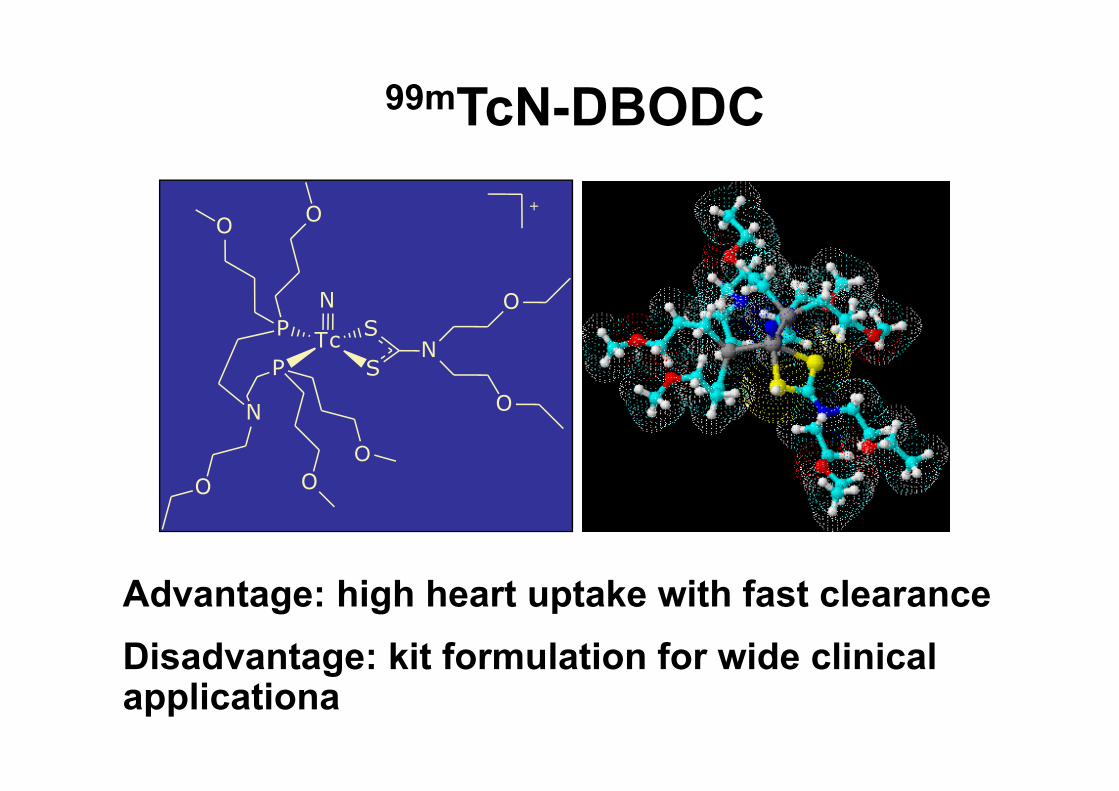
99mTcN-DBODC
Advantage: high heart uptake with fast clearanceDisadvantage: kit formulation for wide clinical applicationa
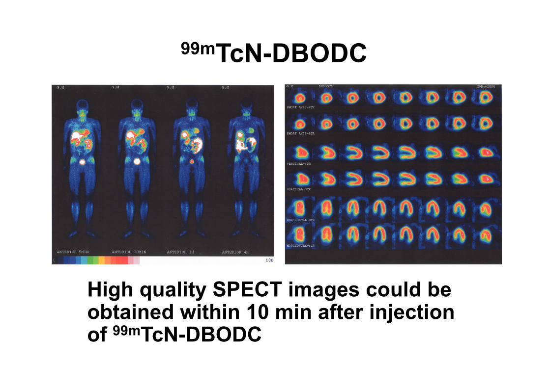
99mTcN-DBODC
High quality SPECT images could be obtained within 10 min after injection of 99mTcN-DBODC
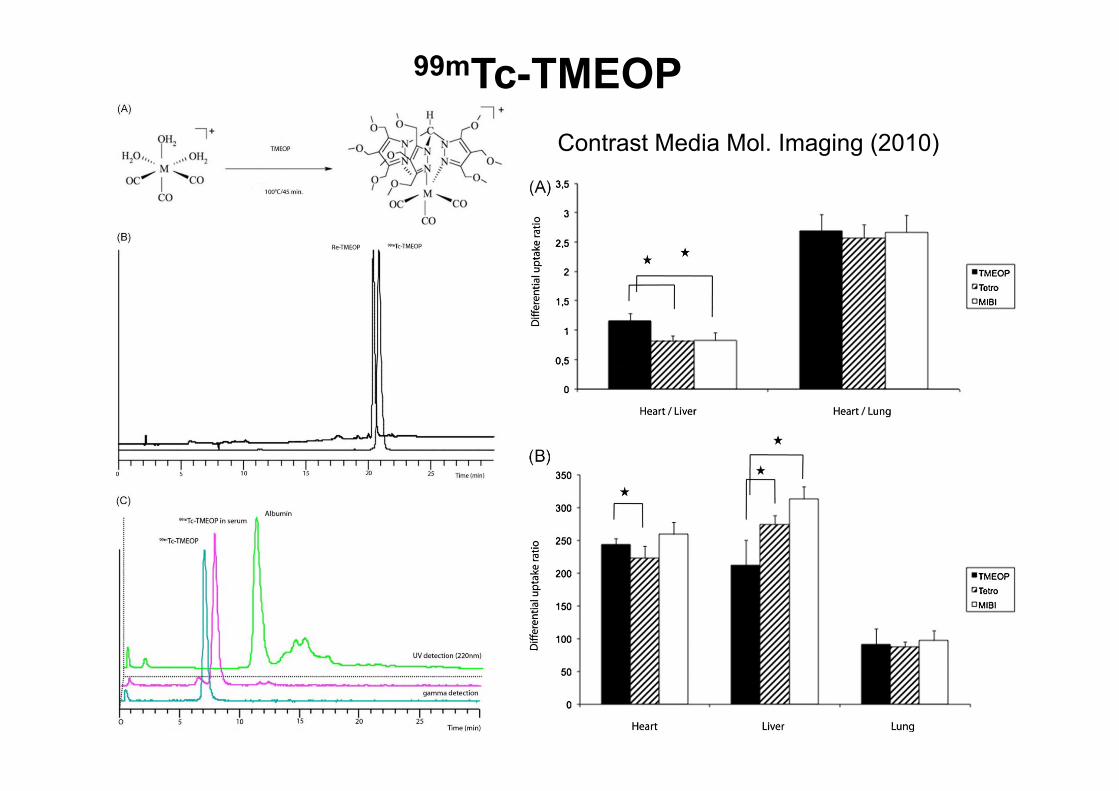
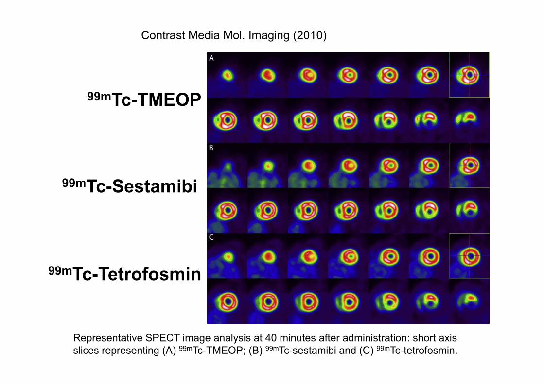
99mTc-TMEOPContrast Media Mol. Imaging (2010)
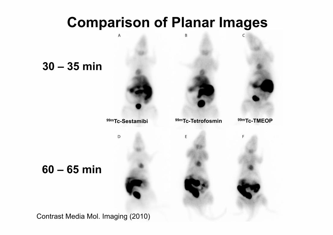
99mTc-Sestamibi 99mTc-Tetrofosmin 99mTc-TMEOP
30 – 35 min
60 – 65 min
Contrast Media Mol. Imaging (2010)
Comparison of Planar Images
Contrast Media Mol. Imaging (2010)
Representative SPECT image analysis at 40 minutes after administration: short axis slices representing (A) 99mTc-TMEOP; (B) 99mTc-sestamibi and (C) 99mTc-tetrofosmin.
99mTc-TMEOP
99mTc-Sestamibi
99mTc-Tetrofosmin
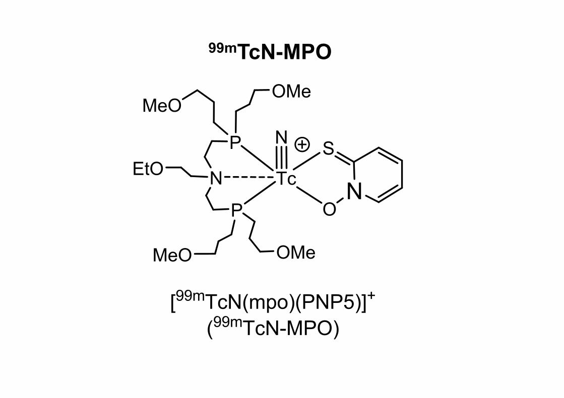
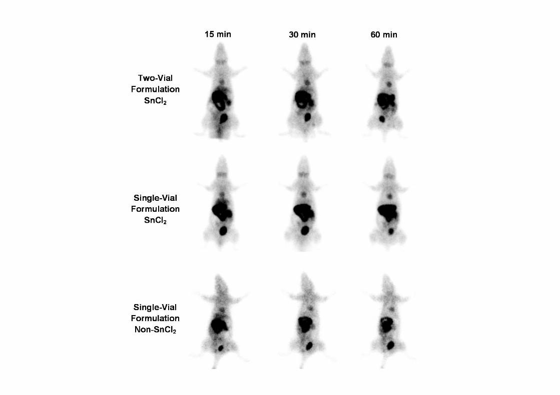
99mTcN-MPO
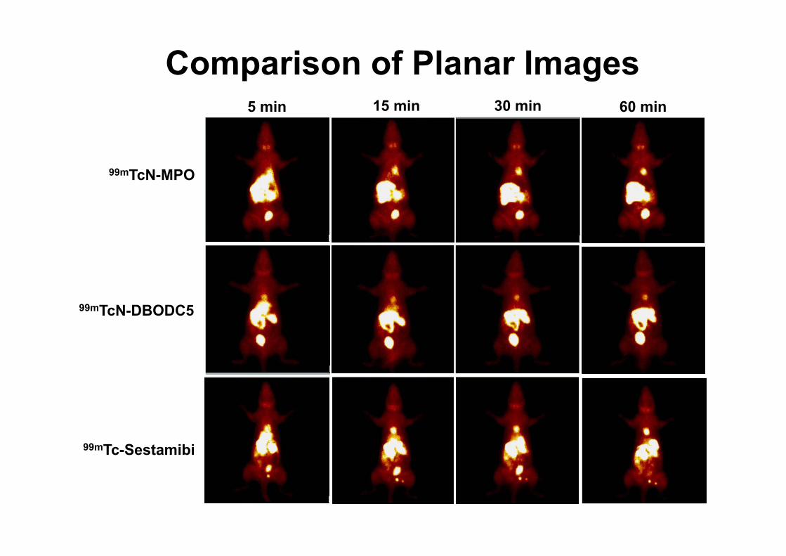
Comparison of Planar Images
99mTc-Sestamibi
99mTcN-DBODC5
99mTcN-MPO
5 min 15 min 30 min 60 min

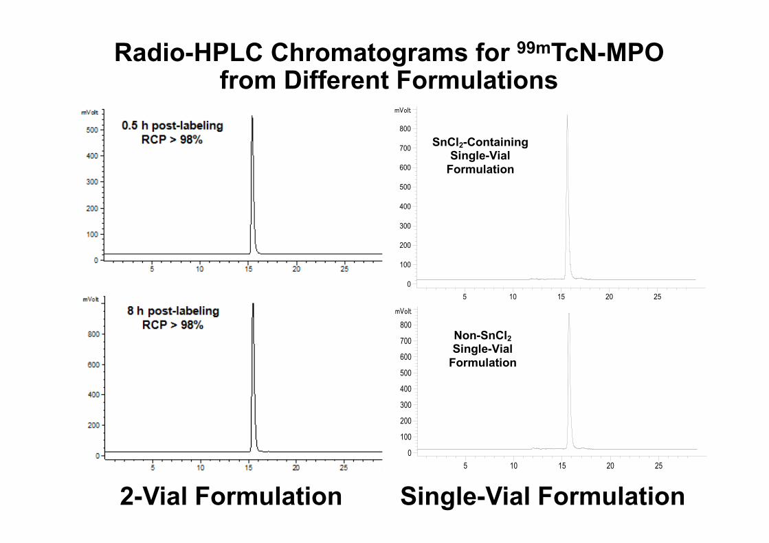
Radio-HPLC Chromatograms for 99mTcN-MPO from Different Formulations
2-Vial Formulation Single-Vial Formulation
5 10 15 20 25
mVolt
0
100
200
300
400
500
600
700
800
5 10 15 20 25
mVolt
0
100
200
300
400
500
600
700
800
Non-SnCl2 Single-Vial
Formulation
SnCl2-Containing Single-Vial
Formulation
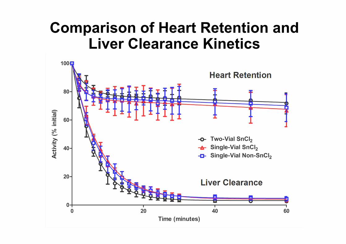
Comparison of Heart Retention and Liver Clearance Kinetics
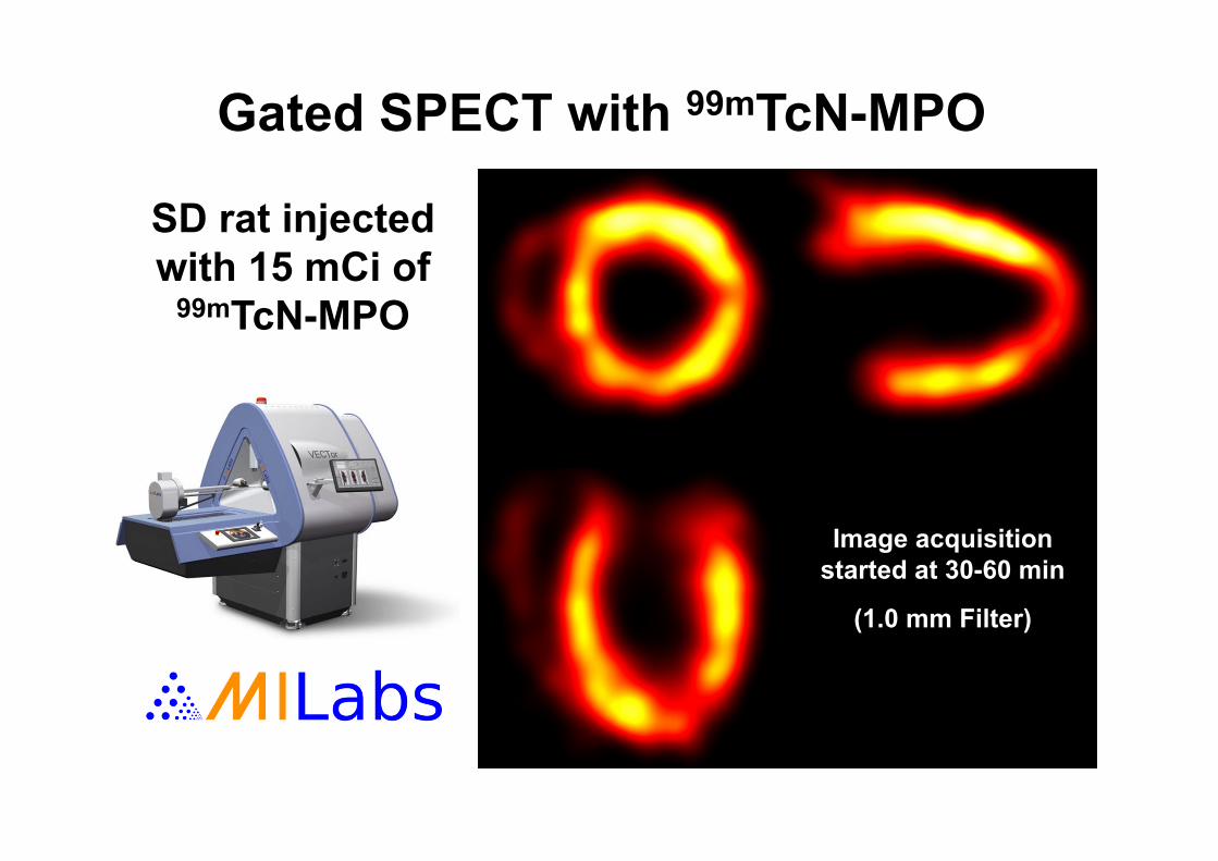
SD rat injected with 15 mCi of
99mTcN-MPO
Gated SPECT with 99mTcN-MPO
Image acquisition started at 30-60 min
(1.0 mm Filter)
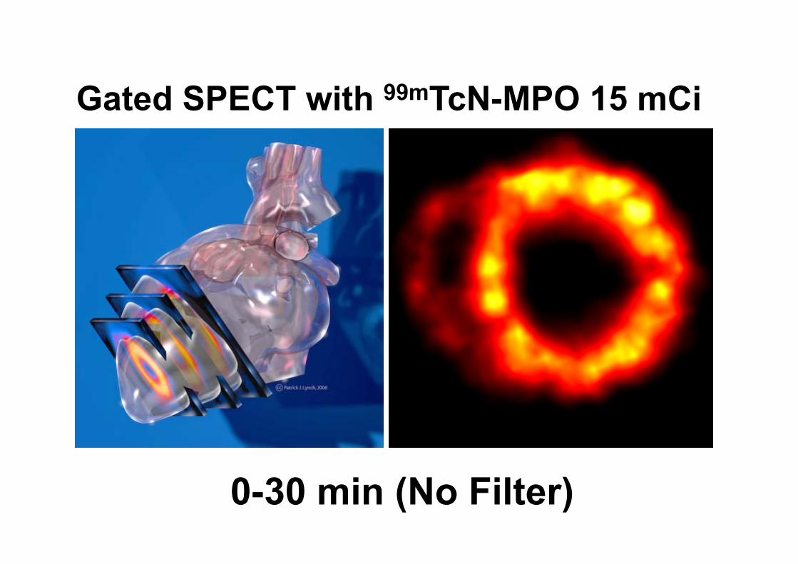
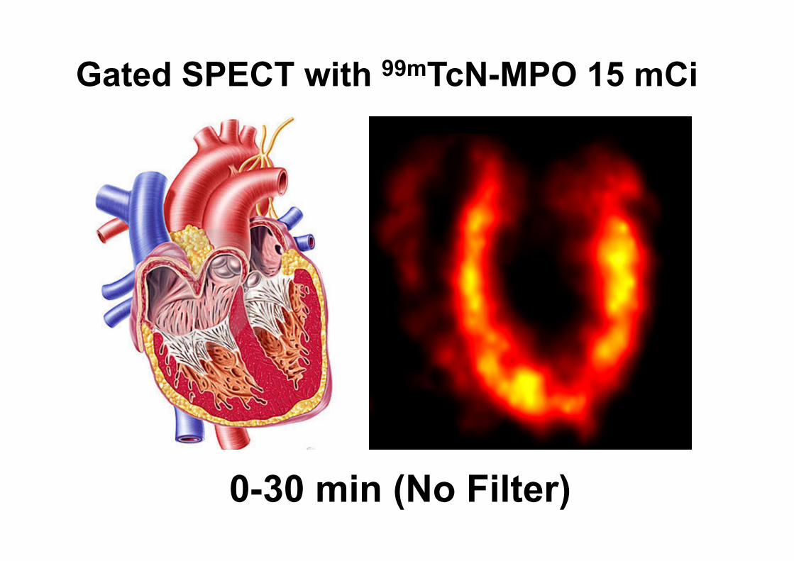
Gated SPECT with 99mTcN-MPO 15 mCi
0-30 min (No Filter)
0-30 min (No Filter)
Gated SPECT with 99mTcN-MPO 15 mCi
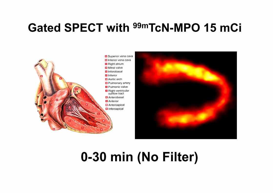
0-30 min (No Filter)
Gated SPECT with 99mTcN-MPO 15 mCi
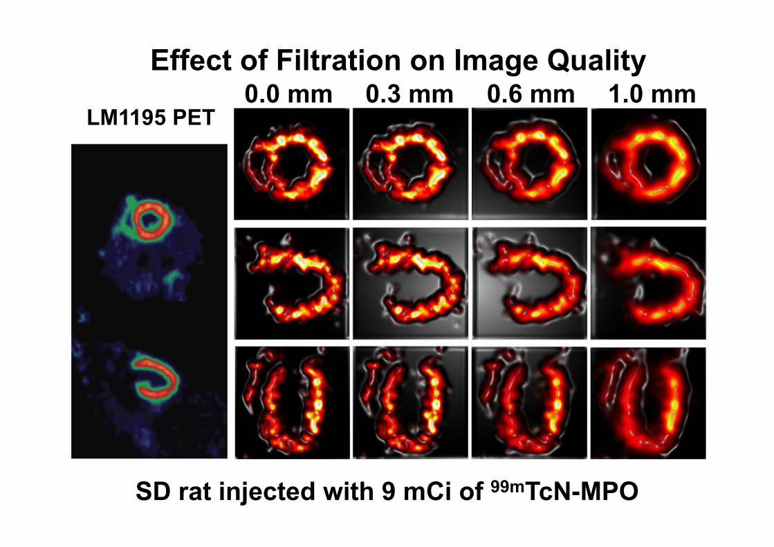
Effect of Filtration on Image Quality0.0 mm 0.3 mm 0.6 mm 1.0 mm
LM1195 PET
SD rat injected with 9 mCi of 99mTcN-MPO
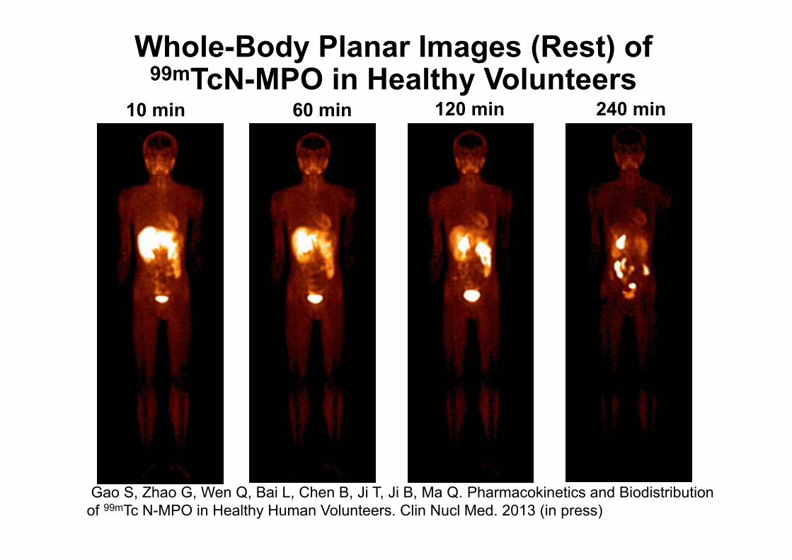
Whole-Body Planar Images (Rest) of 99mTcN-MPO in Healthy Volunteers
10 min 60 min 120 min 240 min
Gao S, Zhao G, Wen Q, Bai L, Chen B, Ji T, Ji B, Ma Q. Pharmacokinetics and Biodistribution of 99mTc N-MPO in Healthy Human Volunteers. Clin Nucl Med. 2013 (in press)
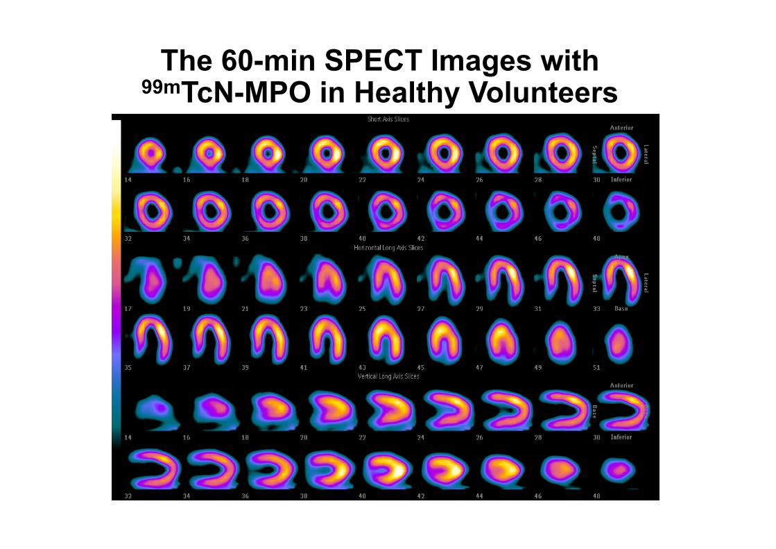
The 60-min SPECT Images with 99mTcN-MPO in Healthy Volunteers
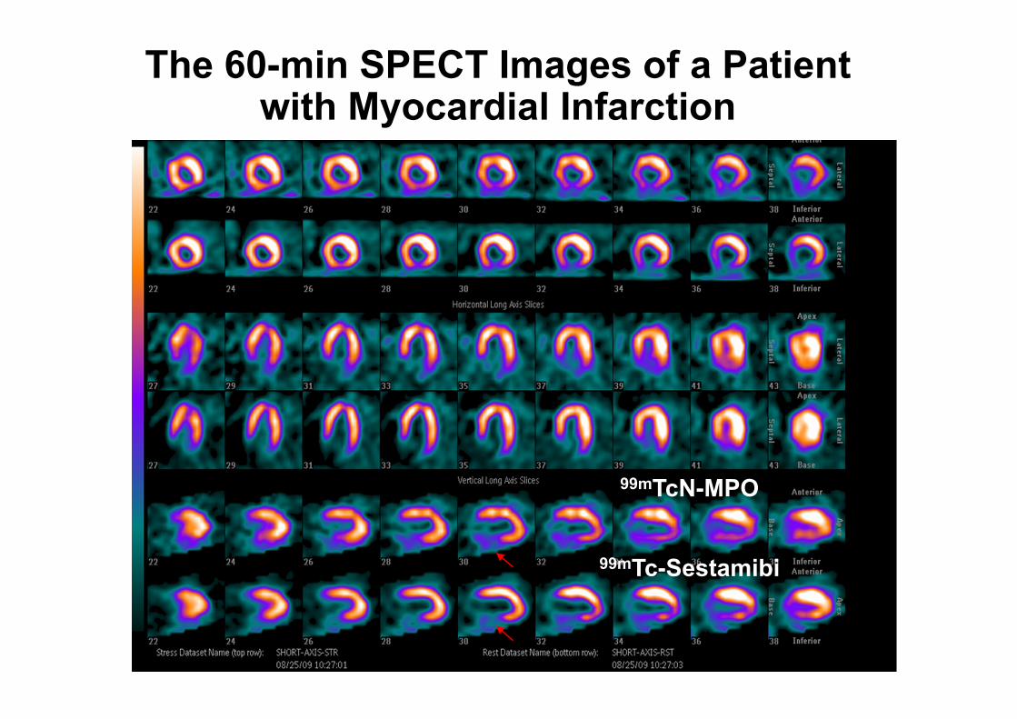
The 60-min SPECT Images of a Patient with Myocardial Infarction
99mTc-Sestamibi
99mTcN-MPO
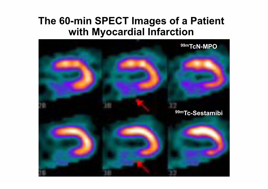
The 60-min SPECT Images of a Patient with Myocardial Infarction
99mTc-Sestamibi
99mTcN-MPO
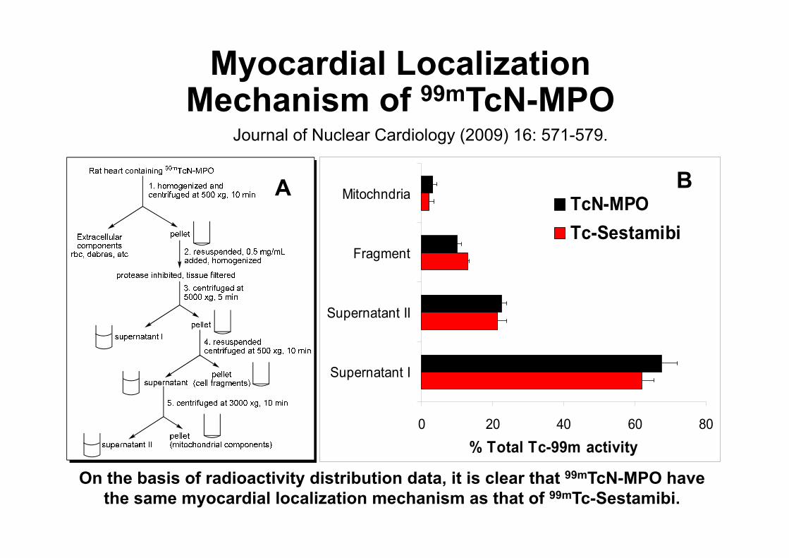
Myocardial Localization Mechanism of 99mTcN-MPO
On the basis of radioactivity distribution data, it is clear that 99mTcN-MPO have the same myocardial localization mechanism as that of 99mTc-Sestamibi.
0 20 40 60 80
Supernatant I
Supernatant II
Fragment
Mitochndria
% Total Tc-99m activity
TcN-MPOTc-Sestamibi
A B
Journal of Nuclear Cardiology (2009) 16: 571-579.
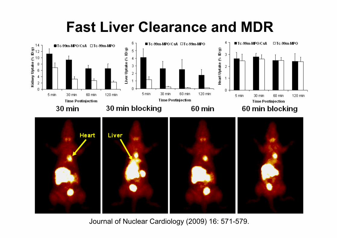
Fast Liver Clearance and MDR
Journal of Nuclear Cardiology (2009) 16: 571-579.

99mTc-Teboroxime & Analogs
5 10 15 20 25
mVolt
0
200
400
600
800
5 10 15 20 25
mVolt
50
100
150
200
250
300
350
400
5 10 15 20 25
mVolt
0
100
200
300
400
500
0 5 10 15 20 25
mVolt
0
100
200
300
400
500
600
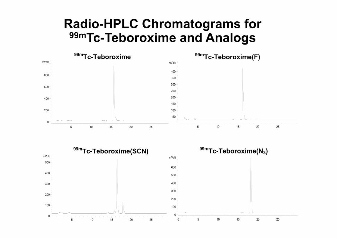
99mTc-Teboroxime 99mTc-Teboroxime(F)
99mTc-Teboroxime(N3) 99mTc-Teboroxime(SCN)
Radio-HPLC Chromatograms for 99mTc-Teboroxime and Analogs
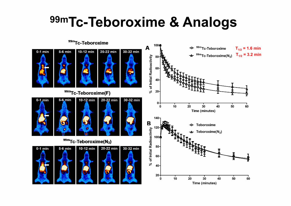
99mTc-Teboroxime & AnalogsT1/2 = 1.6 minT1/2 = 3.2 min
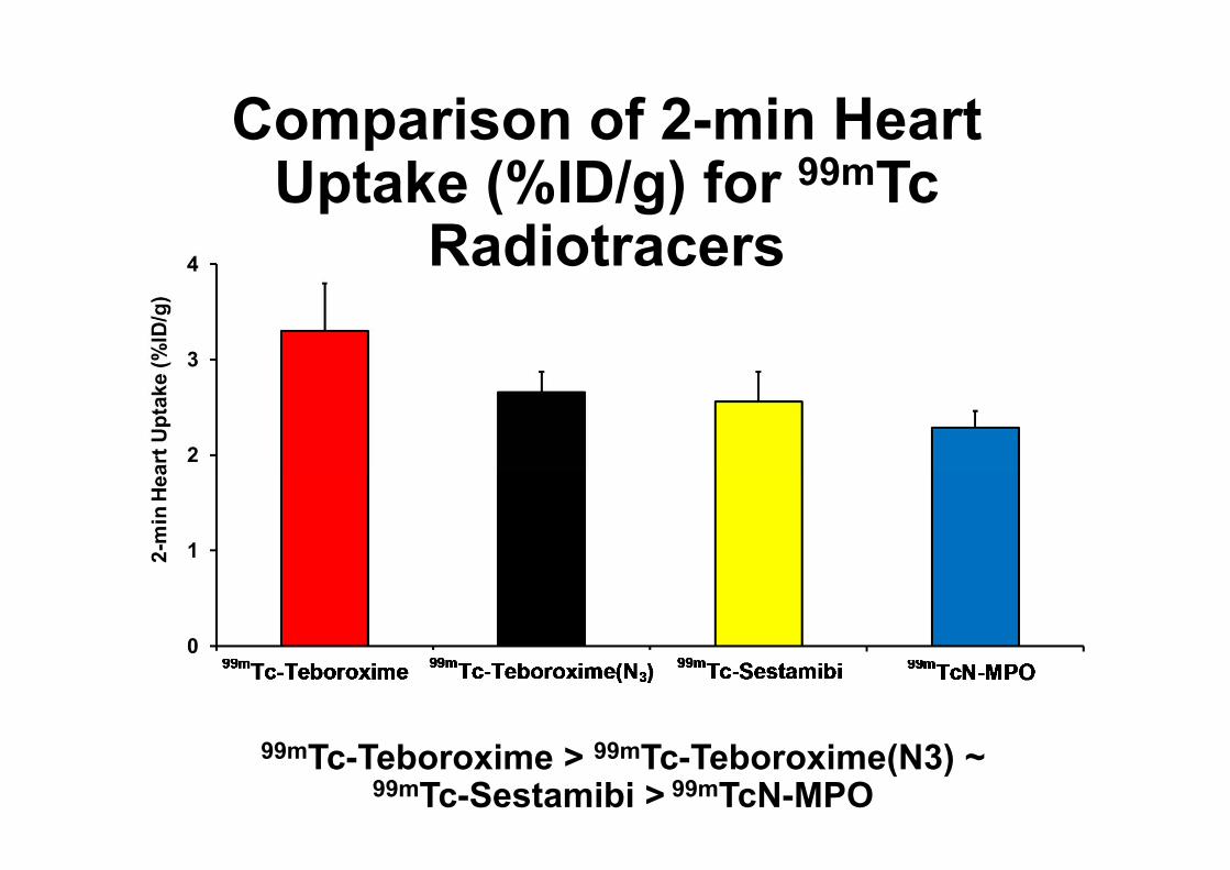
Comparison of 2-min Heart Uptake (%ID/g) for 99mTc
Radiotracers
99mTc-Teboroxime > 99mTc-Teboroxime(N3) ~ 99mTc-Sestamibi > 99mTcN-MPO
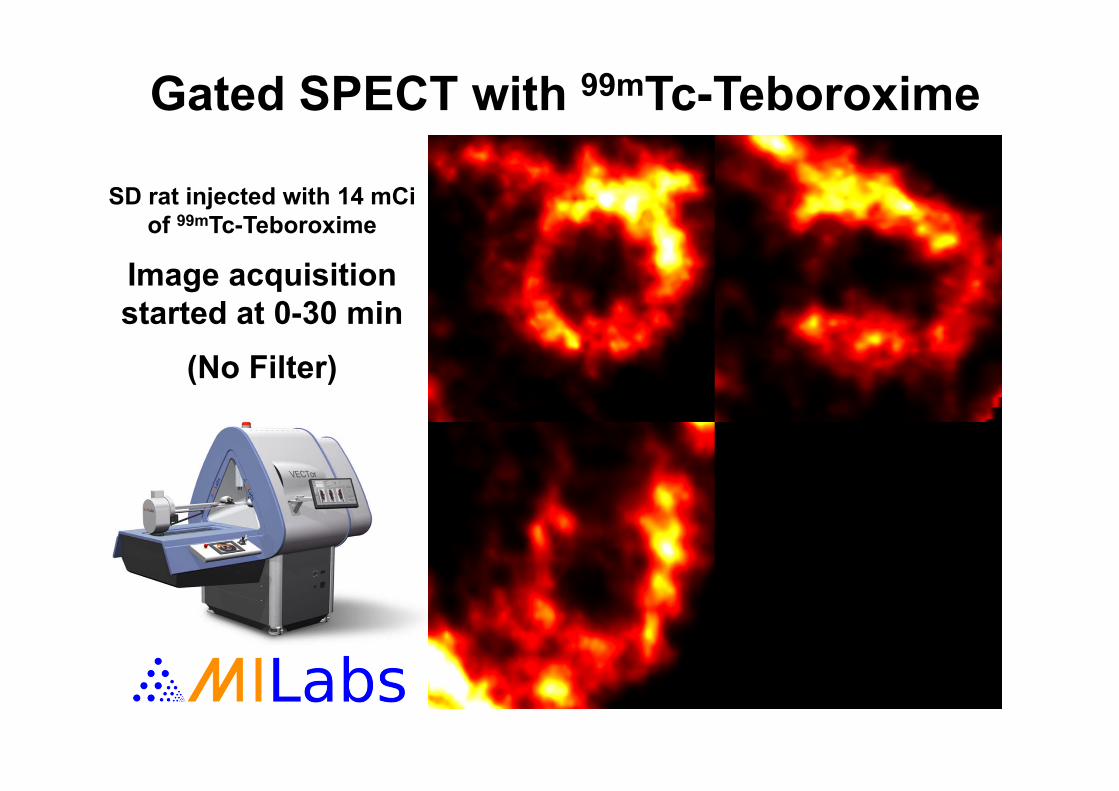
Gated SPECT with 99mTc-Teboroxime
SD rat injected with 14 mCi of 99mTc-Teboroxime
Image acquisition started at 0-30 min
(No Filter)

AcknowledgementAll postdoctoral fellows, visiting scholars and
graduate students over the last 10 years.