Embed Size (px)
Citation preview
May 2012 Volume 22, Number 5
a l s o I n s I d e :
Physician Follow-up Bolsters IVC Filter Retrieval Rate
QPID Search System Quickly Extracts Data in Emergency Departments
Tech Savvy Teachers are Goal of RSNA Workshop
Communication Skills Can Make or Break a Patient-Physician Relationship
Rsna 2012 advance Registration and Housing open May 9—see Page 23
Imag
e co
urte
sy o
f GE
Hea
lthca
re
From Idea to Patent, Radiologist Inventors Share their Insight
edIToR
David M. Hovsepian, M.D.
R&e FoundaTIon ConTRIbuTIng edIToR
C. Leon Partain, M.D., Ph.D.
exeCuTIve edIToRs
Lynn Tefft HoffMarijo Millette
ManagIng edIToR
Beth Burmahl
edIToRIal advIsoRs
Mark G. Watson Executive Director
Roberta E. Arnold, M.A., M.H.P.E. Assistant Executive Director Publications and Communications
edIToRIal boaRd
David M. Hovsepian, M.D. Chair
Colin P. Derdeyn, M.D.Kavita Garg, M.D.Bruce G. Haffty, M.D.Nazia Jafri, M.D.Bonnie N. Joe, M.D., Ph.D.Edward Y. Lee, M.D., M.P.H.Kerry M. Link, M.D.Barry A. Siegel, M.D.Gary J. Whitman, M.D.William T. Thorwarth Jr., M.D. Board Liaison
gRaPHIC desIgneRs
Adam IndykKen Ejka
ConTRIbuTIng WRITeRs
Richard DarganFelicia DechterPaul LaTour
2012 Rsna boaRd oF dIReCToRs
N. Reed Dunnick, M.D. Chair
Ronald L. Arenson, M.D. Liaison for Annual Meeting and Technology
Richard L. Baron, M.D. Liaison for Education
William T. Thorwarth Jr., M.D. Liaison for Publications and Communications
Richard L. Ehman, M.D. Liaison for Science
Vijay M. Rao, M.D. Liaison-designate for Annual Meeting and Technology
George S. Bisset III, M.D. President
Sarah S. Donaldson, M.D President-elect and Secretary-Treasurer
UP FRONT 1 First Impression
2 MOC News
4 My Turn
FeaTURes 5 Physician Follow-up Bolsters IVC Filter Retrieval Rate
9 From Idea to Patent, Radiologist Inventors Share their Insight
11 Tech Savvy Teachers are Goal of RSNA Workshop
13 Communication Skills Can Make or Break a Patient-Physician Relationship
RadiOlOgy’s FUTURe 7 QPID Search System Quickly Extracts Data
in Emergency Departments
15 R&E Foundation Donors
News yOU CaN Use 17 Journal Highlights
18 Radiology in Public Focus
20 The Value of Membership
21 Education and Funding Opportunities
22 Residents & Fellows Corner
23 Annual Meeting Watch
24 RSNA.org
now in an easy-to-swallow tablet.O More images O interactive media O Videos
search “RSNA News” on the android or app store.
MKT 252_News_Tablet_Edition_Ad.indd 1 12-03-30 1:26 PM
For more than 20 years, RSNA News has provided high-quality, timely coverage of radiology research and education and critical issues facing the specialty, along with compre-hensive information about RSNA programs, products and other member benefits.
MAY 2012 • Volume 22, NumbeR 5
7
5
13
Follow us for exclusive news, annual meeting offers and more!
Access the RSNA News tablet edition on the App Store and Android market.
9
may 2012 | RSNA News 2
news you can use
1 RSNA News | may 2012
FIRSt IMPReSSIon
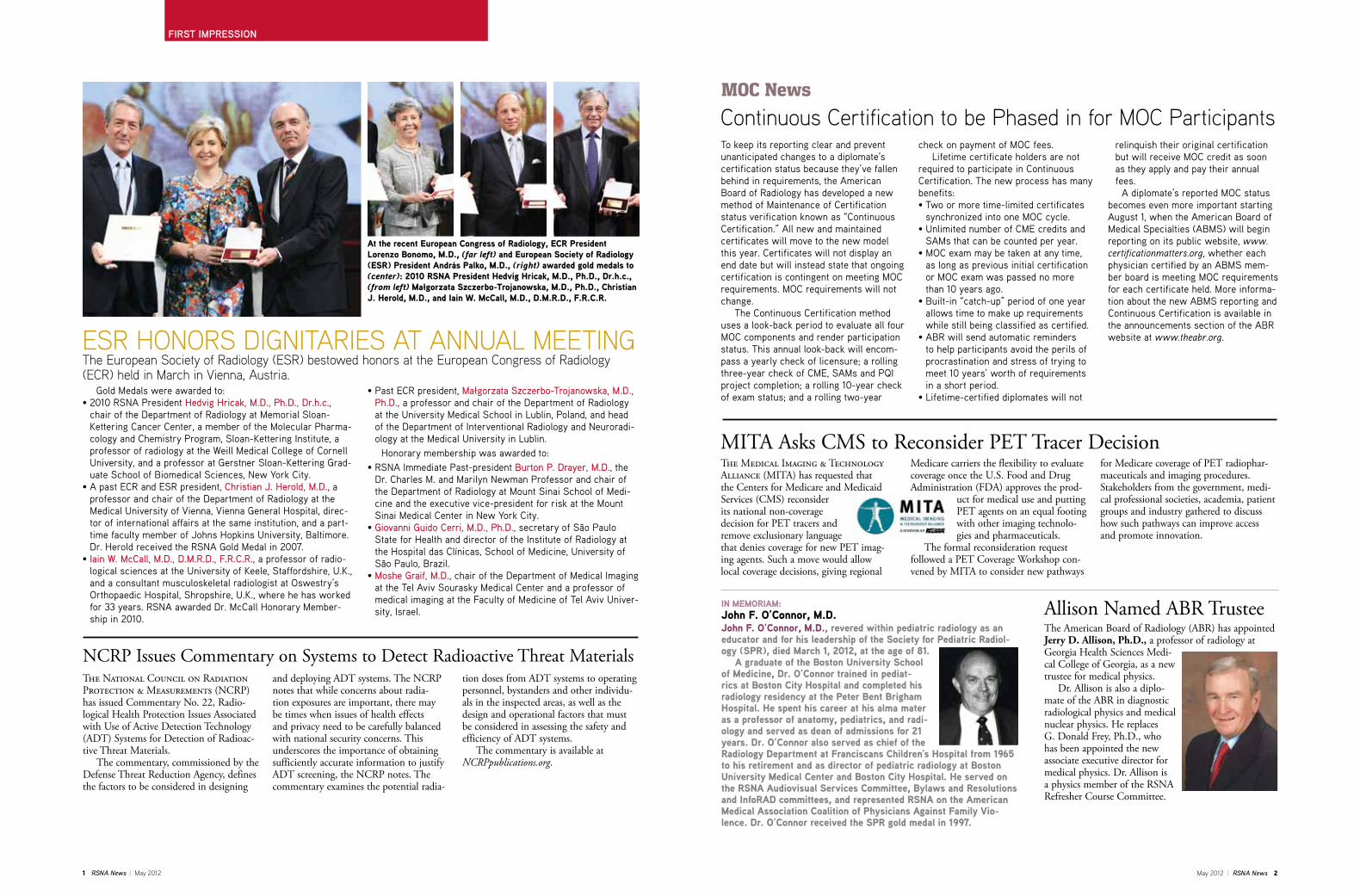
eSR HoNoRS DigNitARieS At ANNuAl meetiNgthe european Society of Radiology (eSR) bestowed honors at the european Congress of Radiology (eCR) held in march in Vienna, Austria.
at the recent european Congress of Radiology, eCR President lorenzo bonomo, M.d., (far left) and european society of Radiology (esR) President andrás Palko, M.d., (right) awarded gold medals to (center): 2010 Rsna President Hedvig Hricak, M.d., Ph.d., dr.h.c., (from left) Małgorzata szczerbo-Trojanowska, M.d., Ph.d., Christian J. Herold, M.d., and Iain W. McCall, M.d., d.M.R.d., F.R.C.R.
Gold Medals were awarded to:• 2010 RSNA President Hedvig Hricak, M.d., Ph.d., dr.h.c.,
chair of the Department of Radiology at Memorial Sloan-Kettering Cancer Center, a member of the Molecular Pharma-cology and Chemistry Program, Sloan-Kettering Institute, a professor of radiology at the Weill Medical College of Cornell University, and a professor at Gerstner Sloan-Kettering Grad-uate School of Biomedical Sciences, New York City.
• A past ECR and ESR president, Christian J. Herold, M.d., a professor and chair of the Department of Radiology at the Medical University of Vienna, Vienna General Hospital, direc-tor of international affairs at the same institution, and a part-time faculty member of Johns Hopkins University, Baltimore. Dr. Herold received the RSNA Gold Medal in 2007.
• iain w. McCall, M.d., d.M.R.d., F.R.C.R., a professor of radio-logical sciences at the University of Keele, Staffordshire, U.K., and a consultant musculoskeletal radiologist at Oswestry’s Orthopaedic Hospital, Shropshire, U.K., where he has worked for 33 years. RSNA awarded Dr. McCall Honorary Member-ship in 2010.
• Past ECR president, Małgorzata szczerbo-Trojanowska, M.d., Ph.d., a professor and chair of the Department of Radiology at the University Medical School in Lublin, Poland, and head of the Department of Interventional Radiology and Neuroradi-ology at the Medical University in Lublin.
Honorary membership was awarded to:• RSNA Immediate Past-president Burton P. drayer, M.d., the
Dr. Charles M. and Marilyn Newman Professor and chair of the Department of Radiology at Mount Sinai School of Medi-cine and the executive vice-president for risk at the Mount Sinai Medical Center in New York City.
• giovanni guido Cerri, M.d., Ph.d., secretary of São Paulo State for Health and director of the Institute of Radiology at the Hospital das Clínicas, School of Medicine, University of São Paulo, Brazil.
• Moshe graif, M.d., chair of the Department of Medical Imaging at the Tel Aviv Sourasky Medical Center and a professor of medical imaging at the Faculty of Medicine of Tel Aviv Univer-sity, Israel.
NCRP Issues Commentary on Systems to Detect Radioactive Threat MaterialsThe National Council on Radiation Protection & Measurements (NCRP) has issued Commentary No. 22, Radio-logical Health Protection Issues Associated with Use of Active Detection Technology (ADT) Systems for Detection of Radioac-tive Threat Materials. The commentary, commissioned by the Defense Threat Reduction Agency, defines the factors to be considered in designing
and deploying ADT systems. The NCRP notes that while concerns about radia-tion exposures are important, there may be times when issues of health effects and privacy need to be carefully balanced with national security concerns. This underscores the importance of obtaining sufficiently accurate information to justify ADT screening, the NCRP notes. The commentary examines the potential radia-
tion doses from ADT systems to operating personnel, bystanders and other individu-als in the inspected areas, as well as the design and operational factors that must be considered in assessing the safety and efficiency of ADT systems. The commentary is available at NCRPpublications.org.
In MeMoRIaM: John F. o’Connor, M.d.John F. o’Connor, M.d., revered within pediatric radiology as an educator and for his leadership of the society for Pediatric Radiol-ogy (sPR), died March 1, 2012, at the age of 81. a graduate of the boston university school of Medicine, dr. o’Connor trained in pediat-rics at boston City Hospital and completed his radiology residency at the Peter bent brigham Hospital. He spent his career at his alma mater as a professor of anatomy, pediatrics, and radi-ology and served as dean of admissions for 21 years. dr. o’Connor also served as chief of the Radiology department at Franciscans Children’s Hospital from 1965 to his retirement and as director of pediatric radiology at boston university Medical Center and boston City Hospital. He served on the Rsna audiovisual services Committee, bylaws and Resolutions and InfoRad committees, and represented Rsna on the american Medical association Coalition of Physicians against Family vio-lence. dr. o’Connor received the sPR gold medal in 1997.
The American Board of Radiology (ABR) has appointed Jerry D. Allison, Ph.D., a professor of radiology at Georgia Health Sciences Medi-cal College of Georgia, as a new trustee for medical physics. Dr. Allison is also a diplo-mate of the ABR in diagnostic radiological physics and medical nuclear physics. He replaces G. Donald Frey, Ph.D., who has been appointed the new associate executive director for medical physics. Dr. Allison is a physics member of the RSNA Refresher Course Committee.
Allison Named ABR Trustee
Continuous Certification to be Phased in for moC ParticipantsTo keep its reporting clear and prevent unanticipated changes to a diplomate’s certification status because they’ve fallen behind in requirements, the American Board of Radiology has developed a new method of Maintenance of Certification status verification known as “Continuous Certification.” All new and maintained certificates will move to the new model this year. Certificates will not display an end date but will instead state that ongoing certification is contingent on meeting MOC requirements. MOC requirements will not change. The Continuous Certification method uses a look-back period to evaluate all four MOC components and render participation status. This annual look-back will encom-pass a yearly check of licensure; a rolling three-year check of CME, SAMs and PQI project completion; a rolling 10-year check of exam status; and a rolling two-year
check on payment of MOC fees. Lifetime certificate holders are not required to participate in Continuous Certification. The new process has many benefits:• Two or more time-limited certificates
synchronized into one MOC cycle.• Unlimited number of CME credits and
SAMs that can be counted per year.• MOC exam may be taken at any time,
as long as previous initial certification or MOC exam was passed no more than 10 years ago.
• Built-in “catch-up” period of one year allows time to make up requirements while still being classified as certified.
• ABR will send automatic reminders to help participants avoid the perils of procrastination and stress of trying to meet 10 years’ worth of requirements in a short period.
• Lifetime-certified diplomates will not
relinquish their original certification but will receive MOC credit as soon as they apply and pay their annual fees.
A diplomate’s reported MOC status becomes even more important starting August 1, when the American Board of Medical Specialties (ABMS) will begin reporting on its public website, www.certificationmatters.org, whether each physician certified by an ABMS mem-ber board is meeting MOC requirements for each certificate held. More informa-tion about the new ABMS reporting and Continuous Certification is available in the announcements section of the ABR website at www.theabr.org.
MOC News
MITA Asks CMS to Reconsider PET Tracer DecisionThe Medical Imaging & Technology Alliance (MITA) has requested that the Centers for Medicare and Medicaid Services (CMS) reconsider its national non-coverage decision for PET tracers and remove exclusionary language that denies coverage for new PET imag-ing agents. Such a move would allow local coverage decisions, giving regional
Medicare carriers the flexibility to evaluate coverage once the U.S. Food and Drug Administration (FDA) approves the prod-
uct for medical use and putting PET agents on an equal footing with other imaging technolo-gies and pharmaceuticals.
The formal reconsideration request followed a PET Coverage Workshop con-vened by MITA to consider new pathways
for Medicare coverage of PET radiophar-maceuticals and imaging procedures. Stakeholders from the government, medi-cal professional societies, academia, patient groups and industry gathered to discuss how such pathways can improve access and promote innovation.
news you can use
3 RSNA News | may 2012 may 2012 | RSNA News 4
FIRSt IMPReSSIon
RSNA NewsMay 2012 • Volume 22, Number 5 Published monthly by the Radiological Society of North America, Inc. 820 Jorie Blvd., Oak Brook, IL 60523-2251. Printed in the USA.
Postmaster: Send address correction “changes” to: RSNA News, 820 Jorie Blvd., Oak Brook, IL 60523-2251Non-member subscription rate is $20 per year; $10 of active members’ dues is allo-cated to a subscription of RSNA News.
Contents of RSNA News copy-righted ©2012, RSNA. RSNA is a registered trademark of the Radiological Society of North America, Inc.
leTTeRs To THe [email protected] 1-630-571-7837 fax
[email protected] 1-888-600-0064 1-630-590-7770
RePRInTs and [email protected] 1-630-571-7829 1-630-590-7724 fax
[email protected] Jim Drew, Director 1-630-571-7819
Rsna MeMbeRsHIP1-877-rsna-mem
My Turn
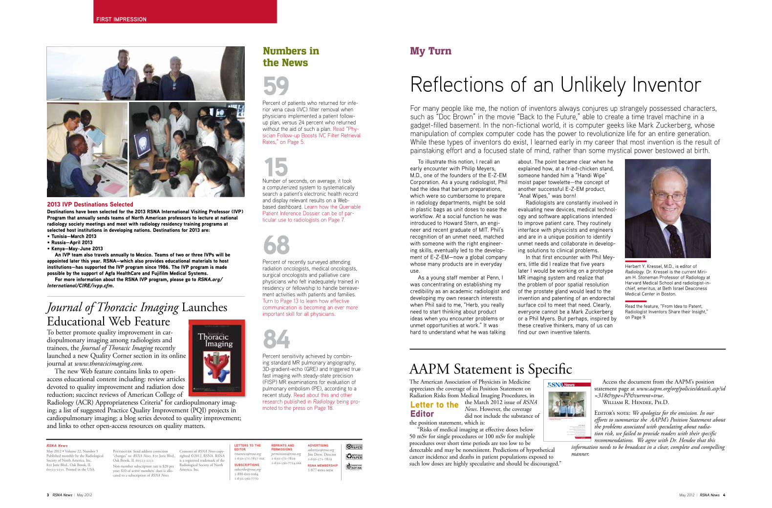
For many people like me, the notion of inventors always conjures up strangely possessed characters, such as “Doc brown” in the movie “back to the Future,” able to create a time travel machine in a gadget-filled basement. in the non-fictional world, it is computer geeks like mark Zuckerberg, whose manipulation of complex computer code has the power to revolutionize life for an entire generation. While these types of inventors do exist, i learned early in my career that most invention is the result of painstaking effort and a focused state of mind, rather than some mystical power bestowed at birth. To illustrate this notion, I recall an early encounter with Philip Meyers, M.D., one of the founders of the E-Z-EM Corporation. As a young radiologist, Phil had the idea that barium preparations, which were so cumbersome to prepare in radiology departments, might be sold in plastic bags as unit doses to ease the workflow. At a social function he was introduced to Howard Stern, an engi-neer and recent graduate of MIT. Phil’s recognition of an unmet need, matched with someone with the right engineer-ing skills, eventually led to the develop-ment of E-Z-EM—now a global company whose many products are in everyday use. As a young staff member at Penn, I was concentrating on establishing my credibility as an academic radiologist and developing my own research interests when Phil said to me, “Herb, you really need to start thinking about product ideas when you encounter problems or unmet opportunities at work.” It was hard to understand what he was talking
about. The point became clear when he explained how, at a fried-chicken stand, someone handed him a “Handi Wipe” moist paper towelette—the concept of another successful E-Z-EM product, “Anal Wipes,” was born! Radiologists are constantly involved in evaluating new devices, medical technol-ogy and software applications intended to improve patient care. They routinely interface with physicists and engineers and are in a unique position to identify unmet needs and collaborate in develop-ing solutions to clinical problems. In that first encounter with Phil Mey-ers, little did I realize that five years later I would be working on a prototype MR imaging system and realize that the problem of poor spatial resolution of the prostate gland would lead to the invention and patenting of an endorectal surface coil to meet that need. Clearly, everyone cannot be a Mark Zuckerberg or a Phil Myers. But perhaps, inspired by these creative thinkers, many of us can find our own inventive talents.
Reflections of an unlikely inventor
Numbers in the News
59Percent of patients who returned for infe-rior vena cava (iVC) filter removal when physicians implemented a patient follow-up plan, versus 24 percent who returned without the aid of such a plan. Read “Phy-sician Follow-up boosts iVC Filter Retrieval Rates,” on Page 5.
15Number of seconds, on average, it took a computerized system to systematically search a patient’s electronic health record and display relevant results on a Web-based dashboard. learn how the Queriable Patient inference Dossier can be of par-ticular use to radiologists on Page 7.
68Percent of recently surveyed attending radiation oncologists, medical oncologists, surgical oncologists and palliative care physicians who felt inadequately trained in residency or fellowship to handle bereave-ment activities with patients and families. turn to Page 13 to learn how effective communication is becoming an ever more important skill for all physicians.
84Percent sensitivity achieved by combin-ing standard mR pulmonary angiography, 3D-gradient-echo (gRe) and triggered true fast imaging with steady-state precision (FiSP) mR examinations for evaluation of pulmonary embolism (Pe), according to a recent study. Read about this and other research published in Radiology being pro-moted to the press on Page 18.
2013 IvP destinations selecteddestinations have been selected for the 2013 Rsna International visiting Professor (IvP) Program that annually sends teams of north american professors to lecture at national radiology society meetings and meet with radiology residency training programs at selected host institutions in developing nations. destinations for 2013 are:• Tunisia—March 2013• Russia—April 2013• Kenya—May-June 2013 an IvP team also travels annually to Mexico. Teams of two or three IvPs will be appointed later this year. Rsna—which also provides educational materials to host institutions—has supported the IvP program since 1986. The IvP program is made possible by the support of agfa HealthCare and Fujifilm Medical systems. For more information about the Rsna IvP program, please go to RSNA.org/International/CIRE/ivpp.cfm.
Journal of Thoracic Imaging Launches Educational Web FeatureTo better promote quality improvement in car-diopulmonary imaging among radiologists and trainees, the Journal of Thoracic Imaging recently launched a new Quality Corner section in its online journal at www.thoracicimaging.com. The new Web feature contains links to open-access educational content including: review articles devoted to quality improvement and radiation dose reduction; succinct reviews of American College of Radiology (ACR) Appropriateness Criteria® for cardiopulmonary imag-ing; a list of suggested Practice Quality Improvement (PQI) projects in cardiopulmonary imaging; a blog series devoted to quality improvement; and links to other open-access resources on quality matters.
Herbert Y. Kressel, M.D., is editor of Radiology. Dr. Kressel is the current Miri-am H. Stoneman Professor of Radiology at Harvard Medical School and radiologist-in-chief, emeritus, at Beth Israel Deaconess Medical Center in Boston.
Read the feature, “From Idea to Patent, Radiologist Inventors Share their Insight,” on Page 9.
The American Association of Physicists in Medicine appreciates the coverage of its Position Statement on Radiation Risks from Medical Imaging Procedures, in
the March 2012 issue of RSNA News. However, the coverage did not include the substance of
the position statement, which is: “Risks of medical imaging at effective doses below 50 mSv for single procedures or 100 mSv for multiple procedures over short time periods are too low to be detectable and may be nonexistent. Predictions of hypothetical cancer incidence and deaths in patient populations exposed to such low doses are highly speculative and should be discouraged.”
Access the document from the AAPM’s position statement page at www.aapm.org/org/policies/details.asp?id=318&type=PP¤t=true. William R. Hendee, Ph.D.
Editor’s note: We apologize for the omission. In our efforts to summarize the AAPM’s Position Statement about the problems associated with speculating about radia-tion risk, we failed to provide readers with their specific recommendations. We agree with Dr. Hendee that this
information needs to be broadcast in a clear, complete and compelling manner.
letter to the editor
March 2012 Volume 22, Number 3
a l s o I n s I d e :
New Tools Help Radiologists Manage Pediatric Radiation Dose in CT
New Elastography Techniques Improve Thyroid Nodule Assessment
From Smartphones to Samurais, Radiologists Explore the Mobile Future
New Academy Poised to Create Radiology’s Most Effective Leaders
submit abstracts by March 31 see Page 22
Visiting Professors Inspired by Resourcefulness of Myanmar Radiologists
AAPM Statement is Specific
news you can use
5 RSNA News | may 2012 may 2012 | RSNA News 6
FeatuRe
Physician Follow-up Boosts IVC Filter Retrieval Rates
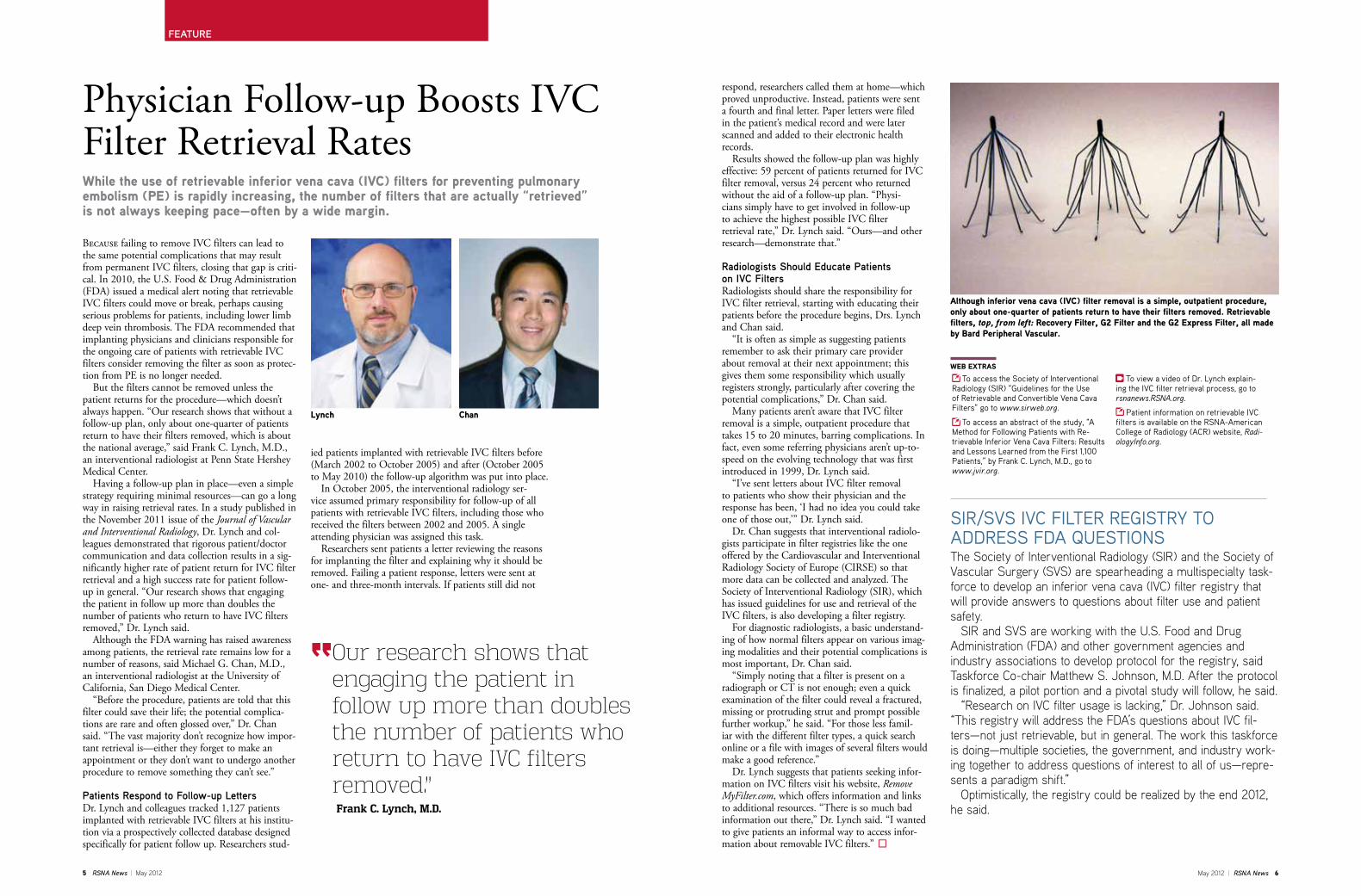
Because failing to remove IVC filters can lead to the same potential complications that may result from permanent IVC filters, closing that gap is criti-cal. In 2010, the U.S. Food & Drug Administration (FDA) issued a medical alert noting that retrievable IVC filters could move or break, perhaps causing serious problems for patients, including lower limb deep vein thrombosis. The FDA recommended that implanting physicians and clinicians responsible for the ongoing care of patients with retrievable IVC filters consider removing the filter as soon as protec-tion from PE is no longer needed. But the filters cannot be removed unless the patient returns for the procedure—which doesn’t always happen. “Our research shows that without a follow-up plan, only about one-quarter of patients return to have their filters removed, which is about the national average,” said Frank C. Lynch, M.D., an interventional radiologist at Penn State Hershey Medical Center. Having a follow-up plan in place—even a simple strategy requiring minimal resources—can go a long way in raising retrieval rates. In a study published in the November 2011 issue of the Journal of Vascular and Interventional Radiology, Dr. Lynch and col-leagues demonstrated that rigorous patient/doctor communication and data collection results in a sig-nificantly higher rate of patient return for IVC filter retrieval and a high success rate for patient follow-up in general. “Our research shows that engaging the patient in follow up more than doubles the number of patients who return to have IVC filters removed,” Dr. Lynch said. Although the FDA warning has raised awareness among patients, the retrieval rate remains low for a number of reasons, said Michael G. Chan, M.D., an interventional radiologist at the University of California, San Diego Medical Center. “Before the procedure, patients are told that this filter could save their life; the potential complica-tions are rare and often glossed over,” Dr. Chan said. “The vast majority don’t recognize how impor-tant retrieval is—either they forget to make an appointment or they don’t want to undergo another procedure to remove something they can’t see.”
Patients Respond to Follow-up LettersDr. Lynch and colleagues tracked 1,127 patients implanted with retrievable IVC filters at his institu-tion via a prospectively collected database designed specifically for patient follow up. Researchers stud-
While the use of retrievable inferior vena cava (IvC) filters for preventing pulmonary embolism (Pe) is rapidly increasing, the number of filters that are actually “retrieved” is not always keeping pace—often by a wide margin.
ied patients implanted with retrievable IVC filters before (March 2002 to October 2005) and after (October 2005 to May 2010) the follow-up algorithm was put into place. In October 2005, the interventional radiology ser-vice assumed primary responsibility for follow-up of all patients with retrievable IVC filters, including those who received the filters between 2002 and 2005. A single attending physician was assigned this task. Researchers sent patients a letter reviewing the reasons for implanting the filter and explaining why it should be removed. Failing a patient response, letters were sent at one- and three-month intervals. If patients still did not
respond, researchers called them at home—which proved unproductive. Instead, patients were sent a fourth and final letter. Paper letters were filed in the patient’s medical record and were later scanned and added to their electronic health records. Results showed the follow-up plan was highly effective: 59 percent of patients returned for IVC filter removal, versus 24 percent who returned without the aid of a follow-up plan. “Physi-cians simply have to get involved in follow-up to achieve the highest possible IVC filter retrieval rate,” Dr. Lynch said. “Ours—and other research—demonstrate that.”
Radiologists Should educate Patients on IVC FiltersRadiologists should share the responsibility for IVC filter retrieval, starting with educating their patients before the procedure begins, Drs. Lynch and Chan said. “It is often as simple as suggesting patients remember to ask their primary care provider about removal at their next appointment; this gives them some responsibility which usually registers strongly, particularly after covering the potential complications,” Dr. Chan said. Many patients aren’t aware that IVC filter removal is a simple, outpatient procedure that takes 15 to 20 minutes, barring complications. In fact, even some referring physicians aren’t up-to-speed on the evolving technology that was first introduced in 1999, Dr. Lynch said. “I’ve sent letters about IVC filter removal to patients who show their physician and the response has been, ‘I had no idea you could take one of those out,’” Dr. Lynch said. Dr. Chan suggests that interventional radiolo-gists participate in filter registries like the one offered by the Cardiovascular and Interventional Radiology Society of Europe (CIRSE) so that more data can be collected and analyzed. The Society of Interventional Radiology (SIR), which has issued guidelines for use and retrieval of the IVC filters, is also developing a filter registry. For diagnostic radiologists, a basic understand-ing of how normal filters appear on various imag-ing modalities and their potential complications is most important, Dr. Chan said. “Simply noting that a filter is present on a radiograph or CT is not enough; even a quick examination of the filter could reveal a fractured, missing or protruding strut and prompt possible further workup,” he said. “For those less famil-iar with the different filter types, a quick search online or a file with images of several filters would make a good reference.” Dr. Lynch suggests that patients seeking infor-mation on IVC filters visit his website, Remove MyFilter.com, which offers information and links to additional resources. “There is so much bad information out there,” Dr. Lynch said. “I wanted to give patients an informal way to access infor-mation about removable IVC filters.” q
“ Our research shows that engaging the patient in follow up more than doubles the number of patients who return to have IVC filters removed.”Frank C. Lynch, M.D.
Chanlynch
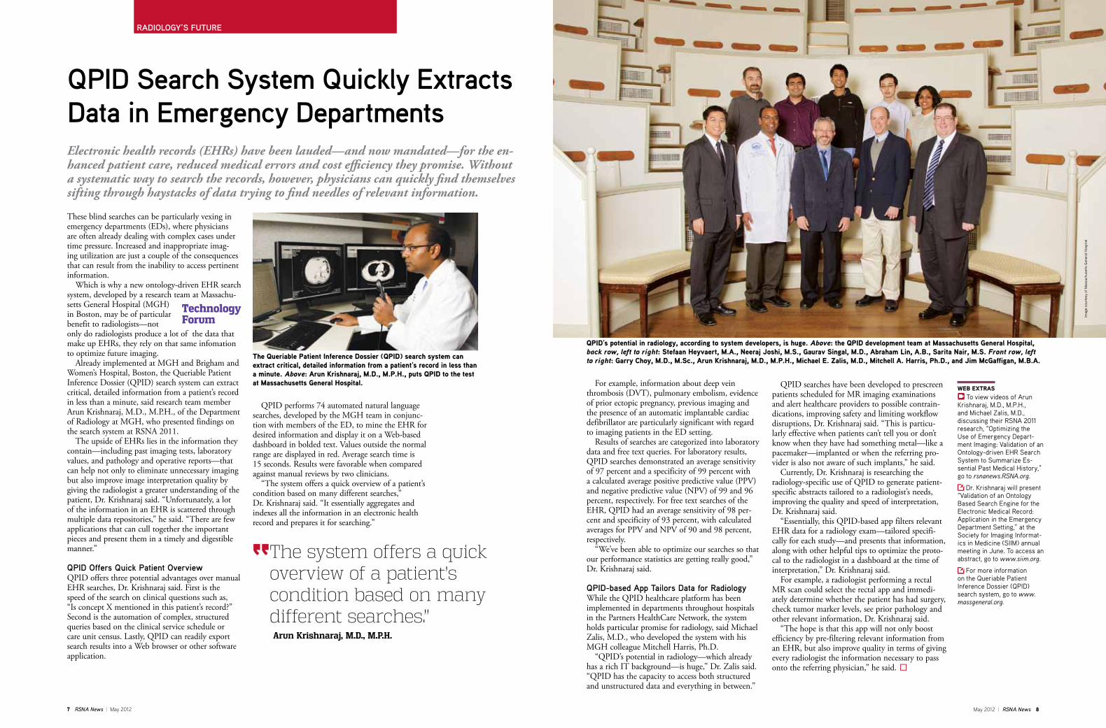
although inferior vena cava (IvC) filter removal is a simple, outpatient procedure, only about one-quarter of patients return to have their filters removed. Retrievable filters, top, from left: Recovery Filter, g2 Filter and the g2 express Filter, all made by bard Peripheral vascular.
To access the Society of Interventional Radiology (SIR) “Guidelines for the Use of Retrievable and Convertible Vena Cava Filters” go to www.sirweb.org.
To access an abstract of the study, “A Method for Following Patients with Re-trievable Inferior Vena Cava Filters: Results and Lessons Learned from the First 1,100 Patients,” by Frank C. Lynch, M.D., go to www.jvir.org.
Web exTRas
To view a video of Dr. Lynch explain-ing the IVC filter retrieval process, go to rsnanews.RSNA.org.
Patient information on retrievable IVC filters is available on the RSNA-American College of Radiology (ACR) website, Radi-ologyInfo.org.
SIR/SVS IVC FILTER REGISTRY TO ADDRESS FDA QUESTIONSthe Society of interventional Radiology (SiR) and the Society of Vascular Surgery (SVS) are spearheading a multispecialty task-force to develop an inferior vena cava (iVC) filter registry that will provide answers to questions about filter use and patient safety. SiR and SVS are working with the u.S. Food and Drug Administration (FDA) and other government agencies and industry associations to develop protocol for the registry, said taskforce Co-chair matthew S. Johnson, m.D. After the protocol is finalized, a pilot portion and a pivotal study will follow, he said. “Research on iVC filter usage is lacking,” Dr. Johnson said. “this registry will address the FDA’s questions about iVC fil-ters—not just retrievable, but in general. the work this taskforce is doing—multiple societies, the government, and industry work-ing together to address questions of interest to all of us—repre-sents a paradigm shift.” optimistically, the registry could be realized by the end 2012, he said.
news you can use
7 RSNA News | may 2012 may 2012 | RSNA News 8
RadIoLogy’S FutuRe
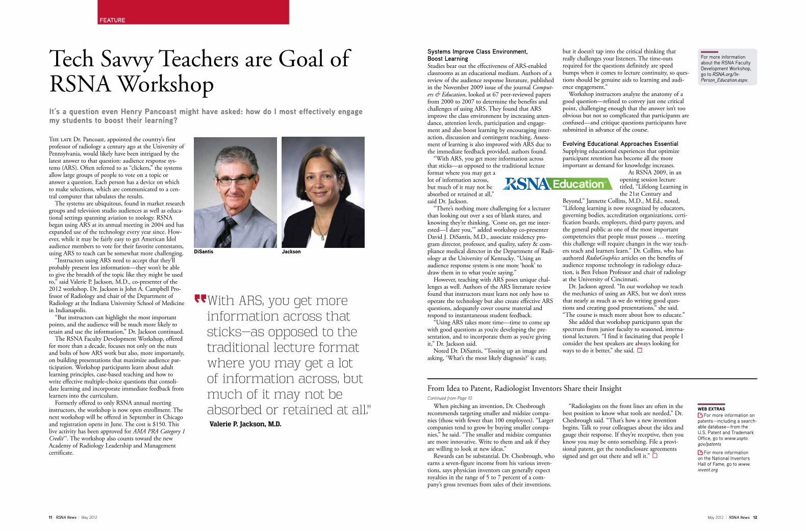
QPId’s potential in radiology, according to system developers, is huge. Above: the QPId development team at Massachusetts general Hospital, back row, left to right: stefaan Heyvaert, M.a., neeraj Joshi, M.s., gaurav singal, M.d., abraham lin, a.b., sarita nair, M.s. Front row, left to right: Garry Choy, M.D., M.Sc., Arun Krishnaraj, M.D., M.P.H., Michael E. Zalis, M.D., Mitchell A. Harris, Ph.D., and Jim McGaffigan, M.B.A.
“ The system offers a quick overview of a patient’s condition based on many different searches.”Arun Krishnaraj, M.D., M.P.H.
QPId Search System Quickly extracts data in emergency departments
These blind searches can be particularly vexing in emergency departments (EDs), where physicians are often already dealing with complex cases under time pressure. Increased and inappropriate imag-ing utilization are just a couple of the consequences that can result from the inability to access pertinent information. Which is why a new ontology-driven EHR search system, developed by a research team at Massachu-setts General Hospital (MGH) in Boston, may be of particular benefit to radiologists—not only do radiologists produce a lot of the data that make up EHRs, they rely on that same infomation to optimize future imaging. Already implemented at MGH and Brigham and Women’s Hospital, Boston, the Queriable Patient Inference Dossier (QPID) search system can extract critical, detailed information from a patient’s record in less than a minute, said research team member Arun Krishnaraj, M.D., M.P.H., of the Department of Radiology at MGH, who presented findings on the search system at RSNA 2011. The upside of EHRs lies in the information they contain—including past imaging tests, laboratory values, and pathology and operative reports—that can help not only to eliminate unnecessary imaging but also improve image interpretation quality by giving the radiologist a greater understanding of the patient, Dr. Krishnaraj said. “Unfortunately, a lot of the information in an EHR is scattered through multiple data repositories,” he said. “There are few applications that can cull together the important pieces and present them in a timely and digestible manner.”
QPId offers Quick Patient overviewQPID offers three potential advantages over manual EHR searches, Dr. Krishnaraj said. First is the speed of the search on clinical questions such as, “Is concept X mentioned in this patient’s record?” Second is the automation of complex, structured queries based on the clinical service schedule or care unit census. Lastly, QPID can readily export search results into a Web browser or other software application.
Electronic health records (EHRs) have been lauded—and now mandated—for the en-hanced patient care, reduced medical errors and cost efficiency they promise. Without a systematic way to search the records, however, physicians can quickly find themselves sifting through haystacks of data trying to find needles of relevant information.
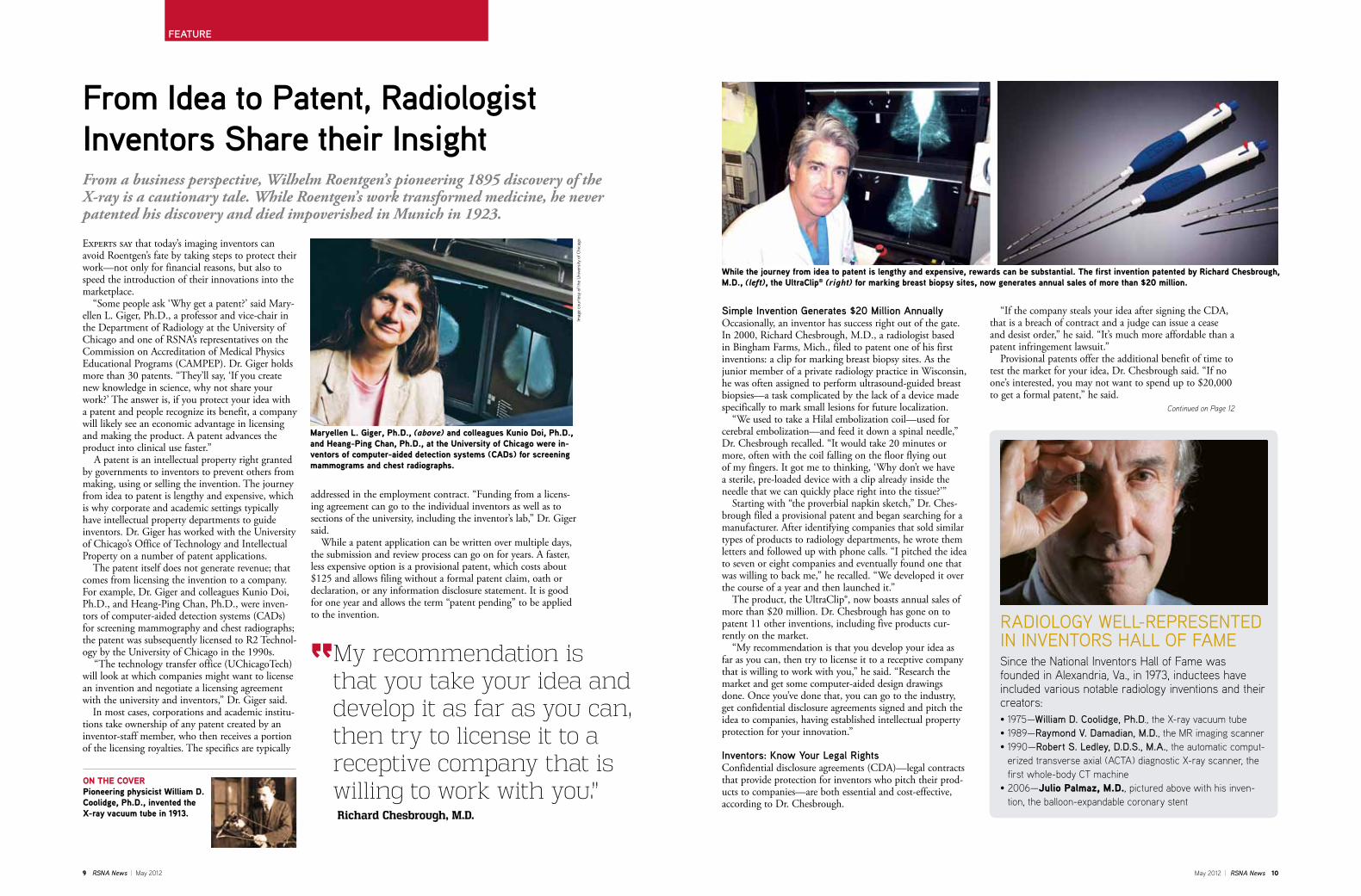
The Queriable Patient Inference dossier (QPId) search system can extract critical, detailed information from a patient’s record in less than a minute. Above: Arun Krishnaraj, M.D., M.P.H., puts QPID to the test at Massachusetts general Hospital.
QPID performs 74 automated natural language searches, developed by the MGH team in conjunc-tion with members of the ED, to mine the EHR for desired information and display it on a Web-based dashboard in bolded text. Values outside the normal range are displayed in red. Average search time is 15 seconds. Results were favorable when compared against manual reviews by two clinicians. “The system offers a quick overview of a patient’s condition based on many different searches,” Dr. Krishnaraj said. “It essentially aggregates and indexes all the information in an electronic health record and prepares it for searching.”
For example, information about deep vein thrombosis (DVT), pulmonary embolism, evidence of prior ectopic pregnancy, previous imaging and the presence of an automatic implantable cardiac defibrillator are particularly significant with regard to imaging patients in the ED setting. Results of searches are categorized into laboratory data and free text queries. For laboratory results, QPID searches demonstrated an average sensitivity of 97 percent and a specificity of 99 percent with a calculated average positive predictive value (PPV) and negative predictive value (NPV) of 99 and 96 percent, respectively. For free text searches of the EHR, QPID had an average sensitivity of 98 per-cent and specificity of 93 percent, with calculated averages for PPV and NPV of 90 and 98 percent, respectively. “We’ve been able to optimize our searches so that our performance statistics are getting really good,” Dr. Krishnaraj said.
QPId-based app tailors data for Radiology While the QPID healthcare platform has been implemented in departments throughout hospitals in the Partners HealthCare Network, the system holds particular promise for radiology, said Michael Zalis, M.D., who developed the system with his MGH colleague Mitchell Harris, Ph.D. “QPID’s potential in radiology—which already has a rich IT background—is huge,” Dr. Zalis said. “QPID has the capacity to access both structured and unstructured data and everything in between.”
QPID searches have been developed to prescreen patients scheduled for MR imaging examinations and alert healthcare providers to possible contrain-dications, improving safety and limiting workflow disruptions, Dr. Krishnaraj said. “This is particu-larly effective when patients can’t tell you or don’t know when they have had something metal—like a pacemaker—implanted or when the referring pro-vider is also not aware of such implants,” he said. Currently, Dr. Krishnaraj is researching the radiology-specific use of QPID to generate patient-specific abstracts tailored to a radiologist’s needs, improving the quality and speed of interpretation, Dr. Krishnaraj said. “Essentially, this QPID-based app filters relevant EHR data for a radiology exam—tailored specifi-cally for each study—and presents that information, along with other helpful tips to optimize the proto-cal to the radiologist in a dashboard at the time of interpretation,” Dr. Krishnaraj said. For example, a radiologist performing a rectal MR scan could select the rectal app and immedi-ately determine whether the patient has had surgery, check tumor marker levels, see prior pathology and other relevant information, Dr. Krishnaraj said. “The hope is that this app will not only boost efficiency by pre-filtering relevant information from an EHR, but also improve quality in terms of giving every radiologist the information necessary to pass onto the referring physician,” he said. q
Web exTRasTo view videos of Arun
Krishnaraj, M.D., M.P.H., and Michael Zalis, M.D., discussing their RSNA 2011 research, “Optimizing the Use of Emergency Depart-ment Imaging: Validation of an Ontology-driven EHR Search System to Summarize Es-sential Past Medical History,” go to rsnanews.RSNA.org.
Dr. Krishnaraj will present “Validation of an Ontology Based Search Engine for the Electronic Medical Record: Application in the Emergency Department Setting,” at the Society for Imaging Informat-ics in Medicine (SIIM) annual meeting in June. To access an abstract, go to www.siim.org.
For more information on the Queriable Patient Inference Dossier (QPID) search system, go to www.massgeneral.org.
Technology Forum Im
age
cour
tesy
of M
assa
chus
etts
Gen
eral
Hos
pita
l
news you can use
9 RSNA News | may 2012 may 2012 | RSNA News 10
FeatuRe
Experts say that today’s imaging inventors can avoid Roentgen’s fate by taking steps to protect their work—not only for financial reasons, but also to speed the introduction of their innovations into the marketplace. “Some people ask ‘Why get a patent?’ said Mary-ellen L. Giger, Ph.D., a professor and vice-chair in the Department of Radiology at the University of Chicago and one of RSNA’s representatives on the Commission on Accreditation of Medical Physics Educational Programs (CAMPEP). Dr. Giger holds more than 30 patents. “They’ll say, ‘If you create new knowledge in science, why not share your work?’ The answer is, if you protect your idea with a patent and people recognize its benefit, a company will likely see an economic advantage in licensing and making the product. A patent advances the product into clinical use faster.” A patent is an intellectual property right granted by governments to inventors to prevent others from making, using or selling the invention. The journey from idea to patent is lengthy and expensive, which is why corporate and academic settings typically have intellectual property departments to guide inventors. Dr. Giger has worked with the University of Chicago’s Office of Technology and Intellectual Property on a number of patent applications. The patent itself does not generate revenue; that comes from licensing the invention to a company. For example, Dr. Giger and colleagues Kunio Doi, Ph.D., and Heang-Ping Chan, Ph.D., were inven-tors of computer-aided detection systems (CADs) for screening mammography and chest radiographs; the patent was subsequently licensed to R2 Technol-ogy by the University of Chicago in the 1990s. “The technology transfer office (UChicagoTech) will look at which companies might want to license an invention and negotiate a licensing agreement with the university and inventors,” Dr. Giger said. In most cases, corporations and academic institu-tions take ownership of any patent created by an inventor-staff member, who then receives a portion of the licensing royalties. The specifics are typically
From Idea to Patent, Radiologist Inventors Share their InsightFrom a business perspective, Wilhelm Roentgen’s pioneering 1895 discovery of the X-ray is a cautionary tale. While Roentgen’s work transformed medicine, he never patented his discovery and died impoverished in Munich in 1923.
addressed in the employment contract. “Funding from a licens-ing agreement can go to the individual inventors as well as to sections of the university, including the inventor’s lab,” Dr. Giger said. While a patent application can be written over multiple days, the submission and review process can go on for years. A faster, less expensive option is a provisional patent, which costs about $125 and allows filing without a formal patent claim, oath or declaration, or any information disclosure statement. It is good for one year and allows the term “patent pending” to be applied to the invention.
Simple Invention generates $20 Million annuallyOccasionally, an inventor has success right out of the gate. In 2000, Richard Chesbrough, M.D., a radiologist based in Bingham Farms, Mich., filed to patent one of his first inventions: a clip for marking breast biopsy sites. As the junior member of a private radiology practice in Wisconsin, he was often assigned to perform ultrasound-guided breast biopsies—a task complicated by the lack of a device made specifically to mark small lesions for future localization. “We used to take a Hilal embolization coil—used for cerebral embolization—and feed it down a spinal needle,” Dr. Chesbrough recalled. “It would take 20 minutes or more, often with the coil falling on the floor flying out of my fingers. It got me to thinking, ‘Why don’t we have a sterile, pre-loaded device with a clip already inside the needle that we can quickly place right into the tissue?’” Starting with “the proverbial napkin sketch,” Dr. Ches-brough filed a provisional patent and began searching for a manufacturer. After identifying companies that sold similar types of products to radiology departments, he wrote them letters and followed up with phone calls. “I pitched the idea to seven or eight companies and eventually found one that was willing to back me,” he recalled. “We developed it over the course of a year and then launched it.” The product, the UltraClip®, now boasts annual sales of more than $20 million. Dr. Chesbrough has gone on to patent 11 other inventions, including five products cur-rently on the market. “My recommendation is that you develop your idea as far as you can, then try to license it to a receptive company that is willing to work with you,” he said. “Research the market and get some computer-aided design drawings done. Once you’ve done that, you can go to the industry, get confidential disclosure agreements signed and pitch the idea to companies, having established intellectual property protection for your innovation.”
Inventors: Know your Legal Rights Confidential disclosure agreements (CDA)—legal contracts that provide protection for inventors who pitch their prod-ucts to companies—are both essential and cost-effective, according to Dr. Chesbrough.
While the journey from idea to patent is lengthy and expensive, rewards can be substantial. The first invention patented by Richard Chesbrough, M.d., (left), the ultraClip® (right) for marking breast biopsy sites, now generates annual sales of more than $20 million.
Maryellen l. giger, Ph.d., (above) and colleagues Kunio Doi, Ph.D., and Heang-Ping Chan, Ph.d., at the university of Chicago were in-ventors of computer-aided detection systems (Cads) for screening mammograms and chest radiographs.
“ My recommendation is that you take your idea and develop it as far as you can, then try to license it to a receptive company that is willing to work with you.”Richard Chesbrough, M.D.
Since the National inventors Hall of Fame was founded in Alexandria, Va., in 1973, inductees have included various notable radiology inventions and their creators:
RADIOLOGY WELL-REPRESENTED IN INVENTORS HALL OF FAME
• 1975—william d. Coolidge, Ph.d., the X-ray vacuum tube• 1989—Raymond V. damadian, M.d., the mR imaging scanner• 1990—Robert s. ledley, d.d.s., M.a., the automatic comput-
erized transverse axial (ACtA) diagnostic X-ray scanner, the first whole-body Ct machine
• 2006—Julio Palmaz, M.d., pictured above with his inven-tion, the balloon-expandable coronary stent
on THe CoveRPioneering physicist William d. Coolidge, Ph.d., invented the x-ray vacuum tube in 1913.
Continued on Page 12
“If the company steals your idea after signing the CDA, that is a breach of contract and a judge can issue a cease and desist order,” he said. “It’s much more affordable than a patent infringement lawsuit.” Provisional patents offer the additional benefit of time to test the market for your idea, Dr. Chesbrough said. “If no one’s interested, you may not want to spend up to $20,000 to get a formal patent,” he said.
Imag
e co
urte
sy o
f the
Uni
vers
ity o
f Chi
cago
newS you Can uSe
11 RSNA News | may 2012
FeatuRe
may 2012 | RSNA News 12
Tech Savvy Teachers are Goal of RSNA Workshop
The late Dr. Pancoast, appointed the country’s first professor of radiology a century ago at the University of Pennsylvania, would likely have been intrigued by the latest answer to that question: audience response sys-tems (ARS). Often referred to as “clickers,” the systems allow large groups of people to vote on a topic or answer a question. Each person has a device on which to make selections, which are communicated to a cen-tral computer that tabulates the results. The systems are ubiquitous, found in market research groups and television studio audiences as well as educa-tional settings spanning aviation to zoology. RSNA began using ARS at its annual meeting in 2004 and has expanded use of the technology every year since. How-ever, while it may be fairly easy to get American Idol audience members to vote for their favorite contestants, using ARS to teach can be somewhat more challenging. “Instructors using ARS need to accept that they’ll probably present less information—they won’t be able to give the breadth of the topic like they might be used to,” said Valerie P. Jackson, M.D., co-presenter of the 2012 workshop. Dr. Jackson is John A. Campbell Pro-fessor of Radiology and chair of the Department of Radiology at the Indiana University School of Medicine in Indianapolis. “But instructors can highlight the most important points, and the audience will be much more likely to retain and use the information,” Dr. Jackson continued. The RSNA Faculty Development Workshop, offered for more than a decade, focuses not only on the nuts and bolts of how ARS work but also, more importantly, on building presentations that maximize audience par-ticipation. Workshop participants learn about adult learning principles, case-based teaching and how to write effective multiple-choice questions that consoli-date learning and incorporate immediate feedback from learners into the curriculum. Formerly offered to only RSNA annual meeting instructors, the workshop is now open enrollment. The next workshop will be offered in September in Chicago and registration opens in June. The cost is $150. This live activity has been approved for AMA PRA Category 1 Credit™. The workshop also counts toward the new Academy of Radiology Leadership and Management certificate.
It’s a question even Henry Pancoast might have asked: how do I most effectively engage my students to boost their learning?
Jacksondisantis
Systems Improve Class environment, Boost LearningStudies bear out the effectiveness of ARS-enabled classrooms as an educational medium. Authors of a review of the audience response literature, published in the November 2009 issue of the journal Comput-ers & Education, looked at 67 peer-reviewed papers from 2000 to 2007 to determine the benefits and challenges of using ARS. They found that ARS improve the class environment by increasing atten-dance, attention levels, participation and engage-ment and also boost learning by encouraging inter-action, discussion and contingent teaching. Assess-ment of learning is also improved with ARS due to the immediate feedback provided, authors found. “With ARS, you get more information across that sticks—as opposed to the traditional lecture format where you may get a lot of information across, but much of it may not be absorbed or retained at all,” said Dr. Jackson. “There’s nothing more challenging for a lecturer than looking out over a sea of blank stares, and knowing they’re thinking, ‘Come on, get me inter-ested—I dare you,’” added workshop co-presenter David J. DiSantis, M.D., associate residency pro-gram director, professor, and quality, safety & com-pliance medical director in the Department of Radi-ology at the University of Kentucky. “Using an audience response system is one more ‘hook’ to draw them in to what you’re saying.” However, teaching with ARS poses unique chal-lenges as well. Authors of the ARS literature review found that instructors must learn not only how to operate the technology but also create effective ARS questions, adequately cover course material and respond to instantaneous student feedback. “Using ARS takes more time—time to come up with good questions as you’re developing the pre-sentation, and to incorporate them as you’re giving it,” Dr. Jackson said. Noted Dr. DiSantis, “Tossing up an image and asking, ‘What’s the most likely diagnosis?’ is easy,
but it doesn’t tap into the critical thinking that really challenges your listeners. The time-outs required for the questions definitely are speed bumps when it comes to lecture continuity, so ques-tions should be genuine aids to learning and audi-ence engagement.” Workshop instructors analyze the anatomy of a good question—refined to convey just one critical point, challenging enough that the answer isn’t too obvious but not so complicated that participants are confused—and critique questions participants have submitted in advance of the course.
evolving educational approaches essentialSupplying educational experiences that optimize participant retention has become all the more important as demand for knowledge increases.
At RSNA 2009, in an opening session lecture titled, “Lifelong Learning in the 21st Century and
Beyond,” Jannette Collins, M.D., M.Ed., noted, “Lifelong learning is now recognized by educators, governing bodies, accreditation organizations, certi-fication boards, employers, third-party payers, and the general public as one of the most important competencies that people must possess … meeting this challenge will require changes in the way teach-ers teach and learners learn.” Dr. Collins, who has authored RadioGraphics articles on the benefits of audience response technology in radiology educa-tion, is Ben Felson Professor and chair of radiology at the University of Cincinnati. Dr. Jackson agreed. “In our workshop we teach the mechanics of using an ARS, but we don’t stress that nearly as much as we do writing good ques-tions and creating good presentations,” she said. “The course is much more about how to educate.” She added that workshop participants span the spectrum from junior faculty to seasoned, interna-tional lecturers. “I find it fascinating that people I consider the best speakers are always looking for ways to do it better,” she said. q
“ With ARS, you get more information across that sticks—as opposed to the traditional lecture format where you may get a lot of information across, but much of it may not be absorbed or retained at all.”Valerie P. Jackson, M.D.
When pitching an invention, Dr. Chesbrough recommends targeting smaller and midsize compa-nies (those with fewer than 100 employees). “Larger companies tend to grow by buying smaller compa-nies,” he said. “The smaller and midsize companies are more innovative. Write to them and ask if they are willing to look at new ideas.” Rewards can be substantial. Dr. Chesbrough, who earns a seven-figure income from his various inven-tions, says physician inventors can generally expect royalties in the range of 5 to 7 percent of a com-pany’s gross revenues from sales of their inventions.
Web exTRasFor more information on
patents—including a search-able database—from the U.S. Patent and Trademark Office, go to www.uspto.gov/patents
For more information on the National Inventors Hall of Fame, go to www.invent.org
Continued from Page 10
From Idea to Patent, Radiologist Inventors Share their Insight
“Radiologists on the front lines are often in the best position to know what tools are needed,” Dr. Chesbrough said. “That’s how a new invention begins. Talk to your colleagues about the idea and gauge their response. If they’re receptive, then you know you may be onto something. File a provi-sional patent, get the nondisclosure agreements signed and get out there and sell it.” q
For more information about the RSNA Faculty Development Workshop, go to RSNA.org/In- Person_Education.aspx.
may 2012 | RSNA News 1413 RSNA News | may 2012
newS you Can uSeFeatuRe
Communication Skills Can Make or Break a Patient-Physician Relationship
The woman immediately broke down in tears and ran out of the room. As this case demonstrates, how a doctor commu-nicates to a patient can make all the difference in setting the tone for how a patient will react and begin to grasp the impact. “A doctor’s attitude or behavior can absolutely make or break their relationship with a patient,” said Michael Linver, M.D., director of mammogra-phy at X-Ray Associates of New Mexico in Albu-querque, who provided the true example of a former colleague from his own practice. Dr. Linver pre-sented “Talking with Patients: Ways to Gain Their Trust” at RSNA 2011. “Sensitivity and empathy should always be part of any radiologist/patient interchange.” As patients continue to take a more active role in their own healthcare, all physicians—particularly radiologists—need to develop the communication skills necessary to create satisfying patient/physician relationships. No longer isolated in the back room interpreting exams, radiologists are increasingly being asked to take on a more direct role in patient care. Efective patient communications skills are not only desirable, they’re necessary, experts say. “With the exception of interventional radiology, there has traditionally been little opportunity for real one-on-one interaction between patient and radiologist,” Dr. Linver said. “That changed dramat-ically in the past 10 years with the advent of the breast imaging specialist.” Because many radiologists lack communication training and are accustomed to working behind the scenes, some are likely to feel ill-equipped to step into the role of “communicator.” Fortunately, radiology organizations including RSNA are developing and fortifying efforts to improve patient/physician communications that could go a long way toward making imaging more valuable in the eyes of the public (See sidebar).
treat Patients like FamilyWhen engaging with patients, radiologists need to remember that names and images belong to an actual person. “I treat every patient as a member of my own family—it is the way I would want to be treated if I were a patient,” Dr. Linver said. There should always be an attempt to go over the report with the patient by phone or in person, and, if in person, show actual images to the patient—which creates an even stronger bond. Other simple
Many years ago, a relatively new breast imager preparing to give a patient the results of an abnormal mammogram walked into the exam room, and without introducing himself, blurted out, “Well, you’ve got cancer.”
actions include providing a letter to patients to inform them when their results will be ready, stepping into the exam room to greet a patient before a radiologic procedure and following up with a patient after recommending additional studies. Radiologists should also strive to obtain better histories directly from patients, Dr. Linver said. “This allows for a more accurate interpretation by the radiologist and shows the patient that the radiologist really cares about the patient as a person, not just as a collection of images.” Always use a reassuring voice and be very kind when patients ask questions. “The doctor should explain the situa-tion in a language the patient can understand and in a way that is as reassuring as is possible, always trying to find some-thing to be positive about,” Dr. Linver said. Even a physician who is informing a patient she has cancer should not leave the person without hope. “She will never sur-vive the entire breast cancer treatment process without that,” he said.
Kusanolinver
Radiation oncologists Lack Communication SupportResults of one new study underscore the lack of specialized train-ing in communication skills among physicians who likely need it the most. Two-thirds of cancer care physicians do not feel they received adequate training in this area during their residency or fellowship, according to research presented at the 2011 American Society for Radiation Oncology (ASTRO) annual meeting. “Talking with patients is one of the most important and under-valued services we deliver in healthcare,” said lead author Aaron S. Kusano, M.D., S.M., a resident in the Department of Radiation Oncology at the University of Washington School of Medicine, Seattle. “If done well, a critical bond with the patient can be established, creating a level of trust and comfort.” The urgency of this issue was echoed by senior author, Charles R. Thomas, Jr., M.D., a professor and chair of the Department of Radiation Oncology at the Knight Cancer Institute of the Oregon Health and Science University, Portland. The study, an anonymous online survey, examined the fre-quency and scope of bereavement practices among cancer care and palliative care physicians in the northwestern U.S. It was completed by 162 attending radiation oncologists, medical oncol-ogists, surgical oncologists and palliative care physicians directly involved in patient care in fall 2010.
Results showed that 70 percent of cancer care physicians were routinely engaged in at least one bereavement activity, with send-ing a condolence letter by far the most common form of commu-nication. Other physician-initiated activities included calling fam-ilies or attending a funeral service following a patient’s death. Yet 68 percent of respondents felt inadequately trained in resi-dency or fellowship to address bereavement activities; physicians were less likely to perform follow up with the bereaved if they lacked bereavement support services or felt uncomfortable about what to say. “Every physician will encounter death and when we choose to reach out to a family, our training should equip us to feel comfortable in doing so,” said Dr. Kusano. Several factors made an individual more likely to perform active follow up, including being a medical oncologist (versus a radia-tion oncologist or palliative care physician), having access to a pal-liative care program and feeling responsible for writing a condo-lence letter. The most common barriers to bereavement follow up were lack of time and uncertainty as to which family member to contact. “I think there is an important message here to all physicians,” Dr. Kusano said. “Through anecdotes and small studies, we know that many families/survivors appreciate follow up by the physi-cian, but what physicians actually do needs much more study.” q
“ Talking with patients is one of the most important and undervalued services we deliver in healthcare.”Aaron S. Kusano, M.D., S.M.
long at the forefront of patient-cen-tered radiology, RSNA is ramping up efforts in 2012 on a number of fronts. themed “Patients First,” RSNA 2012 will offer pro-gramming focusing on patient-centered care, including communication with patients and pro-viding dose-optimized imaging. RSNA will again offer the refresher courses,
“Vignette-based Disclosure of medical error in Radiology,” “Patient-centered Radiology: it’s good business” and “What the Referring Physician Needs
to Know.” the RSNA board of Directors has also appointed
a Patient-Centered Radiology Steering Committee to guide future patient-centered initiatives.
in addition, the RSNA-American College of Radiology (ACR) website Radiologyinfo.org—the radiology information resource for patients—is regularly updated with new informa-tion and videos educating patients on radiology procedures. Stay updated on RSNA 2012 pro-gramming at RSNA2012.RSNA.org.
RSNA INITIATIVES PUT PATIENTS FIRST
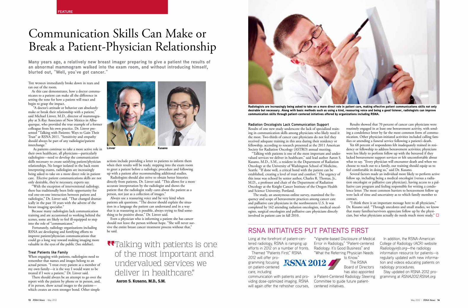
Radiologists are increasingly being asked to take on a more direct role in patient care, making effective patient communications skills not only desirable but necessary. along with basic methods such as using a kind, reassuring voice and being a good listener, radiologists can improve communication skills through patient-centered initiatives offered by organizations including Rsna.
may 2012 | RSNA News 16
RadIoLogy’S FutuRe
15 RSNA News | may 2012
RESEARCH & EDUCATION FOUNDATION DONORSthe R&e Foundation thanks the following donors for gifts made January 24, 2012 – February 24, 2012.
Individual donorsdonors who give $1,500 or more per year qualify for the RSna Presidents Circle. their names are shown in bold face.
$2,500 - $4,999Valerie P. Jackson, M.d.$1,500 - $2,499$1,500 – $2,499Phyllis & Leonard Berlin, M.d.Lisa & Jonathan Breslau, M.d.tracey Hillier, M.d., F.R.C.P.C. & Sukhvinder S. dhillon, M.R.C.P., F.R.C.R.
Betty & o. wayne Houser, M.d.Beverly & Michael HuckmanLyne noel de tilly, M.d. & edward e. Kassel, M.d.
In memory of Derek C. Harwood-Nash, M.D., D.Sc.
Leon e. Kinasiewicz, M.d.Marten KlopSusan & Kenyon K. Kopecky, M.d.david C. Levin, M.d.deborah Levine, M.d. & alex Jesurum, Ph.d.
John H. Rees, M.d.Marilyn J. Siegel, M.d. & Barry a. Siegel, M.d.
John w. thomas, M.d.Linda J. warren, M.d.warren a. wass, M.d.Corine yee, M.d. & Michael t. oliver, M.d.
In memory of Mr. & Mrs. William Yee and Mr. Thomas F. Oliver
$500 – $1,499Richard S. Colvin, M.D. In honor of Robert E. Campbell, M.D.Jennifer R. Cranny, M.D.Heike E. Daldrup-Link, M.D., Ph.D. & Thomas M. Link, M.D.
gary w. Swenson, M.d.$250 or lessAnnie & Mitchell D. Achee, M.D.Arifudin Achmad, M.D.Kamran Ahrar, M.D.Adenike O. Akhigbe, M.B.B.S. & Kingsley O. Akhigbe, M.B.B.S.
Amjad Ali, M.D.Ildefonso G. Almonte, M.D.Lisa A. Altieri, M.D.Michael D. Ames, M.D.Brian L. Anderson, M.D.Charles M. Anderson, M.D., Ph.D.Shannon E. Ardoin, M.D. & Gregory Ardoin
Ronald C. Arildsen, M.D.M. Julie Armada, M.D.
Valerie & Derek C. Armstrong, M.D.Deborah Arnold, M.D. & Philip ArnoldRony Avritscher, M.D.Filip Banovac, M.D.Jody M. Barber, M.D.Patricia A. Barry, M.D. & John Cosgrove, M.D.
Kimberly N. & J. Edward Bass, M.D.Jayson L. Benjert, D.O.Jonas J. Berman, M.D.Kalpana & Shailesh M. Bhatt, M.D.Royce J. Biddle, M.D.Jacques Blanca, M.D., Ph.D.Ernesto Blanco, M.D.Carolyn H. & George T. Bolton, D.V.M., M.D.
Mitra B. Boodram, M.D.Erin M. Bowman, M.D. & Jason BowmanJ.D. & Ronald M. Boyd, M.D.Luther W. Brady Jr., M.D.Oleg E. Bronov, M.D.Robert T. Brown, M.D.Sara R. & Stephen D. Brown, M.D.Kelly A. Brozetti, M.D.Erin Yourtz & Lawrence D. Bub, M.D.James T. Buratto, M.D.Laura & John K. Campbell, M.D.Erin McBride Cannington & Royce Cannington, M.D.
Jose A. Cardenas, M.D.Suzette G. Casal, M.D.Won Chae, M.D.Tilden L. Childs III, M.D.Joseph M. Chin, M.D.Cristina G. Ortega & Cesar Cisneros Victorino, M.D.
Winston F. Clarke, M.D.Karen & Steven M. Cohen, M.D.Rivka R. Colen, M.D.Angel R. Colon, M.D.Cody A. Cox, M.D.Gordon L. Cross, M.D.Julianna M. Czum, M.D.Lisa & Jay M. Daly, M.D.Cliff R. Davis, M.D.Jennifer & Mark S. Dixon, M.D.Peter Drescher, M.D., M.S.Andre J.G. Duerinckx, M.D., Ph.D. & David Adams
Karen & Brian L. Dunkley, M.D.Christopher G. Eckel, M.D.Karl Engelhard, M.D.Tam Huynh & Jeremy J. Erasmus, M.D.Birgit B. Ertl-Wagner, M.D. & Bernd Wagner
Janet & David R. Erwin, M.D.Lawrence C. Fan, M.D.Michael E. Farber, M.D.Kristin & Scott A. Fargher, M.D.Jerrold S. Feit, M.D.Frieda Feldman, M.D. & Rubem Pochaczevsky, M.D.
Aimee & John C. Feldman, M.D.Kristin M. Foley, M.D.Cheryl & Michael J. Foley, M.D.Robert P. Fortunato, M.D., M.B.A.Terry S. & Daniel R. Fox, M.D.Therese & Irwin M. Freundlich, M.D.Angela M. Fried, M.D. & C. Stephen Djedjos, M.D.
Elizabeth Orvoen-Frija, M.D. & Guy Frija, M.D.
Karen S. Frush, M.D. & Donald P. Frush, M.D.
Colleen & Michael W. Gabriele, M.D.Pablo A. Gamboa, M.D.Connie J. Gapinski, M.D., Ph.D.Janet B. Garrett, M.D.Megan M. Gau, M.D.ayca gazelle, M.d. & g. Scott gazelle, M.d., M.P.H., Ph.d.
Susan A. & Richard W. Geldmeier, M.D.Peter W. Gendall, M.D.Hilary & Anthony Gentile In memory of my mother Mrs. Moira Wilde
Michael S. Gibson, M.D.Joann M. Gierbolini, M.D.Maryellyn Gilfeather, M.D.Susan L. Goldfine, M.D.Lori & Monte E. Golditch, M.D.Rod Golovoy, M.D.Igor Goykhman, D.O.Valerie T. Greco-Hunt, M.D.Larry D. Greenfield, M.D.W. Lawrence Greif, M.D.Sharon & Irwin grossman, M.d.Kurt T. Grozinger, M.D.Dale W. Gunn, M.D.Jean Pierre Gurret, M.D.Susan & Adam R. Guttentag, M.D.Soterios Gyftopoulos, M.D.Amos Q. Habib, M.D.Elizabeth L. Hadley, M.D.Ronald L. Hainen, M.D.Joan McEnery & Kevin D. Halista, M.D.Cynthia W. Hanemann, M.D.Katherine & Jonathan M. Hard, M.D.Melissa & Christopher O. Harker, M.D.Dashartha Harsewak, M.D.
Mizuki & Hiroto Hatabu, M.D., Ph.D.Kathleen Golueke-Heredia & Sergio L. Heredia, M.D.
In memory of Dr. Peter & Florence Golueke
Michelle & Richard J. Hicks, M.D.Michael T. Hirleman, M.D.Jill Holsinger, M.D. & Philip HolsingerStephanie P. Holz, M.D. & Timothy D. Holz
Glenda K. Holzman, M.D. & Brian Holzman
Rachel B. Hulen, M.D.
Chien-Fu Hung, M.D.Kim Hwang, M.D.Shawna Hassett-Iannuccilli & Jason D. Iannuccilli, M.D.
Marcy B. Jagust, M.D.Bradley A. Johnson, M.D.Jeffrey L. Johnson, M.D.John O. Johnson, M.D.Marvin W. Johnson, M.D.Nancy & Thomas G. Johnson, M.D.Ronald C. Joseph, M.D.Jonathan R. Joudanin, M.D.Christian H. Julien, M.D.Brian T. Kaineg, M.D.Vivien G. Kane, M.D.Raveen Kaur, M.D. & Kulwant GillAdrienne & Alan D. Kaye, M.D.Delia M. Keating, M.D.C. Stephen Keklak, M.D.Ian A. Kellman, M.D.Kevin M. Kelly, M.D.Ajit K. Khanuja, M.D.Stephane Khazoom, M.D.Bahram Kiani, M.D.Marie I. & Fabian Kiessling, M.D.Jacobo Kirsch, M.D.
Suzanne Freitag & Philip D. Kousoubris, M.D.
Mary W. & Stanton S. Kremsky, M.D.Ruben Krishnananthan, M.D., M.B.B.S.George S. Krol, M.D.Anthony L. Kudirka, M.D.Neeraj Lalwani, M.D.Phobe M. & James T. Lambeth, M.D.Kent T. Lancaster, M.D.Kevin C. Lau, M.B.B.S.David S. Leder, M.D.Choon K. Lee, M.D.Henri E. Lemmers, M.D.Louise Nolet & Jacques J. Levesque, M.D.
Cynthia & Paolo P. Lim, M.D.Tae-Hwan Lim, M.D.Christopher K. Lin, M.D.Noam Littman, M.D.Ee-Loon Tham & Simon S. Lo, M.D.Alan F. Lobo, M.D.Richard J. Loges III, M.D.Connie S. & Rodolfo A. Lopez, M.D.Dean D. Maglinte, M.D.Sheila S. Manion, M.D.Paul C. Marinelli, M.D.Diego R. Martin, M.D., Ph.D.Eric M. Martin, M.D., Ph.D.Janine R. Martyn, M.D.Michael F. Mastromatteo, M.D.Ani Ketabgian & Edward F. Math, M.D.John R. Mathieson, M.D.David A. May, M.D.Peter McClure, M.D.Maura & Vincent G. McDermott, M.D.Rekha Meesa, M.D. & Sudheer MeesaSimone Pimenta & Paulo Sergio R. Mendlovitz, M.D.
Corinne Balleyguier, M.D. & Yves M. Menu, M.D.
Lucy Houlihan & Timothy J. Mickus, M.D.
John M. Milbourn, M.D.William D. Miller, M.D.Yoshimi Anzai, M.D. & Satoshi Minoshima, M.D., Ph.D.
Ari D. Mintz, M.D.Shachi & Malay K. Mody, M.D.William M. Molpus, M.D.Joseph Morrell Jr., M.D.Mario Moya, M.D.Alexandra L. Muschenheim, M.D. In memory of Elmer C. Paulson, M.D.Kirk V. Myers, D.O.Helen R. Nadel, M.D. & Tevy GoodmanAgnieszka Napora, M.D.Luigi Natale, M.D.Richard A. Neff, M.D.Jared W. Nelson, M.D.
Sibongile Mkhize & Charles M. Nkabinde, M.B.B.Ch., M.Med.
Don B. Norwood Jr., M.D., M.B.A.John I. Nwankwo, M.D.Bette J. Nyhlen, M.D.Bernardo M. Olhagaray-Rivera, M.D.Elder A. Oliveros, M.D.Fukiat Ongseng, M.D.Eleanor L. Ormsby, M.D. & Bernard L. Ormsby
Michelle & Richard R. Ozmun Jr., M.D.Alexis Pagacz, M.D.Diana & Aloyzas K. Pakalniskis, M.D.Jose K. Palma, M.S., M.D.Vincent Palumbo, M.D.Sonali S. & Salil P. Parikh, M.D.Gail C. & Billy J. Parkhill Jr., M.D.Harold S. Parnes, M.D.Christine L. Petersen, M.D.Rebecca T. Peterson, M.D.Melissa & Kevin D. Phillips, M.D.Cynthia & Uwe Piepgras, M.D.Mabelle & Robert J. Pizzutiello Jr., M.D.Michelle Dombrowski & Stephen D. Plichta Jr., M.D.
Jeffrey S. Pollak, M.D.Elizabeth & Donald J. Ponec, M.D.Deepak S. Prasad, M.B.B.S.Stefan T. Rau, M.D.Michele C. & James G. Ravenel, M.D.Rajaram E. Reddy, M.D.Claudia S. Reynders, M.D.David H. Riggans, M.D.Joshua E. Robertson, M.D.
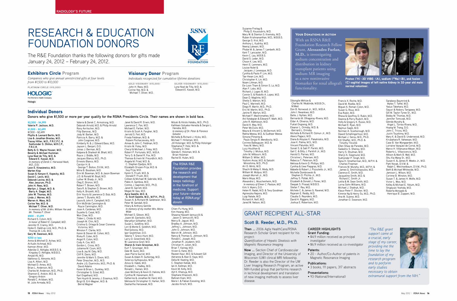
With an RSNA R&E Foundation Research Fellow Grant, Alessandro Furlan, M.D., is investigating sodium concentration and distribution in kidney transplant patients using sodium MR imaging as a new noninvasive biomarker for renal allograft functionality.
Your Donations in Action
Proton (1H) -3d vIbe- (a), sodium (23na) (b), and fusion (C) sagittal images of left native kidney in 32-year old male normal volunteer.
exhibitors Circle ProgramCompanies who give annual unrestricted gifts at four levels from $1,500 to $10,000
PlaTInuM CIRCle ($10,000)
Hologic
sIlveR vIsIonaRy ($10,000)Lyne Noel de Tilly, M.D. & Edward E. Kassel, M.D.
visionary donor ProgramIndividuals recognized for cumulative lifetime donations
gold vIsIonaRy ($15,000)John H. Rees, M.D.Corine Yee, M.D. & Michael T. Oliver, M.D.
The RsNa R&e Foundation provides the research and development that keeps radiology in the forefront of medicine. support your future—donate today at RSNA.org/donate.
Francis X. Roche, M.D.David M. Roelke, M.D.Miguel A. Roman-Colon, M.D.Susan G. Roux, M.D.Eva Rubin, M.D.Rhesa & Geoffrey D. Rubin, M.D.Deanna & Perry Rudich, M.D.Holly & Gregory K. Russo, M.D.Michael E. Ryan, M.D.Najmus Saqib, M.D.Norman A. Scarborough, M.D.Dawid Schellingerhout, M.D.Michael J. Serou, M.D., Ph.D.Kitt Shaffer, M.D., Ph.D. & Timothy Titcomb
Ellen Shaw de Paredes, M.D.Sadashiv S. Shenoy, M.D.Harry S. Shulman, M.D.Sheryl A. Siegmund, M.D.Sukhjinder P. Singh, M.D.Dana H. Smetherman, M.D., M.P.H. & Charles Freeman
Frances M. Murphy, M.D., M.P.H. & James G. Smirniotopoulos, M.D.
Clarence E. Smith, M.D.Jacqueline Smith, M.D. & Richard C. Smith Jr.
Letita & W. Sean Smith, M.D.Lorna Sohn Williams, M.D.Michael J. Stadnyk, M.D.Klaus-Peter F. Stocker, M.D.Kimmie Ng & Henry Su, M.D., Ph.D.Ana M. Suarez, M.D.Jonathan O. Swanson, M.D.
Sarabess Baumrind & Myles T. Taffel, M.D.
Yasuo Takehara, M.D.Sayuri & Noboru Tanigawa, M.D.Gill M. Taylor-Tyree Sr., M.D.Bobby M. Thomas, M.D.Jennifer Tomczak, M.D.Glenda Murphy & Angel L. Torres-Rivera, M.D.
Hoang M. Trang, D.O.John C. Tricou, M.D.Juichi Tsushima, M.D.Mary K. & David B. Underwood, M.D.Kirubahara R. Vaheesan, M.D.Case B. Van Wyngaarden, M.D.Lorraine Vazquez de Corral, M.D.Marco H. Villanueva-Meyer, M.D.Roberto L. Villavicencio, M.D.Allison & Mark B. Wall, M.D.Shu Xia Wang Sr., M.D.Susan K. & James R. Weeks Jr., M.D.Patti Novak, D.V.M. & Ralph C. Weichselbaum, D.V.M., Ph.D.
Beth & Michael Whiteside, M.D.Jamison L. Wilson, M.D.Corinne B. Winston, M.D.Susan T. & James H. Wolfe, M.D.Victor L. Woo, M.D.Kelley & Michael E. Yokum, M.D.Shigeyuki Yoshida, M.D.Roberta Morgado & Henrique B. Zuppani, M.D.
then ... 2006 Agfa HealthCare/RSNA Research Scholar grant recipient for his project:Quantification of Hepatic Steatosis with Magnetic Resonance Imaging
now ... Section Chief of Cardiovascular imaging, and Director of the university of Wisconsin (uW) clinical mRi fellowship. Dr. Reeder is also the Director of the uW liver imaging Research Program, an active NiH-funded group that performs research in technical development and translation of new imaging methods to assess liver disease.
Scott B. Reeder, M.d., Ph.d. “The R&E grant support came at a crucial, early stage of my career, providing me the time to lay the foundation of my research program and to perform early studies necessary to obtain extramural support from the NIH.”
GRANT RECIPIENT ALL-STAR
CaReeR HIgHLIgHtSgrant Funding:• $4.9 million received as principal
investigator• $6.9 million received as co-investigator
Patents:• 20 - Author/Co-Author of patents in
magnetic Resonance imaging
Publications:• 3 books, 111 papers, 317 abstracts
Presentations:• 93 (National/International)
a b C
may 2012 | RSNA News 18
news you can use
17 RSNA News | may 2012 | RSNA News 18
newS you Can uSe
17 RSNA News |
Whole-body CT is increasingly recognized as the emerging standard for providing rapid and accurate diagnoses to trauma victims with multiple, severe injuries, posing new challenges to radiologists in providing effi-cient and timely interpretations. In the May-June issue of RadioGraphics (RSNA.org/RadioGraphics) David Dreizin, M.D., and Felipe Munera, M.D., of the University of Miami Health System, discuss the use of 64-section whole-body CT angiogra-phy (CTA) in evaluating patients with blunt polytrauma. Specifically, the authors discuss:• Potential indications for whole-body CTA• The importance of the use of trauma scoring• The value of whole-body CTA in depicting important, not-to-miss inju-
ries at each anatomic level• The benefit of reviewing multiplanar reformation and 3D images for
timely and accurate interpretation• Potential pitfalls that should be avoided, as well as ongoing controver-
sies and future trends. “Utilization of 64-section whole-body CT angiography in the trauma setting is expected to continue to become more widespread in the foreseeable future, underscoring the need for radiologists to develop strategies for efficient and accurate interpretation, be mindful of the rational use of whole-body CT with consideration of radiation risk, and be knowledgeable of injuries that should be promptly communicated to surgical or interventional colleagues,” the authors write.
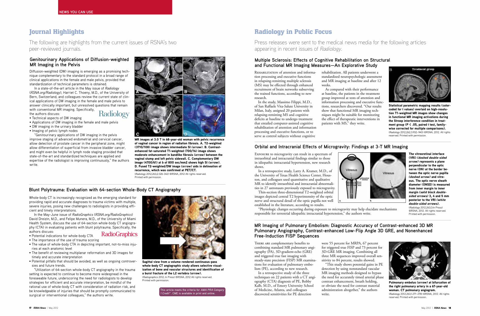
sagittal view from a volume-rendered continuous-pass whole-body CT angiographic study allows selective visual-ization of bone and vascular structures and identification of a burst fracture of the l2 vertebra (arrow).(Radiographics 2012; In Press) ©RSNA, 2012 All rights reserved. Printed with permission.
Blunt Polytrauma: evaluation with 64-section whole-Body Ct angiography
The following are highlights from the current issues of RSNA’s two peer-reviewed journals.
Journal Highlights
genitourinary applications of diffusion-weighted MR Imaging in the Pelvis
MR images at 3.0 T in 68-year-old woman with pelvic recurrence of vaginal cancer in region of radiation fibrosis. a. T2-weighted (3710/108) image shows intermediate sI (arrow). b. Contrast-enhanced fat-saturated T1-weighted (700/16) image shows moderate enhancement in bandlike fibrosis (arrow) between the vaginal stump and left pelvic sidewall. C. Complementary dW image (4700/61) at b of 800 sec/mm2 shows high sI (arrow). d. Fused T2-weighted/dW image (arrow) aids in delineation of recurrence, which was confirmed at PeT/CT.(Radiology 2012;263;2:326-342) ©RSNA, 2012 All rights reserved. Printed with permission.
Diffusion-weighted (DW) imaging is emerging as a promising tech-nique complementary to the standard protocol in a broad range of clinical applications in the female and male pelvis, provided that standardization of technical parameters is obtained. In a state-of-the-art article in the May issue of Radiology (RSNA.org/Radiology), Harriet C. Thoeny, M.D., of the University of Bern, Switzerland, and colleagues review the current state of clin-ical applications of DW imaging in the female and male pelvis to answer clinically important, but unresolved questions that remain with conventional MR imaging. Specifically, the authors discuss:• Technical aspects of DW imaging• Applications of DW imaging in the female and male pelvis• DW imaging in the urinary bladder• Imaging of pelvic lymph nodes “Genitourinary applications of DW imaging in the pelvis improve staging of advanced endometrial and cervical cancer, allow detection of prostate cancer in the peripheral zone, might allow differentiation of superficial from invasive bladder cancer, and might even be helpful in lymph node staging provided that state-of-the-art and standardized techniques are applied and expertise of the radiologist is improving continuously,” the authors write.
Press releases were sent to the medical news media for the following articles appearing in recent issues of Radiology.
Radiology in Public Focus
Multiple Sclerosis: effects of Cognitive Rehabilitation on Structural and Functional MR Imaging Measures—an explorative StudyRehabilitation of attention and informa-tion processing and executive functions in relapsing-remitting multiple sclerosis (MS) may be effected through enhanced recruitment of brain networks subserving the trained functions, according to new research. In the study, Massimo Filippi, M.D., of San Raffaele Vita-Salute University in Milan, Italy, assigned 20 patients with relapsing-remitting MS and cognitive deficits at baseline to undergo treatment that entailed computer-assisted cognitive rehabilitation of attention and information processing and executive functions, or to serve as control subjects without cognitive
Exposure to microgravity can result in a spectrum of intraorbital and intracranial findings similar to those in idiopathic intracranial hypertension, new research shows. In a retrospective study, Larry A. Kramer, M.D., of the University of Texas Health Science Center, Hous-ton, and colleagues used quantitative and qualitative MR to identify intraorbital and intracranial abnormali-ties in 27 astronauts previously exposed to microgravity. Thin-section three-dimensional T2-weighted orbital images depicted central T2 hyperintensity of the optic nerve and structural detail of the optic papilla not well established in the literature, according to results. “Physiologic changes occurring during exposure to microgravity may help elucidate mechanisms responsible for terrestrial idiopathic intracranial hypertension,” the authors write.
orbital and Intracranial effects of Microgravity: Findings at 3-t MR Imaging
this article meets the criteria for AMA PRA Category 1 Credit™. CMe is available in print and online.
statistical parametric mapping results (color-coded for t values) overlaid on high-resolu-tion T1-weighted MR images show changes in functional MR imaging activations during the stroop interference condition in treat-ment group (P < .05, paired t test, family-wise corrected for multiple comparisons). (Radiology 2012;262;3:932–940) ©RSNA, 2012. All rights reserved. Printed with permission.
rehabilitation. All patients underwent a standardized neuropsychologic assessmentand MR imaging at baseline and after 12 weeks. As compared with their performance at baseline, the patients in the treatment group improved at tests of attention and information processing and executive func-tions, researchers discovered. “Our results show that functional MR imaging tech-niques might be suitable for monitoring the effect of therapeutic interventions in patients with MS,” they write.
MR Imaging of Pulmonary embolism: diagnostic accuracy of Contrast-enhanced 3d MR Pulmonary angiography, Contrast-enhanced Low-Flip angle 3d gRe, and nonenhanced Free-Induction FISP SequencesThere are complementary benefits to combining standard MR pulmonary angi-ography (PA), 3D-gradient-echo (GRE) and triggered true fast imaging with steady-state precision (FISP) MR examina-tions for evaluation of pulmonary embo-lism (PE), according to new research. In a retrospective study of the three techniques on 22 patients with a CT angi-ography (CTA) diagnosis of PE, Bobby Kalb, M.D., of Emory University School of Medicine, Atlanta, and colleagues discovered sensitivities for PE detection
were 55 percent for MRPA, 67 percent for triggered true FISP and 73 percent for 3D-GRE MR imaging. Combining all three MR sequences improved overall sen-sitivity to 84 percent, results showed. “This study shows potential gains in PE detection by using nonstandard vascular MR imaging methods designed to bypass the need for accurately timed arterial phase contrast enhancement, breath holding, or obviate the need for contrast material administration altogether,” the authors write.
The vitreoretinal interface (vRI) (dashed double-sided arrow) represents a plane perpendicular to the optic nerve (on) at the border be-tween the optic nerve papilla (dashed arrow) and vitre-ous. The optic nerve sheath diameter (onsd) is measured from inner margin to inner margin (solid black double-sided arrows) 3, 4 and 5 mm posterior to the vRI (white double-sided arrows).(Radiology 2012;263;3:In Press) ©RSNA, 2012. All rights reserved. Printed with permission.
Pulmonary embolus (arrow) at bifurcation of the right pulmonary artery in a 67-year-old woman. CT pulmonary angiogram.(Radiology 2012;263;1:271–278) ©RSNA, 2012. All rights reserved. Printed with permission.
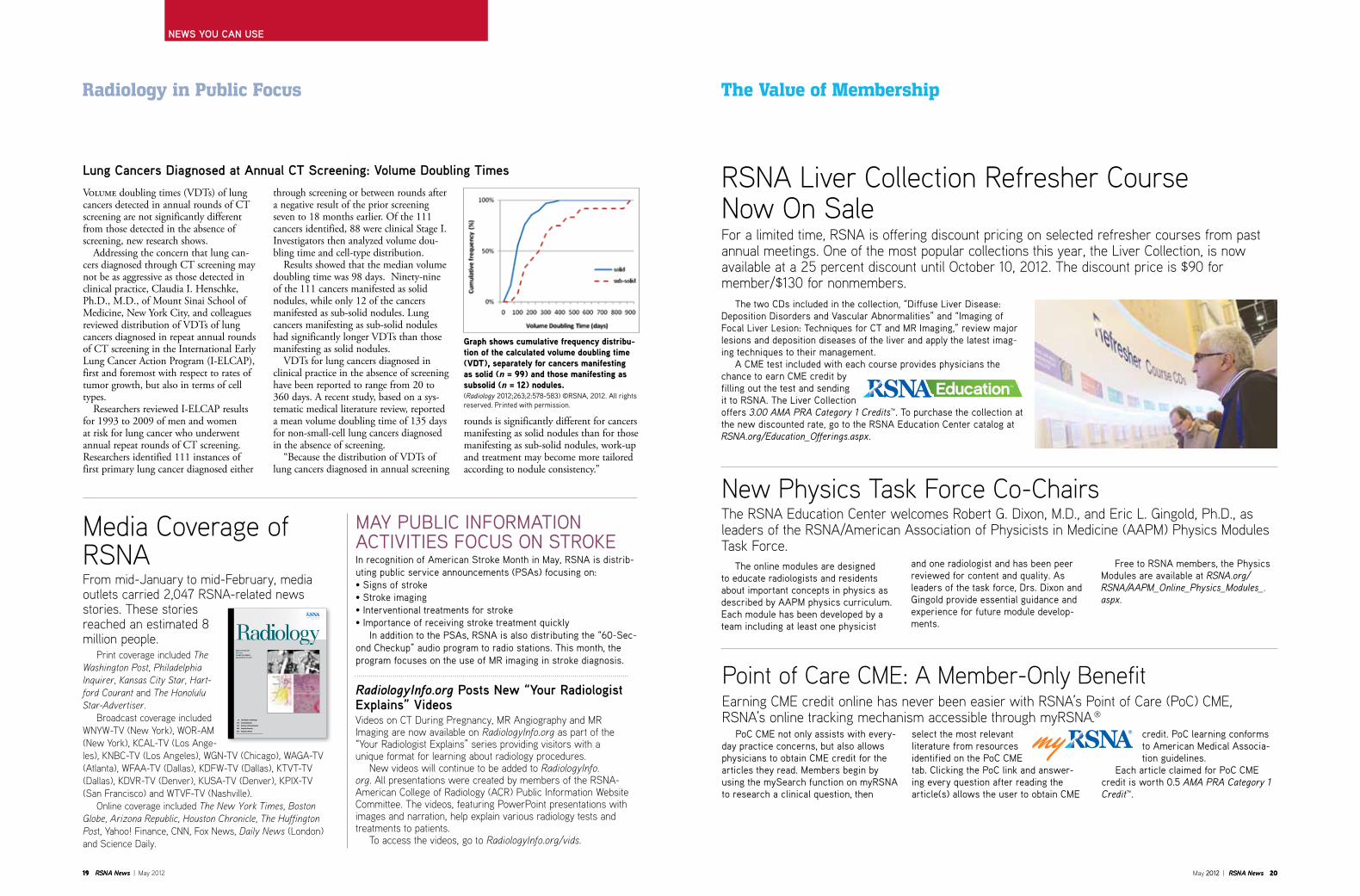
news you can use
19 RSNA News | may 2012 may 2012 | RSNA News 20 2012 | RSNA News 20
newS you Can uSe
19 RSNA News |
The Value of Membership
For a limited time, RSNA is offering discount pricing on selected refresher courses from past annual meetings. one of the most popular collections this year, the liver Collection, is now available at a 25 percent discount until October 10, 2012. The discount price is $90 for member/$130 for nonmembers.
RSNA liver Collection Refresher Course Now on Sale
The two CDs included in the collection, “Diffuse Liver Disease: Deposition Disorders and Vascular Abnormalities” and “Imaging of Focal Liver Lesion: Techniques for CT and MR Imaging,” review major lesions and deposition diseases of the liver and apply the latest imag-ing techniques to their management. A CME test included with each course provides physicians the chance to earn CME credit by filling out the test and sending it to RSNA. The Liver Collection offers 3.00 AMA PRA Category 1 Credits™. To purchase the collection at the new discounted rate, go to the RSNA Education Center catalog at RSNA.org/Education_Offerings.aspx.
Radiology in Public Focus
media Coverage of RSNAFrom mid-January to mid-February, media outlets carried 2,047 RSNA-related news stories. these stories reached an estimated 8 million people. Print coverage included The Washington Post, Philadelphia Inquirer, Kansas City Star, Hart-ford Courant and The Honolulu Star-Advertiser. broadcast coverage included WNYW-tV (New York), WoR-Am (New York), KCAl-tV (los Ange-les), KNbC-tV (los Angeles), WgN-tV (Chicago), WAgA-tV (Atlanta), WFAA-tV (Dallas), KDFW-tV (Dallas), KtVt-tV (Dallas), KDVR-tV (Denver), KuSA-tV (Denver), KPiX-tV (San Francisco) and WtVF-tV (Nashville). online coverage included The New York Times, Boston Globe, Arizona Republic, Houston Chronicle, The Huffington Post, Yahoo! Finance, CNN, Fox News, Daily News (london) and Science Daily.
RADIOLOGY.RSNA.ORG
MAY 2012 VOLUME 263 NUMBER 2
RADLAX 263 (2) 313–622
3A This Month in Radiology
316 Communications
318 Reviews and Commentary
364 Original Research
610 Diagnosis Please
MAY PUBLIC INFORMATION ACTIVITIES FOCUS ON STROKEIn recognition of American Stroke Month in May, RSNA is distrib-uting public service announcements (PSAs) focusing on:• Signs of stroke• Stroke imaging• Interventional treatments for stroke• Importance of receiving stroke treatment quickly In addition to the PSAs, RSNA is also distributing the “60-Sec-ond Checkup” audio program to radio stations. This month, the program focuses on the use of MR imaging in stroke diagnosis.
Lung Cancers diagnosed at annual Ct Screening: Volume doubling times
Volume doubling times (VDTs) of lung cancers detected in annual rounds of CT screening are not significantly different from those detected in the absence of screening, new research shows. Addressing the concern that lung can-cers diagnosed through CT screening may not be as aggressive as those detected in clinical practice, Claudia I. Henschke, Ph.D., M.D., of Mount Sinai School of Medicine, New York City, and colleagues reviewed distribution of VDTs of lung cancers diagnosed in repeat annual rounds of CT screening in the International Early Lung Cancer Action Program (I-ELCAP), first and foremost with respect to rates of tumor growth, but also in terms of cell types. Researchers reviewed I-ELCAP results for 1993 to 2009 of men and women at risk for lung cancer who underwent annual repeat rounds of CT screening. Researchers identified 111 instances of first primary lung cancer diagnosed either
through screening or between rounds after a negative result of the prior screening seven to 18 months earlier. Of the 111 cancers identified, 88 were clinical Stage I. Investigators then analyzed volume dou-bling time and cell-type distribution. Results showed that the median volume doubling time was 98 days. Ninety-nine of the 111 cancers manifested as solid nodules, while only 12 of the cancers manifested as sub-solid nodules. Lung cancers manifesting as sub -solid nodules had significantly longer VDTs than those manifest ing as solid nodules. VDTs for lung cancers diagnosed in clinical practice in the absence of screening have been reported to range from 20 to 360 days. A recent study, based on a sys-tematic medical literature review, reported a mean volume doubling time of 135 days for non-small-cell lung cancers diagnosed in the absence of screening. “Because the distribution of VDTs of lung cancers diagnosed in annual screening
graph shows cumulative frequency distribu-tion of the calculated volume doubling time (vdT), separately for cancers manifesting as solid (n = 99) and those manifesting as subsolid (n = 12) nodules.(Radiology 2012;263;2:578-583) ©RSNA, 2012. All rights reserved. Printed with permission.
the RSNA education Center welcomes Robert g. Dixon, m.D., and eric l. gingold, Ph.D., as leaders of the RSNA/American Association of Physicists in Medicine (AAPM) Physics Modules task Force.
New Physics task Force Co-Chairs
The online modules are designed to educate radiologists and residents about important concepts in physics as described by AAPM physics curriculum. Each module has been developed by a team including at least one physicist
and one radiologist and has been peer reviewed for content and quality. As leaders of the task force, Drs. Dixon and Gingold provide essential guidance and experience for future module develop-ments.
Free to RSNA members, the Physics Modules are available at RSNA.org/RSNA/AAPM_Online_Physics_Modules_.aspx.
earning Cme credit online has never been easier with RSNA’s Point of Care (PoC) Cme, RSNA’s online tracking mechanism accessible through myRSNA.®
Point of Care Cme: A member-only benefit
PoC CME not only assists with every-day practice concerns, but also allows physicians to obtain CME credit for the articles they read. Members begin by using the mySearch function on myRSNA to research a clinical question, then
select the most relevant literature from resources identified on the PoC CME tab. Clicking the PoC link and answer-ing every question after reading the article(s) allows the user to obtain CME
credit. PoC learning conforms to American Medical Associa-tion guidelines.
Each article claimed for PoC CME credit is worth 0.5 AMA PRA Category 1 Credit™.
rounds is signif icantly different for cancers man ifesting as solid nodules than for those manifesting as sub-solid nodules, work-up and treatment may become more tailored according to nodule consistency.”
RadiologyInfo.org Posts new “your Radiologist explains” VideosVideos on Ct During Pregnancy, mR Angiography and mR imaging are now available on RadiologyInfo.org as part of the “Your Radiologist explains” series providing visitors with a unique format for learning about radiology procedures. New videos will continue to be added to RadiologyInfo.org. All presentations were created by members of the RSNA-American College of Radiology (ACR) Public information Website Committee. the videos, featuring PowerPoint presentations with images and narration, help explain various radiology tests and treatments to patients. to access the videos, go to RadiologyInfo.org/vids.
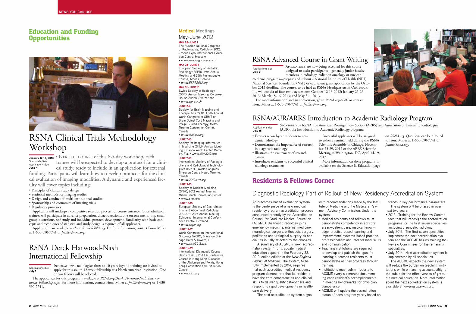
may 2012 | RSNA News 22 | RSNA News 22
newS you Can uSe
21 RSNA News | may 2012
Education and Funding Opportunities
Medical Meetingsmay-June 2012May 28-June 1The Russian National Congress of Radiologists, Radiology 2012, Crocus Expo International Exhibi-tion Centre, Moscow • www.radiology-congress.ruMay 28- June 1European Society of Pediatric Radiology (ESPR), 49th Annual Meeting and 35th Postgraduate Course, Athens, Greece • www.ESPR2012.orgMay 31- June 2Swiss Society of Radiology (SSR), Annual Meeting, Congress House Zurich, Switzerland • www.sgr-ssr.chJune 2-4Society for Brain Mapping and Therapeutics (SBMT), 9th Annual World Congress of SBMT on Brain Spinal Cord Mapping and Image Guided Therapy, Metro Toronto Convention Center, Canada • www.ibmisps.orgJune 7-10Society for Imaging Informatics in Medicine (SIIM), Annual Meet-ing, Orlando World Center Marri-ott, Fla. • www.siim2012.orgJune 7-10International Society of Radiogra-phers and Radiological Technolo-gists (ISRRT), World Congress, Sheraton Centre Hotel, Toronto, Canada • www.2012isrrt.orgJune 9-13Society of Nuclear Medicine (SNM), 2012 Annual Meeting, Miami Beach Convention Center • www.snm.orgJune 12-15European Society of Gastrointes-tinal and Abdominal Radiology (ESGAR), 23rd Annual Meeting, Edinburgh International Confer-ence Centre, Scotland • www.esgar.orgJune 14-17World Congress on Interventional Oncology (WCIO), Sheraton Chi-cago Hotel & Towers, Ill. • www.wcio2012.org.June 16-19International Diagnostic Course Davos (IDKD), 2nd IDKD Intensive Course in Hong Kong, Diseases of the Abdomen and Pelvis, Hong Kong Convention and Exhibition Centre • www.idkd.org
RSNA Clinical Trials Methodology Workshop
Over the course of this 6½-day workshop, each trainee will be expected to develop a protocol for a clini-cal study, ready to include in an application for external
funding. Participants will learn how to develop protocols for the clini-cal evaluation of imaging modalities. A dynamic and experienced fac-ulty will cover topics including: • Principles of clinical study design• Statistical methods for imaging studies• Design and conduct of multi-institutional studies• Sponsorship and economics of imaging trials• Regulatory processes Applicants will undergo a competitive selection process for course entrance. Once admitted, trainees will participate in advance preparation, didactic sessions, one-on-one mentoring, small group discussions, self-study and individual protocol development. Familiarity with basic con-cepts and techniques of statistics and study design is required of all applicants. Applications are available at clinicaltrials.RSNA.org. For for information, contact Fiona Miller at 1-630-590-7741 or [email protected].
January 12-18, 2013 Scottsdale/Ariz. Applications due June 4
International radiologists three to 10 years beyond training are invited to apply for this six- to 12-week fellowship at a North American institution. One or two fellows will be selected.
The application for this program is available at RSNA.org/Derek_Harwood-Nash_Interna-tional_Fellowship.aspx. For more information, contact Fiona Miller at [email protected] or 1-630-590-7741.
RSNA Derek Harwood-Nash International FellowshipApplications due July 1
RSNA/AUR/ARRS Introduction to Academic Radiology ProgramSponsored by RSNA, the American Roentgen Ray Society (ARRS) and Association of University Radiologists (AUR), the Introduction to Academic Radiology program:Applications due
July 15
RSNA Advanced Course in Grant WritingApplications are now being accepted for this course designed to assist participants—generally junior faculty members in radiology, radiation oncology or nuclear
medicine programs—prepare and submit a National Institutes of Health (NIH), National Sciences Foundation (NSF) or equivalent grant application by the Octo-ber 2013 deadline. The course, to be held at RSNA Headquarters in Oak Brook, Ill., will consist of four two-day sessions: October 12-13 2012; January 25-26, 2013; March 15-16, 2013; and May 3-4, 2013. For more information and an application, go to RSNA.org/AGW or contact Fiona Miller at 1-630-590-7741 or [email protected].
Applications due July 31
• Exposes second-year residents to aca-demic radiology
• Demonstrates the importance of research in diagnostic radiology
• Illustrates the excitement of research careers
• Introduces residents to successful clinical radiology researchers
Successful applicants will be assigned to either a seminar held during the RSNA Scientific Assembly in Chicago, Novem-ber 25-29, 2012 or the ARRS Scientific Meeting in Washington, DC, April 14-19, 2013. More information on these programs is available on the Science & Education page
on RSNA.org. Questions can be directed to Fiona Miller at 1-630-590-7741 or [email protected].
Residents & Fellows Corner
An outcomes-based evaluation system is the centerpiece of a new medical residency program accreditation process announced recently by the Accreditation Council for Graduate Medical Education (ACGME). Diagnostic radiology joins emergency medicine, internal medicine, neurological surgery, orthopedic surgery, pediatrics and urological surgery as spe-cialties initially affected by the changes. A summary of ACGME’s “next accred-itation system” for graduate medical education appears in the February 22, 2012, online edition of the New England Journal of Medicine. The system, to be fully implemented by 2014, requires that each accredited medical residency program demonstrate that its residents have the core competencies and clinical skills to deliver quality patient care and respond to rapid developments in health-care delivery. The next accreditation system aligns
with recommendations made by the Insti-tute of Medicine and the Medicare Pay-ment Advisory Commission. Under the system: • Medical residents and fellows must
demonstrate competency in six core areas—patient care, medical knowl-edge, practice-based learning and improvement, systems-based practice, professionalism and interpersonal skills and communication.
• Teaching institutions are required to develop and publish the specific learning outcomes residents must demonstrate as they progress through training.
• Institutions must submit reports to ACGME every six months document-ing each resident’s accomplishments in meeting benchmarks for physician competence.
• ACGME will update the accreditation status of each program yearly based on
trends in key performance parameters. The system will be phased in over next two years:• 2012—Training for the Review Commit-
tees that will redesign the accreditation programs for the first seven specialties including diagnostic radiology.
• July 2013—The first seven specialties implement the next accreditation sys-tem and the ACGME begins training the Review Committees for the remaining specialties.
• July 2014—Next accreditation system is implemented by all specialties.
The ACGME expects the new system will reduce the burden on teaching insti-tutions while enhancing accountability to the public for the effectiveness of gradu-ate medical education. More information about the next accreditation system is available at www.acgme-nas.org.
Diagnostic Radiology Part of Rollout of New Residency Accreditation System
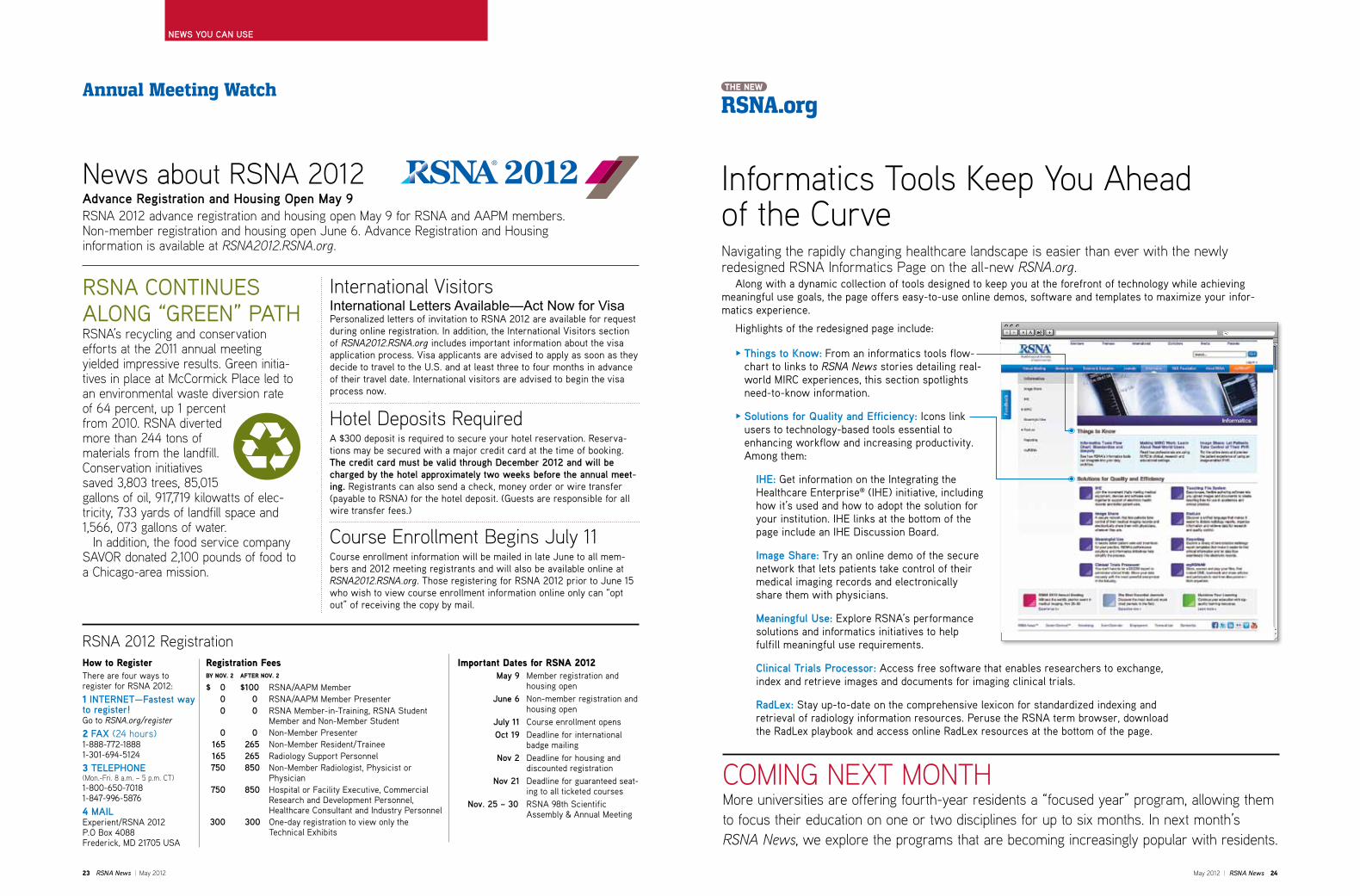
may 2012 | RSNA News 24
news you can use
23 RSNA News | may 2012
informatics tools Keep You Ahead of the CurveNavigating the rapidly changing healthcare landscape is easier than ever with the newly redesigned RSNA informatics Page on the all-new RSNA.org. Along with a dynamic collection of tools designed to keep you at the forefront of technology while achieving meaningful use goals, the page offers easy-to-use online demos, software and templates to maximize your infor-matics experience.
Highlights of the redesigned page include:
ComiNg NeXt moNtHmore universities are offering fourth-year residents a “focused year” program, allowing them to focus their education on one or two disciplines for up to six months. in next month’s RSNA News, we explore the programs that are becoming increasingly popular with residents.
RSNA.orgTHe neW Annual Meeting Watch
Registration Feesby nov. 2 aFTeR nov. 2
$ 0 $ 100 RSNA/AAPM Member 0 0 RSNA/AAPM Member Presenter 0 0 RSNA Member-in-Training, RSNA Student
Member and Non-Member Student 0 0 Non-Member Presenter 165 265 Non-Member Resident/Trainee 165 265 Radiology Support Personnel 750 850 Non-Member Radiologist, Physicist or
Physician 750 850 Hospital or Facility Executive, Commercial
Research and Development Personnel, Healthcare Consultant and Industry Personnel
300 300 One-day registration to view only the Technical Exhibits
Important dates for Rsna 2012 May 9 Member registration and
housing open June 6 Non-member registration and
housing open July 11 Course enrollment opens oct 19 Deadline for international
badge mailing nov 2 Deadline for housing and
discounted registration nov 21 Deadline for guaranteed seat-
ing to all ticketed courses nov. 25 – 30 RSNA 98th Scientific
Assembly & Annual Meeting
RSNA 2012 RegistrationHow to RegisterThere are four ways to register for RSNA 2012:
1 InteRnet—Fastest way to register!Go to RSNA.org/register2 Fax (24 hours)1-888-772-18881-301-694-5124
3 teLePHone (mon.-Fri. 8 a.m. – 5 p.m. Ct)1-800-650-70181-847-996-5876
4 MaILExperient/RSNA 2012P.O Box 4088Frederick, MD 21705 USA
News about RSNA 2012advance Registration and Housing open May 9RSNA 2012 advance registration and housing open may 9 for RSNA and AAPm members. Non-member registration and housing open June 6. Advance Registration and Housing information is available at RSNA2012.RSNA.org.
O things to Know: From an informatics tools flow-chart to links to RSNA News stories detailing real-world MIRC experiences, this section spotlights need-to-know information.
O Solutions for Quality and efficiency: Icons link users to technology-based tools essential to enhancing workflow and increasing productivity. Among them:
IHe: Get information on the Integrating the Healthcare Enterprise® (IHE) initiative, including how it’s used and how to adopt the solution for your institution. IHE links at the bottom of the page include an IHE Discussion Board.
Image Share: Try an online demo of the secure network that lets patients take control of their medical imaging records and electronically share them with physicians.
Meaningful use: Explore RSNA’s performance solutions and informatics initiatives to help fulfill meaningful use requirements.
Clinical trials Processor: Access free software that enables researchers to exchange, index and retrieve images and documents for imaging clinical trials.
RadLex: Stay up-to-date on the comprehensive lexicon for standardized indexing and retrieval of radiology information resources. Peruse the RSNA term browser, download the RadLex playbook and access online RadLex resources at the bottom of the page.
Hotel Deposits RequiredA $300 deposit is required to secure your hotel reservation. Reserva-tions may be secured with a major credit card at the time of booking. the credit card must be valid through december 2012 and will be charged by the hotel approximately two weeks before the annual meet-ing. Registrants can also send a check, money order or wire transfer (payable to RSNA) for the hotel deposit. (Guests are responsible for all wire transfer fees.)
Course enrollment begins July 11Course enrollment information will be mailed in late June to all mem-bers and 2012 meeting registrants and will also be available online at RSNA2012.RSNA.org. Those registering for RSNA 2012 prior to June 15 who wish to view course enrollment information online only can “opt out” of receiving the copy by mail.
international VisitorsInternational Letters Available—Act Now for VisaPersonalized letters of invitation to RSNA 2012 are available for request during online registration. In addition, the International Visitors section of RSNA2012.RSNA.org includes important information about the visa application process. Visa applicants are advised to apply as soon as they decide to travel to the U.S. and at least three to four months in advance of their travel date. International visitors are advised to begin the visa process now.
RSNA CONTINUES ALONG “GREEN” PATHRSNA’s recycling and conservation efforts at the 2011 annual meeting yielded impressive results. green initia-tives in place at mcCormick Place led to an environmental waste diversion rate of 64 percent, up 1 percent from 2010. RSNA diverted more than 244 tons of materials from the landfill. Conservation initiatives saved 3,803 trees, 85,015 gallons of oil, 917,719 kilowatts of elec-tricity, 733 yards of landfill space and 1,566, 073 gallons of water. in addition, the food service company SAVoR donated 2,100 pounds of food to a Chicago-area mission.
Feed
back
RSNA News »
RSNA News™ AdvertisingCareer Connect™ Event Calendar Employment Terms of Use Contact Us
Copyright © 2011 Radiological Society of North America | TEL 1-630-571-2670 | TOLL FREE U.S. and Canada: 1-800-381-6660 | Membership 1-877-RSNA-MEM (776-2636)
About RSNAAnnual Meeting Membership Science & Education InformaticsJournals R&E Foundation
Search...
Welcome John Smith, MD Log out »
Go »
Trainees International Exhibitors Patients MediaMembers
News
RSNA Workshop Offers Fertile Ground for Clinical TrialsMarch 1, 2012
As a starting junior faculty member, James R. Fink, M.D., wanted to pursue studies in advanced MR imaging in neuro-oncology but lacked the training in evidence-based and hypothesis...
Personalized Image-guided Therapy is Next Frontier in Cancer TreatmentMarch 1, 2012
Along with making significant headway in the fight against cancer, image-guided drug delivery (IGDD) is expanding the concept of personalized medicine, according to an RSNA 2010 presenter...
Self-selected Radiology Mentors Yield Greater SatisfactionMarch 1, 2012
While formal mentoring programs are considerably beneficial to radiology residents overall, those allowed to self-select mentors are more likely to be satisfied with the relationship,...
RSNA 2012 Online Abstract Submission Now OpenMarch 1, 2012
The online system to submit abstracts for RSNA 2012 is now active. The submission deadline is 12 p.m. Central Time on March 31, 2012. Abstracts....
Access the RSNA News tablet edition on the App Store and Android MarketMarch 1, 2012
Users of tablet PCs can now download the RSNA News app by searching “RSNA News” on the Android Market or App Store, or going directly…
New Self-Assessment Modules (SAMs) Available from RSNAMarch 1, 2012
Each month, new online Self-Assessment Modules (SAMs) are added to RSNA.org under the Education tab. These online SAMs help physicians...
Announcements
It’s Better When You BelongJoin a global community of leaders in radiologic education and research. Join now »
Maximize Your LearningContinue your education with top-quality learning resources. Learn more »
myRSNA—Store, Access and SharePersonalize your workspace—access files, articles and CME from anywhere. Log in »
RSNA 2012—Patients FirstWitness the world’s premier event in radiologic science and technology.Experience it »
RSNA Buyers GuideShop radiology-related equipment, services, devices and software. Browse now »
RSNA Journals »
Event Calendar »
Journal Features
Perfusion CT Demonstrates Therapy-related ChangesMarch 1, 2012
Perfusion CT may depict therapy-induced changes in patients with unresectable lung ade-nocarcinoma and can identify response to treatment on the whole tumor mass....
Lung Abnormalities at Multimodality Imaging after Radiation Therapy March 1, 2012
Three-dimensional (3D) conformal radiation therapy and stereotactic body radiation therapy are designed to deliver the maximum therapeutic radiation dose to the tumor allowing improved ...
Histologic, Molecular, Cytogenetic Features of Ovarian CancersMarch 1, 2012
Ovarian epithelial carcinoma (OEC), the most common ovarian malignancy, is a heterogeneous disease with several histologic subtypes that show characteristic cytogenetic features,...
The Most Essential JournalsDiscover the most read and most cited peer-reviewed journals in the field. Subscribe now »
myRSNA®
Feed
back
RSNA News »
RSNA News™ AdvertisingCareer Co ect Event Calendar Employment Terms of Usf e Contact Us
Copyright © 2011 Radiological Society of North America | TEL 1-630-571-2670 | TOLL FREE U.S. and Canada: 1-800-381-6660 | Membership 1-877-RSNA-MEM (776-2636)
About RSNAAnnual Meeting Membership Science & Education InformaticsJournals R&E Foundation
Search...
Welcome John Smith, MD Log out »
Go »
Trainees International Exhibitors Patients MediaMembers
News
RSNA Workshop Offers Fertile Groundfor Clinical TrialsMarch 1, 2012
As a starting junior faculty member, James R. Fr ink, M.D., wanted to pursue studies in advanced MR imaging in neuro-oncology but lacked the training in evidence-based and hypothesis...
Personalized Image-guided Therapy isNext Frontier in Cancer TreatmentMarch 1, 2012
Along with making significant headway in the fight against cancer, imagerr -guided drug delivery(IGDD) is expanding the concept of personalized medicine, according to an RSNA 2010 presenter...
Self-selected Radiology Mentors YieldGreater SatisfactionMarch 1, 2012
While formal mentoring programs areconsiderably beneficial to radiology residentsoverall, those allowed to self-select mentors aremore likely to be satisfied with the relationship,...
RSNA 2012 Online AbstractSubmission Now OpenMarch 1, 2012
The online system to submit abstracts for RSNA ff2012 is now active. The submission deadline is 12p.m. Central Time on March 31, 2012. Abstracts....
Access the RSNA News tablet edition onthe App Store and Android MarketkkMarch 1, 2012
Users of tablet PCs can now download the RSNANews app by searching “RSNA News” on the Android Market or App Store, or going directly…
New Self-Assessment Modules (SAMs)Available from RSNAMarch 1, 2012
Each month, new online Self-Assessment Modules (SAMs) are added to RSNA.org under the Education tab. These online SAMs help physicians...
Announcements
It’s Better When You BelongYYJoin a global community of leaders inradiologic education and research. Join now »
Maximize Your LearningYYContinue your education with top-quality learning resources.Learn more »
myRSNA—Store, Access and SharePersonalize your workspace—access files, articles and CME from anywhere. Log in »
RSNA 2012—Patients FirstWitness the world’s premier event ’in radiologic science and technology.Experience it »
RSNA Buyers GuideShop radiology-related equipment,services, devices and softwff are.Browse now »
RSNA Journals »
Event Calendar »
Journal Features
Perfusion CT DemonstratesTherapy-related ChangesMarch 1, 2012
Perfusion CT may depicT t therapy-induced changes in patients with unresectable lung ade-nocarcinoma and can identify response totreatment on the whole tumor mass....
Lung Abnormalities at MultimodalityImaging after Radiation Therapy March 1, 2012
Three-dimensional (3D) conformal radiationfftherapy and stereotactic body radiation therapy are designed to deliver the maximum therapeuticradiation dose to the tumor allowing improved ...
Histologic, Molecular,rr Cytogenetic Features of Ovarian Cancercc sMarch 1, 2012
Ovarian epithelial carcinoma (OEC), the most common ovarian malignancy, is a heterogeneousyydisease with several histologic subtypes thatshow characteristic cytogenetic features,...
The Most Essential JournalsDiscover the most read and most cited peer-reviewed journals in the field. Subscribe now »
myRSNA®
YOU SPOKE.WE LISTENED.The all new RSNA.orgdesigned for you.
• Easier to navigate
• Fresh and late-breaking content
• Resources tailored to yourprofessional needs
WEB485-R02/12
Web Launch Journals Ad.indd 1 2/7/12 9:44 AM