Embed Size (px)
Citation preview
Free Radical Scavenging Activity of Aerial Parts of Artemisia pallens in Mediating Hepatoprotective Activity of RIF+INH Intoxicated Rats Praveen Kumar Ashok1,a*, Poonam Rishishwar2, Kumud Upadhyaya3
1Faculty of Pharmacy, GRD (PG) IMT, Dehradun.248009 2Faculty of Pharmaceutical Sciences Shri Venkateshwara University Gajraula, Uttar Pradesh
3Faculty of Pharmacy, Kumaun University, Bhimtal, Nanital.243136 [email protected]*, Tel.: +918279392039
Keywords: Artemisia pallens, free radical, hepatoprotective, SGOT, SGPT.
Abstract. To investigate the free radical scavenging & hepatoprotective activity of phenolic rich fraction of Artemisia pallens on RIF+INH induced oxidative stress in Sprague dawley rats. Free radical scavenging and hepatoprotective activity was evaluated by using DPPH, Nitric oxide, Superoxide radical and Hydroxyl radical assay models and induced RIF+INH intoxicated rats. The total phenolic content was found to be 312.60 µg gallic acid equivalents (GAE)/gm of dry extract. The total flavonoid content was found to be 322.20 µg rutin equivalents (RUE)/gm of dry extract. In the current study, free radical scavenging activity was evaluated by using 1,1-diphenyl-2-picrylhydrazyl (DPPH), nitric oxide, superoxide radical and hydroxyl radical scavenging activity were found to be (42.25 ± 0.95) and (09.16 ± 1.62) µg/ml, (101.62 ± 1.64) and (32.41 ± 1.24) µg/ml, (72.62 ± 1.86) and (10.28 ± 1.96) µg/ml, (33.82 ± 1.12) and (12.82 ± 1.86) µg/ml, respectively. There was also a dose dependent increase in reductive ability of Artemisia pallens extract with increase in concentration and were further investigated in invivo hepatoprotective activity experiment against toxicity induced by RIF+INH. The free radical scavenging and hepatoprotective activity may be attributed to the presence of phenolic compounds and histology of the liver section of the animals treated with the extracts showed the presence of normal hepatic cords, absence of necrosis and fatty infiltration, which further evidenced the hepatoprotective activity of Artemisia pallens.
Introduction Liver cells possess variety of compensative mechanisms to deal with reactive oxygen species
(ROS) and their effect; among these are the induction of variety of antioxidant proteins comparable to superoxide dismutase (SOD), catalose, gluthathione peroxidase (GSHPx) and therefore the tripeptide glutathione (GSH). Therefore, oxidative stress, caused primarily by ROS, is additionally related to hepatic diseases[1]. Tuberculosis (TB) may be a unsafe disease, that gradually swallows the life human being. The world prevalence of T.B. was thirty second (1.86 billion people) and therefore the international case mortality was twenty third[2].
Dietary antioxidant intake is also a very important strategy for inhibiting or delaying the oxidisation of susceptible cellular substrates, and is therefore relevant to disease prevention in several paradigms. phenolic compounds like flavonoids, phenolic acids, diterpenes and tannins have received attention for their high antioxidative activity[3]. Oxidative damage caused by free polygenic disorder, cancer and liver disease[4]. Antioxidant compounds reduce the action of reactive oxygen species (ROS) in damaged tissues throughout the recovery method[5].
In view of severe undesirable side result of synthetic agents, there's growing focus to follow systematic research methodology and to evaluate scientific basis for the normal herbal medicines that are claimed to possess hepatoprotective activity. The utilization of medicinal plants in modern drugs suffers from the very fact that although many plants are utilized in the world to prevent or to cure diseases, scientific evidence in terms of recent medication is lacking in most cases. But nowadays it's necessary to provide scientific proof as whether or not to justify the utilization of plant or its active principles[6].
International Journal of Pharmacology, Phytochemistry and Ethnomedicine Submitted: 2019-01-07ISSN: 2297-6922, Vol. 14, pp 1-15 Revised: 2019-08-26doi:10.18052/www.scipress.com/IJPPE.14.1 Accepted: 2019-11-19CC BY 4.0. Published by SciPress Ltd, Switzerland, 2019 Online: 2019-11-28
This paper is an open access paper published under the terms and conditions of the Creative Commons Attribution license (CC BY)(https://creativecommons.org/licenses/by/4.0)
Artemisia pallens (Compositea), Indian name-Davana (Hindi) may be a medicative plant found in south india as Maharashtra, Andhra Pradesh, Karnataka and Tamilnadu. It’s the foremost vital aromatic plants utilized in the perfumery and cosmetic industries and india is major exporters of Artemisia pallens oil to the rest of the world. Several pharmacological activities of Artemisia pallens are reported: perfumenes and as an antifungal and antibacterial drug agent, analgesic and anti inflammatory activity[7]. Davana is wide utilized in iraqi and Indian folk drugs for the treatment of diabetes mellitus[8].
In the present investigation, an effort has been made to assess the antihepatotoxic effects of Davana on isoniazid and rifampicin induced hepatotoxicity was studied with relevance changes within the levels of diagnostic marker enzymes.
Material and Methods Chemical and Drugs
Isoniazid and rifampicin were obtained by M/s Lupin Pharmaceutical Ltd., Mumbai. 1,1-diphenyl-2-picryl-hydrazyl (DPPH) and Bovine serum albumin were obtained from M/s Sigma chemicals company, St. Louis, MO, USA. Reduced nicotinamide adenine dinucleotide (NADH), phenazine methosulphate (PMS), Nitroblue tetrazolium (NBT), Sodium nitroprusside (SNP), ascorbic acid, and thiobarbituric acid (TBA) was obtained from Spectrochem Pvt. Ltd., Mumbai. Folin-Ciocalteu’s phenol reagent (FCR) was purchased from Sisco Research Laboratories Pvt. Ltd., Mumbai. All other chemicals and solvents were used in high analytical grade. Plant Material and Preparation of Extract
Aerial parts of Artemisia pallens were collected from the South india (Salem, Tamilnadu) within the month of September 2011 throughout that the plant grows wide below natural condition. Voucher specimen 222849 is preserved in N.B.R.I. Lucknow india once ethanobotanical identification of species. Aerial parts were shade dried at room temperature for 07 days then powdered in a very mechanical grinder. The material powdered (200gm) was successively extracted with pet. ether (60-800c) followed by methyl alcohol exploitation soxhlet extraction equipment. Pet. Ether and methyl alcohol content were evaporated using Buchi Rotavapor (Buchi Labortahnik, Switzerland) at 40c. Finally, solution was freeze-dried in a Hetolyophilizer (HITOSICC, Denmark) and also the dried extract was hold on during a vaccum desecrator. The yied of the pet. ether and wood spirit extract was about 17.2% and 51.1%, respectively. Preliminary phytochemical study of the methyl alcohol extract of Artemisia pallens indicated the presence of steroid, sesquitriterpenoid, flavonoids, polyphenolic, saponin and phenol. Preparation of phenolic fraction
5 gms of obtained crude extract was dissolved in 100 ml water and sequentially extracted thrice-using 200 ml hexane and ethyl acetate. Then solvent in the each fractions were removed using rotary evaporator to obtain ethyl acetate fraction as phenolic fraction. Determination of total phenol compounds
Total phenolic compounds were determined by the Folin-Ciocalteu technique, that was adapted from swain and Hillis[9]. 150 µl of extract / fraction, 2400µl of nanopure water and 150µl of 0.25N Folin-Ciocalteu chemical agent were combined then mixed well. The reaction mixture was allowed to react for 3 min. then 300µl of 1N Na2CO3 solution was added and mixed well. The solution was incubated at room temperature within the dark for 2 hr. The absorbance was measured at 725ɳm employing a spectrophometer and therefore the results were expressed in milligram of gallic acid equivalents (GAE) per gram of extract / fraction. Determination of total flavonoid compound
Total flavonoid compound was calculated by aluminium chloride colorimetric method[10]. To sumup, a number of dilations of rutin were obtained to prepare a calibration curve. Thus, the same dilutions from the sample were also prepared and separately mixed with 95% ethanol, 0.1ml of 10% aluminium chloride, 0.1M of 1M sodium acetate as well as 2.8ml of distilled water. Following
2 IJPPE Volume 14
incubation for 30 min. at room temperature, absorbance of the reaction mixture was measured at wavelength of 415nm with a schimadzu UV-visible double beam spectrophotometer. The total flavonoid content of the extract was expressed as rutin equivalents (mg/gm extract). In-vitro Free Radical Scavenging Activity DPPH radical assay
The stable 1, 1-diphenyl-2-picrylhydrazyl (DPPH) radical scavenging activity was determined by the method of Blois[11]. The samples and references dissolved in methanol (85%) were mixed with DPPH solution (1.5X10⁻⁴M). Remaining DPPH amount was measured at 517ɳm using UV-Visible double beam spectrophotometer. Ascorbic acid (AA) was employed as the reference. Analysis was run in triplicates and inhibition of DPPH (I%) was calculated as given below: I% = [(Ablank-Asample)/Ablank] X 100 Where as Ablank is the absorbance of the control reaction (containing all reagents except the test sample) and Asample is the absorbance of extract / reference.
Reduction of 1, 1, Diphenyl -2-Picryl hydrazyl – (DPPH) free radical
NO2
NO2
NN NO2[ H ]
NO2N N
NO2
NO2
H
DPPH Reduced form of DPPH Nitric oxide radical scavenging
Aqueous sodium nitro prusside solution, interacts with oxygen to produce nitrite ions to generate nitric oxide, which may be quantified by the Griess Illosvoy reaction[12]. The reaction mixture contained 1ml of 10 mm sodium nitroprusside was mixed with 1ml solution of various concentration. A. pallens extract or standard ascorbic acid in phosphate buffer (PH 7.4) and the reaction mixture were incubated at 250c for 150 min. From the incubated mixture, 1 ml was taken out and 1 ml of Greiss reagent (1% sulphanilamide, 2% O-phosphoric acid and 0.1% naphthyl ethylene diamine dihydrochloride) was added to it. Absorbance of chromophore structure by the iodination methods of the nitrite with sulfanilamide and subsequent coupling with naphthyl ethylene diamine dihydrochloride was measured at 546ɳm and percentage inhibition was calculated by comparing the results of the test with those of the control using the above formula. Superoxide anion scavenging activity
Measurement of anion radicals scavenging activity of APE was supported the method described[13]. Superoxide radicals are formed in PMS – NADH systems by chemical reaction of NADH and assayed by the reduction of NBT. In these experiments, the superoxide radicals were generated in 3 milliliter of Tris- HCl buffer (16 millimetre, pH 8.0) containing 1 milliliter of NBT (50 μM), 1 milliliter NADH (78 μM) and APE (25 – 50 μg). The reaction was started by adding 1 milliliter of PMS solution (10 μM) to the mixture. The mixture was incubated at 25° C for 5 min, the absorbance was scan at 560 nm employing a spectrophotometer (Schimadzu UV-Vis 1700) against blank samples exploitation l- vitamin C as a control. Reduced absorbance of the reaction mixture indicated increasing superoxide anion scavenging activity. The percentage inhibition was calculated by comparison the results of the test with those of the control using the above formula.
International Journal of Pharmacology, Phytochemistry and EthnomedicineVol. 14
3
Hydroxyl radical scavenging activity Measurement of hydroxyl radicals scavenging activity of APE was supported the method
described [14]. The mixture of 3 ml containing 1 ml FeSO4 (1.5 mM), 0.7 ml hydrogen peroxide (6 mM), 0.3 ml sodium salicylate (20 mM) and different concentrations of the extracts (0.5-2.5 µg/ml) were taken. Therefore, incubation for about 1h at 37˚C, the absorbance of mixture complex was measured at 562 nm. vitamin C was used as a standard. The percentage of inhibition of hydroxyl radicals scavenging activity was calculated by comparing the results of the test with those of the control using the above formula. Hepatoprotective Activity Animals
Studies were carried out using Sprague –dawley rats weighing about 100- 300 gm. They were obtained from the central Animal house of GRD (PG) IMT, Dehradun, Uttarakhand. The rats were housed in polyacrylic cages with not more than 6 animals per cage and maintained under standard laboratory conditions and relative humidity 44-56% with dark and light cycle of 12± 1 h. They were allowed free access to standard dry pellet diet and water. All procedure describe were reviewed and approved by the institutional committee Ethic no.1145/ a/07/CPCSEA/2011/02 for ethical use of animals [15]. Experimental protocol
Rifampicin and Isoniazid induced hepatotoxicity by the method of Ravinder pal [16]. The Animals (100-300 gm) was divided into 6 Group of 6 animals each. Group I
Control animals received 1% carboxymethyl cellulose in distilled water (10 ml/kg.b.wt.) orally and this served as solvent control. Group II
Animal received Rifampicin +Isoniazid (RIF+INH)(50mg/kg body wt. each, p.o). RIF and INH solution was prepared separately in sterile distilled water, the pH of RIF solution was adjusted to 3.0 with 0.1mol/L HCL. RIF+INH were administered orally for 28 days, After 28 day’s hepatotoxicity was confirmed with help of bio-chemical parameter. Group III
Animals received RIF+ INH (50 mg/kg body wt. each, p.o) and extract of Artemisia pallens (100mg/kg body wt. p.o) for 28 days. Group IV
Animals received RIF+INH (50mg/kg body wt. each p.o) and extract of Artemisia pallens (200mg\kg body wt. p.o) for 28 days. Group V
Animals received RIF+INH (50 mg/kg body wt. each, p.o) and extract of Artemisia pallens (400mg/kg body wt. p.o) for 28 days. Group VI
Animals received (RIF+INH) (50mg/kg body wt. each, p.o) and silymarin (100 mg/kg body wt. p.o) for 28 days.
At the end of 28th day, Twelve hours after the last oral treatment, blood was collected from retro-orbital plexus under ether anesthesia. Blood samples were allowed to clot and the serum was separated by centrifugation at 3000 rpm at 370C and was used for biochemical estimation. All animals were then sacrificed by cervical dislocation and liver tissues were dissected out to check the antioxidant status and histopathological observation. Estimation of biochemical assessment
Blood samples of the animals were taken and left for 45 min at room temperature to obtain serum separation. Then, the samples were subjected to centrifuge for 15 min at 3000rpm and
4 IJPPE Volume 14
biochemical parameters [serum glutamic oxaloacetic transaminase (SGOT), serum glutamic pyruvic transaminase (SGPT) activities, ALP, total bilirubin, albumin and total protein] relevant to liver metabolism according to the method reported[17]. All the tests were measured by using commercially available kits from Span Diagnostics Ltd. Mumbai, India.
Statistical Analysis All results were shown as Mean±SEM of three individual measurements. 50% inhibitory
concentrations (IC50) were calculated by plotting the data in the graph as concentration versus percentage inhibition using Graph Pad Prism software, version 5. For hepatoprotective activity, data were calculated by one way ANOVA followed by Dunnett’s post hoc test using Graph Pad Prism software, version 5. P< 0.05 was considered as statistical significance.
Results Total phenol and total flavonoid contents of plant extracts are correlated with their antioxidant
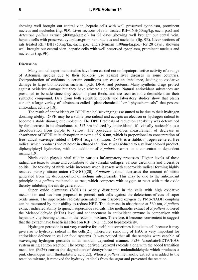
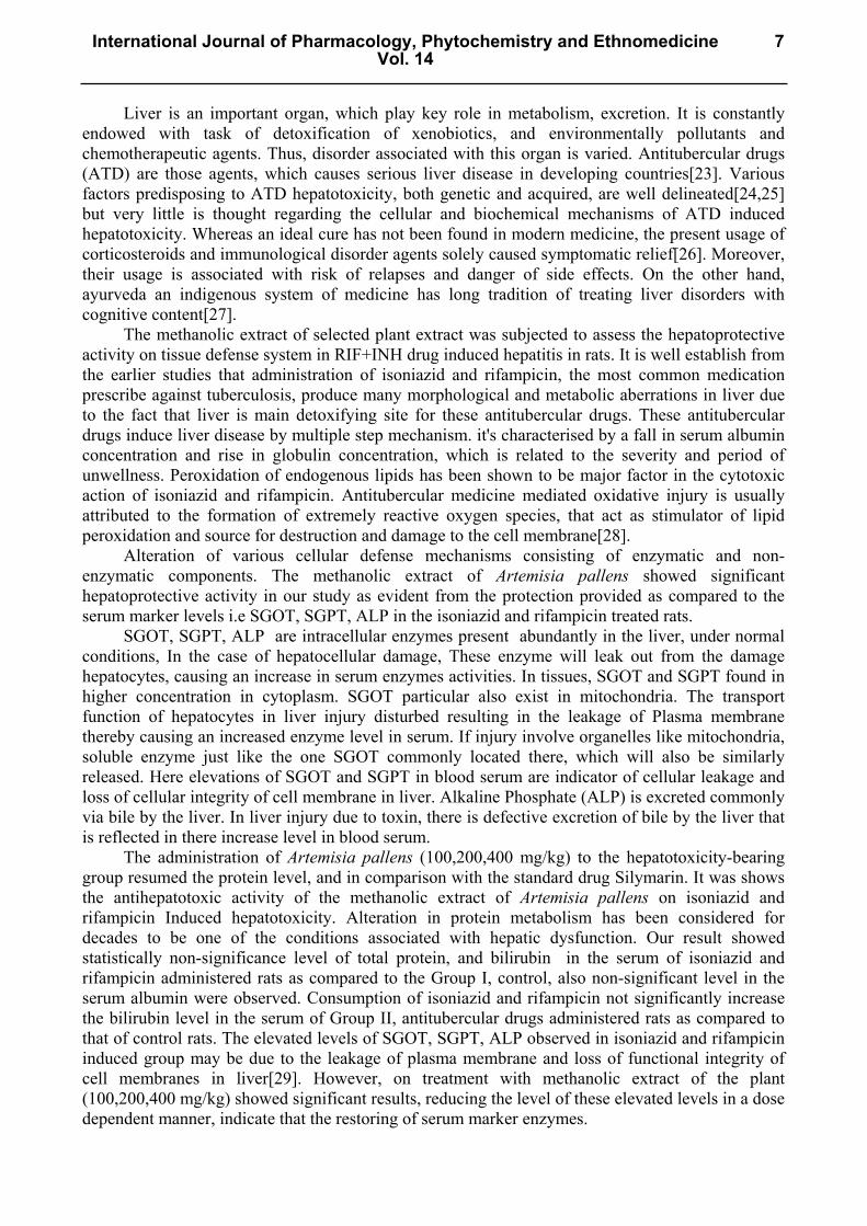
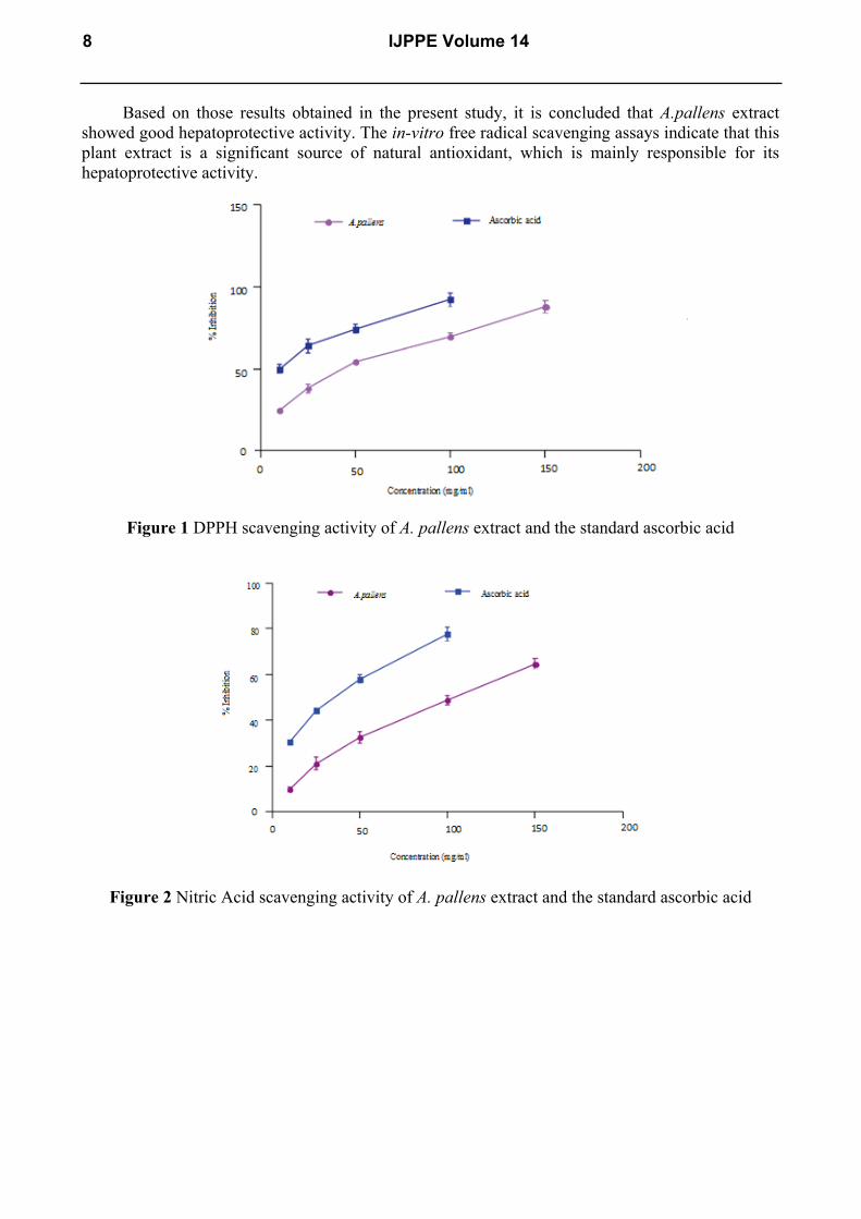
activity, as phenolics are strong antioxidants and hepatoprotective. Considering this, total phenol and flavonoid contents in the methanol extracts were determined and they were found to contain quite less amount of total phenol as gallic acid equivalent and total flavonoid contents as rutin equivalent. The A. pallens extract showed the dose dependent free radical scavenging activity in all in vitro assay models (Figure 1-4). IC50 values of A. pallens extract and standard ascorbic acid for DPPH, nitric oxide, superoxide radical scavenging, and hydroxyl radical were found to be (42.25 ± 0.95) and (09.16 ± 1.62) µg/ml, (101.62 ± 1.64) and (32.41 ± 1.24) µg/ml, (72.62 ± 1.86) and (10.28 ± 1.96) µg/ml, (33.82 ± 1.12) and (12.82 ± 1.86) µg/ml, respectively (Figure 5). There was also a dose dependant increase in reductive ability with increase in concentration of A.pallens extract and ascorbic acid. The aerial parts extract revealed content total phenolic and total flavonoid compounds (312.60 ± 1.24) µg and (322.20 ± 1.39) in 1 000 µg of A.pallens extract. Serum enzymes
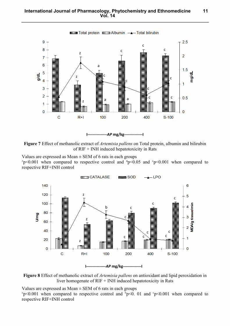
Since oxidative stress is one of the major contributors in occurrence of hepatic problems, preliminary antioxidant screening of the plant extracts, which will be further examined in hepatoprotective experiments in vivo, is considered to be necessary. The active extracts in antioxidant tests are given privilege for hepatoprotective assays. The effect of A.pallens extract on SGOT, SGPT, ALP, total bilirubin level, albumin and total protein levels in RIF+INH intoxicated rats are summarized in fig. 6-7. There was a significant increased in SGOT, SGPT, ALP, total bilirubin level, albumin and the total protein level was significantly decreased in the RIF+INH intoxicated rats, when compared with those of normal group. Treatment with A.pallens extract showed a significant decrease in all elevated serum marker levels and reversed the altered total protein level to almost normal. Comparative results were also found with the standard drug silymarin.
Toxic effect of RIF+INH significantly decrease the enzymic activities (CAT, SOD) and increase LPO level of liver homogenate in the RIF+INH intoxicated rats, when compared with those of normal group (figure 8). There was significantly increase the enzymatic antioxidant system and the elevated LPO level were found to be decreased towards the normal value in A.pallens extract as well as silymarin treated rats. Histopathological observation of liver section of normal control group showing normal hepatic cells with well preserved cytoplasm; well brought out central vien; prominent nucleus and nucleolus (fig. 9A). Liver sections of RIF+INH (50mg/kg, each, p.o.) treated rats showing; massive fatty changes ,necrosis ballooning degeneration, and irritation of lymphocytes and kuffer cells around the central vien and the loss of cellular boundaries(fig. 9B). Liver sections of rats treated RIF+INH (50mg/kg, each p.o.) and Artemisia pallens extracts (100mg/kg,p.o.) for 28 days , showing well brought out central vien ,hepatic cells with well preserved cytoplasm, prominent nucleus and nucleolus (fig. 9C). Liver sections of rats treated RIF+INH (50mg/kg, each, p.o.) and Artemisia pallens extracts (200mg/kg,p.o.) for 28 days ,
International Journal of Pharmacology, Phytochemistry and EthnomedicineVol. 14
5
showing well brought out central vien ,hepatic cells with well preserved cytoplasm, prominent nucleus and nucleolus (fig. 9D). Liver sections of rats treated RIF+INH(50mg/kg, each, p.o.) and Artemisia pallens extract (400mg/kg,p.o.) for 28 days ,showing well brought out cental vein, hepatic cells with preserved cytoplasm,prominent nucleus and nucleolus (fig. 9E). Liver sections of rats treated RIF+INH (50mg/kg, each, p.o.) and silymarin (100mg/kg,p.o.) for 28 days , showing well brought out central vien ,hepatic cells with well preserved cytoplasm, prominent nucleus and nucleolus (fig. 9F).
Discussion Many animal experiment studies have been carried out on hepatoprotective activity of a range
of Artemisia species due to their folkloric use against liver diseases in some countries. Overproduction of oxidants in certain conditions can cause an imbalance, leading to oxidative damage to large biomolecules such as lipids, DNA, and proteins. Many synthetic drugs protect against oxidative damage but they have adverse side effects. Natural antioxidant substances are presumed to be safe since they occur in plant foods, and are seen as more desirable than their synthetic compound. Data from both scientific reports and laboratory studies show that plants contain a large variety of substances called ‘‘plant chemicals’’ or ‘‘phytochemicals’’ that possess antioxidant activity[18].
The result of antioxidants on DPPH radical scavenging is assumed to be due to their hydrogen donating ability. DPPH may be a stable free radical and accepts an electron or hydrogen radical to become a stable diamagnetic molecule. The DPPH radicals of reduction capability was determined by the decrease in its absorbance at 517 nm induced by antioxidants. it's visually noticeable as a discolouration from purple to yellow. The procedure involves measurement of decrease in absorbance of DPPH at its absorption maxima of 516 nm, which is proportional to concentration of free radical scavenger added to DPPH reagent solution. DPPH is a stable, nitrogen-centered free radical which produces violet color in ethanol solution. It was reduced to a yellow colored product, diphenylpicryl hydrazine, with the addition of A.pallens extract in a concentration-dependent manner[19].
Nitric oxide plays a vital role in various inflammatory processes. Higher levels of these radical are toxic to tissue and contribute to the vascular collapse, various carcinoma and ulcerative colitis. The toxicity of nitric oxide increases when it reacts with superoxide radical forming highly reactive peroxy nitrate anion (ONOO-)[20]. A.pallens extract decreases the amount of nitrite generated from the decomposition of sodium nitroprusside. This may be due to the antioxidant principle in A.pallens methanolic extract, which competes with oxygen to react with nitric oxide thereby inhibiting the nitrite generation.
Super oxide dismutase (SOD) is widely distributed in the cells with high oxidative metabolism and has been proposed to protect such cells against the deleterious effects of super oxide anion. The superoxide radicals generated from dissolved oxygen by PMS-NADH coupling can be measured by their ability to reduce NBT. The decrease in absorbance at 560 nm, A.pallens extract indicated ability to quench superoxide radicals. The methanolic extract of A.pallens lowered the Melanoaldehyde (MDA) level and enhancement in antioxidant enzyme in comparison with hepatotoxicity bearing animals in the reaction mixture. Therefore, it becomes convenient to suggest that the extract have beneficial effect on RIF+INH induced hepatotoxicity.
Hydrogen peroxide is not very reactive for itself, but sometimes is toxic to cell because it may give rise to hydroxyl radical in the cells[21]. Therefore, removing of H2O2 is very important for antioxidant defence in cell or food systems. It was noticed that all the samples were capable of scavenging hydrogen peroxide in an amount dependent manner. Fe3+ /ascorbate/EDTA/H2O2 system using Fenton reaction. The oxygen derived hydroxyl radicals along with the added transition metal ion (Fe2+) causes the degradation of deoxyribose into malondialdehyde which produces a pink chromogen with thiobarbituric acid[22]. When A.pallens methanolic extract was added to the reaction mixture, it removed the hydroxyl radicals from the sugar and prevented the reaction.
6 IJPPE Volume 14
Liver is an important organ, which play key role in metabolism, excretion. It is constantly endowed with task of detoxification of xenobiotics, and environmentally pollutants and chemotherapeutic agents. Thus, disorder associated with this organ is varied. Antitubercular drugs (ATD) are those agents, which causes serious liver disease in developing countries[23]. Various factors predisposing to ATD hepatotoxicity, both genetic and acquired, are well delineated[24,25] but very little is thought regarding the cellular and biochemical mechanisms of ATD induced hepatotoxicity. Whereas an ideal cure has not been found in modern medicine, the present usage of corticosteroids and immunological disorder agents solely caused symptomatic relief[26]. Moreover, their usage is associated with risk of relapses and danger of side effects. On the other hand, ayurveda an indigenous system of medicine has long tradition of treating liver disorders with cognitive content[27].
The methanolic extract of selected plant extract was subjected to assess the hepatoprotective activity on tissue defense system in RIF+INH drug induced hepatitis in rats. It is well establish from the earlier studies that administration of isoniazid and rifampicin, the most common medication prescribe against tuberculosis, produce many morphological and metabolic aberrations in liver due to the fact that liver is main detoxifying site for these antitubercular drugs. These antitubercular drugs induce liver disease by multiple step mechanism. it's characterised by a fall in serum albumin concentration and rise in globulin concentration, which is related to the severity and period of unwellness. Peroxidation of endogenous lipids has been shown to be major factor in the cytotoxic action of isoniazid and rifampicin. Antitubercular medicine mediated oxidative injury is usually attributed to the formation of extremely reactive oxygen species, that act as stimulator of lipid peroxidation and source for destruction and damage to the cell membrane[28].
Alteration of various cellular defense mechanisms consisting of enzymatic and non- enzymatic components. The methanolic extract of Artemisia pallens showed significant hepatoprotective activity in our study as evident from the protection provided as compared to the serum marker levels i.e SGOT, SGPT, ALP in the isoniazid and rifampicin treated rats.
SGOT, SGPT, ALP are intracellular enzymes present abundantly in the liver, under normal conditions, In the case of hepatocellular damage, These enzyme will leak out from the damage hepatocytes, causing an increase in serum enzymes activities. In tissues, SGOT and SGPT found in higher concentration in cytoplasm. SGOT particular also exist in mitochondria. The transport function of hepatocytes in liver injury disturbed resulting in the leakage of Plasma membrane thereby causing an increased enzyme level in serum. If injury involve organelles like mitochondria, soluble enzyme just like the one SGOT commonly located there, which will also be similarly released. Here elevations of SGOT and SGPT in blood serum are indicator of cellular leakage and loss of cellular integrity of cell membrane in liver. Alkaline Phosphate (ALP) is excreted commonly via bile by the liver. In liver injury due to toxin, there is defective excretion of bile by the liver that is reflected in there increase level in blood serum.
The administration of Artemisia pallens (100,200,400 mg/kg) to the hepatotoxicity-bearing group resumed the protein level, and in comparison with the standard drug Silymarin. It was shows the antihepatotoxic activity of the methanolic extract of Artemisia pallens on isoniazid and rifampicin Induced hepatotoxicity. Alteration in protein metabolism has been considered for decades to be one of the conditions associated with hepatic dysfunction. Our result showed statistically non-significance level of total protein, and bilirubin in the serum of isoniazid and rifampicin administered rats as compared to the Group I, control, also non-significant level in the serum albumin were observed. Consumption of isoniazid and rifampicin not significantly increase the bilirubin level in the serum of Group II, antitubercular drugs administered rats as compared to that of control rats. The elevated levels of SGOT, SGPT, ALP observed in isoniazid and rifampicin induced group may be due to the leakage of plasma membrane and loss of functional integrity of cell membranes in liver[29]. However, on treatment with methanolic extract of the plant (100,200,400 mg/kg) showed significant results, reducing the level of these elevated levels in a dose dependent manner, indicate that the restoring of serum marker enzymes.
International Journal of Pharmacology, Phytochemistry and EthnomedicineVol. 14
7
Based on those results obtained in the present study, it is concluded that A.pallens extract showed good hepatoprotective activity. The in-vitro free radical scavenging assays indicate that this plant extract is a significant source of natural antioxidant, which is mainly responsible for its hepatoprotective activity.
Figure 1 DPPH scavenging activity of A. pallens extract and the standard ascorbic acid
Figure 2 Nitric Acid scavenging activity of A. pallens extract and the standard ascorbic acid
8 IJPPE Volume 14
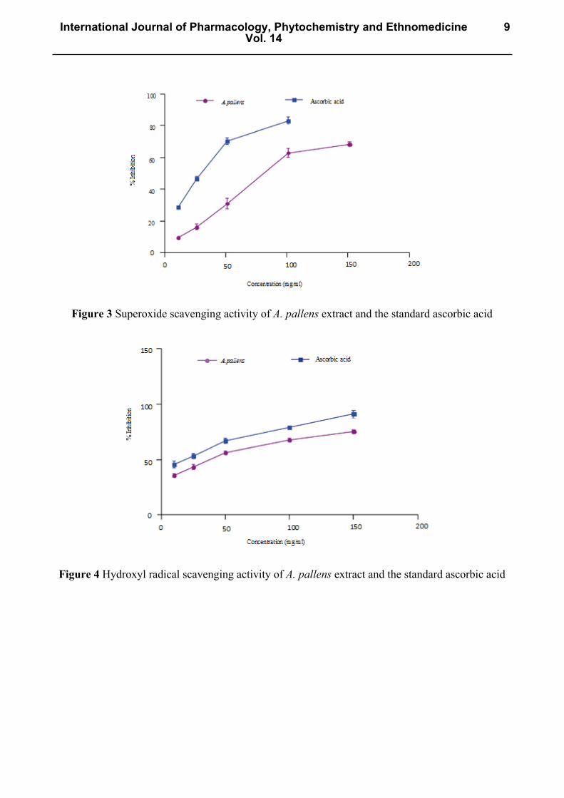
Figure 3 Superoxide scavenging activity of A. pallens extract and the standard ascorbic acid
Figure 4 Hydroxyl radical scavenging activity of A. pallens extract and the standard ascorbic acid
International Journal of Pharmacology, Phytochemistry and EthnomedicineVol. 14
9
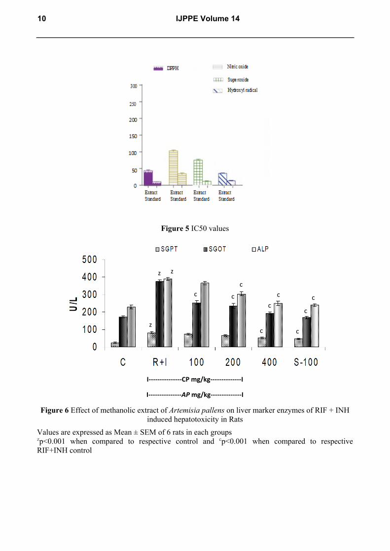
Figure 5 IC50 values
I---------------CP mg/kg--------------I
I---------------AP mg/kg--------------I
Figure 6 Effect of methanolic extract of Artemisia pallens on liver marker enzymes of RIF + INH induced hepatotoxicity in Rats
Values are expressed as Mean ± SEM of 6 rats in each groups
zp<0.001 when compared to respective control and cp<0.001 when compared to respective RIF+INH control
c
c
c
z z
c c z
c c
c
c
10 IJPPE Volume 14
I---------------AP mg/kg--------------I
Figure 7 Effect of methanolic extract of Artemisia pallens on Total protein, albumin and bilirubin of RIF + INH induced hepatotoxicity in Rats
Values are expressed as Mean ± SEM of 6 rats in each groups zp<0.001 when compared to respective control and ap<0.05 and cp<0.001 when compared to respective RIF+INH control
I---------------AP mg/kg--------------I
Figure 8 Effect of methanolic extract of Artemisia pallens on antioxidant and lipid peroxidation in liver homogenate of RIF + INH induced hepatotoxicity in Rats
Values are expressed as Mean ± SEM of 6 rats in each groups zp<0.001 when compared to respective control and bp<0. 01 and cp<0.001 when compared to respective RIF+INH control
z
z
z a
c
c c
c
c
c
c
c
c
a
a
z
z
z
b
c
c
c
c c
c
c c
c
International Journal of Pharmacology, Phytochemistry and EthnomedicineVol. 14
11
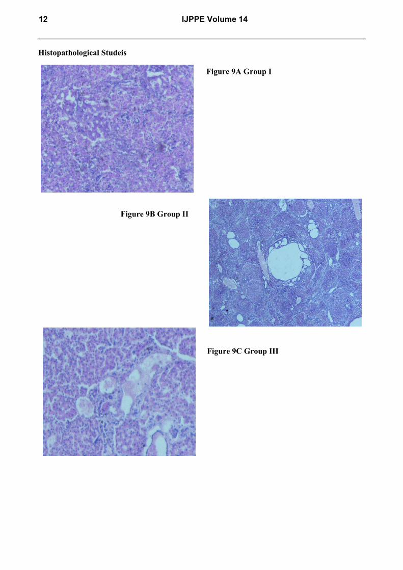
Histopathological Studeis
Figure 9A Group I
Figure 9B Group II
Figure 9C Group III
12 IJPPE Volume 14
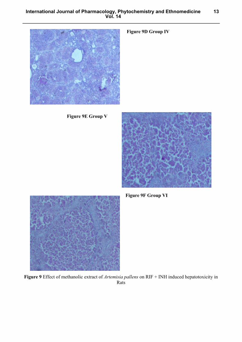
Figure 9D Group IV
Figure 9E Group V
Figure 9F Group VI
Figure 9 Effect of methanolic extract of Artemisia pallens on RIF + INH induced hepatotoxicity in Rats
International Journal of Pharmacology, Phytochemistry and EthnomedicineVol. 14
13
References [1] Valdis-Arzate A, Luna A, Bucio L, Licona C, Clemens DL, Souza V, et al. Hepatocyte
growth factor protects hepatocytes against oxidative injury induced by ethanol metabolism. Free Radic Biol Med. 47(2009) 424-30
[2] Mindie, HN., Gabriel, G. Does isoniazid cause more serious hepatotoxicity in Hapatitis B virus carriers? Am. J. Gastroenterol. 97(2002) 1092-1093.
[3] Rice-Evans, CA., Miller, NJ., Payanga, G. Structure-antioxidant activity relationships of flavonoids and phenolic acids. Free Rad. Biol. Med. 20 (1996) 933-956.
[4] Halliwell, B., Gutteridge, J.M.C. Oxygen toxicity, oxygen radicals, transition metals and disease. Biochem. J. (1984) 219: 1-4.
[5] Barros, L., Baptista, P., Ferreira ICFR. Effect of Lactarius piperatus fraiting body maturity stage on antioxidant activity measured by several biochemical assays. Food Chem. Toxicol. 45 (2007) 1731-1737.
[6] Singh RP., Padmavathi B., Rao AR. Modulatory influence of Adhatoda vesica (Justica adhatoda) leaf extract on the enzyme of xenobiotic metabolism, antioxidant status and lipid peroxidation in mice. Mol cell Biochem. 213 (2000) 99-109.
[7] Ashok PK., Upadhyaya K. Analgesic and anti-inflammatory properties of Artemisia pallens wall ex.DC. The Pharma Res. 3 (2010) 249-256.
[8] Subramoniam A., Pushpangadan P., Rajasekharan S., Evans DA., Latha PG., Valsarj R. Effect of Artemisia pallens wall on blood glucose levels in normal and alloxan-induced diabetic rats. J. Ethnopharmacol. 50(1996) 13-17.
[9] Swain, T., Hillis, W.E. The phenolic constituents of prunus domestica I-the quantitative analysis of phenolic constituents. J. Sci. Food Agric. 10(1959) 63-68.
[10] Woisky R, Salatina A. Analysis of propolis: Some parameters and procedures for chemical quality control. J. Apicol Res. 37(1998) 99-105.
[11] Blois MS. Antioxidant determinations by the use of a stable free radical. Nature 181(1958) 1199-200.
[12] Sakat SS., Javekar AR., Gambhire MN. In vitro antioxidant and anti-inflammatory activity of methanol extract of oxalis corniculata Linn. Int J Pharm Pharm Sci. 2(1) (2010) 146-155.
[13] Liu F, Ooi VE, Chang ST. Free radical scavenging activities of mushroom polysaccharide extracts. Life Sci. 60(1997) 763-71.
[14] Elizabeth, K. Rao, MNA. Oxygen radical scavenging activity of Curcumin. Int. J. Pharmacol. 58 (1990) S237-S40.
[15] Zimmerman, M. Ethical guidelines for investigations of experimental pain in conscious animals. Pain. 16(1983) 109-110.
[16] Ravinder Pal, Kim Vaiphei, Arbab Sikander. Effect of garlic ion isoniazid and rifampicin-induced hepatic injury in rats. World J. of Gastroenterology. 28:12(4) (2006) 636-639.
[17] Wilkinson JH, Baron DN, Moss DW, Wolter PG. Standardization of clinical enzyme assays: reference method for aspartate and alanine transaminases. J Clin Pathol. 25 (1972) 940–5.
[18] Uttara B., Singh AV., Zamboni P., Mahajan RT. Oxidative stress and neurodegenerative disease: A review of upstream and downstream antioxidant therapeutics option. Curr neuropharmacol. 7(1) (2009) 65-74.
14 IJPPE Volume 14
[19] Jothyl SL., Zuraini Z., Sasidharan S. Phytochemicals screening DPPH free radical scavenging and xanthine oxidose inhibitory activities of cassia fistula seeds extract. J. Med Plant Res. 5(10) (2011) 1941-1947.
[20] Rajan S., Mahalakshmi S., Deepa VM., Sathya k., Shajitha S., Thirunalasundari T. Antioxidant potentials of Punica granatum fruit rind extract. Int. J. Pharm Pharm Sci. 3(3) (2011) 82-88.
[21] Halliwell, B. Reactive oxygen species in living systems: Source, biochemistry, and role in human disease. American Journal of Medicine. 91(1991) 14–22.
[22] Battu GR., Ethadi SR., Veda PG., Swathi PK., Chandrika K., Rao VA. Evaluation of antioxidant and anti-inflammatory activity of Euphorbia heyneana spreng. Asian Pac. J. Trop Bio Med. (2011) 191-194.
[23] AcharyaSK, DasarathyS, KumerTL, SushmaS, PrasannaKS, TandonA,etal. Fulminan the patitis in a tropical population: clinical course, cause and early predictors of outcome. Hepatology. 23 (1996) 1448–1455.
[24] Huang YS, Chern HD, Su WJ, Wu J C, Lai S L, Yang S Y, etal. Polymorphism of N-acetyltransferase 2gene as a susceptibility Risk factor of antituberculosis drug induced hepatitis. Hepatologyn. 35 (2002) 883–889.
[25] Roy B, Chowdhury A, Kundu S, Santra A, Dey B, Chakraborty M,etal. Increased risk of antituberculosis drug induced hepatotoxicity in individual swithglutathiones- transferase M1‘null’ mutation. J. Gastroenterol Hepatol. 16 (2001) 1033–1037.
[26] Handa S.S. and Sharma A. Hepatoprotective activity of andrographolide against galactosamine and paracetamol intoxication in rats. Indian Journal of Medical Research [B]. 92 (1986) 284–292.
[27] De Vita, VT. Jr. Principles of chemotherapy. In. V.T.Devita Jr. S.Hellman and S.A.Rosenberg (eds). Cancer Principle and Pratice of Oncology. 4 (1993) 276-292.
[28] Georgieva, N., Gadjeva, V., Tolekova, A. New isonicotinoylhydrazones with ssa protect against oxidative-hepatic injury of isoniazid. TJS. 2 (2004) 37–43.
[29] Singh K., Khanna AK., Chandan R. Hepatoprotective activity of ellagic acid against carbon tetrachloride induced hepatotoxicity in rats. Indian J Experim. Biol. 37 (1999) 1025-1026.
International Journal of Pharmacology, Phytochemistry and EthnomedicineVol. 14
15