Embed Size (px)
Citation preview
1 [email protected] | Tel: +353 1 832 2843 | ©NOAH 2012-2014
Fracture Repair in Animals Fracture Repair in Dogs & Cats
Contents ................................................................................................................................................................ 1
Fracture Repair in Animals ...................................................................................................................... 1
Broken Bones Happen......................................................................................................................... 3
The Parts of Bone ................................................................................................................................ 3
Leg Structure ....................................................................................................................................... 3
The Bones ............................................................................................................................................ 4
The Biggest Bone – The Femur ....................................................................................................... 4
The Radius, Ulna, Tibia, and Fibula ................................................................................................. 4
Your Pet's Fingers & Toes ................................................................................................................ 5
Understand Broken Bones .................................................................................................................. 5
Broken Bones: Growing Bones ........................................................................................................ 5
Broken Bones: Inflammation .......................................................................................................... 6
Broken Bones: Reparation .............................................................................................................. 6
Broken Bones: Remodeling ............................................................................................................. 6
Summary of Fracture Healing ......................................................................................................... 7
Assisting the Healing Process .......................................................................................................... 7
Jaw Fractures ...................................................................................................................................... 8
Upper Jaw ....................................................................................................................................... 8
Lower Jaw ........................................................................................................................................ 8
Diagnosis ......................................................................................................................................... 8
Favourable or Unfavourable Findings ............................................................................................. 8
The Job of Repair............................................................................................................................. 8
Jaw Healing ..................................................................................................................................... 9
Emergency: How to React ................................................................................................................... 9
2 [email protected] | Tel: +353 1 832 2843 | ©NOAH 2012-2014
Theories on Strains ............................................................................................................................. 9
Perren's Theory on Strains .............................................................................................................. 9
Carter and Blenman's Theory On Strains ...................................................................................... 10
Limiting Mechanical Stimulus ........................................................................................................... 10
External Fixators............................................................................................................................ 10
Internal Fixation ............................................................................................................................ 11
How We Choose Treatment .............................................................................................................. 11
Pain Management ............................................................................................................................. 11
After Care .......................................................................................................................................... 12
Physical Therapy ........................................................................................................................... 12
Time Needed ................................................................................................................................. 12
3 [email protected] | Tel: +353 1 832 2843 | ©NOAH 2012-2014
Broken Bones Happen Fractures can occur in a number of ways, and so there are terms associated with the type of fracture that might occur. For example; salter is a type of fracture that occurs through the growth plate of the bone. This part of bone is located at the top or bottom of a long bone such as the femur or tibia. These are usually problematic in immature animals that are still growing. As we reach adulthood this part of bone hardens. Salter fractures can interrupt both normal bone growth, and can greatly influence repair. A greenstick fracture is an incomplete break that is a result like a bend in the bone. Only a part of the bone may be broken partially such as part of the cortical bone. It too, is an occurrence in young animals more so than adult. A complete fracture in a long bone is one that breaks the entire bone through the full circumference. This can result in two or more pieces. More pieces can be fragments and these are usually sequestered bone pieces. Complete fractures can be discussed based on their shape. A transverse fracture is one where the break is straight across the bone at a right angle to the length of the bone in question. An oblique fracture is where the break runs diagonal and this will usually result in the creation of two bone fragments. Comminuted breaks are usually where there is more than one fracture thus a result of three or more pieces of varying shapes. There can be the occurrence of open fractures where bone can penetrate the skin resulting in a wound. Broken bone can penetrate the skin to become exposed, or for example, a bullet can break bone creating a wound from outside in. No wound is usually termed a closed fracture.
The Parts of Bone The periosteum is the part of the bone that is the outer shell so to speak. It's a soft tissue that is necessary for bone growth, repair, and remodelling. The next internal layer is called the cortical bone. This is termed "boney" tissue, as it's the hard shell that is the bulk of thickness of bone. Cancellous bone is the soft mesh-like tissue found inside the long bones at each end. Finally, bone marrow is the fatty tissue that fills the hollow interior of bones. Marrow contains blood cells, blood vessels, other cells along with chemicals that are vital to good health and necessary in healing.
Leg Structure The leg bones of dogs & cats are comparable to the bones of the legs and arms of people. When we talk about the anatomy of legs there are unique terms of identification used. On the feet of the front legs, the three unique toe bones are identified as the 1st, 2nd & 3rd phalanges. These are met by four main bones (the metacarpal bones) which can be compared to the human hand. Above the hand comes the carpus (wrist), made up of the carpal bones. The forearm contains the radius and ulna, and finally the upper arm contains the humerus and scapula (shoulder blade). The canine's back legs contain a similar series of bones to humans, but take different names as they are also different shapes, sizes and position. Toes are referenced in the same manner as the front legs (1st, 2nd & 3rd phalange). They are met by the metatarsal bones, the main bones of the foot. Tarsal bones make up what is called the hock (ankle). These are linked to the tibia and fibula. The tibia is often called the shin bone. The thigh holds the largest bone in the body, the femur. Finally, the hips are the last series of bones associated with the rear legs. The hips (pelvis) are made up of the ilium, acetabulum, and the ischium. The acetabulum is the part that holds the hip socket; the joint established between the hip and the femur. Joints allow for the movement of the static structure that is built from bone. The carpus joint at the wrist area of the front leg is the joining of the small carpal bones with that of the radius. The elbow joint is made up of the carpus, ulna, and humerus bones, whereas the shoulder joint is at the humerus and scapula. The ankle, or tarsus, is the small tarsal bones meeting with the tibia. The knee (stifle) is where the tibia and femur meet, and contains the patella (kneecap) that allows for the joint to flex and extend. The hip joint is where the femur meets with the acetabulum.
4 [email protected] | Tel: +353 1 832 2843 | ©NOAH 2012-2014
The Bones
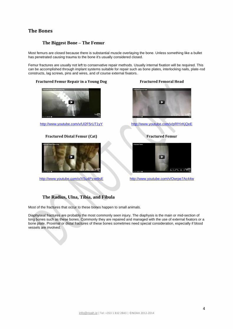
The Biggest Bone – The Femur
Most femurs are closed because there is substantial muscle overlaying the bone. Unless something like a bullet has penetrated causing trauma to the bone it's usually considered closed. Femur fractures are usually not left to conservative repair methods. Usually internal fixation will be required. This can be accomplished through implant systems suitable for repair such as bone plates, interlocking nails, plate-rod constructs, lag screws, pins and wires, and of course external fixators.
Fractured Femur Repair in a Young Dog
http://www.youtube.com/v/Ul2F5rUT1yY
Fractured Femoral Head
http://www.youtube.com/v/pRlYirKjQeE
Fractured Distal Femur (Cat)
http://www.youtube.com/v/XSu4Pzxe9sE
Fractured Femur
http://www.youtube.com/v/Owrpe7Ac44w
The Radius, Ulna, Tibia, and Fibula
Most of the fractures that occur to these bones happen to small animals. Diaphyseal fractures are probably the most commonly seen injury. The diaphysis is the main or mid-section of long bones such as these bones. Commonly they are repaired and managed with the use of external fixators or a bone plate. Proximal or distal fractures of these bones sometimes need special consideration, especially if blood vessels are involved.
5 [email protected] | Tel: +353 1 832 2843 | ©NOAH 2012-2014
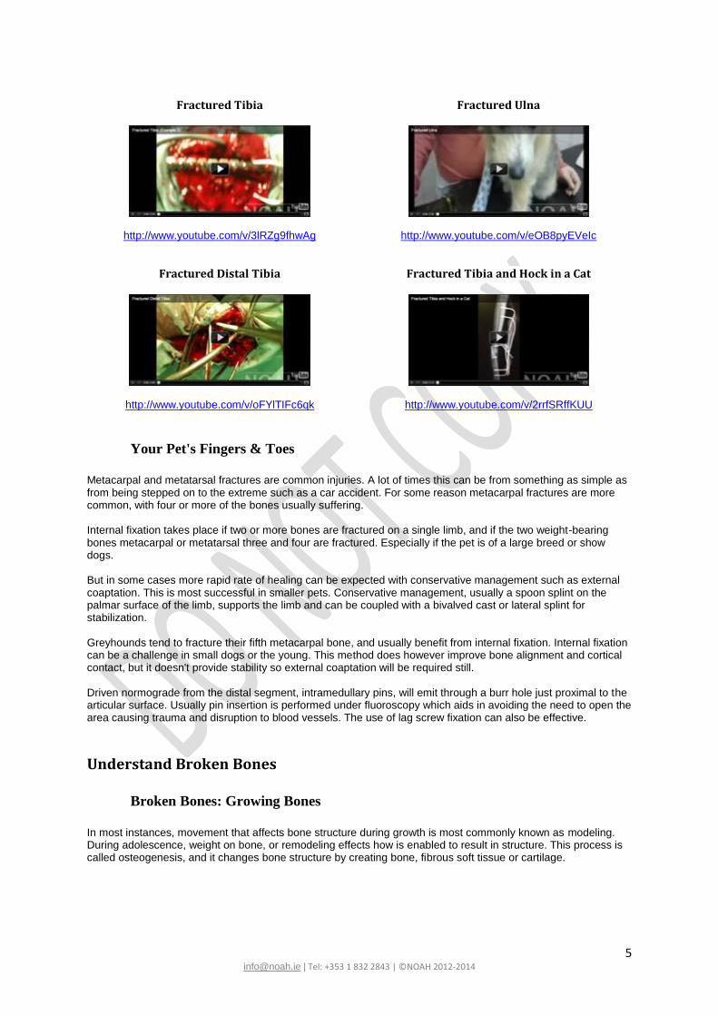
Fractured Tibia
http://www.youtube.com/v/3lRZg9fhwAg
Fractured Ulna
http://www.youtube.com/v/eOB8pyEVeIc
Fractured Distal Tibia
http://www.youtube.com/v/oFYlTIFc6qk
Fractured Tibia and Hock in a Cat
http://www.youtube.com/v/2rrfSRffKUU
Your Pet's Fingers & Toes
Metacarpal and metatarsal fractures are common injuries. A lot of times this can be from something as simple as from being stepped on to the extreme such as a car accident. For some reason metacarpal fractures are more common, with four or more of the bones usually suffering. Internal fixation takes place if two or more bones are fractured on a single limb, and if the two weight-bearing bones metacarpal or metatarsal three and four are fractured. Especially if the pet is of a large breed or show dogs. But in some cases more rapid rate of healing can be expected with conservative management such as external coaptation. This is most successful in smaller pets. Conservative management, usually a spoon splint on the palmar surface of the limb, supports the limb and can be coupled with a bivalved cast or lateral splint for stabilization. Greyhounds tend to fracture their fifth metacarpal bone, and usually benefit from internal fixation. Internal fixation can be a challenge in small dogs or the young. This method does however improve bone alignment and cortical contact, but it doesn't provide stability so external coaptation will be required still. Driven normograde from the distal segment, intramedullary pins, will emit through a burr hole just proximal to the articular surface. Usually pin insertion is performed under fluoroscopy which aids in avoiding the need to open the area causing trauma and disruption to blood vessels. The use of lag screw fixation can also be effective.
Understand Broken Bones
Broken Bones: Growing Bones
In most instances, movement that affects bone structure during growth is most commonly known as modeling. During adolescence, weight on bone, or remodeling effects how is enabled to result in structure. This process is called osteogenesis, and it changes bone structure by creating bone, fibrous soft tissue or cartilage.
6 [email protected] | Tel: +353 1 832 2843 | ©NOAH 2012-2014
Broken Bones: Inflammation
Inflammation occurs instantaneously when a bone fractures. A blood clot or hematoma forms at the fracture site. This hematoma provides two important factors important for fracture healing. Firstly the hematoma provides a small amount of mechanical stability to the fracture site. Secondly, this blood clot brings special cells called osteoblasts, and chondrocytes to the fracture site in large numbers so they can begin producing a matrix. This in turn starts the process of bone modeling. Also brought to the site are other cells called macrophages and osteoclasts. These cells come to the site of fracture to remove damaged and dead tissue. Bone fracture usually disrupts the periosteum surrounding the bone. The periosteum is specialised coating of cells that act as connecting tissue, and they exist as a covering on all bones. Precursor cells from the periosteum will be introduced into the fracture site to also aid in reconstruction.
Broken Bones: Reparation
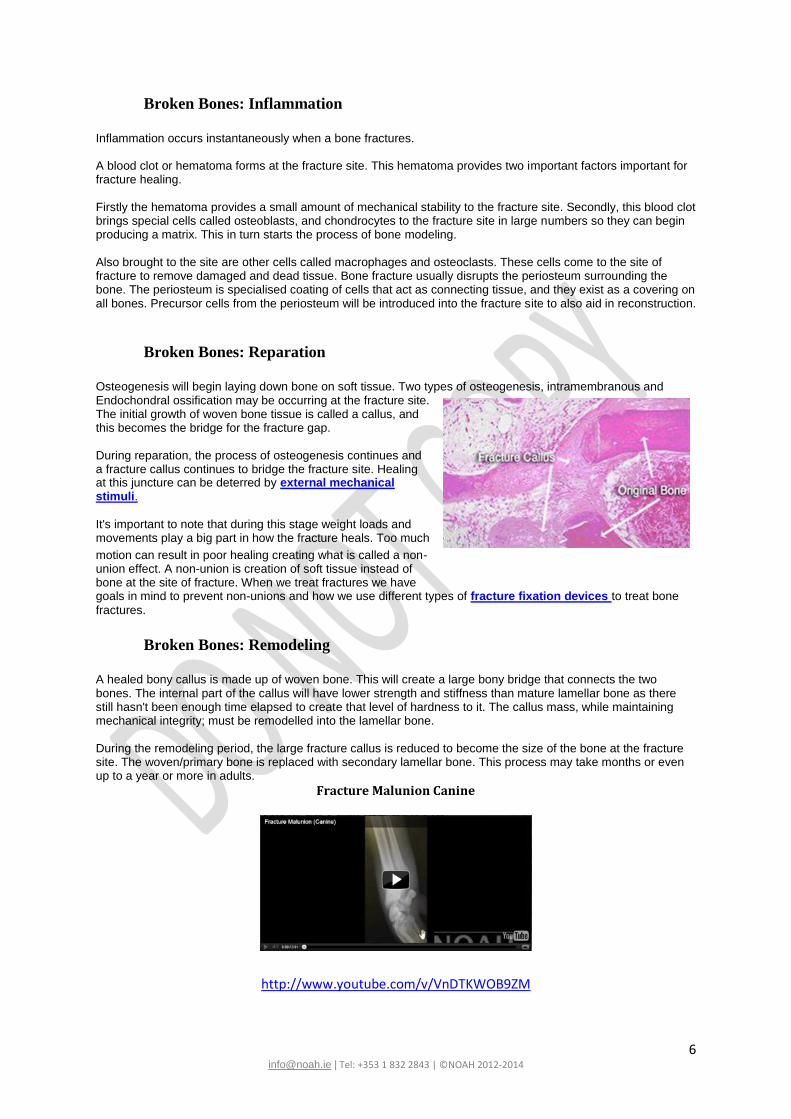
Osteogenesis will begin laying down bone on soft tissue. Two types of osteogenesis, intramembranous and Endochondral ossification may be occurring at the fracture site. The initial growth of woven bone tissue is called a callus, and this becomes the bridge for the fracture gap. During reparation, the process of osteogenesis continues and a fracture callus continues to bridge the fracture site. Healing at this juncture can be deterred by external mechanical stimuli.
It's important to note that during this stage weight loads and movements play a big part in how the fracture heals. Too much
motion can result in poor healing creating what is called a non-union effect. A non-union is creation of soft tissue instead of bone at the site of fracture. When we treat fractures we have goals in mind to prevent non-unions and how we use different types of fracture fixation devices to treat bone
fractures.
Broken Bones: Remodeling
A healed bony callus is made up of woven bone. This will create a large bony bridge that connects the two bones. The internal part of the callus will have lower strength and stiffness than mature lamellar bone as there still hasn't been enough time elapsed to create that level of hardness to it. The callus mass, while maintaining mechanical integrity; must be remodelled into the lamellar bone. During the remodeling period, the large fracture callus is reduced to become the size of the bone at the fracture site. The woven/primary bone is replaced with secondary lamellar bone. This process may take months or even up to a year or more in adults.
Fracture Malunion Canine
http://www.youtube.com/v/VnDTKWOB9ZM
Example of a Callus forming 1
7 [email protected] | Tel: +353 1 832 2843 | ©NOAH 2012-2014
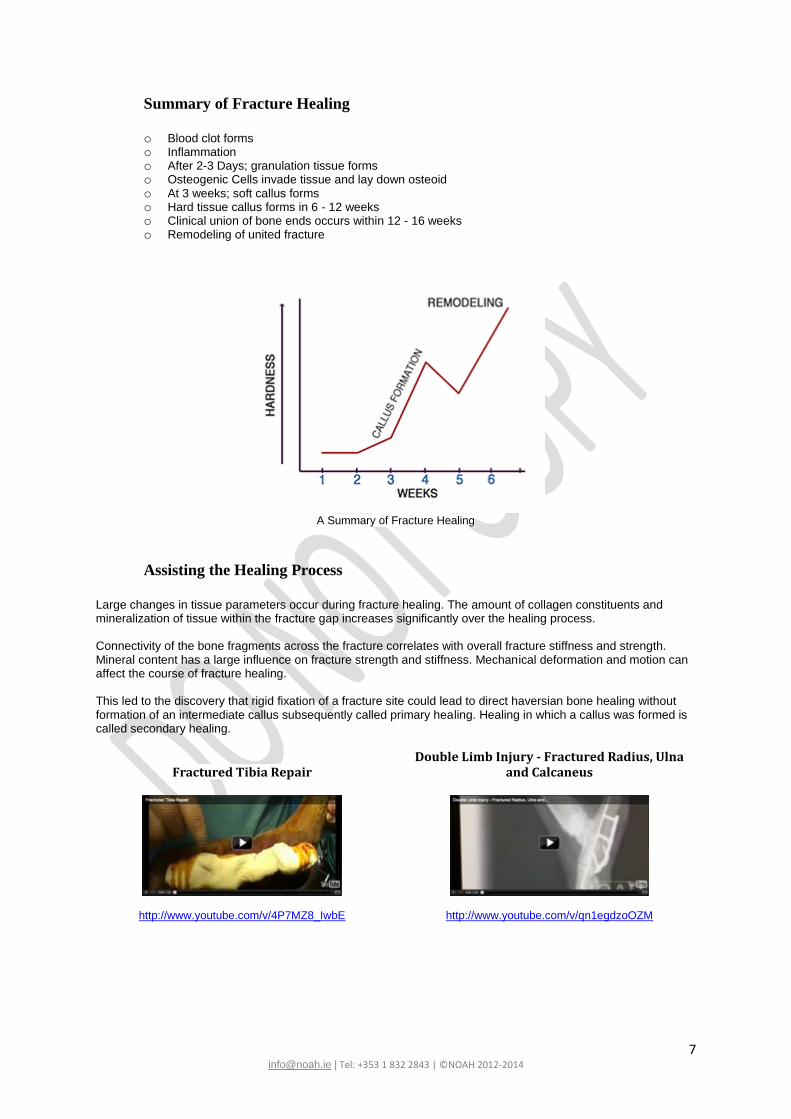
Summary of Fracture Healing
o Blood clot forms o Inflammation o After 2-3 Days; granulation tissue forms o Osteogenic Cells invade tissue and lay down osteoid o At 3 weeks; soft callus forms o Hard tissue callus forms in 6 - 12 weeks o Clinical union of bone ends occurs within 12 - 16 weeks o Remodeling of united fracture
A Summary of Fracture Healing
Assisting the Healing Process
Large changes in tissue parameters occur during fracture healing. The amount of collagen constituents and mineralization of tissue within the fracture gap increases significantly over the healing process. Connectivity of the bone fragments across the fracture correlates with overall fracture stiffness and strength. Mineral content has a large influence on fracture strength and stiffness. Mechanical deformation and motion can affect the course of fracture healing. This led to the discovery that rigid fixation of a fracture site could lead to direct haversian bone healing without formation of an intermediate callus subsequently called primary healing. Healing in which a callus was formed is called secondary healing.
Fractured Tibia Repair
http://www.youtube.com/v/4P7MZ8_IwbE
Double Limb Injury - Fractured Radius, Ulna and Calcaneus
http://www.youtube.com/v/qn1egdzoOZM
8 [email protected] | Tel: +353 1 832 2843 | ©NOAH 2012-2014
Jaw Fractures
Upper Jaw
The maxilla is the upper part of the jaw. Fractures that occur to the maxilla can be obvious or subtle. In many instances facial muscles and swelling at the site require the necessity to perform x-rays, also called radiographs. A series of dental radiographs, much like when people see their dentist, should be taken to determine the extent of damage and better aid in the understanding of what is the best course of treatment. Car accidents or accidents where the animal gets hit by a blunt object or actual abuse cases are the most common causes for jaw trauma and jaw fractures. Animals can also acquire injuries sustained through fights with other animals. Dental extraction complications, metabolic diseases, oral cysts, and tumours can also be a cause behind fractures of the jaw.
Lower Jaw
The lower jaw can sustain fractures in very much the same ways as the upper jaw. These fractures are frequently called mandibular, symphyseal or temporomandibular joint injuries. Where these differ to upper jaw fractures is that sometimes this type of fracture is actually quite a visible injury, but they can also elude the best by being a very subtle injury. Repair terminology refers to mandibular injuries as either "favorable" or "unfavorable". This very much relates to the interpretation of the injury and if a foreseen repair and healing process can be determined.
Diagnosis
Jaw fractures in most Veterinary practices are diagnosed with the standard Veterinary radiograph equipment. Sometimes the necessity for higher detail is required and this can be achieved with specialized dental radiography. In these cases, a more accurate diagnosis can be achieved. Once a diagnosis can be arrived at so can the optimal plan of repair along with the prognosis for the animal. Animals that have continually growing teeth, such as rabbits or rodents, Jaw injuries are life threatening injuries. To repair such fractures in these animals there is an absolute precision necessary to alignment. If correct anatomic alignment does not occur, abnormal teeth relationships will develop. These are also called malocclusions and will most likely result in factors causing the animal to not want to eat due to pain and ultimately can result in loss of life.
Favourable or Unfavourable Findings
Fractures that run perpendicular to the mandible tend to be unfavourable. Also an injury that causes the downward motion resulting from the actions of the digastric muscle and upward motion resulting from the actions of the opposing muscle group to come apart at the mandibular fracture line can be deemed an unfavourable fracture. There must be a repair of the fracture that stabilises this, and allows conducive healing to occur. If there is to be constant motion of the fracture line, as a result of opening and closing the mouth, this is not ideal for healing. The circumstance of a favourable jaw fracture is one that does not have a fracture line resulting in displacement.
The Job of Repair
Injuries where loss of blood supply, bone fragmentation and/or nerve damage occur might result in a mandibulectomy (the entire removal of the mandible). With these fractures the severity of bone fragmentation, the degree of contamination, and the analysis of blood supply are all necessary to determine if repair can be favorable. Tooth root injury and/or neurovascular damage within the mandibular or infraorbital canals is also of serious concern when mapping a proper treatment. In part, the concept of movements made by the jaw, also termed the biomechanics, are of great importance in planning repair. The common methods for repair are best accomplished by the use of wire reinforced composite splints. For injuries that are of minor displacement, sometimes the simplicity of tape muzzles can garner adequate results.
9 [email protected] | Tel: +353 1 832 2843 | ©NOAH 2012-2014
It is of great importance that the repair plan allows the pet to eat with comfort but also maintain normal occlusion. Normal occlusion is a dental term that refers to the alignment and placement of the teeth within the jaw that allows for normal functioning. If this is incorrectly repaired or perhaps heals incorrectly, a malocclusion will result, which is best interpreted as the wrong alignment or placement of the teeth. Of course we wish to never see this happen.
Jaw Healing
The outcome for a jaw fracture is typically good. Of course there are exceptions to this as mentioned in the other sections, but the best attempts at repair and treatment are our goal. A broken maxilla most often can be easily repaired with long-term favorable results. Mandibular fractures have a variable prognosis because of how injury occurred and the extent of the damage inflicted. Minor trauma usually results in a very positive prognosis. Periodontal disease in older dogs that suffer the injury of a jaw fracture during tooth extractions may have a poor, guarded or fair prognosis. It helps to brush your dog’s teeth, coupled with annual teeth cleanings.
Emergency: How to React What do you do when you've learned your dog or cat has broken its bone? Immediately, it's important to immobilize the pet so they cannot move. This is done because should there be any sequestered fragments of bone they will not be able to move. The right way to temporarily immobilize a fracture is to place a splint. A splint must start at the joint above the bone and extend to the joint below the bone to prevent it from moving. If the tibia was to have suffered a fracture it's important to place the splint from the stifle (knee) to the tarsus (ankle). It's fairly easy to immobilize bones from the knee or elbow. The jaw might be easily immobilized by a muzzle or a self-made muzzle from gauze tied loosely around the jaw, and then extended behind the ears so the animal cannot remove the tie. Be careful, and sensitive to the aspect that your pet could be in pain, and any movement or attempts might result where they could bite. It's nothing personal, they're in pain and don't know how to tell you. Sometimes it's best to have help holding the animal while another makes attempts at administering this type of emergency medicine. Immobilization will also aid in pain management as it will hurt a lot less if the ends of pieces, which can be sharp, do not cause further tissue damage to nerves, muscles, and blood vessels. Before you can get to the Veterinary clinic or hospital is important to confine your pet in a very small space. If you have a crate that allows your pet to move, try and confine the space further using a large pillow against the back or a small box to shorten the space. Essentially you want space for them to lay and not move or try to move. Once situated for transport, try to get to a Veterinary surgeon as soon as possible. Reaction time and proper treatment can aid in proper and speedy recovery. It's also important because other dangerous medical conditions could have occurred from the break, such as a damaged blood vessel. Do not give your pet any medications or apply any therapy unless you receive directions from a Veterinary professional. Your pet will be evaluated by a Veterinary professional who will determine if the animal is stable, needs pain management, or sedation. Sedation is usually administered to further assist Veterinary professionals in obtaining X-rays, professionally known as radiograph. These are integral in creating a treatment plan of a fracture.
Theories on Strains
Perren's Theory on Strains
Interfragmentary strain theory postulates that changes in fracture gap tissue can be related to strain magnitudes. Perren theorized that the magnitude of interfragmentary strain would determine the subsequent differentiation of fracture gap tissue. Perren theorized that extreme interfragmentary strain would lead to non-union. Intermediary strains lead to initial fibrous tissue formation. Mild strains lead to cartilage formation and an endochonral ossification formation. Minute strains lead to direct bone formation and primary fracture healing.
10 [email protected] | Tel: +353 1 832 2843 | ©NOAH 2012-2014
Strain issues he surmised, results in an inability to form a fracture gap. In addition to the removal of strain effects on initial formation, Perren believed that once set in progress, tissues formed would stiffen the fracture gap which would lead to lower strains, which would allow formation of the next stiffest tissue and the cycle would repeat until all bone was formed.
Carter and Blenman's Theory on Strains
Carter and Blenman's theory differs from Perren's theory in that it not only predicts how the magnitude of mechanical stimulus will affect fracture tissue differentiation, but also differentiates the possible results obtained from each type of mechanical stimulus. It is known that vascular supply to tissues was the primary factor in determining tissue differentiation. Both the magnitude and type of mechanical stress, basically hydrostatic pressure versus octahedral shear stress, will affect the type of tissue forming within fracture sites. If a good vascular supply was available to tissues, Carter and Blenman believed that the following sequence would occur:
Osteogenic stimulus occurs
If minimum or no stressors present, coupled with a good blood supply results in bone forming directly
If high hydrostatic compressive stresses are present; fibro cartilage will form
If high tensile or shear stresses are present; fibrous tissue will form
Alongside fibro cartilage formation, further shear stresses will lead to eventual bone formation Without a blood supply or within a poor fracture gap with poor vascularity, Carter and Blenman believed that producing bone would not be possible because bone is a highly metabolic tissue that requires a good blood supply.
Limiting Mechanical Stimulus Both Perren's and Carter and Blenman's theories suggest that too high of a mechanical stimulus will prevent bone formation and lead to a non-union. Both also suggest that very low magnitudes of strain and stress will lead to direct bone formation. Clinical results suggest that the theories are correct concluding that both high stress or strain will produce fibrous non-union, but varying stressors produce results that are less clear. For these reasons it's widely believed that some mechanical rigidity is needed for complex unstable fractures to prevent gap tissue stresses from becoming too high and allowing bone formation to heal the fracture. The fracture fixation devices include external fixators, internal plates, intramedullary rods and casts. Most fractures can be treated by immobilization with something as simple as a cast that will help reduce strains at the fracture site. External coaptation is a splint or cast and is applied to the outside of the limb. This can adequately resist bending forces thus resisting torsion and compression forces. Concepts such as splints can reduce strain, but compound fractures usually must be treated surgically with fracture fixation devices. There are three categories of devices such as those entirely within the skin called internal fixation, or on the outside of the skin called external fixation, and there are other internal devices such as a plate that is screwed to the outside of the bone, a rod that goes down the centre of the bone or wire coupled with other devices and possible external fixation such as splints. The choice of what device to use is based on clinical experience and surgical ease in placing the device.
External Fixators
External fixators will deflect and carry most of the load. Fibro cartilage will form and begin to share load with the fixator. It is because the external fixator holds a stiffness that remains the same, that the fracture gap stiffness is increased and the overall construct stiffness increases thus reducing strain in the fracture gap. This then allows the cartilage tissue to become calcified and turn to bone. Adjusting the stiffness of the fracture fixation device has been proven to enable clinical predictions of strains to the fracture gap and thus ensures the fracture healing process. External fixation is achieved by an external frame with pins placed through the skin and into bone pieces above and below the fracture site. External fixation is appropriate mainly for long bone fixation and it's most common application in complex, compound tibial fractures. In this case, it may not be possible to plate the fracture if many fragments exist.
11 [email protected] | Tel: +353 1 832 2843 | ©NOAH 2012-2014
It is important to note that the mechanical stability at a fracture gap that can be achieved using external fixators depends on the fixator's constructive stiffness, which in turn depends upon the geometric configuration of the fixator. Under axial load and to some extent under bending loads fixator stiffness is determined by bending of the pins placed in the bone.
Internal Fixation
Plates
Plate fixation has the ability to achieve much more mechanical rigidity at the fracture site and no external pins at the site of fixation. What can be seen as a possible disadvantage is the decision of whether or not to remove the plate when the fracture has healed. Also there is a risk for bone resorption under the plate. Utilizing plates in internal fixation procedures one positions the plate insuring the fracture gap is between two sets of holes, place screws through those holes and then through both cortices if possible in additional holes. The screws are angled towards the fracture gap creating compression across the gap.
Rods
Another common method of fracture fixation is intramedullary rod fixation. This is most often used for femoral shaft fracture. The femoral shaft is slightly bowed, and takes on loads resulting in mechanical stressors. Intramedullary rods can carry much of the initial loads imposed on the bone.
How We Choose Treatment As surgeons one of our responsibilities is interpreting the correct fixation devices to assist the mechanical properties and fracture healing under different stressors. The choice of fracture fixation is determined by a number of factors in addition to mechanical rigidity including ease of surgical implantation, anatomic site, patient compliance, risk of infection and complications and ease of removal. For example, in a dog tibia fracture tested under bending, axial distraction and torsion, plate fixation produced the highest rigidity. How rigidity affects bone formation during fracture healing shows that better and faster bone formation is generally seen with plates. Mechanical stimulus has a big effect on tissue adaptation, with the ability to affect the whole course of tissue differentiation from bone to cartilage to fibrous tissue. This greatly affects fracture treatments. There is a need to surgically provide adequate mechanical stability at the fracture site in order to reduce fracture tissue strains and allow bone formation. It's very important in young animals to be conscious that no damage occurs to the growth plates to avoid any further complications.
Pain Management
Studies have shown that the administration of preoperative Non-Steroidal Anti-Inflammatory Drugs (NSAIDs) can have a good analgesic effect. Drugs like carprofen, commonly known as Rimadyl has been used before in fracture cases with great success. Epidural anaesthesia is highly recommended for surgical procedures caudal to the diaphragm such as femoral fractures or pelvic. Again preoperative analgesics coupled with NSAIDs can produce improved pain management. NSAIDs can be a substantial part of the initial treatment process to help inflammation. They can be very synergistic when utilizing other strong analgesics such as opioids and local anaesthetics. Just as preoperative carprofen combined with epidural anaesthesia administered using Mepivacaine produces great postoperative results. Pets should be tested however for increased plasma concentrations of urea, creatinine, or prolonged bleeding times and should be considered for potential side effects when establishing pain management and analgesia to be used.
12 [email protected] | Tel: +353 1 832 2843 | ©NOAH 2012-2014
After Care It's important to follow all instructions provided to you from your Veterinary professionals. If your pet has a splint or cast, or a bandage from surgery to help with swelling and pain, so it's important to monitor and tend to maintenance of these treatment solutions. Problems can result from simple bandaging. If such problems occur, it's important to contact your Veterinary professionals. One should never assume a splint or cast is a cheap & easy option. These tools need frequent evaluations and changes to avoid complications that can occur over the healing process. Monitor bandages for slipping or for damage where the pet might chew at it. Changes in position or areas removed by chewing could cause it to lose its integrity and serious problems can occur with healing. Usually these will need replacing. If the end of the bandage is open on a leg, it's important to regularly check the two central toenails twice daily. Look and feel them. They should be close together and not be enlarged to where they look bigger than the day you left the Veterinary clinic or hospital. Spreading apart indicates toe swelling and can result in serious complications, and so it's important to get to your clinic or hospital to have this assessed. Of course it's important to keep bandages, casts and splints clean and dry. Place a plastic baggy with a loose rubber band to go out in rainy or wet weather and then remove it when back indoors. Should the bandage get wet or have a bad odour coming from it, it will need to be changed as a serious skin problem can develop. Do not modify, change or add to the bandaging, cast or splint. If you're concerned, a phone call to your Veterinarian is very easy to do and will produce the best results. It's important to restrict activity of your pet to encourage good healing. Confine your pet as directed, and it's best to keep your pet on carpeted floors. Helpful things like baby gates and crates can also help avoid stairs and slippery floors. Do not allow jumping on or off furniture. No playing, running or jumping, no matter how sorry you feel, it's important. Use short leashes for dogs when going out. Sometimes your pet will need help getting up and down. Help your pet stand and if needed help it ease its way down without impact. Sometimes light assistance in walking and movement might also be necessary. Sometimes a sling under the animal can aid if support is needed in movement. There are several sling-type products available that can be purchased through your Veterinary clinic or hospital. It can also be required of you to aid in feeding your pet through syringe injection should your pet's jaw be wired shut.
Physical Therapy
Over the healing period many things can happen. Muscles, nerves, and blood vessels can be damaged as mentioned in the other parts of this site, this can result in pain and poor function. A leg can be weak or hurt to stand on. Sometimes when a leg is not used for a long period of time joints can stiffen, and muscles can get smaller or what's called atrophy. Bone healing can actually be delayed as well. Physical therapy in pets has proved very successful and be used during fracture healing to improve comfort and leg use without harming bone healing. Some simple methods of physical therapy can be done at home, and some more advanced techniques like the use of water tanks, treadmills, or combined can be provided at your Veterinary clinic or hospital under the guidance of trained professionals in this area. Careful coordination between your Veterinary surgeon and physical therapist can result in excellent results in the healing process. Cold therapy in the first week after injury to the fracture site will reduce inflammation, swelling and pain. This will make your pet more comfortable and allow earlier function and use to occur. During the first month after injury flexing and extending the joints of the injured leg will maintain joint health while your pet is not using the leg. The range of flexion and extension will start off small, as it's important to assist the movement slow and gentle without creating pain. Further extension and flexion will progress as healing improves and eventually normal range of joint motion will be established. Over the years massage therapy (after inflammation has subsided) can prevent tough scar tissue from developing. Scar tissue can inhibit normal movement. Massage can also offer pain relief in the intermediate period of healing.
Time Needed
Healing is a lengthy process. Younger pets can heal faster than old. Bones from low-impact trauma heal faster than high-energy trauma fractures such as being hit by a car. Trauma where tissues such as muscle and blood vessels have been damaged will heal more slowly. Repairs made with minimal surgical intervention heal faster than traumas that require a lot of surgical treatment. It's important to consider these factors when estimating healing times. A surgeon should be able to tell you what
13 [email protected] | Tel: +353 1 832 2843 | ©NOAH 2012-2014
to expect with healing, but in general fractures need a minimum of four weeks in young animals and sometimes eight or more in older cats and dogs in order to return to normal activities. It is vitally important to always attend follow-up appointments and conduct therapy as instructed to insure that proper healing is taking place as well as establishing a good record of healing with your Veterinary professionals.
The information on this site is brought to you by the specialists at
NOAH provides an orthopaedic referral service
for injured or ill dogs and cats from the
Republic of Ireland and Northern Ireland