Embed Size (px)
Citation preview
Yonsei Med J http://www.eymj.org Volume 56 Number 4 July 20151106
Fracture Healing Effects of Locally-Administered Adipose Tissue-Derived Cells
Sheen-Woo Lee,1 Tae Joo Jeon,2 and Sandip Biswal31Department of Radiology, Gil Hospital, Gachon University School of Medicine and Science, Incheon;
2Department of Nuclear Medicine, Gangnam Severance Hospital, Yonsei University College of Medicine, Seoul, Korea; 3Department of Radiology, Stanford University School of Medicine, Stanford, CA, USA.
Received: July 30, 2014Revised: September 2, 2014Accepted: September 24, 2014Corresponding author: Dr. Tae Joo Jeon, Department of Nuclear Medicine, Yonsei University College of Medicine, 211 Eonju-ro, Gangnam-gu, Seoul 135-720, Korea.Tel: 82-2-2019-3740, Fax: 82-2-3462-5472E-mail: [email protected]
∙ The authors have no financial conflicts of interest.
© Copyright:Yonsei University College of Medicine 2015
This is an Open Access article distributed under the terms of the Creative Commons Attribution Non-Commercial License (http://creativecommons.org/ licenses/by-nc/3.0) which permits unrestricted non-commercial use, distribution, and reproduction in any medium, provided the original work is properly cited.
Purpose: Although the applications of adipose tissue-derived cells (ADCs) in re-generative medicine have been investigated, the role of ADCs in fracture healing remains unclear. In this study, we examined the fracture-healing effects and survival of transplanted ADCs using micro-computed tomography (CT) and biolumines-cence imaging (BLI). Materials and Methods: Luciferase-expressing ADCs were suspended in solubilized basement membrane preparation (SBMP) and xenografted on defects in the right femur of nude mice (n=5). SBMP alone was grafted on a de-fect in the contralateral femur. Serial in vivo micro-CT and BLI were performed for 20 days. Ex vivo BLI images of both femurs were obtained. Differences in the Houn-sfield unit (HU), HUratio, and luciferase activities were compared using Wilcoxon signed-rank tests and non-parametric longitudinal analyses (p<0.05). Results: In vivo BLI revealed a signal drop on day 2, reconstitution on day 5, and continuous decrement thereafter. Ex vivo BLI revealed residual activity in the ADC-implanted and adjacent areas. No activity was detected in the contralateral femur. The overall increment rate of normalized HUs was higher for ADC-treated femurs than for SBMP-treated femurs. Cell migration to distant injury sites was not detected. Con-clusion: Enhanced bone density in the implant area suggests that ADCs have frac-ture-healing effects.
Key Words: Fracture, stem cells, computed tomography, imaging
INTRODUCTION
Despite substantial advances in the field of orthopedics and related medicine, 5‒10% of fractures that occur annually in the United States end in non-union.1 Fracture healing is a complex process that is orchestrated by numerous factors, in-cluding cytokines, chemokines, osteoblasts, osteoclasts, fibroblasts, and inflamma-tory cells.2,3 Mesenchymal stem cells (MSCs) have been studied extensively for their ability to accelerate bone regeneration and healing.4,5 MSCs occur in various tissues, including bone marrow, adipose tissue, periosteum, fetal blood, and synovi-um.6-8 Although bone marrow-derived MSCs (BMMSCs) have been widely stud-
Original Article http://dx.doi.org/10.3349/ymj.2015.56.4.1106pISSN: 0513-5796, eISSN: 1976-2437 Yonsei Med J 56(4):1106-1113, 2015
Fracture-Healing Effects of ADCs
Yonsei Med J http://www.eymj.org Volume 56 Number 4 July 2015 1107
minced for 5 min. The tissues were then digested with 0.075% collagenase (Sigma-Aldrich, St. Louis, MO, USA) at 37°C for 1 h. Neutralized cells were centrifuged, and mature adi-pocytes and fibro-vascular fractions were discarded. Pelleted stromal cells were then passed through a 100-μm cell strainer before plating. The cells were cultured in Dulbecco’s modi-fied Eagle medium, containing 10% fetal bovine serum, 100 IU/mL penicillin, 100 IU/mL streptomycin, and 0.25 µg/mL amphotericin at 37°C in an atmosphere of 5% CO2. The pas-sage of ADCs used in this study was 2 or 3. A preliminary in vitro BLI of cultured ADCs was performed to confirm lu-ciferase expression before implantation into animal models.
Surgery and local administration of ADC in the fracture modelFive nude mice (NU/NU), aged 8‒10 weeks, were purchased from the Charles River Laboratory (Wilmington, MA, USA). The animals were placed in a supine position and adminis-tered isoflurane (an inhalational anesthetic). A surgical inci-sion was made along the lateral side of the bilateral thighs to expose the femoral shafts. A hole with 0.5 mm diameter was drilled in the femora using a small trephine bit, under sa-line irrigation.
The harvested reporter-expressing ADCs (6×10⁵) were suspended in solubilized basement membrane preparation (SBMP) (MatrigelTM, BD Biosciences, San Jose, CA, USA) and inserted into the hole in the right femur shaft. Subse-quently, the muscles and skin were sutured. In addition, SB-MP alone was administered into a hole of the same size in the contralateral femur, using the same protocol.
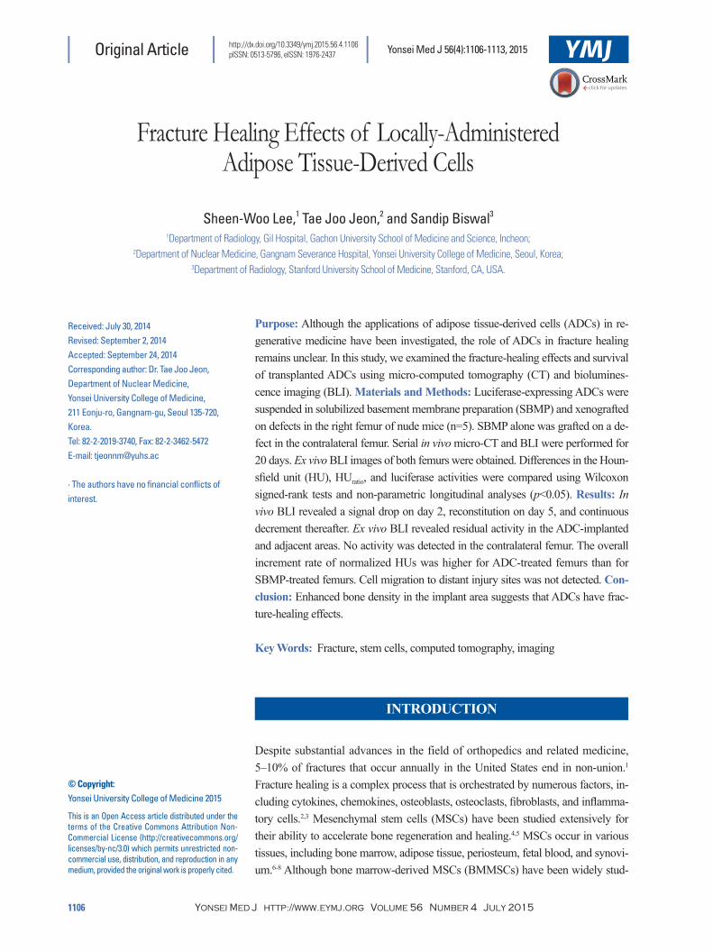
In vivo and ex vivo BLIApproximately 3 mg of luciferin (30 mg/mL solution of D-luciferin; Biosynth International, IL, USA) was adminis-tered via intraperitoneal injection. BLI was performed 10 min after the administration of luciferin using a cooled charge-coupled device camera (IVIS; Xenogen, CA, USA). The acquisition time was 5 min, and five mice were imaged simultaneously in the prone and bilateral decubitus posi-tions. BLI was performed on days 0, 2, 5, 7, 9, 12, 14, 16, and 20 after the operation and administration of ADCs. All BLI data from mice with implanted ADC were obtained from left decubitus images. The BLI data were quantified by drawing a 3.0-cm sized region of interest (ROI) to include the entire BLI signal detected on day 0. An ROI of the same size was used to measure BLI signals during the follow-up period to avoid loss of BLI activity from the areas near the
ied, these cells have limited applications in fracture healing owing to low yields and invasive harvesting procedures.9 On the other hand, adipose tissue might be an ideal source of MSCs because of its abundance in the body. In addition, adipose tissue-derived MSCs (ADMSCs) may be easily har-vested. Therefore, ADMSCs are being studied extensively for potential applications in the field of stem cell therapeutics.7,10
MSCs from diverse tissues and organs have been introduc-ed via systemic or local routes to treat fractures. Many stud-ies have demonstrated that systemically introduced BMMSCs and ADMSCs migrate to the fracture site and promote bone healing.11-14 However, other studies showed that only genet-ically modified cells that expressed bone morphogenic pro-tein-2 had significant bone regeneration effects, while AD-MSCs alone failed to promote bone healing.10,15 Therefore, the exact role of MSCs in fracture healing remains contro-versial and requires further investigation.
Recent advances in imaging technologies such as micro-computed tomography (CT) have made it possible to assess the healing process of the skeletal system by measuring the Hounsfield unit (HU) of bone tissue. Bioluminescence im-aging (BLI) has enabled us to monitor viable grafted cells expressing genetically engineered reporter genes such as green fluorescent protein (GFP) and luciferase.14 In our study, we harvested adipose tissue derived cells (ADCs) of trans-genic mice and achieved stable levels of luciferase activity without degradation during the entire follow-up period.
The main purpose of this study was to evaluate the bone-healing effects and survival or proliferation rates of locally administered ADCs during the early phase of fracture heal-ing. In addition, we attempted to evaluate the trafficking abil-ity of locally administered ADCs to remote fracture sites us-ing micro-CT and BLI.
MATERIALS AND METHODS
Mesenchymal stem cells isolation and cultureAll experiments were approved by the Institutional animal care and use committee of the Stanford University. To obtain steady reporter gene signals, we isolated ADCs from trans-genic mice carrying the β-actin promoter gene and express-ing GFP and firefly luciferase, instead of transfecting report-er genes into harvested ADCs. Subcutaneous adipose tissue was obtained from the region between the lower anterior ab-dominal wall and the inguinal area of transgenic mice, wash-ed in phosphate buffered saline several times, and finely
Sheen-Woo Lee, et al.
Yonsei Med J http://www.eymj.org Volume 56 Number 4 July 20151108
was evaluated by measuring the HU of the bone in a 0.2285 mm3-sized cylindrical volume of interest (VOI). The HU data obtained from the VOIs of bilateral femoral holes were plotted versus time. To minimize bias due to differences in the initial baseline HUs between the control and ADC-treated sites, we used another parameter, the HUratio. The HUratio was calculated by dividing the HU at each time point with the baseline HU. The HUs and HUratio of both the left and right femurs during the follow-up period were compared to eval-uate the bone-healing effects of ADCs.
Statistical analysisStatistical differences in the BLI signals during the follow-up period were assessed using the Mann-Whitney U test. The HUs and HUratio of the ADC- and SBMP-treated (con-trol) sites were compared and evaluated using the Wilcoxon signed-rank test. To analyze whether the differences be-tween the HUratio of the SBMP- and ADC-treated sites in-creased significantly at each time point, a non-parametric longitudinal analysis was performed. Differences were con-sidered statistically significant if p<0.05. All statistical eval-uations were performed using the SAS statistical software (version 9.2; SAS Institute Inc., Cary, NC, USA).
hole in the femur (Fig. 1). A time versus activity curve was obtained from this data to evaluate changes in luciferase ac-tivity with time. Bioluminescence activity was expressed in terms of average radiance (photon/cm2/s/steradian).
After the last CT follow-up, the animals were adminis-tered an intraperitoneal injection of D-luciferin and sacrificed after 10 min. ADC-treated and control femora were har-vested, and the soft tissue overlying the femoral defect was excised. BLI data wererecorded for the bones, with the en-trance to the hole facing the camera.
Micro-CTCT images of both femurs were obtained using a dedicated small animal CT scanner (eXplore RS micro-CT system, GE Medical Systems, Raleigh, NC, USA). The animals were placed on the scanner table in a prone position under inhalation anesthesia and exposed to a cone beam (80 kVp, 450 µA) for 400 ms. Image acquisition and reconstruction were performed using the eXplore Evolver and eXplore Re-construction Interface software, respectively. Micro-CT im-ages were obtained on days 0, 3, 5, 7, 12, 14, 16, and 20 after the operation and implantation of ADCs. The resolution of the reconstructed image (voxel) was 0.45×0.45×0.45 mm3 (Fig. 2). The change in bone density in the femoral defect
Fig. 1. Bioluminescence imaging of adipose tissue-derived cell-implanted fracture model. A region of interest (ROI) that included nearly the entire signal was drawn on the image recorded in the left lateral decubitus position.
ImageMin=-8207.6
Max=5.2759e+06p/sec/cm2/sr
ROI 1=2.4045e+07ROI 3=1.8715e+07
ROI 4=6.3893e+07ROI 5=3.4858e+07
×106
ROI 2=1.0286e+
5
4
3
2
1
Color barMin=26380
Max=5.2759e+06
Fracture-Healing Effects of ADCs
Yonsei Med J http://www.eymj.org Volume 56 Number 4 July 2015 1109
To determine whether the locally administered ADCs mi-grated to remote fracture sites, serial in vivo and ex vivo BLI were performed. No bioluminescence activity was detected in the left femur (control side) in both the ex vivo and in vivo studies, although ex vivo BLI of the right femora of all five mice showed persistent luciferase activities to various de-
RESULTS
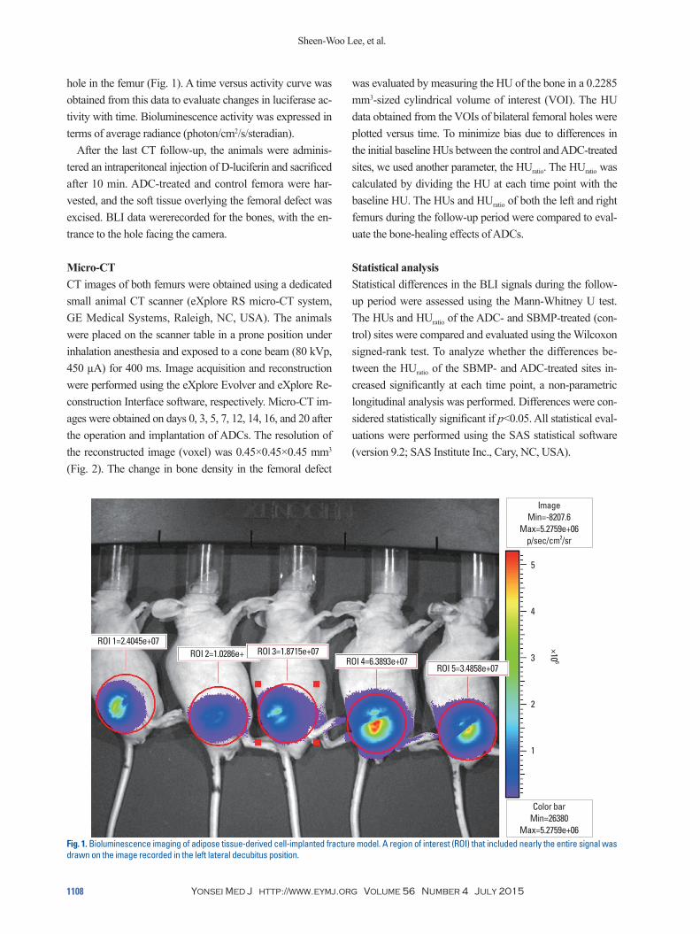
Second or third passage ADCs harvested from transgenic mice expressing two reporter genes–GFP and firefly lucif-erase–showed relevant bioluminescence activities in a pre-liminary in vitro BLI. Post transplantation, initial in vivo BLI of the five femur fracture models revealed strong biolumi-nescence activities; however, an abrupt decrease in activity was noted on day 2, followed by an increase on day 5. How-ever, the Mann-Whitney U test showed no significant differ-ences in the activities between days 0 and 5 (p=0.841). After day 5, bioluminescence activity decreased continuously with time. However, the time versus activity curve revealed a rela-tively rapid decrease in the activity until day 9 and a slow decrease thereafter, until day 14. This was followed by a st-eady level of activity in the remaining 6 days (Fig. 3). There were no significant differences in the bioluminescence ac-tivities between days 9 and 20. During the follow-up period, no significant increase in the BLI signal could be detected, which might be indicative of proliferation of implanted ADCs.
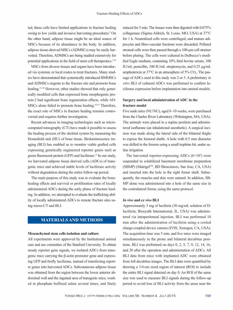
Fig. 2. Micro-computed tomography (CT) of bilateral femurs with holes (diameter, 0.5 mm). Baseline micro-CT images of control (A) and adipose tissue-de-rived cell (ADC)-implanted sides (B) showed relatively low bone densities in the holes. Final micro-CT images on day 20 showed increased bone densities in both the control (C) and ADC-implanted sides (D).
A
C
B
D
Fig. 3. The time vs. activity curve for firefly luciferase-expressing ADCs showed rapid decrement on post-operative day (POD) 7, followed by slow decrement until POD 12, before reaching a plateau. BLI, bioluminescence imaging; ADCs, adipose tissue-derived cells.
7e+7
6e+7
5e+7
4e+7
3e+7
2e+7
1e+7
0
-1e+7
BLI a
ctivi
ty (p
hoto
n/cm
2 /sec
/ste
radi
an)
POD
0
POD
2
POD
5
POD
7
POD
9
POD
12
POD
14
POD
16
POD
20
BLI activity of ADC
Sheen-Woo Lee, et al.
Yonsei Med J http://www.eymj.org Volume 56 Number 4 July 20151110
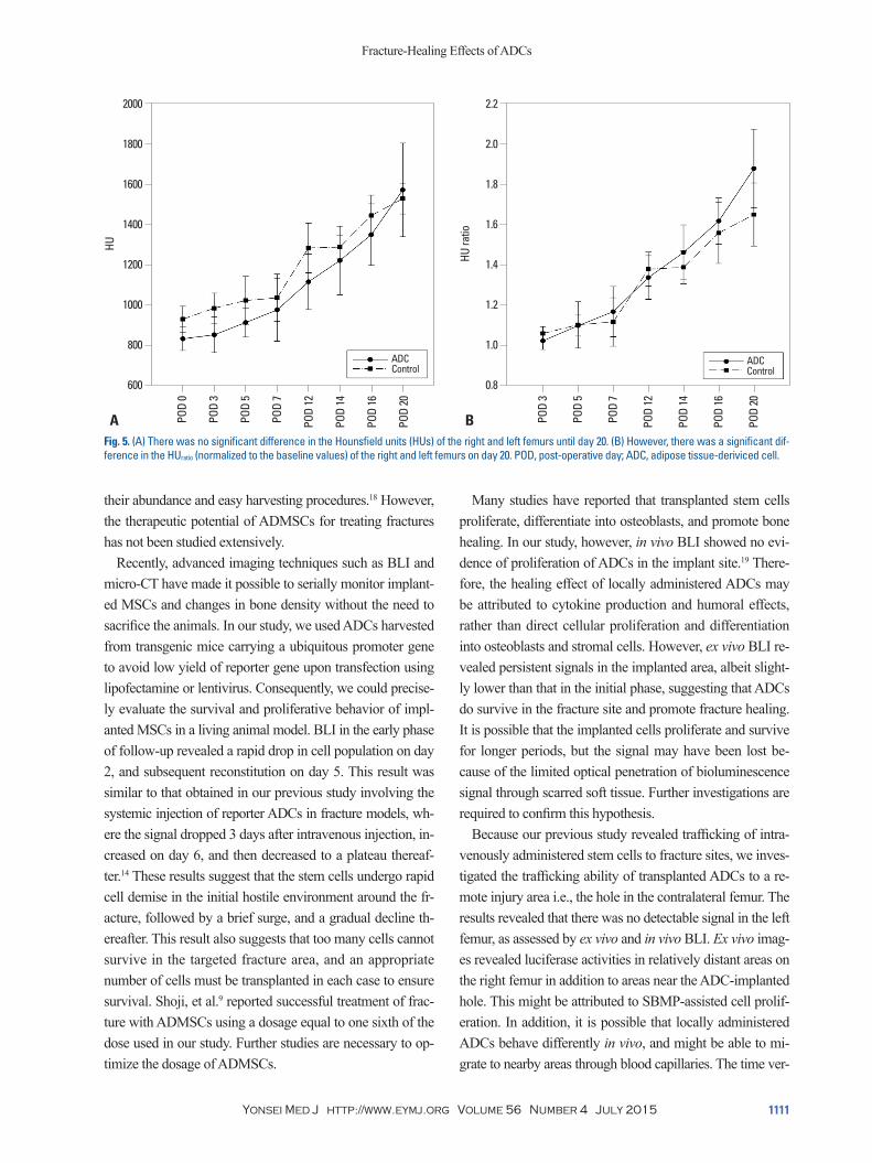
Micro-CT imaging of the femoral holes revealed higher HU values for the SBMP-treated side than for the ADC-tr-eated side from baseline to day 16; serial HUs increased ste-adily with time. However, there was no significant difference in the HUs between the two sides at each time point (Fig. 5A). The addition of the ADC pellet to SBMP may have in-creased the semi-liquid volume of the injectate in the ADC-SBMP group, compared with the SBMP-only group. This increased liquid volume might have washed away the he-matoma and calcifications in the bone defect, resulting in a lower HU on the ADC-treated side. Therefore, to minimize bias caused by the difference in baseline HUs between the two groups, HUratio, which represents the relative change of HUs at follow-up time points with respect to baseline HUs, was calculated and compared in the same manner. The HUratio graph revealed an increasing difference in the HUratio between the two groups (Fig. 5B), and a higher increment ratio in the ADC-treated group than in the control group. We also compared the differences between the HUratio of the two groups (inter-group gap) on days 16 and 20. The gap on day 20 was significantly larger than that on day 16 and earlier time points. These results were confirmed by performing a non-parametric longitudinal analysis (p<0.001). These re-sults imply that the ADC-treated side showed accelerated bone healing with time.
DISCUSSION
Although fractures are fairly common, treatment is not al-ways successful because of various factors such as injuries to surrounding soft tissues, poor blood supply, and osteopo-rosis.16 Therefore, fracture treatment in complicated cases such as in the elderly and in patients of osteoporosis and im-mune suppression, has been difficult despite advances in surgery and regenerative medicine.2 Among the various new therapeutic approaches that have been proposed for the treatment of fractures, stem cells are promising candidates that activate bone healing. In particular, MSCs are known to be involved in the bone healing process. It has been sug-gested that MSCs are recruited to the injury site, where they proliferate and differentiate into osteoblasts.5 Initially, most studies investigated the bone-healing properties of BM-MSCs. However, these cells have limited applications as therapeutic agents because of their invasive harvesting pro-cedures and relatively low yields.17 Therefore, ADMSCs were investigated as an alternative to BMMSCs owing to
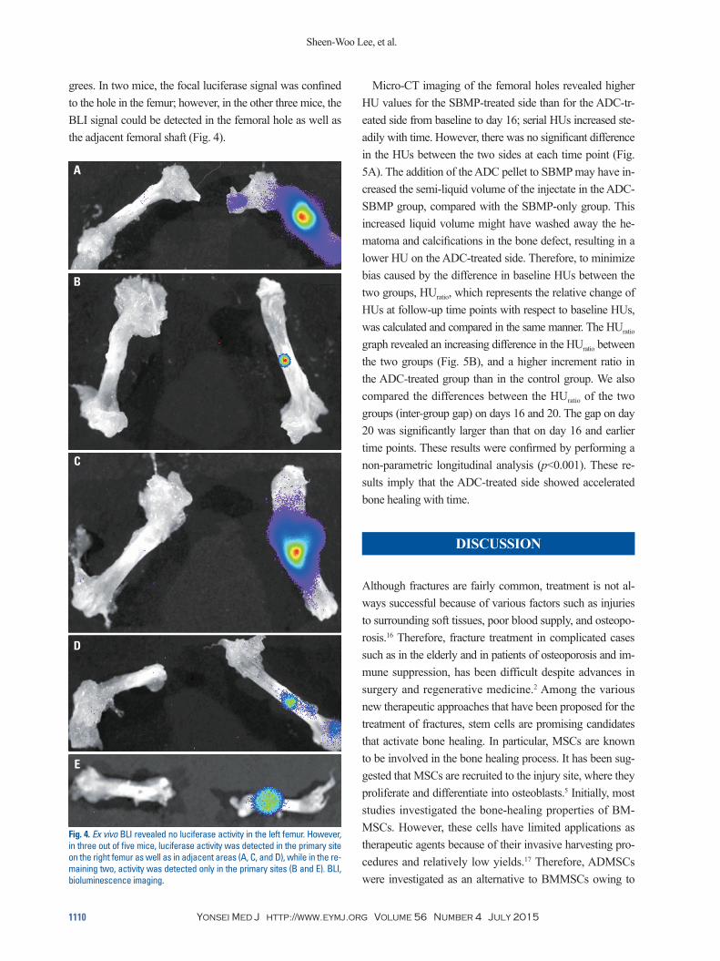
grees. In two mice, the focal luciferase signal was confined to the hole in the femur; however, in the other three mice, the BLI signal could be detected in the femoral hole as well as the adjacent femoral shaft (Fig. 4).
Fig. 4. Ex vivo BLI revealed no luciferase activity in the left femur. However, in three out of five mice, luciferase activity was detected in the primary site on the right femur as well as in adjacent areas (A, C, and D), while in the re-maining two, activity was detected only in the primary sites (B and E). BLI, bioluminescence imaging.
A
E
D
B
C
Fracture-Healing Effects of ADCs
Yonsei Med J http://www.eymj.org Volume 56 Number 4 July 2015 1111
Many studies have reported that transplanted stem cells proliferate, differentiate into osteoblasts, and promote bone healing. In our study, however, in vivo BLI showed no evi-dence of proliferation of ADCs in the implant site.19 There-fore, the healing effect of locally administered ADCs may be attributed to cytokine production and humoral effects, rather than direct cellular proliferation and differentiation into osteoblasts and stromal cells. However, ex vivo BLI re-vealed persistent signals in the implanted area, albeit slight-ly lower than that in the initial phase, suggesting that ADCs do survive in the fracture site and promote fracture healing. It is possible that the implanted cells proliferate and survive for longer periods, but the signal may have been lost be-cause of the limited optical penetration of bioluminescence signal through scarred soft tissue. Further investigations are required to confirm this hypothesis.
Because our previous study revealed trafficking of intra-venously administered stem cells to fracture sites, we inves-tigated the trafficking ability of transplanted ADCs to a re-mote injury area i.e., the hole in the contralateral femur. The results revealed that there was no detectable signal in the left femur, as assessed by ex vivo and in vivo BLI. Ex vivo imag-es revealed luciferase activities in relatively distant areas on the right femur in addition to areas near the ADC-implanted hole. This might be attributed to SBMP-assisted cell prolif-eration. In addition, it is possible that locally administered ADCs behave differently in vivo, and might be able to mi-grate to nearby areas through blood capillaries. The time ver-
their abundance and easy harvesting procedures.18 However, the therapeutic potential of ADMSCs for treating fractures has not been studied extensively.
Recently, advanced imaging techniques such as BLI and micro-CT have made it possible to serially monitor implant-ed MSCs and changes in bone density without the need to sacrifice the animals. In our study, we used ADCs harvested from transgenic mice carrying a ubiquitous promoter gene to avoid low yield of reporter gene upon transfection using lipofectamine or lentivirus. Consequently, we could precise-ly evaluate the survival and proliferative behavior of impl-anted MSCs in a living animal model. BLI in the early phase of follow-up revealed a rapid drop in cell population on day 2, and subsequent reconstitution on day 5. This result was similar to that obtained in our previous study involving the systemic injection of reporter ADCs in fracture models, wh-ere the signal dropped 3 days after intravenous injection, in-creased on day 6, and then decreased to a plateau thereaf-ter.14 These results suggest that the stem cells undergo rapid cell demise in the initial hostile environment around the fr-acture, followed by a brief surge, and a gradual decline th-ereafter. This result also suggests that too many cells cannot survive in the targeted fracture area, and an appropriate number of cells must be transplanted in each case to ensure survival. Shoji, et al.9 reported successful treatment of frac-ture with ADMSCs using a dosage equal to one sixth of the dose used in our study. Further studies are necessary to op-timize the dosage of ADMSCs.
Fig. 5. (A) There was no significant difference in the Hounsfield units (HUs) of the right and left femurs until day 20. (B) However, there was a significant dif-ference in the HUratio (normalized to the baseline values) of the right and left femurs on day 20. POD, post-operative day; ADC, adipose tissue-deriviced cell.
2000
1800
1600
1400
1200
1000
800
600
2.2
2.0
1.8
1.6
1.4
1.2
1.0
0.8
HU
HU ra
tio
POD
0
POD
3
POD
5
POD
7
POD
12
POD
14
POD
16
POD
20
POD
3
POD
5
POD
7
POD
12
POD
14
POD
16
POD
20
ADCControl
ADCControl
A B
Sheen-Woo Lee, et al.
Yonsei Med J http://www.eymj.org Volume 56 Number 4 July 20151112
improvements in the sensitivity of BLI and reporter gene technology might enable the monitoring of locally adminis-tered ADCs for longer time intervals.
In summary, BLI data revealed that the levels of locally administered ADCs fluctuated in the early postoperative phase and reached a plateau thereafter, without providing definite evidence of cell proliferation or migration to remote sites of injury. Despite the relatively short follow-up period, micro-CT revealed that the implanted ADCs enhanced the rate of fracture healing during the study period, as suggested by the incremental patterns of the HUs and HUratio. Our re-sults suggest that ADCs play a role in fracture healing; this ef-fect might be mediated by cytokines or other humoral effects.
ACKNOWLEDGEMENTS
We gratefully acknowledge Dr. Contag (Stanford University, USA) for providing the transgenic mice.
This work was supported by academic promotion grant of the Korean Society of Nuclear Medicine (2010).
REFERENCES
1. Brighton CT, Shaman P, Heppenstall RB, Esterhai JL Jr, Pollack SR, Friedenberg ZB. Tibial nonunion treated with direct current, capacitive coupling, or bone graft. Clin Orthop Relat Res 1995: 223-34.
2. Giannoudis P, Psarakis S, Kontakis G. Can we accelerate fracture healing? A critical analysis of the literature. Injury 2007;38 Suppl 1:S81-9.
3. Tsiridis E, Upadhyay N, Giannoudis P. Molecular aspects of frac-ture healing: which are the important molecules? Injury 2007;38 Suppl 1:S11-25.
4. Bielby R, Jones E, McGonagle D. The role of mesenchymal stem cells in maintenance and repair of bone. Injury 2007;38 Suppl 1: S26-32.
5. Knight MN, Hankenson KD. Mesenchymal Stem Cells in Bone Regeneration. Adv Wound Care (New Rochelle) 2013;2:306-16.
6. De Bari C, Dell’Accio F, Tylzanowski P, Luyten FP. Multipotent mesenchymal stem cells from adult human synovial membrane. Arthritis Rheum 2001;44:1928-42.
7. Zuk PA, Zhu M, Ashjian P, De Ugarte DA, Huang JI, Mizuno H, et al. Human adipose tissue is a source of multipotent stem cells. Mol Biol Cell 2002;13:4279-95.
8. Kern S, Eichler H, Stoeve J, Klüter H, Bieback K. Comparative analysis of mesenchymal stem cells from bone marrow, umbilical cord blood, or adipose tissue. Stem Cells 2006;24:1294-301.
9. Shoji T, Ii M, Mifune Y, Matsumoto T, Kawamoto A, Kwon SM, et al. Local transplantation of human multipotent adipose-derived stem cells accelerates fracture healing via enhanced osteogenesis and angiogenesis. Lab Invest 2010;90:637-49.
sus activity curves and the ex vivo BLI images suggest that the detection of activity in areas adjacent to the hole in the right femur may be attributed to local migration of implant-ed cells rather than proliferation. If the implanted cells had proliferated, bioluminescence activity would have increased at more than one instance during the follow-up period.
Using high-resolution micro-CT, we evaluated the differ-ences in early bone changes between the ADC-treated and control sides. Although the final HU value of the ADC-treat-ed side was higher than that of the control side, there was no significant difference between the serial HUs of the ADC-treated and control femora; this may be attributed to the low-er baseline HU value of the ADC-treated side than the con-trol side (Fig. 5A). Next, we compared the HUratio of both sides to minimize bias arising from the different baseline val-ues. We found that the HUratio of the ADC-treated side was higher than that of the control side from day 7 onwards, and the rate of increment on the ADC-treated side was higher than that on the control side, suggesting that ADC-SBMP treatment promotes bone healing. These results contradict those obtained in a study by Lyons, et al.,20 which suggested that MSCs might adversely affect healing by creating a bar-rier to host response. Because of the low levels of biolumi-nescence activity on day 20, we did not continue the micro-CT analysis for a longer follow-up period. Instead, we an-alyzed luciferase activity using ex vivo BLI. Therefore, com-plementary research that includes long-term monitoring of ADCs is necessary. It may be noted here that a study by Shoji, et al.9 revealed that ADMSCs might have long-term healing effects on fractured bones.
There are several limitations to our study. Most important-ly, the follow-up period was too short to evaluate the bone union status of the fracture models. However, the main aim of our study was to evaluate the behavior of locally admin-istered ADCs in a living animal and to monitor early chang-es in bone density using micro-CT. Therefore, when the in vivo BLI revealed diminished activity, we had to sacrifice the animals to determine whether the ADCs had survived in the femoral holes. A second limitation was that we could not perform an immunohistochemical analysis of the speci-mens owing to a delay in tissue preservation to perform ex vivo BLI. However, many previous studies have investigat-ed the histological characteristics of MSCs. Therefore, we reasoned that demonstrating the presence of surviving ADCs by ex vivo BLI might be more useful for future studies. Be-cause of the limited sensitivity of BLI, follow-up for a lon-ger period was not possible in our study. However, future

Fracture-Healing Effects of ADCs
Yonsei Med J http://www.eymj.org Volume 56 Number 4 July 2015 1113
16. Bigham-Sadegh A, Oryan A. Basic concepts regarding fracture healing and the current options and future directions in managing bone fractures. Int Wound J 2014 Feb 21 [Epub]. http://dx.doi.org/10.1111/iwj.12231.
17. Schubert T, Lafont S, Beaurin G, Grisay G, Behets C, Gianello P, et al. Critical size bone defect reconstruction by an autologous 3D osteogenic-like tissue derived from differentiated adipose MSCs. Biomaterials 2013;34:4428-38.
18. Schubert T, Xhema D, Vériter S, Schubert M, Behets C, Delloye C, et al. The enhanced performance of bone allografts using osteo-genic-differentiated adipose-derived mesenchymal stem cells. Bio-materials 2011;32:8880-91.
19. Bruder SP, Kurth AA, Shea M, Hayes WC, Jaiswal N, Kadiyala S. Bone regeneration by implantation of purified, culture-expanded human mesenchymal stem cells. J Orthop Res 1998;16:155-62.
20. Lyons FG, Al-Munajjed AA, Kieran SM, Toner ME, Murphy CM, Duffy GP, et al. The healing of bony defects by cell-free collagen-based scaffolds compared to stem cell-seeded tissue engineered constructs. Biomaterials 2010;31:9232-43.
10. Peterson B, Zhang J, Iglesias R, Kabo M, Hedrick M, Benhaim P, et al. Healing of critically sized femoral defects, using genetically modified mesenchymal stem cells from human adipose tissue. Tis-sue Eng 2005;11:120-9.
11. De Ugarte DA, Morizono K, Elbarbary A, Alfonso Z, Zuk PA, Zhu M, et al. Comparison of multi-lineage cells from human adipose tissue and bone marrow. Cells Tissues Organs 2003;174:101-9.
12. Bruder SP, Jaiswal N, Ricalton NS, Mosca JD, Kraus KH, Kadiya-la S. Mesenchymal stem cells in osteobiology and applied bone re-generation. Clin Orthop Relat Res 1998;(355 Suppl):S247-56.
13. Cowan CM, Shi YY, Aalami OO, Chou YF, Mari C, Thomas R, et al. Adipose-derived adult stromal cells heal critical-size mouse cal-varial defects. Nat Biotechnol 2004;22:560-7.
14. Lee SW, Padmanabhan P, Ray P, Gambhir SS, Doyle T, Contag C, et al. Stem cell-mediated accelerated bone healing observed with in vivo molecular and small animal imaging technologies in a model of skeletal injury. J Orthop Res 2009;27:295-302.
15. Li H, Dai K, Tang T, Zhang X, Yan M, Lou J. Bone regeneration by implantation of adipose-derived stromal cells expressing BMP-2. Biochem Biophys Res Commun 2007;356:836-42.