Embed Size (px)
Citation preview

Foundations in Microbiology
Sixth Edition
Chapter 17
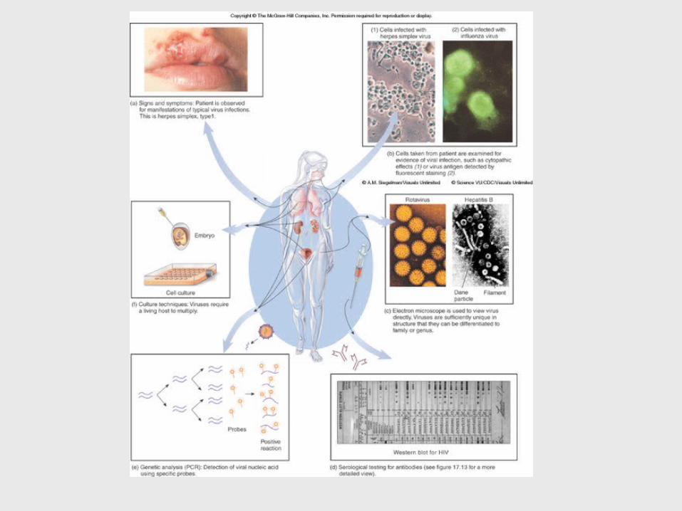
Diagnosing Infections
Lecture PowerPoint to accompany
Talaro
Copyright © The McGraw-Hill Companies, Inc. Permission required for reproduction or display.

Survey of Microbial Disease
Methods of identifying unknown microbes fall into three categories:
1. Phenotypic - observable microscopic and macroscopic characteristics
2. Genotypic – genetic make up
3. Immunological – serology; antibody-antigen reactions

Phenotypic Methods
• Microscopic morphology – fresh or stained microorganisms from specimen; shape, size, stain reaction, cell structures
• Macroscopic morphology – colony appearance; texture, size, shape, pigment, growth requirements
• Physiological/biochemical characteristics – detection of presence or absence of particular enzymes or metabolic pathways
• Chemical analysis – analyze specific chemical composition; cell wall peptides, cell membrane lipids
Genotypic Methods
• Assess genetic make-up.
• Culture is not necessary.
• Precise, automated methods, quick results

Immunological Methods
• Specific antibodies are used to detect antigens.

Specimen Collection and Laboratory Methods
• Sampling body sites or fluids for suspected infectious agent
• Results depend on specimen collection, handling, transport and storage.
• Aseptic procedures should be used.

Insert figure 17.1Sampling sites


Phenotypic Methods
• Observation – – macroscopic - cultivation – colony
appearance, growth requirements, appropriate media
– microscopic - differential and special stains – Gram, AFB, fluorescent antibody stains
• Direct antigen/antibody testing
• Biochemical testing – physiological reactions to nutrients as evidence of the absence or presence of enzymes


• Important to consider whether microbe recovered and identified is actually causing the disease or simply normal flora

Genotypic Methods
• DNA analysis – Assess the proportion of G + C nucleotides
relative to A + T content.– Determine DNA or ribosomal RNA sequences
using probes and polymerase chain reactions.

Immunological Methods
• Serology – attempts to detect signs of infection in a patient by identifying specific antibodies in vitro
• Visible reactions include precipitates, color changes, or the release of radioactivity.
• Tests can be used to identify and to determine the amount of antibody in serum – titer.


Agglutination and Precipitation Reactions
• Agglutination testing – antibody cross links whole-cell antigens, forming complexes that settle out and form visible clumps– blood typing, some bacterial and viral diseases
• Precipitation tests – soluble antigen is made insoluble by an antibody – syphilis,
• Western blot – immunoelectrophoresis; separates antigens into bands – HIV


Insert figure 17.10Cellular\molecular view

Complement Fixation
• Detect antibodies that fix complement and lyse target cells– antigen, antibody, complement, and sensitized
sheep RBCs– If complement is fixed by the Ag-Ab, the RBCs
will not be lysed.


Immunoassays
• Extremely sensitive to detect trace antigens and antibodies
• Radioimmunoassay (RIA) – antigens and antibodies labeled with radioactive isotopes
• Enzyme-linked immunosorbent assay (ELISA) – enzyme-antibody complex produces a colored product when an enzyme-substrate reaction occurs


In vivo Testing
• Antigens are introduced directly into the body to determine the presence or absence of antibodies.– tuberculin skin test, allergy testing