Embed Size (px)
Citation preview
Foundations in Microbiology
Chapter
4
PowerPoint to accompany
Fifth Edition
Talaro
Copyright The McGraw-Hill Companies, Inc. Permission required for reproduction or display.
Prokaryotic Profiles: the Bacteria and the Archaea
Chapter 4
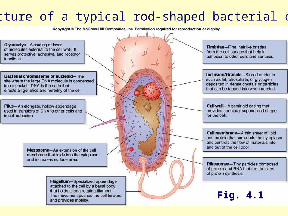
3Fig. 4.1
Stucture of a typical rod-shaped bacterial cell
4
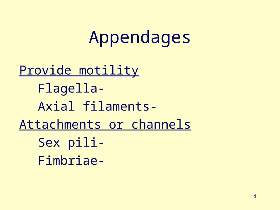
Appendages
Provide motility
Flagella-
Axial filaments-
Attachments or channels
Sex pili-
Fimbriae-
5
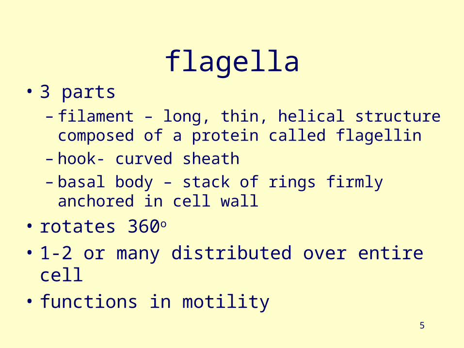
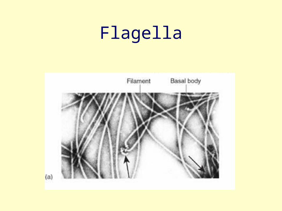
flagella• 3 parts
– filament – long, thin, helical structure composed of a protein called flagellin
– hook- curved sheath– basal body – stack of rings firmly anchored in cell
wall
• rotates 360o
• 1-2 or many distributed over entire cell
• functions in motility
Flagella
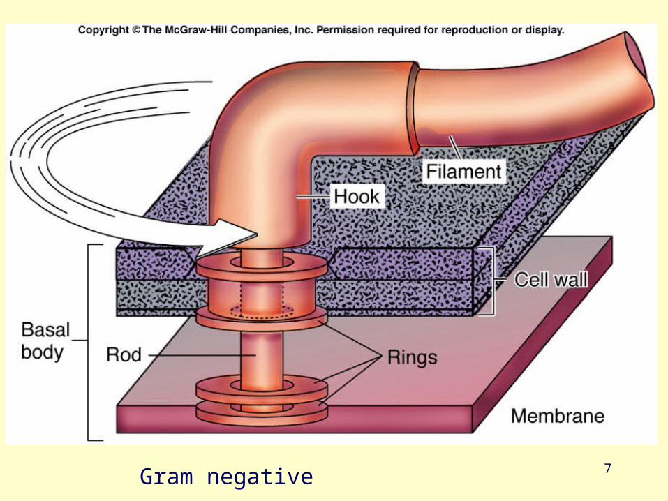
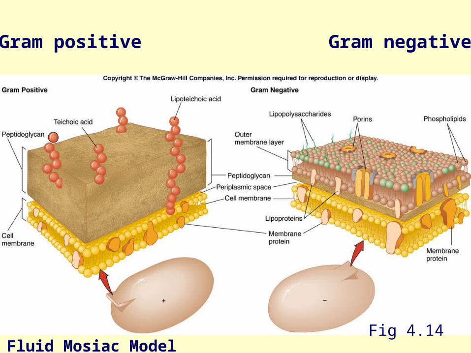
7Gram negative
8
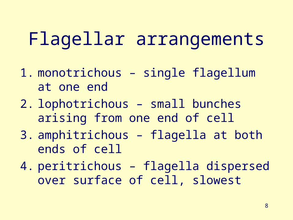
Flagellar arrangements
1. monotrichous – single flagellum at one end
2. lophotrichous – small bunches arising from one end of cell
3. amphitrichous – flagella at both ends of cell
4. peritrichous – flagella dispersed over surface of cell, slowest
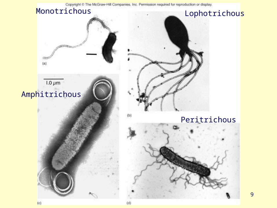
9
Monotrichous Lophotrichous
Amphitrichous
Peritrichous
10
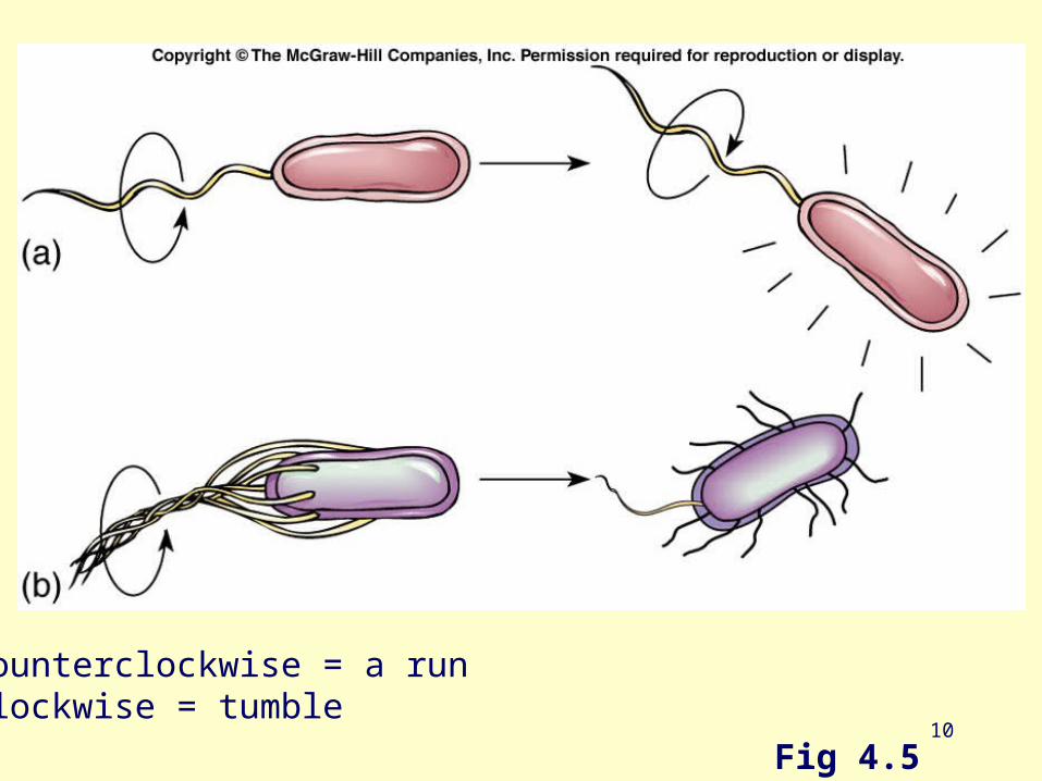
Fig 4.5
Counterclockwise = a runClockwise = tumble
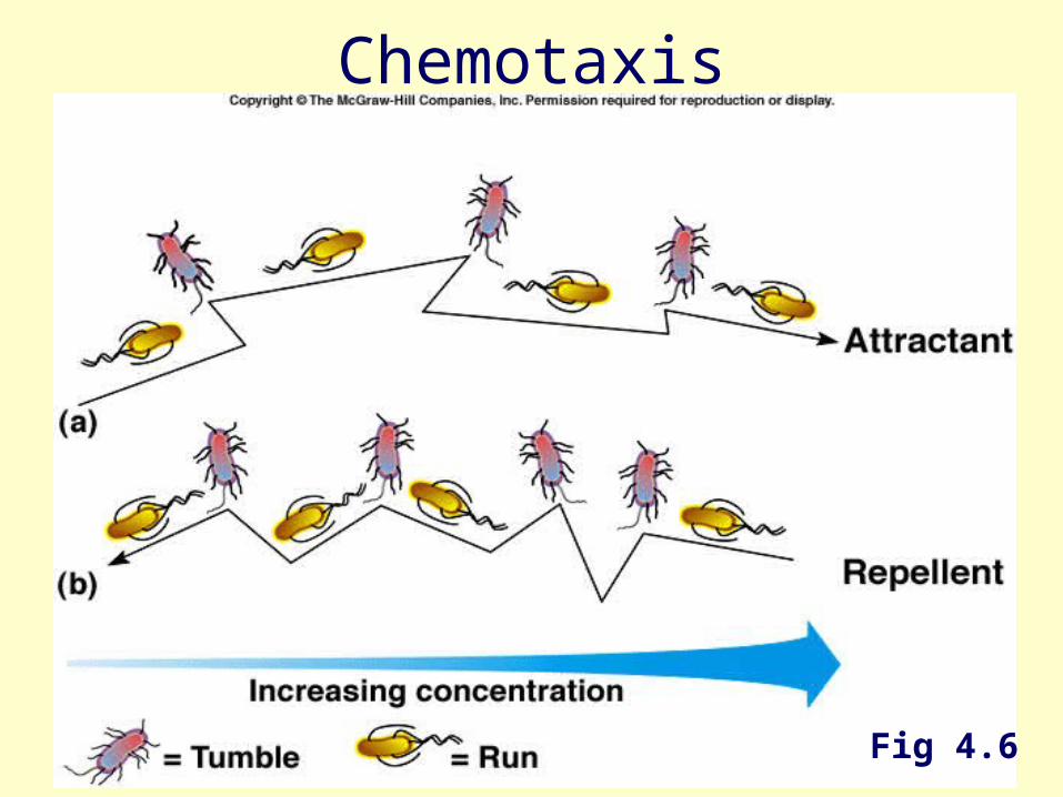
11Fig 4.6
Chemotaxis
12
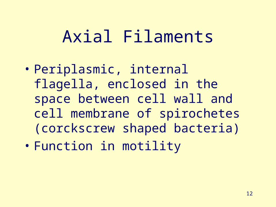
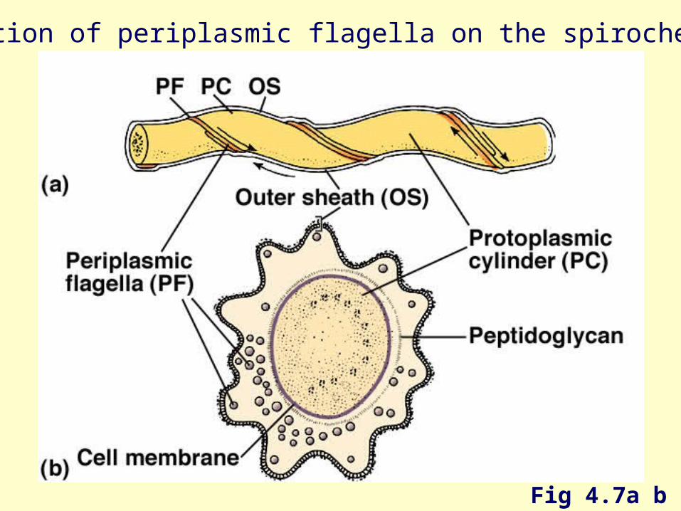
Axial Filaments
• Periplasmic, internal flagella, enclosed in the space between cell wall and cell membrane of spirochetes (corckscrew shaped bacteria)
• Function in motility
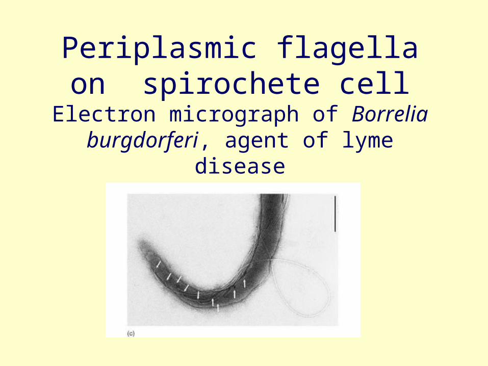
Periplasmic flagella on spirochete cell
Electron micrograph of Borrelia burgdorferi, agent of lyme disease
14
Fig 4.7a b
Orientation of periplasmic flagella on the spirochete cell
15
Fimbrae
• fine hairlike bristles from the cell surface
• function in adhesion to other cells and surfaces
16
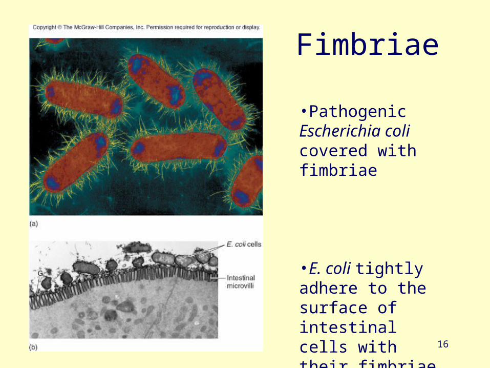
Fimbriae
•Pathogenic Escherichia coli covered with fimbriae
•E. coli tightly adhere to the surface of intestinal cells with their fimbriae
17
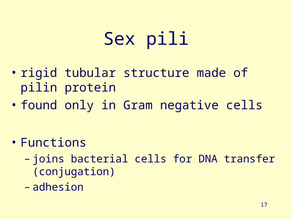
Sex pili
• rigid tubular structure made of pilin protein
• found only in Gram negative cells
• Functions – joins bacterial cells for DNA transfer (conjugation)– adhesion

18
Conjugation
19
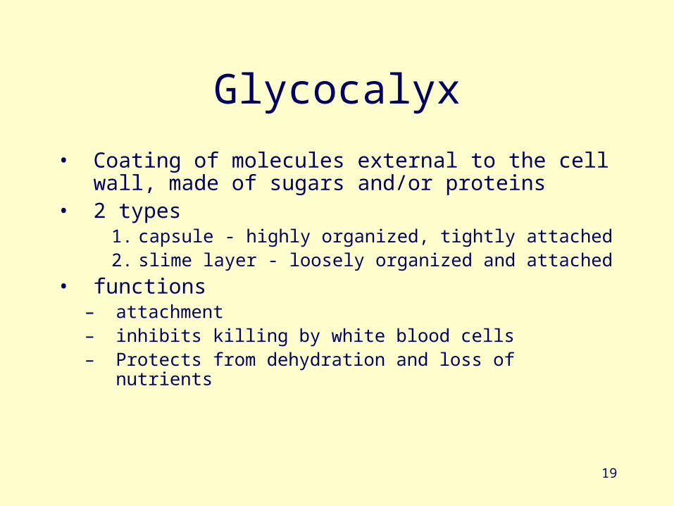
Glycocalyx
• Coating of molecules external to the cell wall, made of sugars and/or proteins
• 2 types1. capsule - highly organized, tightly attached2. slime layer - loosely organized and attached
• functions– attachment– inhibits killing by white blood cells– Protects from dehydration and loss of nutrients
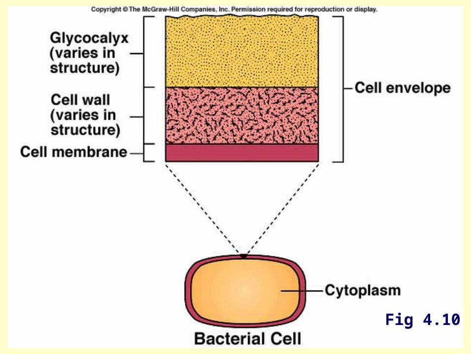
20Fig 4.10
21
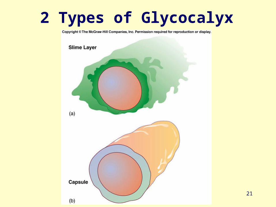
2 Types of Glycocalyx
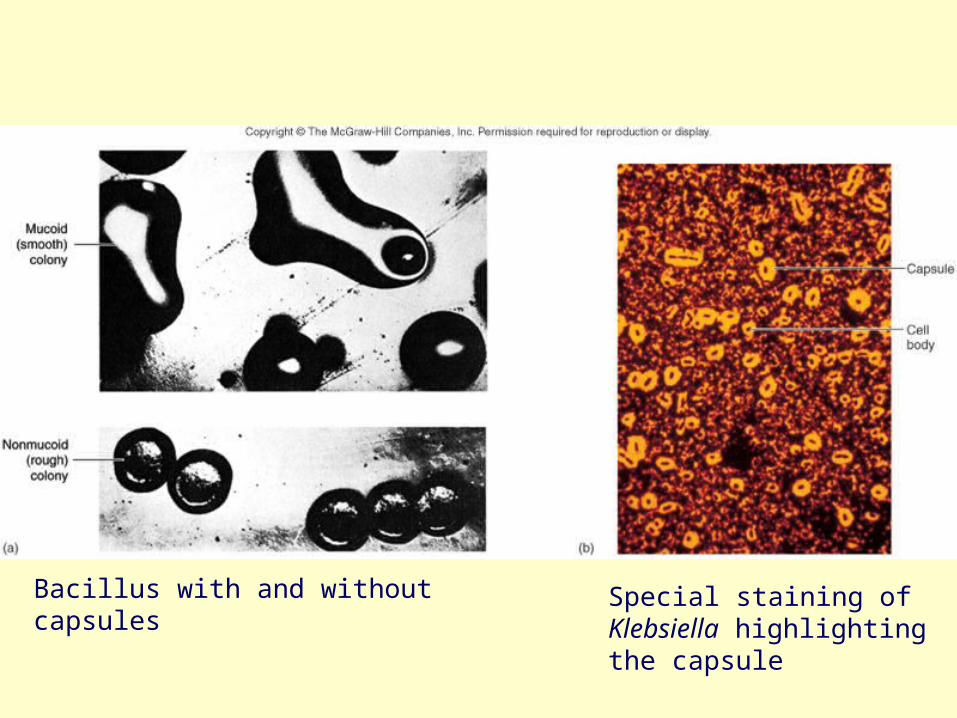
Fig. 4.12
Bacillus with and without capsules Special staining of Klebsiella highlighting the capsule
23
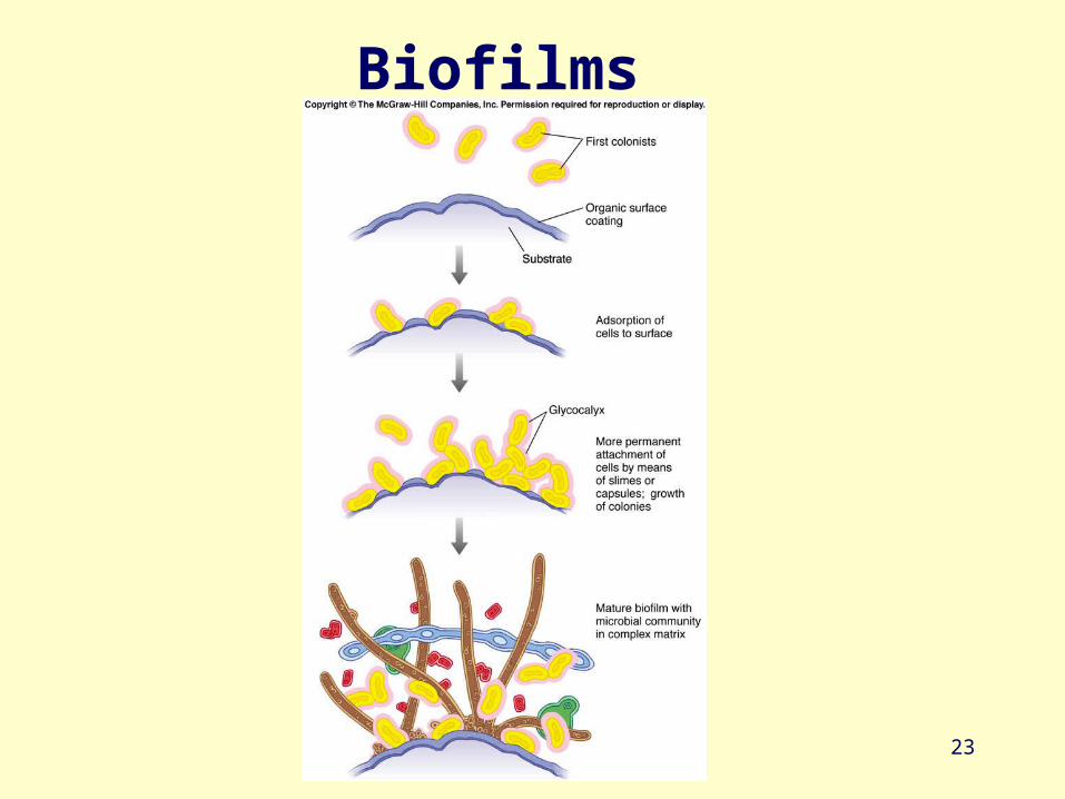
Biofilms
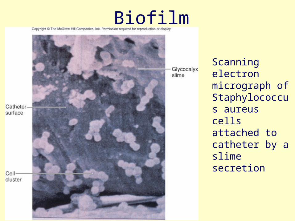
Fig. 4.13
Biofilm
Scanning electron micrograph of Staphylococcus aureus cells attached to catheter by a slime secretion

Cell wall composition
26
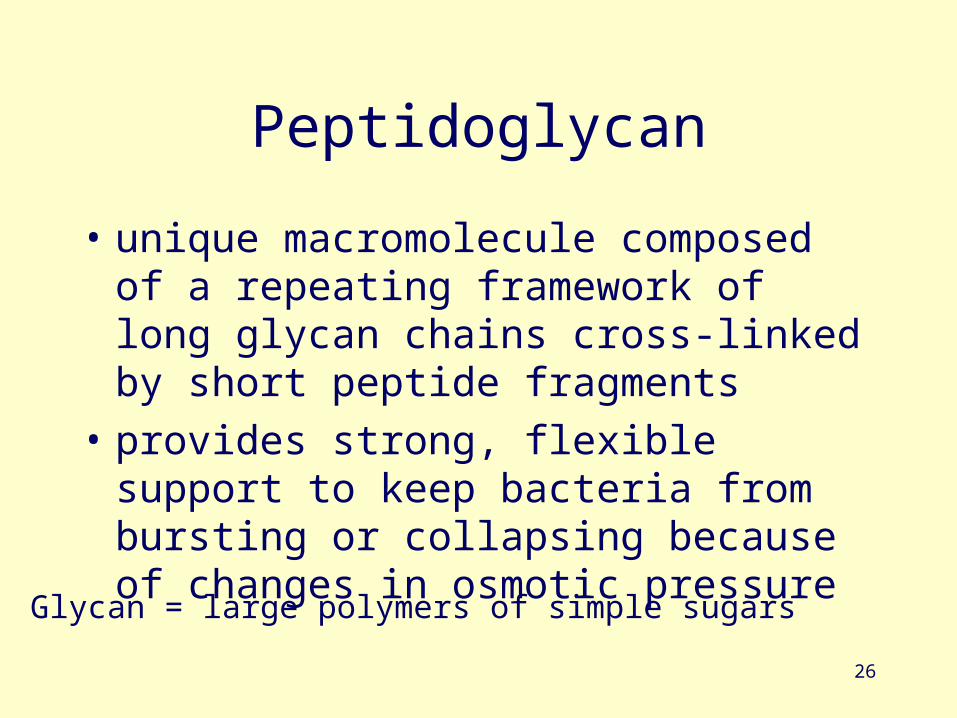
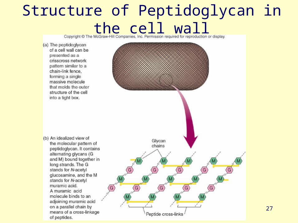
Peptidoglycan
• unique macromolecule composed of a repeating framework of long glycan chains cross-linked by short peptide fragments
• provides strong, flexible support to keep bacteria from bursting or collapsing because of changes in osmotic pressure
Glycan = large polymers of simple sugars
27
Structure of Peptidoglycan in the cell wall
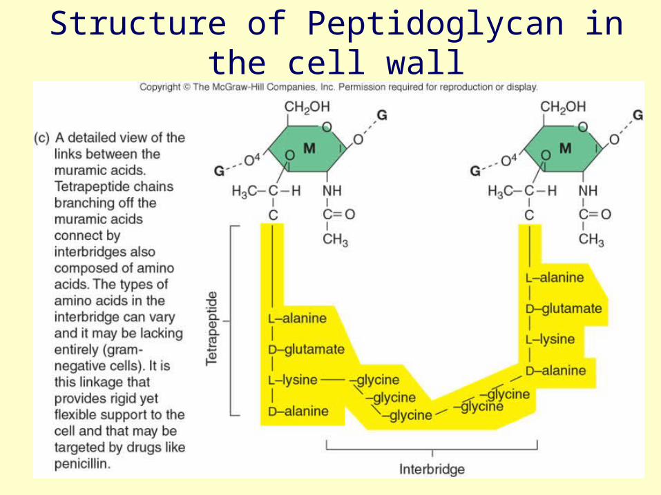
Fig. 4.14c
Structure of Peptidoglycan in the cell wall
29
Peptidoglycan
• Lysozyme (enzyme in tears and salivia) hydrolyzes the bonds in glycan chains of invading bacteria, causing the wall to break down
• Several drugs including penicillin target the peptide cross-links in the peptidoglycan
30
4 groups based on cell wall composition
1. Gram positive cells
2. Gram negative cells
3. Bacteria without cell walls-mycoplasms
4. Bacteria with chemically unique cell walls
31
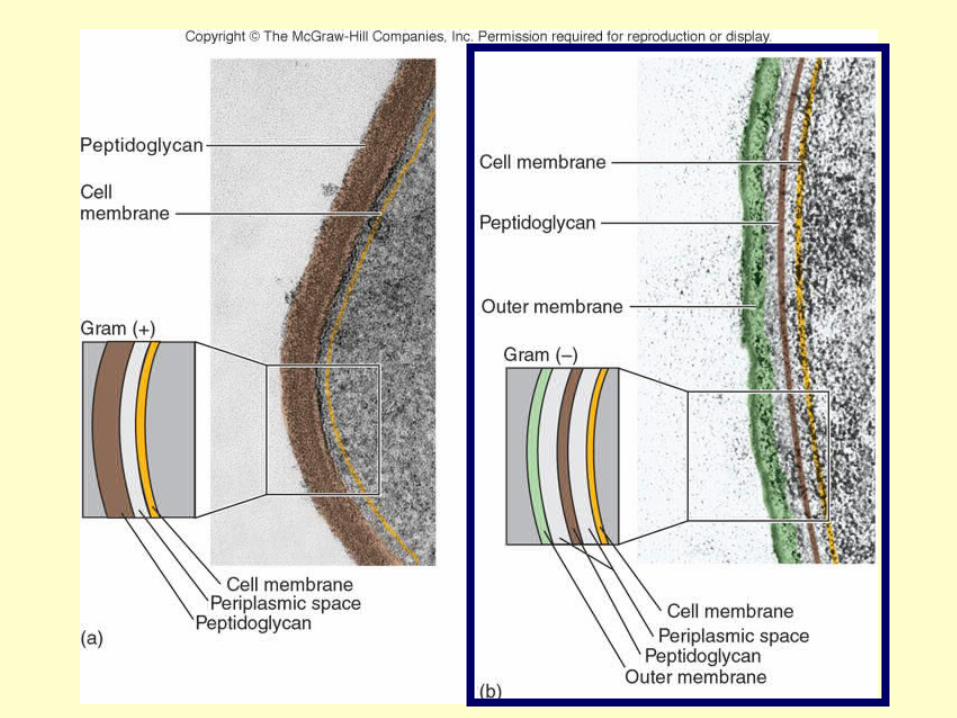
Gram positive Gram negative
Fig 4.14Fluid Mosiac Model
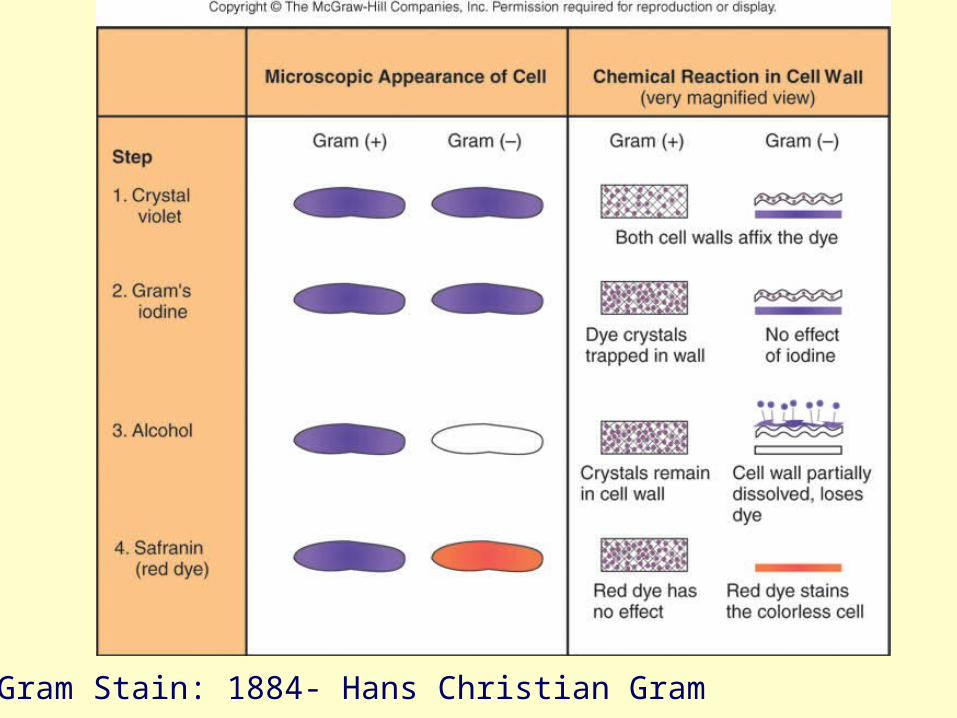
The Gram Stain: 1884- Hans Christian Gram
33
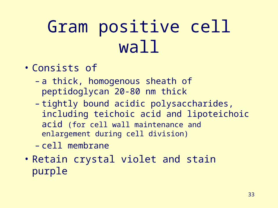
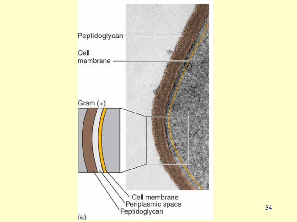
Gram positive cell wall
• Consists of – a thick, homogenous sheath of peptidoglycan
20-80 nm thick– tightly bound acidic polysaccharides, including
teichoic acid and lipoteichoic acid (for cell wall maintenance and enlargement during cell division)
– cell membrane
• Retain crystal violet and stain purple
34
35
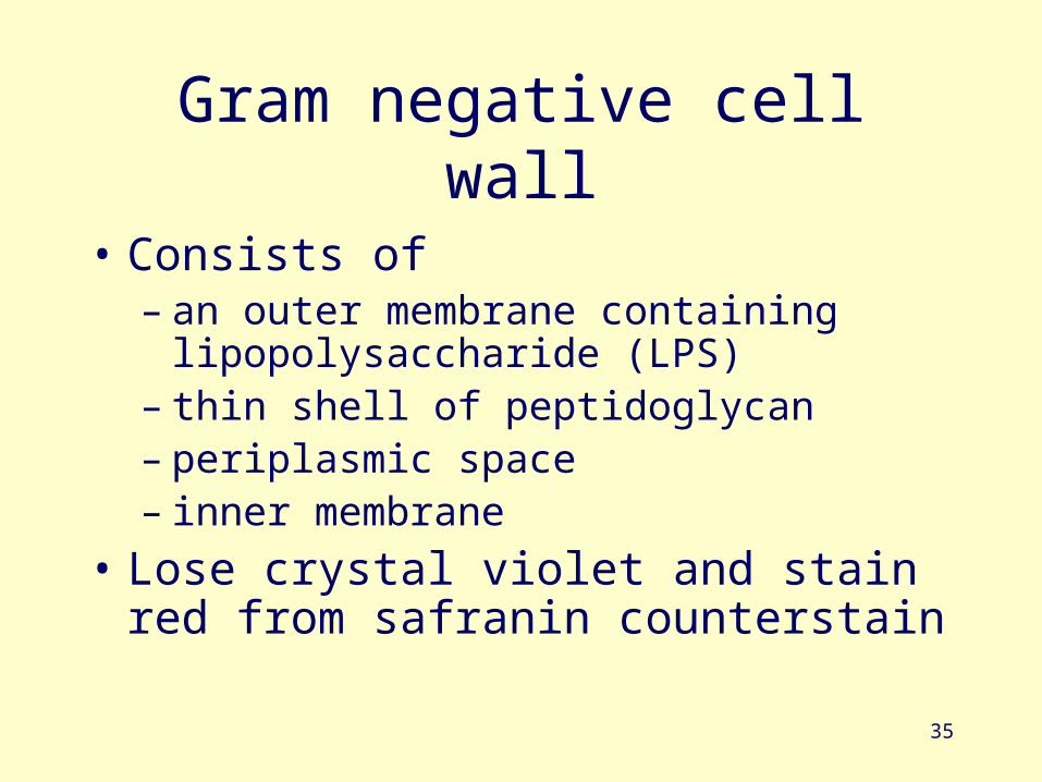
Gram negative cell wall
• Consists of– an outer membrane containing
lipopolysaccharide (LPS)– thin shell of peptidoglycan– periplasmic space– inner membrane
• Lose crystal violet and stain red from safranin counterstain
36

Basic Internal Components
38
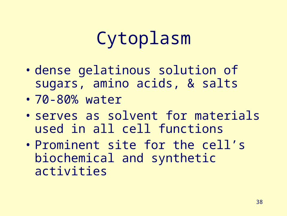
Cytoplasm
• dense gelatinous solution of sugars, amino acids, & salts
• 70-80% water• serves as solvent for materials used in all
cell functions• Prominent site for the cell’s biochemical
and synthetic activities
39
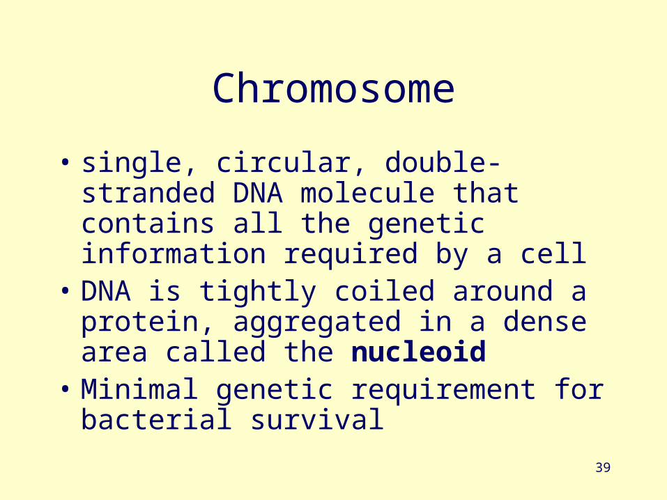
Chromosome
• single, circular, double-stranded DNA molecule that contains all the genetic information required by a cell
• DNA is tightly coiled around a protein, aggregated in a dense area called the nucleoid
• Minimal genetic requirement for bacterial survival
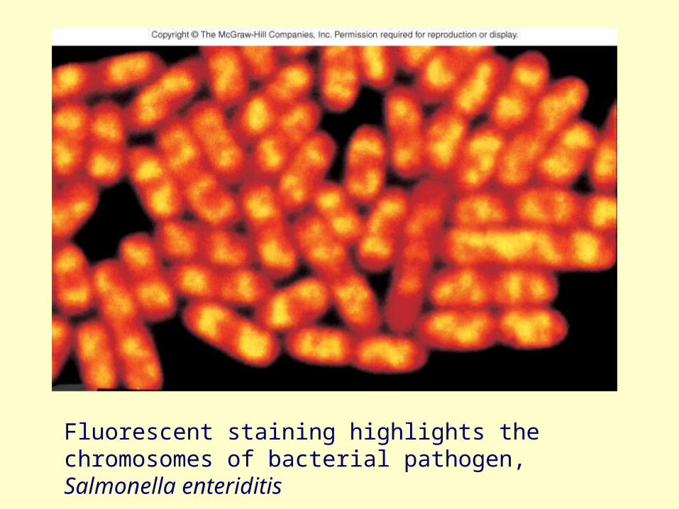
Fig. 4.18
Fluorescent staining highlights the chromosomes of bacterial pathogen, Salmonella enteriditis
41
plasmids
• small circular, double-stranded DNA• free or integrated into the chromosome• duplicated and passed on to offspring• not essential to bacterial growth & metabolism• may encode antibiotic resistance, tolerance to
toxic metals, enzymes & toxins• used in genetic engineering- readily manipulated
& transferred from cell to cell
42
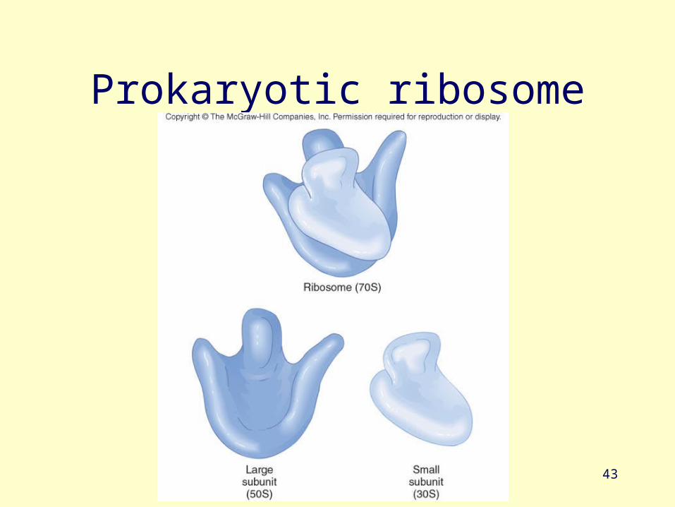
ribosomes• made of 60% ribosomal RNA & 40%
protein
• consist of 2 subunits: large & small
• procaryotic differ from eucaryotic ribosomes in size & number of proteins
• site of protein synthesis
• All cells have ribosomes.
43
Prokaryotic ribosome
44
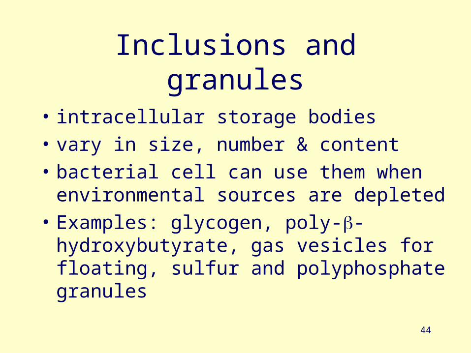
Inclusions and granules
• intracellular storage bodies
• vary in size, number & content
• bacterial cell can use them when environmental sources are depleted
• Examples: glycogen, poly--hydroxybutyrate, gas vesicles for floating, sulfur and polyphosphate granules
45
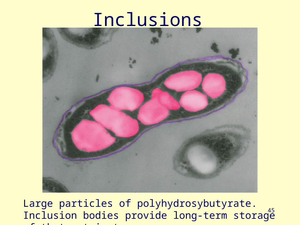
Inclusions
Large particles of polyhydrosybutyrate. Inclusion bodies provide long-term storage of that nutrient
46
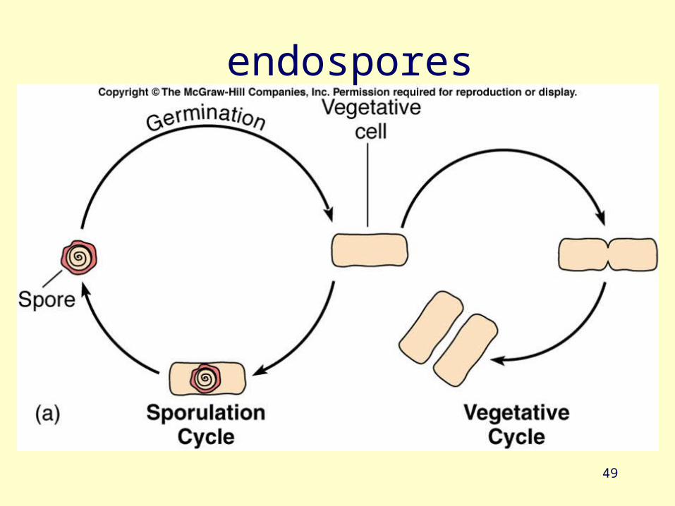
endospores• Resting, dormant cells• produced by some G+ genera: Clostridium, Bacillus
& Sporosarcina• Have a 2-phase life cycle – vegetative cell & an
endospore• sporulation -formation of endospores
• germination- return to vegetative growth • hardiest of all life forms• withstand extremes in heat, drying, freezing,
radiation & chemicals not a means of reproduction
47
endospores
• heat resistance linked to high levels of calcium & dipicolinic acid
• dehydrated, metabolically inactive• thick coat• longevity verges on immortality 25 to 250
million years.• pressurized steam at 120oC for 20-30
minutes will destroy (the autoclave)
48
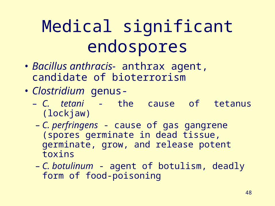
Medical significant endospores
• Bacillus anthracis- anthrax agent, candidate of bioterrorism
• Clostridium genus-– C. tetani - the cause of tetanus (lockjaw)– C. perfringens - cause of gas gangrene (spores
germinate in dead tissue, germinate, grow, and release potent toxins
– C. botulinum - agent of botulism, deadly form of food-poisoning
49
endospores
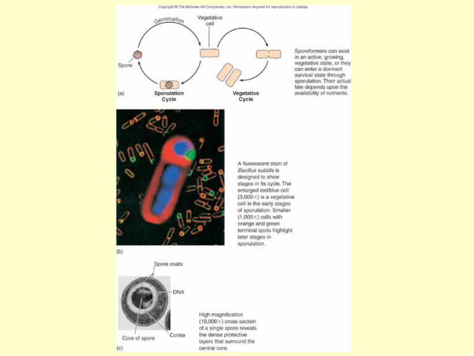
Fig. 4.21
51
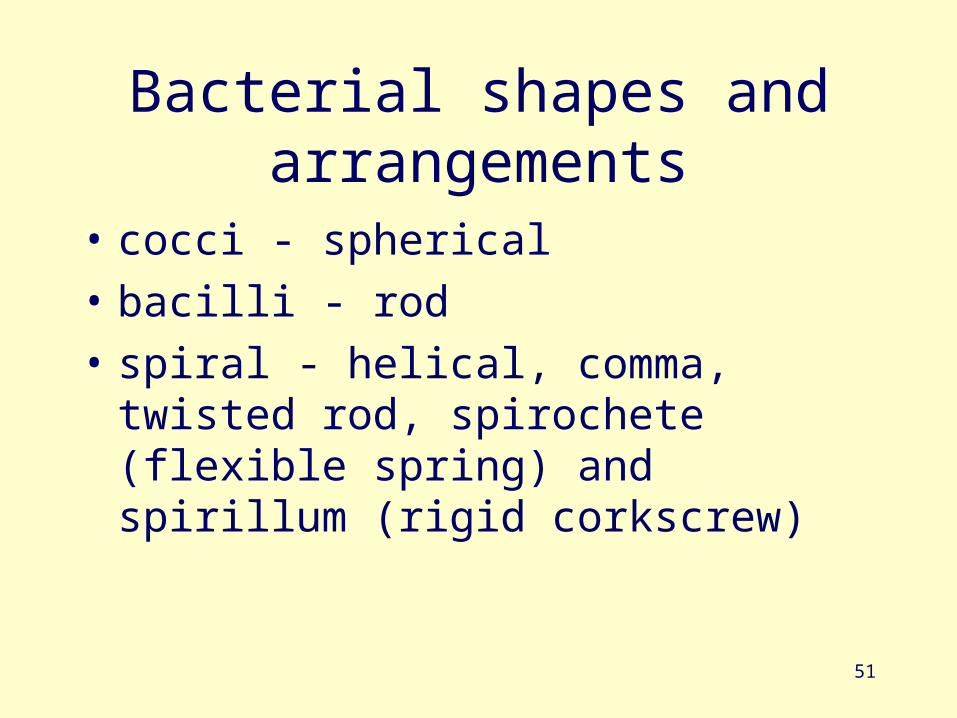
Bacterial shapes and arrangements
• cocci - spherical
• bacilli - rod
• spiral - helical, comma, twisted rod, spirochete (flexible spring) and spirillum (rigid corkscrew)
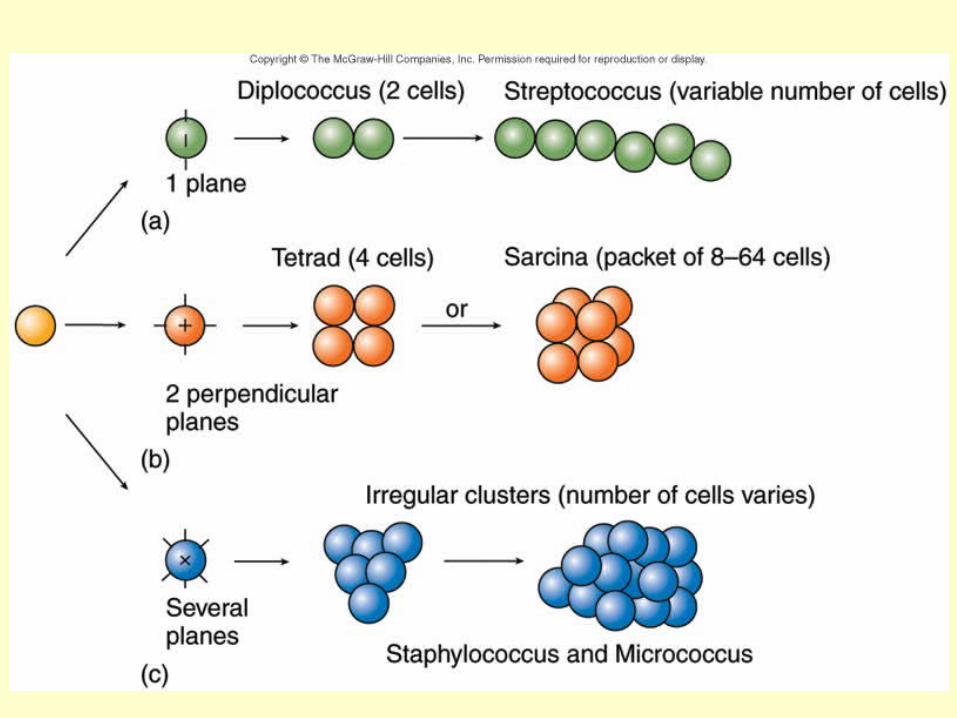
Fig. 4.25
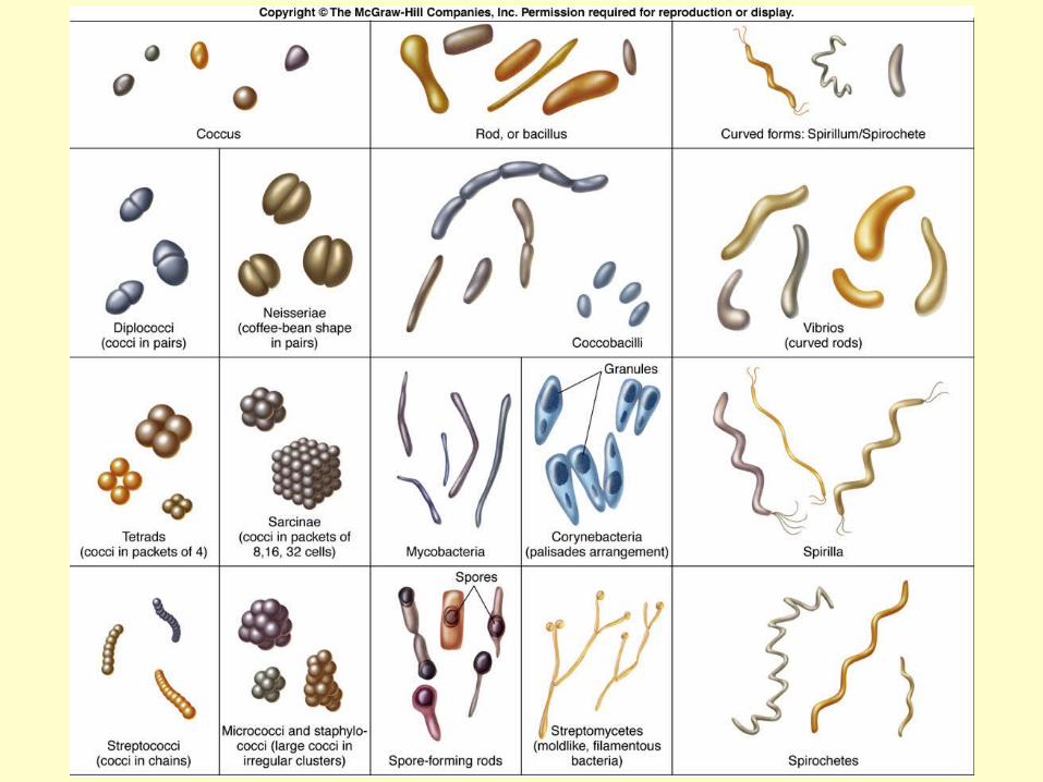
53
54
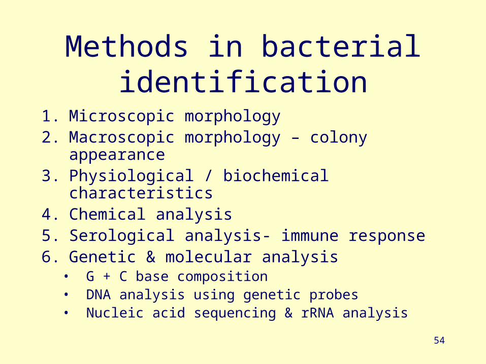
Methods in bacterial identification
1. Microscopic morphology2. Macroscopic morphology – colony appearance3. Physiological / biochemical characteristics4. Chemical analysis5. Serological analysis- immune response6. Genetic & molecular analysis
• G + C base composition• DNA analysis using genetic probes• Nucleic acid sequencing & rRNA analysis
55
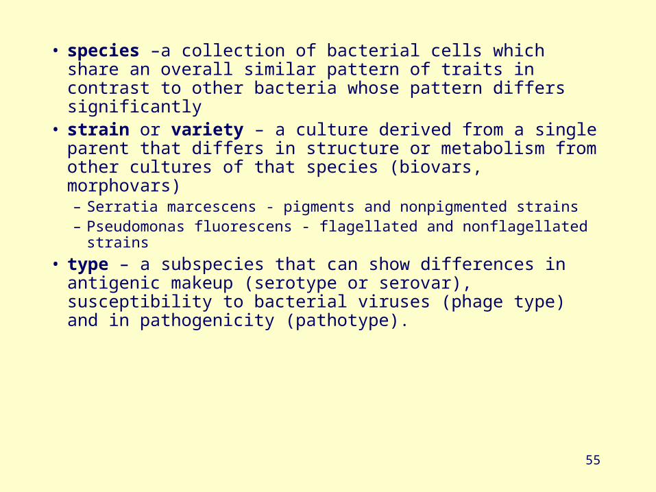
• species –a collection of bacterial cells which share an overall similar pattern of traits in contrast to other bacteria whose pattern differs significantly
• strain or variety – a culture derived from a single parent that differs in structure or metabolism from other cultures of that species (biovars, morphovars)– Serratia marcescens - pigments and nonpigmented strains– Pseudomonas fluorescens - flagellated and nonflagellated strains
• type – a subspecies that can show differences in antigenic makeup (serotype or serovar), susceptibility to bacterial viruses (phage type) and in pathogenicity (pathotype).

Prokaryotes with unusual characteristics
57
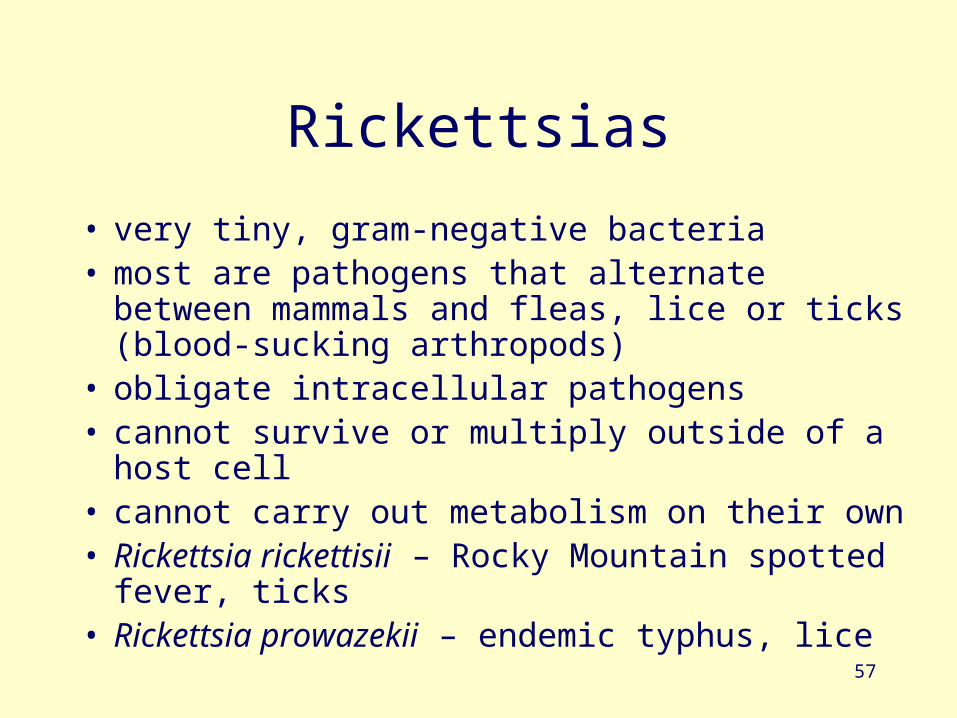
Rickettsias
• very tiny, gram-negative bacteria• most are pathogens that alternate between mammals
and fleas, lice or ticks (blood-sucking arthropods)• obligate intracellular pathogens • cannot survive or multiply outside of a host cell• cannot carry out metabolism on their own • Rickettsia rickettisii – Rocky Mountain spotted
fever, ticks• Rickettsia prowazekii – endemic typhus, lice
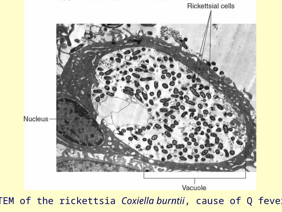
TEM of the rickettsia Coxiella burntii, cause of Q fever
59
Chlamydias
• tiny• obligate intracellular parasites (require host
cells for growth and metabolism)
• not transmitted by arthropods• Chlamydia trachomatis – severe eye
infection and one of the most common sexually transmitted diseases
• Chlamydia pneumoniae – lung infections
60
Mycoplasmas
• Bacteria that naturally lack a cell wall• stabilized by sterols, resistant to lysis• extremely small• range in shape from filamentous to coccus or
doughnut shaped– Pleomorphism - extreme variation of shape
• Mycoplasma pneumoniae – atypical pneumonia in humans, adheres to epithelial cells in the lung
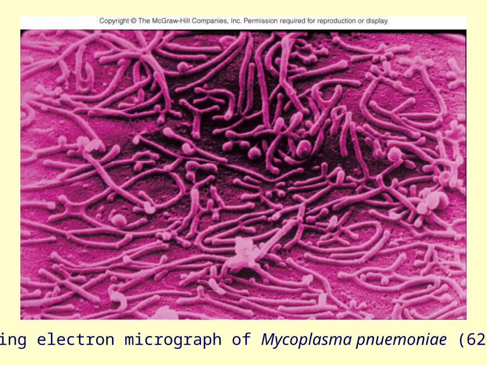
Fig. 4.32
Scanning electron micrograph of Mycoplasma pnuemoniae (62,000x)
62
Free-living nonpathogenic bacteria
• Photosynthetic bacteria– Cyanobacteria (oldest)– Green & purple sulfur bacteria
• Gliding, fruiting bacteria
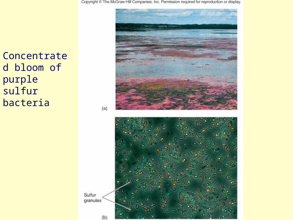
Fig. 4.34
Concentrated bloom of purple sulfur bacteria
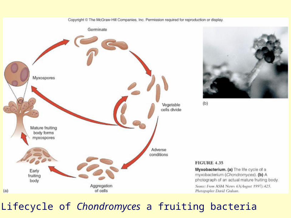
Fig. 4.35
Lifecycle of Chondromyces a fruiting bacteria
65
Archaea: the other prokaryotes• constitute third Domain Archaea• Unusual biochemistry and genetics that make them
different than bacteria• seem more closely related to Domain Eukarya than to
bacteria• contain unique genetic sequences in their rRNA• have unique membrane lipids & cell wall construction• live in the most extreme habitats in nature,
extremophiles• adapted to heat, salt, acid, pH, pressure & atmosphere• includes: methane producers, hyperthermophiles,
extreme halophiles, and sulfur reducers
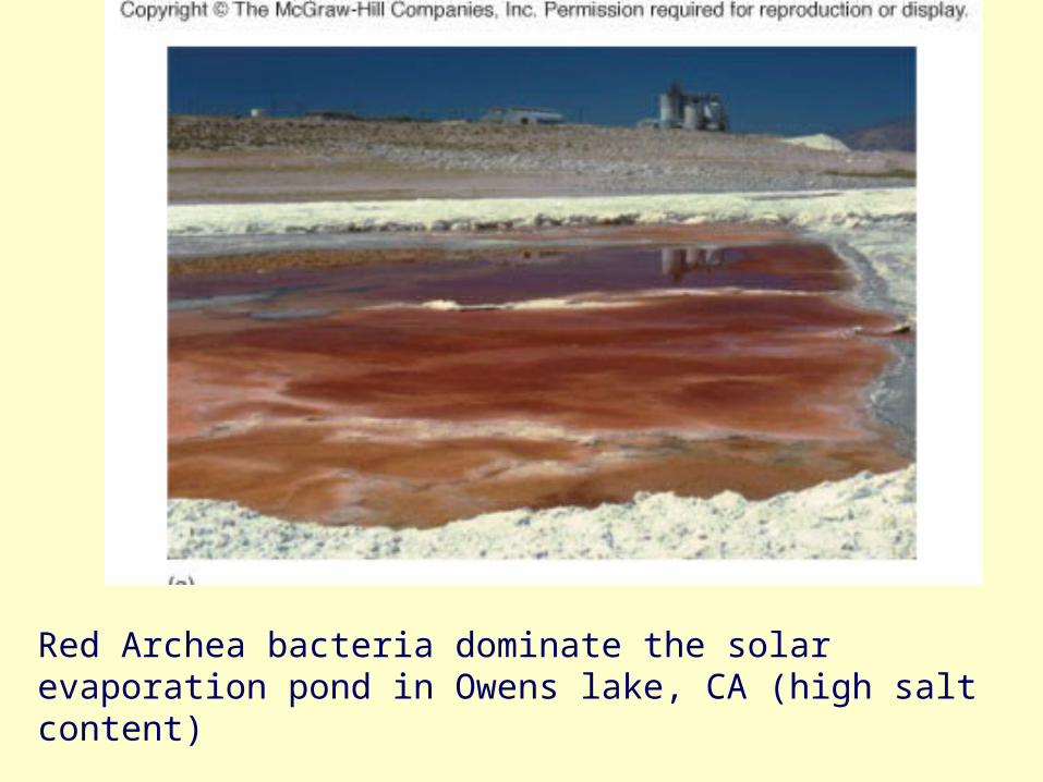
Red Archea bacteria dominate the solar evaporation pond in Owens lake, CA (high salt content)
Fig. 4.30