Embed Size (px)
Citation preview

Foundations in Microbiology
Seventh Edition
Chapter 21
Miscellaneous Bacterial Agents of Disease
Lecture PowerPoint to accompany
Talaro
Copyright © The McGraw-Hill Companies, Inc. Permission required for reproduction or display.
2
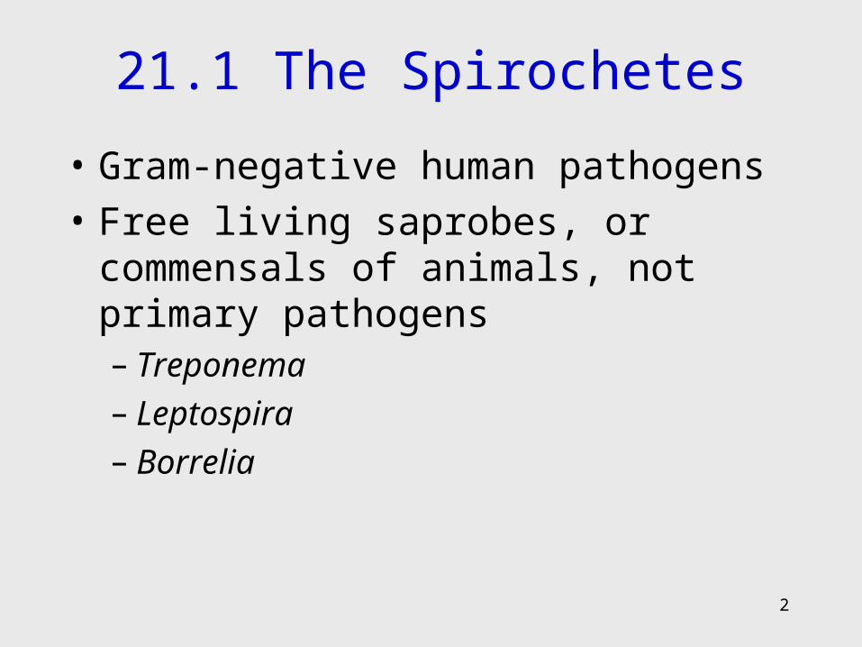
21.1 The Spirochetes
• Gram-negative human pathogens
• Free living saprobes, or commensals of animals, not primary pathogens– Treponema– Leptospira– Borrelia
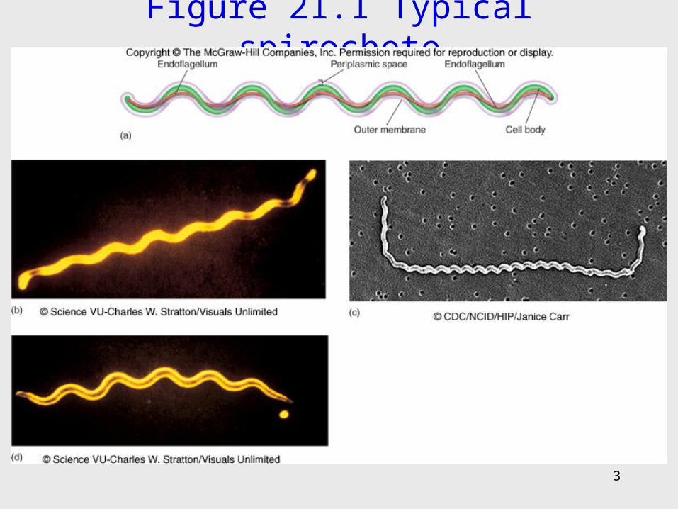
Figure 21.1 Typical spirochete
3
4
Genus Treponema
• Thin, regular, coiled cells
• Live in the oral cavity, intestinal tract, and perigenital regions of humans and animals
• Pathogens are strict parasites with complex growth requirements
• Require live cells for cultivation
5
Treponema Pallidum: The Spirochete of Syphilis
• Human is the natural host
• Extremely fastidious and sensitive; cannot survive long outside of the host
• Sexually transmitted and transplacental
6
Pathogenesis and Host Response
• Spirochete binds to epithelium (mucous membrane or abraded skin), multiplies, and penetrates capillaries
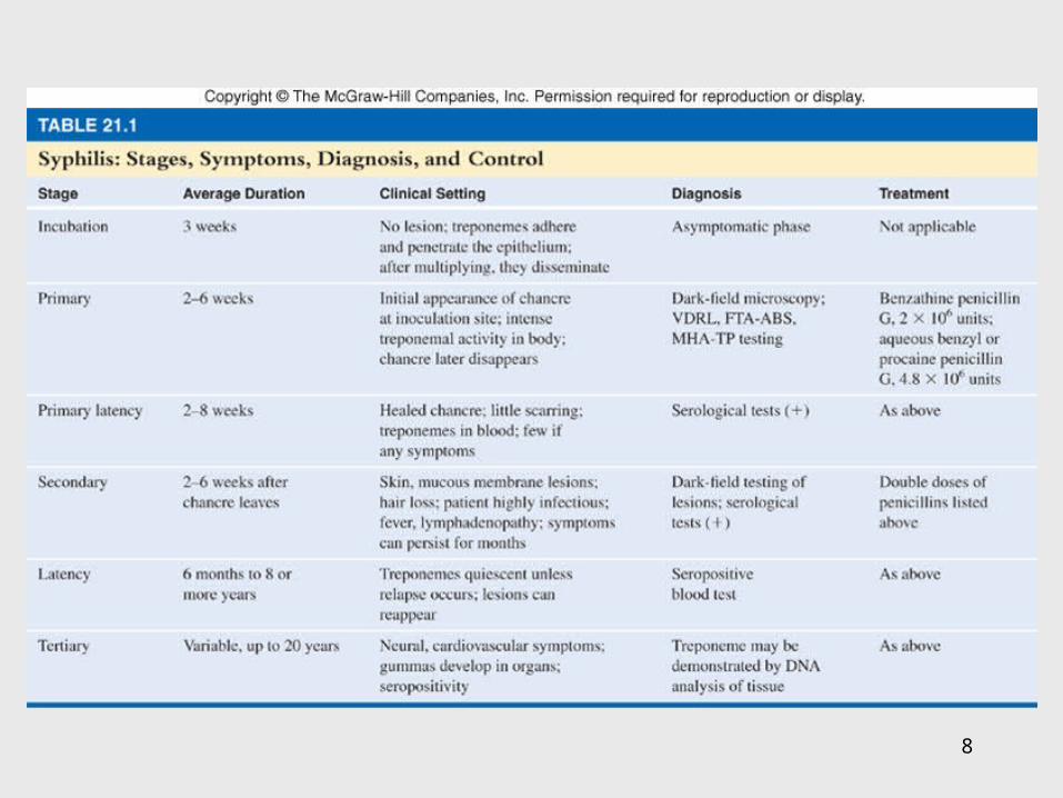
• Moves into circulation and multiplies • Untreated syphilis marked by 3 clinical stages:
– Primary, secondary, tertiary
• Spirochete appears in lesions and blood during first 2 stages – communicable
7
• Primary syphilis – appearance of hard chancre at site of inoculation; chancre heals spontaneously
• Secondary syphilis – fever, headache, sore throat, red or brown rash on skin, palms, and soles; rash disappears spontaneously
• Tertiary syphilis – about 30% of infections enter in tertiary stage; can last for 20 years or longer; numerous pathologic complications occur in susceptible tissues and organs– Neural, cardiovascular symptoms, gummas develop
• Congenital syphilis – nasal discharge, skin eruptions, bone deformation, nervous system abnormalities
8
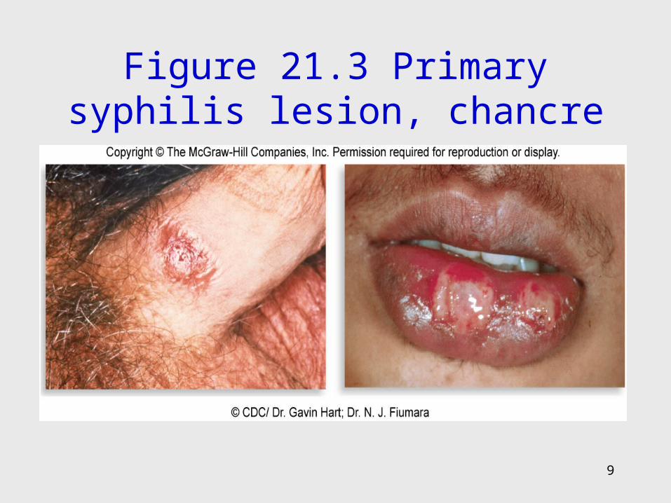
Figure 21.3 Primary syphilis lesion, chancre
9
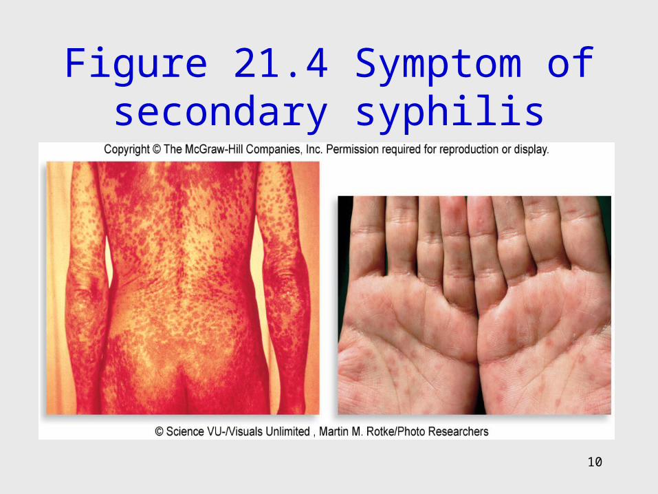
Figure 21.4 Symptom of secondary syphilis
10
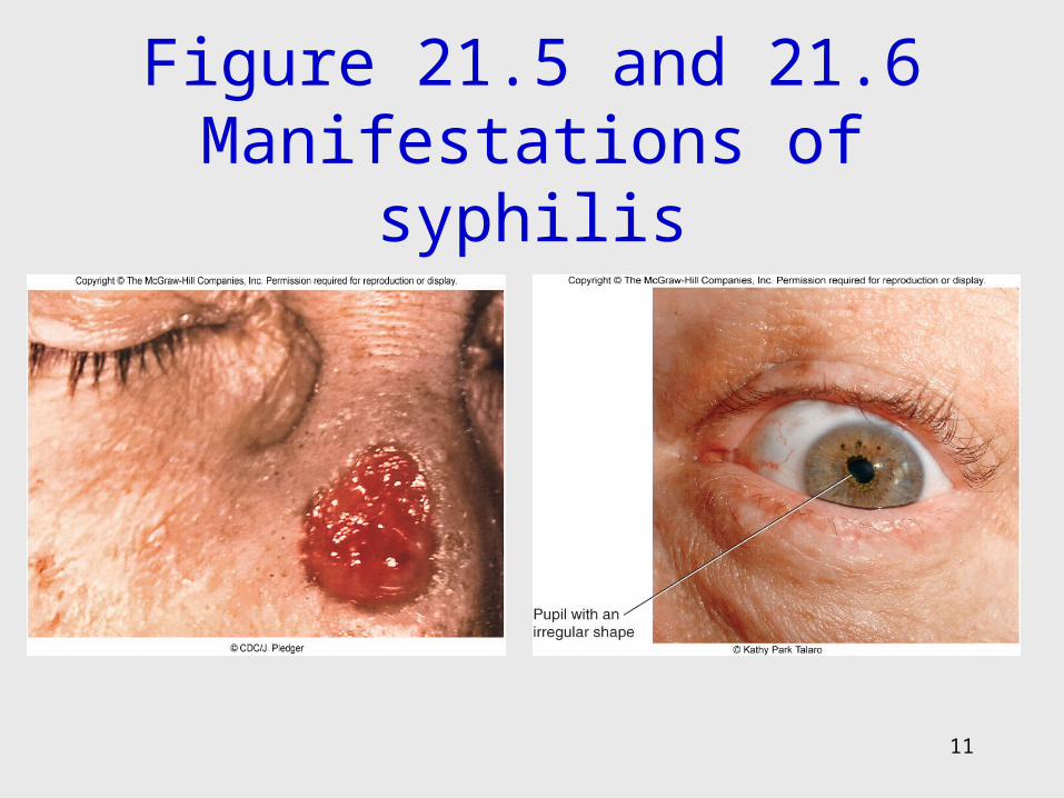
Figure 21.5 and 21.6 Manifestations of syphilis
11
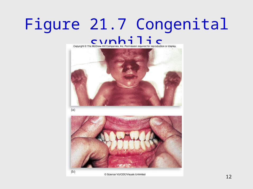
Figure 21.7 Congenital syphilis
12
13
Diagnosis and Treatment
• Stages of syphilis mimic other diseases
• Consider symptoms, history, microscopic, and serological testing– RPR, VDRL, FTA-ABS
• Treatment: penicillin G
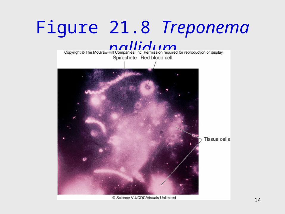
Figure 21.8 Treponema pallidum
14
15
Nonsyphilitic Treponematoses
• Resemble syphilis; rarely transmitted sexually or congenitally; cutaneous and bone diseases endemic to specific regions
• Bejel – T. pallidum subspecies endemicum; deforming childhood infection of the mouth, nasal cavity, body, and hands
• Yaws – T. pallidum subspecies pertenue; invasion of skin cut, causing a primary ulcer that seeds a second crop of lesions
• Pinta – T. carateum; superficial skin lesion that depigments and scars the skin
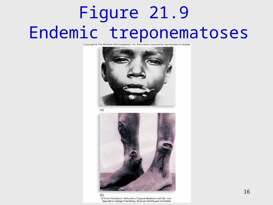
Figure 21.9 Endemic treponematoses
16
17
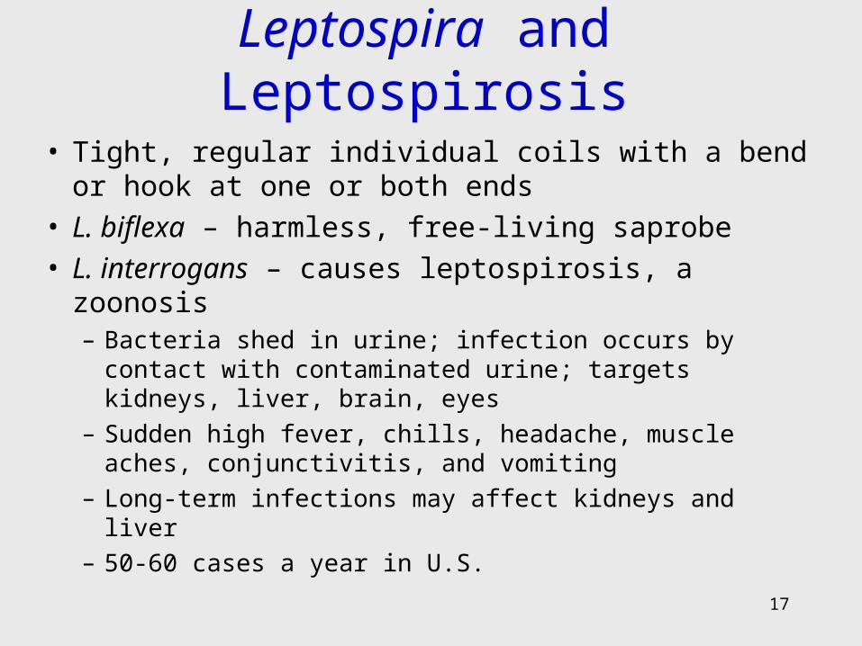
Leptospira and Leptospirosis
• Tight, regular individual coils with a bend or hook at one or both ends
• L. biflexa – harmless, free-living saprobe• L. interrogans – causes leptospirosis, a zoonosis
– Bacteria shed in urine; infection occurs by contact with contaminated urine; targets kidneys, liver, brain, eyes
– Sudden high fever, chills, headache, muscle aches, conjunctivitis, and vomiting
– Long-term infections may affect kidneys and liver
– 50-60 cases a year in U.S.
18
Borrelia: Arthropod-Borne Spirochetes
• Large, 3-10 coils irregularly spaced
• Borrelioses transmitted by arthropod vector
• B. hermsii – relapsing fever
• B. burgdorferi – Lyme disease
19
B. Hermsii – Relapsing Fever
• Mammalian reservoirs – squirrels, chipmunks, wild rodents
• Tick-borne• After 2-15-day incubation, patients have high fever,
shaking, chills, headache, and fatigue• Nausea, vomiting, muscle aches, abdominal pain;
extensive damage to liver, spleen, heart, kidneys, and cranial nerves
• Parasite changes and immune system tries to control it – Recurrent relapses
• Tetracycline
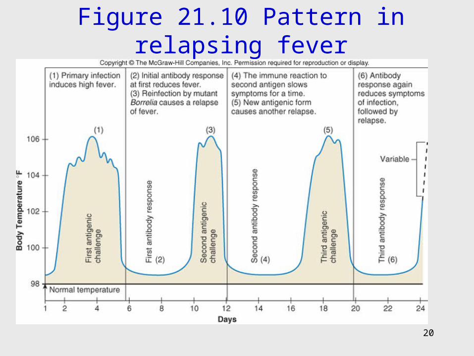
Figure 21.10 Pattern in relapsing fever
20
21
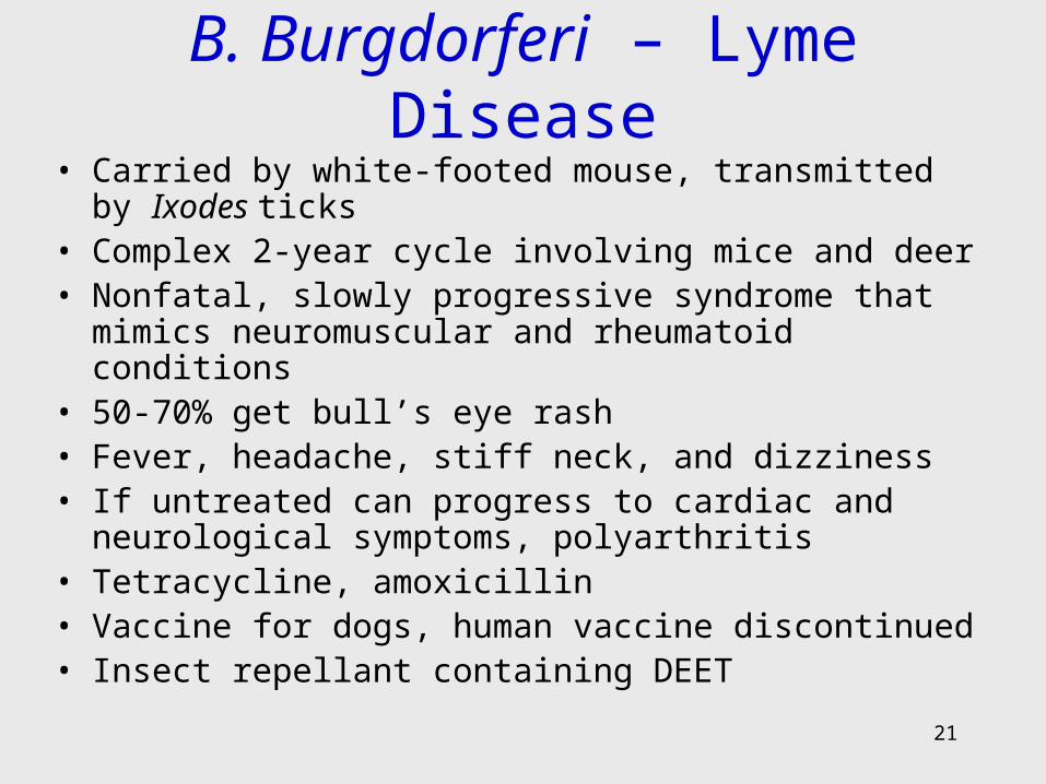
B. Burgdorferi – Lyme Disease• Carried by white-footed mouse, transmitted by Ixodes ticks• Complex 2-year cycle involving mice and deer• Nonfatal, slowly progressive syndrome that mimics
neuromuscular and rheumatoid conditions• 50-70% get bull’s eye rash• Fever, headache, stiff neck, and dizziness• If untreated can progress to cardiac and neurological
symptoms, polyarthritis• Tetracycline, amoxicillin• Vaccine for dogs, human vaccine discontinued• Insect repellant containing DEET
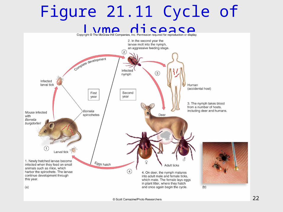
Figure 21.11 Cycle of Lyme disease
22
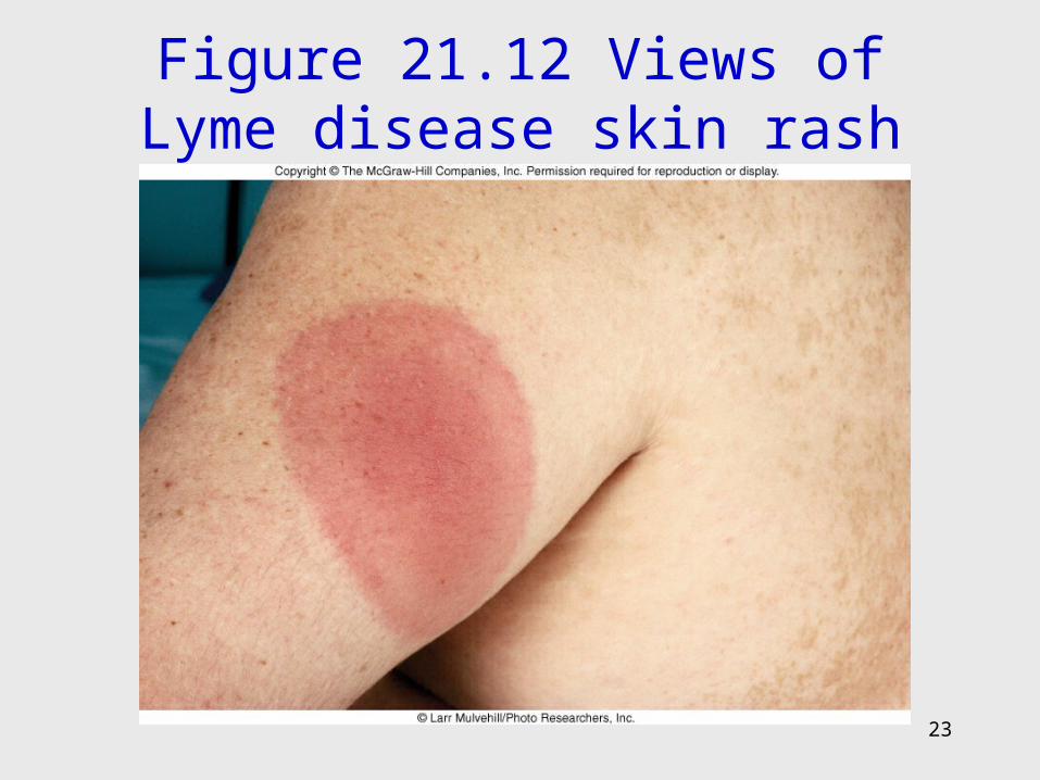
Figure 21.12 Views of Lyme disease skin rash
23
24
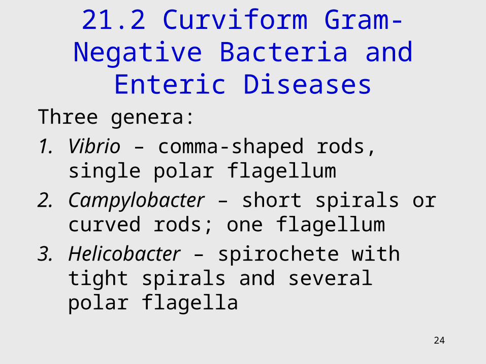
21.2 Curviform Gram-Negative Bacteria and Enteric Diseases
Three genera:
1. Vibrio – comma-shaped rods, single polar flagellum
2. Campylobacter – short spirals or curved rods; one flagellum
3. Helicobacter – spirochete with tight spirals and several polar flagella
25
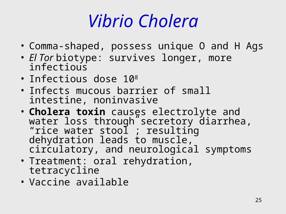
Vibrio Cholera• Comma-shaped, possess unique O and H Ags• El Tor biotype: survives longer, more infectious• Infectious dose 108
• Infects mucous barrier of small intestine, noninvasive
• Cholera toxin causes electrolyte and water loss through secretory diarrhea, “rice water stool”; resulting dehydration leads to muscle, circulatory, and neurological symptoms
• Treatment: oral rehydration, tetracycline• Vaccine available
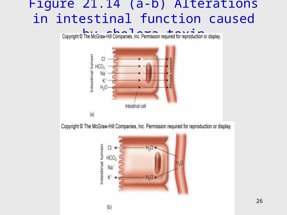
Figure 21.14 (a-b) Alterations in intestinal function caused by cholera toxin
26
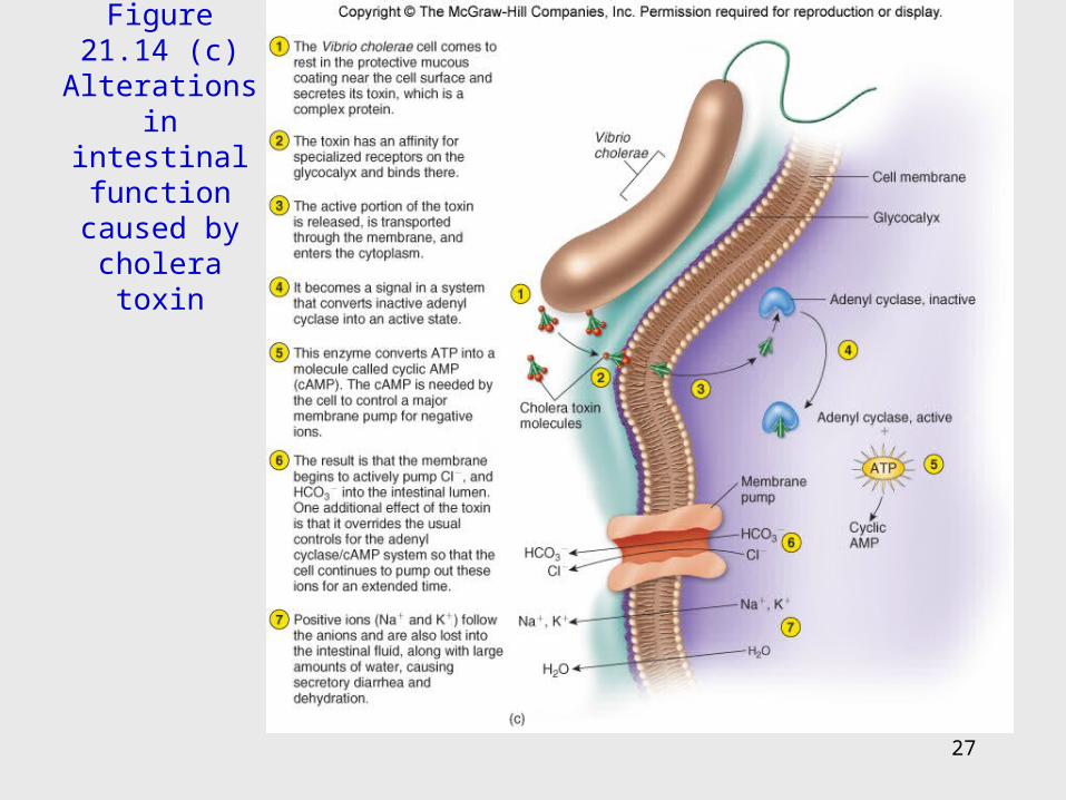
Figure 21.14 (c) Alterations in
intestinal function caused by cholera toxin
27
28
Pathogens Carried by Seafood
• Salt-tolerant inhabitants of coastal waters, associate with marine invertebrates
• Vibrio parahaemolyticus – gastroenteritis from raw seafood; symptoms similar to cholera
• Vibrio vulnificus – gastroenteritis from raw oysters; serious complications in persons with diabetes or liver disease
• Treatment – fluid and electrolyte replacement; occasionally antimicrobials
29
Diseases of the Campylobacter Vibrios
• Campylobacters – slender, curved, or spiral bacilli, often S-shaped or gull-winged pairs
• Polar flagella• Common residents of the intestinal tract,
genitourinary tract, the oral cavity of birds and mammals
• Most important:– Campylobacter jejuni– C. fetus

30
Campylobacter Jejuni Enteritis• Important cause of bacterial gastroenteritis• Transmitted by beverages and food• Reach mucosa at the last segment of small intestine
near colon; adhere, burrow through mucus and multiply
• Heat-labile enterotoxin CJT stimulates a secretory diarrhea like that of cholera
• Symptoms of headache, fever, abdominal pain, bloody or watery diarrhea
• Treatment with rehydration and electrolyte balance therapy
31
• Campylobacter fetus – opportunistic pathogen that infects debilitated persons or women late in pregnancy
• Meningitis, pneumonia, arthritis, septicemia in the newborn
32
Helicobacter Pylori: Gastric Pathogen
• Curved cells discovered in 1979 in stomach biopsied specimens
• Causes 90% of stomach and duodenal ulcers; apparent cofactor in stomach cancer
• People with type O blood have a 1.5-2X higher rate of ulcers
• Produces urease which converts urea into ammonium and bicarbonate

Figure 21.16 The causative agent of stomach ulcers
33

34
21.3 Medically Important Bacteria of Unique Morphology and Biology
35
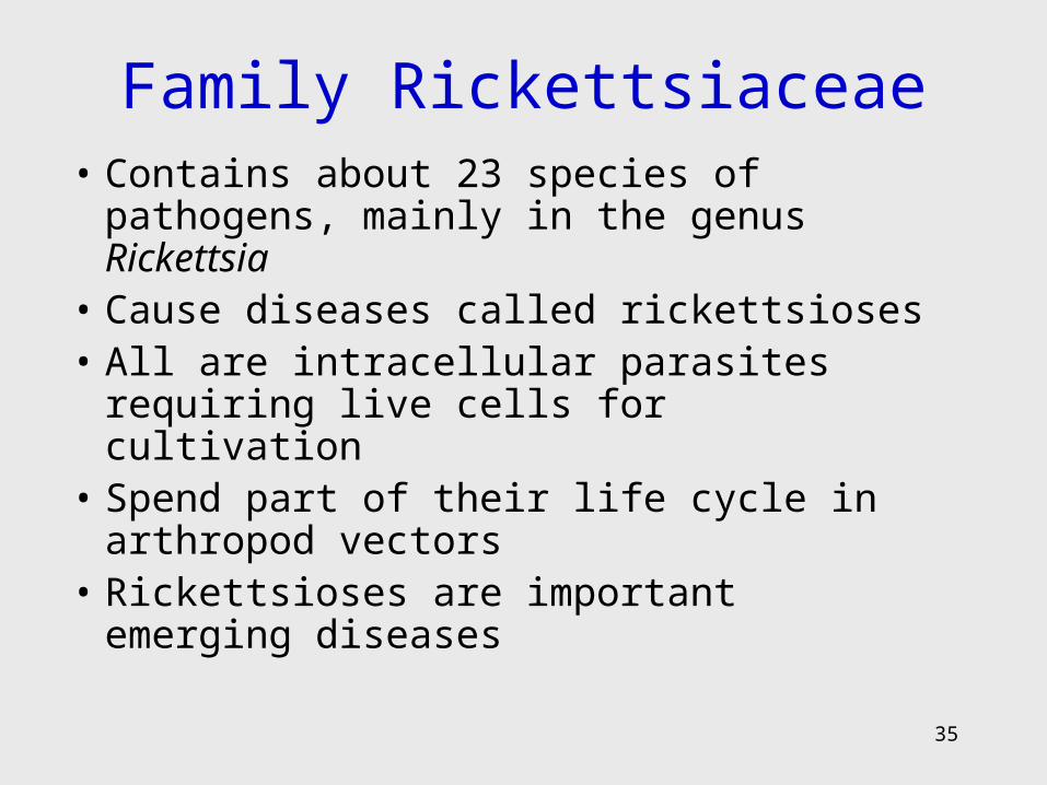
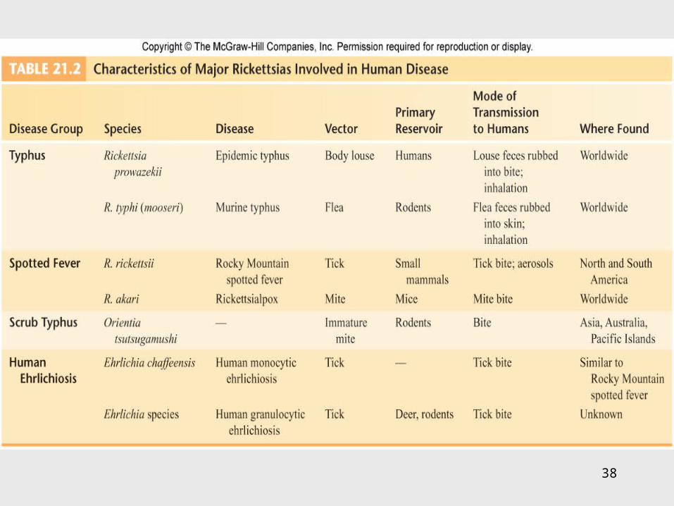
Family Rickettsiaceae• Contains about 23 species of pathogens,
mainly in the genus Rickettsia• Cause diseases called rickettsioses• All are intracellular parasites requiring live
cells for cultivation• Spend part of their life cycle in arthropod
vectors• Rickettsioses are important emerging
diseases
36
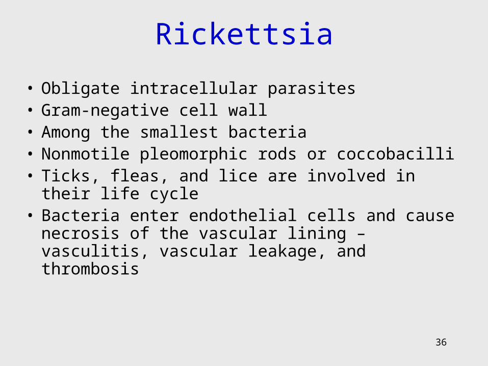
Rickettsia
• Obligate intracellular parasites• Gram-negative cell wall• Among the smallest bacteria• Nonmotile pleomorphic rods or coccobacilli• Ticks, fleas, and lice are involved in their life cycle • Bacteria enter endothelial cells and cause necrosis of
the vascular lining – vasculitis, vascular leakage, and thrombosis
37
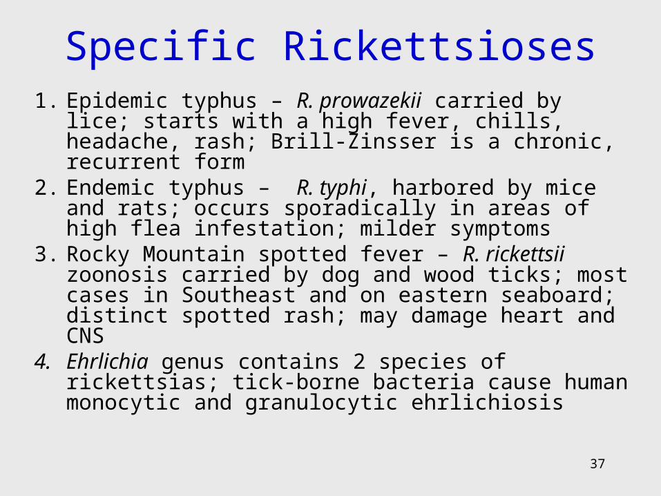
Specific Rickettsioses1. Epidemic typhus – R. prowazekii carried by lice;
starts with a high fever, chills, headache, rash; Brill-Zinsser is a chronic, recurrent form
2. Endemic typhus – R. typhi, harbored by mice and rats; occurs sporadically in areas of high flea infestation; milder symptoms
3. Rocky Mountain spotted fever – R. rickettsii zoonosis carried by dog and wood ticks; most cases in Southeast and on eastern seaboard; distinct spotted rash; may damage heart and CNS
4. Ehrlichia genus contains 2 species of rickettsias; tick-borne bacteria cause human monocytic and granulocytic ehrlichiosis
38
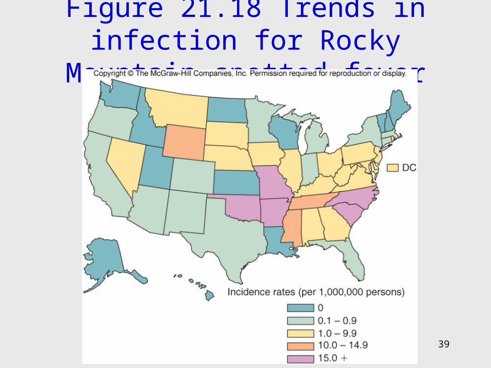
Figure 21.18 Trends in infection for Rocky Mountain spotted fever
39
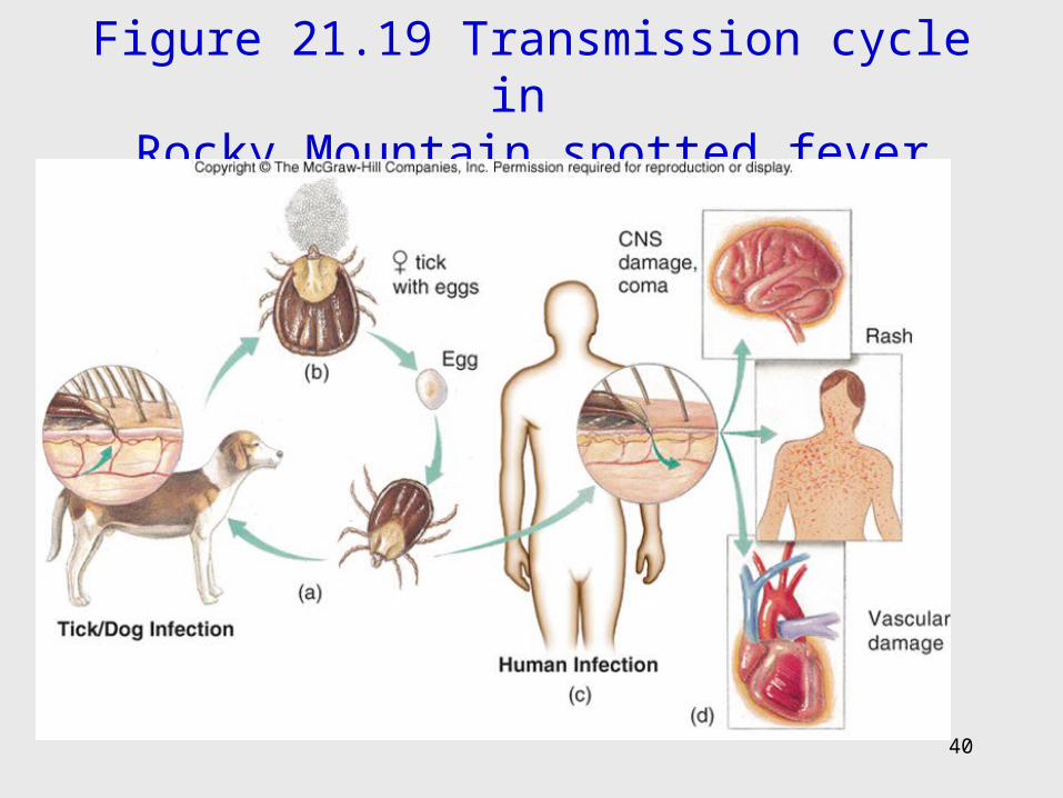
Figure 21.19 Transmission cycle in Rocky Mountain spotted fever
40
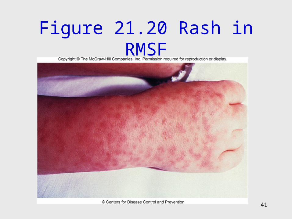
Figure 21.20 Rash in RMSF
41

42
Related to the Rickettsioses
• Coxiella burnetti
• Bartonella sp.
43
Coxiella Burnetti• Causes Q fever• Intracellular parasite • Produces an unusual resistant spore• Harbored by a wide assortment of vertebrates and
arthropods• Infectious material includes urine, feces, milk, and
airborne particles• Usually inhaled causing pneumonitis, fever, hepatitis• Tetracycline treatment• Vaccine available
44
Bartonella Species• Small gram-negative, fastidious, cultured
on blood agar• Cause:
– Trench fever, spread by lice– Cat-scratch disease, a lymphatic infection
associated with a clawing injury by cats– Bacillary angiomatosus in AIDS patients
• Tetracycline, erythromycin, and rifampin
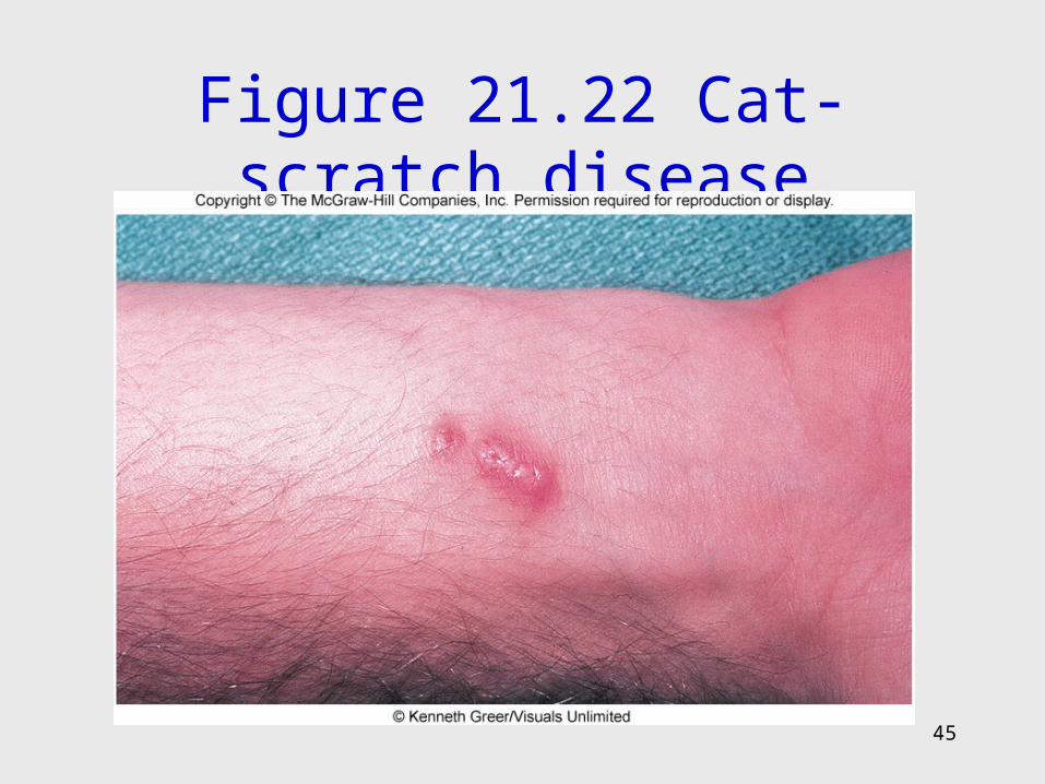
Figure 21.22 Cat-scratch disease
45
46
The Chlamydiaceae
• Obligate intracellular parasites• Small, gram-negative cell wall• Alternate between 2 stages:
– Elementary body – small metabolically inactive, extracellular, infectious form released by the infected host
– Reticulate body – noninfectious, actively dividing form, grows within host cell vacuoles
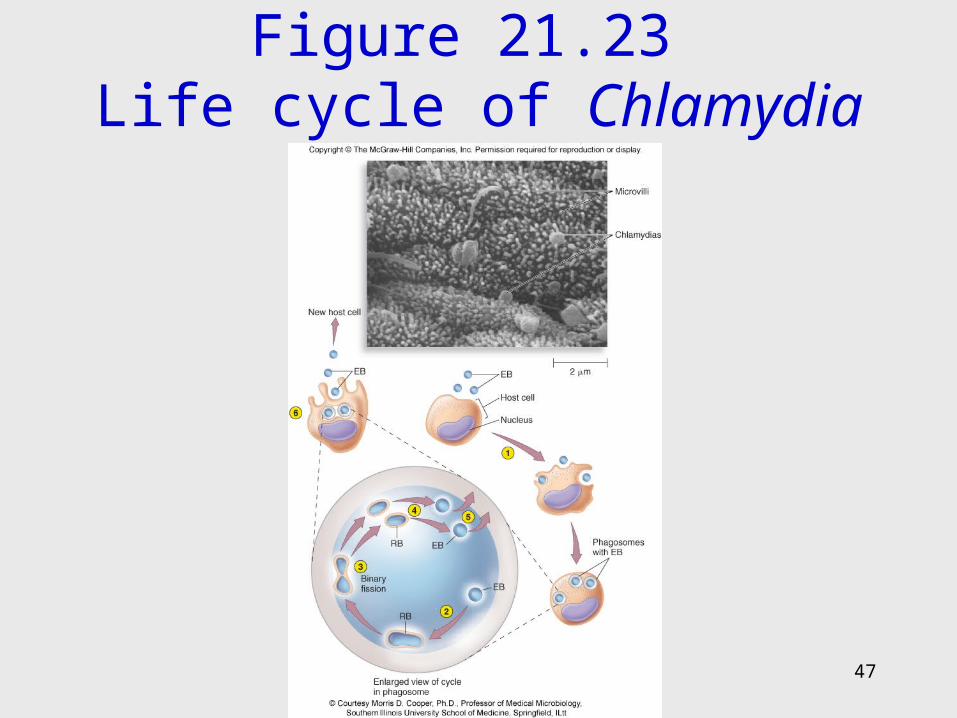
Figure 21.23 Life cycle of Chlamydia
47
48
Chlamydia Trachomatis• Human reservoir• 2 strains• Trachoma – attacks the mucous membranes of the
eyes, genitourinary tract, and lungs– Ocular trachoma – severe infection, deforms eyelid
and cornea, may cause blindness– Inclusion conjunctivitis – occurs as baby passes
through birth canal; prevented by prophylaxis– STD – second most prevalent STD; urethritis,
cervicitis, salpingitis (PID), infertility, scarring• Lymphogranuloma venereum – disfiguring disease of
the external genitalia and pelvic lymphatics
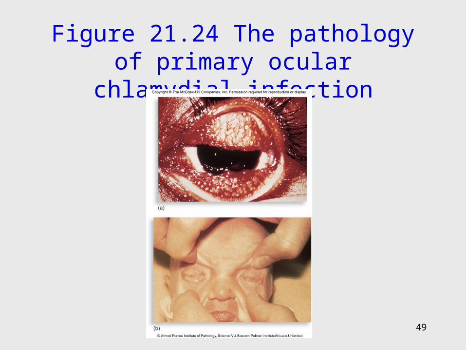
Figure 21.24 The pathology of primary ocular chlamydial infection
49
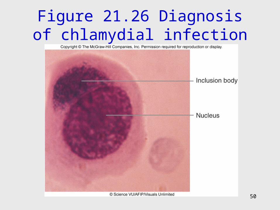
Figure 21.26 Diagnosis of chlamydial infection
50
51
Chlamydophila – A New Genus
• Contains members that used to be members of genus Chlamydia
• Chlamydophila pneumoniae – causes an atypical pneumonia that is serious in asthma patients
• C. psittaci – causes ornithosis, a zoonosis transmitted to humans from bird vectors; highly communicable among all birds; pneumonia or flulike infection with fever, lung congestion
52
21.4 Molliculites and Other Cell-Wall-Deficient Bacteria
• Called mycoplasmas• Naturally lack cell walls, highly pleomorphic• Require special lipids from host membranes• Treated with tetracycline, erthyromycin• M. pneumoniae – primary atypical pneumonia;
pathogen slowly spreads over interior respiratory surfaces, causing fever, chest pain, and sore throat
• M. genitalium and Ureplasma urealyticum – weak sexually transmitted pathogens
53
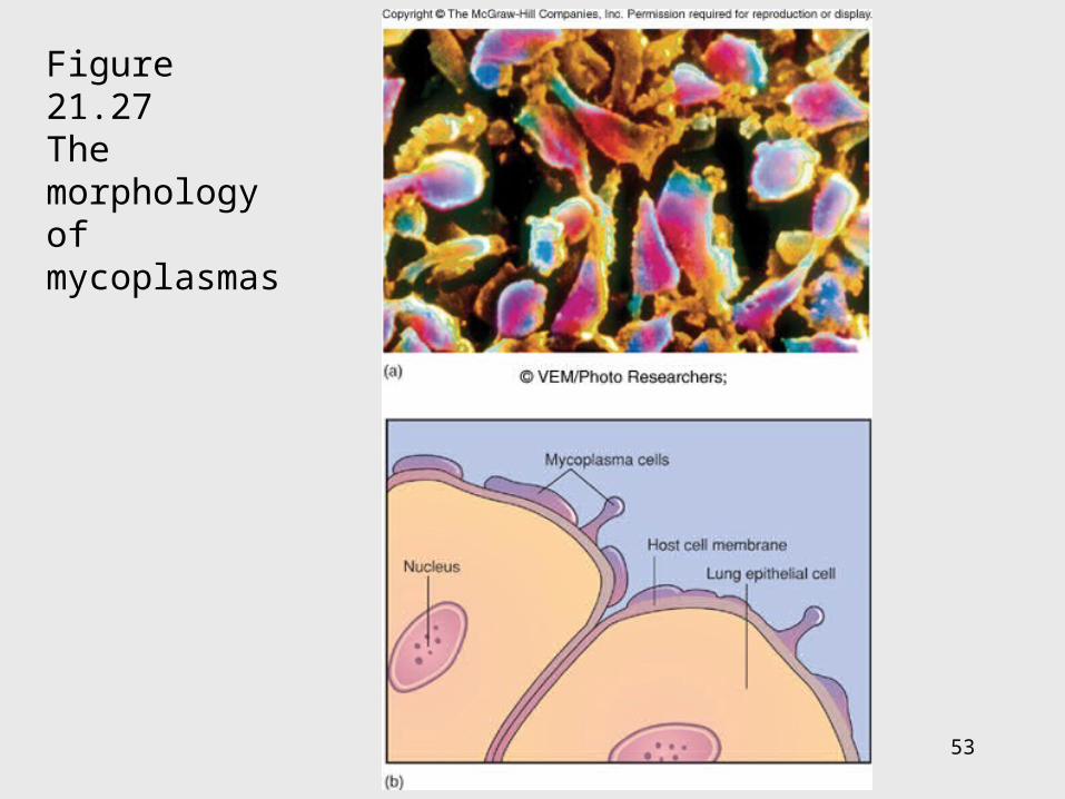
Figure 21.27 The morphology of mycoplasmas
54
Bacteria That Have Lost Their Cell Walls
• Exposure to certain drugs or enzymes can result in cell wall-deficient bacteria called L forms or
L-phase
• Induced or occur spontaneously
• May be involved in some chronic diseases– L- phase variants of group A streptococci, Proteus, and
Corynebacterium, Mycobacterium avium paratuberculosis
55
21.5 Bacteria in Dental Disease
• Oral cavity is a complex, dynamic ecosystem, containing 400 species
• Dental caries – slow progressive infection of irregular areas of enamel surface
1. Begins with colonization by slime-forming species of Streptococcus and cross adherence with Actinomyces
2. Process forms layer of thick, adherent material (plaque) that harbors masses of bacteria which produce acid that dissolves enamel
56
3. If plaque is allowed to stay, secondary invaders appear – Lactobacillus, Bacteroides, Fusobacterium, Porphyromonas, Treponema
4. Acid dissolves tooth enamel leading to caries and tooth damage

Figure 21.28 The anatomy of a tooth
57
58
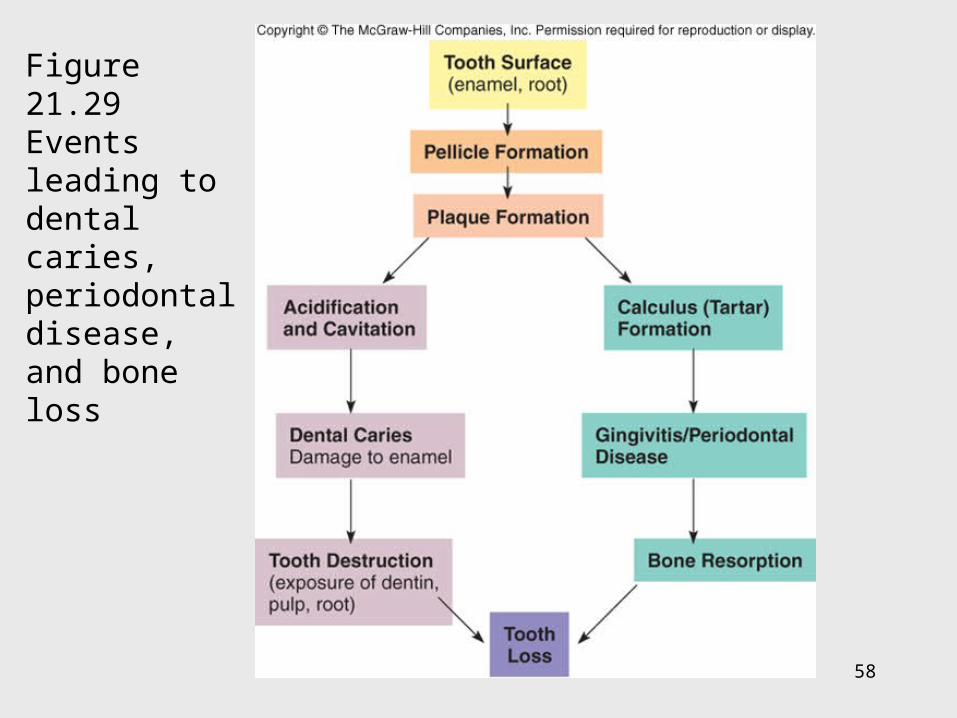
Figure 21.29Events leading to dental caries, periodontal disease, and bone loss
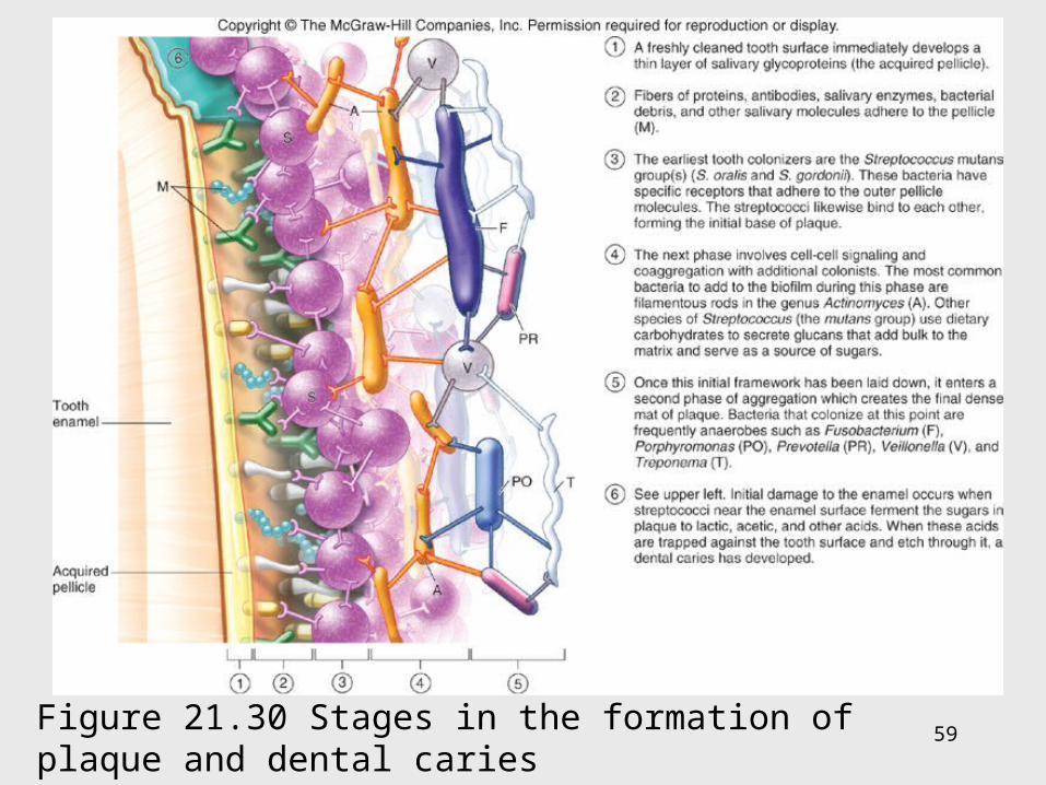
59Figure 21.30 Stages in the formation of plaque and dental caries
60
Periodontal Disease
• Soft tissue disease• When plaque becomes calcified into calculus
above and below the gingiva• This irritates tender gingiva causing inflammation
– Gingivitis• Pockets between tooth and gingiva are invaded by
bacteria (spirochetes and gram-negative bacilli)• Tooth socket may be involved (peridontitis)• Tooth may be lost
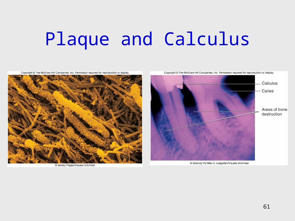
Plaque and Calculus
61
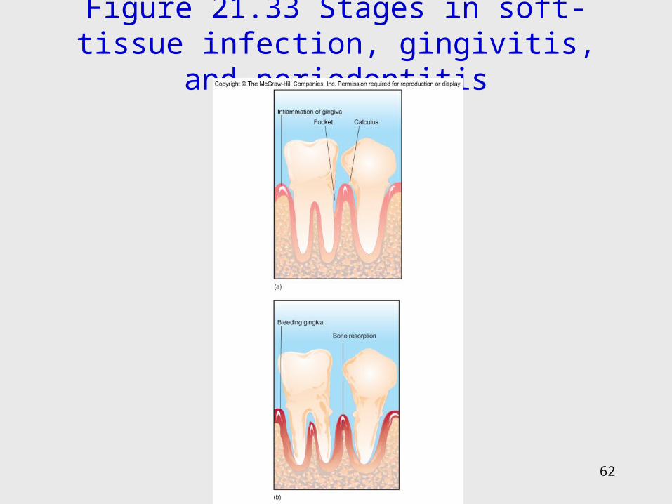
Figure 21.33 Stages in soft-tissue infection, gingivitis, and periodontitis
62
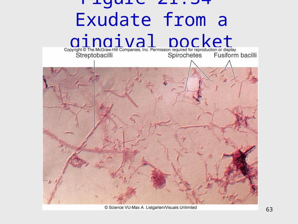
Figure 21.34 Exudate from a gingival pocket
63