Embed Size (px)
Citation preview
Geological Survey of New South WalesQuarterly Notes
Geological Survey of New South Wales
June 2011 No 136
© State of New South Wales through the Division of Resources and Energy, 2011
Papers in Quarterly Notes are subject to external review. External reviewer for this issue was Dr Steve Hill, University of Adelaide. His assistance is appreciated.
Quarterly Notes is published to give wide circulation to results of studies in the Geological Survey of New South Wales. Papers are also welcome that arise from team studies with external researchers. Contact: [email protected]
ISSN 0155-3410
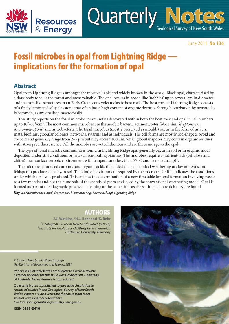
1J.J. Watkins, 2H.J. Behr and 2K. Behr1 Geological Survey of New South Wales (retired)
2 Institute for Geology and Lithospheric Dynamics, Göttingen University, Germany
AUTHORS
Fossil microbes in opal from Lightning Ridge — implications for the formation of opal
AbstractOpal from Lightning Ridge is amongst the most valuable and widely known in the world. Black opal, characterised by a dark body tone, is the rarest and most valuable. The opal occurs in geode-like ‘nobbies’ up to several cm in diameter and in seam-like structures in an Early Cretaceous volcaniclastic host rock. The host rock at Lightning Ridge consists of a finely laminated silty claystone that often has a high content of organic detritus. Strong bioturbation by nematodes is common, as are opalised macrofossils.
This study reports on the fossil microbe communities discovered within both the host rock and opal in cell numbers up to 107–108/cm3. The most common microbes are the aerobic bacteria actinomycetes (Nocardia, Streptomyces, Micromonospora) and myxobacteria. The fossil microbes (mostly preserved as moulds) occur in the form of mycels, mats, biofilms, globular colonies, networks, swarms and as individuals. The cell forms are mostly rod-shaped, ovoid and coccoid and generally range from 2–5 µm but may exceed 100 µm. Small globular spores may contain organic residues with strong red fluorescence. All the microbes are autochthonous and are the same age as the opal.
The type of fossil microbe communities found in Lightning Ridge opal generally occur in soil or in organic muds deposited under still conditions or in a surface-fouling biomass. The microbes require a nutrient-rich (cellulose and chitin) near-surface aerobic environment with temperatures less than 35 °C and near-neutral pH.
The microbes produced carbonic and organic acids that aided the biochemical weathering of clay minerals and feldspar to produce silica hydrosol. The kind of environment required by the microbes for life indicates the conditions under which opal was produced. This enables the determination of a new timetable for opal formation involving weeks to a few months and not the hundreds of thousands of years envisaged by the conventional weathering model. Opal is formed as part of the diagenetic process — forming at the same time as the sediments in which they are found.Key words: microbes, opal, Cretaceous, bioweathering, bacteria, fungi, Lightning Ridge

2 June 2011
IntroductionPrecious opal consists of amorphous silica spheres of uniform size (0.14–0.30 µm diameter) arranged in a regular pattern. The regular array of spheres and voids diffracts white light by breaking it into the complete range of colours. The colour observed is dependent on the layer spacing, which is determined by the sphere size. Opal with little or no colour is referred to as ‘potch’. In potch opal, the silica spheres are either of assorted sizes or too small to produce blue colouration, even when arranged in a regular array. Precious opal and potch opal can occur in the same sample and the process of formation of both types of opal is genetically, spatially and temporally connected. Photograph 1 shows a bulbous-shaped area of precious opal squeezed out under pressure from an area of potch, and intruded diapirically into black opal. The chemical formula of opal can be expressed as SiO2.nH2O. Water content varies from 2–20 per cent.
The information contained in this publication is based on knowledge and understanding at the time of writing (April 2011). However, because of advances in knowledge, users are reminded of the need to ensure that information upon which they rely is up to date and to check currency of the information with the appropriate officer of the Division of Resources and Energy, or the user’s independent adviser.
Production co-ordination Geneve Cox and general editing :
Geological editor: Simone Meakin
Geospatial information: Cheryl Hormann Phillip Carter
Layout: Carey Martin
Cover photograph: 'Nobby' showing concave-upwards meniscus, above which spectacular blue rods of opal have grown. (Photographer J.J. Watkins).
Contents
Abstract 1
Introduction 2
Geological setting 3
Descriptions of the microbes 6
Habitats of the microbes 13
Bioweathering 17
Structural control and the formation of precious opal 18
Discussion and conclusions 19
Acknowledgements 19
References 20
Externally published papers written by GSNSW staff and collaborators 21
Photograph 1. Thin section photomicrograph of precious opal squeezed out under pressure and intruded as a small diapir into black potch. The base of the precious opal and the feeder is potch opal. (Photographer H.J. Behr).

3Quarterly Notes
The process of opal formation has been a matter of conjecture and several conceptual models have evolved over time. The weathering model (Darragh et al. 1966, 1976; Senior 1975; Barnes et al. 1992; Watkins 1984, 1999; Robertson & Scott 1990; Townsend 2002) suggests that the weathering of the host sedimentary rock (mostly sandstone) during the Late Cretaceous or Paleogene produced a silica rich solution that travelled down the profile and precipitated opal within the underlying claystone unit. Watkins (1984) suggested that local and regional structures were important in localising economic deposits. Pecover (1996, 2003) however proposed that opal was deposited from deeply sourced hydrothermal fluid. The fluid ascended along faults and silica was deposited actively and rapidly in fractures, at temperatures and pressures well above those typically involved in surface processes (>100 °C). The model suggested that opal was precipitated as a result of cooling.
The present study commenced in 1999 when Professor Hans (Ted) Behr announced at the first Lightning Ridge Opal Symposium that he had observed large numbers of microbe communities in potch opal (Behr et al. 2000; Watkins 2000). Over 320 samples of both opal and host rock were collected from the opal mining fields at Lightning Ridge and Coober Pedy and studied in polished thin sections with optical microscopy and oil-immersion objectives. Additional studies were undertaken using a field emission scanning electron microscope (Zeiss LEO Gemini 1530, QUANTA 200F) at Göttingen University. The identification of the microbes was undertaken by Dr Karen Behr, a microbiologist from Göttingen University, and was based on their morphology only, as cultivation in the laboratory was unsuccessful.
Professor Hans Behr passed away in 2002.
Geological settingThe Lightning Ridge opal fields lie on the southern margin of the Great Australian Basin, near the border between New South Wales and Queensland (Figure 1). Opal is found at shallow depths, usually less than 30 m, in the deeply weathered portion of the Early Cretaceous Griman Creek Formation of the Rolling Downs Group. The Griman Creek Formation consists of the Coocoran Claystone Member (0–4 m thick) overlying the Wallangulla Sandstone Member (1–20 m thick). Within the Wallangulla Sandstone Member are discontinuous lenses of the Finch Claystone facies (0.2–2 m thick). The Early Cretaceous sequence is in part unconformably overlain by a sequence of predominantly quartzose fluviatile gravels and sands (0.2–4 m) of Neogene age. The latter unit is often heavily silicified to silcrete. An unnamed unit of colluvium is also sporadically
developed beneath the Neogene sediments. Opal occurs in nodules (‘nobbies’) and seams, mostly within the upper part of the Finch Claytone facies. Opal clasts have also been recovered from both the unnamed colluvium and the Neogene sediments.
The Finch Claystone facies consists dominantly of claystone with minor siltstone and sandstone with centimetre-scale bedding and millimetre-scale laminations. The silt is composed of quartz, pyroclastic fragments of volcanic glass and feldspar, and clay minerals. Biotite, pyroxene, anatase, and rutile are accessory minerals. The occurrence of quartz phenocrysts with growth embayments and zoning is indicative of a rhyolitic volcanic provenance. The fragments of volcanic glass contain longitudinal feldspar crystals (15–25 µm) which are arranged in parallel flow textures. These pilotaxitic textures are indicative of trachytic volcanism and alkali feldspar (Photograph 2). The clay minerals are mostly smectite
Photograph 2. Scanning Electron Microscope (SEM) image of sample of Finch Claystone facies in which the silty clasts are supported by an opal matrix with deformed microbe moulds. Two pyroclastic fragments with pilotaxitic feldspar laths (opalised) and dissolved glass-matrix. (Image by H.J. Behr).

4 June 2011
N.T.
S.A.
Tas.
Vic.
N.S.W
Qld
156°00’E152°00’E148°00’E144°00’E140°00’E136°00’E132°00’E
12°00’S
16°00’S
20°00’S
24°00’S
28°00’S
32°00’S
36°00’S
40°00’S
CarpentariaBasin
SuratBasin
2011_05_0038
Euroka Arch
Nebine
R
idge
Winton
Quilpie
Yowah
Hebel
Lightning RidgeCoocoran
White CliffsAndamooka
CooberPedy Stuart Creek
MintabieLambina
0 500 1000km
REFERENCEMajor precious opal deposits
Precious opal deposits
Great Australian Basin
Cenozoic sediments
Cretaceous sedimentary rocks
margin of G
reat Australian Basin
Tasman Sea
Coral Sea
PacificOcean
Great SouthernOcean
Bass Strait
Gulf ofCarpentaria
Figure 1. The Great Australian Basin showing major opal fields. Lightning Ridge is located in northern New South Wales on the southern edge of the basin. Digital elevation model image ©Commonwealth of Australia (Geoscience Australia) 2011. This material is released under the Creative Commons Attribution 3.0 Australia Licence.

5Quarterly Notes
The Wallangulla Sandstone Member is a fine- to medium-grained feldspathic sandstone unit with common claystone pellet rip-up clasts at its base. It comprises a stacked fluvial sequence of distributary palaeochannels and thick units (3–10 m) show a close spatial association with productive opal fields. The base of the unit contains a 0–0.3 m thick silicified layer that discontinuously overlies the Finch Claystone facies and is known as the ‘steel band’. The Wallangulla Sandstone Member grades up into the Coocoran Claystone Member which, like the Finch Claystone facies, has common bioturbation and open and infilled vertical tubes.
During the Early Cretaceous, the Lightning Ridge area had a predominantly cold climate and may have been on or close to the Antarctic Circle. Sedimentology and fossils indicate that the area was probably a low-lying coastal plain. Mud enriched in organic matter was deposited in estuaries, shallow ponds, oxbow lakes, and as overbank deposits. Primitive coniferous forests dominated the interfluvial areas. There is no evidence of marine transgressions.
Boreham (1996) estimated that approximately 1200 m of sediment was deposited on top of the Griman Creek Formation and subsequently removed 5–10 million years later following rapid uplift. Byrnes (1975) however, estimated 100 to 500 m of sediment was deposited locally on top of the Griman Creek Formation at Lightning Ridge based on the deformation of ‘Chinese hat’ nobbies. There is evidence within the sequence that overpressure was frequently developed in the Finch Claystone facies, particularly in areas with high clay contents or below stacked fluvial sequences of the Wallangulla Sandstone Member. The over-pressuring resulted in the formation of diapirs or breccia pipes (‘blows’). Opal fragments were transported in the brecciated material. Collapse breccias pipes are also present and form cone-shaped collapse structures. The breccia pipes are typically filled with coarse-grained sand, pieces of rounded and angular host rock, clasts of opal and Neogene gravels.
and often show transitions to volcanic glass fragments. The feldspar crystals were partly weathered prior to the deposition, and are now completely altered to kaolinite. Syn-depositional erosion channels (>20 cm deep, and some metres wide) occur in the Finch Claystone facies. They are filled with redeposited sediments, plant litter, macrofossils, opalised wood and redeposited nobbies. Bioturbation by earthworms is common. Tubular vertical trace structures (1 cm diameter, up to 30 cm length) are developed in the lower part of the unit. Photograph 3 shows soil aggregates (peds) up to 4 mm in diameter recovered from the Finch Claystone facies. The peds occur in an opalised domain with plant fragments and a potch infilled worm tube. The unit contains a range of well-preserved opalised macrofossils including plants, molluscs, crustaceans, fish, turtles, plesiosaurs, crocodiles, pterosaurs, dinosaurs, birds and mammals. The macrofossils occur as isolated pieces or concentrated in fossil beds (Smith 1999).
Photograph 3. Thin section photomicrograph of soil aggregates (‘peds’) up to 4 mm diameter with plant remains in an opalised domain. Potch-infilled worm tube at base. Potch appears yellow–white. (Photographer H. J. Behr).

6 June 2011
Branching and mycelial bacteria (actinomycetes)StreptomycesFilaments of Streptomyces measure 0.5–1 µm in diameter with undetermined but long lengths. The most striking specimens grow in opal nobbies on the surface of potch laminae (Photograph 4) and in biomats where there is a clear differentiation of substratum mycelia and aerial mycelia.
MicromonosporaMicromonospora generates substrate mycelia with
hyphae up to 100 µm with diameters of 0.5 µm. Filamentous and parallel branches of fragile threads support single spores (<1 µm) (Photograph 5). Some species show longitudinal pairs of spherical spores that
Descriptions of the microbesThe fossil microbes in potch opal at Lightning Ridge have been identified from their morphology. Several genera of bacteria as well as slime moulds, fungi and nematodes have been identified and described. The microbe communities have been preserved mostly as external moulds in numbers comparable with soil habitats, i.e., multi-millions of microbes per cubic centimetre. The most common microbes are the aerobic bacteria actinomycetes (Streptomyces, Micromonospora and Nocardia) and myxobacteria. The state of preservation of the microbes is generally very good as the opal has been an excellent natural preserving material. Microbes have also been preserved in the host rock (Finch Claystone facies) but were only rarely observed in precious opal.
Photograph 4. Bunches of Streptomyces on black potch. The aerobial mycel are enveloped by clear potch (SEM image by H.J. Behr).
Photograph 5. Substrate mycelia of Micromonospora with hyphae up to 100 µm and 0.5 µm diameter with spores attached (SEM image by H.J. Behr).

7Quarterly Notes
Photograph 6. Hyphae of Micromonospora that grew within the silica hydrosol in the larger pore spaces between ordered silica spheres before final solidification. The hyphae follow planes which are crossing each other in angles of 60o that are developed by the stacking of the silica spheres (SEM image by H.J. Behr).
Photograph 7. Micromonospora with branched sporophores. (SEM image by H.J. Behr).
are attached to the hyphae with arched sporophores. These filamentous species are restricted to nobby-type opal and grew within the silica hydrosol in the larger pore spaces between ordered silica spheres before solidification. They follow planes that cross each other at angles of 60° (Photograph 6), developed by the stacking of the silica spheres. Some species developed branched sporophores (Photograph 7) while others
seem to only occur in silicified domains of the host rock where they form short and knotty mycels (10 µm) covered with branched clusters of grape-shaped sporophores. The growth of Micromonospora hyphae has kept pace with the accumulation of the silica spheres forming the potch opal and the hyphae provide a record of the time taken for the accumulation of silica hydrosol in the nobby cavity.

8 June 2011
NocardiaNocardia is abundant in all samples where it forms mostly substrate mycelia with branches. Mass populations of branched mycelia are very frequent in large potch domains and in veinlets (Photograph 8). Rods and near-spherical elements produced by fragmentation are also common.
Streptosporangia Streptosporangia generally occurs with short substrates and aerial mycelium with filament diameters <1 µm. Its spores are contained within globular sporangia (5 µm diameter) and they are especially common in potch laminae within nobbies.
Coryneform bacteriaCoryneform bacteria develop with small, thickset and pleomorph substrate mycelia (10 µm long) with short and club-shaped swollen branches (Photograph 9). They occur in very dense populations at the boundary between the host rock and potch domains, where they undertake the process of host rock dissolution.
Photograph 9. Coryneform bacteria in the darker patches at the bottom with swarms of actinomycetes in the lighter grey area towards the top. Dense populations of coryneform bacteria are common at the boundary between the host rock and potch domains where they undertake the process of host rock dissolution (SEM image by H.J. Behr).
Photograph 8. Veinlet of potch opal filled with suspended Nocardia. Large populations of branched mycelia are very common in large potch domains and in veinlets. (SEM image by H.J. Behr).

9Quarterly Notes
CorynebacteriumCorynebacterium forms club shaped swollen rods <5 µm in length with irregularly bulging segments. They develop a ‘V’ shape after cell division by snapping and are concentrated in potch domains.
ArthrobacterArthrobacter is similar to Corynebacterium and forms mass populations at the base of nobbies and in border zones of veinlets. It is also common on surfaces of smectite and within pore spaces in the host rock. It occurs as irregular rods that branch to a smaller single coccoid form (pleomorph) <1 µm across. Agglomerations of the rods accumulate in bars of opalised slime (Photograph 10).
Gliding bacteriaCytophagaCytophaga forms slender rod-shaped to filamentous cells with pointed ends (0.5 µm diameter, 5–10 µm length). Two or three cells often clump together or they form packages of parallel arranged cells that attach to the cell walls of plant matter. The cells typically leave tracks in the viscous silica-gel that indicate movement direction and cluster around and destroy small pieces of host rock (Photograph 11). Cytophaga is concentrated in domains with high contents of plant material particularly in ‘steelband’. It is rare in nobbies.
Photograph 10. Arthrobacter. Agglomerations of vegetative cells (SEM image by H.J. Behr).
Photograph 11. Cytophaga at the contact between potch (right) and siltstone (SEM image by H.J. Behr).

10 June 2011
SporocytophagaSporocytophaga occurs in very large numbers in potch as slender rods of 0.5 µm diameter and as spherical resting cells (microcysts) measuring about 1.5 µm.
MyxobacteriaMyxobacteria appear as slender cells about 3 µm long with tapered ends in dense and often radial population swarms up to several millimetres in size. Large populations of the cells occur in potch and cells are also common in potch cement in the host rock along the grain boundaries and embayments of smectite, feldspar and volcanic glass (Photograph 12). The tube-shaped tracks of the gliding myxobacteria are often preserved together with fruiting bodies of various shapes.
ThermoactinomycesRare aerial mycelia and spores of Thermoactinomyces are found in partially filled nobby cavities. These soil microbes are able to live in temperatures of up to 65 °C, generally produced by self-heating of plant debris.
Free living aerobic nitrogen-fixing bacteriaBeijerinckiaBeijerinckia casings occur in a range of different sizes (around 0.8 µm) and as large lipid globules. The organism produces extensive slime concentrations and are frequently found in samples from Coober Pedy.
AzotobacterAzotobacter forms large pleomorphic vegetative cells with diameters of 2–4 µm and produces resting cells (cysts) of about 3 µm in diameter.
Other generaOther microbes are present but their identification is uncertain. Possible other genera include Caulobacter, Hyphomicrobium and Microbispora.
Photograph 12. Thin section photomicrograph of population swarms of myxobacteria along the grain boundaries and embayments of a feldspar grain (centre) and quartz grains (top and bottom). (Photographer H.J. Behr).

11Quarterly Notes
Slime mouldsAcellular slime moulds (Division Myxomycota)The acellular slime moulds form multinucleate streaming masses of protoplasma of indefinite form (plasmodium). No cell walls are developed. If the food supply is exhausted, fruiting bodies with spores are formed (sorocarp). Fruiting bodies can be identified in the potch domains.
Cellular slime moulds (Acrasiomycetes)The cellular slime moulds form long (>100 µm) stalks supporting globular sporophores in the potch (Photograph 13). They grow upward from sludge-like plasmodium from bacterial mats and consist of individual amoeboid cells (Photograph 14).
Photograph 13. Thin section photomicrograph of stalk supporting a globular sporophore from a slime mould in an opalised domain below part of a nematode worm tube. (Photographer H.J. Behr).
Photograph 14. Cellular slime moulds with stalks (>100 µm) grow from a sludge-like plasmodium on bacterial mats (SEM image by H.J. Behr).

12 June 2011
FungiMany nobbies contain filamentous fungi covering the surfaces of potch laminae. The subsurface rhizoid filaments are intensely branched, while the aerial filaments grew vertically in parallel palisades up to 2 cm in height (Photograph 15). They can be recognised macroscopally as the filaments are coated with black manganese.
Fungal hyphae of coenocytic mycelia (without cell walls, diameter 5 µm) occur rarely in potch domains where they covered the walls of cavities (proto nobbies) before the silica hydrosol was introduced.
Nematodes (round worms)Curved open tubes are evidence of bioturbation by earthworms and are common in most samples. The tubes are mostly about 80 µm in diameter and 400 µm in length and cross areas of potch (Photograph 16). The tubes were filled with a brown amorphous siliceous material before solidification of the silica hydrosol. Clusters of spiral nematodes are preserved on the edges of many nobbies.
Photograph 15. Thin section photomicrograph of branching fungal hyphae that grew vertically in parallel palisades up to 2 cm in height. They can be recognised macroscopically because the filaments are coated with black manganese. (Photographer H.J. Behr).
Photograph 16. Thin section photomicrograph of open tubes (produced by nematodes) about 80 µm in diameter crossing areas of potch. (Photographer H.J. Behr).

13Quarterly Notes
Habitats of the microbesThe microbe populations identified in this study have lived in the silica hydrosol and died in the final stages of formation of the opal. All the microbes are autochthonous and of the same age as the opal. Microbe activity and opal formation are cogenetic. The environmental conditions under which the microbes lived have been well described (Gray & Parkinson 1967; Alexander 1977; Fletcher et al. 1987; Atlas & Bartha 1998) and help to constrain the conditions under which opal formation took place.
The types of microbal communities identified belong to the group of aerobic soil microbes. Many are still extant and live in soil or in organic mud deposited under still conditions or in a surface-fouling biomass. The microbes require a nutrient-rich (cellulose and chitin), near-surface, aerobic environment with temperatures less than 35 °C and near-neutral pH.
The cell numbers found in many of the samples correspond to those in the upper parts of humic soil. The same communities, however, can also occur in deposits of organic muds deposited in rivers and lakes, and in some marine habitats. Resting cells and ectospores can be washed out, transported in rivers over longer distances and develop new rich colonies in better habitats after sedimentation. The lack of any definite preserved plant root zones indicates the absence of autochthonous vegetation while the common presence of crushed and ground detrital plant remains suggests transport processes were involved.
The lack of phototrophic organisms such as green algae or cyanobacteria suggests that the main microbe activity did not occur immediately at the soil surface but at shallow depths. The presence of the fossilised aerobic soil microbes together with opalised soil peds (Photograph 3) indicate that opal formation probably took place some centimetres to tens of centimetres below the surface.
The main microbial habitats determined in this study in order of decreasing nutrient availability are as follows:1. Heterogeneously distributed plant remains in
the host rocks that are colonised by Cytophaga, Sporocytophaga, myxobacteria and Caulobacter. The plant material has been partially to completely decomposed and replaced by opal. The matrix in the surrounding sediment has also been replaced by opal (Photograph 17).
2. Joints supplied with silica hydrosol carrying nutrients. These habitats represent culture media for the microbes and cause blooming of species such as Streptomyces, myxobacteria and Nocardia (Photograph 8). Further joints propagate into the host rock containing suspensions of microbes as the microbes progressively weather out and invade the host rock.
3. Areas of smectite-bearing clay matrix within the host rock that have been penetrated by the enhanced growth of isolated colonies of actinomycetes and coryneform bacteria
4. Highly porous areas of the host rock where silica hydrosol has provided sites for the growth of large microbes. Thick biomats, thin biofilms and thin opal seams occur in this habitat with the slime moulds and minor fungi.
5. Surfaces and edges of feldspar crystals and pyroclastic fragments that provide microenvironments for the attachment and growth of colonies of microbes. At the base of the colonies, bowl-shaped erosion structures are present (Photograph 12). The microbes physically break the grain texture. The loose fragments become relocated, coated with microbes and reduced in size.
Photograph 17. Thin section photomicrograph of crushed plant remains completely replaced by potch with suspended microbes. Only a few cell structures have survived. (Photographer H.J. Behr).

14 June 2011
6. The nobby habitat (Figure 2, Photograph 18). These sites record the formation of the nobby-type opal and the interactions between the microbes, the host rock and the silica source. The sites were former cavities produced by the leaching of carbonate nodules in eluvial soil horizons and record a history of nobby opal formation that commences with the growth of thick (up to >0.5 cm) microbial mats at the base and thinner mats on the walls (Photograph 19) (Stage 1, Figure 2). The mats are milky-grey and can often be identified macroscopically. They are often micro-stratified and have a fibrous structure composed of fragments of filaments, spores, fruiting bodies, cells, and insoluble minerals, and contain microbe communities such as Nocardia and Arthrobacter. The surfaces of the mats are overgrown by biofilms with well preserved microbe structures.
In Stage 2 (Figure 2) pieces of host rock can break away and offer new habitats that get partly eroded, dissolved and replaced by opalised organic structures and potch. During the second stage silica precipitation also begins. Laminae of black, white, grey and blue coloured potch in the scale of tens of microns up to some millimetres have been deposited and provide a medium for the growth of microbial mats (Photograph 20). These horizontal laminae can be used to reconstruct the original position of the nobby. If enough open volume is available at the top of laminae, communities of very large fungal hyphae such as Nocardia are able to grow.
Figure 2. Schematic representation of the nobby habitat.
2011_05_0037
5
4
3
2
1
ab
cd
efg
h
i
j
k
l
m
n
Stage 1. Thick microbial mats at the base with thinner mats on the walls. The mats have a fibrous structure composed of fragments of filaments, spores, fruiting bodies, cells and insoluble minerals. They contain microbe communities such as Nocardia (a) and Arthrobacter (b).
Stage 2. Multiple filling stage with laminated potch. Microbe mats developed on broken host rock (c); surface of silica hydrosol overgrown with Streptomyces (d) and (g), fungal hyphae of Norcadia (e) and Micromonospora (f).
Stage 3. Infilling with a volume of homogeneous and mostly translucent potch, commonly with fragile Micromonospora (h), Streptomyces and fungal hyphae.
Stage 4. Filled with black potch containing colonies of Cytophaga (i) and Streptomyces (j) with curved sporophores filled with a dark violet–brown material.
Stage 5. Thin bars of precious opal (k) cap the block opal in Stage 4. This stage can also have a cavity (l) containing fluid and gas inclusions. The upper parts of the nobby habitat may also have clusters of spiralic nematodes on the boundary (m) and worm tubes (n).

15Quarterly Notes
Photograph 18. Nobby habitat showing multiple filling events with concave-up meniscus and an open cavity at the top. (Photographer Dave Barnes).
Photograph 19. Thin section photomicrograph of a nobby with a thick microbial mat at its base and black opal with white suspended fragments of altered host rock in the centre. Convex embayments at the nobby–host rock boundary are due to microbial weathering. (Photographer J.J. Watkins).
Most of the laminae are habitats of very small actinomycetes and Streptosporangia. The presence of the microbal mats in the nobby habitat demonstrates that the laminae represent different filling events of the nobby cavity with silica hydrosol (Photograph 21).
The third stage (Stage 3, Figure 2) is characterised by an infilling of a volume of homogeneous and translucent potch, often with fragile Micromonospora, Streptomyces and fungal hyphae (Photograph 22).
Photograph 20. Laminae of potch with biofilms of microbes at the top of each lamina. The base of each lamina is sharp and demonstrates that the laminae represent different filling events of the nobby cavity with silica hydrosol (SEM image by H.J. Behr).

16 June 2011
Stage 4 (Figure 2) involves a filling of black potch that contains colonies of Streptomyces with curved sporophores in a state of decay and filled with dark violet–brown material (Photograph 23).
The black potch may be capped by small bars of precious opal in Stage 5 (Figure 2). This stage can also have a cavity containing fluid and gas inclusions that are residual fluids from the former silica hydrosol. The cavities show a concave-upwards meniscus indicating that nobby habitats are located in a low pressure (normal atmospheric) environment.
7. The seam habitat. Seams of opal up to several centimetres thick are developed along horizontal and sub-horizontal partings, compaction cracks and erosion horizons. Strongly laminated sequences of microbial communities occur in this habitat (Photograph 24). The biofilms and biomats in the seams are often crushed and fragmented (Photograph 25). Viscous flows of the silica hydrosol in the seams produce banding and irregular flow structures which are not of microbial origin.
Photograph 21. Thin section photomicrograph of microbial mats developed at the top of each filling event in translucent potch. (Photographer J.J. Watkins).
Photograph 22. Thin section photomicrograph of homogeneous and translucent potch with Micromonospora, Streptomyces and fungal hyphae growing out from the boundary. (Photographer J.J. Watkins).
Photograph 23. Black potch containing colonies of Streptomyces filled with a dark material (SEM image by H.J. Behr).

17Quarterly Notes
Photograph 24. Thin section photomicrograph of a seam habitat with strongly laminated sequences of microbial mats and potch. (Photographer H.J. Behr).
Photograph 25. Thin section photomicrograph of sub-horizontal crushed biomats at the base of a seam cemented with potch opal from Coober Pedy. (Photographer H.J. Behr).
Bioweathering The solubility of silica is very low in low temperature meteoric groundwater (Iler 1979) and hence the standard weathering model requires a very long growth time (i.e., hundreds of thousands of years) and large volumes of water for the formation of opal. However the presence of well preserved microbes is evidence for a short time span for opal formation, as otherwise the organic structures would decompose and be destroyed.
The involvement of microbes in the weathering process is now generally accepted and has been well described (Robert & Berthelin 1986; Emerson et al. 1986; Huang & Violante 1986; Bennet & Casey 1994; Pittman & Lewan 1994; Banfield & Nelson 1997; McIntosh & Groat 1997; Reith et al. 2008). Those authors refer to the intensification of weathering and alteration of alumino-silicates by microbial activity that produces carbonic acids and organic acids. Organic acids (e.g. acetic, citric, fulvic, humic acids) are produced by microbes that dominate in the upper parts of the soil profiles while carbonic acid becomes more important in lower parts of the profile (Kodama et al. 1983). The optimal growth temperature for microbes is 20–35 °C but Lightning Ridge was in a cold climate location at the time of opal formation. Lower temperatures imply slow-speed processes and under those conditions the microbes produced higher amounts of organic acids. Organic acids accelerate weathering by forming soluble aluminium and iron complexes with organic ligands (chelation) in eluvial horizons (Robert & Berthelin 1986; Emerson et al. 1986; Huang & Violante 1986; Bennet & Casey 1994; Pittman & Lewan 1994; Banfield & Nelson 1997; McIntosh & Groat 1997).
Microbial activity has played an important catalytic role in the bioweathering process to produce silica hydrosol from the host rock (Finch Claystone facies). Fluctuations of pH produced by microbe activity and buffered by smectite were probably the most important controlling factors in these systems (Lagaly et al. 1999; Williams & Crerar 1985; Farmer 1982). Iler (1979) also emphasised the importance of salinity for the precipitation of silica and the formation of opal.

18 June 2011
Structural control and the formation of precious opal The spatial association of opal deposits with lineaments (faults and joints) at Lightning Ridge is well accepted and has been discussed by many authors including Watkins (1984), Aracic (2003) and Pecover (2003). The lineaments can be interpreted from air photos and other remotely sensed imagery and are used as a first order prospecting tool to locate prospective areas for further work.
The examination of over 300 samples of opal and host rock has shown a high preservation rate for microbes in potch. Only rarely have microbes been observed in precious opal. The difference between precious opal and potch (apart from colour) is, in part, the absence of microbes in precious opal. A mechanism is required to both filter out the microbial material and move some of the silica hydrosol to favorable sites for precious opal formation.
Diagenetic loading from deposits of storm-generated thick sandstone units (Wallangulla Sandstone Member) into areas actively producing silica hydrosol could force the silica hydrosol under pressure into sites in and adjacent to faults, joints, shrinkage cracks and cavities. Meshes of tangled microbes may produce a filtered silica hydrosol available for the formation of precious opal. Photograph 1 shows an example of this process where a bulbous-shaped area of precious opal has been derived from a pressurised domain of potch opal and intruded diapirically into black opal. The faulting associated with the diagenetic loading explains the observed spatial association between faults, thick sandstone units and deposits of precious opal.
The presence of clasts of seam opal in the Wallangulla Sandstone Member from Lightning Ridge (Photograph 26) and from the opal host rock at Coober Pedy (Early Cretaceous Bulldog Shale) (Photograph 27) demonstrate that the opal was being formed, eroded and redeposited as part of a dynamic process during the Early Cretaceous.
Photograph 26. Clast of seam opal (grey–blue) with rounded pebble clasts of claystone in a small cut-and-fill structure in the Early Cretaceous Wallangulla Sandstone Member. (Photographer J.J. Watkins).
Photograph 27. Intraformational clasts of seam opal in the Early Cretaceous Bulldog Shale from Coober Pedy. (Photographer J.J. Watkins).
19Quarterly Notes
The preservation status of the microbes is high. The growth of Micromonospora hyphae have kept pace with the accumulation of the silica spheres and provide a record of the time taken for the accumulation of silica hydrosol. The time taken for the formation of opal is therefore probably of the order of weeks to months and not the hundreds of thousands of years required by the conventional weathering model.
Precious opal always postdates the potch events in the samples examined in this study. If multiple phases of potch are present, then several phases of precious opal are possible. Precious opal forms the latest precipitate at the top of nobbies and at the edges of the opal domain. Potch opal and precious opal are genetically, spatially and temporally connected. Their formation is part of the diagenetic process that may also be responsible for filtering the microbal-rich silica hydrosol to produce deposits of precious opal spatially below thick deposits of sandstone and near structures such as faults.
AcknowledgementsMembers of the Lightning Ridge Miners’ Association and Coober Pedy Miners’ Association are gratefully acknowledged for providing access to numerous mines, together with valuable insights and helpful discussion.Dr Steven Hill, Senior Lecturer at the University of Adelaide is thanked for his constructive comments on the manuscript.
Discussion and conclusionsThis study has identified and classified fossil microbes that are present in both potch and host rock samples collected from over 300 sites from opal workings at Lightning Ridge.
The microbe populations identified have lived in the silica hydrosol and died in the final stages of opal formation. The microbes are autochthonous and the same age as the opal. Microbe activity and opal formation are cogenetic. The microbes belong to a group of microorganisms that inhabit the near-surface environment of soils and muddy sediments and occupied a range of habitats including plant remains, joints, smectite-bearing clay matrix, porous areas within the host rock, grain surfaces and edges, open cavities (the nobby environment) and seams. The environmental conditions under which the microbes lived have been well described and constrain the conditions under which opal formation took place.
Bioweathering of the host rock components by carbonic and organic acids from microbal action has aided the production of silica hydrosol to form opal. The opal has formed in the Early Cretaceous at temperatures less than 35 °C, at atmospheric pressure and at a near neutral pH. The Early Cretaceous age is consistent with the occurrence of clasts of seam opal found in host rocks at both Lightning Ridge and Coober Pedy.

20 June 2011
& M. Schnitzer (eds) Interaction of soil minerals with natural organics and microbes, pp. 475–486. Soil Science Society of America Special Publication 17. Madison, Wisconsin.
Iler R.K. 1979. The chemistry of silica. John Wiley & Sons, New York.
Kodama H., Schnitzer M. & Jaakkimainen M. 1983. Chlorite and biotite weathering by fulvic acid solution in closed and open systems. Canadian Journal of Soil Science, 63 619–629.
Lagaly G., Schulz O. & Zimenl D. 1999. Dispersions and emulsions. Steinkopf Darmstadt, Germany.
McIntosh J.M. & Groat L.A. (eds) 1997. Biological–mineralogical interactions. Mineralogical Association of Canada, Short Course Volume 25.
Pecover S.R. 1996. A new genetic model for the origin of opal in Cretaceous sediments of the Great Australian Basin, pp. 450–454. Geological Society of Australia Abstracts 43.
Pecover S. 2003. Opal geology at Lightning Ridge — a miner’s perspective. In: Exploring for Opal symposium. The University of Sydney Earth Resources Foundation and the Lightning Ridge Miners’ Association. Sydney.
Pittman E.D. & Lewan M.D. (eds) 1994. Organic Acids in Geological Processes. Springer Berlin–Heidelberg.
Reith F., Dürr M., Welch S., & Rogers S.L. 2008. Geomicrobiology of the regolith. In: K. Scott & C. Pain (eds), Regolith Geoscience. CSIRO Publishing, Melbourne.
Robert M. & Berthelin J. 1986. Role of biological and biochemical factors in soil mineral weathering. In: P.M. Huang & M. Schnitzer (eds), Interaction of soil minerals with natural organics and microbes, pp. 453–489. Soil Science Society of America Special Publication 17. Madison, Wisconsin.
Robertson R.S. & Scott D.C. 1990. Geology of the Coober Pedy precious stones field, pp. 1–55. Report of Investigations 56. Department of Mines and Energy of South Australia.
Senior B.R. 1975. Precious opal in Queensland. The Australian Gemmologist 12(5) 155.
Smith E. 1999. Black opal fossils of Lightning Ridge: Treasures from the Rainbow Billabong. Kangaroo Press, Sydney.
Townsend I.J. 2002. Coober Pedy opal field. Australian Geological Convention, Abstracts, 67 271.
Watkins J.J. 1984. Future prospects for opal mining in the Lightning Ridge Region. Geological Survey of New South Wales, Report GS 1984/119.
Watkins J.J. 1999. Controls on opal formation at Lightning Ridge. First National Opal Mining Symposium, Abstracts. Lightning Ridge (unpubl.).
Watkins J.J. 2000. The Opal Bug. New South Wales Department of Mineral Resources. Minfo 65 26–29.
Williams L.A. & Crerar D. 1985. Silica diagenesis, II. General mechanisms. Journal of Sedimentary Petrology 55 3.
ReferencesAlexander M. 1977. Soil microbiology. John Wiley & Sons, Sydney.
Aracic S. 2003. Opal geology at Lightning Ridge — a miner’s perspective. In: Exploring for opal symposium. The University of Sydney Earth Resources Foundation and the Lightning Ridge Miners’ Association, Sydney.
Atlas R.M. & Bartha R. (eds) 1998. Microbial Ecology. Benjamin/Cumming Science Publishing, California.
Banfield J. & Nelson K. 1997. Geomicrobiology: Interaction between microbes and minerals. In: Reviews in Mineralogy 35, 448.
Barnes L.C., Townsend I.J., Robertson R.S. & Scott D.C. 1992. Opal, South Australia s gemstone. Handbook No. 5, Department of Mines and Energy, Geological Survey of South Australia.
Behr H.J., Behr K. & Watkins J.J. 2000. Cretaceous microbes — producer of black opal at Lightning Ridge, NSW, Australia. 15th Australian Geological Convention, Geological Society of Australia Abstracts 59.
Bennett P. C. & Casey W. 1994. Chemistry and mechanisms of low temperature dissolution of silicate by organic acids. In: Pittman E. D. & Lewan M. D. (eds.). Organic acids in geological processes. Springer-Verlag, New York, pp. 162–200.
Boreham C. 1996. The significance of Mid-Cretaceous burial and uplift on the maturation and petroleum generation in the Bowen and Surat Basins, eastern Australia. Geological Society of Australia Abstracts 43, 104–113.
Byrnes J.G. 1975. Chinese hat nobbies may be guilielmites. Geological Survey of New South Wales, Mineralogical Report GS 1975/5.
Darrah P. J., Gaskin A. J. & Sanders J.V. 1976. Opals. Scientific American 234(4) 84–95.
Darrah P.J., Gaskin A.J., Terrell B.C. & Sanders J.V. 1966. Origin of precious opal. Nature 209 (5018) 13–16.
Emerson W.W., Foster R.C. & Oades J.M. 1986. Inorganic mineral complexes in relation to soil aggregation and structure. In: P.M. Huang & M. Schnitzer (eds) Interaction of soil minerals with natural organics and microbes, pp. 525–545. Soil Science Society of America Special Publication 17. Madison, Wisconsin.
Farmer V.C. 1982. Significance of the presence of allophane and imogolite in Podzol Bs horizons for podzolization mechanisms: A review. Soil Science & Plant Nutrition 28 571–578.
Fletcher M., Gray T.R.G. & Jones J.G. (eds) 1987. Ecology of microbial communities. Cambridge University Press, Cambridge.
Gray T.R.G. & Parkinson D. (eds) 1967. The ecology of soil bacteria. Liverpool University Press, Liverpool.
Huang P.M. & Violante. A. 1986. Interactions of soil minerals with natural organics and microbes. In: P.M. Huang

21Quarterly Notes
Published papers 2011
A selection of externally published papers written by GSNSW staff and collaboratorsBruce M.C. 2011. Geodiversity of the southern Barrington Tops lava field, New South Wales: A study in petrology and geochemistry. Proceedings of the Linnean Society of New South Wales 132, 55–69.
Forster D.B., Carr G.R. & Downes P.M. 2011. Lead isotope systematics of ore systems of the Macquarie Arc — implications for arc substrate. Gondwana Research 19, 686–705.
Glen R., Quinn C. & Xiao W. 2011. Island Arcs: their role in the growth of accretionary orogens and mineral endowment. Gondwana Research 19, 567–570.
Glen R.A., Saeed A., Quinn C.D. & Griffin W.L. 2011. U/Pb and Hf isotope data from zircons in the Macquarie Arc, Lachlan Orogen: implications for arc evolution and Ordovician palaeogeography along part of the east Gondwana margin. Gondwana Research 19, 670–685.
Greenfield J.E., Musgrave R.J., Bruce M.C., Gilmore P.J. & Mills K.J. 2011. The Mount Wright Arc: A Cambrian subduction system developed on the continental margin of East Gondwana, Koonenberry Belt, eastern Australia. Gondwana Research 19, 650–669.
Meakin S. 2011. Geodiversity of the Lightning Ridge area and implications for geotourism. Proceedings of the Linnean Society of New South Wales 132, 71–82.
Musgrave R.J. & Fussell M.S. 2011. Paleosecular variation during the Kiaman Superchron: a 1150-year record from glacial varves of the Seaham Formation, New South Wales. Australian Journal of Earth Sciences 58, 375–389.
Percival, I.G., Cooper, R.A. Zhen, Y.Y., Simes, J.E. & Wright, A.J. 2011. Recent discoveries and a review of the Ordovician faunas of New Zealand, pp. 421–428. In: J.C. Gutiérrez-Marco, I. Rábano & D. García-Bellido (eds), Ordovician of the World. Cuadernos del Museo Geominero, 14. Instituto Geológico y Minero de España, Madrid.
Percival, I.G., Popov, L.E., Zhan, R.B. & Ghobadi Pour, M. 2011. Patterns of origination and dispersal of Middle to Late Ordovician brachiopods: examples from South China, East Gondwana, and Kazakh terranes, pp 413–419. In: J.C. Gutiérrez-Marco, I. Rábano & D. García-Bellido (eds), Ordovician of the World. Cuadernos del Museo Geominero, 14. Instituto Geológico y Minero de España, Madrid.
Percival, I.G. & Quinn, C.D. 2011. Reassessment of Lower Palaeozoic geology west of the Catombal Range, Wellington region, central New South Wales. Proceedings of the Linnean Society of New South Wales 132, 221–235.
Wan B., Xiao W.-J., Windley B.F., Zhang L.-C., Han C.-M. & Quinn C.D. 2011. Contrasting styles of mineralization in the Chinese Altai and East Junggar, NW China: Implications for the accretionary history of the southern Altaids. Journal of the Geological Society [London]. in press.

Industry & Investment NSW 516 High Street, Maitland NSW 2320 PO Box 344 Hunter Region Mail Centre NSW 2310. T: 1300 736 122 T: (02) 4931 6666
Quarterly notesFuture papers:
‘Review of Cambrian and Ordovician stratigraphy in New South Wales’ by I.G. Percival, C.D. Quinn & R.A. Glen
‘Early Permian fossils of the Dalwood Group near Paterson, New South Wales’ by N.S. Meakin, P.A. Flitcroft and L. Sherwin
‘A report on the Sydney Basin Regional Deep Seismic Project’ by M. Fahey & A. Kobussen
www.dpi.nsw.gov.au 1068
2 0
6/20
11$150.00 (in
c GST)
Representing many years of work, the Koonenberry Project is one of the largest ever undertaken by the Geological Survey of New South Wales. The aim, to further investigate and map the ecomonic potential of this under-explored north-western corner of the state, has resulted in revised interpretations of the region’s tectonic development with direct implications for the belt’s metallogenesis and exploration potential.
Exploration companies have expressed considerable interest in the region with licences already granted for gold and copper exploration.
The project comprises more than 20 maps, detailed explanatory notes (over 500 pages and a DVD), and a
second DVD with all the original datasets.
For technical information contact:john.green� [email protected]@industry.nsw.gov.au
Geological Survey of New South Wales
To order: email: [email protected] telephone: +61 -2-4931 6666, or fax: +61 -2-4931 6789
New 1:100 000 map releases
Koonenberry mapping project
$19.80(inc GST)
***

Industry & Investment NSW 516 High Street, Maitland NSW 2320 PO Box 344 Hunter Region Mail Centre NSW 2310. T: 1300 736 122 T: (02) 4931 6666
Please send me the following geoscience products ...Title Quantity Price
_______________________________________________________________________________________________________________________________________ _____________________ _____________________
_______________________________________________________________________________________________________________________________________ _____________________ _____________________
_______________________________________________________________________________________________________________________________________ _____________________ _____________________
_______________________________________________________________________________________________________________________________________ _____________________ _____________________
_______________________________________________________________________________________________________________________________________ _____________________ _____________________
_______________________________________________________________________________________________________________________________________ _____________________ _____________________
_______________________________________________________________________________________________________________________________________ _____________________ _____________________
_______________________________________________________________________________________________________________________________________ _____________________ _____________________
_______________________________________________________________________________________________________________________________________ _____________________ _____________________
Postage & handling domestic $8 (1-2 items), $10 (3-5 items), $12 (5+ items); express $13; international $45 ____________
Title/First Name: _______________________________________________________________________________ Surname: _____________________________________________________________________
Job title: ___________________________________________________________________________ Organisation: _____________________________________________________________________________
Address: ___________________________________________________________________________________ City/state: ___________________________________________________________________________
Postcode: ________________________________ Country: ____________________________________________ Telephone: ___________________________________Fax : _______________________
Email : ____________________________________________________________________________________________________________________ Mobile : _______________________________________________
Payment Payment can be made by credit card, cheque or money order (made payable to NSW Department of Industry & Investment)
Card number:
VVC : etad yripxe draC on back of card: Total payment: $ ______________
Cardholder’s signature : ___________________________________ Date : _____________________
Our Privacy Policy: The personal information shown on this brochure and/or material provided by you will be held in a database and may be used in later promotional activities undertaken by future conferences.Please tick this box if you do not wish for your details to be used by the Division of Resources and Energy.
www.industry.nsw.gov.au/minerals
GEOSCIENCE PRODUCT ORDER FORM
Name (on card): ____________________________________________________
Return toInformation and Customer Services CounterDivision of Resources and EnergyPO Box 344 Hunter Region Mail Centre NSW Australia 2310 F: 61 2 4931 6789 T: 61 2 4931 6666 E: [email protected]
/
/ / /

Division of Resources and Energy 516 High Street, Maitland NSW 2320 PO Box 344 Hunter Region Mail Centre NSW 2310. T: 1300 736 122 T: (02) 4931 6666
www.dpi.nsw.gov.au 1068
2 0
6/20
11
Walgett
CanberraSydney
The second edition Angledool geological map and accompanying explanatory notes will soon be available!
The opal � elds of Lightning Ridge lie in the Angledool 1:250 000 map sheet area. Descriptions of the regional
geological setting and geological units are presented in the full colour explanatory notes. The oldest exposed
rocks in the region are Early Cretaceous, and host the opal and a rich diversity of fossils. Overlying Cenozoic alluvial
sequences have been mapped in detail.
The map and notes will be released at the National Opal Symposium in Lightning Ridge on July 25–27, 2011.
For technical information contact:[email protected]� [email protected]
To order: email: [email protected] telephone: +61 -2-4931 6666, or fax: +61 -2-4931 6789
Angledool1:250 000geological sheetSH/55-7