Embed Size (px)
Citation preview
Bachelor thesis
1
Foreign body giant cells in the foreign body reaction to implanted
biomaterials, a systematic review
By: Tom Schepers (s2340124) Supervisor: prof. dr. R.A. Bank
Abstract
Foreign body giant cells (FBGC) are important cells in the foreign body response (FBR), as they are known to be able to degenerate biomaterials,
by oxidation, hydrolysis and MMP secretion. It remains difficult to discriminate between FBGC and other giant cells such as osteoclasts,
therefore several established markers will be discussed. Also several targets for the inhibition of FBGC will be discussed as this migth prove to
become clinically relevant in order to suppress biomaterial degeneration by FBGC. The reaction of FBGC to several different biomaterials will
be discussed as well as there are no reviews discussing this elaborately. Also the comparison of FBGC to mononuclear macrophages was made.
The review concludes that FBGC formation varies between different species of animals, different biomaterials, different compositions of the
same biomaterial, different surfaces of the biomaterial, as well as different sites of implantation of the biomaterial. Contreversial results have
been found with regard to differences between FBGC and mononuclear macrophages with regard to phagocytosis and the secretion of degerative
enzymes. However in gerenal mononuclear macrophages seem to display phagocytosis whereas FBGC are found to surround the biomaterial.
FBGC were found to be able to secrete degenerative enzymes, cytokines, chemokines, ECM, and angiogenic mediators.
Introduction
One of the first contributions to the field of
biomaterials was the implantation of a lens by Harold
Ridley in 1949 [1]. Since then the field has grown
tremendously, with the global market of biomaterials
worth an estimated 150–200 billion US dollar in 2012.
The largest markets include the US, Japan, Germany,
France, Italy, UK, Brazil, China, Canada and Spain
[2].
However, with the invention of biomaterials the arms
race against the response of the host to the biomaterial
began. This response of the host to the implanted
biomaterial has been revered to as the foreign body
reaction (FBR). The FBR includes encapsulation and
calcification of the biomaterial, and can even lead to
device failure [3]. The FBR has been described in
detail numerous times, one of these description has
been provided by Anderson [4]. The FBR will be
explained in further detail later on in this review.
Countless different biomaterials have been used, of
which metals, ceramics and polymers form the main
groups [5]. Modifications are made constantly to these
biomaterials in order to reduce the FBR to the material
and therefore lower the risk of device failure.
One of the notable cells involved in the FBR are
macrophages. Macrophages can be divided in two
main groups being the M1 macrophages and the M2
macrophages, these are involved in inflammation and
tissue repair, respectively [6, 7]. Macrophages display
high plasticity in their reaction, depending on their
micro-environment and its stimuli [6]. One of the main
functions of macrophages is phagocytosis, however
upon encountering a biomaterial they have the ability
to fuse and form foreign body giant cells (FBGC), a
concept known as frustrated phagocytosis [8].
However fusion of macrophages has also been found
under phagocytic circumstances, so the term frustrated
phagocytosis is a bit controversial [8].
Bachelor thesis
2
These FBGC are considered to be a trademark of the
FBR and are associated with biomaterial degeneration,
for example by surface cracking of polyether urethane
(PU) [9]. FBGC have been found at the tissue-material
interface of biomaterials [10]. And FBGC are capable
of being present on to the biomaterial for the entire
lifetime of the biomaterial [11, 12]. They have been
reported to cover around 25% of the biomaterial and
grow up to 1 mm in diameter, consisting of hundreds
of nuclei [9].
These FBGC are known to derive from macrophages
during the inflammatory response of the FBR to an
implanted biomaterial [4]. FBGC have been described
as early as the 1930’s to derive from macrophages, and
are known to be able to secrete matrix
metalloproteinases (MMPs) [4, 13, 14]. These MMPs
degenerate the biomaterial [15, 16]. Since FBGC fulfil
a significant role in the degeneration of biomaterials
during the FBR, it is important to understand their role
in the FBR and the mechanisms by which they
degenerate biomaterials.
The role of these FBGC in the FBR and their reaction
to different biomaterials remains relatively unclear.
This reviews aims to provide an overview of what is
known about the reaction of FBGC to different
biomaterials as well as their overall role in the FBR.
Possible targets of FBGC to reduce their activity in the
FBR will also be discussed. This will ultimately
provide valuable insights for the biomaterial field, as
how to possibly reduce the degeneration of
biomaterials by FBGC.
Bachelor thesis
3
Foreign body reaction
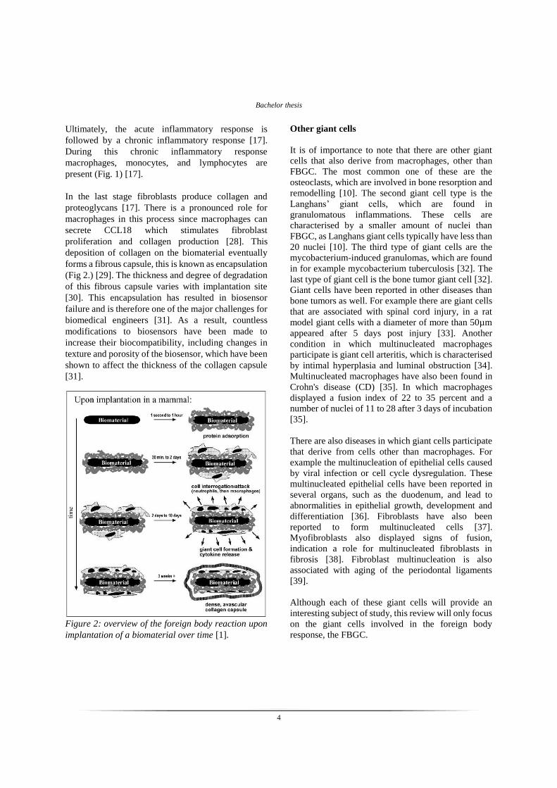
Implantation of medical devices or implants trigger a
variety of reactions from the host. Together these
reactions are known as the FBR [17]. Anderson
provides an overview of the FBR consisting of the
following events: acute inflammation, chronic
inflammation, granulation of the tissue, fibrous
capsule formation and the formation of foreign body
giant cells (FBGC) (Fig. 2) [4].
The first step after implantation of a biomaterial is the
accumulation of proteins on the surface of the
biomaterial [18]. Upon interaction with blood of the
host primarily fibrinogen, fibronectin, haemoglobin
immunoglobulin G, (IgG), and albumin will absorb to
the surface of the biomaterial. Subsequently these
proteins will be replaced by bigger molecules such as
kininogen and factor XII, this effect is referred to as
the Vroman effect [19, 20, 21]. The composition and
structure of this protein layer is believed to affect the
surrounding tissue, and is even associated with the
induction of the FBR because in normal wound
healing this protein layer has not been observed [1,
22].
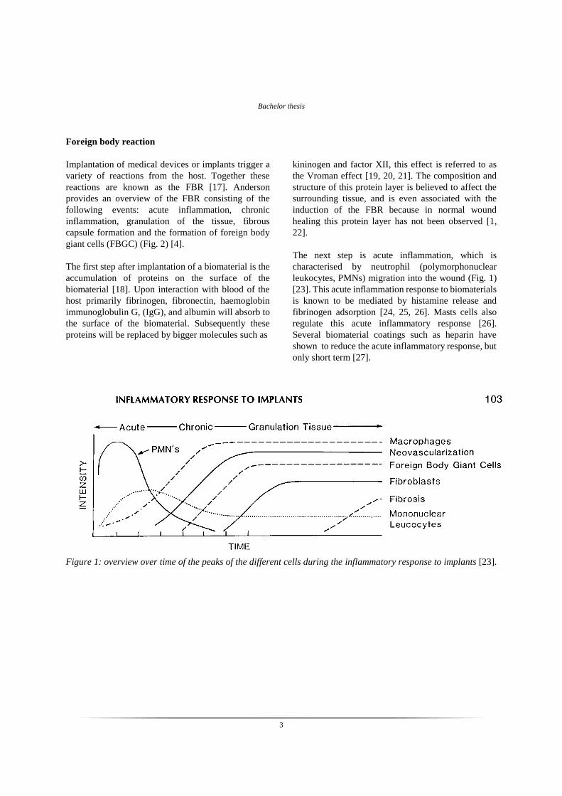
The next step is acute inflammation, which is
characterised by neutrophil (polymorphonuclear
leukocytes, PMNs) migration into the wound (Fig. 1)
[23]. This acute inflammation response to biomaterials
is known to be mediated by histamine release and
fibrinogen adsorption [24, 25, 26]. Masts cells also
regulate this acute inflammatory response [26].
Several biomaterial coatings such as heparin have
shown to reduce the acute inflammatory response, but
only short term [27].
Figure 1: overview over time of the peaks of the different cells during the inflammatory response to implants [23].
Bachelor thesis
4
Ultimately, the acute inflammatory response is
followed by a chronic inflammatory response [17].
During this chronic inflammatory response
macrophages, monocytes, and lymphocytes are
present (Fig. 1) [17].
In the last stage fibroblasts produce collagen and
proteoglycans [17]. There is a pronounced role for
macrophages in this process since macrophages can
secrete CCL18 which stimulates fibroblast
proliferation and collagen production [28]. This
deposition of collagen on the biomaterial eventually
forms a fibrous capsule, this is known as encapsulation
(Fig 2.) [29]. The thickness and degree of degradation
of this fibrous capsule varies with implantation site
[30]. This encapsulation has resulted in biosensor
failure and is therefore one of the major challenges for
biomedical engineers [31]. As a result, countless
modifications to biosensors have been made to
increase their biocompatibility, including changes in
texture and porosity of the biosensor, which have been
shown to affect the thickness of the collagen capsule
[31].
Figure 2: overview of the foreign body reaction upon
implantation of a biomaterial over time [1].
Other giant cells
It is of importance to note that there are other giant
cells that also derive from macrophages, other than
FBGC. The most common one of these are the
osteoclasts, which are involved in bone resorption and
remodelling [10]. The second giant cell type is the
Langhans’ giant cells, which are found in
granulomatous inflammations. These cells are
characterised by a smaller amount of nuclei than
FBGC, as Langhans giant cells typically have less than
20 nuclei [10]. The third type of giant cells are the
mycobacterium-induced granulomas, which are found
in for example mycobacterium tuberculosis [32]. The
last type of giant cell is the bone tumor giant cell [32].
Giant cells have been reported in other diseases than
bone tumors as well. For example there are giant cells
that are associated with spinal cord injury, in a rat
model giant cells with a diameter of more than 50µm
appeared after 5 days post injury [33]. Another
condition in which multinucleated macrophages
participate is giant cell arteritis, which is characterised
by intimal hyperplasia and luminal obstruction [34].
Multinucleated macrophages have also been found in
Crohn's disease (CD) [35]. In which macrophages
displayed a fusion index of 22 to 35 percent and a
number of nuclei of 11 to 28 after 3 days of incubation
[35].
There are also diseases in which giant cells participate
that derive from cells other than macrophages. For
example the multinucleation of epithelial cells caused
by viral infection or cell cycle dysregulation. These
multinucleated epithelial cells have been reported in
several organs, such as the duodenum, and lead to
abnormalities in epithelial growth, development and
differentiation [36]. Fibroblasts have also been
reported to form multinucleated cells [37].
Myofibroblasts also displayed signs of fusion,
indication a role for multinucleated fibroblasts in
fibrosis [38]. Fibroblast multinucleation is also
associated with aging of the periodontal ligaments
[39].
Although each of these giant cells will provide an
interesting subject of study, this review will only focus
on the giant cells involved in the foreign body
response, the FBGC.
Bachelor thesis
5
FBGC formation
During the acute inflammatory response binding of
protein ligands such as fibrinogen, fibronectin,
vitronectin, IgG and complement fragment iC3b to β2
integrin results in monocyte adhesion [4, 32]. These
monocytes express different membrane receptors
important in fusion of macrophages to form foreign
body giant cells (FBGC) including the mannose
receptor, β1 integrin and CCl2 [40, 41].
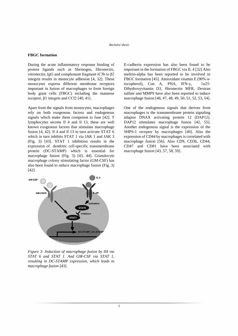
Apart from the signals from monocytes, macrophages
rely on both exogenous factors and endogenous
signals which make them competent to fuse [42]. T
lymphocytes secrete Il 4 and Il 13, these are well
known exogenous factors that stimulate macrophage
fusion [4, 42]. Il 4 and Il 13 in turn activate STAT 6
which in turn inhibits STAT 1 via JAK 1 and JAK 3
(Fig. 3) [43]. STAT 1 inhibition results in the
expression of dendritic cell-specific transmembrane
protein (DC-STAMP) which is essential for
macrophage fusion (Fig. 3) [43, 44]. Granulocyte
macrophage colony stimulating factor (GM-CSF) has
also been found to induce macrophage fusion (Fig. 3)
[42].
Figure 3: Induction of macrophage fusion by Il4 via
STAT 6 and STAT 1. And GM-CSF via STAT 1,
resulting in DC-STAMP expression, which leads to
macrophage fusion [43].
E-cadherin expression has also been found to be
important in the formation of FBGC via IL 4 [32] Also
meltrin-alpha has been reported to be involved in
FBGC formation [45]. Antioxidant vitamin E (90% α-
tocopherol), Con A, PHA, IFN-y, 1α25-
Dihydroxyvitamin D3, fibronectin MFR, Dextran
sulfate and MMP9 have also been reported to induce
macrophage fusion [46, 47, 48, 49, 50, 51, 52, 53, 54].
One of the endogenous signals that derives from
macrophages is the transmembrane protein signaling
adaptor DNAX activating protein 12 (DAP12).
DAP12 stimulates macrophage fusion [42, 55].
Another endogenous signal is the expression of the
SHPS-1 receptor by macrophages [40]. Also the
expression of CD44 by macrophages is correlated with
macrophage fusion [56]. Also CD9, CD36, CD44,
CD47 and CD81 have been associated with
macrophage fusion [43, 57, 58, 59].
Bachelor thesis
6
Markers of FBGC
Besides numerous factors that induce FBGC
formation, there are also quite a few markers that
FBGC express, that have been reported. These
markers could be important in correctly identifying
multinucleated macrophages. Since it has been stated
in the literature that it is difficult differentiating
multinucleated giant cells from for example
osteoclasts which also derive from macrophages [42,
60]. It is important to note that there is a distinct
difference in function as FBGC are unable to resorb
bone whereas this is the main function of osteoclasts
[47]. To make discriminating between the two even
more difficult, TRAP and the vitronectin receptor have
been reported as a marker for osteoclasts but have also
been found to be expressed by multinucleated
macrophages [60, 61]. However there are also studies
that report that TRAP is not expressed by FBGC [62].
So it still is rather unclear which markers can actually
be referred to as FBGC specific. Or even whether there
is an plasticity in the expression of markers by FBGC
leading to these opposing results.
However several markers for FBGC have been
reported that could help correctly identify
multinucleated giant cells. There has been found that
Na-K-adenosinetriphosphatases (Na-K-ATPases) are
expressed on the plasma membranes of both FBGC as
well as osteoclasts [63]. However on the plasma
membrane of FBGC the location of these Na-K-
ATPases is at the non-adhesive side of the cell [63].
This suggest a functionality in polarisation of the
position of these Na-K-ATPases [63]. FBGC also
express a high number of calcitonin receptors, which
are also believed to be involved in the regulation of
local immune reactions [63].
Furthermore McNally et al found that IL4 stimulated
FBGC strongly express HLA-DR, CD98, CD86, and
B7-H1 (PD-L1) [62]. They also express αX integrin
(CD11c), CD68, and dendritic cell-specific
intercellular adhesion molecule-3-grabbing non-
integrin (DC-SIGN) [62]. According to McNally et al,
these molecules might provide interactions with
lymphocytes, such as PD-L1, which is believed to be
able to down regulate lymphocytes when it binds to its
co-receptor on lymphocytes [62]. Suggesting a
regulatory role for FBGC.
Ym1 and ALOX15 are two markers of M2
macrophages, these markers were found to be
significantly higher expressed in FBGC than in M2
macrophages or osteoclasts, on both mRNA and
protein levels [47].
Bachelor thesis
7
Reaction of FBGC to biomaterials
To my knowledge there are no reviews describing the
differences in the response of FBGC to different
biomaterials. In this section the reaction of FBGC to
several biomaterials will be discussed as this reaction
can vary between different biomaterials as well as
different implantation sites and implantation
techniques [30]. An extensive overview of the
differences in the reaction of FBGC to different
biomaterials could therefore prove to be important.
Effect of implantation site
Bakker et al studied the effect of implantation site on
FBGC in male Wistar rats. At implant sites containing
bone and muscle tissue they found multinucleated
cells of up to 20 nuclei after the first week of
implantation, the number of nuclei increased to around
200 after 13 weeks of implantation. In comparison
after 1 week of implantation, tympanic membrane and
submucosal implants demonstrated FBGC with a
maximum of 10 nuclei. For the submucosal implants
this number increased to 30 nuclei after 13 weeks of
implantation. And after 13 weeks of implantation the
tympanic membrane implants the giant cells did not
display more than 20 nuclei [30].
Luttikhuizen et al, showed that this difference in FBR
on different implantation sites correlates to cytokine
and MMP expression in mice [64]. Degeneration of
the biomaterial and leukocyte migration were higher
in supra-epicardially implanted collagen when
compared to subcutaneously implanted collagen. This
difference correlated with the expression of cytokines,
including higher expression of IL-1 and IL-6 but lower
expression of IL-10 creating a predominantly
inflammatory milieu for supra-epicardially implanted
collagen. Supra-epicardially implanted collagen also
displayed higher expression CXCL1/KC and
CXCL2/MIP2 resulting in PMN migration. Gene
expression of collagen degrading MMPs did not differ
significantly between the two sites of implantation,
however MMP activity was higher on supra-
epicardially implanted collagen. The expression of
MMP9, which is known to attract macrophage and
induce fusion, was also higher supra-epicardially [64].
Effect of the surface of biomaterials on FBGC
Anderson states that in general rough or porous
implants contain a higher amounts of FBGC than flat
surfaces such as those of breast implants where only a
one- to two-cell layer of macrophages and FBGC is
found [10, 65]. However Anderson presents no data to
verifies this statement, so one has to be sceptical about
his statements.
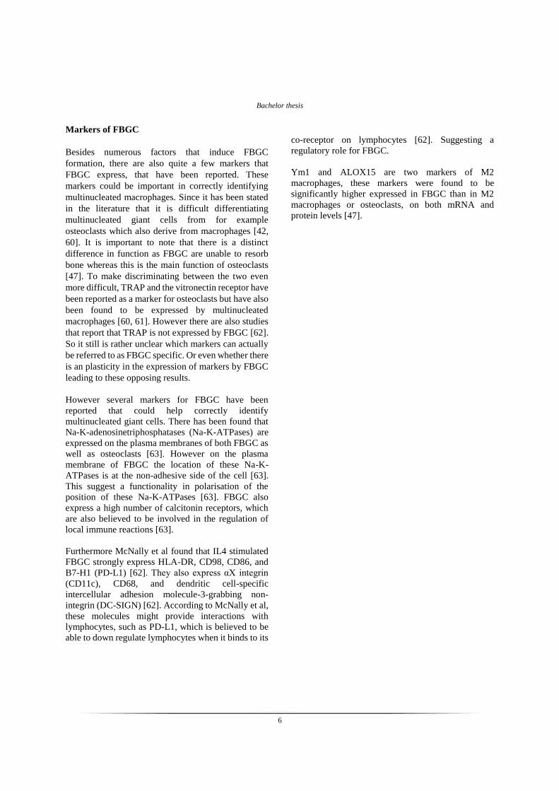
Taylor et al do show that rough surfaces of premium
electrical grade Teflon (PTFE) display higher
macrophage adhesion than smooth surfaces (Fig. 4).
Resulting in higher formation of FBGC on the textured
PTFE and higher secretion of acid phosphatase, than
on the smooth surface. But the fibrous capsule is
reduced in thickness on the textured surface when
compared to the smooth surface [66].
Figure 4:Macrophage adhesion on textured (left) and
smooth (right) premium electrical grade eflon (PTFE)
surface after 3 days of implantation (160X) [66].
Effect of species
It is also known that the FBR differs between species.
In a comparative study it was discovered that mice
display less phagocytosis but more calcification of
implanted biomaterial, when compared to rats [67].
These differences between different species should be
taken into account when researching the FBR in model
species.
Bachelor thesis
8
Glass substrates
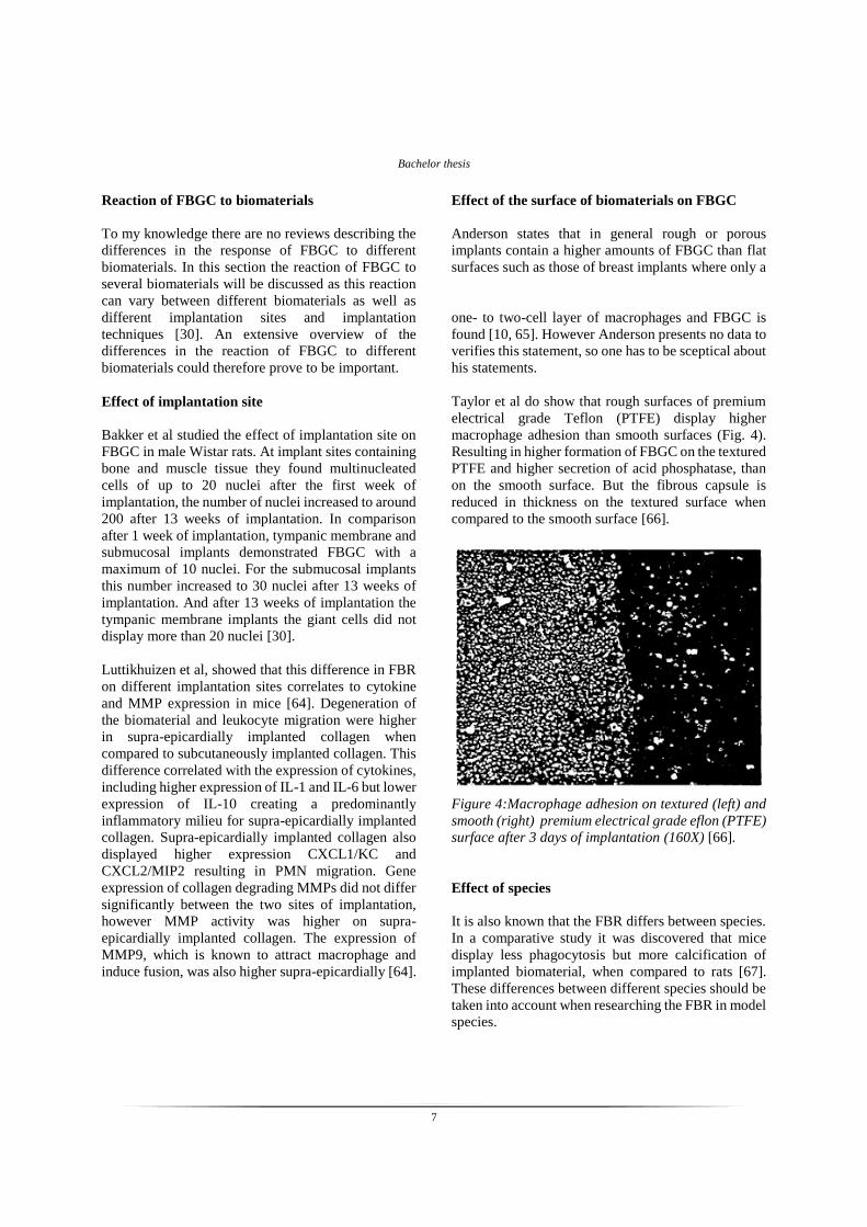
In a study performed by Jenney et al, molecules of the
alkyl family were bound to silane-modified glass in
order to investigate their FBGC formation properties.
These molecules ranged from methyl (C1) till
octadecyl (C18) (Fig. 5) [68].
Figure 5: overview of the alkylsilane-modified glass
substrates (C1, C3, C6, C10, C14 and C18) including
clean glass and silane-modified glass(DM) [68].
There was found that clean glass substrates display
low FBGC formation and density. The tetradecyl
(C14) and octadecyl (C18) showed a decrease in cell
density over the course of 10 days, resulting in a low
cell density on day 10 and therefore low FBGC
formation (Fig. 6) .
In comparison the silane-modified glass and silane-
modified glass containing methyl, propyl, hexyl or
decyl displayed high FBGC formation and density
(Fig. 6) [68]. This effect was found both for
unstimulated monocyte derived FBGC as well as IL4
induced monocyte derived FBGC [68].
Figure 6: density of IL4 induced FBGC on the
alkylsilane-modified glass substrates after 10 days of
culturing [68].
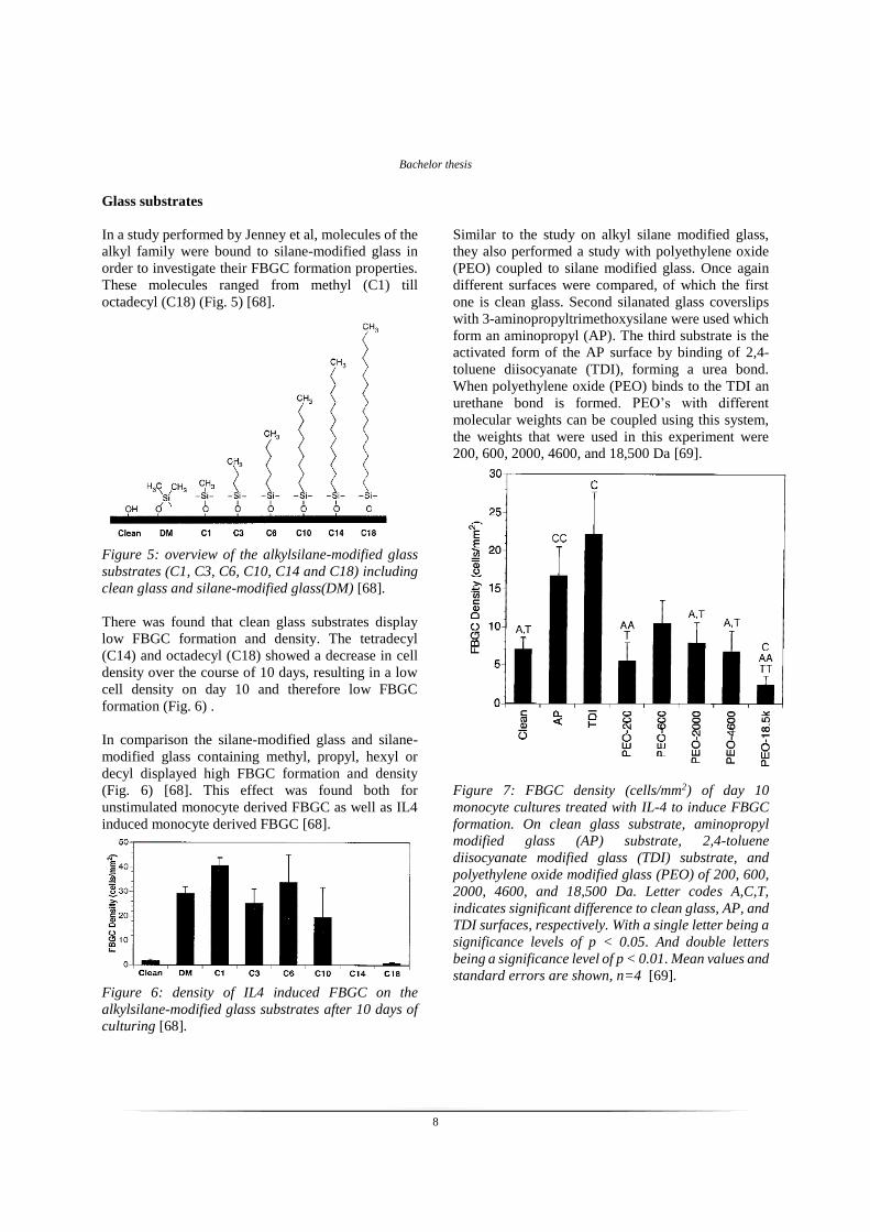
Similar to the study on alkyl silane modified glass,
they also performed a study with polyethylene oxide
(PEO) coupled to silane modified glass. Once again
different surfaces were compared, of which the first
one is clean glass. Second silanated glass coverslips
with 3-aminopropyltrimethoxysilane were used which
form an aminopropyl (AP). The third substrate is the
activated form of the AP surface by binding of 2,4-
toluene diisocyanate (TDI), forming a urea bond.
When polyethylene oxide (PEO) binds to the TDI an
urethane bond is formed. PEO’s with different
molecular weights can be coupled using this system,
the weights that were used in this experiment were
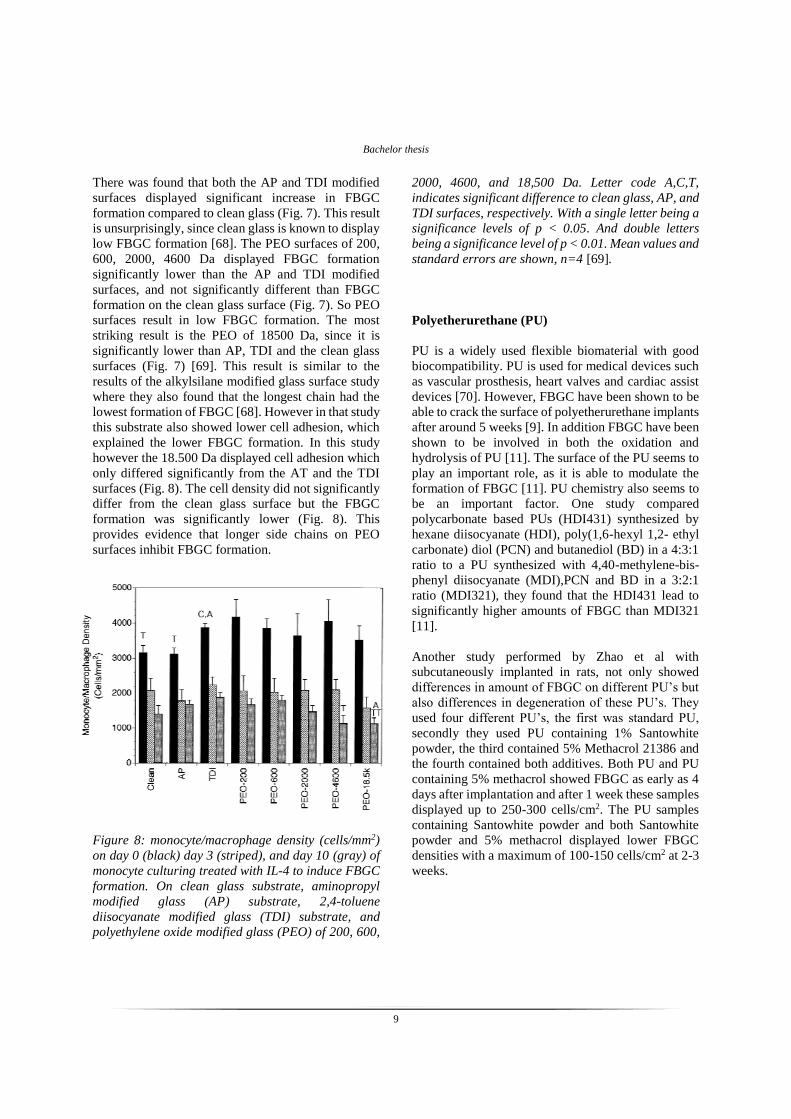
200, 600, 2000, 4600, and 18,500 Da [69].
Figure 7: FBGC density (cells/mm2) of day 10
monocyte cultures treated with IL-4 to induce FBGC
formation. On clean glass substrate, aminopropyl
modified glass (AP) substrate, 2,4-toluene
diisocyanate modified glass (TDI) substrate, and
polyethylene oxide modified glass (PEO) of 200, 600,
2000, 4600, and 18,500 Da. Letter codes A,C,T,
indicates significant difference to clean glass, AP, and
TDI surfaces, respectively. With a single letter being a
significance levels of p < 0.05. And double letters
being a significance level of p < 0.01. Mean values and
standard errors are shown, n=4 [69].
Bachelor thesis
9
There was found that both the AP and TDI modified
surfaces displayed significant increase in FBGC
formation compared to clean glass (Fig. 7). This result
is unsurprisingly, since clean glass is known to display
low FBGC formation [68]. The PEO surfaces of 200,
600, 2000, 4600 Da displayed FBGC formation
significantly lower than the AP and TDI modified
surfaces, and not significantly different than FBGC
formation on the clean glass surface (Fig. 7). So PEO
surfaces result in low FBGC formation. The most
striking result is the PEO of 18500 Da, since it is
significantly lower than AP, TDI and the clean glass
surfaces (Fig. 7) [69]. This result is similar to the
results of the alkylsilane modified glass surface study
where they also found that the longest chain had the
lowest formation of FBGC [68]. However in that study
this substrate also showed lower cell adhesion, which
explained the lower FBGC formation. In this study
however the 18.500 Da displayed cell adhesion which
only differed significantly from the AT and the TDI
surfaces (Fig. 8). The cell density did not significantly
differ from the clean glass surface but the FBGC
formation was significantly lower (Fig. 8). This
provides evidence that longer side chains on PEO
surfaces inhibit FBGC formation.
Figure 8: monocyte/macrophage density (cells/mm2)
on day 0 (black) day 3 (striped), and day 10 (gray) of
monocyte culturing treated with IL-4 to induce FBGC
formation. On clean glass substrate, aminopropyl
modified glass (AP) substrate, 2,4-toluene
diisocyanate modified glass (TDI) substrate, and
polyethylene oxide modified glass (PEO) of 200, 600,
2000, 4600, and 18,500 Da. Letter code A,C,T,
indicates significant difference to clean glass, AP, and
TDI surfaces, respectively. With a single letter being a
significance levels of p < 0.05. And double letters
being a significance level of p < 0.01. Mean values and
standard errors are shown, n=4 [69].
Polyetherurethane (PU)
PU is a widely used flexible biomaterial with good
biocompatibility. PU is used for medical devices such
as vascular prosthesis, heart valves and cardiac assist
devices [70]. However, FBGC have been shown to be
able to crack the surface of polyetherurethane implants
after around 5 weeks [9]. In addition FBGC have been
shown to be involved in both the oxidation and
hydrolysis of PU [11]. The surface of the PU seems to
play an important role, as it is able to modulate the
formation of FBGC [11]. PU chemistry also seems to
be an important factor. One study compared
polycarbonate based PUs (HDI431) synthesized by
hexane diisocyanate (HDI), poly(1,6-hexyl 1,2- ethyl
carbonate) diol (PCN) and butanediol (BD) in a 4:3:1
ratio to a PU synthesized with 4,40-methylene-bis-
phenyl diisocyanate (MDI),PCN and BD in a 3:2:1
ratio (MDI321), they found that the HDI431 lead to
significantly higher amounts of FBGC than MDI321
[11].
Another study performed by Zhao et al with
subcutaneously implanted in rats, not only showed
differences in amount of FBGC on different PU’s but
also differences in degeneration of these PU’s. They
used four different PU’s, the first was standard PU,
secondly they used PU containing 1% Santowhite
powder, the third contained 5% Methacrol 21386 and
the fourth contained both additives. Both PU and PU
containing 5% methacrol showed FBGC as early as 4
days after implantation and after 1 week these samples
displayed up to 250-300 cells/cm2. The PU samples
containing Santowhite powder and both Santowhite
powder and 5% methacrol displayed lower FBGC
densities with a maximum of 100-150 cells/cm2 at 2-3
weeks.
Bachelor thesis
10
On the standard PU sample cracking of the surface
started to appear after 3 weeks, in comparison the PU’s
containing the additives this cracking seemed lower.
However on both samples containing 5% methacrol
severe pitting of the sample appeared after as early as
2 weeks. This cracking of the surface was believed to
be induced by oxidative degeneration, caused by
oxygen radical release of FBGC showing their
degenerative function. 5% methacrol is believed to
cause pitting in acidic environments. It is known that
FBGC can cause a highly acidic zone (pH 3.5) when
attached to the surface of the biomaterial, this acidic
environment caused by the FBGC may have caused
the pitting of the 5% methacrol samples [71].
Candidate substances to inhibit FBGC formation on
PU include IL-4 neutralizing antibody (IL4Ab), which
has shown an inhibition of FBGC formation on
subcutaneous implanted PU in mice [72]
Secretion of cytokines by FBGC
So far it remains relatively unclear whether FBGC are
capable of the secretion of cytokines and whether or
not FBGC have a role in the inflammatory response
during the FBR. However it is known that in giant cell
arteritis other multinucleated giant cells promote
neoangiogensis through the production of angiogenic
cytokines such as vascular endothelial growth factor
(VEGF) [73]. This suggest that FBGC are able to
secrete cytokines. It has also been suggested that
FBGC are capable of producing cytokines, such as
connective tissue growth factor that would induce a
fibrogenic phenotype in wound healing [65].
FBGC gene expression plasticity
Luttikhuizen et al performed a study in which they
compared gene expression of FBGC on different
substrates including, polyethylene terephthalate
(Dacron), cross-linked sheep collagen (HDSC), and
bioactive PCLdiUPy [74].
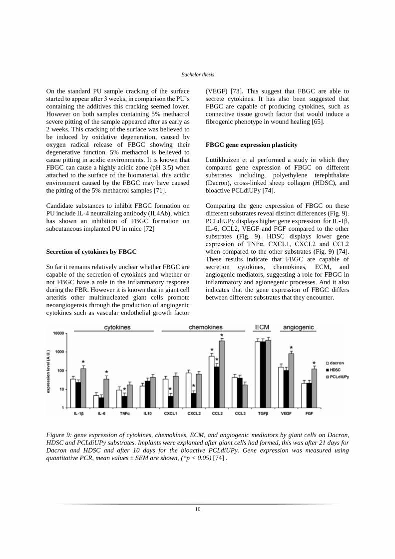
Comparing the gene expression of FBGC on these
different substrates reveal distinct differences (Fig. 9).
PCLdiUPy displays higher gene expression for IL-1β,
IL-6, CCL2, VEGF and FGF compared to the other
substrates (Fig. 9). HDSC displays lower gene
expression of TNFα, CXCL1, CXCL2 and CCL2
when compared to the other substrates (Fig. 9) [74].
These results indicate that FBGC are capable of
secretion cytokines, chemokines, ECM, and
angiogenic mediators, suggesting a role for FBGC in
inflammatory and agionegenic processes. And it also
indicates that the gene expression of FBGC differs
between different substrates that they encounter.
Figure 9: gene expression of cytokines, chemokines, ECM, and angiogenic mediators by giant cells on Dacron,
HDSC and PCLdiUPy substrates. Implants were explanted after giant cells had formed, this was after 21 days for
Dacron and HDSC and after 10 days for the bioactive PCLdiUPy. Gene expression was measured using
quantitative PCR, mean values ± SEM are shown, (*p < 0.05) [74] .
Bachelor thesis
11
Role of FBGC in inflammatory response
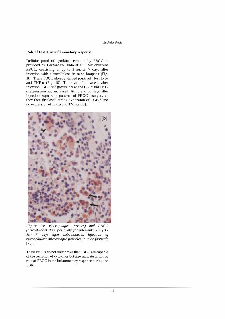
Definite proof of cytokine secretion by FBGC is
provided by Hernandez-Pando et al. They observed
FBGC, consisting of up to 3 nuclei, 7 days after
injection with nitrocellulose in mice footpads (Fig.
10). These FBGC already stained positively for IL-1α
and TNF-α (Fig. 10). Three and four weeks after
injection FBGC had grown in size and IL-1α and TNF-
α expression had increased. At 45 and 60 days after
injection expression patterns of FBGC changed, as
they then displayed strong expression of TGF-β and
no expression of IL-1α and TNF-α [75].
Figure 10: Macrophages (arrows) and FBGC
(arrowheads) stain positively for interleukin-1α (IL-
1α) 7 days after subcutaneous injection of
nitrocellulose microscopic particles in mice footpads
[75].
These results do not only prove that FBGC are capable
of the secretion of cytokines but also indicate an active
role of FBGC in the inflammatory response during the
FBR.
Bachelor thesis
12
FBGC and Diabetes Mellitus
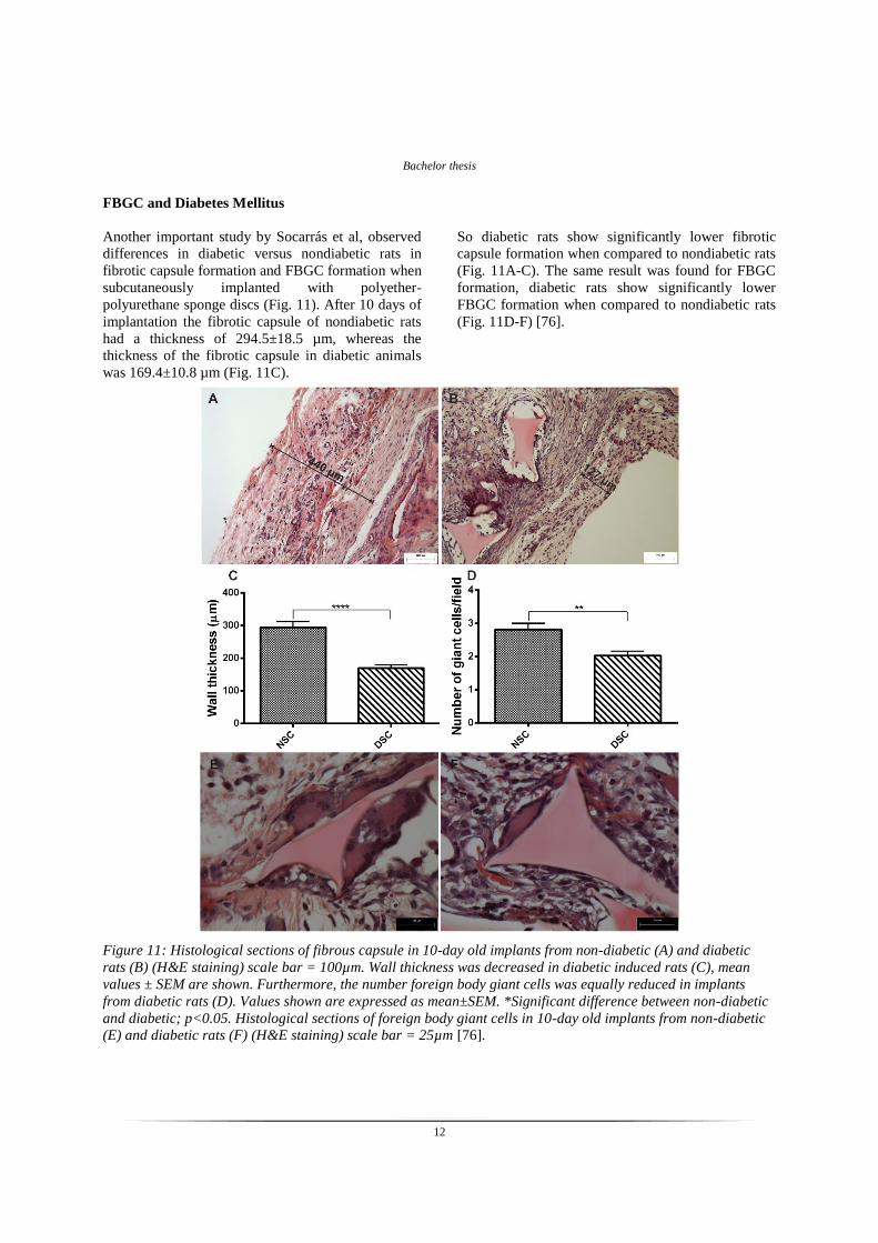
Another important study by Socarrás et al, observed
differences in diabetic versus nondiabetic rats in
fibrotic capsule formation and FBGC formation when
subcutaneously implanted with polyether-
polyurethane sponge discs (Fig. 11). After 10 days of
implantation the fibrotic capsule of nondiabetic rats
had a thickness of 294.5±18.5 µm, whereas the
thickness of the fibrotic capsule in diabetic animals
was 169.4±10.8 µm (Fig. 11C).
So diabetic rats show significantly lower fibrotic
capsule formation when compared to nondiabetic rats
(Fig. 11A-C). The same result was found for FBGC
formation, diabetic rats show significantly lower
FBGC formation when compared to nondiabetic rats
(Fig. 11D-F) [76].
Figure 11: Histological sections of fibrous capsule in 10-day old implants from non-diabetic (A) and diabetic
rats (B) (H&E staining) scale bar = 100µm. Wall thickness was decreased in diabetic induced rats (C), mean
values ± SEM are shown. Furthermore, the number foreign body giant cells was equally reduced in implants
from diabetic rats (D). Values shown are expressed as mean±SEM. *Significant difference between non-diabetic
and diabetic; p<0.05. Histological sections of foreign body giant cells in 10-day old implants from non-diabetic
(E) and diabetic rats (F) (H&E staining) scale bar = 25µm [76].
Bachelor thesis
13
Polydimethylsiloxane (PDMS)
PDMS is a widely used biomaterial which is
incorporated in biomedical devices such as stents. Its
elastomeric properties, biocompatibility, gas
permeability, optical transparency, and relatively low
costs make it an attractive material [77, 78].
When PDMS was compared to low density
polyethylene (LDPE), PDMS was found to display a
higher activation of complement factors and more
adhesion of macrophages resulting in higher FBGC
formation [21]. After 3 weeks the PDMS contained
more FBGC with more than 20 nuclei in comparison
to the LDPE. Also the phagocytic abilities of the
FBGC on PDMS were lower than on LDPE [79].
PDMS surfaces also displayed significantly higher
macrophage fusion than Bionate 80A (PCU),
Elasthane 80A (PEU) and PurSil20 80A (PEU-S)
surfaces after 7 days and after 10 days of culturing
[80]. However PDMS and other hydrogels have been
found to display a lower density of FBGC, as well as
a lower coverage of the surface with FBGC than
Pellethane® a thermoplastic PU [81]. So tremendous
differences can be found of FBGC formation, density
and coverage, between different substrates.
Xenograft
Furthermore FBGC reactions have been found in
patients with lower eyelid retractions using tarSys™
xenograft [82].
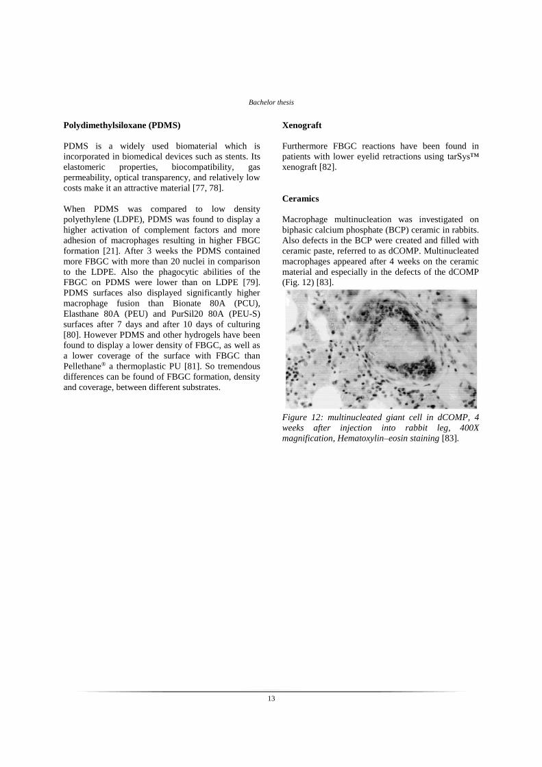
Ceramics
Macrophage multinucleation was investigated on
biphasic calcium phosphate (BCP) ceramic in rabbits.
Also defects in the BCP were created and filled with
ceramic paste, referred to as dCOMP. Multinucleated
macrophages appeared after 4 weeks on the ceramic
material and especially in the defects of the dCOMP
(Fig. 12) [83].
Figure 12: multinucleated giant cell in dCOMP, 4
weeks after injection into rabbit leg, 400X
magnification, Hematoxylin–eosin staining [83].
Bachelor thesis
14
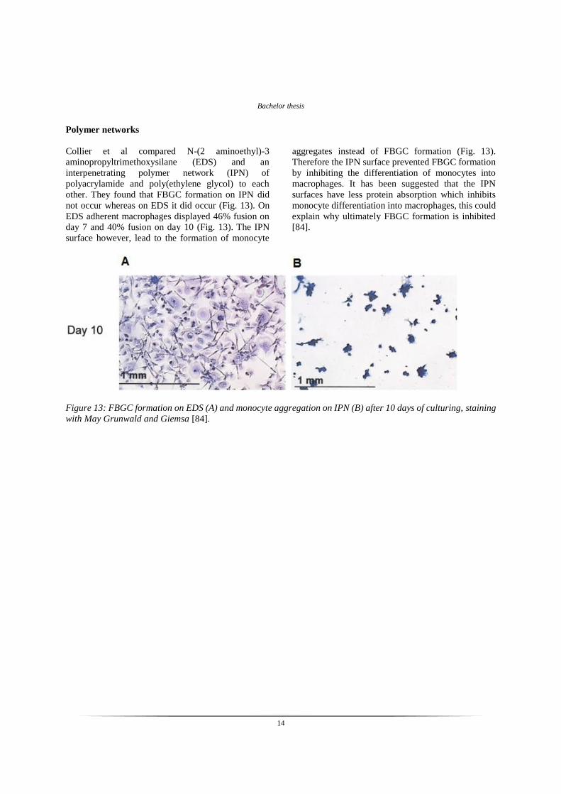
Polymer networks
Collier et al compared N-(2 aminoethyl)-3
aminopropyltrimethoxysilane (EDS) and an
interpenetrating polymer network (IPN) of
polyacrylamide and poly(ethylene glycol) to each
other. They found that FBGC formation on IPN did
not occur whereas on EDS it did occur (Fig. 13). On
EDS adherent macrophages displayed 46% fusion on
day 7 and 40% fusion on day 10 (Fig. 13). The IPN
surface however, lead to the formation of monocyte
aggregates instead of FBGC formation (Fig. 13).
Therefore the IPN surface prevented FBGC formation
by inhibiting the differentiation of monocytes into
macrophages. It has been suggested that the IPN
surfaces have less protein absorption which inhibits
monocyte differentiation into macrophages, this could
explain why ultimately FBGC formation is inhibited
[84].
Figure 13: FBGC formation on EDS (A) and monocyte aggregation on IPN (B) after 10 days of culturing, staining
with May Grunwald and Giemsa [84].
Bachelor thesis
15
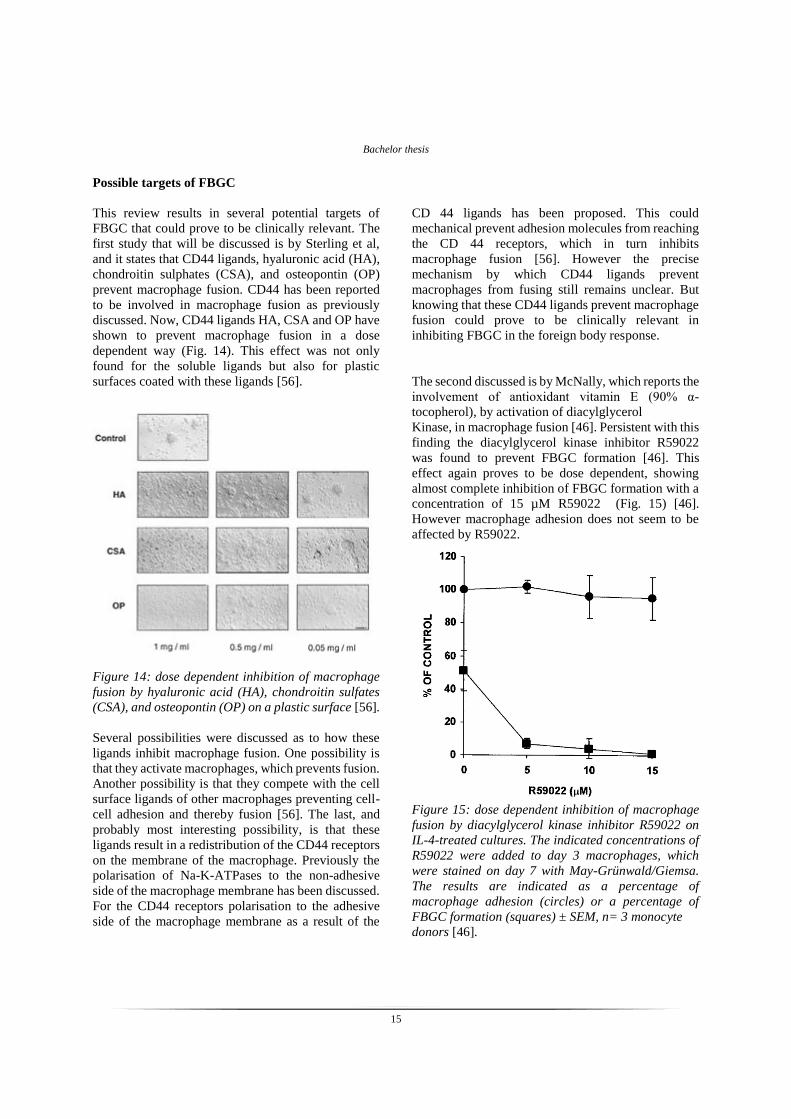
Possible targets of FBGC
This review results in several potential targets of
FBGC that could prove to be clinically relevant. The
first study that will be discussed is by Sterling et al,
and it states that CD44 ligands, hyaluronic acid (HA),
chondroitin sulphates (CSA), and osteopontin (OP)
prevent macrophage fusion. CD44 has been reported
to be involved in macrophage fusion as previously
discussed. Now, CD44 ligands HA, CSA and OP have
shown to prevent macrophage fusion in a dose
dependent way (Fig. 14). This effect was not only
found for the soluble ligands but also for plastic
surfaces coated with these ligands [56].
Figure 14: dose dependent inhibition of macrophage
fusion by hyaluronic acid (HA), chondroitin sulfates
(CSA), and osteopontin (OP) on a plastic surface [56].
Several possibilities were discussed as to how these
ligands inhibit macrophage fusion. One possibility is
that they activate macrophages, which prevents fusion.
Another possibility is that they compete with the cell
surface ligands of other macrophages preventing cell-
cell adhesion and thereby fusion [56]. The last, and
probably most interesting possibility, is that these
ligands result in a redistribution of the CD44 receptors
on the membrane of the macrophage. Previously the
polarisation of Na-K-ATPases to the non-adhesive
side of the macrophage membrane has been discussed.
For the CD44 receptors polarisation to the adhesive
side of the macrophage membrane as a result of the
CD 44 ligands has been proposed. This could
mechanical prevent adhesion molecules from reaching
the CD 44 receptors, which in turn inhibits
macrophage fusion [56]. However the precise
mechanism by which CD44 ligands prevent
macrophages from fusing still remains unclear. But
knowing that these CD44 ligands prevent macrophage
fusion could prove to be clinically relevant in
inhibiting FBGC in the foreign body response.
The second discussed is by McNally, which reports the
involvement of antioxidant vitamin E (90% α-
tocopherol), by activation of diacylglycerol
Kinase, in macrophage fusion [46]. Persistent with this
finding the diacylglycerol kinase inhibitor R59022
was found to prevent FBGC formation [46]. This
effect again proves to be dose dependent, showing
almost complete inhibition of FBGC formation with a
concentration of 15 µM R59022 (Fig. 15) [46].
However macrophage adhesion does not seem to be
affected by R59022.
Figure 15: dose dependent inhibition of macrophage
fusion by diacylglycerol kinase inhibitor R59022 on
IL-4-treated cultures. The indicated concentrations of
R59022 were added to day 3 macrophages, which
were stained on day 7 with May-Grünwald/Giemsa.
The results are indicated as a percentage of
macrophage adhesion (circles) or a percentage of
FBGC formation (squares) ± SEM, n= 3 monocyte
donors [46].
Bachelor thesis
16
These results could indicate a prominent role for
phosphorylation of diacylglycerol in FBGC formation.
The R59022 inhibitor could therefore prove to be
clinically relevant in preventing macrophage fusion.
The third study that presents a candidate for inhibition
of macrophage fusion is the study performed by Zhao
et al on PU coatings, where they found that Santowhite
powder coatings resulted in inhibition of FBGC
formation [71]. In addition the studies by Jenny et al,
also proposed inhibitory effects of coating. They
found that tetradecyl (C14) and octadecyl (C18) alkyl
coatings on glass resulted in inhibition of FBGC
formation [68]. As well as glass coated with 18.500 Da
PEO [69]. These results indicate that different coatings
of biomaterials could definitely have an effect on the
foreign body response against these biomaterials,
especially the formation of FBGC on the surface of
these biomaterials.
Figure 16: Macrophage fusion of human monocyte
culture in 10 % FCS RPMI medium containing 10
µg/ml of Con A. 1 mM ferric citrate was added after
0h and 12 hours. Each bar represents the mean ± SEM
of 3 experiments. * p < 0.05, ** p < 0.02 [50].
The fourth study that will be discussed is by Tahara et
al, where the researchers found that macrophage
fusion by con A can be inhibited by addition of 1mM
ferric citrate (Fig. 16). However this effect is only
short term as the effect is significant directly after
addition of ferric citrate up to 12 hours after addition
(Fig. 16). After 24 hours the effect is not significant
anymore [50]. This reveals a short term effect of ferric
citrate on FBGC formation. Because this effect is only
short term, the clinical applications would be limited.
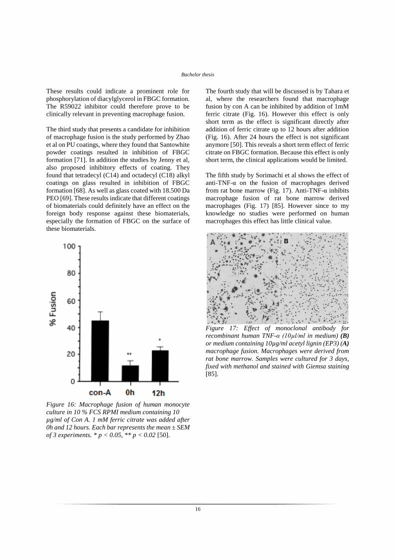
The fifth study by Sorimachi et al shows the effect of
anti-TNF-α on the fusion of macrophages derived
from rat bone marrow (Fig. 17). Anti-TNF-α inhibits
macrophage fusion of rat bone marrow derived
macrophages (Fig. 17) [85]. However since to my
knowledge no studies were performed on human
macrophages this effect has little clinical value.
Figure 17: Effect of monoclonal antibody for
recombinant human TNF-α (10µl/ml in medium) (B)
or medium containing 10µg/ml acetyl lignin (EP3) (A)
macrophage fusion. Macrophages were derived from
rat bone marrow. Samples were cultured for 3 days,
fixed with methanol and stained with Giemsa staining
[85].

Bachelor thesis
17
The sixth study that will be discussed that poses a
candidate for inhibition of macrophage fusion is a
study performed by Katsuyama et al. They show that
interleukin-1 receptor-associated kinase-4 (IRAK4)
inhibits FBGC formation [47]. IRAK4 is arguably the
best potential candidate for clinical use. Since it has
been shown that IRAK4 deficient mice display normal
osteoclast formation and bone mineral density [47]. In
comparison IRAK4 was found to inhibit macrophage
fusion [47]. In addition IRAK4 deficiency restored
macrophage fusion [47]. Normal osteoclast function in
combination with FBGC formation inhibition
classifies IRAK4 as a potential therapeutic target for
inhibition of macrophage fusion during the FBR.
In the last study that will be discussed, Tsai et al show
osteopontin (OPN) as another possible candidate for
inhibition of FBGC formation, both in vitro and in
vivo. OPN knock out mice displayed an increase in
FBGC formation. And in vitro results show a dose
dependent inhibition of FBGC formation by OPN
(Fig. 18). However a relatively high dose of OPN is
required for prevention of fusion (Fig. 18) [86].
Figure 18: macrophage fusion in vitro. Fusion
percentage is the number of nuclei within FBGC
(more than 3 nuclei)/total number of counted nuclei
*100%. * is significant at p=0.0001. N=3 wells per
sample, mean values and SD are shown [86].
FBGC versus mononuclear macrophages
Earlier the differences in the Ym1 and ALOX15
markers between FBGC and M2 macrophages were
discussed, stating that Ym1 and ALOX15 expression
is significantly higher in FBGC than in M2
mononuclear macrophages [39]. However there are
more differences between FBGC and mononuclear
macrophages that have been reported.
Mononuclear macrophages in vitro possess higher
locomotive ability than multinucleated macrophages.
As far as phagocytosis activity goes, mononuclear
macrophages display a higher rate of phagocytosis of
smaller particles whereas giant cells are typically
found to surround larger particles, indication
differences in phagocytosis function [30].
Other studies support the finding that mononuclear
macrophages display phagocytosis of biomaterials,
whereas this is not found for FBGC. Multiple reports
of intracellular biomaterial particles in mononuclear
macrophages support their phagocytic abilities
towards biomaterials [83, 87]. Intracellular
biomaterial particles have however not been reported
in FBGC, questioning their phagocytic abilities.
In a comparative study between mononuclear and
multinuclear macrophages performed by
Papadimitriou et al, it was found that multinucleated
macrophages have significantly higher protein content
than mononuclear macrophages. However no
significant differences in succinate dehydrogenase
activity, non-specific esterase activity or acid
phosphatase activity were found between
mononuclear macrophages and giant cells. All values
were compared per nuclei in order to equally compare
mononuclear and multinuclear macrophages [88].
The most striking result is that succinate
dehydrogenase activity, non-specific esterase activity
and acid phosphatase activity of FGBC were found to
be equal to those of mononuclear macrophages. This
excludes the role of FBGC in the FBR as being more
efficient in breaking down biomaterials. However the
increased functionality of FBGC with regard to these
degrading enzymes might not so much be limited to
the amount of secreted enzyme, but could very well be
in the focussed delivery of these enzymes by FBGC.
Since FBGC might be able to deliver these enzymes

Bachelor thesis
18
on a smaller surface area than multiple mononuclear
macrophages could. This is yet another example that
the functionality of FBGC in the FBR remains unclear.
Another comparative study between mononuclear and
multinuclear macrophages performed by Enelow et al,
revealed different results. They measured oxidative
activity with two different techniques. First oxidative
activity was measured by cytochrome-c reduction
measurements, this revealed a 2.2 fold increase in
oxidative activity of multinucleated giant cells as
appose to mononuclear macrophages. With giant cells
producing 34.3 ± 8.4 nmol of superoxide/µg of cellular
protein and mononuclear macrophages producing 16.2
± 4.5 nmol of superoxide/µg of cellular protein.
Secondly, fluorescence intensity revealed that giant
cells had 1.7 times brighter fluorescence per unit of
cytoplasm compared to mononuclear macrophages.
These results indicate that giant cells have an
increased superoxide anion production compared to
mononuclear macrophages [89].
In another study however, it has been shown that
FBGC are capable of an 20-30 fold higher secretion of
oxygen free radicals per cell in comparison to normal
macrophages in response to zymosan [90]. So notable
differences in oxygen free radical production by
FBGC have been reported in the literature.
Enelow et al, not only investigated the superoxide
anion production, but also compared the phagocytosis
and killing of Candidae albicans (a gastrointestinal
yeast) of mononuclear macrophages and giant cells.
They found no significant differences in the number of
Candidae phagocytized nor in the number of cells
displaying phagocytic activity [89]. However they did
find significant differences in killing of Candidae
between macrophages and giant cells. As giant cells
killed 35.1% ± 2.0% of phagocytized organisms,
whereas macrophages killed 22.9% ± 1.8% of
phagocytized organisms. This experiment reveals the
phagocytic activity of multinucleated giant cells, as
well as their ability to kill phagocytized organisms
(Fig. 19) [89].
Figure 19: Multinucleated giant cell phagocytizing
Candidae. Giant cell nuclei are indicated with open
arrows, candidal "ghosts" are indicated with arrows
and candidae surviving at time of fixation are
indicated with arrowheads. Bars = 10 um [89].
However controversial results have been found
regarding the phagocytosis of multinucleated giant
cells. As Papadimitriou et al observed an decrease in
both yeast and staphylococci ingestion by
macrophages as they fuse (Fig. 20). As the number of
nuclei increases the phagocytic ability decreases (Fig.
20) [91]. So whereas Enelow et al, found that
multinucleated giant cells phagocytize Candidae,
Papadimitriou et al found that phagocytic ability
degreases with the amount of nuclei.
Figure 20: progressive decrease in phagocytosis of
yeast and staphylococci with increase in number of
nuclei of giant cells [91].
Bachelor thesis
19
Conclusion
To summarize, FBGC are believed to cause oxidation
and hydrolysis of biomaterials. This is supported by
the fact that FBGC showed to release up to 30 times
more oxygen free radicals than mononuclear
macrophages. They are also believed to be able to
secrete cytokines chemokines, ECM, and angiogenic
mediators.
Furthermore there have been differences found in
FBGC formation between different species of animals,
different biomaterials, different compositions of the
same biomaterial, different surfaces of the biomaterial,
as well as different sites of implantation of the
biomaterial.
Controversial results were found in the literature with
regard to phagocytic ability and the secretion of
degeneration enzymes of giant cells in comparison to
mononuclear macrophages. However in gerenal
mononuclear macrophages seem to display
phagocytosis whereas FBGC typically surround the
biomaterial.
Future research
This review concludes that controversial results were
found in the literature with regard to FBGC. This
implies that further research should be conducted. In
this part of the review, future prospects for research on
FBGC will be discussed.
The first option that will be discussed builds on the
results of Jenny et al, they investigated the effect of
silane modified glass with alkyl and PEO side chains
on FBGC formation [68, 69]. They found that longer
side chains inhibited FBGC formation, either through
inhibition of macrophage adhesion or direct inhibition
of FBGC formation. Also different surface textures
were found to influence FBGC formation. However
the a combination of these experiments has not yet
been performed. Surfaces modified with cone like
shapes of different lengths could prove the effect of
both surface texture and side chain length on FBGC
formation. It is known that cone like shaped surfaces
effect cell attachment of fibroblasts [92]. Cone shape
modified surfaces might also prove to be effective in
the inhibition of FBGC formation. The spacing of the
cones can be modified as well [92]. This could
simulate a more smooth surface instead of textured,
which was found to inhibit macrophage adhesion [66].
By providing side chains to the surface by means of
modifying the surface with cone shapes, and applying
enough spacing between the cones, FBGC formation
might be inhibited. As this would combine the effects
of side chains and limit the macrophage adhesion of a
textured surface. So testing these effects might prove
to be a significant contribution to our knowledge about
FBGC formation.
The second option for further research builds on the
effect of diabetes on FBGC formation found by
Socarrás et al. They showed that diabetic rats
displayed inhibition of FBGC formation [76]. This
effect might be due to the increase in blood glucose in
diabetes. This increase in blood glucose could in turn
affect the protein absorption to biomaterial surfaces.
Which could in turn affect FBGC formation. Whether
this inhibition of FBGC formation in diabetes is due to
the increase in blood glucose could be investigated in
a simple way by testing FBGC formation on glucose
coated biomaterials. If FBGC formation on glucose
coated biomaterials is lower, than this could provide
evidence for FBGC formation inhibition due to
differences in protein absorption.
The third option for further experiments would be on
FBGC phagocytosis. Even though many articles claim
phagocytic ability of FBGC, extensive search through
the literature provided only one example which could
prove phagocytosis by FBGC [89]. Which is
surprising since experimentally phagocytosis is simple
to prove. If one adds latex beads to IL4 stimulated
macrophages, phagocytosis should be simple to prove
using confocal microscopy. If beads are found within
FBGC in such an experiment, it would provide
evidence that FBGC are capable of phagocytosis.
The last option for further research is one following
my personal curiosity to this subject, which is FBGC
formation in HIV patients. Macrophages can be
infected by HIV, multinucleated giant cells are also
believed to be vulnerable to HIV infection [93].
Macrophages from early HIV patients were found to
be able to fuse and form multinucleated giant cells
[94]. However giant cell formation in advanced HIV
patients is more rare [94]. It would be interesting to
investigate the FBGC formation of HIV patients, and
their FBR to implants. As their FBR might be less
severe due to the HIV infection of macrophages.
Bachelor thesis
20
References
[1] B.D. Ratner, (2004) Biomaterials: where we
have been and where we are going, Annual
Review of Biomedical Engineering 6:41–75.
[2] C. P. Bergmann et al, (2013) Dental ceramics,
Chapter 2 biomaterials. Springer-Verlag Berlin
Heidelberg: ISBN 978-3-642-38223-9.
[3] Y. Onuki et al, (2008) A Review of the
Biocompatibility of Implantable Devices:
Current Challenges to Overcome Foreign Body
Response. Journal of Diabetes Science and
Technology 2: 1003-1015.
[4] J.M. Anderson et al, (2008) Foreign body
reaction to biomaterials. Seminars in
Immunology 20:86-100.
[5] R. Singh et al, (2007) Corrosion degradation
and prevention by surface modification of
biometallic materials. Journal of Materials
Science: Materials in Medicine 18:725-751.
[6] D.T.A. Ploeger et al, (2013) Cell plasticity in
wound healing: paracrine factors of M1/ M2
polarized macrophages influence the
phenotypical state of dermal fibroblasts. Cell
Communication and Signaling 11:29.
[7] L. Zhang et al, (2014) Inflammatory response
of macrophages in infection. Hepatobiliary &
Pancreatic Diseases International 13:138-152.
[8] S.M. Jay et al, (2010) Macrophage fusion
leading to foreign body giant cell formation
persists under phagocytic stimulation by
microspheres in in vitro and in vivo in mouse
models. Journal of Biomedical Materials
Research 93: 189–199.
[9] Q. Zhao et al, (1991) Foreign-body giant cells
and polyurethane biostability : In vivo
correlation of cell adhesion and surface
cracking. Journal of Biomedical Materials
Research 25: 177–183.
[10] J.M. Anderson, (2000) Multinucleated giant
cells. Current Opinion in Hematology 7:40–47.
[11] J.P. Santerre et al, (2005) Understanding the
biodegradation of polyurethanes: From
classical implants to tissue engineering
materials. Biomaterials 26: 7457–7470.
[12] J.M. Anderson, (1993) Mechanisms of
inflammation and infection with implanted
devices. Cardiovascular Pathology 2: 33S-41S,
[13] C.E. Forkner, (1930) The origen and fate of
two types of multinucleated giant cells in the
circulating blood. Journal of experimental
medicine 52:279-297.
[14] J. A. Jones et al, (2007) Matrix
metalloproteinases and their inhibitors in the
foreign body reaction on biomaterials. Journal
of Biomedical Materials Research Part A 84:
158–166.
[15] S. A. van Putten et al, (2013) Macrophage
phenotypes in the collagen-induced foreign
body reaction in rats. Acta Biomaterialia 9:
6502–6510.
[16] A. Simionescu et al, (1996) biochemical
pathways of tissue degeneration in
bioprosthetic cardiac valves, the role of matrix
metalloproteinaes. ASAIO journal 42: M561-
M567.
[17] J.M. Anderson, (2001) Biological responses to
materials. Annual Review of Materials
Research 31: 81-110.
[18] W.J. Hu et al, (2001) Molecular basis of
biomaterial-mediated foreign body reactions.
Blood 98: 1231-1238.
[19] D. A. Pankowsky et al, (1990) Morphologic
characteristics of adsorbed human plasma
proteins on vascular grafts and biomaterials.
Journal of vascular surgery 11: 599–606.
[20] L. Vroman et al, (1980) Interaction of high
molecular weight kininogen, factor XII, and
fibrinogen in plasma at interfaces. Blood 55:
156-159.
[21] M. Bélanger et al, (2001) Hemocompatibility,
biocompatibility, inflammatory and in vivo
studies of primary reference materials low-
density polyethylene and
polydimethylsiloxane: A Review. Journal of
Biomedical Materials Research (Applied
Biomaterials) 58: 467–477.
[22] H. Elwing, (1998) Protein absorption and
ellipsometry in biomaterial research.
Biomaterials 19: 397–406.
[23] J.M. Anderson, (1988) Inflammatory response
to implants. ASAIO Journal 34: 101-107.
Bachelor thesis
21
[24] J. Zdolsek et al, (2007) Histamine release and
fibrinogen adsorption mediate acute
inflammatory responses to biomaterial implants
in humans. Journal of Translational Medicine
5:31.
[25] L. Tang et al, (1993) Fibrin(ogen) mediates
acute inflammatory responses. The Journal of
Experimental Medicine 178 : 2147-2156.
[26] L. Tang et al, (1998) Mast cells mediate acute
inflammatory responses to implanted
biomaterials. PNAS USA 95: 8841–8846.
[27] A. W. Bridges et al, (2008) Reduced acute
inflammatory responses to microgel conformal
coatings. biomaterials 29: 4605–4615.
[28] A. Prasse et al, (2006) A vicious circle of
alveolar macrophages and fibroblasts
perpetuates pulmonary fibrosis via CCL18.
American Journal of Respiratory and Critical
Care Medicine 173: 781–792.
[29] P. Rujitanaroj et al, (2013) Controlling fibrous
capsule formation through long-term down-
regulation of collagen type I (COL1A1)
expression by nanofiber-mediated siRNA gene
silencing. Acta Biomaterialia 9: 4513-4524.
[30] D. Bakker et al, (1988) Effect of implantation
site on phagocyte/polymer interaction and
fibrous capsule formation. Biomaterials 9: 14–
23.
[31] N. Wisniewski et al, (2000) Characterization of
implantable biosensor membrane biofouling.
Fresenius Journal of Analytical Chemistry 366:
611-621.
[32] W.G. Brodbeck et al, (2009) Giant cell
formation and function. Current Opinion in
Hematology 16: 53–57.
[33] A. Leskovar et al, (2001) Giant multinucleated
macrophages occur in acute spinal cord injury.
Cell and Tissue Research 304:311–315.
[34] K-H Ly et al, (2010) Pathogenesis of giant cell
arteritis: More than just an inflammatory
condition?. Autoimmunity Reviews 9: 635–645.
[35] S. Fais et al, (1995) Inability of normal human
intestinal macrophages to form multinucleated
giant cells in response to cytokines. Gut 37:
798-801.
[36] J.K. Schoolmeester et al, (2012) Multinucleated
epithelial giant cells in the duodenum.
International Journal of Surgical Pathology 21
202–204.
[37] D.J. Holt et al, (2011) Multinucleated giant
cells from fibroblast cultures. Biomaterials, 32:
3977–3987.
[38] N.G. El-labban et al, (1983) Myofibroblasts in
central giant cell granuloma of the jaw: an
ultrastructural study. Histopathology 7: 907-
918.
[39] M. Cho et al, (1984) Formation of
multinucleated fibroblasts in the periodontal
ligaments of old mice. The anatomical record
208: 185-196.
[40] T.R. Kyriakides et al, (2004) The CC
chemokine ligand, CCL2/MCP1, participates in
macrophage fusion and foreign body giant cell
formation. American Journal of Pathology 165:
2157–2166.
[41] A.K. Mcnally et al, (2002) β1 and β2 Integrins
mediate adhesion during macrophage fusion
and multinucleated foreign body giant cell
formation. American Journal of Pathology 160:
621–630.
[42] L. Helming et al, (2009) Molecular mediators
of macrophage fusion. Trends in cell biology
19: 514–522.
[43] T. Miyamoto, (2013) STATs and macrophage
fusion,” JAK-STAT 2: e24777.
[44] M. Yagi et al, (2005) DC-STAMP is essential
for cell–cell fusion in osteoclasts and foreign
body giant cells. The journal of experimental
medicine 202: 345–351
[45] E. Abe et al, (1999) Meltrin-alpha, a fusion
protein involved in multinucleated giant cell
and osteoclast formation. Calcified Tissue
International 64: 508-515
[46] A. K. McNally et al, (2003) Foreign body-type
multinucleated giant cell formation is potently
induced by α-tocopherol and prevented by the
diacylglycerol kinase inhibitor R59022.
American Journal of Pathology 163: 1147–
1156.
[47] E. Katsuyama et al, (2015) Interleukin-1
Receptor-associated Kinase-4 (IRAK4)
promotes inflammatory osteolysis by activating
Bachelor thesis
22
osteoclasts and inhibiting formation of foreign
body giant cells. The Journal of Biological
Chemistry 290:716–726
[48] S. MacLauchlan et al, (2009) Macrophage
fusion, giant cell formation, and the foreign
body response require matrix metalloproteinase
9. Journal of Leukocyte Biology 85: 617–626.
[49] C. Saginario et al, (1998) MFR, a Putative
Receptor Mediating the Fusion of
Macrophages. Molecular and cellular biology
18: 6213–6223.
[50] K. Tahara et al, (2003) Suppressive effect of
iron on concanavalin A-induced multinucleated
giant cell formation by human monocytes.
Immunological investigations 32:229–243
[51] J.B. Weinberg et al, (1984) Recombinant
human y-interferon induces human monocyte
polykaryon formation. PNAS USA 81: 4554-
4557
[52] E. Abe et al, (1983) 1α25-Dihydroxyvitamin
D3 promotes fusion of mouse alveolar
macrophages both by a direct mechanism and
by a spleen cell-mediated indirect mechanism.
Cell biology 80: 5583-5587
[53] K. Sorimachi et al, (1991) Multinucleation of
macrophage with Dextran sulfate in vitro.
Agricultural and Biological Chemistry 55: 241-
242
[54] W.J. Kao et al, (2001) Fibronectin modulates
macrophage adhesion and FBGC formation:
The role of RGD, PHSRN, and PRRARV
domains. Journal of Biomedical Materials
Research 55: 79–88.
[55] L. Helming et al, (2008) Essential role of
DAP12 signaling in macrophage programming
into a fusion competent state. Science Signaling
1: ra11.
[56] H. Sterling et al, (1998) CD44 Occupancy
Prevents Macrophage Multinucleation. The
Journal of Cell Biology 143: 837–847.
[57] X. Han et al, (2000) CD47, a ligand for the
macrophage fusion receptor, Participates in
macrophage multinucleation. The Journal of
Biological Chemistry 275: 37984–37992.
[58] L. Helming et al, (2009) The scavenger
receptor CD36 plays a role in cytokine-induced
macrophage fusion. Journal of Cell Science
122, 453-459.
[59] R.S. Hulme et al, (2014) Distinct regions of the
large extracellular domain of tetraspanin CD9
are involved in the control of human
multinucleated giant cell formation. PloS One
9: e116289.
[60] U. Anazawa et al, (2004) Ultrastructural
cytochemical and ultrastructural morphological
differences between human multinucleated
giant cells elicited by wear particles from hip
prostheses and artificial ligaments at the knee.
Ultrastructural Pathology, 28:353–359.
[61] Y. Kadoya et al, (1994) The expression of
osteoclast markers on foreign body giant cells.
Bone and Mineral 27: 85-96.
[62] A.K. McNally et al, (2011) Foreign body-type
multinucleated giant cells induced by
interleukin-4 express select lymphocyte co-
stimulatory molecules and are phenotypically
distinct from osteoclasts and dendritic cells.
Experimental and Molecular Pathology 91:
673–681.
[63] A. Vignery et al, (1991) Multinucleated rat
alveolar macrophages express functional
receptors for calcitonin. American Journal of
Physiology - Renal Physiology 261: F1026-
F1032.
[64] D.T. Luttikhuizen et al, (2006) The correlation
between difference in foreign body reaction
between implant locations and cytokine and
MMP expression. Biomaterials 27: 5763–5770.
[65] J.M. Anderson et al, (2011) Biocompatibility of
implants: lymphocyte/macrophage. Semin
Immunopathol 33:221–233.
[66] S.R. Taylor et al, (1983) Effect of surface
texture on the soft tissue response to polymer
implants. Journal of Biomedical Materials
Research 17: 205-227.
[67] I.M.S.L. Khouw et al, (2000) The foreign body
reaction to a biodegradable biomaterial differs
between rats and mice. Journal of Biomedical
Materials Research Part A, 52: 439–446.
[68] C.R.Jenney et al, (1999) Alkylsilane-modified
surfaces: Inhibition of human macrophage
adhesion and foreign body giant cell formation.
Bachelor thesis
23
Journal of Biomedical Materials Research Part
A, 46: 11–21.
[69] C.R. Jenny et al, (1999) Effects of surface-
coupled polyethylene oxide on human
macrophage adhesion and foreign body giant
cell formation in vitro. Journal of Biomedical
Materials Research Part A, 44: 206–216.
[70] S. Riescher et al, (2014)
Titaniumcarboxonitride layer increased
biocompatibility of medical
polyetherurethanes. Journal of Biomedical
Materials Research Part B, applied
biomaterials 102:141-148
[71] Q. H. Zhao et al, (1992) Theoretical analysis on
cell size distribution and kinetics of foreign-
body giant cell formation in vivo on
polyurethane elastomers. Journal of
Biomedical Materials Research 26: 1019-1038.
[72] W.J. Kao et al, (1995) Role for interleukin-4 in
foreign-body giant cell formation on a
poly(etherurethane urea) in vivo. Journal of
Biomedical Materials Research 29: 1267-1275.
[73] M. Kaiser et al, (1999) Formation of New Vasa
Vasorum in Vasculitis Production of
Angiogenic Cytokines by Multinucleated giant
cells. American Journal of Pathology, 155:
765–774.
[74] D.T. Luttikhuizen et al, (2007) Material
dependent differences in inflammatory gene
expression by giant cells during the foreign
body reaction. Journal of Biomedical Materials
Research Part A 83: 879–886.
[75] R. Hernandez-Pando et al, (2000)
Inflammatory cytokine production by
immunological and foreign body
multinucleated giant cells. Immunology 100:
352-358.
[76] T. O. Socarrás et al, (2014) Foreign body
response to subcutaneous implants in diabetic
rats. PLoS One 9: e110945.
[77] R.Y.Kannan et al, (2005) Current Status of
Prosthetic Bypass Grafts: A Review. Journal of
Biomedical Materials Research Part B:
Applied Biomaterials 74: 570 –581.
[78] J Zhou et al, (2012) Surface modification for
PDMS-based microfluidic devices.
Electrophoresis, 33, 89–104.
[79] K. L. Spilizewski et al, (1987) In vivo
leucocyte interactions with the NHLBI-DTB
primary reference materials: Polyethylene and
silica-free polydimethylsiloxane. Biomaterials
8: 12–17.
[80] M. Dadsetan et al, (2004) Surface chemistry
mediates adhesive structure,
cytoskeletalorganization, and fusion of
macrophages. Journal of Biomedical Materials
Research Part A; 71: 439–448.
[81] J.H.Park et al, (2003) Hydrogels based on
poly(ethylene oxide) and poly(tetramethylene
oxide) or poly(dimethyl siloxane). III. In vivo
biocompatibility and biostability. Journal of
Biomedical Materials Research Part A, 64:
309–319.
[82] W.R. Munday et al, (2014) Foreign body giant
cell reaction to tarSys™ xenograft. Journal of
Cutaneous Pathology 41: 771–774, 2014.
[83] A. Dupraz et al, (1998) Long-term bone
response to particulate injectable ceramic.
Journal of Biomedical Materials Research Part
A, 42: 368–375.
[84] T.O. Collier et al, (2004) Inhibition of
macrophage development and foreign body
giant cell formation by hydrophilic
interpenetrating polymer network. Journal of
Biomedical Materials Research Part A; 69:
644–650.
[85] K. Sorimachi et al, (1995) The involovement of
tumor necrosis factor in the multinucleation of
macrophages. Cell biology international 19:
547-549.
[86] A.T. Tsai et al, (2005) The role of osteopontin
in foreign body giant cell formation.
Biomaterials 26: 5835–5843.
[87] B. van Minnen et al, (2008) In vivo resorption
of a biodegradable polyurethane foam, based
on 1,4-butanediisocyanate: A three-year
subcutaneous implantation study. Journal of
Biomedical Materials Research Part A, 85
:972–982.
[88] J.M. Papadimitriou et al, (1986) Evidence that
multinucleate giant cells are examples of
mononuclear phagocytic differentiation.
Journal of pathology 148: 149-157.
Bachelor thesis
24
[89] R.I. Enelow et al, (1992) Cytokine-induced
human multinucleated giant cells have
enhanced candidacidal activity and oxidative
capacity compared with macrophages. The
Journal of Infectious Diseases 166: 664-668.
[90] H. Kreipe et al, (1988) Multinucleated Giant
Cells Generated in Vitro. American Journal of
Pathology 130: 232-243.
[91] J.M. Papadimitriou et al, (1975) An analysis of
the phagocytic potential of multinucleate
foreign body giant cells. The American Journal
of Pathology 78:343-358.
[92] P. Chandra et al, (2010) UV laser-ablated
surface textures as potential regulator of
cellular response. Biointerphases 5: 53-59
[93] T.H. Burdo et al, (2013)
Monocyte/macrophages and theirrole in HIV
neuropathogenesis. Immunological Reviews
254: 102–113.
[94] B. Ruibal-Ares et al, (1997) Macrophages,
multinucleated giant cells, and apoptosis in
HIV+ patients and normal blood donors.
Clinical immunology and immunopathology 82
: 102–116.