Embed Size (px)
Citation preview
ARTICLE IN PRESS
0021-9290/$ - se
doi:10.1016/j.jb
�CorrespondE-mail addr
Journal of Biomechanics 40 (2007) 2096–2106
www.elsevier.com/locate/jbiomech
www.JBiomech.com
Force-induced activation of Talin and its possible role in focaladhesion mechanotransduction
Seung E. Leea, Roger D. Kamma,b, Mohammad R.K. Mofradc,�
aDepartment of Mechanical Engineering, Massachusetts Institute of Technology, Cambridge, MA 02139, USAbBiological Engineering Division, Massachusetts Institute of Technology, Cambridge, MA 02139, USA
cMolecular Cell Biomechanics Laboratory, Department of Bioengineering and Graduate Group in Biophysics, University of California, Berkeley,
CA 94720, USA
Accepted 10 April 2007
Abstract
It is now well established that cells can sense mechanical force, but the mechanisms by which force is transduced into a biochemical
signal remain poorly understood. One example is the recruitment of vinculin to reinforce initial contacts between a cell and the
extracellular matrix (ECM) due to tensile force. Talin, an essential linking protein in an initial contact, contains at least one vinculin-
binding site (VBS) that is cryptic and inactive in the native state. The N-terminal five-helix bundle of talin rod is a stable structure with a
known cryptic VBS1. The perturbation of this stable structure through elevated temperature or destabilizing mutation activates vinculin
binding. Although the disruption of this subdomain by transmitted mechanical force is a potential cue for the force-induced focal
adhesion strengthening, the molecular basis for this mechanism remains elusive. Here, molecular dynamics (MD) is employed to
demonstrate a force-induced conformational change that exposes the cryptic vinculin-binding residues of VBS1 to solvent under applied
force along a realistic pulling direction. VBS1 undergoes a rotation of 62.079.51 relative to its native state as its vinculin-binding residues
are released from the tight hydrophobic core. Charged and polar residues on the VBS1 surface are the site of force transmission that
strongly interact with an adjacent a-helix, and in effect, apply torque to the VBS1 to cause its rotation. Activation was observed with
mean force of 13.278.0 pN during constant velocity simulation and with steady force greater than 18.0 pN.
r 2007 Elsevier Ltd. All rights reserved.
Keywords: Mechanotransduction; Talin; Vinculin; Focal adhesion
1. Introduction
Living cells respond to mechanical stimulation in avariety of ways that shape their phenotype in health anddisease. Although the biochemical signaling pathwaysactivated by mechanical stimuli have been extensivelystudied, little is known of the basic mechanisms. Onehypothesis is that forces transmitted via individual proteinseither at the site of cell adhesion to its surroundings orwithin the stress-bearing members of the cytoskeletoncause conformational changes that alter their bindingaffinity to other intracellular molecules. This alteredequilibrium state can subsequently initiate a biochemical
e front matter r 2007 Elsevier Ltd. All rights reserved.
iomech.2007.04.006
ing author. Tel.: +1510 643 8165; fax: +1 510 642 5835.
ess: [email protected] (M.R.K. Mofrad).
signaling cascade or produce more immediate and localstructural changes; see reviews (Kamm and Kaazempur-Mofrad, 2004; Vogel, 2006; Vogel and Sheetz, 2006).Several examples have been proposed to support thishypothesis; see e.g. the study of mechanosensitive ionchannels (Sotomayor and Schulten, 2004) and steeredmolecular dynamics (MD) of fibronectin (Gao et al., 2002).Yet, the mechanism by which intracellular proteins areactivated by force remains open to debate.One key mechanosensing protein in focal adhesions is
talin, a cytoplasmic protein with a globular head and anelongated rod that provides an essential structural linkbetween integrins and the actin cytoskeleton (Critchley,2000). The globular head of talin binds to b-integrin(Calderwood, 2004) and can also bind to and activatephosphatidylinositol 4 phosphate 5-kinase type g (PIPKI-g)
ARTICLE IN PRESSS.E. Lee et al. / Journal of Biomechanics 40 (2007) 2096–2106 2097
(Di Paolo et al., 2002; Ling et al., 2002). This, in turn,locally increases the production of phosphatidylinositol 4,5bisphosphate (PIP2) (Ling et al., 2002), which is known toactivate a number of focal adhesion proteins (e.g. vinculinand talin), hence promoting focal adhesion assembly (Linget al., 2002). The talin rod can bind to b-integrin (Xinget al., 2001) and F-actin (Hemmings et al., 1996), andcontains 11 vinculin-binding sites (VBSs), each of which isan amphipathic a-helix (Bass et al., 1999; Gingras et al.,2006). Vinculin is a cytoplasmic protein that may functionas a structural reinforcement. It consists of a globular head,a proline-rich neck region, and a rod-like tail domain,which contains binding sites for many other cytoplasmicproteins (Winkler et al., 1996; Bakolitsa et al., 1999). Cellswith disrupted talin function fail to form focal adhesionsand exhibit spreading defects (Priddle et al., 1998). Cellswith vinculin disruption, however, can still form focaladhesions, but display reduced ability to spread andincreased cell motility (Xu et al., 1998).
Since mechanical force is needed for vinculin recruitmentto focal adhesions (Galbraith et al., 2002), force-inducedactivation of cryptic VBSs on talin through conformationalchange may be the mechanosensing pathway leading torecruitment (Vogel, 2006). Such recruitment could also leadto reinforcement of the focal adhesion. Indeed, talin1 iscritical in force-dependent vinculin recruitment to adhesionsites independent of Src family kinase and focal adhesionkinase activities (Giannone et al., 2003). Jiang et al. (2003)identified that the initial contact that a cell makes with theextracellular matrix (ECM) consists of ECM-integrin-talin-F-actin linkages.
Some of the talin VBSs are inactive and unable to bindto the vinculin subdomain (Vh1; residues 1–258) (Patelet al., 2006). Vh1 is a subdomain of vinculin head thatcontains the binding site for talin and is used in varioustalin-binding experiments (Izard et al., 2004; Patel et al.,2006). The first vinculin-binding site (VBS1; residues606–636) is the fourth helix (H4) of a stable N-terminalfive-helix bundle (TAL5) of talin rod (Papagrigoriou et al.,2004). VBS1 has hydrophobic residues that, upon bindingto Vh1, become deeply embedded in a hydrophobic core ofthe Vh1 (Izard et al., 2004). The same vinculin-bindingresidues form a tight hydrophobic core within TAL5(Papagrigoriou et al., 2004). Experiments have shown thatisolated TAL5 has a low binding affinity for Vh1, whereasa four-helix bundle with helix-5 (H5) removed from TAL5(Patel et al., 2006), a mutated TAL5 with an unstablehydrophobic core (Papagrigoriou et al., 2004), or the wild-type TAL5molecule in elevated temperature solvent (Patelet al., 2006) can each disrupt TAL5 stability and stronglybind to Vh1.
MD has been used to study the force response on variousmolecular structures (Lu et al., 1998; Gao et al., 2002;Carrion-Vazquez et al., 2003). Mostly, unfolding pullingforces were applied between two atoms (e.g. C- andN-termini), and the results were compared with thecorresponding Atomic Force Microscopy (AFM) experi-
ments (Marszalek et al., 1999; Oberhauser et al., 2002). Theunfolding force applied on fibronectin causes its crypticbinding site to get exposed to the solvent (Gao et al., 2002).However, the force being transmitted within a typicalforce-bearing intracelluar protein is likely to be through thehydrogen bonds between secondary structures rather thanthrough the termini. Indeed, the applied force through thetermini is transmitted through the hydrogen bonds betweenthe b-sheets, and force drops are observed as thesehydrogen bonds are broken (Lu et al., 1998; Gao et al.,2002). However, no MD studies with realistic forceapplication on the secondary structures were reported.Here, using computational methods, we demonstrate
that realistically transmitted force acting on the focaladhesion protein talin leads to a conformational changethat exposes the cryptic vinculin-binding residues of VBS1.This then enables force-induced recruitment of vinculin, acritical early step in the process of focal adhesionreinforcement. Sequence homology of VBS1 with otherVBSs suggests that the proposed mechanism may be ageneral force-induced activation mechanism of crypticVBSs, and perhaps even be one of the general mechan-otransduction mechanisms of helical bundles.
2. Methods
2.1. TAL5 simulation with EEF1
The structure of TAL5 was obtained by removing the C-terminal four-
helix-bundle from TAL9 (PDB ID: 1SJ8) (Papagrigoriou et al., 2004). The
location and the assumed orientation of TAL9 within talin are shown in
Fig. 1A. The longest principal length of TAL5 is aligned along the y-axis
and the cross product of the vectors along H1 and H5 is aligned along the
z-axis (Fig. 1B). Commercial molecular dynamics software, CHARMM
(Harvard University, Cambridge, MA) (Brooks et al., 1983), was used
with Effective Energy Function (EEF1) (Lazaridis and Karplus, 1999)
solvent model and the CHARMM19 force field (Neria et al., 1996). The
crystal structure was minimized by alternating the Steepest Decent and
Adopted Basis Newton Raphson methods with 3000 steps. Bond lengths
between hydrogen and heavy atoms were fixed using SHAKE constraint
(Krautler et al., 2001), and a 2fs time step was used. Heating of the
molecule to 300K occurred over 40 ps, followed by a 560ps equilibration
period at 300K.
Umbrella sampling (Torrie, 1974) module of CHARMM with
parabolic potential force constant of 5.0 kcal/mol-A2 imposed on the
reference reaction coordinates. One atom of each of the four residues of
H5 (Q635, Q646, E650, and Q653) was harmonically constrained in space
(k ¼ 0.2 kcal/mol-A2). Forces were applied along a reaction coordinate
defined as distance along a line from the center of mass of the H1 atom
selection (side chain atoms of T498, S501, and S502) to a dummy atom
with neutral charge and no mass located at coordinate (25.0, �17.0, 4.0 A)
(Fig. 1B). At each reference distance separated by 0.1 A, an 800 ps
canonical ensemble calculation was performed with Nose-Hoover (Nose,
1984; Hoover, 1985) thermostat for constant temperature control at
300K. Nuclear-Oberhauser-Effect (NOE) constraints were imposed to the
backbone hydrogen bonding pairs within H1 to prevent unraveling. In
order to start the simulations with intact VBS1–H1 interaction, NOE
constraints were imposed to polar sidechains between VBS1 and H1
during equilibration, which were removed at the beginning of the
production runs. It should be noted that the sequential stepping (0.1 A)
used is smaller than the fluctuation along the reaction coordinate around
the reference value (�71 A), and therefore, the trajectory from the
umbrella sampling simulation is similar to a constant velocity MD
ARTICLE IN PRESS
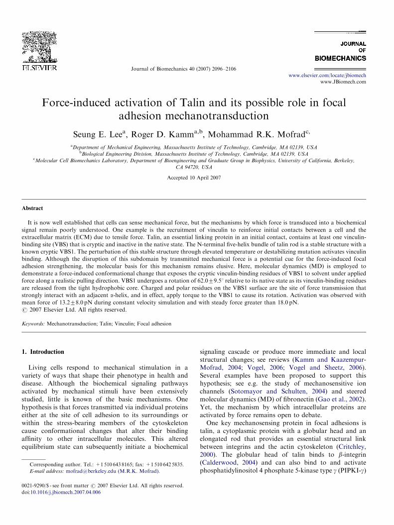
Fig. 1. (A) Crystal structure of TAL9 (PDB ID: 1SJ8) in ribbon representation is shown superimposed on a hypothetical talin model. Three VBSs within
TAL9 are shown in red, and H1 is shown in blue. Since the talin rod has tandem repeats of helical bundles, TAL9 is aligned such that the centers of mass of
the two helical bundles lie on the talin rod axis. (B) Detailed view of the N-terminal five-helix bundle (TAL5) used in the TAL5 simulations. Each of the
five helices is shown in a different color: H1 (blue), H2 (yellow), H3 (tan), H4 (or VBS1; red), and H5 (green). Some important polar residues are shown in
stick representations. A dummy atom with no mass or charge is shown in white. H5 polar side chains (black sticks) are harmonically constrained in space
(constraints shown as triangles). H1 polar side chains (yellow sticks) are pulled toward the dummy atom (effective pulling direction shown as an arrow).
S.E. Lee et al. / Journal of Biomechanics 40 (2007) 2096–21062098
calculation (Izrailev et al., 1999) with effective pulling velocity of
0.125 A/ns.
Constant force simulations with force magnitude varying between
F ¼ 15.0 to 25.0 pN were performed using the same TAL5 model
described above. Constant force of F/(no. of atoms on which the force
is applied) was applied to each of the side chain atoms on H1 (T498, S501,
and S502) toward the positive x-direction, such that total force applied
is F.
2.2. Mutational study on TAL5
Three mutated TAL5 structures were constructed using the MMTSB
toolset (Feig et al., 2004): (i) H5 residues N636 and Q639 mutated to
alanines; (ii) VBS1 residues R606, Q610, K613, E621 and R624 all
mutated to alanines; (iii) H1 residues N500, Q504, Q507, D514, and D515
all mutated to alanines. Umbrella sampling simulations identical to those
described above are performed on each mutated structure.
2.3. Explicit water simulation on TAL5
TAL5 was solvated in an orthorhombic solvent box with each face at
least 10 A away from TAL5 resulting in 23,775 atoms. Periodic boundary
conditions were imposed using the image module. Electrostatic charge of
the solvated system was neutralized by replacing seven water molecules
with sodium ions. An all-hydrogen representation was used with
CHARMM27 force fields (MacKerell et al., 1998). The SHAKE
constraint and 2fs time steps was used. SHIFT truncation was imposed
with a cutoff distance of 12 A for non-bonded interactions, which has been
found to exhibit reasonable accuracy in explicit water simulations (Beck et
al., 2005). Long range non-bonded interactions, beyond the cutoff
distance, were not taken into account in our explicit water simulations.
The model was thoroughly minimized. The system was heated to 300K in
40 ps and equilibrated for 960 ps. An umbrella sampling potential of
5.0 kcal/mol-A2 was used. H5 sidechain atoms (Q646, E650, and Q653)
were harmonically constrained with force constant of 1.5 kcal/mol-A2.
Using a reference distance step size of 0.2 A, and 400 ps simulation at each
step a canonical ensemble simulation was performed with Nose-Hoover at
each reference distance, which is equivalent to 0.5 A/ns pulling rate. A
constant force simulation with F ¼ 50.0 pN was also performed. All
explicit solvent simulations were performed on DataStar IBM p655 at San
Diego Supercomputing Center (SDSC).
3. Results
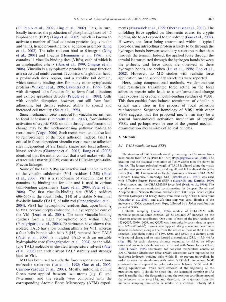
TAL5 forms a stable structure with cryptic VBS1, whichcannot bind to Vh1 in intact TAL5, but elevatedtemperature can effectively disrupt its stability and allowit to strongly bind to Vh1 (Patel et al., 2006). Explicit watersimulations at 300, 360, and 420K were performed toinvestigate what constitutes TAL5 stability and thedestabilizing effects of elevated temperature. Hydrophobicresidues of H5 (L651, A647, V644, and A640) form a tightgroove-fitting interaction with hydrophobic residues ofVBS1 (A611, L615, A618, and L622) (Fig. 2). Thisinteraction prevents VBS1 hydrophobic residues, whichare the vinculin-binding residues, from becoming exposedto the solvent. The trajectories of elevated temperaturesimulations (360 and 420K) did not differ much from thoseat room temperature (300K) other than the expectedincrease in thermal fluctuation. The RMSD of backboneatoms from crystal structures and average distances withfluctuations of VBS1-H1, VBS1-H2, VBS1-H3, and VBS1-H5 for three simulations are shown in Table 1. H5 and H3closely interact with VBS1, and with other helices to alesser extent (Fig. 2B). It is likely that this interaction mustbe disrupted in elevated temperature or force-inducedactivation of VBS1.In the TAL5 simulations displaying VBS1 activation, the
hydrophobic residues of VBS1 (L608, L609, L615, V619,L622, and L623) form a tight hydrophobic core with thehydrophobic residues of H3 (V577, I580, L584, M587,V591, and L594) and the hydrophobic residues of H5(L637, A640, V644, A647, L651, and I655) before
ARTICLE IN PRESSS.E. Lee et al. / Journal of Biomechanics 40 (2007) 2096–2106 2099
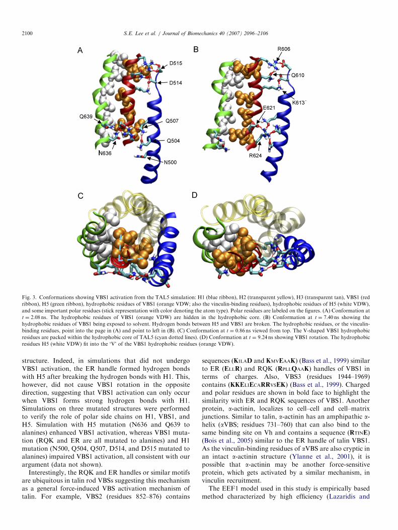
extension. Polar and charged residues on VBS1 interactwith H1 and H5 through hydrogen bonds and salt bridges.RQK (R606, Q610, and K613) cluster on VBS1 interactsstrongly with H1 (D514 and D515) by forming salt bridges(Figs. 1B and 3B). ER (E621 and R624) cluster on VBS1interacts with H5 (N636 and Q639) and more strongly withH1 (N500, Q504, and Q507) as N500, Q504, and Q507 ofH1 surround and form hydrogen bonds with R624 of VBS1(Figs. 1B and 3B). As force is applied to TAL5, it istransmitted through these hydrogen bonds. Since VBS1-H1interaction is stronger, the transmitted force applies atorque through the RQK and ER clusters on VBS1 and theVBS1–H5 interaction is broken. As shown in Fig. 3, thehydrophobic contact formed by VBS1 with H3 and H5eventually slips, and the hydrophobic residues of VBS1 areexposed to the solvent as VBS1 undergoes a rigid bodyrotation (Fig. 3B and D). The hydrophobic residues of H5fit into the V-shaped groove of VBS1 as one side of the ‘V’(L608, L615, and L622) gets exposed to the solvent and theother side (L609, V619, and L623) forms a new hydro-phobic core with H5 and H3 (Fig. 3C and D) (Supple-mentary Movies 1, 2 and 3).
Fig. 2. (A) TAL5 in the same orientation as Figure 1B with hydrophobic
residues shown in surface representation for H1 (pink), H3 (cyan), VBS1
(orange), and H5 (white). Exposed ribbon sites are polar residues, whose
side chains are not shown for clarity. VBS1 and H5 form a tight groove-
fitting contact, which stabilizes VBS1 in TAL5’s hydrophobic core
preventing VBS1 from being accessible for vinculin binding. (B) VBS1
and H5 are shown separately with labels for residues participating in the
groove-fitting interaction.
Table 1
RMSD from crystal structure and average distances between helices from elev
Distance between
VBS1-H1 (A)
Distance between
VBS1-H2 (A)
T ¼ 300K 14.0370.26 14.3870.25
T ¼ 360K 13.7970.26 14.4370.25
T ¼ 420K 13.5170.35 14.3370.28
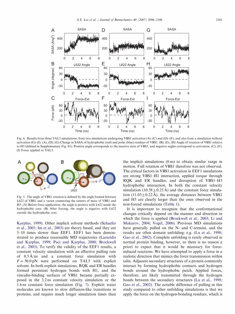
Solvent accessible surface area (SASA) of the hydro-phobic residues of VBS1 (Fig. 4A, D, and G) show howmuch of these residues are exposed. The extent of VBS1rotation is shown by measuring the angle made by L622(only selected as a reference, which is within the hydro-phobic core of TAL5 and gets exposed to solvent later)with the vector connecting the centers of mass of VBS1 andH5 (Fig. 4B, E, and H; angle definition in Fig. 5). VBS1activation is defined by L622 angle becoming negative,since this is the clearest measure of helix rotation to exposeVBS1 for possible binding. An increase in SASA is alsoindicative of activation, although this measure is alsoinfluenced by exposure of VBS1 residues internally, causedby H1 peeling away from VBS1. Note that the force peaks(Fig. 4C) are sharp, but the corresponding changes inrotation angle are more gradual and tend to lag behind thereduction in force. This may be due to rotation beingdiffusive in nature, occurring subsequent to the drop inforce impeding rotation. Results from two VBS1 activatedsimulations and one non-activated simulation (for compar-ison) is shown in Fig. 4. By visual inspection on the VBS1rotation plots displaying negative angles (e.g. Fig. 4B, E,and H), 71.4% of the TAL5 simulations (n ¼ 20 out of 28)exhibited the VBS1 activation. Analyzing only the simula-tions with VBS1 activation, 157.5770.9 A2 of hydrophobicSASA of VBS1 was exposed to solvent, VBS1 rotated by62.079.51, and a mean force of 13.278.0 pN was requiredfor activation.Activation of VBS1 follows disruption of the tight
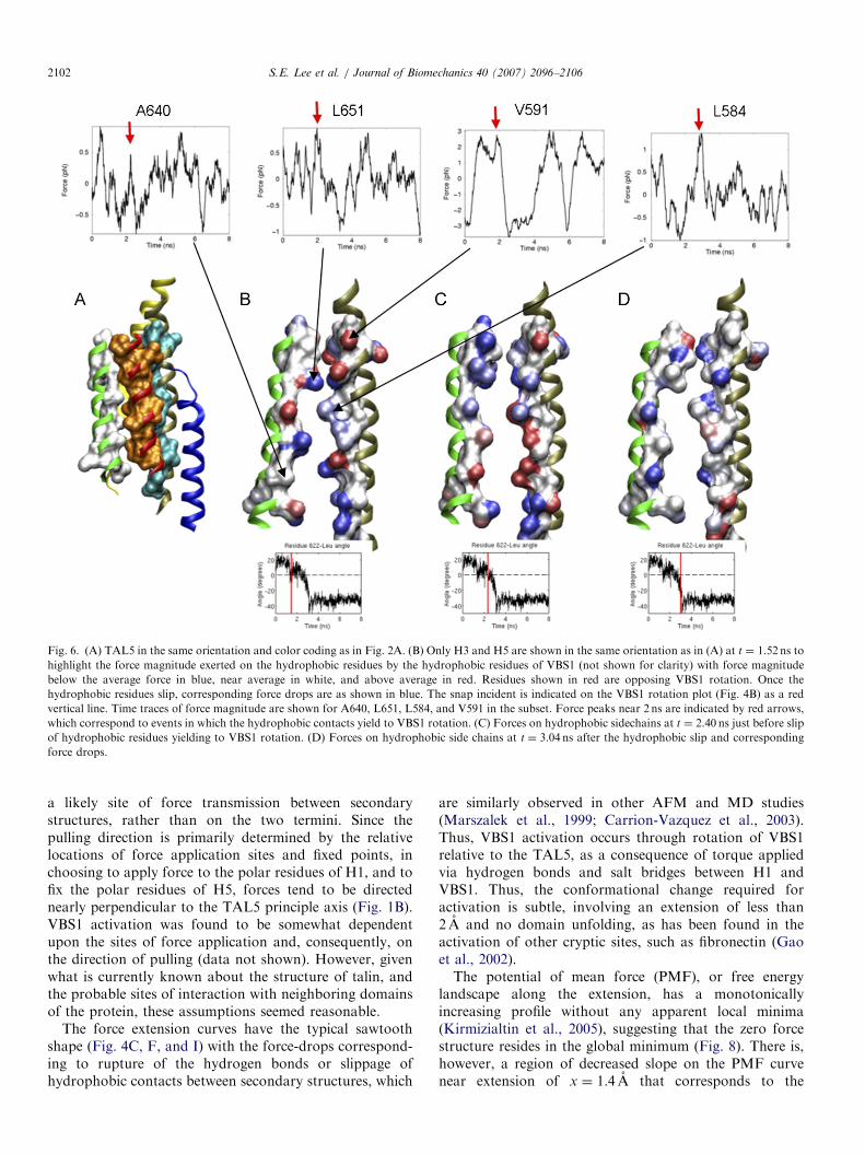
hydrophobic interaction of VBS1 with H3 and H5 ratherthan resulting from hydrogen-bond breakage. Rotationdue to the applied torque through RQK and ER handles isopposed by the hydrophobic contacts from H3 and H5(Fig. 6). Non-bonded components of the interaction forceon the hydrophobic sidechains of H3 and H5 experienceforce drops that correspond to the yielding of VBS1 torotation (Fig. 6B). The identified sidechains opposingVBS1 rotation exhibit simultaneous drops in forcemagnitude near 2 ns. Time traces of the non-bonded forceon A640, L651, L584, and V591 are shown in the subset ofFig. 6B (Supplementary Movie 4).
4. Discussion and conclusion
VBS1 activation in TAL5 is triggered by torquetransmitted through the RQK and ER handles (Fig. 3Aand B). Polar side groups of H5 (N636 and Q639) opposeVBS1 activation by stabilizing the non-extended TAL5
ated temperature explicit water simulations
Distance between
VBS1-H3 (A)
Distance between
VBS1-H5 (A)
RMSD (A)
10.4670.21 8.0470.20 1.9635
10.5770.20 8.1070.20 2.3258
10.8670.26 8.4470.24 2.7919
ARTICLE IN PRESS
Fig. 3. Conformations showing VBS1 activation from the TAL5 simulation: H1 (blue ribbon), H2 (transparent yellow), H3 (transparent tan), VBS1 (red
ribbon), H5 (green ribbon), hydrophobic residues of VBS1 (orange VDW; also the vinculin-binding residues), hydrophobic residues of H5 (white VDW),
and some important polar residues (stick representation with color denoting the atom type). Polar residues are labeled on the figures. (A) Conformation at
t ¼ 2.08 ns. The hydrophobic residues of VBS1 (orange VDW) are hidden in the hydrophobic core. (B) Conformation at t ¼ 7.40 ns showing the
hydrophobic residues of VBS1 being exposed to solvent. Hydrogen bonds between H5 and VBS1 are broken. The hydrophobic residues, or the vinculin-
binding residues, point into the page in (A) and point to left in (B). (C) Conformation at t ¼ 0.86 ns viewed from top. The V-shaped VBS1 hydrophobic
residues are packed within the hydrophobic core of TAL5 (cyan dotted lines). (D) Conformation at t ¼ 9.24 ns showing VBS1 rotation. The hydrophobic
residues H5 (white VDW) fit into the ‘V’ of the VBS1 hydrophobic residues (orange VDW).
S.E. Lee et al. / Journal of Biomechanics 40 (2007) 2096–21062100
structure. Indeed, in simulations that did not undergoVBS1 activation, the ER handle formed hydrogen bondswith H5 after breaking the hydrogen bonds with H1. This,however, did not cause VBS1 rotation in the oppositedirection, suggesting that VBS1 activation can only occurwhen VBS1 forms strong hydrogen bonds with H1.Simulations on three mutated structures were performedto verify the role of polar side chains on H1, VBS1, andH5. Simulation with H5 mutation (N636 and Q639 toalanines) enhanced VBS1 activation, whereas VBS1 muta-tion (RQK and ER are all mutated to alanines) and H1mutation (N500, Q504, Q507, D514, and D515 mutated toalanines) impaired VBS1 activation, all consistent with ourargument (data not shown).
Interestingly, the RQK and ER handles or similar motifsare ubiquitous in talin rod VBSs suggesting this mechanismas a general force-induced VBS activation mechanism oftalin. For example, VBS2 (residues 852–876) contains
sequences (KILAD and KMVEAAK) (Bass et al., 1999) similarto ER (ELLR) and RQK (RPLLQAAK) handles of VBS1 interms of charges. Also, VBS3 (residues 1944–1969)contains (KKELIECARRVSEK) (Bass et al., 1999). Chargedand polar residues are shown in bold face to highlight thesimilarity with ER and RQK sequences of VBS1. Anotherprotein, a-actinin, localizes to cell–cell and cell–matrixjunctions. Similar to talin, a-actinin has an amphipathic a-helix (aVBS; residues 731–760) that can also bind to thesame binding site on Vh and contains a sequence (RTINE)(Bois et al., 2005) similar to the ER handle of talin VBS1.As the vinculin-binding residues of aVBS are also cryptic inan intact a-actinin structure (Ylanne et al., 2001), it ispossible that a-actinin may be another force-sensitiveprotein, which gets activated by a similar mechanism, invinculin recruitment.The EEF1 model used in this study is empirically based
method characterized by high efficiency (Lazaridis and
ARTICLE IN PRESS
0 2 4 6 8
200
400
SA
SA
(A
ng
2)
SASA
0 2 4 6 8
200
400
SASA
0 2 4 6 8
200
400
SASA
0 2 4 6 8
-30
0
30
60
Angle
(degre
e)
L622 Angle
0 2 4 6 8
-30
0
30
60L622 Angle
0 2 4 6 8
-30
0
30
60L622 Angle
0 2 4 6 8
-30
0
30
60
Time (ns)
Forc
e (
pN
)
Force-Ext
0 2 4 6 8
-30
0
30
60
Time (ns)
Force-Ext
0 2 4 6 8
-30
0
30
60
Time (ns)
Force-Ext
Fig. 4. Results from three TAL5 simulations: from two simulations undergoing VBS1 activation (A)–(C) and (D)–(F), and also from a simulation without
activation (G)–(I). (A), (D), (G) Change in SASA of hydrophobic (red) and polar (blue) residues of VBS1. (B), (E), (H) Angle of rotation of VBS1 relative
to H5 (defined in Supplementary Fig. S1). Positive angle corresponds to the inactive state of VBS1, and negative angles correspond to activation. (C), (F),
(I) Force applied to TAL5.
Fig. 5. The angle of VBS1 rotation is defined by the angle formed between
L622 of VBS1 and a vector connecting the centers of mass of VBS1 and
H5. (A) Before force application, the angle is positive with L622 inside the
hydrophobic core. (B) After forcing, the angle is negative with L622
outside the hydrophobic core.
S.E. Lee et al. / Journal of Biomechanics 40 (2007) 2096–2106 2101
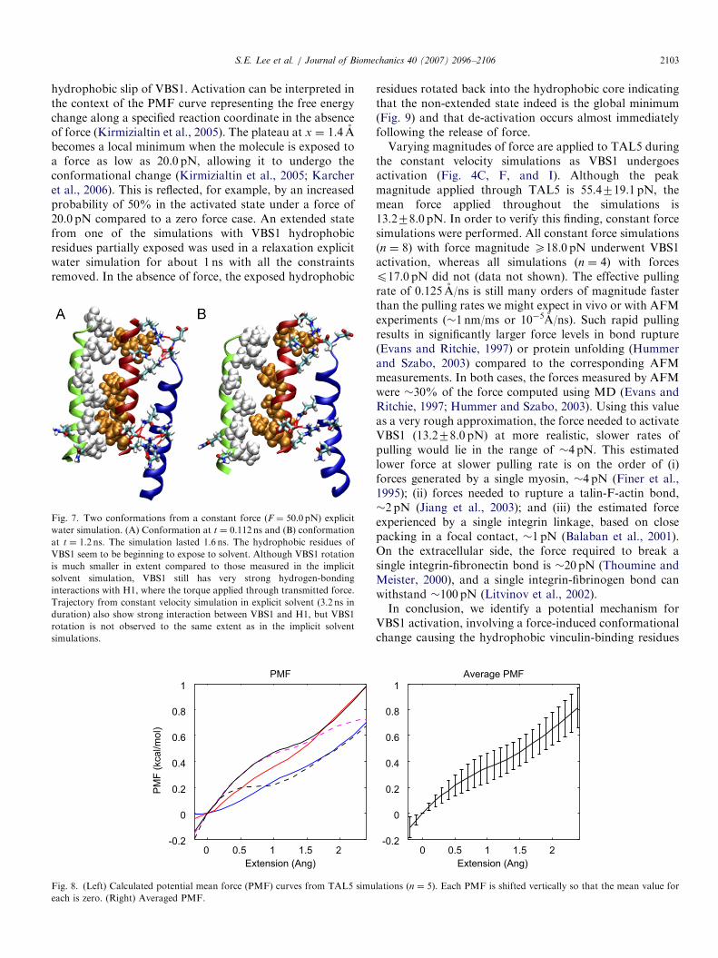
Karplus, 1999). Other implicit solvent methods (Schaeferet al., 2001; Im et al., 2003) are theory based, and they are5–10 times slower than EEF1. EEF1 has been demon-strated to produce reasonable MD trajectories (Lazaridisand Karplus, 1999; Paci and Karplus, 2000; Brockwellet al., 2003). To verify the validity of the EEF1 results, aconstant velocity simulation with an effective pulling rateof 0.5 A/ns and a constant force simulation withF ¼ 50.0 pN were performed on TAL5 with explicitsolvent. In both explicit simulations, RQK and ER handlesformed persistent hydrogen bonds with H1, and thevinculin-binding surfaces of VBS1 became partially ex-posed in the 3.2 ns constant velocity simulation or the1.6 ns constant force simulation (Fig. 7). Explicit watermolecules are known to slow diffusion-like transitions inproteins, and require much longer simulation times than
the implicit simulations (8 ns) to obtain similar range inmotion. Full rotation of VBS1 therefore was not observed.The critical factors in VBS1 activation in EEF1 simulationsare strong VBS1–H1 interaction, applied torque throughRQK and ER handles, and disruption of VBS1–H5hydrophobic interaction. In both the constant velocitysimulation (10.5870.25 A) and the constant force simula-tion (11.0370.22 A), the average distances between VBS1and H5 are clearly larger than the ones observed in thenon-forced simulations (Table 1).It is important to recognize that the conformational
changes critically depend on the manner and direction inwhich the force is applied (Brockwell et al., 2003; Li andMakarov, 2004; Vogel, 2006). Previous MD simulationshave generally pulled on the N- and C-termini, and theresults are often domain unfolding; e.g. (Lu et al., 1998;Gao et al., 2002). Complete unfolding is rarely observed innormal protein binding, however, so there is no reason apriori to expect that it would be necessary for force-induced reactions. We have attempted to apply a force in arealistic direction that mimics the force transmission withintalin. Adjacent secondary structures of a protein commonlyinteract by forming hydrophobic contacts and hydrogenbonds around the hydrophobic patch. Applied forces,therefore, are likely transmitted through the hydrogenbonds between the secondary structures (Lu et al., 1998;Gao et al., 2002). The notable difference of pulling in thisstudy compared to other unfolding simulations is that weapply the force on the hydrogen-bonding residues, which is
ARTICLE IN PRESS
Fig. 6. (A) TAL5 in the same orientation and color coding as in Fig. 2A. (B) Only H3 and H5 are shown in the same orientation as in (A) at t ¼ 1.52 ns to
highlight the force magnitude exerted on the hydrophobic residues by the hydrophobic residues of VBS1 (not shown for clarity) with force magnitude
below the average force in blue, near average in white, and above average in red. Residues shown in red are opposing VBS1 rotation. Once the
hydrophobic residues slip, corresponding force drops are as shown in blue. The snap incident is indicated on the VBS1 rotation plot (Fig. 4B) as a red
vertical line. Time traces of force magnitude are shown for A640, L651, L584, and V591 in the subset. Force peaks near 2 ns are indicated by red arrows,
which correspond to events in which the hydrophobic contacts yield to VBS1 rotation. (C) Forces on hydrophobic sidechains at t ¼ 2.40 ns just before slip
of hydrophobic residues yielding to VBS1 rotation. (D) Forces on hydrophobic side chains at t ¼ 3.04 ns after the hydrophobic slip and corresponding
force drops.
S.E. Lee et al. / Journal of Biomechanics 40 (2007) 2096–21062102
a likely site of force transmission between secondarystructures, rather than on the two termini. Since thepulling direction is primarily determined by the relativelocations of force application sites and fixed points, inchoosing to apply force to the polar residues of H1, and tofix the polar residues of H5, forces tend to be directednearly perpendicular to the TAL5 principle axis (Fig. 1B).VBS1 activation was found to be somewhat dependentupon the sites of force application and, consequently, onthe direction of pulling (data not shown). However, givenwhat is currently known about the structure of talin, andthe probable sites of interaction with neighboring domainsof the protein, these assumptions seemed reasonable.
The force extension curves have the typical sawtoothshape (Fig. 4C, F, and I) with the force-drops correspond-ing to rupture of the hydrogen bonds or slippage ofhydrophobic contacts between secondary structures, which
are similarly observed in other AFM and MD studies(Marszalek et al., 1999; Carrion-Vazquez et al., 2003).Thus, VBS1 activation occurs through rotation of VBS1relative to the TAL5, as a consequence of torque appliedvia hydrogen bonds and salt bridges between H1 andVBS1. Thus, the conformational change required foractivation is subtle, involving an extension of less than2 A and no domain unfolding, as has been found in theactivation of other cryptic sites, such as fibronectin (Gaoet al., 2002).The potential of mean force (PMF), or free energy
landscape along the extension, has a monotonicallyincreasing profile without any apparent local minima(Kirmizialtin et al., 2005), suggesting that the zero forcestructure resides in the global minimum (Fig. 8). There is,however, a region of decreased slope on the PMF curvenear extension of x ¼ 1.4 A that corresponds to the
ARTICLE IN PRESSS.E. Lee et al. / Journal of Biomechanics 40 (2007) 2096–2106 2103
hydrophobic slip of VBS1. Activation can be interpreted inthe context of the PMF curve representing the free energychange along a specified reaction coordinate in the absenceof force (Kirmizialtin et al., 2005). The plateau at x ¼ 1.4 Abecomes a local minimum when the molecule is exposed toa force as low as 20.0 pN, allowing it to undergo theconformational change (Kirmizialtin et al., 2005; Karcheret al., 2006). This is reflected, for example, by an increasedprobability of 50% in the activated state under a force of20.0 pN compared to a zero force case. An extended statefrom one of the simulations with VBS1 hydrophobicresidues partially exposed was used in a relaxation explicitwater simulation for about 1 ns with all the constraintsremoved. In the absence of force, the exposed hydrophobic
Fig. 7. Two conformations from a constant force (F ¼ 50.0 pN) explicit
water simulation. (A) Conformation at t ¼ 0.112 ns and (B) conformation
at t ¼ 1.2 ns. The simulation lasted 1.6 ns. The hydrophobic residues of
VBS1 seem to be beginning to expose to solvent. Although VBS1 rotation
is much smaller in extent compared to those measured in the implicit
solvent simulation, VBS1 still has very strong hydrogen-bonding
interactions with H1, where the torque applied through transmitted force.
Trajectory from constant velocity simulation in explicit solvent (3.2 ns in
duration) also show strong interaction between VBS1 and H1, but VBS1
rotation is not observed to the same extent as in the implicit solvent
simulations.
0 0.5 1 1.5 2-0.2
0
0.2
0.4
0.6
0.8
1
Extension (Ang)
PM
F (
kcal/m
ol)
PMF
Fig. 8. (Left) Calculated potential mean force (PMF) curves from TAL5 simu
each is zero. (Right) Averaged PMF.
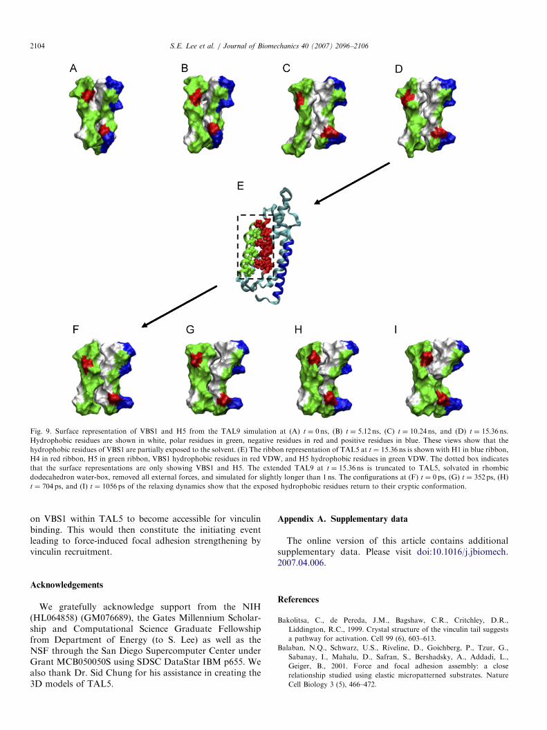
residues rotated back into the hydrophobic core indicatingthat the non-extended state indeed is the global minimum(Fig. 9) and that de-activation occurs almost immediatelyfollowing the release of force.Varying magnitudes of force are applied to TAL5 during
the constant velocity simulations as VBS1 undergoesactivation (Fig. 4C, F, and I). Although the peakmagnitude applied through TAL5 is 55.4719.1 pN, themean force applied throughout the simulations is13.278.0 pN. In order to verify this finding, constant forcesimulations were performed. All constant force simulations(n ¼ 8) with force magnitude X18.0 pN underwent VBS1activation, whereas all simulations (n ¼ 4) with forcesp17.0 pN did not (data not shown). The effective pullingrate of 0.125 A/ns is still many orders of magnitude fasterthan the pulling rates we might expect in vivo or with AFMexperiments (�1 nm/ms or 10�5A/ns). Such rapid pullingresults in significantly larger force levels in bond rupture(Evans and Ritchie, 1997) or protein unfolding (Hummerand Szabo, 2003) compared to the corresponding AFMmeasurements. In both cases, the forces measured by AFMwere �30% of the force computed using MD (Evans andRitchie, 1997; Hummer and Szabo, 2003). Using this valueas a very rough approximation, the force needed to activateVBS1 (13.278.0 pN) at more realistic, slower rates ofpulling would lie in the range of �4 pN. This estimatedlower force at slower pulling rate is on the order of (i)forces generated by a single myosin, �4 pN (Finer et al.,1995); (ii) forces needed to rupture a talin-F-actin bond,�2 pN (Jiang et al., 2003); and (iii) the estimated forceexperienced by a single integrin linkage, based on closepacking in a focal contact, �1 pN (Balaban et al., 2001).On the extracellular side, the force required to break asingle integrin-fibronectin bond is �20 pN (Thoumine andMeister, 2000), and a single integrin-fibrinogen bond canwithstand �100 pN (Litvinov et al., 2002).In conclusion, we identify a potential mechanism for
VBS1 activation, involving a force-induced conformationalchange causing the hydrophobic vinculin-binding residues
0 0.5 1 1.5 2-0.2
0
0.2
0.4
0.6
0.8
1
Extension (Ang)
Average PMF
lations (n ¼ 5). Each PMF is shifted vertically so that the mean value for
ARTICLE IN PRESS
Fig. 9. Surface representation of VBS1 and H5 from the TAL9 simulation at (A) t ¼ 0 ns, (B) t ¼ 5.12 ns, (C) t ¼ 10.24 ns, and (D) t ¼ 15.36 ns.
Hydrophobic residues are shown in white, polar residues in green, negative residues in red and positive residues in blue. These views show that the
hydrophobic residues of VBS1 are partially exposed to the solvent. (E) The ribbon representation of TAL5 at t ¼ 15.36 ns is shown with H1 in blue ribbon,
H4 in red ribbon, H5 in green ribbon, VBS1 hydrophobic residues in red VDW, and H5 hydrophobic residues in green VDW. The dotted box indicates
that the surface representations are only showing VBS1 and H5. The extended TAL9 at t ¼ 15.36 ns is truncated to TAL5, solvated in rhombic
dodecahedron water-box, removed all external forces, and simulated for slightly longer than 1 ns. The configurations at (F) t ¼ 0 ps, (G) t ¼ 352ps, (H)
t ¼ 704 ps, and (I) t ¼ 1056ps of the relaxing dynamics show that the exposed hydrophobic residues return to their cryptic conformation.
S.E. Lee et al. / Journal of Biomechanics 40 (2007) 2096–21062104
on VBS1 within TAL5 to become accessible for vinculinbinding. This would then constitute the initiating eventleading to force-induced focal adhesion strengthening byvinculin recruitment.
Acknowledgements
We gratefully acknowledge support from the NIH(HL064858) (GM076689), the Gates Millennium Scholar-ship and Computational Science Graduate Fellowshipfrom Department of Energy (to S. Lee) as well as theNSF through the San Diego Supercomputer Center underGrant MCB050050S using SDSC DataStar IBM p655. Wealso thank Dr. Sid Chung for his assistance in creating the3D models of TAL5.
Appendix A. Supplementary data
The online version of this article contains additionalsupplementary data. Please visit doi:10.1016/j.jbiomech.2007.04.006.
References
Bakolitsa, C., de Pereda, J.M., Bagshaw, C.R., Critchley, D.R.,
Liddington, R.C., 1999. Crystal structure of the vinculin tail suggests
a pathway for activation. Cell 99 (6), 603–613.
Balaban, N.Q., Schwarz, U.S., Riveline, D., Goichberg, P., Tzur, G.,
Sabanay, I., Mahalu, D., Safran, S., Bershadsky, A., Addadi, L.,
Geiger, B., 2001. Force and focal adhesion assembly: a close
relationship studied using elastic micropatterned substrates. Nature
Cell Biology 3 (5), 466–472.
ARTICLE IN PRESSS.E. Lee et al. / Journal of Biomechanics 40 (2007) 2096–2106 2105
Bass, M.D., Smith, B.J., Prigent, S.A., Critchley, D.R., 1999. Talin
contains three similar vinculin-binding sites predicted to form an
amphipathic helix. Biochemical Journal 341, 257–263.
Beck, D.A., Armen, R.S., Daggett, V., 2005. Cutoff size need not strongly
influence molecular dynamics results for solvated polypeptides.
Biochemistry 44 (2), 609–616.
Bois, P.R., Borgon, R.A., Vonrhein, C., Izard, T., 2005. Structural
dynamics of alpha-actinin-vinculin interactions. Molecular Cell
Biology 25 (14), 6112–6122.
Brockwell, D.J., Paci, E., Zinober, R.C., Beddard, G.S., Olmsted, P.D.,
Smith, D.A., Perham, R.N., Radford, S.E., 2003. Pulling geometry
defines the mechanical resistance of a beta-sheet protein. Nature
Structural Biology 10 (9), 731–737.
Brooks, B.R., Bruccoleri, R.E., Olafson, B.D., States, D.J., Swaminathan,
S., Karplus, M., 1983. Charmm—a program for macromolecular
energy, minimization, and dynamics calculations. Journal of Compu-
tational Chemistry 4 (2), 187–217.
Calderwood, D.A., 2004. Talin controls integrin activation. Biochemical
Society Transactions 32, 434–437.
Carrion-Vazquez, M., Li, H., Lu, H., Marszalek, P.E., Oberhauser, A.F.,
Fernandez, J.M., 2003. The mechanical stability of ubiquitin is linkage
dependent. Nature Structural Biology 10 (9), 738–743.
Critchley, D.R., 2000. Focal adhesions—the cytoskeletal connection.
Current Opinion in Cell Biology 12 (1), 133–139.
Di Paolo, G., Pellegrini, L., Letinic, K., Cestra, G., Zoncu, R., Voronov,
S., Chang, S., Guo, J., Wenk, M.R., De Camilli, P., 2002. PI(4,5)P2
generation at sites of cell adhesion: recruitment and regulation of PIP
kinase type 1gamma by the FERM domain of talin. Molecular Biology
of the Cell 13, 337A.
Evans, E., Ritchie, K., 1997. Dynamic strength of molecular adhesion
bonds. Biophysical Journal 72 (4), 1541–1555.
Feig, M., Karanicolas, J., Brooks 3rd, C.L., 2004. MMTSB Tool Set:
enhanced sampling and multiscale modeling methods for applications
in structural biology. Journal of Molecular Graphics and Modelling 22
(5), 377–395.
Finer, J.T., Mehta, A.D., Spudich, J.A., 1995. Characterization of single
actin-myosin interactions. Biophysical Journal 68 (4 Suppl),
291S–296S ; discussion 296S–297S.
Galbraith, C.G., Yamada, K.M., Sheetz, M.P., 2002. The relationship
between force and focal complex development. Journal of Cell Biology
159 (4), 695–705.
Gao, M., Craig, D., Vogel, V., Schulten, K., 2002. Identifying unfolding
intermediates of FN-III(10) by steered molecular dynamics. Journal of
Molecular Biology 323 (5), 939–950.
Giannone, G., Jiang, G., Sutton, D.H., Critchley, D.R., Sheetz, M.P.,
2003. Talin1 is critical for force-dependent reinforcement of initial
integrin-cytoskeleton bonds but not tyrosine kinase activation. Journal
of Cell Biology 163 (2), 409–419.
Gingras, A.R., Vogel, K.P., Steinhoff, H.J., Ziegler, W.H., Patel, B.,
Emsley, J., Critchley, D.R., Roberts, G.C., Barsukov, I.L., 2006.
Structural and dynamic characterization of a vinculin binding site in
the talin rod. Biochemistry 45 (6), 1805–1817.
Hemmings, L., Rees, D.J.G., Ohanian, V., Bolton, S.J., Gilmore, A.P.,
Patel, B., Priddle, H., Trevithick, J.E., Hynes, R.O., Critchley, D.R.,
1996. Talin contains three actin-binding sites each of which is adjacent
to a vinculin-binding site. Journal of Cell Science 109, 2715–2726.
Hoover, W.G., 1985. Canonical dynamics: equilibrium phase-space
distributions. Physical Review A 31 (3), 1695–1697.
Hummer, G., Szabo, A., 2003. Kinetics from nonequilibrium single-
molecule pulling experiments. Biophysical Journal 85 (1), 5–15.
Im, W., Lee, M.S., Brooks 3rd, C.L., 2003. Generalized born model with a
simple smoothing function. Journal of Computational Chemistry 24
(14), 1691–1702.
Izard, T., Evans, G., Borgon, R.A., Rush, C.L., Bricogne, G., Bois,
P.R.J., 2004. Vinculin activation by talin through helical bundle
conversion. Nature 427 (6970), 171–175.
Izrailev, S., Crofts, A.R., Berry, E.A., Schulten, K., 1999. Steered
molecular dynamics simulation of the Rieske subunit motion
in the cytochrome bc(1) complex. Biophysical Journal 77 (4),
1753–1768.
Jiang, G.Y., Giannone, G., Critchley, D.R., Fukumoto, E., Sheetz, M.P.,
2003. Two-piconewton slip bond between fibronectin and the
cytoskeleton depends on talin. Nature 424 (6946), 334–337.
Kamm, R.D., Kaazempur-Mofrad, M.R., 2004. On the molecular basis
for mechanotransduction. Mechanics and Chemistry of Biosystems 1
(3), 201–209.
Karcher, H., Lee, S.E., Kaazempur-Mofrad, M.R., Kamm, R.D., 2006. A
coarse-grained model for force-induced protein deformation and
kinetics. Biophysical Journal 90 (8), 2686–2697.
Kirmizialtin, S., Huang, L., Makarov, D.E., 2005. Topography of the free-
energy landscape probed via mechanical unfolding of proteins. Journal
of Chemical Physics 122 (23), 234915.
Krautler, V., Van Gunsteren, W.F., Hunenberger, P.H., 2001. A fast
SHAKE: algorithm to solve distance constraint equations for small
molecules in molecular dynamics simulations. Journal of Computa-
tional Chemistry 22 (5), 501–508.
Lazaridis, T., Karplus, M., 1999. Effective energy function for proteins in
solution. Proteins 35 (2), 133–152.
Li, P.C., Makarov, D.E., 2004. Simulation of the mechanical unfolding of
ubiquitin: probing different unfolding reaction coordinates by changing
the pulling geometry. Journal of Chemical Physics 121 (10), 4826–4832.
Ling, K., Doughman, R.L., Firestone, A.J., Bunce, M.W., Anderson,
R.A., 2002. Type I gamma phosphatidylinositol phosphate kinase
targets and regulates focal adhesions. Nature 420 (6911), 89–93.
Litvinov, R.I., Shuman, H., Bennett, J.S., Weisel, J.W., 2002. Binding
strength and activation state of single fibrinogen-integrin pairs on
living cells. Proceedings of the National Academy of Sciences of the
United States of America 99 (11), 7426–7431.
Lu, H., Isralewitz, B., Krammer, A., Vogel, V., Schulten, K., 1998.
Unfolding of titin immunoglobulin domains by steered molecular
dynamics simulation. Biophysical Journal 75 (2), 662–671.
MacKerell, A., Bashford, D., Bellott, M., Dunbrack, R., Evanseck, J.,
Field, M., Fischer, S., Gao, J., Guo, H., Ha, S., et al., 1998. All-atom
empirical potential for molecular modeling and dynamics studies of
proteins. Journal of Physical Chemistry 102, 3586–3616.
Marszalek, P.E., Lu, H., Li, H., Carrion-Vazquez, M., Oberhauser, A.F.,
Schulten, K., Fernandez, J.M., 1999. Mechanical unfolding inter-
mediates in titin modules. Nature 402 (6757), 100–103.
Neria, E., Fischer, S., Karplus, M., 1996. Simulation of activation free
energies in molecular dynamics system. Journal of Chemical Physics
105, 1902–1921.
Nose, S., 1984. A molecular dynamics method for simulations in the
canonical ensemble. Molecular Physics 52, 255–268.
Oberhauser, A.F., Badilla-Fernandez, C., Carrion-Vazquez, M., Fernan-
dez, J.M., 2002. The mechanical hierarchies of fibronectin observed
with single-molecule AFM. Journal of Molecular Biology 319 (2),
433–447.
Paci, E., Karplus, M., 2000. Unfolding proteins by external forces and
temperature: the importance of topology and energetics. Proceedings
of the National Academy of Sciences of the United States of America
97 (12), 6521–6526.
Papagrigoriou, E., Gingras, A.R., Barsukov, I.L., Bate, N., Fillingham,
I.J., Patel, B., Frank, R., Ziegler, W.H., Roberts, G.C.K., Critchley,
D.R., Emsley, J., 2004. Activation of a vinculin-binding site in the talin
rod involves rearrangement of a five-helix bundle. EMBO Journal 23
(15), 2942–2951.
Patel, B.C., Gingras, A.R., Bobkov, A.A., Fujimoto, L.M., Zhang, M.,
Liddington, R.C., Mazzeo, D., Emsley, J., Roberts, G.C., Barsukov,
I.L., Critchley, D.R., 2006. The activity of the vinculin binding sites in
talin is influenced by the stability of the helical bundles that make up
the talin rod. Journal of Biological Chemistry.
Priddle, H., Hemmings, L., Monkley, S., Woods, A., Patel, B., Sutton, D.,
Dunn, G.A., Zicha, D., Critchley, D.R., 1998. Disruption of the talin
gene compromises focal adhesion assembly in undifferentiated but not
differentiated embryonic stem cells. Journal of Cell Biology 142 (4),
1121–1133.
ARTICLE IN PRESSS.E. Lee et al. / Journal of Biomechanics 40 (2007) 2096–21062106
Schaefer, M., Bartels, C., Leclerc, F., Karplus, M., 2001. Effective atom
volumes for implicit solvent models: comparison between Voronoi
volumes and minimum fluctuation volumes. Journal of Computational
Chemistry 22 (15), 1857–1879.
Sotomayor, M., Schulten, K., 2004. Molecular dynamics study of gating in
the mechanosensitive channel of small conductance MscS. Biophysical
journal 87 (5), 3050–3065.
Thoumine, O., Meister, J.J., 2000. Dynamics of adhesive rupture between
fibroblasts and fibronectin: microplate manipulations and determinis-
tic model. European Biophysics Journal 29 (6), 409–419.
Torrie, G. M., J.P.V., 1974. Monte carlo free energy estimates using non-
boltzmann sampling: Application to the sub-critical lennard-jones
fluid. Chemical Physics Letters 28, 578–581.
Vogel, V., 2006. Mechanotransduction involving multimodular proteins:
converting force into biochemical signals. Annual Review of Biophy-
sics and Biomolecular Structure 35, 459–488.
Vogel, V., Sheetz, M., 2006. Local force and geometry sensing regulate
cell functions. Nature Reviews Molecular Cell Biology 7 (4),
265–275.
Winkler, J., Lunsdorf, H., Jockusch, B.M., 1996. The ultrastructure of
chicken gizzard vinculin as visualized by high-resolution electron
microscopy. Journal of Structural Biology 116 (2), 270–277.
Xing, B.D., Jedsadayanmata, A., Lam, S.C.T., 2001. Localization of an
integrin binding site to the C terminus of talin. Journal of Biological
Chemistry 276 (48), 44373–44378.
Xu, W.M., Coll, J.L., Adamson, E.D., 1998. Rescue of the mutant
phenotype by reexpression of full-length vinculin in null F9 cells;
effects on cell locomotion by domain deleted vinculin. Journal of Cell
Science 111, 1535–1544.
Ylanne, J., Scheffzek, K., Young, P., Saraste, M., 2001. Crystal structure
of the alpha-actinin rod reveals an extensive torsional twist. Structure 9
(7), 597–604.