Embed Size (px)
Citation preview

Stress-Induced PARP Activation Mediates Recruitment ofDrosophila Mi-2 to Promote Heat Shock Gene ExpressionMagdalena Murawska1, Markus Hassler2, Renate Renkawitz-Pohl3, Andreas Ladurner2,4, Alexander
Brehm1*
1 Institute of Tumor Research and Molecular Biology, Philipps University, Marburg, Germany, 2 Genome Biology and Structural and Computational Biology Unit, European
Molecular Biology Laboratory, Heidelberg, Germany, 3 Developmental Biology, Philipps University, Marburg, Germany, 4 Department of Physiological Chemistry, Adolf-
Butenandt-Institute, Ludwig-Maximilians University, Munich, Germany
Abstract
Eukaryotic cells respond to genomic and environmental stresses, such as DNA damage and heat shock (HS), with thesynthesis of poly-[ADP-ribose] (PAR) at specific chromatin regions, such as DNA breaks or HS genes, by PAR polymerases(PARP). Little is known about the role of this modification during cellular stress responses. We show here that thenucleosome remodeler dMi-2 is recruited to active HS genes in a PARP–dependent manner. dMi-2 binds PAR suggestingthat this physical interaction is important for recruitment. Indeed, a dMi-2 mutant unable to bind PAR does not localise toactive HS loci in vivo. We have identified several dMi-2 regions which bind PAR independently in vitro, including thechromodomains and regions near the N-terminus containing motifs rich in K and R residues. Moreover, upon HS geneactivation, dMi-2 associates with nascent HS gene transcripts, and its catalytic activity is required for efficient transcriptionand co-transcriptional RNA processing. RNA and PAR compete for dMi-2 binding in vitro, suggesting a two step process fordMi-2 association with active HS genes: initial recruitment to the locus via PAR interaction, followed by binding to nascentRNA transcripts. We suggest that stress-induced chromatin PARylation serves to rapidly attract factors that are required foran efficient and timely transcriptional response.
Citation: Murawska M, Hassler M, Renkawitz-Pohl R, Ladurner A, Brehm A (2011) Stress-Induced PARP Activation Mediates Recruitment of Drosophila Mi-2 toPromote Heat Shock Gene Expression. PLoS Genet 7(7): e1002206. doi:10.1371/journal.pgen.1002206
Editor: Asifa Akhtar, Max-Planck-Institute of Immunobiology, Germany
Received February 2, 2011; Accepted June 9, 2011; Published July 28, 2011
Copyright: � 2011 Murawska et al. This is an open-access article distributed under the terms of the Creative Commons Attribution License, which permitsunrestricted use, distribution, and reproduction in any medium, provided the original author and source are credited.
Funding: MM was supported by DFG IRTG 1384, LOEWE cluster Tumor and Inflammation, and DFG TRR81; MH was supported by the DFG and by EMBL. Researchis supported by DFG TRR81 (AB, RR-P), EMBL, and HFSP (AL). The funders had no role in study design, data collection and analysis, decision to publish, orpreparation of the manuscript.
Competing Interests: The authors have declared that no competing interests exist.
* E-mail: [email protected]
Introduction
The activity of eukaryotic genomes is regulated by dynamic
changes in chromatin structure. A multitude of nucleosome
remodeling enzymes, histone modifying activities and chromatin
binding proteins cooperate to establish, maintain and reprogram
chromatin structures that determine genome activity.
Drosophila heat shock (HS) genes provide a textbook example of
how dramatic changes in the organismal and cellular environ-
ment affect chromatin structure in a manner that promotes
transcriptional activation of genes coding for molecular chaper-
ones required during the HS response. Upon temperature shift,
the HS loci of polytene chromosomes form transcriptionally
active ‘‘puffs’’. This rapid chromatin decondensation correlates
with a strong decrease in nucleosome density [1]. Puff formation
can be uncoupled from transcription and much of the
nucleosome loss at the hsp70 gene occurs prior to the first round
of transcription [1,2]. Recently, heat shock factor (HSF), GAGA
factor and poly-[ADP-ribose] polymerase (PARP) have been
shown to be required for the rapid removal of nucleosomes upon
activation of the hsp70 gene [1]. In addition, HS puffs accumulate
PARylated proteins and puff formation depends on PARP
activity [3]. The mechanisms underlying PARP action during
HS gene activation are not clear. It has been suggested that
PARylation may be removing proteins, including histones - which
are themselves a good PARP substrate - thereby promoting
chromatin opening [1]. The accumulation of PARylated proteins
at HS loci has recently been proposed to build up a ‘‘transcription
compartment’’ which hinders the diffusion of proteins into and
out of the compartment, thus favouring factor recycling [4]. In
addition to histone displacement and transcription compartment
formation at HS genes, recent evidence suggests that PARylation
could also act as a signaling scaffold for the recruitment of PAR-
sensing factors during DNA damage. In mammals PARylation at
DNA damage sites can mediate the recruitment of several ATP-
dependent nucleosome remodeling enzymes [5-10]. Here we
sought to address whether and how nucleosome remodelers may
be recruited to PARP activation sites upon environmental stresses
other than DNA damage. We have investigated a paradigm of
environmental stress, the activation of HS loci in Drosophila and
have analyzed the mechanism through which the nucleosome
remodeler dMi-2 is recruited to HS genes.
Mi-2 (CHD3/CHD4) is a conserved ATP-dependent nucleo-
some remodeler. In both vertebrates and invertebrates, it is a
subunit of Nucleosome Remodeling and Deacetylation (NuRD) complex-
es. NuRD complexes repress cell type specific genes during
differentiation [11–13]. dMi-2 is also a subunit of the Drosophila-
specific Mep-1 complex (dMec) which represses neuron-specific genes
during differentiation of the peripheral nervous system [12,14].
PLoS Genetics | www.plosgenetics.org 1 July 2011 | Volume 7 | Issue 7 | e1002206
Mi-2 containing complexes lack subunits with sequence-specific
DNA binding activity. Two main mechanisms for their recruit-
ment to chromatin have been suggested. First, NuRD complexes
contain subunits with methylated DNA binding domains (MBD) which
direct NuRD to methylated DNA [15,16]. This is unlikely to be a
major recruitment mechanism for Drosophila Mi-2 complexes,
however, given the low and transient levels of DNA methylation in
this organism [17]. A second mode of Mi-2 recruitment involves
interactions with DNA bound transcription factors [11,12,14,18–
22]. In addition, SUMOylation of transcription factors can
increase their affinity for Mi-2 complexes [21,22].
Despite its well established role in repression, dMi-2 localises to
actively transcribed chromosome regions suggesting an unexpect-
ed potential function of dMi-2 in transcription [23]. Here we
sought to establish how dMi-2 is recruited to actively transcribed
chromatin and to clarify its role in transcriptional activation using
genetic, biochemical and pharmacological assays. We show that
dMi-2 rapidly associates with activated HS loci, covering the entire
transcribed region of the hsp70 gene. dMi-2 recruitment is not
affected when transcriptional elongation is blocked but is
abrogated when PARP is inhibited. Indeed, we find that dMi-2
specifically binds PARP’s oligomeric product PAR in vitro.
Significantly, a dMi-2 mutant unable to bind PAR is not recruited
to active HS loci in vivo. We have identified several regions of dMi-
2 that bind PAR in vitro. These include the chromodomains and a
series of K/R-rich motifs near the N-terminus. Further, dMi-2
depletion or expression of an inactive enzyme greatly decreases
transcript levels, suggesting that dMi-2 actively supports efficient
HS gene expression. Indeed, dMi-2 associates with nascent hsp70
transcripts in vivo and ablation of dMi-2 function results in
inefficient RNA processing. RNA and PAR compete for dMi-2
binding suggesting a two step process of dMi-2 association with
HS genes: intial recruitment of dMi-2 is effected by its binding to
PAR which is produced prior to the onset of transcription, dMi-2
then switches to interacting with the emerging nascent transcripts.
Taken together, our results uncover PAR binding as a novel
mechanism for the recruitment of the nucleosome remodeler dMi-
2 to targeted sites of PARP activitation upon environmental stress
and demonstrate that dMi-2 acts as a co-activator for the full
transcriptional activation of HS genes. This study provides the first
evidence for an in vivo function of PARylation in promoting the
recruitment of a nucleosome remodeler to support the transcrip-
tion of stress induced genes.
Results
dMi-2 is recruited to HS genes in a PARP–dependentmanner
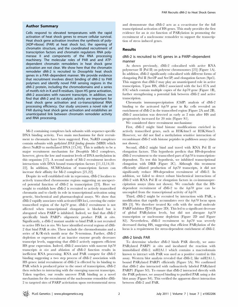
As shown previously, dMi-2 colocalised with active RNA
polymerase II (Pol II) on polytene chromosomes [23] (Figure 1A).
In addition, dMi-2 significantly colocalised with different forms of
elongating Pol II (Ser2P and Ser5P) and elongation factors (Spt5).
This suggests that dMi-2 may play an unanticipated role in active
transcription. Upon HS, dMi-2 associated with the loci 87A and
87C which contain multiple copies of the hsp70 gene (Figure 1B),
further strengthening a potential link between dMi-2 and active
transcription.
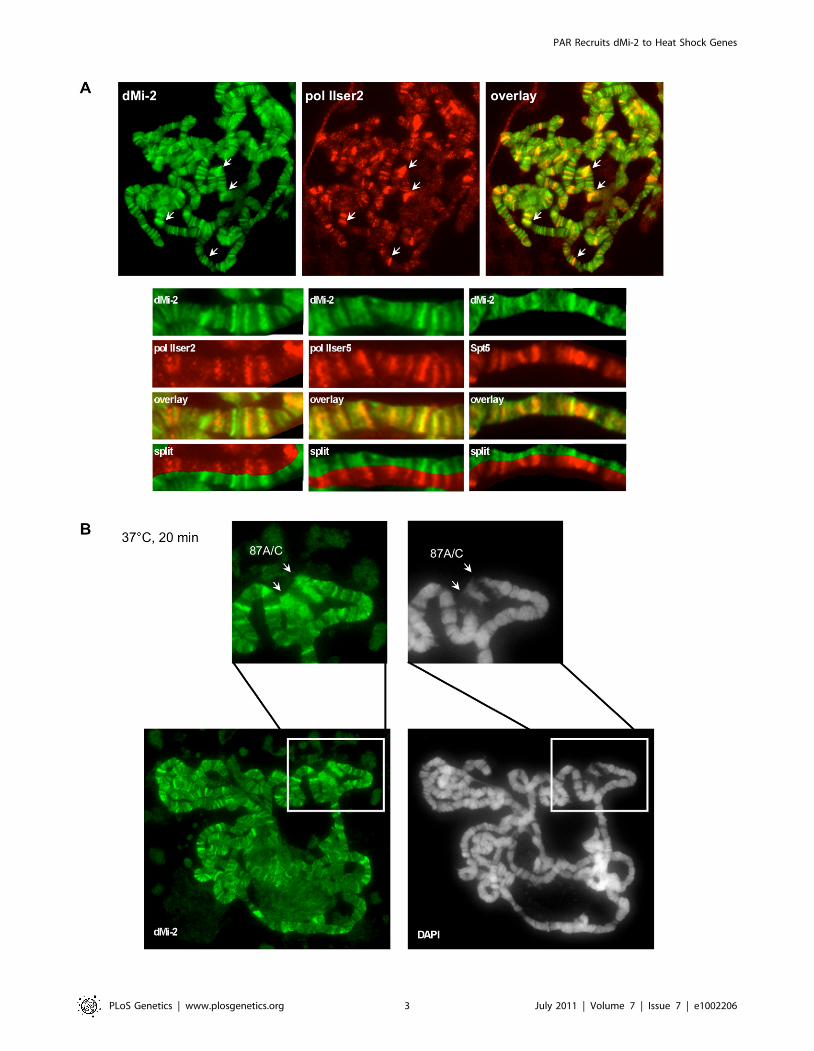
Chromatin immunoprecipitation (ChIP) analysis of dMi-2
binding to the activated hsp70 gene in Kc cells revealed an
enrichment of dMi-2 in the transcribed region (Figure 2A and 2B).
dMi-2 association was detected as early as 2 min after HS and
progressively increased for 20 min (Figure S1).
We considered three recruitment mechanisms:
First, dMi-2 might bind histone modifications enriched in
actively transcribed genes, such as H3K4me3 or H3K36me3.
However, we did not find a methylation sensitive interaction of
recombinant dMi-2 with histone peptides in pulldown assays (data
not shown).
Second, dMi-2 might bind and travel with RNA Pol II or
elongation factors. This hypothesis predicts that HS-dependent
dMi-2 recruitment to the transcribed part of hsp70 is transcription-
dependent. To test this hypothesis, we inhibited transcriptional
elongation with DRB (Figure 2C). Although this treatment
efficiently ablated production of hsp70 transcripts, it did not
significantly reduce HS-dependent recruitment of dMi-2. In
addition, we failed to detect robust biochemical interactions of
dMi-2 with RNA Pol II or elongation factors in co-immunopre-
cipitation assays (data not shown). We conclude that the HS-
dependent recruitment of dMi-2 to the hsp70 gene can be
uncoupled from the transcriptional activity of hsp70.
Third, dMi-2 might be recruited by interaction with PAR, a
modification that rapidly accumulates over the hsp70 locus upon
HS [3]. We therefore treated Kc cells with the small molecule
PARP inhibitor PJ34 (Figure 2D). This led to a significant decrease
of global PARylation levels, but did not abrogate hsp70
transcription or nucleosome depletion (Figure 2D and Figure
S2). Nevertheless, dMi-2 recruitment to hsp70 was severely
decreased during HS, suggesting that efficient PARylation of the
locus is a requirement for stress-dependent enrichment of dMi-2.
dMi-2 binds PARTo determine whether dMi-2 binds PAR directly, we auto-
PARylated PARP1 in vitro and incubated the reaction with
immobilised dMi-2. mH2A1.1 which contains a macrodomain
known to interact with PAR was used as a positive control in this
assay. Western blot analysis revealed that dMi-2, like mH2A1.1,
bound PARylated PARP1 efficiently (Figure 3A). We confirmed
that dMi-2 also interacted with radioactively labeled PARylated
PARP1 (Figure S3). To ensure that dMi-2 interacted directly with
the PAR polymer, we assayed binding to purified PAR using a dot
blot assay (Figure S4). This verified the apparent direct interaction
between dMi-2 and PAR.
Author Summary
Cells respond to elevated temperatures with the rapidactivation of heat shock genes to ensure cellular survival.Heat shock gene activation involves the synthesis of poly-[ADP-ribose] (PAR) at heat shock loci, the opening ofchromatin structure, and the coordinated recruitment oftranscription factors and chromatin regulators RNA poly-merase II and components of the RNA processingmachinery. The molecular roles of PAR and and ATP-dependent chromatin remodelers in heat shock geneactivation are not clear. We show here that the chromatinremodeler dMi-2 is recruited to Drosophila heat shockgenes in a PAR–dependent manner. We provide evidencethat recruitment involves direct binding of dMi-2 to PARpolymers and identify novel PAR sensing regions in thedMi-2 protein, including the chromodomains and a seriesof motifs rich in K and R residues. Upon HS gene activation,dMi-2 associates with nascent transcripts. In addition, wefind that dMi-2 and its catalytic activity are important forheat shock gene activation and co-transcriptional RNAprocessing efficiency. Our study uncovers a novel role ofPAR during heat shock gene activation and establishes anunanticipated link between chromatin remodeler activityand RNA processing.
PAR Recruits dMi-2 to Heat Shock Genes
PLoS Genetics | www.plosgenetics.org 2 July 2011 | Volume 7 | Issue 7 | e1002206
PAR Recruits dMi-2 to Heat Shock Genes
PLoS Genetics | www.plosgenetics.org 3 July 2011 | Volume 7 | Issue 7 | e1002206

Next, we sought to define the dMi-2 region required for PAR
binding. We tested an array of dMi-2 truncation mutants for their
ability to interact with PARylated PARP1 in vitro (Figure 3B). This
revealed that the N-terminal region had a high affinity for PAR.
Within this part of dMi-2, both the PHD finger containing region
N-terminal of the chromodomains (aa 1-485) and (to a lesser
extent) the chromodomains (aa 484-690) were capable of binding
PAR. To verify these results we also tested binding of dMi-2
mutants to PAR in dot blot assays (Figure S4). We conclude that
dMi-2 possesses at least two PAR-sensing regions that can function
independently of each other.
PAR–binding activity is required for dMi-2 recruitment toactive HS loci
To assess the functional importance of dMi-29s PAR bind-
ing activity, we compared recruitment of GFP-dMi-2 fusion
proteins to the activated hsp70 loci in transgenic flies
(Figure 3C). GFP fused to full length dMi-2 and a GFP-dMi-2
fusion lacking the N-terminal PAR-binding regions were
expressed to similar levels in 3rd instar larvae and correctly
localised to salivary gland nuclei (Figure S5). Full length GFP-
dMi-2 was enriched at active HS loci, the PAR binding mutant,
however, failed to accumulate. This supports the notion that
dMi-2 binding to PAR makes an important contribution to the
recruitment of this nucleosome remodeler to the stress-activated
hsp70 gene.
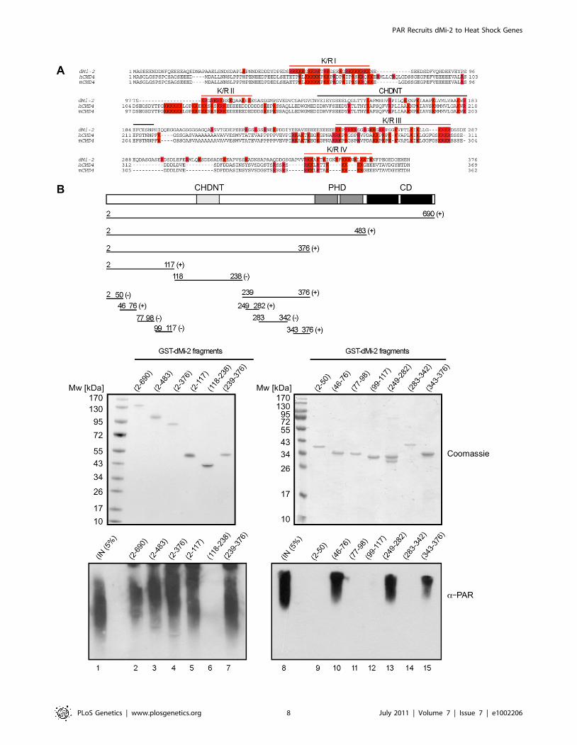
Mapping of the PAR–binding regionsThe N-terminal PAR binding region of dMi-2 contains two
highly conserved domains, a pair of PHD fingers (residues 377 to
484) and a tandem chromodomain (residues 488 to 673). We
generated GST fusions containing these domains and tested their
ability to bind PAR in dot blot assays (Figure S6). This confirmed
that the chromomodomains can bind PAR independently.
However, the PHD fingers did not display PAR binding activity.
We next sought to better define the PAR binding region near
the N-terminus of dMi-2. The N-terminal 375 residues of dMi-2
are characterised by a high content in charged residues (24% D/E,
21% R/K). This general feature is conserved between dMi-2 and
mammalian CHD4 proteins (Figure 4A). In addition, these
proteins share a region with high sequence similarity, the CHDNT
domain (Pfam family PF08073). The function of this domain is not
known. A number of diverse PAR binding motifs have recently
been identified [24–26]. A common feature of these motifs is that
they all contain several R/K residues that are interspersed by
hydrophobic residues which often play critical roles in mediating
PAR binding [24–26]. We subjected different dMi-2 fragments to
the PAR binding assay, including four K/R-rich fragments (K/R I
to IV in Figure 4A). This analysis revealed strong PAR binding
activity for three of the four K/R-rich fragments (K/R I, K/R II
and K/R IV; Figure 4B). By contrast, K/R-rich fragment II and a
fragment encompassing the CHDNT domain failed to interact
with PAR.
Taken together, our results suggest that dMi-2 contains multiple
PAR binding regions in its N-terminus: three are characterised by
a high content of basic amino acid residues (K/R I, K/R III and
K/R IV) and one region containing the tandem chromodomain.
dMi-2 interacts with nascent HS gene transcriptsPARylation of the hsp70 locus has been proposed to assist in the
opening of chromatin structure and to increase access of factors to
DNA and nascent hsp70 transcripts [1]. Given that dMi-2 localises
to the entire transcribed region and given that PAR exhibits
chemical and structural similarity to RNA, we speculated that
dMi-2, once recruited, might interact with nascent hsp70 RNA.
We immunoprecipitated dMi-2 from nuclear extracts of heat
shocked Kc cells and probed for the co-precipitation of nascent
(unprocessed) hsp70 and hsp83 RNA (Figure 5A). Indeed, two
independent dMi-2 antibodies precipitated these transcripts
arguing for a physical, potentially direct interaction. In agreement
with this, dMi-2 bound to single-stranded hsp70 RNA in an
electrophoretic mobility shift assay in vitro (Figure 5B).
Next, we performed competition assays to gain insight into the
relative affinities of dMi-2 for DNA, RNA and PAR and to
determine if dMi-2 can bind to several types of nucleic acid
simultaneously or if binding is competitive. First, we tested dMi-2
binding to RNA and DNA, respectively, in the presence of
increasing amounts of PAR in electrophoretic mobility shift assays
(mass ratios 1:1, 1:2 and 1:4; Figure 5C). In this assay, PAR was
able to compete with RNA and DNA for dMi-2 binding.
However, whereas dMi-2 no longer bound to DNA at a
DNA:PAR mass ratio of 1:2, residual dMi-2/RNA complexes
were still detectable at an RNA:PAR mass ratio of 1:4. This
suggests that dMi-2 has a higher binding affinity for RNA than for
DNA. We confirmed this hypothesis by incubating dMi-2 with
different mass ratios of RNA and DNA (Figure 5C): At a
DNA:RNA mass ratio of 1:1, dMi-2/RNA complexes formed
readily but dMi-2/DNA complexes were not detected. dMi-2/
RNA complexes formed even at DNA:RNA mass ratios of 4:1.
To test if RNA or DNA can compete with dMi-2 for binding to
the branched PAR polymer we performed dot blot assays (Figure
S7). RNA competed with immobilised PAR for binding to dMi-2
whereas DNA failed to do so.
Taken together, our results suggest that dMi-2 has a higher
affinity for binding to RNA and PAR than for binding to DNA. In
addition, dMi-2 appears to bind RNA and PAR in a mutually
exclusive manner. These results are consistent with the hypothesis
that dMi-2 is first recruited to HS loci by interaction with PAR
(which is produced prior to and independent of transcription) and,
once RNA synthesis has been strongly activated, switches to
binding the nascent RNA.
dMi-2 is required for efficient HS gene transcription andprocessing
We hypothesised that dMi-2 binding to nascent RNA might
influence hsp70 transcription or processing. We used transgenic fly
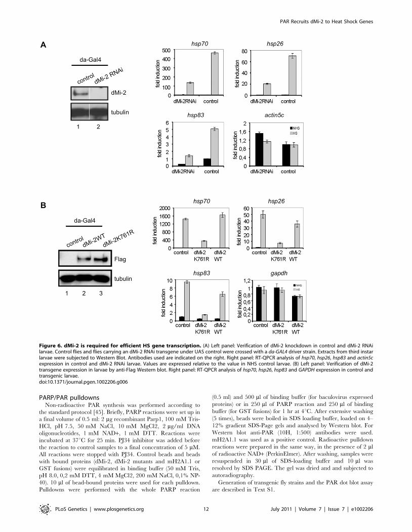
lines to deplete dMi-2 by RNAi (Figure 6A). We subjected
transgenic larvae to HS and determined the HS gene transcription
by RT-QPCR. Although hsp70, hsp26 and hsp83 genes were all
activated by HS, transcript levels were severely reduced in dMi-2
depleted larvae compared to controls. Importantly, transcription
of a housekeeping gene was not significantly affected. We conclude
that dMi-2 makes a positive contribution to transcription and is
essential for full HS gene activation in larvae.
We next determined whether dMi-2 enzymatic activity was
required to activate HS genes. We generated transgenic fly lines
Figure 1. dMi-2 is recruited to HS genes. Immunofluorescence (IF) staining of polytene chromosomes with dMi-2, RNA polymerase II (pol IIser2and pol IIser5), Spt5 antibodies and DAPI as indicated. (A) Untreated chromosomes. Arrows show prominent sites of colocalization. Lower panelsshow magnified sections of individual chromosome arms. (B) Heat shocked chromosomes. Upper panels show magnified section containing thehsp70 loci 87A and 87C (arrows).doi:10.1371/journal.pgen.1002206.g001
PAR Recruits dMi-2 to Heat Shock Genes
PLoS Genetics | www.plosgenetics.org 4 July 2011 | Volume 7 | Issue 7 | e1002206
Figure 2. dMi-2 recruitment to HS genes requires PARP activity. (A and B) ChIP analyses of dMi-2 binding to the hsp70 gene in Kc cells. (A)Upper panel: hsp70 gene and position of amplimers analysed (1: centred at -154, 2: +681). Middle panel: dMi-2 ChIP from cells treated with dsRNAagainst luciferase or dMi-2 as indicated. prom (amplimer 1): promoter; ORF (amplimer 2): open reading frame; NHS: non heat shock; HS: heat shock.
PAR Recruits dMi-2 to Heat Shock Genes
PLoS Genetics | www.plosgenetics.org 5 July 2011 | Volume 7 | Issue 7 | e1002206

overexpressing wild type dMi-2 or a dMi-2 mutant carrying a
point mutation in the ATP binding site (K761R) predicted to
prevent ATP binding (Figure 6B). Indeed, dMi-2K761R could not
hydrolyse ATP in vitro (Figure S8). We subjected 3rd instar larvae
to HS and determined effects on HS gene transcription as before.
Whereas overexpression of wild type dMi-2 had little effect, levels
of HS gene transcripts were greatly reduced in larvae overex-
pressing the enzymatically inactive dMi-2 (Figure 6B). We
conclude that the ATPase activity of dMi-2 is essential for full
HS gene activation.
Next, we sought to assess whether dMi-2 influences RNA
processing. Because dMi-2 depletion and expression of enzymat-
ically inactive dMi-2 resulted in an overall reduction of hsp70
transcript levels we determined the ratio of 39 unprocessed to
total hsp70 RNA as a measure of RNA processing efficiency. We
reasoned that a mere reduction in hsp70 activation (e.g. a
reduction in the number of initiation events per time) would not
change the ratio of unprocessed to total hsp70 RNA. By contrast,
processing defects might give rise to a higher relative proportion
of unprocessed RNA and, therefore, to a higher unprocessed:total
RNA ratio. Depletion of dMi-2 increased the relative proportion
of unprocessed hsp70 RNA (Figure 7A). An even more striking
effect was observed in larvae overexpressing inactive dMi-2,
whereas overexpression of wild type dMi-2 was of little
consequence. Similar effects on 39 RNA processing were
observed with the hsp83 gene (data not shown). Hsp83 is one of
the few HS genes possessing an intron. Therefore, we determined
the ratio of unspliced to total hsp83 transcripts in transgenic
larvae (Figure 7B). Again, we observed a significant increase in
the relative proportion of unspliced RNA in dMi-2-depleted
larvae and in larvae overexpressing inactive enzyme. This
suggests that dMi-2 activity is required for the efficient processing
of HS gene transcripts and that dMi-2 affects both RNA 39 end
cleavage and splicing.
Discussion
dMi-2 associates with active genesMi-2 is strongly linked to transcriptional repression in both
vertebrate and invertebrate organisms. Within NuRD and dMec
complexes it contributes to the repression of cell type-specific
genes [11,14,19–21]. Therefore, the widespread colocalisation of
dMi-2 with active Pol II and elongation factors at many
chromosomal sites is surprising and suggests that dMi-2 might
play an unappreciated role during active transcription, at least (or
specifically) during environmental stresses such as HS. Indeed,
dMi-2 is recruited to HS genes within minutes of HS. This
property is not shared by other chromatin remodelers: Brahma
(BRM) is not enriched at HS puffs and HS gene activation is
independent of BRM function ([27] and data not shown).
Moreover, although imitation switch (ISWI) containing complexes
are important for HS gene transcription, ISWI does not
accumulate to high levels at active HS loci ([28,29] and data not
shown). Recruitment to HS puffs has previously been reported for
Drosophila CHD1 [30]. Thus, accumulation at active HS genes is
shared by at least two members of the CHD family of nucleosome
remodelers but not by SWI/SNF and ISWI proteins.
dMi-2 contributes to efficient HS gene transcriptionDepletion of dMi-2 or a reduction of dMi-2 recruitment does
not significantly perturb hsp70 transcription in Kc cells and,
therefore, dMi-2 is dispensable for HS gene activation in this
system (Figure 2D and data not shown). By contrast, depletion of
dMi-2 in larvae strongly decreases hsp70, hsp26 and hsp83
activation (Figure 6A). It is possible, that the RNAi-mediated
depletion of dMi-2 is more efficient in transgenic flies compared to
cell lines. In addition, it is believed that several factors contributing
to HS gene activation are highly abundant or redundant in Kc
cells but more limiting in other contexts. Accordingly, FACT and
Spt6 are required for a HS gene activation in flies but are not
essential in Kc cells [31,32].
The strong decrease of HS gene activation in dMi-2 RNAi
larvae indicates a positive contribution of dMi-2 to transcription in
vivo. Overexpression of inactive dMi-2 also results in reduced HS
gene transcription implying that its enzymatic activity is critical
(Figure 6B). It is presently unclear whether this reflects a
requirement for dMi-2 catalysed nucleosome remodeling or
whether its activity is directed towards different substrates.
dMi-2 contributes to efficient RNA processingWhile dMi-2 could indirectly influence transcription by
remodeling nucleosomes within the transcribed part of hsp70, its
physical association with nascent HS gene transcripts argues for a
more direct effect. Indeed, dMi-2 is not only required for high HS
gene mRNA levels, but also affects the efficiency of co-
transcriptional 39 end formation and splicing. A role of chromatin
remodelers in splicing has been suggested before: Both CHD1 and
BRG1 bind components of the splicing apparatus [33,34]. CHD1
associates with Pol II and binds nucleosomes containing
H3K4me3, which are enriched near the 59 end of active genes
[34,35]. BRG1 is present at the coding region of genes and
influences splice site choice [33,36]. It has been proposed that
CHD1 and BRG1 physically recruit splicing factors but it is
unclear if their ATPase activities play a role. Indeed, inactive
BRG1 retains the ability to affect exon choice [33,34]. Inefficient
processing of the hsp70 and hsp83 transcripts is not only observed
in larvae expressing reduced levels of dMi-2. Importantly, even
stronger processing defects are generated by overexpression of
inactive dMi-2 (Figure 7). This strongly suggests, for the first time,
that the catalytic activity of a chromatin remodeler is required for
correct co-transcriptional RNA processing. It remains to be
determined whether dMi-2 nucleosome remodeling activity
influences RNA processing indirectly, e.g. by altering Pol II
elongation rates, or whether it has a more direct role.
PAR–dependent recruitment of dMi-2A series of complementary results support our hypothesis that
dMi-2 interacts with PAR polymers that are rapidly synthesized at
activated HS loci. First, the broad distribution of dMi-2 over the
entire transcribed region correlates with the distribution of PAR
polymer [3]. Second, pharmacological inhibition of PARP greatly
decreases dMi-2 binding to activated hsp70. Third, dMi-2 directly
binds PAR polymers in vitro. Fourth, an dMi-2 mutant unable to
bind PAR also fails to localise to active HS loci. As discussed
above, dMi-2 physically associates with nascent HS gene
Lower panel: Verification of RNAi knockdown by Western blot. (B) Upper panel: hsp70 gene and position of amplimers analysed (1: centred at -350; 2:-154; 3: +58; 4: +681; 5: +1702; 6: +2065; 7: +2549). Lower panel: dMi-2 ChIP from NHS (black graph) and HS (gray graph) cells. (C and D) Effect ofelongation inhibitor DRB (C) and PARP inhibitor PJ34 (D) on dMi-2 recruitment to hsp70 gene. hsp70 gene and position of amplimers analysed areshown on top (1: centred at +58; 2: +681; 3: +1702; 4: +2549). Left panels: ChIP analyses of dMi-2 binding to hsp70 gene. Right panels: RT-QPCRanalysis of hsp70 transcription. (D) Rightmost panel: anti-PAR Western blot of extracts from untreated and PJ34 treated Kc cells.doi:10.1371/journal.pgen.1002206.g002
PAR Recruits dMi-2 to Heat Shock Genes
PLoS Genetics | www.plosgenetics.org 6 July 2011 | Volume 7 | Issue 7 | e1002206

Figure 3. dMi-2 binds PAR. (A) PAR was synthesised in vitro by recombinant PARP1 in the presence (+) or absence (-) of PJ34. Reactions were incubatedwith control anti-Flag beads (beads) and beads loaded with dMi-2 or mH2a1.1 as indicated on top. Lanes 1, 2: input; Bound material was analysed byWestern blot using PARP1 (upper panel) and PAR (lower panel) antibodies. (B) Mapping PAR binding regions. Left panel: Schematic representation of dMi-2constructs used. Amino acid boundaries are as follows: dMi-2WT: dMi-2 1-1982; dMi-2DCD: dMi-2 D485-690; dMi-2DN: 691-1982; dMi-2DC: dMi-2 1-1271;dMi-2-CD+ATPase: dMi-2 484-1271; dMi-2-ATPase: dMi-2 691-1271; dMi-2N: dMi-2 1-690; dMi-2(1-485): dMi-2 1-485. Upper right panel: PAR binding assayswith dMi-2 mutants were performed as in (A). dMi-2 mutants are shown on top. Bound material was analysed by anti-PAR Western blot. Lane 1: input. Lowerright panel: Coomasie stained gel showing the dMi-2 constructs used. (C) Polytene chromosomes from transgenic larvae expressing GFP-dMi-2 transgenes(dMi-2WT and dMi-2DN) were analysed by IF using GFP antibody (green) and DAPI (gray). Arrows point to hsp70 HS loci 87A and 87C.doi:10.1371/journal.pgen.1002206.g003
PAR Recruits dMi-2 to Heat Shock Genes
PLoS Genetics | www.plosgenetics.org 7 July 2011 | Volume 7 | Issue 7 | e1002206
PAR Recruits dMi-2 to Heat Shock Genes
PLoS Genetics | www.plosgenetics.org 8 July 2011 | Volume 7 | Issue 7 | e1002206
transcripts and binds RNA in vitro. While this interaction is
potentially important for the efficiency of transcription and
processing, it likely plays a minor role in dMi-2 targeting.
Accordingly, inhibition of transcriptional elongation has no
significant effect on dMi-2 recruitment (Figure 2C).
It is important to note, that while our results argue for an
important role of PAR binding in the recruitment of dMi-2 to HS
loci, we cannot exclude that protein-protein interactions with
histone or non-histone proteins also play a role.
dMi-2 PAR–bindingOur analysis indicates that dMi-2 harbours several PAR binding
motifs in its N-terminal region. Polo and colleagues have recently
demonstrated that human CHD4 is recruited to double stranded
DNA breaks in a PARP-dependent manner [10]. They have
mapped PAR binding activity to the region N-terminal of the
ATPase domain of CHD4. This agrees well with our data and
suggests that the PAR binding function of CHD4/dMi-2 has been
conserved in evolution.
Two structural protein modules directly interact with PAR, the
macrodomain and the PBZ domain; however, these domains are
not present in dMi-2 [5,7,37]. In addition, several shorter PAR
binding motifs have been identified [5,26]. These motifs bear little
sequence similarity but share the presence of several K/R residues
which are interspersed by hydrophobic residues. Our results have
uncovered three K/R-rich regions with PAR binding activity near
the N-terminus of dMi-2. Two of these three K/R-rich regions
(K/R III and K/R IV) consist of interspersed basic and
hydrophobic residues and are therefore reminiscent of the
previously described PAR binding motifs [24,25], the third (K/
R I) lacks hydrophobic residues completely. None of the three K/
R regions matches the consensus PAR binding motifs. It is possible
that a consensus motif should generally be chosen less stringently
and that a high content of K and R-residues in these regions is
sufficient to provide PAR binding activity in vitro. Further
characterisation of these regions will be required to resolve this
issue. In addition to the K/R regions, the tandem chromodomains
of dMi-2 bind PAR in vitro. We have previously shown that the
chromodomains are required for interacting with nucleosomal
DNA in vitro [38]. Our new data suggests that these domains can
interact with different nucleic acids.
Different functions of PAR in HS gene transcriptionSeveral potential molecular functions of PARylation at HS
genes have been suggested. First, PARP activity is required for the
rapid loss of nucleosomes at hsp70 within the first two minutes after
HS [1]. It has been suggested that PARylation of histones aids
rapid nucleosome disassembly [1]. Second, at later stages of the
HS response (20–60 minutes after HS), PARP activity is required
to establish a compartment which restricts the diffusion of factors
such as Pol II and Spt6 and promotes efficient factor recycling [4].
Our results suggest that PARylation carries out a third task,
namely, to recruit factors via their direct interaction with PAR.
The earliest time point when we can detect dMi-2 binding to hsp70
is between 2 and 5 minutes after HS. This places dMi-2
recruitment between the early PARP-dependent nucleosome
removal (0–2 minutes after HS) and effects of the transcription
compartment (20–60 minutes after HS).
PAR versus RNA bindingThe ability of dMi-2 to bind both PAR and RNA and the
finding that RNA can compete for PAR binding to dMi-2 is
consistent with the hypothesis that dMi-2 association with active
HS genes is a two step process (Figure 8). We propose that dMi-2 is
initially recruited via interaction with PAR polymers. Synthesis of
these starts prior to the onset of hsp70 transcription [1]. This results
in a rapid local increase of the dMi-2 concentration. In the second
step, when hsp70 transcripts are produced by elongating RNA
polymerase II at high rates, dMi-2 can switch from binding PAR
to interacting with nascent transcripts.
PAR signals rapid and efficient factor recruitment tochromatin during stress
Severe cellular stresses, such as DNA strand breaks and acute
HS, must be dealt with quickly and efficiently. In both cases, a
multitude of factors are rapidly recruited to orchestrate the
repair of DNA and the massive transcriptional activation of HS
genes, respectively. We postulate that rapid synthesis of PAR
polymers at both DNA damage sites and HS genes affords
an efficient mechanism to recruit chromatin remodelers and
other factors. It has recently been shown that PARylation of
DNA breaks is instrumental in recruiting chromatin remo-
delers, including mammalian dMi-2 homologs, to damaged sites
[8,9,10,39,40,41]. Here, we show that dMi-29s recruitment to
activated HS genes requires PARP activity and that dMi-2 binds
PAR directly. The high local concentration of PAR polymers at
DNA breaks and HS genes might exploit the general affinity of
dMi-2 for nucleic acids. Indeed, dMi-2 binds both DNA and RNA
as well as PAR in vitro ([38] and this study). In this manner, PAR
polymers might act as a scaffold to redirect dMi-2 to chromatin
regions where high levels of dMi-2 activity are required, thus
acting as a stress-dependent, transient affinity site for chromatin
remodeling and possibly RNA processing activities (Figure 8).
Our results highlight a signaling and scaffolding function for
PARP activity during transient environmental stresses other than
DNA damage, suggesting that PARylation carries out important
modulatory functions in the stress-dependent reprogramming of
nuclear activities.
Materials and Methods
Chromatin immunoprecipitation (ChIP)Kc cell HS treatment and ChIP was performed as decribed
using dMi-2C antibody [14,42]. For primer sequences see Dataset
S2. Triplicate mean values of percentage input DNA and standard
deviations are plotted. dMi-2 knockdown by RNAi was described
previously [23]. For RNAi primer sequences see Dataset S4.
Pharmacological treatmentsKc cells were treated with 125 mM DRB (Sigma) to inhibit
transcription and with 5 mM PJ34 (Alexis) to inhibit PARP activity
for 20 min before subjecting cells to HS.
Figure 4. PAR–binding regions of dMi-2. (A) Multiple sequence alignment of N-terminus of dMi-2 and human and mouse CHD4. All K and Ramino acid residues are coloured in red. Red lines indicate the four K/R rich regions. The black line indicates the CHDNT domain. (B) Mapping of PARbinding regions in the N-terminal part of dMi-2. Upper panel: Schematic representation of dMi-2 constructs used. Numbers indicate the amino acidborders of the constructs. (+) and (-) indicate binding to PAR. Middle panel: Coomasie stained gels with purified GST-dMi-2 fragments used for PARPpulldown assays. Lower panel: PAR binding assays with GST-dMi-2 fragments were performed as in Figure 3B. Bound material was analysed by anti-PAR Western blot. Lanes 1 and 8: inputs.doi:10.1371/journal.pgen.1002206.g004
PAR Recruits dMi-2 to Heat Shock Genes
PLoS Genetics | www.plosgenetics.org 9 July 2011 | Volume 7 | Issue 7 | e1002206

PAR Recruits dMi-2 to Heat Shock Genes
PLoS Genetics | www.plosgenetics.org 10 July 2011 | Volume 7 | Issue 7 | e1002206

Polytene chromosomesChromosomes were prepared as before [23]. The following
antibodies were used: Primary antibodies: anti-dMi-2N (rabbit)
1:200, anti-pol II (mouse H5, Covance) 1:50, anti-GFP (rabbit,
Abcam) 1:50, anti-Spt5 (guinea pig) 1:200. Secondary antibodies:
Alexa Fluor 488 goat anti-rabbit 1:200, Alexa Fluor 546 goat anti-
mouse or anti-guinea pig 1:200 (Invitrogen).
Analysis was performed with a Zeiss fluorescence microscope
(Axioplan).
Generation of baculovirus and GST-fusion vectorsFor baculovirus production, dMi-2 mutants (aa 1-691) and (aa
1-485) were generated by PCR using appropriate sets of primers
and cloned with NotI and XbaI into the pVL1392 transfer vector.
Vectors for dMi-2 WT and other mutants were described
previously [38]. dMi-2 GST-fusion fragments were generated by
PCR using appropriate sets of primers and cloned with NotI and
SalI into the pGEX4T1 vector. All constructs were verified by
DNA sequencing. For primer sequences see Dataset S1.
Preparation of protein extracts, purification ofrecombinant proteins, and ATPase assay
Protein extracts from 3rd instar larvae were prepared as
described in [14].
Purification of recombinant dMi-2 and ATPase assays are
described [23]. Recombinant mH2A1.1 was purified as in [43].
GST-fusion proteins were expressed in E.coli BL21(DE3) and
purified with Glutathione Sepharose 4 Fast flow (GE Healthcare)
according to the manufacturer’s instructions.
Electrophoretic mobility shift assays (EMSA)A typical DNA or RNA binding reaction (25 ml) was performed
in the presence of 0.2 mg of dMi-2F and 80 ng of nucleic acid
(DNA or ssRNA) in 40 mM KCl, 20 mM Tris pH 7.6, 1.5 mM
MgCl2, 0.5 mM EGTA, 10% glycerol, BSA (200 ng/ml), 1 mM
DTT (supplemented with 0.4 units of RNAsin). For competition
assays, samples were preincubated for 15 min at 26uC before the
different amounts of competitor (PAR or DNA or RNA) were
added. Reactions were further incubated at 26uC for 75 min.
Products were analyzed on 6% native PAA gel and visualized
with ethidium bromide (EtBr) staining. ssRNA was synthesized
by in vitro transcription using a fragment of hsp70 DNA
as a template. This template (also used for the DNA band-
shift assays) was produced by PCR amplification of cDNA
derived from heat shocked Kc cells using the following primers:
T7-hsp70_f - TAATACGACTCACTATAGGGCCTACGGA-
CTGGACAAGAAC and hsp70_r -AGGGTTGGAGCGCA-
GATCCTTCTTGTAC.
RNA isolation and RT-QPCRTotal RNA was isolated from 3rd instar larvae using PeqGold
total RNA Kit (PeqLab). 10-12 larvae from each cross were pestled
in 400 ml of lysis buffer before loading the material on the column.
1 mg of RNA was reverse transcribed by incubation with 0.3 mg of
random primers (Invitrogene) and 100 U of M-MLV reverse
transcriptase (Invitrogen). cDNA synthesis was performed accord-
ing to the manufacturer’s protocol. cDNA was analyzed by QPCR
using Absolute SybrGreen Mix (Thermo Fisher) and the Mx3000P
real-time detection system (Agilent). For primer sequences used in
RT-QPCR see Dataset S3. All amplifications were performed in
triplicate using 0.6 ml of cDNA per reaction. Triplicate mean
values were calculated according to the DDCt quantification
method using rp49 gene transcription as reference for normaliza-
tion. Relative mRNA levels in uninduced control larvae were set
to 1 and other values were expressed relative to this. The RT-
QPCR results were reproduced several times using independent
fly crosses and representative data sets are shown.
RNA immunoprecipitationRNA immunoprecipitation was performed as described previ-
ously [44]. Briefly, Kc cells were crosslinked as for ChIP. Cells
were washed once with PBS buffer and lysed on ice for 15 min in
FA buffer (50 mM Hepes- KOH, pH 7,6, 140 mM NaCl, 1%
Triton X-100, 0,1% sodium deoxycholate, proteinase inhibitors,
RNAsin (100 u/ml of buffer)). Cells were sonicated, spun down
and chromatin was digested with DNAse I. The chromatin
containing solution was adjusted to 25 mM MgCl2 and 5 mM
CaCl2. 1 ul of DNAse I (Qiagen) was added and reactions were
incubated for 10 min at room temperature and then stopped with
20 mM EDTA. Chromatin was spun down for 10 min
(13000 rpm) at 4uC. 300 ml of chromatin was used for IP. 2 ml
of anti-dMi-2(C) and anti-dMi-2(N) antibodies, 2 ml rabbit IgG,
2 ml rabbit preimmuneserum and beads only (control) and were
used for IP. Samples were incubated over night at 4uC. RNA-
protein complexes were precipitated with 30 ul of 50% protein G
Sepharose beads for 2 hr at 4uC. IPs were washed 5 times in FA
buffer, twice with TE buffer and eluted twice with 100 ml of
elution buffer (100 mm Tris HCl, pH 8,0, 10 mM EDTA, 1%
SDS) – once at room temperature and once at 65uC. All buffers
were supplemented with RNAse inhibitor (RNAsin, Promega). All
samples were digested with proteinase K for 1 hr at 42uC and
decrosslinking was performed at 65uC over night. Immunopre-
cipitated RNA was purified using PeqGold total RNA Kit
(PeqLab), digested with DNAse on the column and eluted with
30 ml of RNAse free dH20. cDNA was synthesized with 10 ml of
eluted RNA and 2 ml of input with random hexamers and
analysed by Q-PCR with appropriate primer pairs.
Figure 5. dMi-2 binds RNA. (A) RNA immunoprecipitation (RIP) of hsp70 and hsp83 unprocessed transcripts from heat shocked Kc cells. RIP wasperformed with two independent dMi-2 antibodies (dMi-2C and dMi-2N), IgG, preimmune serum and protein G beads as indicated. Primer pairs thatspecifically amplify actin5c, rp49 and unprocessed hsp70 and hsp83 transcripts (see Figure 7) were used for RT-QPCR. (B) RNA electrophoretic mobilityshift assay. Single stranded hsp70 RNA was incubated with recombinant dMi-2. Lane 2: 0.1 mg dMi-2, lane 3: 0.2 mg dMi-2, lane 1: no protein. RNA andRNA:protein complexes were resolved by electrophoresis and visualized with ethidium bromide. Position of unbound RNA probe is indicated. (C)Competition mobility shift assays. Upper left panel: Single stranded hsp70 RNA was incubated with 0.2 mg of recombinant dMi-2 in the absence or inthe presence of increasing amounts of PAR polymer, as indicated. Upper right panel: hsp70 DNA was incubated with 0.2 mg of recombinant dMi-2 inthe absence or in the presence of increasing amounts of PAR polymer, as indicated. The following mass ratios of RNA to PAR or DNA to PAR wereused: lane 3 - 1:1, lane 4 -1:2, lane 5 -1:4. Positions of unbound RNA and DNA probes are indicated. Lower left panel: Single stranded hsp70 RNA wasincubated with 0.2 mg of recombinant dMi-2 in the absence or in the presence of increasing amounts of DNA, as indicated. Lower right panel: hsp70DNA was incubated with 0.2 mg of recombinant dMi-2 in the absence or in the presence of increasing amounts of RNA, as indicated. The followingweight ratios of RNA to DNA or DNA to RNA were used: line 3 - 1:1, lane 4 -1:2, lane 5 -1:4. Positions of unbound RNA and DNA probes and dMi-2/DNAand dMi-2/RNA complexes are shown on the right.doi:10.1371/journal.pgen.1002206.g005
PAR Recruits dMi-2 to Heat Shock Genes
PLoS Genetics | www.plosgenetics.org 11 July 2011 | Volume 7 | Issue 7 | e1002206
PARP/PAR pulldownsNon-radioactive PAR synthesis was performed according to
the standard protocol [45]. Briefly, PARP reactions were set up in
a final volume of 0.5 ml: 2 mg recombinant Parp1, 100 mM Tris-
HCl, pH 7.5, 50 mM NaCl, 10 mM MgCl2, 2 mg/ml DNA
oligonucleotides, 1 mM NAD+, 1 mM DTT. Reactions were
incubated at 37uC for 25 min. PJ34 inhibitor was added before
the reaction to control samples to a final concentration of 5 mM.
All reactions were stopped with PJ34. Control beads and beads
with bound proteins (dMi-2, dMi-2 mutants and mH2A1.1 or
GST fusions) were equilibrated in binding buffer (50 mM Tris,
pH 8.0, 0,2 mM DTT, 4 mM MgCl2, 200 mM NaCl, 0,1% NP-
40). 10 ml of bead-bound proteins were used for each pulldown.
Pulldowns were performed with the whole PARP reaction
(0.5 ml) and 500 ml of binding buffer (for baculovirus expressed
proteins) or in 250 ml of PARP reaction and 250 ml of binding
buffer (for GST fusions) for 1 hr at 4uC. After extensive washing
(5 times), beads were boiled in SDS loading buffer, loaded on 4–
12% gradient SDS-Page gels and analysed by Western blot. For
Western blot anti-PAR (10H, 1:500) antibodies were used.
mH2A1.1 was used as a positive control. Radioactive pulldown
reactions were prepared in the same way, in the presence of 2 ml
of radioactive NAD+ (PerkinElmer). After washing, samples were
resuspended in 30 ml of SDS-loading buffer and 10 ml was
resolved by SDS PAGE. The gel was dried and and subjected to
autoradiography.
Generation of transgenic fly strains and the PAR dot blot assay
are described in Text S1.
Figure 6. dMi-2 is required for efficient HS gene transcription. (A) Left panel: Verification of dMi-2 knockdown in control and dMi-2 RNAilarvae. Control flies and flies carrying an dMi-2 RNAi transgene under UAS control were crossed with a da-GAL4 driver strain. Extracts from third instarlarvae were subjected to Western Blot. Antibodies used are indicated on the right. Right panel: RT-QPCR analysis of hsp70, hsp26, hsp83 and actin5cexpression in control and dMi-2 RNAi larvae. Values are expressed relative to the value in NHS control larvae. (B) Left panel: Verification of dMi-2transgene expression in larvae by anti-Flag Western blot. Right panel: RT-QPCR analysis of hsp70, hsp26, hsp83 and GAPDH expression in control andtransgenic larvae.doi:10.1371/journal.pgen.1002206.g006
PAR Recruits dMi-2 to Heat Shock Genes
PLoS Genetics | www.plosgenetics.org 12 July 2011 | Volume 7 | Issue 7 | e1002206

Figure 7. dMi-2 is required for efficient RNA processing. (A and B) Upper panels: Schematic representations of the hsp70 and hsp83 genes.RT-QPCR amplimers, hsp70 cleavage site, hsp83 intron and transcriptional start sites (TSS) are shown. (A) Lower panel: RT-QPCR from control andtransgenic larvae. The ratio between 39 unprocessed and total hsp70 RNA was determined (hsp70: amplimer 2/amplimer 1). The ratio obtained forcontrol larvae was set to 1, other ratios were expressed relative to this. (B) Lower panel: The ratio between unspliced and total hsp83 RNA wasdetermined (hsp83: amplimer 1/amplimer 2) and plotted as in (A).doi:10.1371/journal.pgen.1002206.g007
Figure 8. Model. Upon HS, PARylation of the locus creates binding sites for PAR-sensing regions of dMi-2. dMi-2 is recruited and, subsequently,interacts with nascent transcripts to support transcription and processing. GAF: GAGA Factor, HSE: HS elements.doi:10.1371/journal.pgen.1002206.g008
PAR Recruits dMi-2 to Heat Shock Genes
PLoS Genetics | www.plosgenetics.org 13 July 2011 | Volume 7 | Issue 7 | e1002206

Supporting Information
Dataset S1 Sequences of primers used for cloning.
(DOCX)
Dataset S2 Sequences of primers used for ChIP.
(DOCX)
Dataset S3 Sequences of primers used for RT-QPCR.
(DOCX)
Dataset S4 Sequences of primers used for ds RNA synthesis
(RNAi).
(DOCX)
Text S1 Supporting protocols: Fly strains, transgenesis and dot
blot assay.
(DOC)
Figure S1 Kinetic analysis of dMi-2 binding to hsp70 gene during
heat shock. dMi-2 binding to hsp70 gene was determined by ChIP
under NHS conditions and at different time points following heat
shock as indicated. Amplimers were centered as follows: 1, -154 bp;
2, +58 bp; 3, +681 bp; 4, +1427 bp; 5, + 2549 bp.
(TIF)
Figure S2 H3 ChIP on hsp70 gene upon PJ34 treatment. dMi-2
binding to hsp70 gene was determined by ChIP under NHS
conditions and at different time points in the absence or in the
presence of PJ34, as indicated. Amplimers were centered as follows:
1, -154 bp; 2, +58 bp; 3, +681 bp; 4, +1427 bp; 5, + 2549 bp.
(TIF)
Figure S3 Pulldown with whole PAR reaction. Experiment was
performed as in Figure 3A with a difference that radioactive
NAD+ was used for PAR synthesis. Samples were run on the gel,
gel was dried and exposed overnight on the X-ray film.
(TIF)
Figure S4 PAR binding assay. Dot blot with purified PAR.
BSA or recombinant dMi-2 WT and indicated mutants were
spotted on the nitrocellulose and incubated with PAR. Upon
extensive washes, membrane was subjected to Western Blot
analysis with anti-PAR antibodies. After stripping, membrane
was probed with anti-Flag antibodies to monitor the amount of
proteins spotted.
(TIF)
Figure S5 PAR binding assay. Upper panel: Dot blot with
purified PAR. GST-fusion proteins and GST were spotted on the
nitrocellulose and incubated with PAR. Upon extensive washes
with low salt (150 mM) or high salt (500 mM), membranes were
subjected to Western Blot analysis with anti-PAR antibodies.
Lower panel: Coomasie stained gel with purified proteins used for
PAR binding assay. Chromo - chromodomains of dMi-2 (aa 488-
712), PHDs – PHD fingers of dMi-2 (aa 377-490).
(TIF)
Figure S6 Expression analysis of GFP-tagged transgenes. Left
panel – whole salivary glands from flies crossed to the salivary
gland-specific sgs58ABGAL4 driver were analysed for GFP
expression. Right panel: larval extracts derived from control
(w1118) larvae (line 1) and larvae expressing GFPtagged dMi-
2WT (lane 3) or dMi-2DN transgene (lane 2) crossed to daughterless-
GAL4 driver were analysed by western blot using GFP antibodies.
(TIF)
Figure S7 Competition of PAR binding with RNA and DNA.
(A) Dot blot with purified PAR. dMi-2 WT was spotted on the
nitrocellulose and incubated with PAR. Upon extensive washes
membranes were subjected to Western Blot analysis with anti-PAR
antibodies (upper panel), Ponceau staining indicates the amount of
protein spotted (lower panel). When indicated, membranes were
preincubated with increasing amounts of RNA (lanes: 2,3 and 4)
followed by incubation with PAR. Lane 1: dMi-2 was preincu-
bated with buffer only. (A) Dot blot with purified PAR. dMi-2 WT
was spotted on the nitrocellulose and incubated with PAR. When
indicated, membranes were preincubated with increasing amounts
of DNA (lanes: 2,3 and 4) followed by incubation with PAR. Lane
1: dMi-2 was preincubated with buffer only.
(TIF)
Figure S8 dMi-2 K761R mutant is catalytically inactive. Upper
panel: a Coomasie gel with dMi-2 WT and dMi-2 K761R mutant.
Lower panel: ATPase assay with wild type and mutant form of
dMi-2 in the presence of nucleosomes.
(TIF)
Acknowledgments
We are grateful to F. Winston, K. Basler, and P. O. Hassa for reagents and
to R. Hyland and G. Timinszky for experimental help and discussion.
Author Contributions
Conceived and designed the experiments: MM MH RR-P AL AB.
Performed the experiments: MM MH. Analyzed the data: MM MH RR-P
AL AB. Contributed reagents/materials/analysis tools: RR-P AL AB.
Wrote the paper: MM AL AB.
References
1. Petesch SJ, Lis JT (2008) Rapid, transcription-independent loss of nucleosomesover a large chromatin domain at Hsp70 loci. Cell 134: 74–84.
2. Winegarden NA, Wong KS, Sopta M, Westwood JT (1996) Sodium salicylate
decreases intracellular ATP, induces both heat shock factor binding andchromosomal puffing, but does not induce hsp 70 gene transcription in
Drosophila. J Biol Chem 271: 26971–26980.
3. Tulin A, Spradling A (2003) Chromatin loosening by poly(ADP)-ribosepolymerase (PARP) at Drosophila puff loci. Science 299: 560–562.
4. Zobeck KL, Buckley MS, Zipfel WR, Lis JT (2010) Recruitment Timing andDynamics of Transcription Factors at the Hsp70 Loci in Living Cells. Molecular
Cell 40: 965–975.
5. Krishnakumar R, Kraus WL (2010) The PARP side of the nucleus: molecularactions, physiological outcomes, and clinical targets. Mol Cell 39: 8–24.
6. Till S, Ladurner AG (2009) Sensing NAD metabolites through macro domains.
Front Biosci 14: 3246–3258.
7. Timinszky G, Till S, Hassa PO, Hothorn M, Kustatscher G, et al. (2009) A
macrodomain-containing histone rearranges chromatin upon sensing PARP1
activation. Nat Struct Mol Biol 16: 923–929.
8. Ahel D, Horejsi Z, Wiechens N, Polo SE, Garcia-Wilson E, et al. (2009)
Poly(ADP-ribose)-dependent regulation of DNA repair by the chromatin
remodeling enzyme ALC1. Science 325: 1240–1243.
9. Gottschalk AJ, Timinszky G, Kong SE, Jin J, Cai Y, et al. (2009)Poly(ADP-ribosyl)ation directs recruitment and activation of an ATP-
dependent chromatin remodeler. Proc Natl Acad Sci U S A 106:
13770–13774.
10. Polo SE, Kaidi A, Baskcomb L, Galanty Y, Jackson SP (2010) Regulation of
DNA-damage responses and cell-cycle progression by the chromatin remodel-
ling factor CHD4. Embo J 29: 3130–3139.
11. Fujita N, Jaye DL, Geigerman C, Akyildiz A, Mooney MR, et al. (2004) MTA3and the Mi-2/NuRD complex regulate cell fate during B lymphocyte
differentiation. Cell 119: 75–86.
12. Murawsky CM, Brehm A, Badenhorst P, Lowe N, Becker PB, et al. (2001)Tramtrack69 interacts with the dMi-2 subunit of the Drosophila NuRD
chromatin remodelling complex. EMBO Rep 2: 1089–1094.
13. Marfella CG, Imbalzano AN (2007) The Chd family of chromatin remodelers.Mutat Res 618: 30–40.
14. Kunert N, Wagner E, Murawska M, Klinker H, Kremmer E, et al. (2009) dMec:
a novel Mi-2 chromatin remodelling complex involved in transcriptionalrepression. Embo J 28: 533–544.
15. Feng Q, Zhang Y (2001) The MeCP1 complex represses transcription through
preferential binding, remodeling, and deacetylating methylated nucleosomes.
Genes Dev 15: 827–832.
PAR Recruits dMi-2 to Heat Shock Genes
PLoS Genetics | www.plosgenetics.org 14 July 2011 | Volume 7 | Issue 7 | e1002206

16. Le Guezennec X, Vermeulen M, Brinkman AB, Hoeijmakers WA, Cohen A,
et al. (2006) MBD2/NuRD and MBD3/NuRD, two distinct complexes with
different biochemical and functional properties. Mol Cell Biol 26: 843–851.
17. Lyko F, Beisel C, Marhold J, Paro R (2006) Epigenetic regulation in Drosophila.
Curr Top Microbiol Immunol 310: 23–44.
18. Kehle J, Beuchle D, Treuheit S, Christen B, Kennison JA, et al. (1998) dMi-2, a
hunchback-interacting protein that functions in polycomb repression. Science
282: 1897–1900.
19. Kim J, Sif S, Jones B, Jackson A, Koipally J, et al. (1999) Ikaros DNA-binding
proteins direct formation of chromatin remodeling complexes in lymphocytes.
Immunity 10: 345–355.
20. Koipally J, Renold A, Kim J, Georgopoulos K (1999) Repression by Ikaros and
Aiolos is mediated through histone deacetylase complexes. Embo J 18:
3090–3100.
21. Reddy BA, Bajpe PK, Bassett A, Moshkin YM, Kozhevnikova E, et al. (2010)
Drosophila transcription factor Tramtrack69 binds MEP1 to recruit the
chromatin remodeler NuRD. Mol Cell Biol 30: 5234–5244.
22. Stielow B, Sapetschnig A, Kruger I, Kunert N, Brehm A, et al. (2008)
Identification of SUMO-dependent chromatin-associated transcriptional repres-
sion components by a genome-wide RNAi screen. Mol Cell 29: 742–754.
23. Murawska M, Kunert N, van Vugt J, Langst G, Kremmer E, et al. (2008)
dCHD3, a novel ATP-dependent chromatin remodeler associated with sites of
active transcription. Mol Cell Biol 28: 2745–2757.
24. Gagne JP, Hunter JM, Labrecque B, Chabot B, Poirier GG (2003) A proteomic
approach to the identification of heterogeneous nuclear ribonucleoproteins as a
new family of poly(ADP-ribose)-binding proteins. Biochem J 371: 331–340.
25. Pleschke JM, Kleczkowska HE, Strohm M, Althaus FR (2000) Poly(ADP-ribose)
binds to specific domains in DNA damage checkpoint proteins. J Biol Chem 275:
40974–40980.
26. Zhang Y, Liu S, Mickanin C, Feng Y, Charlat O, et al. RNF146 is a poly(ADP-
ribose)-directed E3 ligase that regulates axin degradation and Wnt signalling.
Nat Cell Biol 13: 623–629.
27. Armstrong JA, Papoulas O, Daubresse G, Sperling AS, Lis JT, et al. (2002) The
Drosophila BRM complex facilitates global transcription by RNA polymerase II.
Embo J 21: 5245–5254.
28. Badenhorst P, Xiao H, Cherbas L, Kwon SY, Voas M, et al. (2005) The
Drosophila nucleosome remodeling factor NURF is required for Ecdysteroid
signaling and metamorphosis. Genes Dev 19: 2540–2545.
29. Deuring R, Fanti L, Armstrong JA, Sarte M, Papoulas O, et al. (2000) The ISWI
chromatin-remodeling protein is required for gene expression and the
maintenance of higher order chromatin structure in vivo. Mol Cell 5: 355–365.
30. Kelley DE, Stokes DG, Perry RP (1999) CHD1 interacts with SSRP1 and
depends on both its chromodomain and its ATPase/helicase-like domain for
proper association with chromatin. Chromosoma 108: 10–25.
31. Ardehali MB, Yao J, Adelman K, Fuda NJ, Petesch SJ, et al. (2009) Spt6
enhances the elongation rate of RNA polymerase II in vivo. Embo J 28:1067–1077.
32. Saunders A, Werner J, Andrulis ED, Nakayama T, Hirose S, et al. (2003)
Tracking FACT and the RNA polymerase II elongation complex throughchromatin in vivo. Science 301: 1094–1096.
33. Batsche E, Yaniv M, Muchardt C (2006) The human SWI/SNF subunit Brm isa regulator of alternative splicing. Nat Struct Mol Biol 13: 22–29.
34. Sims RJ, 3rd, Millhouse S, Chen CF, Lewis BA, Erdjument-Bromage H, et al.
(2007) Recognition of trimethylated histone H3 lysine 4 facilitates therecruitment of transcription postinitiation factors and pre-mRNA splicing. Mol
Cell 28: 665–676.35. Srinivasan S, Armstrong JA, Deuring R, Dahlsveen IK, McNeill H, et al. (2005)
The Drosophila trithorax group protein Kismet facilitates an early step intranscriptional elongation by RNA Polymerase II. Development 132:
1623–1635.
36. Tyagi A, Ryme J, Brodin D, Ostlund Farrants AK, Visa N (2009) SWI/SNFassociates with nascent pre-mRNPs and regulates alternative pre-mRNA
processing. PLoS Genet 5: e1000470. doi:10.1371/journal.pgen.1000470.37. Ahel I, Ahel D, Matsusaka T, Clark AJ, Pines J, et al. (2008) Poly(ADP-ribose)-
binding zinc finger motifs in DNA repair/checkpoint proteins. Nature 451:
81–85.38. Bouazoune K, Mitterweger A, Langst G, Imhof A, Akhtar A, et al. (2002) The
dMi-2 chromodomains are DNA binding modules important for ATP-dependent nucleosome mobilization. Embo J 21: 2430–2440.
39. Chou DM, Adamson B, Dephoure NE, Tan X, Nottke AC, et al. (2010) Achromatin localization screen reveals poly (ADP ribose)-regulated recruitment of
the repressive polycomb and NuRD complexes to sites of DNA damage. Proc
Natl Acad Sci U S A 107: 18475–18480.40. Larsen DH, Poinsignon C, Gudjonsson T, Dinant C, Payne MR, et al. (2010)
The chromatin-remodeling factor CHD4 coordinates signaling and repair afterDNA damage. J Cell Biol 190: 731–740.
41. Smeenk G, Wiegant WW, Vrolijk H, Solari AP, Pastink A, et al. (2010) The
NuRD chromatin-remodeling complex regulates signaling and repair of DNAdamage. J Cell Biol 190: 741–749.
42. Boehm AK, Saunders A, Werner J, Lis JT (2003) Transcription factor andpolymerase recruitment, modification, and movement on dhsp70 in vivo in the
minutes following heat shock. Mol Cell Biol 23: 7628–7637.43. Kustatscher G, Hothorn M, Pugieux C, Scheffzek K, Ladurner AG (2005)
Splicing regulates NAD metabolite binding to histone macroH2A. Nat Struct
Mol Biol 12: 624–625.44. Gilbert C, Svejstrup JQ (2006) RNA immunoprecipitation for determining
RNA-protein associations in vivo. Curr Protoc Mol Biol Chapter 27: Unit 27 24.45. Karras GI, Kustatscher G, Buhecha HR, Allen MD, Pugieux C, et al. (2005)
The macro domain is an ADP-ribose binding module. Embo J 24: 1911–1920.
PAR Recruits dMi-2 to Heat Shock Genes
PLoS Genetics | www.plosgenetics.org 15 July 2011 | Volume 7 | Issue 7 | e1002206