Embed Size (px)
Citation preview
lable at ScienceDirect
Food Hydrocolloids 43 (2015) 360e376
Contents lists avai
Food Hydrocolloids
journal homepage: www.elsevier .com/locate/ foodhyd
Review
Gelatin structure and composition linked to hard capsule dissolution:A review
Anne Duconseille a, Thierry Astruc a, Naira Quintana b, Filip Meersman b,V�eronique Sante-Lhoutellier a, *
a INRA de Clermont-Ferrand-Theix, UR370 QuaPA, F-63122 St Gen�es Champanelle, Franceb Rousselot NV, Meulestedekaai 81, 9000 Gent, Belgium
a r t i c l e i n f o
Article history:Received 8 November 2013Accepted 10 June 2014Available online 17 June 2014
Keywords:Gelatin dissolutionCollagen structureAggregatesCross-linksGelatin molecular weightsHard capsules
* Corresponding author. Tel.: þ33 4 73 62 47 08; faE-mail address: [email protected]
http://dx.doi.org/10.1016/j.foodhyd.2014.06.0060268-005X/© 2014 Elsevier Ltd. All rights reserved.
a b s t r a c t
Gelatin obtained from pig skin constitutes about 50% of world production and is mainly composed ofcollagen extracted from skin by acidic baths and thermal treatments. The gelatin is used to make variousproducts, notably hard gelatin capsules (HGC) which of varying solubility in water. This issue has beenknown for many years and has been, and remains, a subject of study and debate. The main reason for lowgelatin dissolution rates is its tendency to form cross-links in the denatured collagen chains underspecific conditions which stabilize the gel network and prevent dissolution. As it is extracted from animaltissues, gelatin may contain molecules other than collagen (sugars, lipids and other proteins) which mayreact with collagen chains to form covalent bonds. Although this biopolymer has been the subject ofnumerous publications, its structure and composition is not well defined. Indeed, there are many dif-ferences from an article to another. Consequently, the causes of HGC dissolution are not well identifiedand controlled.
© 2014 Elsevier Ltd. All rights reserved.
Contents
1. Introduction . . . . . . . . . . . . . . . . . . . . . . . . . . . . . . . . . . . . . . . . . . . . . . . . . . . . . . . . . . . . . . . . . . . . . . . . . . . . . . . . . . . . . . . . . . . . . . . . . . . . . . . . . . . . . . . . . . . . . . 3612. Collagen composition and structure . . . . . . . . . . . . . . . . . . . . . . . . . . . . . . . . . . . . . . . . . . . . . . . . . . . . . . . . . . . . . . . . . . . . . . . . . . . . . . . . . . . . . . . . . . . . . . . . . 3613. Gelatin composition . . . . . . . . . . . . . . . . . . . . . . . . . . . . . . . . . . . . . . . . . . . . . . . . . . . . . . . . . . . . . . . . . . . . . . . . . . . . . . . . . . . . . . . . . . . . . . . . . . . . . . . . . . . . . . 3624. Gelatin structure and gelation mechanism . . . . . . . . . . . . . . . . . . . . . . . . . . . . . . . . . . . . . . . . . . . . . . . . . . . . . . . . . . . . . . . . . . . . . . . . . . . . . . . . . . . . . . . . . . . 363
4.1. Gelatin structure . . . . . . . . . . . . . . . . . . . . . . . . . . . . . . . . . . . . . . . . . . . . . . . . . . . . . . . . . . . . . . . . . . . . . . . . . . . . . . . . . . . . . . . . . . . . . . . . . . . . . . . . . . . . 3634.2. Nature of interactions . . . . . . . . . . . . . . . . . . . . . . . . . . . . . . . . . . . . . . . . . . . . . . . . . . . . . . . . . . . . . . . . . . . . . . . . . . . . . . . . . . . . . . . . . . . . . . . . . . . . . . . 365
4.2.1. Hydrogen bonds . . . . . . . . . . . . . . . . . . . . . . . . . . . . . . . . . . . . . . . . . . . . . . . . . . . . . . . . . . . . . . . . . . . . . . . . . . . . . . . . . . . . . . . . . . . . . . . . . . . . . 3654.2.2. Hydrophobic interactions . . . . . . . . . . . . . . . . . . . . . . . . . . . . . . . . . . . . . . . . . . . . . . . . . . . . . . . . . . . . . . . . . . . . . . . . . . . . . . . . . . . . . . . . . . . . . 3654.2.3. Electrostatic interactions . . . . . . . . . . . . . . . . . . . . . . . . . . . . . . . . . . . . . . . . . . . . . . . . . . . . . . . . . . . . . . . . . . . . . . . . . . . . . . . . . . . . . . . . . . . . . . . 3664.2.4. Covalent bonds . . . . . . . . . . . . . . . . . . . . . . . . . . . . . . . . . . . . . . . . . . . . . . . . . . . . . . . . . . . . . . . . . . . . . . . . . . . . . . . . . . . . . . . . . . . . . . . . . . . . . . 366
4.3. Parameters affecting cross-link formation . . . . . . . . . . . . . . . . . . . . . . . . . . . . . . . . . . . . . . . . . . . . . . . . . . . . . . . . . . . . . . . . . . . . . . . . . . . . . . . . . . . . . . . 3695. Mechanisms and factors influencing dissolution . . . . . . . . . . . . . . . . . . . . . . . . . . . . . . . . . . . . . . . . . . . . . . . . . . . . . . . . . . . . . . . . . . . . . . . . . . . . . . . . . . . . . . . 370
5.1. Effect of the physicochemical environment on gelatin swelling behavior . . . . . . . . . . . . . . . . . . . . . . . . . . . . . . . . . . . . . . . . . . . . . . . . . . . . . . . . . . . 3705.2. Parameters influencing solubility . . . . . . . . . . . . . . . . . . . . . . . . . . . . . . . . . . . . . . . . . . . . . . . . . . . . . . . . . . . . . . . . . . . . . . . . . . . . . . . . . . . . . . . . . . . . . . . 371
5.2.1. Origin of raw material and concentration . . . . . . . . . . . . . . . . . . . . . . . . . . . . . . . . . . . . . . . . . . . . . . . . . . . . . . . . . . . . . . . . . . . . . . . . . . . . . . . . 3715.2.2. High molecular weight . . . . . . . . . . . . . . . . . . . . . . . . . . . . . . . . . . . . . . . . . . . . . . . . . . . . . . . . . . . . . . . . . . . . . . . . . . . . . . . . . . . . . . . . . . . . . . . 3715.2.3. Aggregates . . . . . . . . . . . . . . . . . . . . . . . . . . . . . . . . . . . . . . . . . . . . . . . . . . . . . . . . . . . . . . . . . . . . . . . . . . . . . . . . . . . . . . . . . . . . . . . . . . . . . . . . . . 3735.2.4. Impact of the manufacturing process . . . . . . . . . . . . . . . . . . . . . . . . . . . . . . . . . . . . . . . . . . . . . . . . . . . . . . . . . . . . . . . . . . . . . . . . . . . . . . . . . . . 3735.2.5. Dissolution test . . . . . . . . . . . . . . . . . . . . . . . . . . . . . . . . . . . . . . . . . . . . . . . . . . . . . . . . . . . . . . . . . . . . . . . . . . . . . . . . . . . . . . . . . . . . . . . . . . . . . 373
x: þ33 4 73 62 42 68.(V. Sante-Lhoutellier).
A. Duconseille et al. / Food Hydrocolloids 43 (2015) 360e376 361
6. Conclusion . . . . . . . . . . . . . . . . . . . . . . . . . . . . . . . . . . . . . . . . . . . . . . . . . . . . . . . . . . . . . . . . . . . . . . . . . . . . . . . . . . . . . . . . . . . . . . . . . . . . . . . . . . . . . . . . . . . . . . . 374Acknowledgments . . . . . . . . . . . . . . . . . . . . . . . . . . . . . . . . . . . . . . . . . . . . . . . . . . . . . . . . . . . . . . . . . . . . . . . . . . . . . . . . . . . . . . . . . . . . . . . . . . . . . . . . . . . . . . . . 375References . . . . . . . . . . . . . . . . . . . . . . . . . . . . . . . . . . . . . . . . . . . . . . . . . . . . . . . . . . . . . . . . . . . . . . . . . . . . . . . . . . . . . . . . . . . . . . . . . . . . . . . . . . . . . . . . . . . . . . . . . 375
1. Introduction
Gelatin derived from animal tissue has been known since an-tiquity and was first used as glue as far back as 6000 BC. During the16th century, at the court of Henry VIII of England, gelatin was aningredient of dishes at every banquet. Over time, its manufacturebecame industrialized and its applications have increased innumber (Schrieber & Gareis, 2007). It is now widely used in thefood, photographic and pharmaceutical industries.
The most abundant sources of gelatin production are pig skin(46%), bovine hides (29.4%) and pig and cattle bones (23.1%). Fishgelatin represented less than 1.5% of total gelatin production in2007 (Gomez-Guill�en et al., 2009). In this review we focus on themost abundant part of the production, i.e. pig skin gelatin, takingaccount of knowledge on all gelatin origins.
This biopolymer consists of proteins (85e92%), mineral salts andwater. It is produced by the partial hydrolysis of collagen (Schrieber& Gareis, 2007). Depending on the raw material used (source andage of the animal), collagen does not have exactly the same struc-ture, composition and properties, and gelatin does not either.Indeed, 28 different types of collagen have been identified (Ricard -Blum, 2010). During the gelatin-making process, proteins areextracted from skin and bone by acid or alkaline baths and thermalpre-treatments. A thermal process is then used to separate proteinsfrom the rest of the raw material (Schrieber & Gareis, 2007).Depending on the manufacturing process, the extract is thendeionized, sterilized and dried, but more steps can be added. Thedried matter obtained is called gelatin. There are two types ofgelatin, A and B, produced from acid and alkaline pre-treatments,respectively.
Gelatin is used as the main ingredient of the hard capsules usedin the pharmaceutical industry. An important property of thesehard capsules is that they melt in water at a temperature above30 �C and easily release drugs they contain in the human digestivetract due to temperature, gastric pH and digestive enzymes. How-ever, to be sold on the market, a hard capsule has to pass thedissolution test in water according to the specifications in theUnited States Pharmacopeia 711 harmonized with the corre-sponding texts of the European Pharmacopoeia and the JapanesePharmacopoeia (U. S. Pharmacopeial Convention, 2012). Sometimesgelatin hard capsules present an insufficient dissolution rate inwater. This dissolution issue has been known since 1974 and wasrevealed by studying chloramphenicol capsules (Khalil, Ali,& AbdelKhalek, 1974). Since then, many publications have dealt with thegelatin dissolution and shown that this issue is still a concern. Themain cause of this poor dissolution is the tendency of gelatin toform cross-links in high relative humidity and temperature con-ditions or in the presence of chemical compounds such as alde-hydes (Ofner, Zhang, Jobeck, & Bowman, 2001). In gastric fluids,cross-linked hard capsules can be dissolved easily in the sameway as non cross-linked hard capsules (Meyer et al., 2000). Thisobservation led to the modification of the United-States Pharma-copeia monograph on gelatin capsule dissolution testing in whichthe use of enzymes in dissolution media is allowed in some cir-cumstances, i.e. in the two-tier test (Cole, Cad, & Benameur, 2008).However, specifications vary according to the pharmacopeia andwater is still generally commonly used as a dissolution medium
(Chiwele, Jones, & Podczeck, 2000). An alternative to gelatin wasdeveloped in the industry with other polymers like HPMC(Hydroxypropyl methylcellulose) most likely to replace gelatin (Al-Tabakha, 2010). However, the properties of HPMC are different fromthose of gelatin and hard gelatin capsules (HGC) are still the secondmost used form of oral dosage after tablets (around 70% for tabletsand around 28% for HGC in 2000) and the trend is increasing,showing that other polymers are not about to replace gelatin inhard capsules (Stegemann, 2002).
Cross-link formation depends on many parameters. The maindifficulty is to order the factors affecting cross-links according totheir nature and impact on dissolution. The rawmaterial used playsan important role in the degree of cross-linking. Indeed, in younganimals, collagen molecules present few cross-links which conferelasticity to skin. But with aging, more cross-links are found in thecollagen network, forming an extremely stable structure. Thegelatin-making process also influences the cross-link degree, moreparticularly during acid or alkaline pre-treatments which partlycleave collagen cross-links to give a denatured collagen structure(Schrieber & Gareis, 2007).
The raw material and the manufacturing process may play animportant role in the dissolution rate of HGC, but other factorslike the presence of various reactive compounds have to be takenin account. Indeed, there are many different molecules in pig skinand, despite pre-treatment and thermal extraction, the extractmay contain not only denatured collagens but also other extra-cellular matrix components such as proteoglycans, elastin orfibronectin which interact with collagen in connective tissue.The latter molecules may create cross-links with the denaturedcollagens and reduce gelatin dissolution. Moreover, sugarsor lipids may also be extracted from the raw material duringthe manufacturing process and be involved in cross-linkformation.
The aim of this review is to provide a state of the art onknowledge of gelatin and identify the factors affecting gelatindissolution. Thus it aims at contributing to better understanding ofthis issue and providing an overview of research in this field.
2. Collagen composition and structure
Skin is mainly composed of type I collagen and, to a lesserextent, type III collagen (Bruckner, 2010)(see Table 1). Collagen iscomposed of three a chains forming a triple-helix structure. The a-chain consists of continuous repetitions of Gly-X-Y amino acid se-quences where X is mostly proline and Y is mostly hydroxyproline(Bailey & Light, 1989). The latter amino acid is specific to thecollagen molecule (Hofman, Hall, Cleaver, & Marshall, 2011).Because of this primary sequence full of proline and hydroxyprolineresidues, which are regularly located in the a-chain in the motifGly-Pro-Hyp, the a-chain adopts a left-handed helix type confor-mation which is unstable in individual state. Indeed, proline andhydroxyproline have rings which force the chain to form a helix dueto steric hindrance (Okuyama, Miyama, Mizuno, & Bachinger,2012). When three chains are linked together they form a verystable right-handed triple helix (Bailey & Light, 1989). This triple-helix is stabilized by intra and inter-chain hydrogen bonds. In thisdense structure, glycine residues are oriented in the center while
Table 1Collagen types and their localizations in animals.
Collagen types/structuralorganization
Localization Abundance in skina References
I FFC Skin, intra muscular, tendon, bone, dentine, cornea 80e85% (Bailey & Light, 1989), (Brinckmann,Notbohm, & Müller, 2005; Riekki et al., 2002)
II FFC Cartilage, disc, vitreous humour e (Bailey & Light, 1989), (Brinckmannet al., 2005; Riekki et al., 2002)
III FFC Skin, intramuscular, vascular, intestine,vessel, uterus
10e15% (Bailey & Light, 1989), (Brinckmannet al., 2005; Riekki et al., 2002)
IV NFC Neuromuscular junction, basement membranes e (Bailey & Light, 1989; Fox, 2008)V FFC Skin, intramuscular, embryonic tissues, cornea, bone 2e4% (Bailey & Light, 1989), (Brinckmann
et al., 2005; Smith, Holbrook, & Madri, 1986)VI BFFC Skin (epidermis), vascular system, bone,
cartilage, cornean/ab (Soderhall et al., 2007), (Bailey & Light, 1989),
(Brinckmann et al., 2005)VII BFFC Skin, amniotic membrane, bladder, oral
mucosa, umbilical cord, amnion0.001% (Bailey & Light, 1989; Brinckmann
et al., 2005; Brucknertuderman,Schnyder, Winterhalter, &Bruckner, 1987; Chagnot et al., 2012)
VIII Network Skin, basement membranes, descemet'smembrane, vessel, bone, brain, heart,kidney, cartilage
n/a (Brinckmann et al., 2005; Sutmuller,Bruijn, & De Heer, 1997)
IX FACIT Articular cartilage,cornea, vitreous e (Brinckmann et al., 2005; Martel-Pelletier,Boileau, Pelletier, & Roughley, 2008)
X NFC Hypertrophic cartilage e (Sweeney, Roberts, Corbo, & Jacenko, 2010)XI FFC Cartilage, intervertebral disc e (Bailey & Light, 1989; Brinckmann et al., 2005)XII FACIT Skin, tendon, cartilage n/a (Brinckmann et al., 2005)XIII MACIT Skin, bone, neuronal structures, endothelial
cells, heart, eye, skeletal musclen/a (Brinckmann et al., 2005; Seppanen
et al., 2006; Ylonen et al., 2005)XIV FACIT Skin, vessel, bone, cartilage, eye, nerve, tendon, uterus n/a (Brinckmann et al., 2005)XV MPC Skin, capillaries, placenta, kidney, heart, ovary, testis n/a (Brinckmann et al., 2005)XVI FACIT Skin, heart, kidney, smooth muscle n/a (Brinckmann et al., 2005)XVII MACIT Hemidesmosome in epithelia, neuronal structures e (Brinckmann et al., 2005; Has & Kern, 2010;
Seppanen et al., 2006)XVIII MPC Perivascular basement membrane, kidney, liver, lung e (Brinckmann et al., 2005)XIX FACIT Skin, central neurons, basement membrane
zone in skeletal muscle, spleen, prostate,kidney, liver, placenta, colon
n/a (Brinckmann et al., 2005; Chagnot et al., 2012;Su, Gorse, Ramirez, & Fox, 2010)
XX FACIT Corneal epithelium (chick) e (Brinckmann et al., 2005; Chagnot et al., 2012)XXI FACIT extracellular matrix of the blood vessel walls,
vessel, heart, stomach, kidney, skeletal muscle, placentae (Brinckmann et al., 2005; Chagnot et al., 2012;
Chou & Li, 2002)XXII FACIT Tissue junctions e (Chagnot et al., 2012; Koch et al., 2004)XXIII MACIT Prostate cancer and distant metastases, heart, retina e (Banyard et al., 2007), (Brinckmann et al., 2005)XXIV FFC Bone, cornea e (Brinckmann et al., 2005;
Chagnot et al., 2012; Matsuo et al., 2008)XXV MACIT Neuronal structures, brain, heart, testis, eye e (Brinckmann et al., 2005; Hashimoto et al., 2002)XXVI BFFC Testis and ovary e (Chagnot et al., 2012; Sato et al., 2002)XXVII FFC Adult cartilage e (Chagnot et al., 2012; Hjorten et al., 2007)XXVIII BFFC Neuronal tissue e (Chagnot et al., 2012; Grimal et al., 2010)
a The abundance of collagen type depends on the age of animals and species. The values given in the table correspond to a human adult.b n/a: no answer.
A. Duconseille et al. / Food Hydrocolloids 43 (2015) 360e376362
the side chains of the X and Y residues are exposed to solvent (VacaChavez, Hellstrand, & Halle, 2006). At the extremity of the triple-helices, the collagen chains form non-helical structures called thetelopeptide regions.
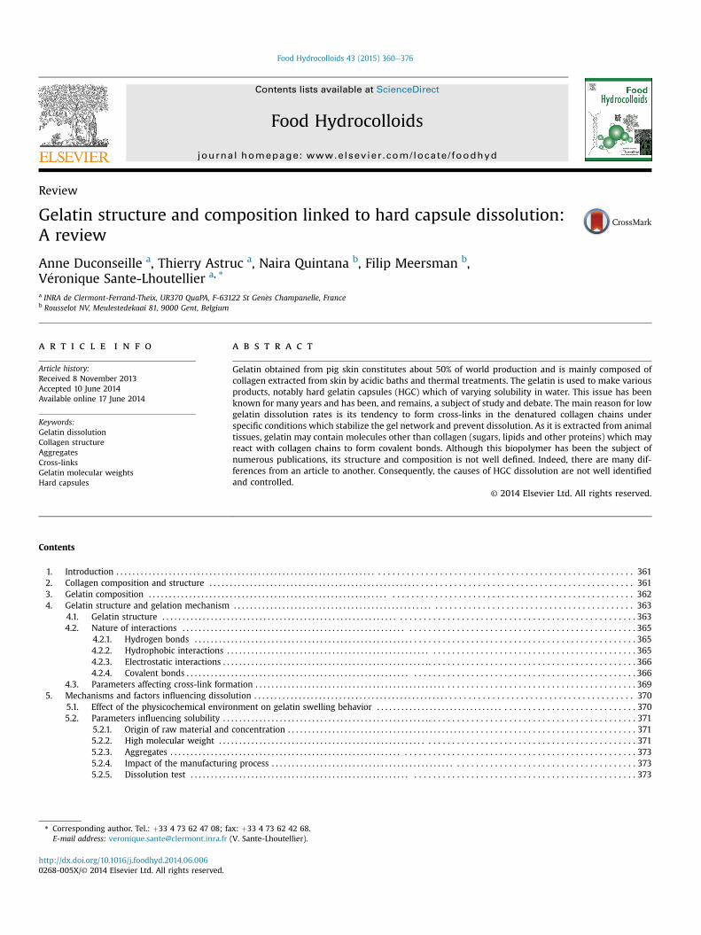
Collagens can have diverse supramolecular organizations,namely fibril-forming collagen (FFC), network-forming collagen(NFC), beaded filament-forming collagen (BFFC), membrane asso-ciated collagens with interrupted triple-helixes (MACIT), fibril-associated collagens with interrupted triple helixes (FACIT) andmultiplexins (MPC) (Chagnot, Listrat, Astruc,& Desvaux, 2012). Themost abundant molecular structure in skin is FFC while FACIT,MACIT, MPC and BFFC are present in lower proportions. Othermolecules like elastin, proteoglycans, laminin or fibronectin arelinked together, forming a network with collagens in the extracel-lular matrix (Fig. 1). In the case of fibrils forming collagen, thetriple-helix structures are linked together by covalent bonds toform a fibril that links to other fibrils to form a collagen fiber(Schrieber & Gareis, 2007). Collagen presents different cross-linkage levels as a function of the type of tissue and the age ofthe animals. Indeed, in dense tissues like bones, collagen is more
cross-linked than in loose tissues, making the matrix more rigid.Likewise, collagen is more cross-linked in old than in young ani-mals, reducing, for instance, skin elasticity (Eyre & Wu, 2005;Schrieber & Gareis, 2007; Shoulders & Raines, 2009).
3. Gelatin composition
As gelatin contains a majority of denatured collagens, its ami-noacid composition is close to that of collagen molecules. However,some variations remain due to the manufacturing process and themolecular organization of gelatin is very different from that ofnative collagen. The transformation of collagen to gelatin leads tochanges in the molecular composition of several aminoacids.Indeed, the alkaline process deaminates glutamine into glutamicacid and asparagine into aspartic acid. Thus the proportion ofaspartic acid and glutamic acid is higher in type B gelatin than intype A (Singh, Manikandan, Venugopal, Rama Rao, 2002; Taheri,bedian Kenari, Gildberg, & Behnam, 2009; Zhou & Regenstein,2006).
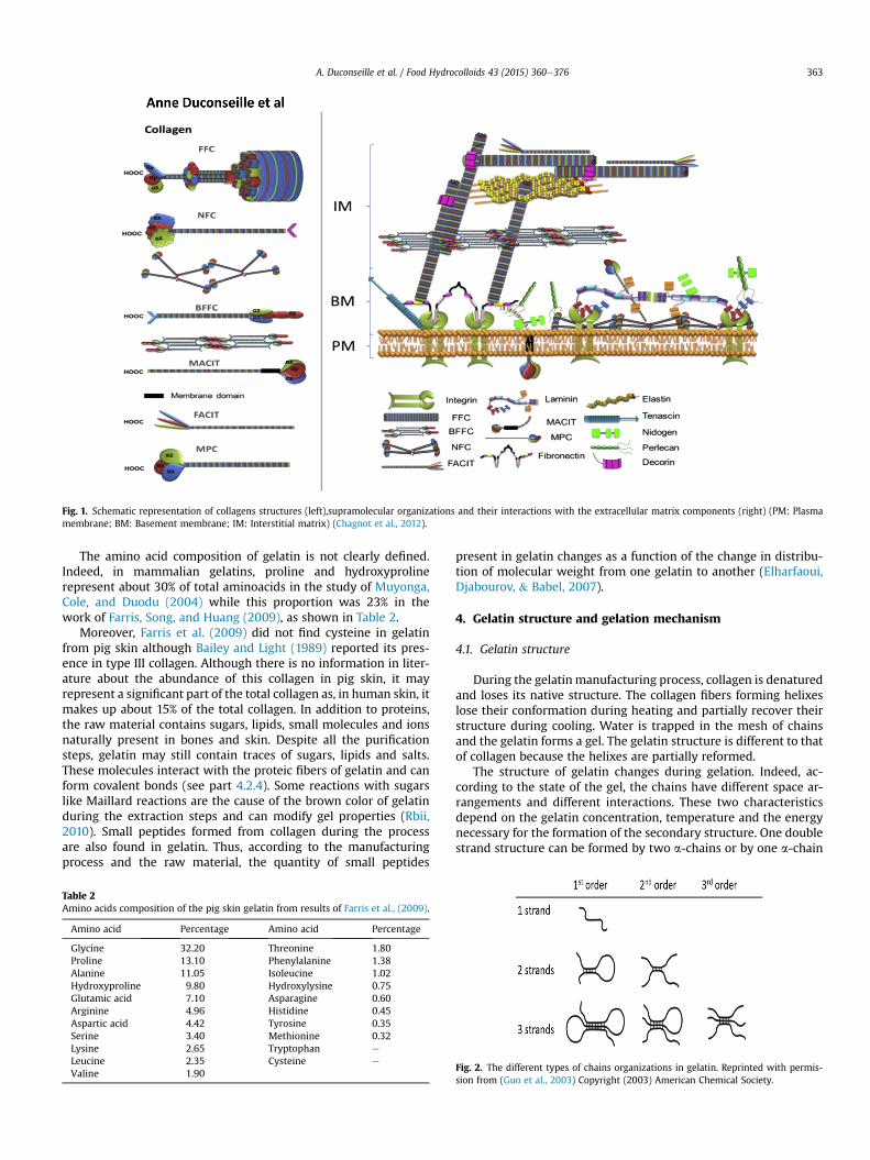
Fig. 1. Schematic representation of collagens structures (left),supramolecular organizations and their interactions with the extracellular matrix components (right) (PM: Plasmamembrane; BM: Basement membrane; IM: Interstitial matrix) (Chagnot et al., 2012).
A. Duconseille et al. / Food Hydrocolloids 43 (2015) 360e376 363
The amino acid composition of gelatin is not clearly defined.Indeed, in mammalian gelatins, proline and hydroxyprolinerepresent about 30% of total aminoacids in the study of Muyonga,Cole, and Duodu (2004) while this proportion was 23% in thework of Farris, Song, and Huang (2009), as shown in Table 2.
Moreover, Farris et al. (2009) did not find cysteine in gelatinfrom pig skin although Bailey and Light (1989) reported its pres-ence in type III collagen. Although there is no information in liter-ature about the abundance of this collagen in pig skin, it mayrepresent a significant part of the total collagen as, in human skin, itmakes up about 15% of the total collagen. In addition to proteins,the raw material contains sugars, lipids, small molecules and ionsnaturally present in bones and skin. Despite all the purificationsteps, gelatin may still contain traces of sugars, lipids and salts.These molecules interact with the proteic fibers of gelatin and canform covalent bonds (see part 4.2.4). Some reactions with sugarslike Maillard reactions are the cause of the brown color of gelatinduring the extraction steps and can modify gel properties (Rbii,2010). Small peptides formed from collagen during the processare also found in gelatin. Thus, according to the manufacturingprocess and the raw material, the quantity of small peptides
Table 2Amino acids composition of the pig skin gelatin from results of Farris et al., (2009).
Amino acid Percentage Amino acid Percentage
Glycine 32.20 Threonine 1.80Proline 13.10 Phenylalanine 1.38Alanine 11.05 Isoleucine 1.02Hydroxyproline 9.80 Hydroxylysine 0.75Glutamic acid 7.10 Asparagine 0.60Arginine 4.96 Histidine 0.45Aspartic acid 4.42 Tyrosine 0.35Serine 3.40 Methionine 0.32Lysine 2.65 Tryptophan e
Leucine 2.35 Cysteine e
Valine 1.90
present in gelatin changes as a function of the change in distribu-tion of molecular weight from one gelatin to another (Elharfaoui,Djabourov, & Babel, 2007).
4. Gelatin structure and gelation mechanism
4.1. Gelatin structure
During the gelatin manufacturing process, collagen is denaturedand loses its native structure. The collagen fibers forming helixeslose their conformation during heating and partially recover theirstructure during cooling. Water is trapped in the mesh of chainsand the gelatin forms a gel. The gelatin structure is different to thatof collagen because the helixes are partially reformed.
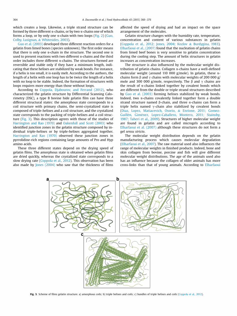
The structure of gelatin changes during gelation. Indeed, ac-cording to the state of the gel, the chains have different space ar-rangements and different interactions. These two characteristicsdepend on the gelatin concentration, temperature and the energynecessary for the formation of the secondary structure. One doublestrand structure can be formed by two a-chains or by one a-chain
Fig. 2. The different types of chains organizations in gelatin. Reprinted with permis-sion from (Guo et al., 2003) Copyright (2003) American Chemical Society.
A. Duconseille et al. / Food Hydrocolloids 43 (2015) 360e376364
which creates a loop. Likewise, a triple strand structure can beformed by three different a-chains, or by two a-chains one of whichforms a loop, or by only one a-chain with two loops (Fig. 2) (Guo,Colby, Lusignan, & Whitesides, 2003).
Guo et al. (2003) developed three different reaction orders for agelatin from limed bones (species unknown). The first order meansthat there is only one a-chain in the structure. The second one isused to present reactions with two different a-chains and the thirdorder includes three different a-chains. The structures formed arereversible and stable only if they have a minimum length, indi-cating that these helixes are stabilized by weak bonds. For instance,if a helix is too small, it is easily melt. According to the authors, thelength of a helix with one loop has to be twice the length of a helixwith no loop to be stable. Indeed, the formation of structures withloops requires more energy than those without loops.
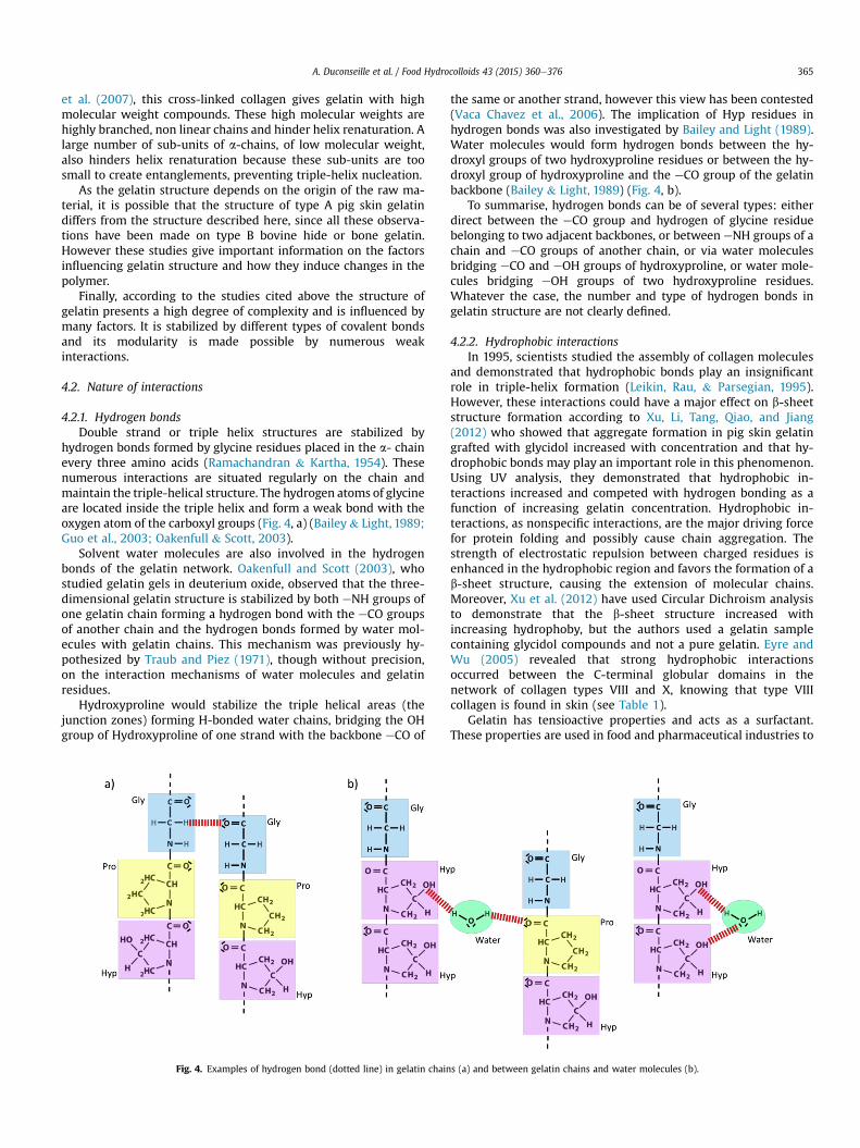
According to Coppola, Djabourov, and Ferrand (2012), whocharacterized the gelatin structure by Differential Scanning Calo-rimetry (DSC), a type B bovine hide gelatin film can have threedifferent structural states: the amorphous state corresponds to acoil structure with primary chains, the semi-crystalized state iscomposed of triple-helixes and a coil structure, and the crystalizedstate corresponds to the packing of triple-helixes and a coil struc-ture (Fig. 3). This description agrees with those of the studies ofHarrington and Rao (1970) and Oakenfull and Scott (2003) whoidentified junction zones in the gelatin structure composed by in-dividual triple-helixes or by triple-helixes aggregated together.Harrington and Rao (1970) observed these junction zones inpyrrolidine-rich regions containing large amounts of Pro and Hypamino acids.
These three different states depend on the drying speed ofgelatin films. The amorphous state is obtained when gelatin filmsare dried quickly, whereas the crystalized state corresponds to aslow drying rate (Coppola et al., 2012). This observation has beenalso made by Jones (2004) who saw that the thickness of films
Fig. 3. Scheme of films gelatin structure. a) amorphous coils; b) triple helixe
affected the speed of drying and had an impact on the spacearrangement of the molecules.
Gelatin structure changes with the humidity rate, temperature,concentration and content of various substances in gelatin(Coppola et al., 2012; Jones, 2004; Kozlov & Burdygina, 1983).Elharfaoui et al., (2007) found that the nucleation of gelatin chainsfrom limed beef bones is very sensitive to gelatin concentrationduring the cooling step. The amount of helix structures in gelatinincreases as concentration increases.
The structure is also influenced by the molecular weight dis-tribution of gelatin chains. Collagen a-chains have a well-definedmolecular weight (around 110 000 g/mole). In gelatin, these a-chains form b and g chains with molecular weights of 200 000 g/mole and 300 000 g/mole, respectively. The b and g chains arethe result of a-chains linked together by covalent bonds whichare different from the double or triple strand structures describedby Guo et al. (2003) forming helixes stabilized by weak bonds.Indeed, two a-chains covalently linked together form a doublestrand structure named b-chain, and three a-chains can form atriple helix named g-chain also stabilized by covalent bonds(Diaz, Lopez, Matiacevich, Osorio, & Enrione, 2011; Gomez-Guill�en, Gim�enez, Lopez-Caballero, Montero, 2011; Stainsby,1987; Taheri et al., 2009). Structures of higher molecular weightare found in gelatin and are called microgels according toElharfaoui et al. (2007) although these structures do not form agel sensu stricto.
The molecular weight distribution depends on the gelatinmanufacturing process which causes molecular degradation(Elharfaoui et al., 2007). The raw material used also influences therange of molecular weights in finished products. Indeed, bone andskin collagen from bovine, porcine and fish will give differentmolecular weight distributions. The age of the animals used alsohas an influence because the collagen of older animals has morecross-links than that of young animals. According to Elharfaoui
s and coils; c) bundles of triple helixes and coils (Coppola et al., 2012).
A. Duconseille et al. / Food Hydrocolloids 43 (2015) 360e376 365
et al. (2007), this cross-linked collagen gives gelatin with highmolecular weight compounds. These high molecular weights arehighly branched, non linear chains and hinder helix renaturation. Alarge number of sub-units of a-chains, of low molecular weight,also hinders helix renaturation because these sub-units are toosmall to create entanglements, preventing triple-helix nucleation.
As the gelatin structure depends on the origin of the raw ma-terial, it is possible that the structure of type A pig skin gelatindiffers from the structure described here, since all these observa-tions have been made on type B bovine hide or bone gelatin.However these studies give important information on the factorsinfluencing gelatin structure and how they induce changes in thepolymer.
Finally, according to the studies cited above the structure ofgelatin presents a high degree of complexity and is influenced bymany factors. It is stabilized by different types of covalent bondsand its modularity is made possible by numerous weakinteractions.
4.2. Nature of interactions
4.2.1. Hydrogen bondsDouble strand or triple helix structures are stabilized by
hydrogen bonds formed by glycine residues placed in the a- chainevery three amino acids (Ramachandran & Kartha, 1954). Thesenumerous interactions are situated regularly on the chain andmaintain the triple-helical structure. The hydrogen atoms of glycineare located inside the triple helix and form a weak bond with theoxygen atom of the carboxyl groups (Fig. 4, a) (Bailey& Light, 1989;Guo et al., 2003; Oakenfull & Scott, 2003).
Solvent water molecules are also involved in the hydrogenbonds of the gelatin network. Oakenfull and Scott (2003), whostudied gelatin gels in deuterium oxide, observed that the three-dimensional gelatin structure is stabilized by both eNH groups ofone gelatin chain forming a hydrogen bond with the eCO groupsof another chain and the hydrogen bonds formed by water mol-ecules with gelatin chains. This mechanism was previously hy-pothesized by Traub and Piez (1971), though without precision,on the interaction mechanisms of water molecules and gelatinresidues.
Hydroxyproline would stabilize the triple helical areas (thejunction zones) forming H-bonded water chains, bridging the OHgroup of Hydroxyproline of one strand with the backbone eCO of
Fig. 4. Examples of hydrogen bond (dotted line) in gelatin chain
the same or another strand, however this view has been contested(Vaca Chavez et al., 2006). The implication of Hyp residues inhydrogen bonds was also investigated by Bailey and Light (1989).Water molecules would form hydrogen bonds between the hy-droxyl groups of two hydroxyproline residues or between the hy-droxyl group of hydroxyproline and the eCO group of the gelatinbackbone (Bailey & Light, 1989) (Fig. 4, b).
To summarise, hydrogen bonds can be of several types: eitherdirect between the eCO group and hydrogen of glycine residuebelonging to two adjacent backbones, or between eNH groups of achain and eCO groups of another chain, or via water moleculesbridging eCO and eOH groups of hydroxyproline, or water mole-cules bridging eOH groups of two hydroxyproline residues.Whatever the case, the number and type of hydrogen bonds ingelatin structure are not clearly defined.
4.2.2. Hydrophobic interactionsIn 1995, scientists studied the assembly of collagen molecules
and demonstrated that hydrophobic bonds play an insignificantrole in triple-helix formation (Leikin, Rau, & Parsegian, 1995).However, these interactions could have a major effect on b-sheetstructure formation according to Xu, Li, Tang, Qiao, and Jiang(2012) who showed that aggregate formation in pig skin gelatingrafted with glycidol increased with concentration and that hy-drophobic bonds may play an important role in this phenomenon.Using UV analysis, they demonstrated that hydrophobic in-teractions increased and competed with hydrogen bonding as afunction of increasing gelatin concentration. Hydrophobic in-teractions, as nonspecific interactions, are the major driving forcefor protein folding and possibly cause chain aggregation. Thestrength of electrostatic repulsion between charged residues isenhanced in the hydrophobic region and favors the formation of ab-sheet structure, causing the extension of molecular chains.Moreover, Xu et al. (2012) have used Circular Dichroism analysisto demonstrate that the b-sheet structure increased withincreasing hydrophoby, but the authors used a gelatin samplecontaining glycidol compounds and not a pure gelatin. Eyre andWu (2005) revealed that strong hydrophobic interactionsoccurred between the C-terminal globular domains in thenetwork of collagen types VIII and X, knowing that type VIIIcollagen is found in skin (see Table 1).
Gelatin has tensioactive properties and acts as a surfactant.These properties are used in food and pharmaceutical industries to
s (a) and between gelatin chains and water molecules (b).
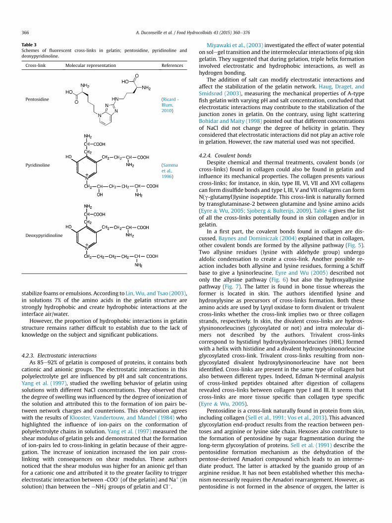
Table 3Schemes of fluorescent cross-links in gelatin; pentosidine, pyridinoline anddeoxypyridinoline.
Cross-link Molecular representation References
Pentosidine (Ricard -Blum,2010)
Pyridinoline (Sammaet al.,1996)
Deoxypyridinoline
A. Duconseille et al. / Food Hydrocolloids 43 (2015) 360e376366
stabilize foams or emulsions. According to Lin,Wu, and Tsao (2003),in solutions 7% of the amino acids in the gelatin structure arestrongly hydrophobic and create hydrophobic interactions at theinterface air/water.
However, the proportion of hydrophobic interactions in gelatinstructure remains rather difficult to establish due to the lack ofknowledge on the subject and significant publications.
4.2.3. Electrostatic interactionsAs 85e92% of gelatin is composed of proteins, it contains both
cationic and anionic groups. The electrostatic interactions in thispolyelectrolyte gel are influenced by pH and salt concentrations.Yang et al. (1997), studied the swelling behavior of gelatin usingsolutions with different NaCl concentrations. They observed thatthe degree of swelling was influenced by the degree of ionization ofthe solution and attributed this to the formation of ion pairs be-tween network charges and counterions. This observation agreeswith the results of Klooster, Vandertouw, and Mandel (1984) whohighlighted the influence of ion-pairs on the conformation ofpolyelectrolyte chains in solution. Yang et al. (1997) measured theshear modulus of gelatin gels and demonstrated that the formationof ion-pairs led to cross-linking in gelatin because of their aggre-gation. The increase of ionization increased the ion pair cross-linking with consequences on shear modulus. These authorsnoticed that the shear modulus was higher for an anionic gel thanfor a cationic one and attributed it to the greater facility to triggerelectrostatic interaction between -COO- (of the gelatin) and Naþ (insolution) than between the eNH3
þ groups of gelatin and Cl�.
Miyawaki et al., (2003) investigated the effect of water potentialon solegel transition and the intermolecular interactions of pig skingelatin. They suggested that during gelation, triple helix formationinvolved electrostatic and hydrophobic interactions, as well ashydrogen bonding.
The addition of salt can modify electrostatic interactions andaffect the stabilization of the gelatin network. Haug, Draget, andSmidsrød (2003), measuring the mechanical properties of A-typefish gelatin with varying pH and salt concentration, concluded thatelectrostatic interactions may contribute to the stabilization of thejunction zones in gelatin. On the contrary, using light scatteringBohidar and Maity (1998) pointed out that different concentrationsof NaCl did not change the degree of helicity in gelatin. Theyconsidered that electrostatic interactions did not play an active rolein gelation. However, the raw material used was not specified.
4.2.4. Covalent bondsDespite chemical and thermal treatments, covalent bonds (or
cross-links) found in collagen could also be found in gelatin andinfluence its mechanical properties. The collagen presents variouscross-links; for instance, in skin, type III, VI, VII and XVI collagenscan form disulfide bonds and type I, III, V and VII collagens can formN(g-glutamyl)lysine isopeptide. This cross-link is naturally formedby transglutaminase-2 between glutamine and lysine amino acids(Eyre & Wu, 2005; Sjoberg & Bulterijs, 2009). Table 4 gives the listof all the cross-links potentially found in skin collagen and/or ingelatin.
In a first part, the covalent bonds found in collagen are dis-cussed. Baynes and Dominiczak (2004) explained that in collagen,other covalent bonds are formed by the allysine pathway (Fig. 5).Two allysine residues (lysine with aldehyde group) undergoaldolic condensation to create a cross-link. Another possible re-action includes both allysine and lysine residues, forming a Schiffbase to give a lysinorleucine. Eyre and Wu (2005) described notonly the allysine pathway (Fig. 6) but also the hydroxyallysinepathway (Fig. 7). The latter is found in bone tissue whereas theformer is located in skin. The authors identified lysine andhydroxylysine as precursors of cross-links formation. Both theseamino acids are used by Lysyl oxidase to form divalent or trivalentcross-links whether the cross-link implies two or three collagenstrands, respectively. In skin, the divalent cross-links are hydrox-ylysinonorleucines (glycosylated or not) and intra molecular di-mers not described by the authors. Trivalent cross-linkscorrespond to hystidinyl hydroxylysinonorleucines (HHL) formedwith a helix with histidine and a divalent hydroxylysinonorleucineglycosylated cross-link. Trivalent cross-links resulting from non-glycosylated divalent hydroxylysinonorleucine have not beenidentified. Cross-links are present in the same type of collagen butalso between different types. Indeed, Edman N-terminal analysisof cross-linked peptides obtained after digestion of collagensrevealed cross-links between collagen type I and III. It seems thatcross-links are more tissue specific than collagen type specific(Eyre & Wu, 2005).
Pentosidine is a cross-link naturally found in protein from skin,including collagen (Sell et al., 1991; Vos et al., 2013). This advancedglycosylation end-product results from the reaction between pen-toses and arginine or lysine side chain. Hexoses also contribute tothe formation of pentosidine by sugar fragmentation during thelong-term glycosylation of proteins. Sell et al. (1991) describe thepentosidine formation mechanism as the dehydration of thepentose-derived Amadori compound which leads to an interme-diate product. The latter is attacked by the guanido group of anarginine residue. It has not been established whether this mecha-nism necessarily requires the Amadori rearrangement. However, aspentosidine is not formed in the absence of oxygen, the latter is
Table 4Cross-links found in collagen and/or gelatin from skin.
Cross-link Presence in collagen from skin Absence in collagenfrom skin
Presence in gelatinfrom skin
Formation process
Disulfide bonds (Bailey & Light, 1989) n/aa Probably: cleaved inalkaline conditions(Smithies, 1965)
Between cysteine residues
N(g-glutamyl)lysine peptide Potentially present in type IIIcollagen (Eyre & Wu, 2005)
n/a Transglutaminase
Aldol cross-link betweentwo lysines
(Baynes & Dominiczak, 2004) n/a n/a Lysyl oxidase pathway
Lysinonorleucine (Baynes & Dominiczak, 2004) n/a n/a Lysyl oxidase pathwayHydroxylysinonorleucine
(glycosylated or not)(Eyre & Wu, 2005) n/a No: cleaved in acidic
conditions (Eyre & Wu, 2005)Lysyl oxidase pathway
Histidino-hydroxylysinonorleucine(HHL)
(Eyre & Wu, 2005;Robins, 2007;Yamauchi, Woodley,& Mechanic, 1988)
n/a No: cleaved in acidicconditions (Eyre & Wu, 2005)
Age-related cross-link;Lysyl oxidase pathway
Pentosidine (Sell et al., 1991;Vos et al., 2013)
n/a Yes (Van den Bosch &Gielens, 2003)
Advanced glycation endproduct
� Lysyl e Pyridinoline ¼Deoxypyridinoline
� Hydroxylysyl e Pyridinoline ¼pyridinoline
(Moriguchi & Fujimoto, 1979;Ricard - Blum, Esterre,& Grimaud, 1993;Robins et al., 2003;Uriarte-Montoyaet al., 2011)
(Eyre & Wu, 2005;Ricard - Blum, 2010;Souberbielle, 2000;Yamauchi et al., 1988)
Yes (Uriarte-Montoyaet al., 2011; Vanden Bosch & Gielens, 2003)
Lysyl oxidase pathway
Desmosine (pyridinium ring) n/a (Baynes & Dominiczak,2004;Ma et al., 2003;Viglio et al., 2000)
Yes (Digenis et al., 1994) In collagen: Lysyl oxidase pathwayIn gelatin: Oxydation
Methylene bond n/a n/a Yes (Digenis et al., 1994) Reaction with aldehydesAminal n/a n/a Yes: formed in gelatin but
cleaved in acidic conditions(Digenis et al., 1994)
Reaction with aldehydes(pH close to 7)
Aminoglycoside bond(ketose sugar)
n/a n/a Yes (Digenis et al., 1994) Reaction with aldose sugars
� Lysyl Pyrrol� Hydroxylysyl Pyrrol
(Scott, Qian et al., 1983) (Eyre & Wu, 2005) n/a Lysyl oxidase pathway
Glucosepane (Monnier et al., 2013;Sjoberg & Bulterijs, 2009)
n/a n/a Age-related cross-link;Advanced glycation endproduct
Arginoline n/a (Eyre, Weis, & Wu, 2010) n/a Lysyl oxidase pathway
a n/a: no answer.
A. Duconseille et al. / Food Hydrocolloids 43 (2015) 360e376 367
necessary. Pentoses involved in pentosidine formation could beformed from sugars with more carbons by oxidative fragmentation.Smaller sugars such as trioses, tetroses, and ketoses could alsocontribute by condensation and/or reverse aldol reactions (Sellet al., 1991).
Pyridinoline and its reduced form (deoxypyridinoline) aretwo pyridinium ring-type cross-links (Robins et al., 2003)formed by the lysyl oxidase pathway (Ricard-Blum, 2010;Samma et al., 1996) (Table 3). These cross-links are naturallylocated in non-helical regions of collagen (called the telopep-tide region) at the extremity of the triple-helices (Souberbielle,2000). Pyridinoline and deoxypyridinoline are also referred inthe literature as hydroxylysyl-pyridinoline and lysyl-pyridinoline, respectively (Ricard-Blum, 2010; Samma et al.,1996). The existence of different names for the same cross-links complicates their inventory. Moreover, the presence ofpyridinoline and deoxypyridinoline in collagen from skin is stillcontroversial (Table 4).
In a second part the covalent bonds found in gelatin aredescribed (Table 4). As gelatin results from the collagen denatur-ation, some cross-links described above remain while other cova-lent bonds are formed due to chemical and environmentalconditions during and after the manufacturing process. Thesecross-links are favoured by high temperature and humidity but alsoby UV-light and chemical compounds like formaldehyde andreducing sugars (Singh, Rama Rao, Venugopal, & Manikandan,2002).
The different mechanisms assumed to be responsible forcross-links formation in gelatin have been described by Digenis,Gold, and Shah (1994). During oxidation, a lysine residue isdeaminated and its free amine function is replaced by an alde-hyde group. This group is then attacked by another free aminefunction of an adjacent lysine residue and an imine bond iscreated. Several aldol type reactions between the imine andother lysines give a desmosine-type cross-link (Fig. 8). Accordingto the authors, desmosine are pyridinium rings which aredifferent from the pyridinoline described by Ricard-Blum et al.(2010) although desmosines and pyridinolines come from thesame pathway, i.e. the allysine pathway. Indeed, in this pathway(in the extracellular matrix), the lysyl oxidase deaminates alysine residue to give an allysine. When several allysines areclose to each other, they interact to form a pyridinium ring(desmosine-type cross-link). However, so far desmosine was notfound in collagen while it was reported in elastin (Baynes &Dominiczak, 2004; Ma, Lieberman, Turino, & Lin, 2003; Viglioet al., 2000) .
Other cross-links have been found in gelatin and the underlyingmechanisms of their formation characterized. A free amine groupof a lysine residue may react with an aldehyde group. This reactiongives a hydroxymethylamine which yields a molecule of water tocreate a secondary aldimine. This imine reacts with another lysineresidue also changed into a hydroxymethylamine to givedimethylene ether. This compound undergoes rearrangements toform a methylene bond between two lysine residues (Fig. 9). The
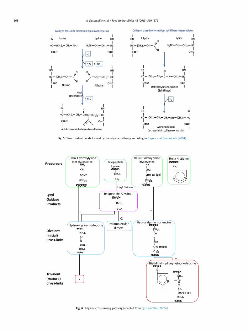
Fig. 6. Allysine cross-linking pathway (adapted from Eyre and Wu (2005)).
Fig. 5. Two covalent bonds formed by the allysine pathway according to Baynes and Dominiczak (2004).
A. Duconseille et al. / Food Hydrocolloids 43 (2015) 360e376368
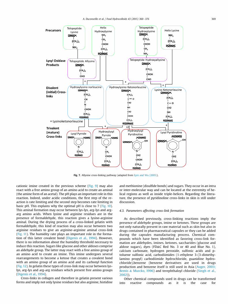
Fig. 7. Allysine cross-linking pathway (adapted from Eyre and Wu (2005)).
A. Duconseille et al. / Food Hydrocolloids 43 (2015) 360e376 369
cationic imine created in the previous scheme (Fig. 9) may alsoreact with a free amino group of an amino acid to create an aminal(the amine form of an acetal). The pH plays an important role in thisreaction. Indeed, under acidic conditions, the first step of the re-action is rate limiting and the second step becomes rate limiting inbasic pH. This explains why the optimal pH is close to 7 (Fig. 10).This aminal formation may occur between lys-lys, arg-lys and arg-arg amino acids. When lysine and arginine residues are in thepresence of formaldehyde, this reaction gives a lysine-arginineaminal. During the drying process of a cross-linked gelatin withformaldehyde, this kind of reaction may also occur between twoarginine residues to give an arginine-arginine aminal cross-link(Fig. 11). The humidity rate plays an important role in the forma-tion of this latter covalent bond (Digenis et al., 1994). However,there is no information about the humidity threshold necessary toinduce this reaction. Sugars like glucose and other aldoses comprisean aldehyde group. The latter may react with a free amino group ofan amino acid to create an imine. This imine undergoes severalrearrangements to become a ketose that creates a covalent bondwith an amino group of an amino acid and its carbonyl function(Fig. 12). In gelatin these types of cross-link may occur between lys-lys, arg-lys and arg-arg residues which present free amino groups(Digenis et al., 1994).
Cross-links in collagen and therefore in gelatin present variousforms and imply not only lysine residues but also arginine, histidine
andmethionine (disulfide bonds) and sugars. They occur in an intraor inter-molecular way and can be located at the extremity of he-lical regions as well as inside triple-helices. Regarding the litera-ture, the presence of pyridinoline cross-links in skin is still underdiscussion.
4.3. Parameters affecting cross-link formation
As described previously, cross-linking reactions imply thepresence of aldehyde groups, imine or ketones. These groups arenot only naturally present in raw material such as skin but also indrugs contained in pharmaceutical capsules or they can be addedduring the capsules manufacturing process. Chemical com-pounds which have been identified as favoring cross-link for-mation are aldehydes, imines, ketones, saccharides (glucose andaldose sugars), dyes (FD&C Red No. 3 or 40 and Blue No. 1),calcium carbonate, hydrogen peroxide, sulfonic acids and p-toluene sulfonic acid, carbodiimides (1-ethylene 3-(3-dimethy-lamino propyl) carbodiimide hydrochloride, guanidine hydro-chloride),benzene (benzene derivatives are used in drugsproduction and benzene itself is still used in Asia (Auger, 2003;Bemis & Murcko, 1996) and terephthaloyl chloride (Singh et al.,2002)).
Other chemical compounds used in drugs can be transformedinto reactive compounds as it is the case for
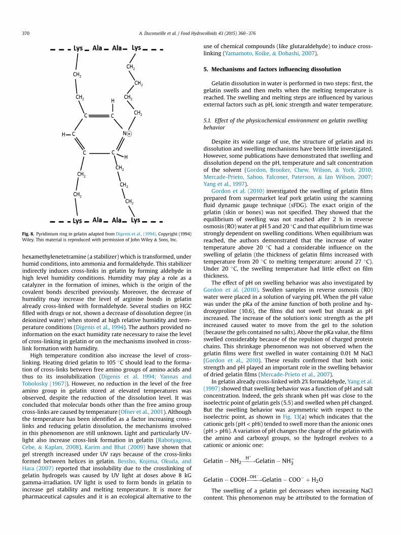
Fig. 8. Pyridinium ring in gelatin adapted from Digenis et al., (1994). Copyright (1994)Wiley. This material is reproduced with permission of John Wiley & Sons, Inc.
A. Duconseille et al. / Food Hydrocolloids 43 (2015) 360e376370
hexamethylenetetramine (a stabilizer) which is transformed, underhumid conditions, into ammonia and formaldehyde. This stabilizerindirectly induces cross-links in gelatin by forming aldehyde inhigh level humidity conditions. Humidity may play a role as acatalyzer in the formation of imines, which is the origin of thecovalent bonds described previously. Moreover, the decrease ofhumidity may increase the level of arginine bonds in gelatinalready cross-linked with formaldehyde. Several studies on HGCfilled with drugs or not, shown a decrease of dissolution degree (indeionized water) when stored at high relative humidity and tem-perature conditions (Digenis et al., 1994). The authors provided noinformation on the exact humidity rate necessary to raise the levelof cross-linking in gelatin or on the mechanisms involved in cross-link formation with humidity.
High temperature condition also increase the level of cross-linking. Heating dried gelatin to 105 �C should lead to the forma-tion of cross-links between free amino groups of amino acids andthus to its insolubilization (Digenis et al. 1994; Yannas andTobolosky (1967)). However, no reduction in the level of the freeamino group in gelatin stored at elevated temperatures wasobserved, despite the reduction of the dissolution level. It wasconcluded that molecular bonds other than the free amino groupcross-links are caused by temperature (Ofner et al., 2001). Althoughthe temperature has been identified as a factor increasing cross-links and reducing gelatin dissolution, the mechanisms involvedin this phenomenon are still unknown. Light and particularly UV-light also increase cross-link formation in gelatin (Rabotyagova,Cebe, & Kaplan, 2008). Karim and Bhat (2009) have shown thatgel strength increased under UV rays because of the cross-linksformed between helices in gelatin. Bessho, Kojima, Okuda, andHara (2007) reported that insolubility due to the crosslinking ofgelatin hydrogels was caused by UV light at doses above 8 kGgamma-irradiation. UV light is used to form bonds in gelatin toincrease gel stability and melting temperature. It is more forpharmaceutical capsules and it is an ecological alternative to the
use of chemical compounds (like glutaraldehyde) to induce cross-linking (Yamamoto, Koike, & Dobashi, 2007).
5. Mechanisms and factors influencing dissolution
Gelatin dissolution in water is performed in two steps: first, thegelatin swells and then melts when the melting temperature isreached. The swelling and melting steps are influenced by variousexternal factors such as pH, ionic strength and water temperature.
5.1. Effect of the physicochemical environment on gelatin swellingbehavior
Despite its wide range of use, the structure of gelatin and itsdissolution and swelling mechanisms have been little investigated.However, some publications have demonstrated that swelling anddissolution depend on the pH, temperature and salt concentrationof the solvent (Gordon, Brooker, Chew, Wilson, & York, 2010;Mercade-Prieto, Sahoo, Falconer, Paterson, & Ian Wilson, 2007;Yang et al., 1997).
Gordon et al. (2010) investigated the swelling of gelatin filmsprepared from supermarket leaf pork gelatin using the scanningfluid dynamic gauge technique (sFDG). The exact origin of thegelatin (skin or bones) was not specified. They showed that theequilibrium of swelling was not reached after 2 h in reverseosmosis (RO) water at pH 5 and 20 �C and that equilibrium timewasstrongly dependent on swelling conditions. When equilibriumwasreached, the authors demonstrated that the increase of watertemperature above 20 �C had a considerable influence on theswelling of gelatin (the thickness of gelatin films increased withtemperature from 20 �C to melting temperature: around 27 �C).Under 20 �C, the swelling temperature had little effect on filmthickness.
The effect of pH on swelling behavior was also investigated byGordon et al. (2010). Swollen samples in reverse osmosis (RO)water were placed in a solution of varying pH. When the pH valuewas under the pKa of the amine function of both proline and hy-droxyproline (10.6), the films did not swell but shrank as pHincreased. The increase of the solution's ionic strength as the pHincreased caused water to move from the gel to the solution(because the gels contained no salts). Above the pKa value, the filmsswelled considerably because of the repulsion of charged proteinchains. This shrinkage phenomenon was not observed when thegelatin films were first swelled in water containing 0.01 M NaCl(Gordon et al., 2010). These results confirmed that both ionicstrength and pH played an important role in the swelling behaviorof dried gelatin films (Mercade-Prieto et al., 2007).
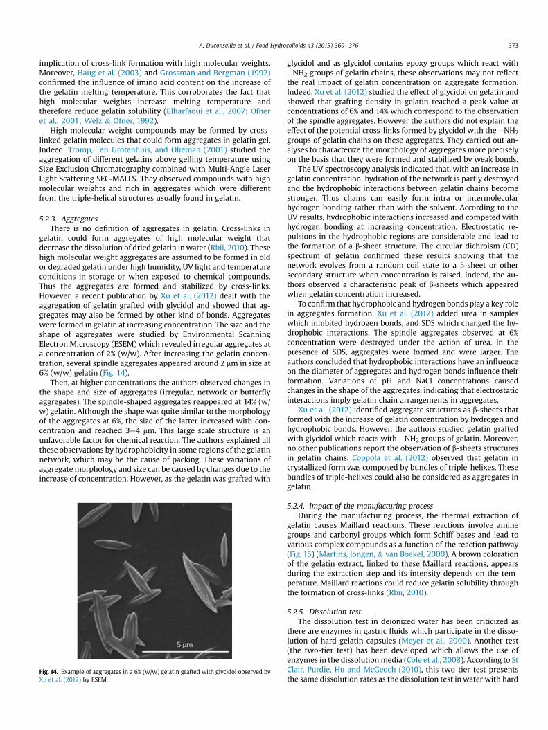
In gelatin already cross-linked with 2% formaldehyde, Yang et al.(1997) showed that swelling behavior was a function of pH and saltconcentration. Indeed, the gels shrank when pH was close to theisoelectric point of gelatin gels (5.5) and swelled when pH changed.But the swelling behavior was asymmetric with respect to theisoelectric point, as shown in Fig. 13(a) which indicates that thecationic gels (pH < pHi) tended to swell more than the anionic ones(pH > pHi). A variation of pH changes the charge of the gelatin withthe amino and carboxyl groups, so the hydrogel evolves to acationic or anionic one:
Gelatin�NH2���!HþGelatin�NHþ
3
Gelatin� COOH����!OH�Gelatin� COO� þ H2O
The swelling of a gelatin gel decreases when increasing NaClcontent. This phenomenon may be attributed to the formation of
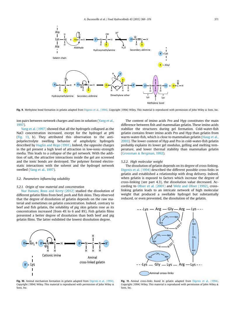
Fig. 9. Methylene bond formation in gelatin adapted from Digenis et al., (1994). Copyright (1994) Wiley. This material is reproduced with permission of John Wiley & Sons, Inc.
A. Duconseille et al. / Food Hydrocolloids 43 (2015) 360e376 371
ion pairs between network charges and ions in solution (Yang et al.,1997).
Yang et al. (1997) showed that all the hydrogels collapsed as theNaCl concentration increased, except for the hydrogel at pHi(Fig. 13, b). They attributed this observation to the anti-polyelectrolyte swelling behavior of ampholytic hydrogelsdescribed by Huglin and Rego (1991). Indeed, the opposite chargesin the gel present a high level of attraction in low-ionic-strengthmedia. This leads to a collapse of the gel network. With the addi-tion of salt, the attractive interactions inside the gel are screenedand the ionic bonds are destroyed. The polymer formed electro-static interactions with the solvent and the hydrogel networkswelled (Yang et al., 1997).
5.2. Parameters influencing solubility
5.2.1. Origin of raw material and concentrationNur Hanani, Roos and Kerry (2012) studied the dissolution of
different gelatin films from beef, pork and fish skins. They observedthat the degree of dissolution of gelatin depends on the raw ma-terial and sometimes on gelatin concentration. Indeed, contrary tobeef and fish gelatin, the solubility of pig skin gelatin rose as itsconcentration increased (from 4% to 6 and 8%). Fish gelatin filmspresented a better degree of dissolution than both beef and piggelatin films. The latter exhibited the lowest dissolution degree.
Fig. 10. Aminal mechanism formation in gelatin adapted from Digenis et al., (1994).Copyright (1994) Wiley. This material is reproduced with permission of John Wiley &Sons, Inc.
The content of imino acids Pro and Hyp constitutes the maindifference between fish and mammalian gelatin. These imino acidsstabilize the structures during gel formation. Cold-water-fishgelatin contains fewer imino acids Pro and Hyp than gelatin fromwarm-water-fish, which is close to mammalian gelatin (Haug et al.,2003). The lower content of Hyp and Pro in cold-water-fish gelatinprobably explains its lower gel modulus, gelling and melting tem-perature, and lower thermal stability than mammalian gelatin(Grossman & Bergman, 1992).
5.2.2. High molecular weightThe dissolution of gelatin depends on its degree of cross-linking.
Digenis et al. (1994) described the different possible cross-links ingelatin and established a relationship with drug delivery. Indeed,when gelatin is exposed to factors which increase the degree ofcross-linking (see part 4.3), the dissolution value decreased. Ac-cording to Ofner et al. (2001) and Welz and Ofner (1992), cross-linking gelatin leads to an intricate network of high molecularweight that produced a swellable hydrogel but substantiallyreduced, or even prevented, the dissolution of the gelatin.
Fig. 11. Aminal cross-links found in gelatin adapted from Digenis et al., (1994).Copyright (1994) Wiley. This material is reproduced with permission of John Wiley &Sons, Inc.
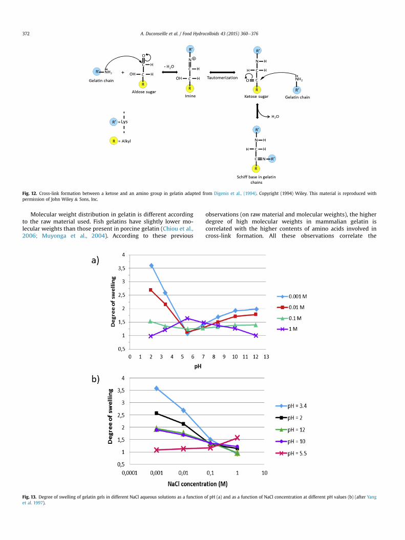
Fig. 12. Cross-link formation between a ketose and an amino group in gelatin adapted from Digenis et al., (1994). Copyright (1994) Wiley. This material is reproduced withpermission of John Wiley & Sons, Inc.
A. Duconseille et al. / Food Hydrocolloids 43 (2015) 360e376372
Molecular weight distribution in gelatin is different accordingto the raw material used. Fish gelatins have slightly lower mo-lecular weights than those present in porcine gelatin (Chiou et al.,2006; Muyonga et al., 2004). According to these previous
Fig. 13. Degree of swelling of gelatin gels in different NaCl aqueous solutions as a functionet al. 1997).
observations (on raw material and molecular weights), the higherdegree of high molecular weights in mammalian gelatin iscorrelated with the higher contents of amino acids involved incross-link formation. All these observations correlate the
of pH (a) and as a function of NaCl concentration at different pH values (b) (after Yang
A. Duconseille et al. / Food Hydrocolloids 43 (2015) 360e376 373
implication of cross-link formation with high molecular weights.Moreover, Haug et al. (2003) and Grossman and Bergman (1992)confirmed the influence of imino acid content on the increase ofthe gelatin melting temperature. This corroborates the fact thathigh molecular weights increase melting temperature andtherefore reduce gelatin solubility (Elharfaoui et al., 2007; Ofneret al., 2001; Welz & Ofner, 1992).
High molecular weight compounds may be formed by cross-linked gelatin molecules that could form aggregates in gelatin gel.Indeed, Tromp, Ten Grotenhuis, and Olieman (2001) studied theaggregation of different gelatins above gelling temperature usingSize Exclusion Chromatography combined with Multi-Angle LaserLight Scattering SEC-MALLS. They observed compounds with highmolecular weights and rich in aggregates which were differentfrom the triple-helical structures usually found in gelatin.
5.2.3. AggregatesThere is no definition of aggregates in gelatin. Cross-links in
gelatin could form aggregates of high molecular weight thatdecrease the dissolution of dried gelatin inwater (Rbii, 2010). Thesehigh molecular weight aggregates are assumed to be formed in oldor degraded gelatin under high humidity, UV light and temperatureconditions in storage or when exposed to chemical compounds.Thus the aggregates are formed and stabilized by cross-links.However, a recent publication by Xu et al. (2012) dealt with theaggregation of gelatin grafted with glycidol and showed that ag-gregates may also be formed by other kind of bonds. Aggregateswere formed in gelatin at increasing concentration. The size and theshape of aggregates were studied by Environmental ScanningElectron Microscopy (ESEM) which revealed irregular aggregates ata concentration of 2% (w/w). After increasing the gelatin concen-tration, several spindle aggregates appeared around 2 mm in size at6% (w/w) gelatin (Fig. 14).
Then, at higher concentrations the authors observed changes inthe shape and size of aggregates (irregular, network or butterflyaggregates). The spindle-shaped aggregates reappeared at 14% (w/w) gelatin. Although the shapewas quite similar to the morphologyof the aggregates at 6%, the size of the latter increased with con-centration and reached 3e4 mm. This large scale structure is anunfavorable factor for chemical reaction. The authors explained allthese observations by hydrophobicity in some regions of the gelatinnetwork, which may be the cause of packing. These variations ofaggregatemorphology and size can be caused by changes due to theincrease of concentration. However, as the gelatin was grafted with
Fig. 14. Example of aggregates in a 6% (w/w) gelatin grafted with glycidol observed byXu et al. (2012) by ESEM.
glycidol and as glycidol contains epoxy groups which react witheNH2 groups of gelatin chains, these observations may not reflectthe real impact of gelatin concentration on aggregate formation.Indeed, Xu et al. (2012) studied the effect of glycidol on gelatin andshowed that grafting density in gelatin reached a peak value atconcentrations of 6% and 14% which correspond to the observationof the spindle aggregates. However the authors did not explain theeffect of the potential cross-links formed by glycidol with theeNH2groups of gelatin chains on these aggregates. They carried out an-alyses to characterize the morphology of aggregates more preciselyon the basis that they were formed and stabilized by weak bonds.
The UV spectroscopy analysis indicated that, with an increase ingelatin concentration, hydration of the network is partly destroyedand the hydrophobic interactions between gelatin chains becomestronger. Thus chains can easily form intra or intermolecularhydrogen bonding rather than with the solvent. According to theUV results, hydrophobic interactions increased and competed withhydrogen bonding at increasing concentration. Electrostatic re-pulsions in the hydrophobic regions are considerable and lead tothe formation of a b-sheet structure. The circular dichroism (CD)spectrum of gelatin confirmed these results showing that thenetwork evolves from a random coil state to a b-sheet or othersecondary structure when concentration is raised. Indeed, the au-thors observed a characteristic peak of b-sheets which appearedwhen gelatin concentration increased.
To confirm that hydrophobic and hydrogen bonds play a key rolein aggregates formation, Xu et al. (2012) added urea in sampleswhich inhibited hydrogen bonds, and SDS which changed the hy-drophobic interactions. The spindle aggregates observed at 6%concentration were destroyed under the action of urea. In thepresence of SDS, aggregates were formed and were larger. Theauthors concluded that hydrophobic interactions have an influenceon the diameter of aggregates and hydrogen bonds influence theirformation. Variations of pH and NaCl concentrations causedchanges in the shape of the aggregates, indicating that electrostaticinteractions imply gelatin chain arrangements in aggregates.
Xu et al. (2012) identified aggregate structures as b-sheets thatformed with the increase of gelatin concentration by hydrogen andhydrophobic bonds. However, the authors studied gelatin graftedwith glycidol which reacts with eNH2 groups of gelatin. Moreover,no other publications report the observation of b-sheets structuresin gelatin chains. Coppola et al. (2012) observed that gelatin incrystallized formwas composed by bundles of triple-helixes. Thesebundles of triple-helixes could also be considered as aggregates ingelatin.
5.2.4. Impact of the manufacturing processDuring the manufacturing process, the thermal extraction of
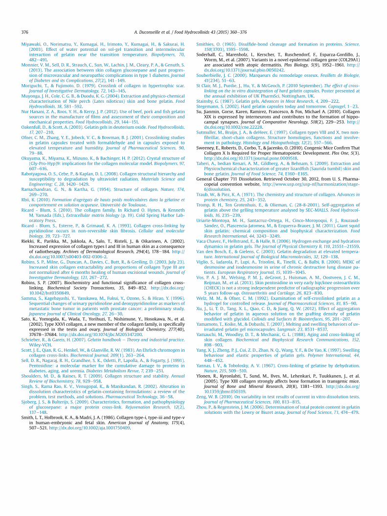
gelatin causes Maillard reactions. These reactions involve aminegroups and carbonyl groups which form Schiff bases and lead tovarious complex compounds as a function of the reaction pathway(Fig. 15) (Martins, Jongen, & van Boekel, 2000). A brown colorationof the gelatin extract, linked to these Maillard reactions, appearsduring the extraction step and its intensity depends on the tem-perature. Maillard reactions could reduce gelatin solubility throughthe formation of cross-links (Rbii, 2010).
5.2.5. Dissolution testThe dissolution test in deionized water has been criticized as
there are enzymes in gastric fluids which participate in the disso-lution of hard gelatin capsules (Meyer et al., 2000). Another test(the two-tier test) has been developed which allows the use ofenzymes in the dissolutionmedia (Cole et al., 2008). According to StClair, Purdie, Hu and McGeoch (2010), this two-tier test presentsthe same dissolution rates as the dissolution test inwater with hard
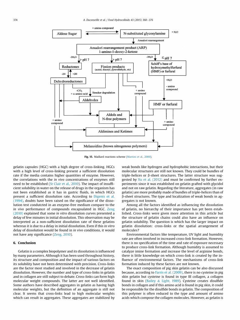
Fig. 15. Maillard reactions scheme (Martins et al., 2000).
A. Duconseille et al. / Food Hydrocolloids 43 (2015) 360e376374
gelatin capsules (HGC) with a high degree of cross-linking. HGCswith a high level of cross-linking present a sufficient dissolutionrate if the media contains higher quantities of enzyme. However,the correlations with the in vivo concentrations of enzymes stillneed to be established (St Clair et al., 2010). The impact of insuffi-cient solubility inwater on the release of drugs in the organism hasnot been established as it has in gastric fluids, in which HGCspresent a sufficient dissolution rate. According to Digenis et al.(1994), doubts have been raised on the significance of the disso-lution test condutcted in an enzyme-free medium compare to thein vivo performance of compounds encapsulated in HGC. Zeng(2010) explained that some in vitro dissolution curves presented adelay of few minutes in initial dissolution. This observation may beinterpreted as a non-sufficient dissolution rate of these gelatinswhereas it is due to a delay in initial dissolution. Even if this in vitrodelay of dissolution would be found in in vivo conditions, it wouldnot have any significance (Zeng, 2010).
6. Conclusion
Gelatin is a complex biopolymer and its dissolution is influencedbymany parameters. Although it has been used throughout history,its structure and composition and the impact of various factors onits solubility have not been determined with precision. Cross-linksare the factor most studied and involved in the decrease of gelatindissolution. However, the number and type of cross-links in gelatinand in collagen are still subject to debate. Cross-links can form highmolecular weight compounds. The latter are not well identified.Some authors have described aggregates in gelatin as having highmolecular weights, but the definition of an aggregate is still notclear. It seems that cross-links lead to high molecular weightswhich can result in aggregates. These aggregates are stabilized by
weak bonds like hydrogen and hydrophobic interactions, but theirmolecular structures are still not known. They could be bundles oftriple-helices or b-sheet structures. The latter structure was sug-gested by Xu et al. (2012) and must be confirmed by further ex-periments since it was established on gelatin grafted with glycidoland not on raw gelatin. Regarding the literature, aggregates (in rawgelatin) are more probably made of bundles of triple-helices than ofb-sheet structures. The type and localization of weak bonds in ag-gregates is not known.
Among all the factors identified as influencing the dissolutionof gelatin, no hierarchy of their importance has yet been estab-lished. Cross-links were given more attention in this article butthe structure of gelatin chains could also have an influence ongelatin solubility. The question is which has the larger impact ongelatin dissolution: cross-links or the spatial arrangement ofmolecules?
Environmental factors like temperature, UV light and humidityrate are often involved in increased cross-link formation. However,there is no specification of the time and rate of exposure necessaryto produce cross-link formation. Although humidity is assumed tocatalyze imine formation and increase the level of arginine bonds,there is little knowledge on which cross-link is created by the in-fluence of environmental factors. The mechanisms of cross-linkformation induced by these factors are not known.
The exact composition of pig skin gelatin can be also discussedbecause, according to Farris et al. (2009), there is no cysteine in pigskin gelatin but cysteine is found in type III collagen, a collagenfound in skin (Bailey & Light, 1989). Cysteine creates disulfidebonds in collagen and if this amino acid is found in pig skin, it couldbe responsible for the disulfide bonds in gelatin. The composition ofthis polymer is often reduced to the type and amount of aminoacids which compose the collagenmolecules. However, as gelatin is

A. Duconseille et al. / Food Hydrocolloids 43 (2015) 360e376 375
extracted from animal tissues by thermal treatments, sugars, lipidsand other proteins can also be found.
The issue of the dissolution of hard gelatin capsules is difficult tounderstand due to the high complexity of gelatin and the manyparameters identified as potentially involved in its solubility. It iseven more complex given the variability of the results obtained bycurrent in vitro dissolution tests.
In the future, an effort should be made to link structural andcompositional properties of gelatinwith its dissolution degree suchas dynamic light scattering to study the size of aggregates orspectroscopy techniques (fluorescence, infra-red or raman) to getnew insights in the chemical functions involved in the structuralproperties.
Acknowledgments
The authors acknowledge the financial support of Rousselot inthe framework of a CIFRE PhD contract (industrial researchagreements).
References
Al-Tabakha, M. M. (2010). HPMC capsules: current status and future prospects.Journal of Pharmacy and Pharmaceutical Sciences, 13, 428e442.
Auger, P. L. (2003). Benz�ene : Est-ce encore un probl�eme en Am�erique du Nord en ced�ebut de mill�enaire? Le M�edecin du Qu�ebec, 38(10), 111e113.
Bailey, A. J., & Light, N. D. (1989). Connective tissue in meat and meat products (19thed.).
Banyard, J., Bao, L., Hofer, M. D., Zurakowski, D., Spivey, K. A., Feldman, A. S., et al.(2007). Collagen XXIII expression is associated with prostate cancer recurrenceand distant metastases. Clinical Cancer Research, 13(9), 2634e2642. http://dx.doi.org/10.1178/1078-0432.ccr-06-2163.
Baynes, J., & Dominiczak, M. (2004). The extracellular matrix. In Medical biochem-istry (2nd ed) (p. 712). Elsevier ltd.
Bemis, G. W., & Murcko, M. A. (1996). The properties of known drugs. 1. Molecularframeworks. Journal of Medicinal Chemistry, 39(15), 2887e2893.
Bessho, M., Kojima, T., Okuda, S., & Hara, M. (2007). Radiation-induced cross-linkingof gelatin by using gamma-rays: insoluble gelatin hydrogel formation. Bulletinof the Chemical Society of Japan, 80, 979e985.
Bohidar, H. B., & Maity, S. (1998). Polarized light scattering study from gelatin so-lutions and gels. European polymer journal, 34, 1361e1370.
Brinckmann, J., Notbohm, H., & Müller, P. K. (2005). Collagen: Primer in structure.processing and assembly: Springer.
Bruckner, P. (2010). Suprastructures of extracellular matrices: paradigms of func-tions controlled by aggregates rather than molecules. Cell and Tissue Research,339, 7e18.
Brucknertuderman, L., Schnyder, U. W., Winterhalter, K. H., & Bruckner, P. (1987).Tissue form of type-vii collagen from human-skin and dermal fibroblasts inculture. European Journal of Biochemistry, 165(3), 607e611. http://dx.doi.org/10.1111/j.1432-1033.1987.tb11483.x.
Chagnot, C., Listrat, A., Astruc, T., & Desvaux, M.l. (2012). Bacterial adhesion to an-imal tissues: protein determinants for recognition of extracellular matrixcomponents. Cellular Microbiology, 14, 1687e1696.
Chiou, B. S., Vena-Bustillos, R. J., Shey, J., Yee, E., Bechtel, P. J., & Imam, S. H. (2006).Rheological and mechanical properties of cross-linked fish gelatins. Polymer, 47,6379e6386.
Chiwele, I., Jones, B. E., & Podczeck, F. (2000). The shell dissolution of various emptyhard capsules. Chemical and Pharmaceutical Bulletin, 48, 951e956.
Chou, M. Y., & Li, H. C. (2002). Genomic organization and characterization of thehuman type XXI collagen (COL21A1) gene. Genomics, 79(3), 395e401. http://dx.doi.org/10.1006/geno.2002.6712.
Cole, E. T., Cad, D., & Benameur, H. (2008). Challenges and opportunities in theencapsulation of liquid and semi-solid formulations into capsules for oraladministration. Advanced Drug Delivery Reviews, 60, 747e756.
Coppola, M., Djabourov, M., & Ferrand, M. (2012). Unified phase diagram of gelatinfilms plasticized by hydrogen bonded liquids. Polymer, 53, 1483e1493.
Diaz, P., Lopez, D., Matiacevich, S., Osorio, F., & Enrione, J. (2011). State diagram ofsalmon (Salmo salar) gelatin films. Journal of the Science of Food and Agriculture,91, 2558e2565.
Digenis, G. A., Gold, T. B., & Shah, V. P. (1994). Cross-linking of gelatin capsules andits relevance to their in vitro-in vivo performance. Journal of PharmaceuticalSciences, 83, 915e921.
Elharfaoui, N., Djabourov, M., & Babel, W. (2007). Molecular weight influence ongelatin gels: structure, enthalpy and rheology. Macromolecular Symposia, 256,149e157.
Eyre, D. R., & Wu, J. J. (2005). Collagen cross-links. Topics in Current Chemistry, 247,207e229.
Eyre, D. R., Weis, M. A., & Wu, J. J. (2010). Maturation of collagen ketoimine cross-links by an alternative mechanism to pyridinoline formation in cartilage.Journal of Biological Chemistry, 285(22), 16675e16682. http://dx.doi.org/10.1074/jbc.M110.111534.
Farris, S., Song, J., & Huang, Q. (2009). Alternative reaction mechanism for the cross-linking of gelatin with glutaraldehyde. Journal of Agricultural and food chemistry,58, 998e1003.
Fox, M. A. (2008). Novel roles for collagens in wiring the vertebrate nervous system.Current Opinion in Cell Biology, 20(5), 508e513. http://dx.doi.org/10.1016/j.ceb.2008.05.003.
G�omez-Guill�en, M. C., Gim�enez, B., L�opez-Caballero, M. E., & Montero, M. P. (2011).Functional and bioactive properties of collagen and gelatin from alternativesources: a review. Food Hydrocolloids, 25, 1813e1827.
G�omez-Guill�en, M. C., P�erez-Mateos, M., G�omez-Estaca, J., L�opez-Caballero, E.,Gim�enez, B., & Montero, P. (2009). Fish gelatin: a renewable material for devel-oping active biodegradable films. Trends in Food Science and Technology, 20, 3e16.
Gordon, P. W., Brooker, A. D. M., Chew, Y. M. J., Wilson, D. I., & York, D. W. (2010).Studies into the swelling of gelatine films using a scanning fluid dynamic gauge.Food and Bioproducts Processing, 88, 357e364.
Grimal, S., Puech, S., Wagener, R., Venteo, S., Carroll, P., & Fichard-Carroll, A. (2010).Collagen XXVIII is a distinctive component of the peripheral nervous systemnodes of Ranvier and surrounds nonmyelinating glial cells. Glia, 58(16),1977e1987. http://dx.doi.org/10.1002/glia.21066.
Grossman, S., & Bergman, M. (1992). Process for the production of gelatin from fishskins (Rep. No. US patent 5,093,474).
Guo, L., Colby, R. H., Lusignan, C. P., & Whitesides, T. H. (2003). Kinetics of triple helixformation in semidilute gelatin solutions. Macromolecules, 36, 9999e10008.
Harrington, W. F., & Rao, N. V. (1970). Collagen structure in solution. I. Kinetics ofhelix regeneration in single-chain gelatins. Biochemistry, 9, 3714e3724.
Has, C., & Kern, J. S. (2010). Collagen XVII. Dermatologic Clinics, 28(1). http://dx.doi.org/10.1016/j.det.2009.10.007, 61-þ.
Hashimoto, T., Wakabayashi, T., Watanabe, A., Kowa, H., Hosoda, R., Nakamura, A.,et al. (2002). CLAC: a novel Alzheimer amyloid plaque component derived froma transmembrane precursor, CLAC-P/collagen type XXV. Embo Journal, 21(7),1524e1534. http://dx.doi.org/10.1093/emboj/21.7.1524.
Haug, I. J., Draget, K. I., & Smidsrød, O. (2003). Physical and rheological properties offish gelatin compared to mammalian gelatin. Food Hydrocolloids, 18, 203e213.
Hjorten, R., Hansen, U., Underwood, R. A., Telfer, H. E., Fernandes, R. J., Krakow, D.,et al. (2007). Type XXVII collagen at the transition of cartilage to boneduring skeletogenesis. Bone, 41(4), 535e542. http://dx.doi.org/10.1016/j.bone.2007.06.024.
Hofman, K., Hall, B., Cleaver, H., & Marshall, S. (2011). High-throughput quantifi-cation of hydroxyproline for determination of collagen. Analytical Biochemistry,417, 289e291.
Huglin, M. B., & Rego, J. M. (1991). Influence of a salt on some properties of hy-drophilic methacrylate hydrogels. Macromolecules, 24, 2556e2563.
Jones, R. T. (2004). Gelatin: manufacture and physico-chemical properties. InF.Podczeck, & B. E. Jones (Eds.), Pharmaceutical capsules (2nd ed) (pp. 23e59).Pharmaceutical Press.
Karim, A. A., & Bhat, R. (2009). Fish gelatin: properties, challenges, and prospects asan alternative to mammalian gelatins. Food Hydrocolloids, 23, 563e576.
Khalil, S. A. H., Ali, L. M. M., & Abdel Khalek, M. M. (1974). Effects of ageing andrelative humidity on drug release. I. Chloramphenicol capsules. Pharmazie, 29,36e37.
Klooster, N. T. M., Vandertouw, F., & Mandel, M. (1984). Solvent effects in poly-electrolyte solutions .1. Potentiometric and viscosimetric titration of poly(-acrylic acid) in methanol and counterion specificity. Macromolecules, 17,2070e2078.
Koch, M., Schulze, J., Hansen, U., Ashwodt, T., Keene, D. R., Brunken, W. J.,Burgeson, R. E., Bruckner, P., et al. (2004). A novel marker of tissue junctions,collagen XXII. Journal of Biological Chemistry, 279(21), 22514e22521. http://dx.doi.org/10.1074/jbc.M400536200.
Kozlov, P. V., & Burdygina, G. I. (1983). The structure and properties of solid gelatinand the principles of their modification. Polymer, 24, 651e666.
Leikin, S., Rau, D. C., & Parsegian, V. A. (1995). Temperature-favoured assembly ofcollagen is driven by hydrophilic not hydrophobic interactions. Nature Struc-tural and Molecular Biology, 2, 205e210.
Lin, S. H., Wu, T. F., & Tsao, H. K. (2003). Interfacial dynamics of a gelatin solutionwith surfactant. Macromolecules, 36, 8786e8795.
Ma, S., Lieberman, S., Turino, G. M., & Lin, Y. Y. (2003). The detection and quanti-tation of free desmosine and isodesmosine in human urine and their peptide-bound forms in sputum. Proceedings of the National Academy of Sciences of theUnited States of America, 100, 12941e12943.
Martel-Pelletier, J., Boileau, C., Pelletier, J.-P., & Roughley, P. J. (2008). Cartilage innormal and osteoarthritis conditions. Best Practice & Research in Clinical Rheu-matology, 22(2), 351e384. http://dx.doi.org/10.1016/j.berh.2008.02.001.
Martins, S. I. F. S., Jongen, W. M. F., & van Boekel, M. A. J. S. (2000). A review ofMaillard reaction in food and implications to kinetic modelling. Trends in FoodScience & Technology, 11, 364e373.
Mercade-Prieto, R., Sahoo, P. K., Falconer, R. J., Paterson, W. R., & Ian Wilson, D.(2007). Polyelectrolyte screening effects on the dissolution of whey protein gelsat high pH conditions. Food Hydrocolloids, 21, 1275e1284.
Meyer, Straughn, Hussain, Mhatre, Bottom, Shah, et al. (2000). The effect of gelatincross-linking on the bioequivalence of hard and soft gelatin acetaminophencapsules. Pharmaceutical Research, 17, 962e966.
A. Duconseille et al. / Food Hydrocolloids 43 (2015) 360e376376
Miyawaki, O., Norimatsu, Y., Kumagai, H., Irimoto, Y., Kumagai, H., & Sakurai, H.(2003). Effect of water potential on sol-gel transition and intermolecularinteraction of gelatin near the transition temperature. Biopolymers, 70,482e491.
Monnier, V. M., Sell, D. R., Strauch, C., Sun, W., Lachin, J. M., Cleary, P. A., & Genuth, S.(2013). The association between skin collagen glucosepane and past progres-sion of microvascular and neuropathic complications in type 1 diabetes. Journalof Diabetes and its Complications, 27(2), 141e149.
Moriguchi, T., & Fujimoto, D. (1979). Crosslink of collagen in hypertrophic scar.Journal of Investigative Dermatology, 72, 143e145.
Muyonga, J. H., Cole, C. G. B., & Duodu, K. G. (2004). Extraction and physico-chemicalcharacterisation of Nile perch (Lates niloticus) skin and bone gelatin. FoodHydrocolloids, 18, 581e592.
Nur Hanani, Z. A., Roos, Y. H., & Kerry, J. P. (2012). Use of beef, pork and fish gelatinsources in the manufacture of films and assessment of their composition andmechanical properties. Food Hydrocolloids, 29, 144e151.
Oakenfull, D., & Scott, A. (2003). Gelatin gels in deuterium oxide. Food Hydrocolloids,17, 207e210.
Ofner, C. M., Zhang, Y. E., Jobeck, V. C., & Bowman, B. J. (2001). Crosslinking studiesin gelatin capsules treated with formaldehyde and in capsules exposed toelevated temperature and humidity. Journal of Pharmaceutical Sciences, 90,79e88.
Okuyama, K., Miyama, K., Mizuno, K., & Bachinger, H. P. (2012). Crystal structure of(Gly-Pro-Hyp)9: implications for the collagen molecular model. Biopolymers, 97,607e616.
Rabotyagova, O. S., Cebe, P., & Kaplan, D. L. (2008). Collagen structural hierarchy andsusceptibility to degradation by ultraviolet radiation. Materials Science andEngineering: C, 28, 1420e1429.
Ramachandran, G. N., & Kartha, G. (1954). Structure of collagen. Nature, 174,269e270.
Rbii, K. (2010). Formation d'agr�egats de hauts poids mol�eculaires dans la g�elatine etcomportement en solution acqueuse. Universit�e de Toulouse.
Ricard - Blum, S. (2010). The collagen family. In Richard O. Hynes, & KennethM. Yamada (Eds.), Extracellular matrix biology (p. 19). Cold Spring Harbor Lab-oratory Press.
Ricard - Blum, S., Esterre, P., & Grimaud, K. A. (1993). Collagen cross-linking bypyridinoline occurs in non-reversible skin fibrosis. Cellular and molecularbiology, 39, 723e727.
Riekki, R., Parikka, M., Jukkola, A., Salo, T., Risteli, J., & Oikarinen, A. (2002).Increased expression of collagen types I and III in human skin as a consequenceof radiotherapy. Archives of Dermatological Research, 294(4), 178e184. http://dx.doi.org/10.1007/s00403-002-0306-2.
Robins, S. P., Milne, G., Duncan, A., Davies, C., Butt, R., & Greiling, D. (2003, July 23).Increased skin collagen extractability and proportions of collagen Type III arenot normalized after 6 months healing of human excisional wounds. Journal ofInvestigative Dermatology, 121, 267e272.
Robins, S. P. (2007). Biochemistry and functional significance of collagen cross-linking. Biochemical Society Transactions, 35, 849e852. http://dx.doi.org/10.1042/bst0350849.
Samma, S., Kagebayashi, Y., Yasukawa, M., Fukui, Y., Ozono, S., & Hirao, Y. (1996).Sequential changes of urinary pyridinoline and deoxypyridinoline as markers ofmetastatic bone tumor in patients with prostate cancer: a preliminary study.Japanese Journal of Clinical Oncology, 27, 26e30.
Sato, K., Yomogida, K., Wada, T., Yorihuzi, T., Nishimune, Y., Hosokawa, N., et al.(2002). Type XXVI collagen, a newmember of the collagen family, is specificallyexpressed in the testis and ovary. Journal of Biological Chemistry, 277(40),37678e37684. http://dx.doi.org/10.1074/jbc.M205347200.
Schrieber, R., & Gareis, H. (2007). Gelatin handbook e Theory and industrial practice.Wiley-VCH.
Scott, J. E., Qian, R. G., Henkel, W., & Glanville, R. W. (1983). An Ehrlich chromogen incollagen cross-links. Biochemical Journal, 209(1), 263e264.
Sell, D. R., Nagaraj, R. H., Grandhee, S. K., Odetti, P., Lapolla, A., & Fogarty, J. (1991).Pentosidine: a molecular marker for the cumulative damage to proteins indiabetes, aging, and uremia. Diabetes Metabolism Revue, 7, 239e251.
Shoulders, M. D., & Raines, R. T. (2009). Collagen structure and stability. AnnualReview of Biochemistry, 78, 929e958.
Singh, S., Rama Rao, K. V., Venugopal, K., & Manikandan, R. (2002). Alteration indissolution characteristics of gelatin-containing formulations: a review of theproblem, test methods, and solutions. Pharmaceutical Technology, 36e58.
Sjoberg, J. S., & Bulterijs, S. (2009). Characteristics, formation, and pathophysiologyof glucosepane: a major protein cross-link. Rejuvenation Research, 12(2),137e148.
Smith, L. T., Holbrook, K. A., & Madri, J. A. (1986). Collagen type-i, type-iii and type-vin human-embryonic and fetal skin. American Journal of Anatomy, 175(4),507e521. http://dx.doi.org/10.1002/aja.1001750409.
Smithies, O. (1965). Disulfide-bond cleavage and formation in proteins. Science,150(3703), 1595e1598.
Soderhall, C., Marenholz, I., Kerscher, T., Ruschendorf, F., Esparza-Gordillo, J.,Worm, M., et al. (2007). Variants in a novel epidermal collagen gene (COL29A1)are associated with atopic dermatitis. Plos Biology, 5(9), 1952e1961. http://dx.doi.org/10.1371/journal.pbio.0050242.
Souberbielle, J. C. (2000). Marqueurs du remodelage osseux. Feuillets de Biologie,41(234), 51e63.
St Clair, M. J., Purdie, J., Hu, Y., & McGeoch, P. (2010 September). The effect of cross-linking on the in vitro disintegration of hard gelatin capsules. Poster presented atthe annual exhibition of UK PharmSci, Nottingham, UK.
Stainsby, G. (1987). Gelatin gels. Advances in Meat Research, 4, 209e222.Stegemann, S. (2002). Hard gelatin capsules today and tomorrow. Capsugel, 1e23.Su, Jianmin, Gorse, Karen, Ramirez, Francesco, & Fox, Michael A. (2010). Collagen
XIX is expressed by interneurons and contributes to the formation of hippo-campal synapses. Journal of Comparative Neurology, 518(2), 229e253. http://dx.doi.org/10.1002/cne.22228.
Sutmuller, M., Bruijn, J. A., & deHeer, E. (1997). Collagen types VIII and X, two non-fibrillar, short-chain collagens. Structure homologies, functions and involve-ment in pathology. Histology and Histopathology, 12(2), 557e566.
Sweeney, E., Roberts, D., Corbo, T., & Jacenko, O. (2010). Congenic Mice Confirm ThatCollagen X Is Required for Proper Hematopoietic Development. Plos One, 5(3).http://dx.doi.org/10.1371/journal.pone.0009518.
Taheri, A., bedian Kenari, A. M., Gildberg, A., & Behnam, S. (2009). Extraction andPhysicochemical characterization of greater lizardfish (Saurida tumbil) skin andbone gelatin. Journal of Food Science, 74, E160eE165.
General Chapter 711 Dissolution. Retrieved October 30, 2012, from U. S. Pharma-copeial convention website, http://www.usp.org/usp-nf/harmonization/stage-6/dissolution.
Traub, W., & Piez, K. A. (1971). The chemistry and structure of collagen. Advances inprotein chemistry, 25, 243e352.
Tromp, R. H., Ten Grotenhuis, E., & Olieman, C. (28-8-2001). Self-aggregation ofgelatin above the gelling temperature analysed by SEC-MALLS. Food Hydrocol-loids, 16, 235e239.
Uriarte-Montoya, M. H., Santacruz-Ortega, H., Cinco-Moroyoqui, F. J., Rouzaud-S�andez, O., Plascencia-Jatomea, M., & Ezquerra-Brauer, J. M. (2011). Giant squidskin gelatin: chemical composition and biophysical characterization. FoodResearch International, 44, 3243e3249.
Vaca Chavez, F., Hellstrand, E., & Halle, B. (2006). Hydrogen exchange and hydrationdynamics in gelatin gels. The Journal of Physical Chemistry B, 110, 21551e21559.
Van den Bosch, E., & Gielens, C. (2003). Gelatin degradation at elevated tempera-ture. International Journal of Biological Macromolecules, 32, 129e138.
Viglio, S., Iadarola, P., Lupi, A., Trisolini, R., Tinelli, C., & Balbi, B. (2000). MEKC ofdesmosine and isodesmosine in urine of chronic destructive lung disease pa-tients. European Respiratory Journal, 15, 1039e1045.
Vos, P. A. J. M., Welsing, P. M. J., deGroot, J., Huisman, A. M., Oostveen, J. C. M.,Reijman, M., et al. (2013). Skin pentosidine in very early hip/knee osteoarthritis(CHECK) is not a strong independent predictor of radiographic progression over5 years follow-up. Osteoarthritis and Cartilage, 26, 823e830.
Welz, M. M., & Ofner, C. M. (1992). Examination of self-crosslinked gelatin as ahydrogel for controlled release. Journal of Pharmaceutical Sciences, 81, 85e90.
Xu, J., Li, T. D., Tang, X. L., Qiao, C. D., & Jiang, Q. W. (2012). Effect of aggregationbehavior of gelatin in aqueous solution on the grafting density of gelatinmodified with glycidol. Colloids and Surfaces B: Biointerfaces, 95, 201e207.
Yamamoto, T., Koike, M., & Dobashi, T. (2007). Melting and swelling behaviors of uv-irradiated gelatin gel microcapsules. Langmuir, 23, 8531e8537.
Yamauchi, M., Woodley, D. T., & Mechanic, G. L. (1988). Aging and cross-linking ofskin collagen. Biochemical and Biophysical Research Communications, 152,898e903.
Yang, X. J., Zheng, P. J., Cui, Z. D., Zhao, N. Q., Wang, Y. F., & De Yao, K. (1997). Swellingbehaviour and elastic properties of gelatin gels. Polymer International, 44,448e452.
Yannas, I. V., & Tobolosky, A. V. (1967). Cross-linking of gelatine by dehydration.Nature, 215, 509e510.
Ylonen, R., Kyronlahti, T., Sund, M., Ilves, M., Lehenkari, P., Tuukkanen, J., et al.(2005). Type XIII collagen strongly affects bone formation in transgenic mice.Journal of Bone and Mineral Research, 20(8), 1381e1393. http://dx.doi.org/10.1359/jbmr.050319.
Zeng, W. B. (2010). On variability in test results of current in vitro dissolution tests.Journal of Pharmaceutical Sciences, 100, 813e815.
Zhou, P., & Regenstein, J. M. (2006). Determination of total protein content in gelatinsolutions with the Lowry or Biuret assay. Journal of Food Science, 71, 474e479.