Embed Size (px)
Citation preview
Evidence-based Series #4-9: Section 1
Follow-up after Primary Therapy for Endometrial Cancer: A Clinical Practice Guideline
M. Fung-Kee-Fung, J. Dodge, L. Elit, H. Lukka, A. Chambers, T. Oliver,
and the Gynecology Cancer Disease Site Group
A Quality Initiative of the Program in Evidence-Based Care, Cancer Care Ontario.
Developed by the Provincial Gynecology Cancer Disease Site Group
Report Date: January 10, 2006 Question
What is the most appropriate strategy for the follow-up of patients with endometrial cancer who are clinically disease free after receiving potentially curative primary treatment? Specifically, do differences in follow-up intervals, diagnostic interventions, clinical setting, or specialty influence patient outcomes related to local or distant recurrence, survival, or quality of life? Target Population
Women without evidence of disease after primary potentially curative treatment for any stage of endometrial cancer comprise the target population. Of particular interest are outcomes from follow-up strategies reported for patients at a lower risk of recurrence (i.e., stage IA or IB, grade 1 or 2) and those at a higher risk of recurrence (i.e., stage IA or IB, grade 3, or stage IC or advanced stage). Recommendations
There is a lack of randomized controlled trial evidence related to the clinical questions. Based on the interpretation of evidence from retrospective studies and expert consensus opinion, the Gynecology Cancer Disease Site Group recommends the following: • It is recommended that all patients receive counselling about the potential symptoms of
recurrence of endometrial cancer, because the majority of recurrences in the identified studies were symptomatic. ▪ Symptomatic signs of possible recurrence can include, but are not limited to,
unexplained vaginal bleeding or discharge, detection of a mass, abdominal distension, persistent pain, especially in the abdomen or pelvic region, fatigue, diarrhea, nausea or vomiting, persistent cough, swelling, or weight loss.
PRACTICE GUIDELINE – page 1
• indi▪
The most appropriate follow-up strategy is likely one based upon the risk of recurrence, with vidual patient preferences for more or less follow-up taken into account. For patients at a surgically or pathologically confirmed low risk of recurrence (i.e., stage IA or IB, grade 1 or 2): A general examination, including a complete history and a pelvic-rectal examination, conducted semi-annually or annually for the first three years and annually for the next two years.
▪ For patients at high risk of recurrence (i.e., stage IA or IB, grade 3, or stage IC or advanced stage). A general examination, including a complete history and a pelvic-rectal examination, every three to six months for the first three years and semi-annually for the next two years.
• Since the majority of patients with recurrence were symptomatic and virtually all recurred within five years, it seems reasonable that patients return to annual population-based general physical and pelvic examination after five years of recurrence-free follow-up. There is insufficient evidence to inform the optimum clinical setting or type of specialist required for follow-up; however, it is recommended that all patients be followed by a health
•
• fects associated with
can include complications with the rectum, urinary bladder, vagina, skin,
Ke
•
•
physical
▪ reported 0% to 4% of recurrences were detected by Pap smear,
care professional who is knowledgeable about the natural history of the disease, and who is comfortable performing speculum and pelvic exams, in order to diagnose or detect a local (vaginal) recurrence. ▪ If a patient is initially followed by a specialist, it seems reasonable that they be followed
by a qualified general practitioner after three to five years of recurrence-free follow-up. • It is recommended that all patients undergo a targeted investigation to rule out recurrence if
symptomatic, since patients with local recurrence are potentially curable with further therapy. • There is insufficient evidence to inform the routine use of Pap smear, chest x-ray, abdominal
ultrasound, computed tomography (CT) scan or CA 125 testing to detect asymptomatic recurrences. Where treatment with radiotherapy is involved, it is recommended that patients be counselled on the potential adverse effects of radiotherapy. Adverse efradiotherapysubcutaneous tissue, bones, and other sites.
y Evidence • Sixteen non-comparative retrospective studies provided the evidence basis for this report.
Twelve studies evaluated follow-up programs, while four studies evaluated the role of the tumour-marker cancer antigen (CA) 125 in detecting disease recurrence. In 12 studies, overall (local and distant) recurrence rates ranged from 8% to 19%, with a weighted mean of 13% (95% confidence interval [CI]; 11%-14%). In four studies that categorized patients by risk of recurrence, recurrence rates ranged from 1% to 3% for low-risk patients and 5% to 16% for high-risk patients.
• In 12 studies, 41% to 100% of all recurrences were symptomatic, the weighted mean being 77% (95% CI; 74%-81%).
• In 9 studies, 68% to 100% of recurrences occurred within approximately three years of follow-up. The number of asymptomatic patients with recurrences detected by a routine follow-up test alone was not consistently reported; however, with the available data, as a percentage of total recurrences: ▪ Seven studies reported 5% to 33% of recurrences were detected by
examination, Four studies
▪ Six studies reported 0% to 14% of recurrences were detected by chest x-ray,
PRACTICE GUIDELINE – page 2

▪ Two studies reported 4% and 13% of recurrences were detected by abdominal ultrasound,
▪ Two studies reported 5% and 21% of recurrences were detected by CT scan, and ▪ One study reported 15% of recurrences in selected patients were detected by CA-125
level.
Contact Information
613-737-8 737-8828
For information about the PEBC and the most current version of all reports, please visit the CCO Web site
Cop ght This evidence-based series is copyrighted by Cancer Care Ontario; the series and the illustrations herein
Care h , any person seeking to apply or consult the evidence-ba ed series is expected to use independent medical judgment in the context of individual clinical circumstances or seek out the supervision of a qualified
clinician. Cancer Care Ontario makes no representation or guarantees of any kind whatsoever regarding their content or use or application and disclaims any for their application or use in any way.
For further information about this series, please contact Dr. Michael Fung Kee Fung, Chair, Gynecology Cancer Disease Site Group; Ottawa General Hospital, 501 Smyth Road, Ottawa, Ontario; Telephone:
560, FAX: 613-
at http://www.cancercare.on.ca/ or contact the PEBC office at: Phone: 905-525-9140, ext. 22055 Fax: 905-522-7681
Funding
The PEBC is supported by Cancer Care Ontario (CCO) and the Ontario Ministry of Health and Long-Term Care. All work produced by the PEBC is editorially independent from its funding agencies.
yri
may not be reproduced without the express written permission of Cancer Care Ontario. Cancer Care Ontario reserves the right at any time, and at its sole discretion, to change or revoke this authorization.
Disclaimer
as been taken in the preparation of the information contained in this document. Nonethelesss
PRACTICE GUIDELINE – page 3
Evidence-based Series #4-9: Section 2
Follow-up after Primary Therapy for Endometrial Cancer: A Systematic Review
M. Fung-Kee-Fung, J. Dodge, L. Elit, H. Lukka, A. Chambers, T. Oliver,
and the Gynecology Cancer Disease Site Group
A Quality Initiative of the Program in Evidence-based Care, Cancer Care Ontario.
Developed by the Provincial Gynecology Cancer Disease Site Group
Report Date: January 10, 2006 QUESTION(S)
What is the most appropriate strategy for the follow-up of patients with endometrial cancer who are clinically disease-free after receiving potentially curative primary treatment? Specifically, do differences in follow-up intervals, diagnostic interventions, clinical setting, or specialty influence patient outcomes related to local or distant recurrence, survival, or quality of life? INTRODUCTION
Endometrial cancer, the most common gynecologic malignancy, accounts for 3,700 new cases a year in Canada, with 1,450 occurring in Ontario (1). The disease presentation is such that the majority of cases are clinically stage I or II with a case fatality ratio of approximately 0.19 or 19% of patients in patients (1). Treatment for stage I or II endometrial cancer generally includes a total abdominal hysterectomy and bilateral salpingo-oophorectomy with or without pelvic and/or para-aortic lymphadenectomy. Surgical pathologic factors that predict survival and disease recurrence include tumour grade, histology, depth of myometrial invasion, presence of lymph node metastasis, and the presence of extrauterine disease (2). Patients who are deemed to be at a higher risk for recurrence (i.e., stage IA or IB, grade 3, or stage IC or advanced stage) may receive postoperative adjuvant radiation therapy in the form of vaginal vault brachytherapy, pelvic external-beam radiation therapy, or other modalities. Randomized trials have shown that in early stage endometrial cancer adjuvant pelvic radiotherapy improves local-regional control but does not improve overall survival (3,4).
The anatomic locations of recurrences are roughly equivalent between local (pelvic) and distant (abdominal and chest) (3-6), with the most common sites being the vaginal vault, pelvis, intra-abdominal region, and lungs (7). There is some controversy surrounding the salvage rate among patients who recur. Published salvage rates range from 10% to 38% (7,8). Radiation also seems to affect the pattern of recurrence—women who receive radiation therapy seem to have fewer local recurrences but not fewer distant recurrences than women (in similar risk categories) who do not receive radiation therapy (3-6).
SYSTEMATIC REVIEW – Page 1

The concept of long-term surveillance of patients treated with curative intent is based on the premise that early detection will result in decreased morbidity and mortality. At present, follow-up protocols to date that have been used in this population have been highly variable, utilizing a number of tests at a variety of intervals (7). There are no formal recommendations regarding the optimal program for monitoring patients. The primary aim of this series is to outline, if possible, an optimal program for following patients based on previously published evidence. Specific components of such a program to be addressed would include optimal intervals for follow-up, optimal location for follow-up (cancer centres, local gynaecologist, etc.), accuracy of the surveillance tests presently being done, and modification of the follow-up program based on an individual patient’s risk of recurrence. METHODS
This systematic review was developed by Cancer Care Ontario’s Program in Evidence-Based Care (PEBC). Evidence was selected and reviewed by members of the PEBC Gynecology Cancer Disease Site Group (DSG) and methodologists.
This systematic review is a convenient and up-to-date source of the best available evidence on the follow-up of patients after potentially curative primary therapy for endometrial cancer. The body of evidence in this review is comprised of retrospective data. That evidence, combined with expert consensus, forms the basis of a clinical practice guideline developed by the Provincial Gynecology Cancer DSG. The systematic review and companion practice guideline are intended to promote evidence-based practice in Ontario, Canada. The PEBC is editorially independent of Cancer Care Ontario and the Ontario Ministry of Health and Long-Term Care. Literature Search Strategy
The literature was searched using MEDLINE (OVID: 1980 through October 2005), EMBASE (OVID: 1980 through October 2005), the Cochrane Library (OVID: Issue 3, 2005), the Canadian Medical Association Infobase, and the National Guideline Clearinghouse. In addition, the proceedings of the meetings of the American Society of Clinical Oncology (1999-2005) and the American Society for Therapeutic Radiology and Oncology (1999-2003) were searched for relevant abstracts. Reference lists of papers that were eligible for inclusion in the systematic review were scanned for additional citations. The literature search of the electronic databases combined disease-specific terms (uterine neoplasms/ or cervical neoplasms/ or endometrial neoplasms/ or (cervix or endometrium or endometrial and cancer or carcinoma)) and (surveillance.ti. or follow$.ti. or strategy.ti. or routine.ti.) for the following study designs: practice guidelines, systematic reviews, meta-analyses, randomized controlled trials, non-randomized comparative cohort studies, prospective single-cohort studies, and retrospective single-cohort studies. Study Selection Criteria
Articles were selected for inclusion in the evidence series if they reported data on follow-up strategies for patients who had received curative treatment for endometrial cancer and who were clinically disease-free at study point. Specifically, studies were to describe the follow-up program, define the entry criteria for the study population, and report outcome data on survival, the number of recurrences found during screening, or on patient preferences. Case reports, letters, editorials, and papers published in a language other than English were not considered for inclusion in the systematic review of the evidence.
In the absence of randomized controlled trials, in order of preference, comparative cohort studies, prospective single-cohort studies, and retrospective single-cohort studies were deemed eligible for inclusion. Practice guidelines, meta-analyses, or systematic reviews explicitly based on evidence related to the guideline question were also eligible for inclusion in the systematic review.
SYSTEMATIC REVIEW – Page 2
Synthesizing the Evidence The recurrence rates of the non-comparative trials were pooled using the formula PRR =
∑(wiRRi) / ∑wi, where PRR is the pooled recurrence rate of the studies, wi is the weight of the ith study, and RRi is the response rate of the ith study. RR was calculated by dividing the number of recurrences by the total number of patients in a study. ‘w’ was determined by the inverse of the variance for a study, with the variance calculated by multiplying the proportion of patients with a recurrence by the proportion of patients with no recurrence, and then dividing the result by the total number of patients in the study. The 95% confidence interval (95% CI) for each PRR was calculated by the formula PRR ± 1.96SEPRR, where SEPRR = √(1/∑wi) (9). RESULTS Literature Search Results
Sixteen retrospective studies were identified and deemed eligible for inclusion in the summary of the evidence (10-25). Twelve studies (10-21) evaluated follow-up programs, while the remaining four studies evaluated the role of the tumour-marker cancer antigen (CA) 125 in detecting disease recurrence (22-25). In addition to the 16 retrospective studies identified, two systematic reviews (26,27) based upon similar retrospective data were also identified and considered eligible for review. The study characteristics and results of the twelve retrospective studies of follow-up programs are summarized in Tables 1 through 5 (10-21). Table 1. Description of participants in follow-up studies.
Surgical stage (% patients)
Histologic grade (% patients) Author Year (Ref)
# of pts. I II III-IV 1 2 3
Lymph node dissection
(% patients)
Adjuvant radiotherapy (% patients)
Morice 2001 (10) 351 71% 20% 8% 38% 55% 8% 77% 43% Owen 1996 (11) 97 86% 2% 11% NR 27% 23% Gadducci 2000 (12) 133 81% 8% 11% 48% 35% 17% 47% 64% Agboola 1997 (13) 432 79% 15% 5% 59% 28% 11% NR NR Gordon 1997 (14) 111 82% 7% 11% NR 14% 50% Ng 1997 (15) 86 64% 12% 13% NR NR NR Salvesen 1997 (16) 249 83% 8% 9% 47% 38% 15% NR 73% Berchuck 1995 (17) 354 100% 0% 45% 41% 14% 55% NR Reddoch 1995 (18) 398 NR NR NR NR Shumsky 1994 (19) 317 82% 11% 7% NR NR NR Podczaski 1992 (20) 300 NR 54% 31% 15% 56% 49% MacDonald 1990 (21) 101 NR NR NR 34% Note: Ref, Reference; # of pts., number of patients; NR, not reported.
Outcomes
Before it is possible to establish the optimal intervals for follow-up for patients who have been treated for endometrial cancer, it is important to determine the time frame for when recurrences tend to occur and the survival for women who have recurrences. Detection of Disease Recurrence
Recurrent disease discovered during follow-up is summarized in Table 2. When the data were pooled across the studies, there was an overall recurrence rate of 13% (95% CI, 11%-14%) with 77% (95% CI, 74% -81%) of recurrences associated with symptoms. In regard to the pooled data on symptomatic recurrences, the study by Macdonald et al (21) likely skews the results somewhat, since all the patients in that study were symptomatic. If removed from the analysis, the rate of symptomatic recurrences becomes 70% (95% CI, 65%-75%), and the rate of asymptomatic recurrences becomes 30% (95% CI, 25%-34%), While there may be some variation in the interpretation of the results, the pooled data shows that approximately 70% or more of all recurrences were symptomatic. The actual range of symptomatic recurrences fell between 41% and 100% of all recurrences reported in the 12 studies. The pooled data also indicated that 61%
SYSTEMATIC REVIEW – Page 3

(95% CI, 56%-65%) of recurrences involved distant metastases, the range being 38% to 86%. The majority of recurrences were detected by approximately three years or less of follow-up, with the range being 68% to 100%.
In four studies data, it was possible to determine recurrence outcome by high or low risk of recurrence (10,12,17,18). There were varying definitions of risk across studies; however, in each case, patients at a lower risk of recurrence had fewer recurrences than patients at a higher risk of recurrence. One study (16), reported that no asymptomatic recurrences were detected among 160 women considered to be at low risk (<60 years, stage IA/IB disease). In the remaining studies, for patients at a low risk of recurrence, the actual recurrence rate was 3% or less of the total number of recurrences. Table 2. Disease recurrence rates, characteristics, and timing of disease recurrence.
Patients with recurrent disease
# (%)
Recurrences diagnosed after
surgery (years)
Author Year (Ref)
# of pts.
Low riska
High riska
Sym
ptom
atic
Asy
mpt
omat
ic
Local Distant
Median
follow-up months (range)
Median time to
recurrence months (range)
% re
curr
ence
s
Year
s fr
om
surg
ery
Morice 2001 (10) 351 9
(3%) 18
(5%) 22
(81%) 5
(19%) 7
(26%) 20
(74%) 42
(12-137) 22
(5-67) 85%
100% 3
5.6 Owen 1996 (11) 97 17
(18%) 11
(65%) 6
(35%) 8
(47%) 9
(53%) >120 (NR)
NR (NR)
82%
2
Gadducci 2000 (12) 133 3
(2%) 21
(16%) 11
(46%) 13
(54%) 6
(25%) 18
(75%) 53
(16-125) 18
(6-64) 100% 5.3
Agboola 1997 (13) 432 50
(12%) 30
(60%) 20
(40%) 19
(38%) 31
(62%) 55
(3-138) 19
(3-194) 80% 3
Gordon 1997 (14) 111 17
(15%) 13
(76%) 4
(24%) 5
(29%) 12
(71%) NR
(NR) 21d, 8 e
(NR) 100% 5
Ng 1997 (15) 86 14
(17%) 12
(86%) 2
(14%) 2
(14%) 12
(86%) 26
(3-90) NR
(NR) NR NR
Salvesen 1997 (16) 249 47
(19%) 42
(89%) 5
(11%) 15
(32%) 32
(68%) 108
(48-192) NR
(NR) 68% 2
Berchuck 1995 (17) 354 12
(3%) 32
(9%) 27
(61%) 17
(39%) 24
(55%) 20
(45%) >60 (NR)
NR (NR)
82% 3
Reddoch 1995 (18) 398 1
(1%) 38
(10%) 16
(41%) 23
(59%) 15
(38%) 24
(62%) 64
(NR) 15
(NR) 100% 3.2
Shumsky 1994 (19) 317 53
(16%) 40
(75%) 13
(25%) 25c
(47%) 28c
(53%) ≤ 120 (NR)
18 b
(3-110) b70%
86% b3 5
Podczaski 1992 (20) 300 47
(16%) 23
(49%) 24
(51%) 29
(62%) 18
(38%) 56
(NR) 13
(2-125) 70% 2
MacDonald1990 (21) 101 19
(19%) 19
(100%) 0
(0%) NR
(NR) NR
(NR) NR
(NR) NR
(NR) 89%
100% 3 5
Pooled data
(95% CI)
Total 2922
13% (11% - 14%)
77% (74% -81%)
23% (19% - 26%
39% (35%-44%)
61% (56%-65%)
-- -- --
Note: Ref, reference; # of pts., number of patients; CI, confidence interval; NR, not reported. a Definitions for risk recurrence outlined in Table 1. b Estimated by reviewer from survival curve. c 6 patients were diagnosed with both local and distant disease.
d Syptomatic e Asymptomatic
SYSTEMATIC REVIEW – Page 4
Survival The details provided by the retrospective studies regarding survival varied considerably.
The most relevant and consistent survival outcome reported was patient survival by symptomatic or asymptomatic disease recurrence. One study (14) reported that, of the 17 recurrences, four were asymptomatic and 13 symptomatic, and a significant survival advantage was seen in patients who were asymptomatic at the time of recurrence (p=0.048). Those results must be interpreted with caution given the retrospective study design and the fact that the analysis was based upon a small sample of 17 patients with recurrence. Five studies reported no differences in survival (11,12,13,19) or recurrence-free survival (16) between patients with symptomatic or asymptomatic recurrences. The remaining studies did not report data for that outcome (10,15,17,18,20,21).
Six studies reported additional information on survival outcomes (10,12,13,17,18,20). Morice et al (10) reported that, among the 27 patients with recurrences, 19 patients had died, six patients were alive with disease progression, and one patient was alive without disease after a median of 12.2 months. One patient with disease recurrence was lost to follow-up.
Gadducci et al (12) reported that survival after recurrence was not related to the initial stage of disease, tumour grade, or myometrial invasion. They did report that survival was longer for women who were diagnosed with a recurrence after 17.5 months compared to women who were diagnosed with a recurrence prior to that time.
The study by Agboola et al (13) reported that 35 of the 50 women who had recurrences had died by the time of analysis. The median follow-up was 54.5 months. The median survival after recurrence was 9.5 months.
Berchuck et al (17) reported that eight of the 44 women with recurrences were alive without evidence of disease. None of the women with poorly differentiated disease recovered from her disease recurrence (0/10), while 33% of the women with well-differentiated (4/12) and 18% of the women with moderately differentiated disease (4/22) recovered from their recurrences. Women with isolated vaginal recurrences were more likely to survive than were women with other patterns of recurrence (p=0.01).
Reddoch et al (18) reported that, of the 39 recurrences detected, after a median follow-up of 64 months, 30 of the women had died of disease, six women were alive with disease, and three women were alive without signs of disease.
Podczaski et al (20) reported that women with recurrences detected soon after treatment fared more poorly than women whose recurrences were detected later after treatment. In addition, they reported that women who did not receive postoperative radiation therapy had a greater one-year actuarial survival than women who did receive radiation therapy (54% versus 37%). The results of those comparisons must be interpreted cautiously because the study was retrospective, and the women who received radiation therapy were more likely to have a poorer prognosis than the women who did not receive radiation therapy. Intervals for Follow-up
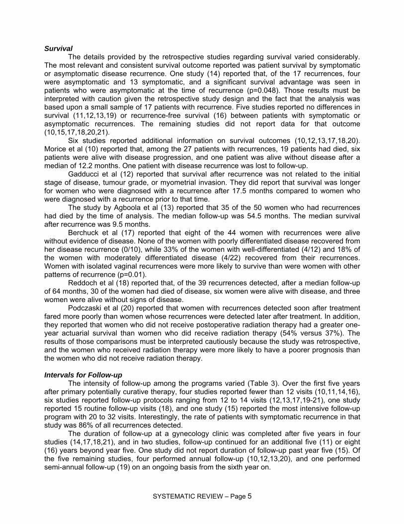
The intensity of follow-up among the programs varied (Table 3). Over the first five years after primary potentially curative therapy, four studies reported fewer than 12 visits (10,11,14,16), six studies reported follow-up protocols ranging from 12 to 14 visits (12,13,17,19-21), one study reported 15 routine follow-up visits (18), and one study (15) reported the most intensive follow-up program with 20 to 32 visits. Interestingly, the rate of patients with symptomatic recurrence in that study was 86% of all recurrences detected.
The duration of follow-up at a gynecology clinic was completed after five years in four studies (14,17,18,21), and in two studies, follow-up continued for an additional five (11) or eight (16) years beyond year five. One study did not report duration of follow-up past year five (15). Of the five remaining studies, four performed annual follow-up (10,12,13,20), and one performed semi-annual follow-up (19) on an ongoing basis from the sixth year on.
SYSTEMATIC REVIEW – Page 5
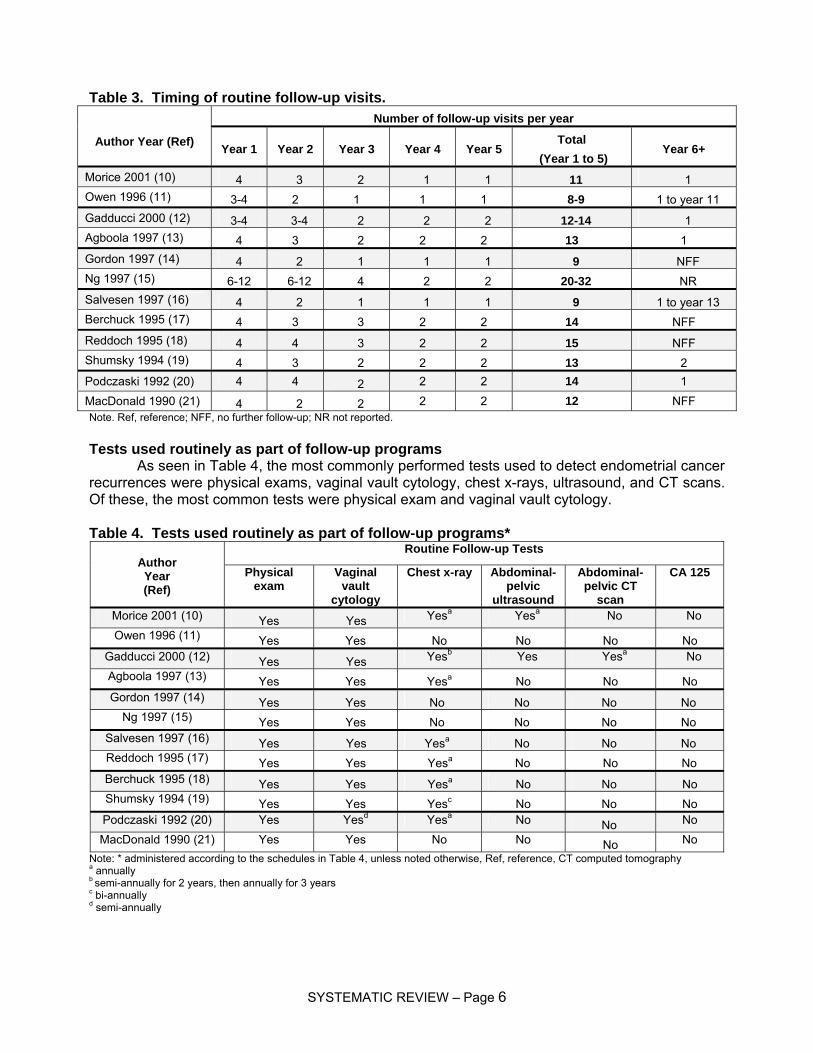
Table 3. Timing of routine follow-up visits. Number of follow-up visits per year
Author Year (Ref) Year 1 Year 2 Year 3 Year 4 Year 5 Total
(Year 1 to 5) Year 6+
Morice 2001 (10) 4 3 2 1 1 11 1 Owen 1996 (11) 3-4 2 1 1 1 8-9 1 to year 11 Gadducci 2000 (12) 3-4 3-4 2 2 2 12-14 1 Agboola 1997 (13) 4 3 2 2 2 13 1 Gordon 1997 (14) 4 2 1 1 1 9 NFF Ng 1997 (15) 6-12 6-12 4 2 2 20-32 NR Salvesen 1997 (16) 4 2 1 1 1 9 1 to year 13 Berchuck 1995 (17) 4 3 3 2 2 14 NFF Reddoch 1995 (18) 4 4 3 2 2 15 NFF Shumsky 1994 (19) 4 3 2 2 2 13 2 Podczaski 1992 (20) 4 4 2 2 2 14 1 MacDonald 1990 (21) 4 2 2 2 2 12 NFF Note. Ref, reference; NFF, no further follow-up; NR not reported. Tests used routinely as part of follow-up programs
As seen in Table 4, the most commonly performed tests used to detect endometrial cancer recurrences were physical exams, vaginal vault cytology, chest x-rays, ultrasound, and CT scans. Of these, the most common tests were physical exam and vaginal vault cytology. Table 4. Tests used routinely as part of follow-up programs*
Routine Follow-up Tests Author Year (Ref)
Physical exam
Vaginal vault
cytology
Chest x-ray Abdominal-pelvic
ultrasound
Abdominal-pelvic CT
scan
CA 125
Morice 2001 (10) Yes Yes Yesa Yesa No No Owen 1996 (11) Yes Yes No No No No
Gadducci 2000 (12) Yes Yes Yesb Yes Yesa No Agboola 1997 (13) Yes Yes Yesa No No No Gordon 1997 (14) Yes Yes No No No No
Ng 1997 (15) Yes Yes No No No No Salvesen 1997 (16) Yes Yes Yesa No No No Reddoch 1995 (17) Yes Yes Yesa No No No Berchuck 1995 (18) Yes Yes Yesa No No No Shumsky 1994 (19) Yes Yes Yesc No No No Podczaski 1992 (20) Yes Yesd Yesa No No No MacDonald 1990 (21) Yes Yes No No No No
Note: * administered according to the schedules in Table 4, unless noted otherwise, Ref, reference, CT computed tomography a annually b semi-annually for 2 years, then annually for 3 years c bi-annually d semi-annually
SYSTEMATIC REVIEW – Page 6
All of the follow-up programs included physical examination and vaginal vault cytology at every visit, with the exception of the study by Podczaski et al (20) where a sample for cytology was obtained at every other visit in the first two years of follow-up. In one study (15), patients were followed up with physical exam and vaginal vault cytology for up to 12 times a year for the first two years. Chest x-rays were performed as part of a follow-up program in eight studies (10,1213,16,17-20), and not included as part of follow-up in four studies (11,14,15,21). The follow-up schedule for routine testing with chest-x-ray was semi-annually in one study (19), annually in six studies (10,13,16,18-20), and bi-annually in one study (19). The use of ultrasound, CT scan, or CA 125 testing was generally not employed as part of routine testing in the studies identified. Two studies used abdominal-pelvic ultrasound scanning for routine follow-up (10,12); in one of these, annual abdominal-pelvic computed tomography (CT) scans were also performed (12). What is the accuracy of the surveillance tests presently being used to follow up patients who have been treated for endometrial cancer?
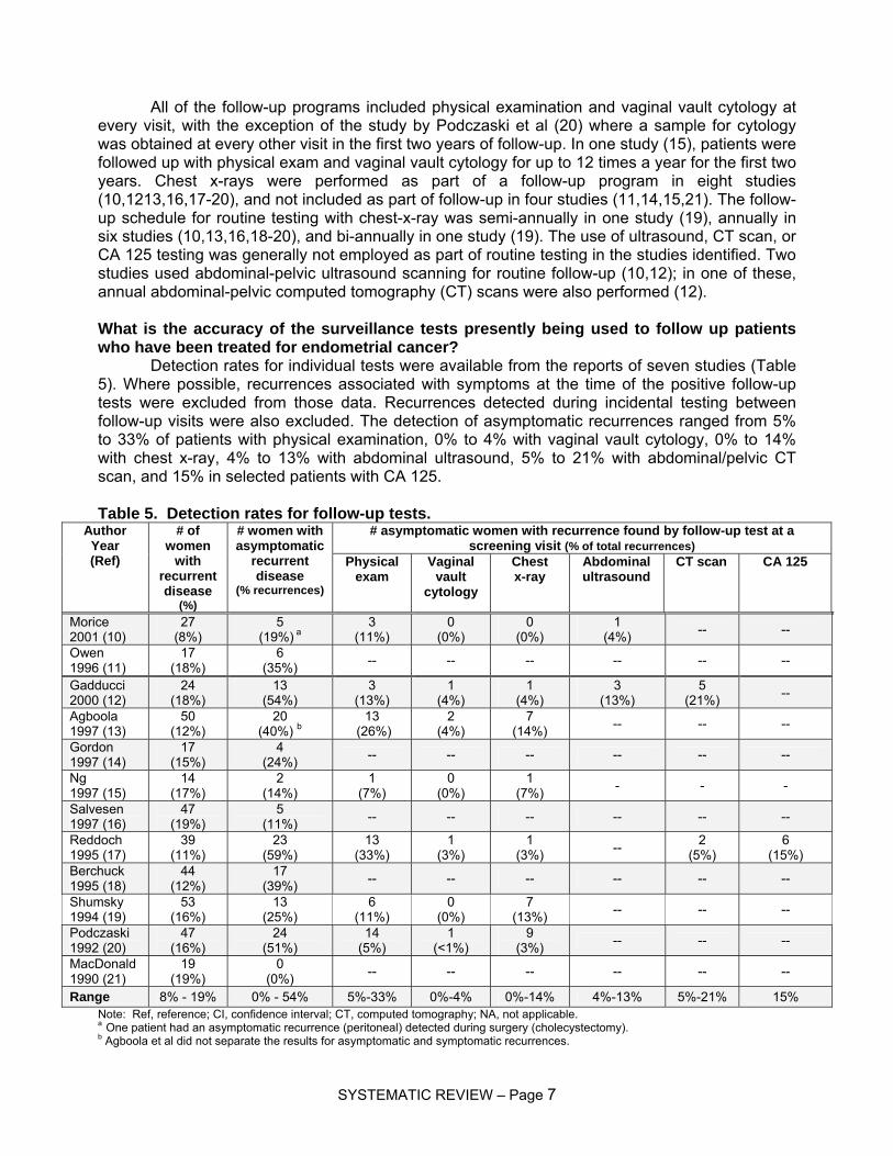
Detection rates for individual tests were available from the reports of seven studies (Table 5). Where possible, recurrences associated with symptoms at the time of the positive follow-up tests were excluded from those data. Recurrences detected during incidental testing between follow-up visits were also excluded. The detection of asymptomatic recurrences ranged from 5% to 33% of patients with physical examination, 0% to 4% with vaginal vault cytology, 0% to 14% with chest x-ray, 4% to 13% with abdominal ultrasound, 5% to 21% with abdominal/pelvic CT scan, and 15% in selected patients with CA 125. Table 5. Detection rates for follow-up tests.
# asymptomatic women with recurrence found by follow-up test at a screening visit (% of total recurrences)
Author Year (Ref)
# of women
with recurrent disease
(%)
# women with asymptomatic
recurrent disease
(% recurrences)
Physical exam
Vaginal vault
cytology
Chest x-ray
Abdominal ultrasound
CT scan CA 125
Morice 2001 (10)
27 (8%)
5 (19%) a
3 (11%)
0 (0%)
0 (0%)
1 (4%) -- --
Owen 1996 (11)
17 (18%)
6 (35%) -- -- -- -- -- --
Gadducci 2000 (12)
24 (18%)
13 (54%)
3 (13%)
1 (4%)
1 (4%)
3 (13%)
5 (21%) --
Agboola 1997 (13)
50 (12%)
20 (40%) b
13 (26%)
2 (4%)
7 (14%) -- -- --
Gordon 1997 (14)
17 (15%)
4 (24%) -- -- -- -- -- --
Ng 1997 (15)
14 (17%)
2 (14%)
1 (7%)
0 (0%)
1 (7%) - - -
Salvesen 1997 (16)
47 (19%)
5 (11%) -- -- -- -- -- --
Reddoch 1995 (17)
39 (11%)
23 (59%)
13 (33%)
1 (3%)
1 (3%) -- 2
(5%) 6
(15%) Berchuck 1995 (18)
44 (12%)
17 (39%) -- -- -- -- -- --
Shumsky 1994 (19)
53 (16%)
13 (25%)
6 (11%)
0 (0%)
7 (13%) -- -- --
Podczaski 1992 (20)
47 (16%)
24 (51%)
14 (5%)
1 (<1%)
9 (3%) -- -- --
MacDonald 1990 (21)
19 (19%)
0 (0%) -- -- -- -- -- --
Range 8% - 19% 0% - 54% 5%-33% 0%-4% 0%-14% 4%-13% 5%-21% 15% Note: Ref, reference; CI, confidence interval; CT, computed tomography; NA, not applicable. a One patient had an asymptomatic recurrence (peritoneal) detected during surgery (cholecystectomy). b Agboola et al did not separate the results for asymptomatic and symptomatic recurrences.
SYSTEMATIC REVIEW – Page 7
Radiation Therapy The fact that that women who receive radiation therapy have different patterns of
recurrence than women who do not receive radiation therapy is widely recognized (3-5). Unfortunately, none of the studies included in this report specifically addressed the potentially different follow-up requirements of women who received radiation therapy compared to women who did not nor can it be certain that patients selected for radiation therapy were comparable to those who were not. The study by Podczaski et al (20) indicated that women who had received radiation therapy received yearly intravenous pyelograms for five years after treatment; however, they did not report differences in recurrences between women who had received radiation compared to those who did not. Owen et al (11) reported that women were followed up at either gynecology and radiation therapy clinics or gynecology clinics. They did not specify whether there were different procedures performed at the clinics. Shumsky et al published another paper in 1997 (28), based on the patients from their 1994 study, that retrospectively reviewed the charts of 435 women who had been treated for endometrial cancer (19). In their subsequent publication, Shumsky et al (28) retrospectively reviewed the same patients but split them into high and low risk of recurrence groups. Shumsky et al suggested that follow-up should be targeted towards women at a high risk of recurrence, because they are at high risk and also to monitor the effects of radiation therapy (assuming that the low-risk patients did not receive radiation therapy). However, Shumsky et al did not outline a possible follow-up regimen for women at high risk of recurrence. Serum CA 125 Levels
In addition to the tests previously mentioned, four studies examined the role of serial tumour markers in the post-treatment surveillance of early-stage endometrial cancer (22-25).
Patsner et al (22) obtained serum CA 125 levels for 125 women with surgical stage I or II endometrial carcinoma before surgery and every three to four months during follow-up. Follow-up visits also included pelvic examinations and Pap smears. Median follow-up time was 18 months (range, 12 to 36 months). Among 123 patients with preoperative CA 125 levels <35 U/ml, 106 (86%) had normal CA 125 levels throughout follow-up and were recurrence free. A total of thirteen women had recurrences (11%)—six patients with normal CA 125 levels during follow-up had vaginal recurrences (five diagnosed because of vaginal bleeding and one by follow-up Pap smear) and seven with elevated CA 125 levels had recurrences at other sites (one pelvic, four abdominal and two pulmonary). Four patients without recurrent disease had elevated CA 125 levels associated with small bowel obstruction as a result of postoperative radiotherapy.
Rose et al (23) conducted a similar study but obtained preoperative CA 125 levels on only 45% of patients (n=236). Twenty-five percent of those with a preoperative CA 125 assessment had elevated levels (>35 U/ml) before surgery. Patients were classified as low risk (stage I, grade 1 or 2, and one-third or less myometrial invasion), medium risk (stage I, grade 1 or 2, and middle- or outer third or less myometrial invasion) or high risk (stage II, III, or IV, grade 3 or serous or clear cell carcinoma). CA 125 was measured as part of a surveillance program that also included pelvic examination, Pap smear, and chest x-ray every three to four months for the first two years, every six months for the next three years, and yearly thereafter. Median follow-up time was 39 months (range, four to 54 months). There were 29 recurrences among 236 patients treated by surgery (12%)—none of 97 in the low-risk group, two of 42 in the medium-risk group (5%), and 27 of 97 in the high-risk group (28%). Fifteen (55%) of the women with recurrent disease in the latter group had follow-up CA 125 levels >35 U/ml. The number of false negatives among the surgical group was not clear.
Among a series of 23 patients with stage I-IV endometrial cancer and elevated pre-treatment tumour markers (CA 125, CA 15.3 or CA 19.9), Lo et al (24) studied 14 women with stage I or II disease who had been treated by surgery plus postoperative radiotherapy. Three of the early-stage patients had elevated CA 125 levels during follow-up but none had recurrent disease. One patient with an elevated CA 19.9 level during follow-up was found to have a pulmonary recurrence. None of the patients with normal serum marker levels had recurrences.
SYSTEMATIC REVIEW – Page 8
Price et al (25) reviewed the serial CA 125 data from 11 women with uterine papillary serous carcinoma (six stage I, two stage II, and three stage III). All had CA 125 values <35 U/ml before surgery. Following the completion of adjuvant chemotherapy, CA 125 was measured every three months for a median follow-up time of 63 months (range, 21-90 months). One patient died of endometrial cancer six months after primary therapy but had normal CA 125 levels. The other 10 patients had no evidence of recurrent disease at their last follow-up visit, but four had elevated CA 125 levels (>35 U/ml) during follow-up. Quality of Life
None of the identified studies evaluated patient preferences for follow-up or addressed the impact of follow-up programs on quality of life (10-25). Two of the studies identified (11,17) made reference to the potential psychological impact of follow-up appointments; however, the studies were not designed to measure the psychological impact of follow-up. The study by Berchuck et al (17) stated that it was difficult to assess “the value of psychological reassurance associated with a normal examination.” Systematic Reviews
Two systematic reviews were identified in the search of the literature (26,27). Both reviews located similar evidence used to inform the present evidence series.
Tjalma et al (26) reported a 13% overall recurrence rate with the probability of recurrence ranging from 7.7% to 18.9%. In their review, approximately 33% of recurrences were local, 57% were distant, and 10% were both local and distant. They reported that approximately 65% of recurrences were symptomatic, greater than 80% of recurrences were detected through clinical examination and symptomology, and greater than 80% of all recurrences occurred within the first three years of primary curative treatment. Tjalma et al (26) reported little value associated with cytology, CA 125, chest-X-ray, intravenous pyelogram, ultrasound, or CT in detecting recurrences in asymptomatic patients in order to improve overall survival. They concluded that a reasonable follow-up strategy for low-risk patients could entail a careful history and clinical examination every six months for three years and annually, starting in year four. The authors did not provide follow-up recommendations for patients at a high risk of recurrence. Cost-effectiveness information was also presented in the systematic review but is not a focus of the present evidence series.
The systematic review by Kew et al (27) reported rates of recurrence ranging from 8.5% to 19% of patients included in the nine retrospective studies identified in their search of the literature. They reported that the methodological quality and the heterogeneity of the studies made comparisons between studies difficult. They concluded that the studies did not show any survival benefit to routine follow-up but reported that small differences in survival might not have been detected given the small number of patients who recurred in the identified studies. The authors of the review did not provide conclusions on the optimum follow-up of patients. DISCUSSION
The primary goal of a surveillance strategy in patients who have been treated for endometrial cancer is to facilitate the early detection of recurrent disease. This detection results in the introduction of salvage treatment, with the overall aim to improve survival or decrease morbidity secondary to the recurrence. A review of the 16 retrospective studies in this series suggests that there is no evidence to support that intensive follow-up schedules with multiple routine diagnostic interventions result in survival benefits any more or less than non-intensive follow-up schedules without multiple routine diagnostic interventions.
From the 12 studies that reported results for specific follow-up schedules that ranged from a low of eight visits to a high of 32 visits over five years, no discernable differences in outcome were detected between any of the follow-up programs.
When considering the use of routine examinations or diagnostic interventions in asymptomatic patients, the reporting of outcomes was inconsistent; however, only physical
SYSTEMATIC REVIEW – Page 9
examination showed some utility in detecting recurrence. This adds some support to the idea that a physical examination that includes a pelvic rectal examination is useful as part of a routine follow-up strategy. In seven studies, physical examination showed the greatest efficacy, with recurrence detection rates ranging from 5% and 33%, while Pap tests detected the least amount of asymptomatic recurrences (≤ 4%). Chest X-ray detected from 0% to 14% of asymptomatic recurrences, but the detection of clinical asymptomatic recurrences in the chest and the impact of that detection on survival have not been clearly elucidated. In four non-comparative studies with limited data (24-27), elevated CA 125 serum levels did not consistently indicate disease recurrence, and in two studies, a high rate of false positives were reported (21% and 40%). Intensive surveillance with CT scans and ultrasounds directed at detecting asymptomatic abdominal extra-pelvic recurrences showed limited benefit when employed on a routine basis.
There was no evidence to inform the role of follow-up by clinical setting or type of specialist on patient outcomes. In spite of this, many patients may continue to be seen by specialists in a cancer centre when there is no evidence to support or refute that outcomes would vary if followed by a qualified general practitioner in the office setting. The key issue is not so much location but that practitioners be skilled in the in the performance of a pelvic rectal examination and assessment grounded in an understanding of the natural history of the disease. Because of the resource implications involved, this issue lends itself ideally to a prospective evaluation of the most efficient location for follow-up. In the breast cancer setting, for instance, a randomized trial detected no significant differences in outcomes for patients followed by family physician versus specialist care (29).
Even though the evidence from the retrospective studies is modest, there are some compelling points to consider in determining the most appropriate follow-up of patients. One is the relatively low risk of patient recurrence. The overall recurrence rate across all of the studies was 13%, and for patients at a low risk of recurrence, rates ranged from 1% to 3%. This means that the majority of patients who were followed did not experience a recurrence, regardless of follow-up; this was especially the case for patients at a low risk of recurrence. It seems reasonable therefore, that patients at a lower risk of recurrence be followed differently than those at a higher risk of recurrence.
Another point relates to the known natural history of disease recurrence for these patients. The data indicated that about two thirds to three quarters of all recurrences were detected through symptoms alone. For these patients, recurrence detection would have occurred regardless of follow-up strategy. In addition, if a patient does experience a recurrence, the data indicate that approximately 60% of the time the recurrence will be distant. The prognosis for patients with a distant recurrence is generally not favourable, regardless of timing of disease detection. For these patients, it is unlikely that early detection through follow-up would result in any survival benefits.
The final point to consider is that most patients had a recurrence at about three years or less after primary potentially curative treatment. At about three years, 70% to 100% of recurrences had occurred in 11 of the 12 studies that reported that data. For the majority of patients, follow-up in years four, five, or beyond, detected very few recurrences, and would seem to be of questionable benefit.
Taken together, it appears that the follow-up programs identified in this series were not particularly effective in improving patient outcomes related to survival, especially after a three-to five-year period. To illustrate, according to the data identified in this systematic review, if 1,000 patients who were at a low risk of recurrence were to be followed, approximately 3% or 30 patients would experience a recurrence, most within three years of primary treatment with curative intent. Of these thirty patients, approximately 20 or more would present with symptomatic disease outside of regular follow-up. This leaves approximately 10 or less asymptomatic patients (≤ 1%) for whom the detection of recurrence through follow-up may be beneficial. Of these ten or so patients, approximately six patients would experience a distant recurrence, for which the early detection of recurrence has shown no overall survival benefit. That leaves approximately four out of 1,000 low-risk patients who could potentially benefit from a follow-up program. Of the four
SYSTEMATIC REVIEW – Page 10

asymptomatic patients with a local recurrence, approximately two patients would not be salvageable, thus leaving approximately two patients who would ultimately benefit in an absolute way (i.e., survival) from a follow-up program as compared, in theory, with no follow-up program at all. For patients at a higher risk of recurrence, assuming a 13% recurrence rate, the number who would benefit from follow-up increases to approximately seven patients.
While the data indicate that the small number of patients who would benefit from a surveillance strategy does not seem to reasonably justify the routine follow-up of all patients, there are good arguments to support the use of follow-up programs. The strongest argument is that the data used to inform the issue is from retrospective studies, and the actual rates and types of recurrence may vary considerably. Until definitive results from randomized controlled trials or large prospective studies become available, it seems prudent that patients be followed according to some type of schedule. Patients may also derive a psychological benefit from some type of follow-up program, but there is insufficient evidence to support or refute that speculation. Finally, standard practice is such that most patients are followed according to some type of follow-up strategy after potentially curative primary therapy, and this practice reflects a more conservative approach. While it is not being suggested that standard practice be discontinued, resource utility is a practical consideration that should not be overlooked when determining the most appropriate follow-up schedule. The follow-up schedules of the identified studies were generally consistent with five year follow-up ranging from eight to 15 visits. It would seem to be reasonable, therefore, that a follow-up program fall within that range of visits but also account for risk of recurrence and the natural history of the disease. Other factors that may impact upon this are patient preferences and resource availability. ONGOING TRIALS
No ongoing trials were identified in the search of the literature. CONCLUSIONS
Based on the interpretation of the evidence from retrospective studies and expert consensus opinion, the Gynecology Cancer DSG concluded that the most appropriate follow-up strategy was likely one based upon the risk of recurrence, the natural history of the disease, and individual patient preferences. Specifically, for patients at low risk of recurrence, a reasonable follow-up schedule could include a general examination that includes a complete history and a pelvic-rectal examination, conducted on a semi-annual to annual basis; a targeted investigation if symptomatic; and counselling about the symptoms of recurrence of endometrial cancer. Counselling is extremely important because, in the retrospective studies reviewed, 41% to 100% of patients with recurrences were symptomatic. The choice of follow-up interval should be decided in large part by patient preference. There is no evidence to suggest that closer follow-up leads to improved detection of recurrence, but patients may derive a psychological benefit with more follow-up as opposed to less follow-up.
For patients at a high risk of recurrence, a reasonable follow-up schedule could include a general examination, which includes a complete history, a pelvic-rectal examination every three to six months for the first three years and semi-annually for the next two years, a targeted investigation if symptomatic, counselling about the signs and symptoms of recurrence, and counselling on the potential adverse effects of radiotherapy. Overall, there is insufficient evidence to inform the routine use of Pap smears, chest x-rays, abdominal ultrasounds, CT scans, or CA 125 levels alone to detect asymptomatic recurrences with the aim of improving survival.
Patients should be followed by a health care professional who is knowledgeable about the natural history of the disease and who is comfortable performing speculum and pelvic exams in order to diagnose or detect a local (vaginal) recurrence, as this type of recurrence is potentially curable. Since most patients tend to recur within a three-year time frame, if a patient is initially followed by specialist, it is reasonable to suggest that patients be followed by a qualified general practitioner after three to five years of recurrence-free follow-up. Formal follow-up to detect
SYSTEMATIC REVIEW – Page 11
recurrences beyond five years is generally not indicated because the majority of recurrences are symptomatic and virtually all recurrences occur before that time. Thus, it appears reasonable to suggest that annual population-based general physical and pelvic examination be conducted for all patients after five years of routine follow-up.
The available retrospective evidence highlights the need for well-conducted studies, preferably randomized controlled trials, to help inform decision making on the most appropriate follow-up for patients. Ideally, after potentially curative primary therapy, a multicentre study would categorize patients as being at a low, intermediate, or high risk of recurrence (with surgical or pathological confirmation) and would randomize patients who were clinically disease free to either a less intensive follow-up schedule with or without multiple diagnostic interventions in asymptomatic patients or a more intensive follow-up schedule with or without multiple diagnostic interventions in asymptomatic patients. All patients would receive counselling on the symptoms of potential recurrence. The study could compare differences in recurrence by type of clinical setting where follow-up is performed and by type of specialist involved in the follow-up. Careful detailing of the type of recurrence, whether symptomatic or asymptomatic, whether distant or local, quality of life, patient preferences, and subsequent survival outcomes would greatly inform the most appropriate follow-up strategy for this patient population. CONFLICT OF INTEREST
Members of the Gynecology Cancer DSG declared that there were no conflicts of interest. JOURNAL REFERENCES
Fung-Kee-Fung M, Dodge J, Elit L, Lukka H, Chambers A, Oliver T; on behalf of the Cancer Care Ontario Program in Evidence-based Care Gynecology Cancer Disease Site Group. Follow-up after primary therapy for endometrial cancer: a systematic review. Gynecol Oncol. 2006 Jun;101(3):520-9. ACKNOWLEDGEMENTS
The Gynecology Cancer DSG would like to thank Dr Michael Fung-Kee-Fung, Dr Jason Dodge, Dr. Laurie Elit, Dr. Himu Lukka, Ms. Alexandra Chambers, and Mr. Tom Oliver for taking the lead in drafting and revising this evidence series report.
For a complete list of the Gynecology Cancer Disease Site Group members, please visit the CCO Web site at http://www.cancercare.on.ca/
SYSTEMATIC REVIEW – Page 12
Funding The PEBC is supported by Cancer Care Ontario (CCO) and the Ontario Ministry of Health and Long-Term Care. All
work produced by the PEBC is editorially independent from its funding agencies.
Copyright This evidence-based series is copyrighted by Cancer Care Ontario; the series and the illustrations herein may not be
reproduced without the express written permission of Cancer Care Ontario. Cancer Care Ontario reserves the right at any time, and at its sole discretion, to change or revoke this authorization.
Disclaimer
Care has been taken in the preparation of the information contained in this document. Nonetheless, any person seeking to apply or consult the evidence-based series is expected to use independent medical judgment in the context of individual clinical circumstances or seek out the supervision of a qualified clinician. Cancer Care Ontario makes no
representation or guarantees of any kind whatsoever regarding their content or use or application and disclaims any for their application or use in any way.
Contact Information
For further information about this series, please contact Dr. Michael Fung-Kee-Fung, Chair, Gynecology Cancer Disease Site Group; Ottawa General Hospital,
501 Smyth Road, Ottawa, Ontario; Telephone: 613-737-8560, FAX: 613-737-8828
For information about the PEBC and the most current version of all reports, please visit the CCO Web site at http://www.cancercare.on.ca/ or contact the PEBC office at:
Phone: 905-525-9140, ext. 22055 Fax: 905-522-7681
SYSTEMATIC REVIEW – Page 13

REFERENCES
1. National Cancer Institute of Canada. Canadian Cancer Statistics 2003. Toronto, Canada; 2003.
2. Creasman WT, Morrow CP, Bundy BN, Homesley HD, Graham JE, Heller PB. Surgical pathologic spread patterns of endometrial cancer. A Gynecologic Oncology Group Study. Cancer. 1987;60:41-2.
3. Keys HM, Roberts JA, Brunetto VL, Zaino RJ, Spirtos NM, Bloss JD, et al. A phase III trial of surgery with or without adjunctive external pelvic radiation therapy in intermediate-risk endometrial adenocarcinoma: a Gynecologic Oncology Group study. Gynecol Oncol. 2004;92:744-51.
4. Creutzberg CL, van Putten WL, Koper PC, Lybeert ML, Jobsen JJ, Warlam-Rodenhuis CC, et al. Surgery and postoperative radiotherapy versus surgery alone for patients with stage-1 endometrial carcinoma: multicentre randomised trial. PORTEC Study Group. Post Operative Radiation Therapy in Endometrial Carcinoma. Lancet. 2000;355:1404-11.
5. Aalders J, Abeler V, Kolstad P, Onsrud M. Postoperative external irradiation and prognostic parameters in stage I endometrial carcinoma. Obstet Gynecol. 1980;56:419-26.
6. Piver MS, Yazigi R, Blumenson L, Tsukada Y. A prospective trial comparing hysterectomy, hysterectomy plus vaginal radium, and uterine radium plus hysterectomy in stage I endometrial carcinoma. Obstet Gynecol. 1979;54:85-9.
7. Menczer J. Endometrial carcinoma. Is routine intensive periodic follow-up of value? Eur J Gynaecol Oncol. 2000;21:461-5.
8. Ackerman I, Malone S, Thomas G, Franssen E, Balogh J, Dembo A. Endometrial carcinoma--relative effectiveness of adjuvant irradiation vs therapy reserved for relapse. Gynecol Oncol. 1996;60:177-83.
9. Lipsey MW, Wilson, DB. Practical meta-analysis. Thousand Oaks (CA): SAGE Publications; 2001. p. 113-6.
10. Morice P, Levy-Piedbois C, Ajaj S, Pautier P, Haie-Meder C, Lhomme C, et al. Value and cost evaluation of routine follow-up for patients with clinical stage I/II endometrial cancer. Eur J Cancer. 2001;37:985-90.
11. Owen P, Duncan ID. Is there any value in the long term follow up of women treated for endometrial cancer? Br J Obstet Gynaecol. 1996;103:710-3.
12. Gadducci A, Cosio S, Fanucchi A, Cristofani R, Genazzani AR. An intensive follow-up does not change survival of patients with clinical stage I endometrial cancer. Anticancer Res. 2000;20:1977-84.
13. Agboola OO, Grunfeld E, Coyle D, Perry GA. Costs and benefits of routine follow-up after curative treatment for endometrial cancer. Can Med Assoc J. 1997;157:879-86.
14. Gordon AF, Owen P, Chien PFW, Duncan ID. A critical evaluation of follow-up of women treated for endometrial adenocarcinoma. J Obstet Gynaecol. 1997;17:386-9.
15. Ng TY, Ngan HYS, Cheng DKL, Wong LC. Vaginal vault cytology in the routine follow-up of patients treated for endometrial carcinoma: Is it useful? Aust NZ J Obstet Gynaecol. 1997;37:104-6.
16. Salvesen HB, Akslen LA, Iversen T, Iversen OE. Recurrence of endometrial carcinoma and the value of routine follow up. Br J Obstet Gynaecol. 1997;104:1302-7.
17. Berchuck A, Anspach C, Evans AC, Soper JT, Rodriguez GC, Dodge R, et al. Postsurgical surveillance of patients with FIGO stage I/II endometrial adenocarcinoma. Gynecol Oncol. 1995;59:20-4.
18. Reddoch JM, Burke TW, Morris M, Tornos C, Levenback C, Gershenson DM. Surveillance for recurrent endometrial carcinoma: development of a follow-up scheme. Gynecol Oncol. 1995;59:221-5.
SYSTEMATIC REVIEW – Page 14
19. Shumsky AG, Stuart GC, Brasher PM, Nation JG, Robertson DI, Sangkarat S. An evaluation of routine follow-up of patients treated for endometrial carcinoma. Gynecol Oncol. 1994;55:229-33.
20. Podczaski E, Kaminski P, Gurski K, MacNeill C, Stryker JA, Singapuri K, et al. Detection and patterns of treatment failure in 300 consecutive cases of "early" endometrial cancer after primary surgery. Gynecol Oncol. 1992;47:323-7.
21. MacDonald JH, Kidd GM. An audit of endometrial carcinoma: the value of routine follow up. J Obstet Gynaecol. 1990;10:548-50.
22. Patsner B, Orr JW, Jr., Mann WJ, Jr. Use of serum CA 125 measurement in posttreatment surveillance of early-stage endometrial carcinoma. Am J Obstet Gynecol. 1990;162:427-9.
23. Rose PG, Sommers RM, Reale FR, Hunter RE, Fournier L, Nelson BE. Serial serum CA 125 measurements for evaluation of recurrence in patients with endometrial carcinoma. Obstet Gynecol. 1994;84:12-6.
24. Lo SS, Khoo US, Cheng DK, Ng TY, Wong LC, Ngan HY. Role of serial tumor markers in the surveillance for recurrence in endometrial cancer. Cancer Detect Prev. 1999;23:397-400.
25. Price FV, Chambers SK, Carcangiu ML, Kohorn EI, Schwartz PE, Chambers JT. CA 125 may not reflect disease status in patients with uterine serous carcinoma. Cancer. 1998;82:1720-5.
26. Tjalma WA, van Dam PA, Makar AP, Cruickshank DJ. The clinical value and the cost-effectiveness of follow-up in endometrial cancer patients. Int J Gynecol Cancer. 2004 Sep-Oct;14(5):931-7.
27. Kew FM, Roberts AP, Cruickshank DJ. The role of routine follow-up after gynecological malignancy. Int J Gynecol Cancer. 2005 May-Jun;15(3):413-9.
28. Shumsky AG, Brasher PM, Stuart GC, Nation JG. Risk-specific follow-up for endometrial carcinoma patients. Gynecol Oncol. 1997 Jun;65(3):379-82.
29. Grunfeld E, Levine MN, Julian JA, Coyle D, Szechtman B, Mirsky D, et al. Randomized trial of long-term follow-up for early-stage breast cancer: A comparison of family physician versus specialist care. J Clin Oncol. 2006 Feb 20;24(6):848-55.
SYSTEMATIC REVIEW – Page 15

Evidence-Based Series #4-9: Section 3
Follow-up after Primary Therapy for Endometrial Cancer: Guideline Development and External Review—Methods and Results
M. Fung-Kee-Fung, J. Dodge, L. Elit, H. Lukka, A. Chambers, T. Oliver,
and the Gynecology Cancer Disease Site Group
A Quality Initiative of the Program in Evidence-Based Care, Cancer Care Ontario.
Developed by the Provincial Gynecology Cancer Disease Site Group
Report Date: January 10, 2006
THE PROGRAM IN EVIDENCE-BASED CARE
The Program in Evidence-based Care (PEBC) is an initiative of the Ontario provincial cancer system, Cancer Care Ontario (CCO) (1). The PEBC mandate is to improve the lives of Ontarians affected by cancer, through the development, dissemination, implementation, and evaluation of evidence-based products designed to facilitate clinical, planning, and policy decisions about cancer care.
The PEBC supports a network of disease-specific panels, called Disease Site Groups (DSGs) and Guideline Development Groups (GDGs), mandated to develop the PEBC products. These panels are comprised of clinicians, methodologists, and community representatives from across the province.
The PEBC is well known for producing evidence-based practice guideline reports, using the methods of the Practice Guidelines Development Cycle (1,2). The PEBC reports consist of a comprehensive systematic review of the clinical evidence on a specific cancer care topic, an interpretation of and consensus agreement on that evidence by our DSGs and GDGs, the resulting clinical recommendations, and an external review by Ontario clinicians in the province for whom the topic is relevant. The PEBC has a formal standardized process to ensure the currency of each clinical practice guideline report, through the routine periodic review and evaluation of the scientific literature and, where appropriate, the integration of that literature with the original clinical practice guideline information. The Evidence-based Series: A New Look to the PEBC Practice Guidelines Each Evidence-based Series is comprised of three sections. • Section 1: Clinical Practice Guideline. This section contains the clinical recommendations
derived from a systematic review of the clinical and scientific literature and its interpretation by the DSG or GDG involved and a formalized external review by Ontario practitioners.
DEVELOPMENT & METHODS – page 1

• Section 2: Systematic Review. This section presents the comprehensive systematic review of the clinical and scientific research on the topic and the conclusions reached by the DSG or GDG.
• Section 3: Guideline Development and External Review: Methods and Results. This section summarizes the guideline development process and the results of the formal external review by Ontario practitioners of the draft version of the clinical practice guideline and systematic review.
DEVELOPMENT OF THIS EVIDENCE-BASED SERIES Developing the Draft Systematic Review and Clinical Practice Guideline
This evidence-based series was developed by the Gynecology Cancer DSG of Cancer Care Ontario’s PEBC. The series is a convenient and up-to-date source of the best available evidence developed through systematic review, evidence synthesis, and input from practitioners in Ontario. The systematic review on follow-up after primary therapy for endometrial cancer is reported in Section 2. On the basis of that evidence and the interpretation by members of the DSG, draft recommendations were circulated to Ontario practitioners on June 25, 2004 for feedback (Table 1). Table 1. Draft recommendations circulated for external review.
Target Population The target population for screening for disease recurrence includes two groups of women without evidence of metastatic disease after primary, curative treatment for endometrial cancer (all stages of disease): 1. Those who underwent a total abdominal hysterectomy and bilateral salpingo-oophorectomy with or without
pelvic lymphadenectomy as primary therapy (i.e., low-risk) 2. Those who underwent a total abdominal hysterectomy and bilateral salpingo-oophorectomy with or without
pelvic lymphadenectomy as primary therapy, with subsequent adjuvant radiotherapy (i.e., high risk) Draft Recommendations The lack of sufficient, high quality evidence precludes definitive recommendations from being made. Instead, the Gynecology Cancer Disease Site Group offers the following opinions based on the evidence reviewed: • There is insufficient evidence to indicate that an intensive surveillance program for women who have been
treated for endometrial cancer results in a survival benefit. • There is no direct evidence that supports the clinical benefit of using routine Pap smears and chest X-rays to
detect asymptomatic recurrences in women who have been treated for endometrial cancer. • There is insufficient evidence to make recommendations regarding intervals for surveillance. However, based
on the Gynecology Cancer Disease Site Group’s interpretation of the existing evidence, management options that clinicians and patients should consider include: ° For women at low-risk (i.e., stage IA, grade 1 or 2 or stage IB, grade 1 or 2):
− An annual well-woman assessment, including a pelvic-rectal examination. − Counselling about the symptoms of recurrence of endometrial cancer, because more than 50% of
recurrences are symptomatic. ° For women at high risk (i.e., stage IA, grade 3, stage IB, grade 3, stage IC, advanced stage):
− A general examination, pelvic-rectal examination, and targeted investigation based on symptoms every three to six months within the first three years.
− Counselling about the symptoms of recurrence of endometrial cancer, because more than 50% of recurrences are symptomatic.
− Counselling about the symptoms suggestive of long-term toxicity associated with radiation therapy. o Women with a suspected recurrence should be referred promptly to a cancer centre for further assessment.
It is the opinion of the Gynecology Cancer Disease Site Group that women who have been treated for endometrial cancer should be followed by a health care professional who is comfortable performing speculum and pelvic exams, in order to diagnose or detect a local (vaginal) recurrence. This type of recurrence is potentially curable.
Practitioner Feedback
Based on the evidence and the draft recommendations presented above, feedback was sought from Ontario clinicians
DEVELOPMENT & METHODS – page 2

Methods Practitioner feedback was obtained through a mailed survey of 172 practitioners in
Ontario (101 family practitioners, 40 medical oncologists, 16 surgeons, 14 gynecologists, and one urologist). The survey consisted of items evaluating the methods, results, and interpretive summary. Written comments were invited. Follow-up reminders were sent at two weeks (post card) and four weeks (complete package mailed again). The Gynecology DSG reviewed the results of the survey. Results
One hundred and twenty six responses were received out of the 172 surveys sent (73.3% response rate). Responses include returned completed surveys as well as phone, fax, and email responses. Of the practitioners who responded, 27 indicated that the report was relevant to their clinical practice and completed the survey (15 family practitioners, five medical oncologists, three surgeons, three gynecologists, and one urologist). Results of the practitioner feedback survey are summarized in Table 2. Table 2. Results of the practitioner feedback survey.
Number (%) Item Strongly
agree or agree
Neither agree nor disagree
Strongly disagree or
disagree The rationale for developing an evidence summary, as stated in the “Introduction” of the report, is clear.
23 (85.2%)
4 (14.8%)
-
There is a need for an evidence summary on this topic. 21 (77.8%) 5 (18.5%) 1 (3.7%) The literature search is relevant and complete in this evidence summary.
17 (63.0%) 10 (37.0) -
I agree with the methodology used to summarize the evidence. 25 (92.6%) 2 (7.4%) - I agree with the overall interpretation of the evidence in the evidence summary.
24 (88.9%) 2 (7.4%) 1 (3.7%)
The “Opinions of the Disease Site Group” section of this evidence summary is useful.
24 (88.9%) 3 (11.1%) -
An evidence summary of this type will be useful for clinical decision making.
22 (81.5%) 4 (14.8%) 1 (3.7%)
At present, there is insufficient evidence to develop a practice guideline on this topic.
14 (53.9%) 5 (19.2%) 7 (26.9%)
There is a need to develop an evidence-based practice guideline on this topic when sufficient evidence becomes available.
22 (81.5%) 3 (11.1%) 2 (7.4%)
Summary of Written Comments
Six (22.2%) respondents (three family practitioners, two medical oncologists, and one surgeon) provided written comments. One practitioner commented that the targeted investigation of symptoms should be applicable for women at both high and low risk, while another practitioner commented that further information regarding the symptoms of recurrence of endometrial cancer and the toxicity associated with radiotherapy would be helpful information in counselling patients. The remaining practitioners provided general comments not requiring revisions to the draft series. Modifications/Actions
In response to the written comments, the Gynecology Cancer DSG made the following changes to the guideline: • The recommendation for women at low risk was revised to include a targeted investigation
for patients who were symptomatic.
DEVELOPMENT & METHODS – page 3

• A statement regarding symptoms of recurrence was added to the introduction, and examples of symptoms associated with radiotherapy were added to the third recommendation.
Report Approval Panel
The evidence series was circulated to the two members of the Report Approval Panel and the Guidelines Coordinator of the PEBC. Feedback was provided by the Panel and the Coordinator and is summarized below. Feedback was reviewed by the Gynecology Cancer DSG, and modifications were made to the series in response. The revised draft was then recirculated to the Panel for final approval. Summary of Written Comments with Modifications Made by the Gynecology Cancer DSG • In the past, the PEBC has suggested that recommendations based primarily on expert
opinion be presented as ‘Opinions’ rather than ‘Recommendations’. However, it may be more helpful to present any proposed action or conclusion as a recommendation while clearly indicating the type of supporting evidence (based on expert consensus, randomized trials, etc.). Response: The draft was revised to reflect that recommendations, not opinions, were being presented on the basis of modest evidence from retrospective studies and through expert consensus opinion.
• The panel questioned whether the interval between follow-up visits should remain constant or was it reasonable to lengthen intervals over time. Response: The recommendations regarding interval of follow-up were revised on the basis that most patients who recur do so within three years of primary curative treatment and very few recurrences are detected past five years.
• The panel questioned if there was a time where follow-up was no longer needed. Response: A recommendation was added to reflect that follow-up to detect recurrences beyond five years is generally not indicated because the majority of recurrences are symptomatic and virtually all recurrences occur before that time. Regarding who performs follow-up, a sub-bullet was added to the recommendations saying that, if a patient is initially followed by a specialist, they may be followed by a qualified general practitioner after three to five years of recurrence-free follow-up.
• The panel requested a more explicit recommendation on the use of diagnostic tests in asymptomatic patients. Response: The recommendation on the use of diagnostic tests in asymptomatic patients was revised to state that there was insufficient evidence to inform (either for or against) their routine use to detect asymptomatic recurrences.
• The panel requested an indication of the specific symptoms associated with recurrence. Response: The specific symptoms associated with recurrence were added as a sub-bullet in the recommendations.
• The difference between a well-woman assessment and a clinical examination, both including pelvic-rectal examinations, was not explicitly reported. Response: The recommendations were revised to read that all patients receive a clinical examination, including a complete history and pelvic-rectal examination, regardless of the risk of recurrence.
• The key evidence indicates that no significant differences in survival were detected between asymptomatic and symptomatic patients; however, Table 4 reports a significant difference in the study reported by Gordon et al. Response: While one study did report a significant survival advantage for patients with asymptomatic recurrences, that advantage was based on only 17 patients who recurred in a
DEVELOPMENT & METHODS – page 4
retrospective study not designed to detect survival advantages. The trial was removed from the Key Evidence section and a discussion of the survival advantage was added to the section on Survival. As a side note, Table 4 was removed because there was considerable overlap between the text and the table as well as overlap between Tables 3 and 4.
• The recurrence rates for low- and high-risk patients reported in the key evidence differ from those reported in Table 3. Response: The reporting of recurrence rates was modified to be consistent in the Key Evidence and Table 3, and was also addressed in the Discussion section.
• The panel suggested that since the Series is understandably and appropriately heavily influenced by ‘expert opinion’, a description of the DSG consensus process would be helpful. Response: While much of the historical information around the DSG consensus process was not documented, the draft was modified to include a more comprehensive Discussion and Conclusions section.
• While very commonly used, some would find use of the term ‘salvage therapy’ to describe ‘second-line therapy’ as inappropriate and potentially offensive to patients. Suggest restating. Response: Because the term `salvage' is the common phraseology for the treatment of patients who recur, to avoid confusion, it was felt that the use of the term was appropriate.
• The Panel commented that the formula used for pooling the detection recurrence rates is no longer considered appropriate. If the pooled data is considered key to the document, a formula that weights the data from each study according to the inverse of the study variance would be appropriate. Otherwise, the pooled data could be deleted from the document. Response: The data were re-analyzed using the appropriate formula, which was explicitly noted in the Synthesizing the Evidence section.
Peer Review
The systematic review was submitted to the Journal of Gynecologic Oncology in November 2005. In December 2005, feedback requiring substantive revisions was provided by the journal. Feedback was reviewed by the Gynecology Cancer DSG, and modifications were made to the series in response. A revised manuscript was then re-submitted to the journal for consideration in January 2006. Reviewer 1: This work is a nice synthesis of the heterogeneous data available regarding follow-up for endometrial cancer after primary therapy. The authors have given appropriate weight and emphasis to the limitations of their analysis. With wide variation in management philosophies for this malignancy, it is a daunting task to make sense of follow-up strategies and outcomes. I congratulate the authors on a job well done. • In the abstract, the 3rd sentence in the "Results" section has a semicolon. If punctuation is
desired there, a comma would be more appropriate. Response: The semicolon was removed from the manuscript
• Appendix 1 is not necessary for the target audience of this manuscript. Response: Appendix 1 was removed from the manuscript
• With the authors' experience and interest in evidence-based management, I think the paper could be strengthened by a brief discussion of how they would design a study to look at the value of (both medically and psychologically) and best approach to follow-up for these women. This is purely optional as it was not a goal of this paper to design such a study; however, a few comments about how we can use the data culled and interpreted by the authors here as a foundation to build an evidence-based approach for follow-up care would
DEVELOPMENT & METHODS – page 5
be of interest to a large portion of the readership of the journal and may spark interest in doing such a study. Response: A statement was added to the Conclusions regarding the authors’ views of a randomized trial that would inform the most appropriate follow-up strategy for this patient population.
Reviewer 2: The authors performed an extensive literature review and identified 16 retrospective studies that provide information about the follow-up of women treated for endometrial cancer. This represents an important undertaking as significant resources are expended during the surveillance of treated women without careful analysis of existing data. • It is unfortunate that all available sources consisted of retrospective analyses—and,
therefore, have inherent limitations. Response: None
• The most useful portion of the manuscript was the collating of data into tables 3, 4, 6, and 7. Table 1 and Appendix 1 are unnecessary. Table 2 has limited value, and Table 5 could be condensed and incorporated into Table 6. Response: Table 1 and Appendix 1 were removed, Tables 2 and 5 were felt to be important to fully inform readers but were revised to improve clarity and relevance.
• Given the assumptions listed in the introduction regarding the value of surveillance—interventions are cost effective, natural history is known, and salvage therapy is available--I was disappointed that the discussion section did not systematically address these issues. If the data do not support these assumptions, then the authors should argue that no surveillance is indicated. In the end, we are left with a large compilation of data that leads to no conclusion or recommendation. What was the point? What do the authors do? Response: The manuscript was revised to exclude cost effectiveness as a consideration as it is not a focus of the series, and the conclusions were rewritten to address the optimum follow-up schedule based on the available evidence.
• Several of the series reviewed derived recommended modifications to their pre-existing empiric surveillance protocols based upon the analysis of recurrence data. The current manuscript does not describe these modifications or assess the legitimacy of the modifications. However, the manuscript proposes the same surveillance schema in its "conclusions." Response: The manuscript deviates from the identified studies in that patients were not followed according to risk of recurrence in the studies but are in the present series. The evidence was not deemed strong enough to deviate any further from the follow-up programs listed in the majority of the studies. This point was expanded upon in the revised discussion section.
• If reference 28 is a letter to the editor, don't list it. Similarly, if reference 29 is a limited description of 21 cases, take it of the list. Response: The references were removed from the manuscript.
• The authors did a very nice job of consolidating data from multiple disparate sources into their tables. I found it difficult to follow the text which provided a sentence by sentence rehash of the tabular data. The discussion would be stronger if it focused on a summary analysis of the tables, rather than a study by study synopsis. Response: The discussion was revised to focus on the results of the evidence identified and to highlight the interpretation of that evidence in informing the most appropriate follow-up of this patient population.
• The analysis of the validity of routine testing was particularly weak [see pg.16]. Response: The discussion was revised to expand upon the analysis of the validity of routine testing.
DEVELOPMENT & METHODS – page 6

Reviewer 3: The paper is giving a systematic review of studies of follow-up protocols after primary therapy for endometrial cancer. The presentation is interesting, timely and the abstract, introduction and discussion well written and balanced. The result part and number of tables are relevant but should be substantially shortened to pinpoint the main findings. These main findings should be presented in a total of two tables, and further tables could be as supplementary data on the web, in order to shorten the manuscript. Response: Table 1 and Appendix 1 were removed, Tables 2 and 5 were felt to be important to fully inform readers, but were revised to improve clarity and relevance, and the manuscript was edited for greater brevity where possible. • The author state that: Surgical pathologic factors that predict survival and disease
recurrence include tumour grade, histology, depth of myometrial invasion, presence of lymph node metastasis, and the presence of extrauterine disease, factors that are all incorporated into the Fédération Internationale de Gynécologie Obstétrique (FIGO) staging system. FIGO 1988 surgical stag is not influenced by tumour grade and subtype, as the author also state in the appendix. The text should be modified to reflect this. Response: The reference to the FIGO 1988 staging system was removed from the text.
• The authors state that: Although endometrial cancer has a relatively low case fatality ratio of 0.19 (in patients with stage I or II disease), one can expect a 5% to 20% recurrence rate among high-risk patients. The reported recurrence rate among high-risk patients seem low. Do the authors mean high-risk patients among Stage I/II patients, many of which would be classified as low-risk patients. Please clarify. Response: The 5% to 20% recurrence rate did refer to high risk stage I or II patients. That information was included in the text.
• Table 1: Should be improved and changed to reflect that: FIGO stage IIA is treated as FIGO stage I, and is not necessarily high risk. Serous papilllary/clear cell carcinomas are always high risk. The risk of a grade 2 tumour stage IC depends on whether proper lymph node staging has been performed. Many of the categories can be grouped together to improve the reading of the table. Response: Table 1 was removed from the manuscript. Patients were considered at a higher risk of recurrence if they had disease other than stage IA or IB, grade 1 or 2.
The Gynecology Cancer DSG believes that the current iteration of the evidence series
satisfies the criterion for internal PEBC approval and is appropriate for publication in a peer-reviewed journal. All evidence-based series approved by the Report Approval Panel are posted on the CCO Web site (www.cancercare.on.ca), and most are submitted for publication to a peer-reviewed journal. RELATED PRINT AND ELECTRONIC PUBLICATIONS Fung-Kee-Fung M, Dodge J, Elit L, Lukka H, Chambers A, Oliver T; on behalf of the Cancer Care Ontario Program in Evidence-based Care Gynecology Cancer Disease Site Group. Follow-up after primary therapy for endometrial cancer: a systematic review. Gynecol Oncol. 2006 Jun;101(3):520-9.
DEVELOPMENT & METHODS – page 7

Funding The PEBC is supported by Cancer Care Ontario (CCO) and the Ontario Ministry of Health and Long-Term
Care. All work produced by the PEBC is editorially independent from its funding agencies.
Copyright This evidence-based series is copyrighted by Cancer Care Ontario; the series and the illustrations herein
may not be reproduced without the express written permission of Cancer Care Ontario. Cancer Care Ontario reserves the right at any time, and at its sole discretion, to change or revoke this authorization.
Disclaimer
Care has been taken in the preparation of the information contained in this document. Nonetheless, any person seeking to apply or consult the evidence-based series is expected to use independent medical judgment in the context of individual clinical circumstances or seek out the supervision of a qualified
clinician. Cancer Care Ontario makes no representation or guarantees of any kind whatsoever regarding their content or use or application and disclaims any for their application or use in any way.
Contact Information
For further information about this series, please contact Dr. Michael Fung Kee Fung, Chair, Gynecology Cancer Disease Site Group; Ottawa General Hospital,
501 Smyth Road, Ottawa, Ontario; Telephone: 613-737-8560, FAX: 613-737-8828
For information about the PEBC and the most current version of all reports, please visit the CCO Web site at http://www.cancercare.on.ca/ or contact the PEBC office at:
Phone: 905-525-9140, ext. 22055 Fax: 905-522-7681
DEVELOPMENT & METHODS – page 8

REFERENCES
1. Browman GP, Levine MN, Mohide EA, Hayward RSA, Pritchard KI, Gafni A, et al. The practice guidelines development cycle: a conceptual tool for practice guidelines development and implementation. J Clin Oncol. 1995;13:502-12.
2. Browman GP, Newman TE, Mohide EA, Graham ID, Levine MN, Pritchard KI, et al. Progress of clinical oncology guidelines development using the practice guidelines development cycle: the role of practitioner feedback. J Clin Oncol. 1998;16(3):1226-31.
DEVELOPMENT & METHODS – page 9