Embed Size (px)
Citation preview
HAL Id: hal-02187756https://hal.archives-ouvertes.fr/hal-02187756
Submitted on 2 Dec 2020
HAL is a multi-disciplinary open accessarchive for the deposit and dissemination of sci-entific research documents, whether they are pub-lished or not. The documents may come fromteaching and research institutions in France orabroad, or from public or private research centers.
L’archive ouverte pluridisciplinaire HAL, estdestinée au dépôt et à la diffusion de documentsscientifiques de niveau recherche, publiés ou non,émanant des établissements d’enseignement et derecherche français ou étrangers, des laboratoirespublics ou privés.
Fluorescent Zr(IV) Metal–Organic Frameworks Basedon an Excited-State Intramolecular Proton
Transfer-Type LigandVirgile Trannoy, Nathalie Guillou, Carine Livage, Catherine Roch-Marchal,Mohamed Haouas, Anne Léaustic, Clémence Allain, Gilles Clavier, Pei Yu,
Thomas Devic
To cite this version:Virgile Trannoy, Nathalie Guillou, Carine Livage, Catherine Roch-Marchal, Mohamed Haouas, etal.. Fluorescent Zr(IV) Metal–Organic Frameworks Based on an Excited-State Intramolecular ProtonTransfer-Type Ligand. Inorganic Chemistry, American Chemical Society, 2019, 58 (10), pp.6918-6926.�10.1021/acs.inorgchem.9b00388�. �hal-02187756�
Fluorescent Zr(IV) Metal Organic Frameworks based on an Ex-cited state intramolecular proton transfer (ESIPT)-type ligand
Virgile Trannoy†,‡,§*, Nathalie Guillou†, Carine Livage†, Catherine Roch-Marchal†, Mohamed Haouas†,
Anne Léaustic‡, Clémence Allain§, Gilles Clavier§, Pei Yu‡* and Thomas Devic∥ *
† ILV, Université de Versailles St Quentin, UMR CNRS 8180, Université Paris-Saclay, 78035 Versailles, France ‡ ICMMO, Université Paris-Sud, UMR CNRS 8182, Université Paris-Saclay, 91405 Orsay Cedex, France
§ PPSM, ENS Cachan, UMR CNRS 8531, Université Paris-Saclay, 94235 Cachan, France
∥ Institut des Matériaux Jean Rouxel (IMN), Université de Nantes, UMR CNRS 6502, 2 rue de la Houssinière, BP 32229,
44322 Nantes cedex 3, France
KEYWORDS: Metal-Organic Frameworks, Fluorescence, ESIPT, Zirconium, Structure analysis, Stability
ABSTRACT: We report here the preparation of a series of Zr(IV) Metal Organic Frameworks of the MIL-140 structure type incor-
porating a ligand exhibiting an intense Excited State Intramolecular Proton Transfer (ESIPT) fluorescence. These solids were obtained
by systematically varying the substitution rate of 4,4'-biphenyldicarboxylate by 2,2'-bipyridine-3,3'-diol-5,5'-dicarboxylate, and thor-
oughly characterized by complementary techniques, including high resolution powder X-ray diffraction, solid state NMR spectros-
copy, nitrogen sorption experiments and time-resolved fluorescence. We showed that the incorporation of the ESIPT-type ligand
induces an increase of the hydrophilicity, leading ultimately to a higher sensitivity toward hydrolysis, a phenomenon rarely observed
in this structure type considered as one of the most stable among the Zr carboxylate MOFs. Eventually, the optimization of the amount
of fluorescent ligand within the structure allowed combining a decent microposity (SBET > 750 m2.g-1) and a high stability even in
boiling water, together with a high fluorescence quantum yield (> 30%).
INTRODUCTION
Porous coordination polymers or Metal-Organic Frame-
works (MOFs), with their wide variety of chemical composition
and structural diversity are now considered promising candi-
dates not only for purely sorption-related applications, but also
in other areas where their tunable optical or electronic proper-
ties are definite advantages. Specifically, light emitting MOFs
have been proposed for optical sensing, lighting, imaging, or
luminescence thermometry.1-7 Although lanthanide-based emit-
ters have been widely studied, organic luminophores are also
appealing, especially because of their greater sensitivity to
guest molecules. Nevertheless, this sensitivity is often affected
by low quantum yields of fluorescence, a consequence of short
luminophore-luminophore contacts leading to intra-framework
quenching phenomena.8-11 In order to create highly emissive
MOFs, we have focused our attention on the use of ligands pre-
senting an ESIPT-type fluorescence (Excited State Intramolec-
ular Proton Transfer).12-13
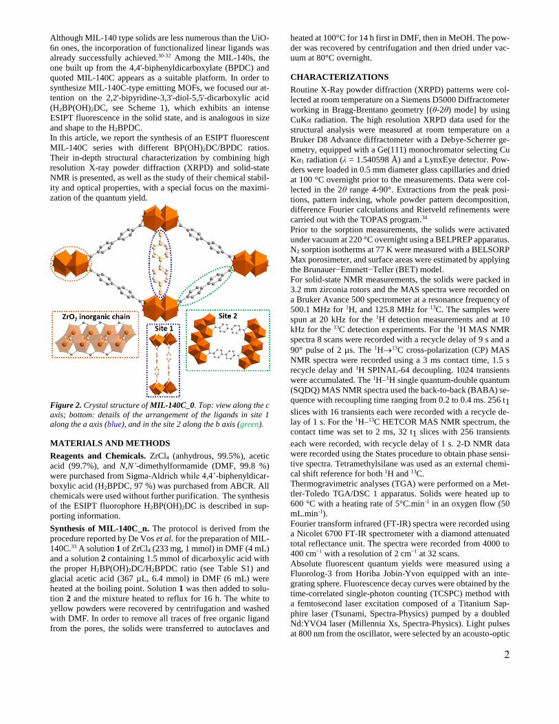
ESIPT is an ultrafast photochemical process and its resulting
emission characterized by an “abnormally” large Stokes shift
and a very environment-specific sensitivity induced by the in-
tramolecular hydrogen bond. A four-level photo-cycle (E E*
K* K) diagram is shown in Figure 1 to illustrate the main
photochemical and photophysical processes involved in the
case of an archetypal molecule of this family, the 2-(2-hydrox-
yphenyl)-benzothiazole (HBT). Unlike many organic emitters,
self-quenching in ESIPT-type fluorophores is minimized due to
the absence of spectral overlapping between absorption and
emission. This unique feature of ESIPT emission is appealing
for the design of efficient fluorescent materials.12-13 ESIPT
chromophores-based MOFs are thus prime candidates for po-
tential applications in a wide variety of fields such as chemical
sensors, fluorescence imaging or light emitting diodes. To date,
only a very limited number of porous materials with ESIPT-
type fluorescence have been reported, showing far above aver-
age fluorescent quantum yields.2,14-23 Most of these solids are
based on divalent cations (Mg, Zn, Ca, Sr, Ba) and carboxylate
linkers, and are therefore prone to hydrolysis.24
Figure 1. Diagram of the ESIPT process. Adapted from ref 13. Cop-
yright 2016 The Royal Society of Chemistry.
Group IV metal ions are well known for their strong affinity
with oxygenated complexing groups, resulting in high metal-
ligand bond stability toward hydrolysis.25-27 Based on this con-
sideration, we focused our attention on Zr(IV)-dicarboxylate
MOFs. While the UiO-6n series exhibits the well-known fcc
structure type with twelve organically linked Zr6 oxo-clusters
leading to octahedral and tetrahedral cages, the pseudo-poly-
morphs MIL-140s are built up from chains of edge sharing ZrO7
polyhedra connected through the ligands to define triangular
channels.28-29 In this structure type, the organic moieties are lo-
cated on two independent crystallographic sites in a 1 to 1 ratio
(Figure 2). MIL-140s present lower porosities than their UiO-
6n analogues, but higher chemical and mechanical stabilities.29
E* (S1)
E (S0)Enol form
En
ol
em
issio
n
Keto
em
issio
n
K* (S1)
K (S0)Keto form
hν
enol form cis-keto form
2
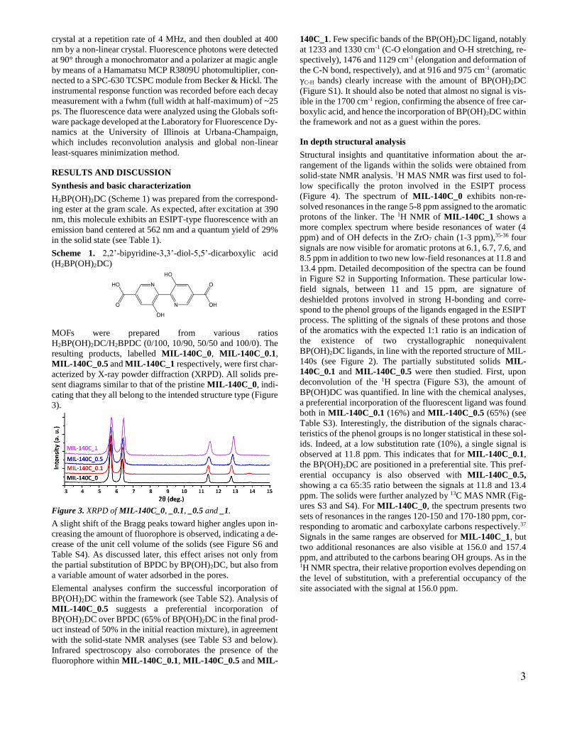
Although MIL-140 type solids are less numerous than the UiO-
6n ones, the incorporation of functionalized linear ligands was
already successfully achieved.30-32 Among the MIL-140s, the
one built up from the 4,4'-biphenyldicarboxylate (BPDC) and
quoted MIL-140C appears as a suitable platform. In order to
synthesize MIL-140C-type emitting MOFs, we focused our at-
tention on the 2,2'-bipyridine-3,3'-diol-5,5'-dicarboxylic acid
(H2BP(OH)2DC, see Scheme 1), which exhibits an intense
ESIPT fluorescence in the solid state, and is analogous in size
and shape to the H2BPDC.
In this article, we report the synthesis of an ESIPT fluorescent
MIL-140C series with different BP(OH)2DC/BPDC ratios.
Their in-depth structural characterization by combining high
resolution X-ray powder diffraction (XRPD) and solid-state
NMR is presented, as well as the study of their chemical stabil-
ity and optical properties, with a special focus on the maximi-
zation of the quantum yield.
Figure 2. Crystal structure of MIL-140C_0. Top: view along the c
axis; bottom: details of the arrangement of the ligands in site 1
along the a axis (blue), and in the site 2 along the b axis (green).
MATERIALS AND METHODS
Reagents and Chemicals. ZrCl4 (anhydrous, 99.5%), acetic
acid (99.7%), and N,N’-dimethylformamide (DMF, 99.8 %)
were purchased from Sigma-Aldrich while 4,4’-biphenyldicar-
boxylic acid (H2BPDC, 97 %) was purchased from ABCR. All
chemicals were used without further purification. The synthesis
of the ESIPT fluorophore H2BP(OH)2DC is described in sup-
porting information.
Synthesis of MIL-140C_n. The protocol is derived from the
procedure reported by De Vos et al. for the preparation of MIL-
140C.33 A solution 1 of ZrCl4 (233 mg, 1 mmol) in DMF (4 mL)
and a solution 2 containing 1.5 mmol of dicarboxylic acid with
the proper H2BP(OH)2DC/H2BPDC ratio (see Table S1) and
glacial acetic acid (367 µL, 6.4 mmol) in DMF (6 mL) were
heated at the boiling point. Solution 1 was then added to solu-
tion 2 and the mixture heated to reflux for 16 h. The white to
yellow powders were recovered by centrifugation and washed
with DMF. In order to remove all traces of free organic ligand
from the pores, the solids were transferred to autoclaves and
heated at 100°C for 14 h first in DMF, then in MeOH. The pow-
der was recovered by centrifugation and then dried under vac-
uum at 80°C overnight.
CHARACTERIZATIONS
Routine X-Ray powder diffraction (XRPD) patterns were col-
lected at room temperature on a Siemens D5000 Diffractometer
working in Bragg-Brentano geometry [(θ-2θ) mode] by using
CuKα radiation. The high resolution XRPD data used for the
structural analysis were measured at room temperature on a
Bruker D8 Advance diffractometer with a Debye-Scherrer ge-
ometry, equipped with a Ge(111) monochromator selecting Cu
Kα1 radiation (λ = 1.540598 Å) and a LynxEye detector. Pow-
ders were loaded in 0.5 mm diameter glass capillaries and dried
at 100 °C overnight prior to the measurements. Data were col-
lected in the 2θ range 4-90°. Extractions from the peak posi-
tions, pattern indexing, whole powder pattern decomposition,
difference Fourier calculations and Rietveld refinements were
carried out with the TOPAS program.34
Prior to the sorption measurements, the solids were activated
under vacuum at 220 °C overnight using a BELPREP apparatus.
N2 sorption isotherms at 77 K were measured with a BELSORP
Max porosimeter, and surface areas were estimated by applying
the Brunauer−Emmett−Teller (BET) model.
For solid-state NMR measurements, the solids were packed in
3.2 mm zirconia rotors and the MAS spectra were recorded on
a Bruker Avance 500 spectrometer at a resonance frequency of
500.1 MHz for 1H, and 125.8 MHz for 13C. The samples were
spun at 20 kHz for the 1H detection measurements and at 10
kHz for the 13C detection experiments. For the 1H MAS NMR
spectra 8 scans were recorded with a recycle delay of 9 s and a
90° pulse of 2 µs. The 1H13C cross-polarization (CP) MAS
NMR spectra were recorded using a 3 ms contact time, 1.5 s
recycle delay and 1H SPINAL-64 decoupling. 1024 transients
were accumulated. The 1H–1H single quantum-double quantum
(SQDQ) MAS NMR spectra used the back-to-back (BABA) se-
quence with recoupling time ranging from 0.2 to 0.4 ms. 256 t1
slices with 16 transients each were recorded with a recycle de-
lay of 1 s. For the 1H–13C HETCOR MAS NMR spectrum, the
contact time was set to 2 ms, 32 t1 slices with 256 transients
each were recorded, with recycle delay of 1 s. 2-D NMR data
were recorded using the States procedure to obtain phase sensi-
tive spectra. Tetramethylsilane was used as an external chemi-
cal shift reference for both 1H and 13C.
Thermogravimetric analyses (TGA) were performed on a Met-
tler-Toledo TGA/DSC 1 apparatus. Solids were heated up to
600 °C with a heating rate of 5°C.min-1 in an oxygen flow (50
mL.min-1).
Fourier transform infrared (FT-IR) spectra were recorded using
a Nicolet 6700 FT-IR spectrometer with a diamond attenuated
total reflectance unit. The spectra were recorded from 4000 to
400 cm−1 with a resolution of 2 cm−1 at 32 scans.
Absolute fluorescent quantum yields were measured using a
Fluorolog-3 from Horiba Jobin-Yvon equipped with an inte-
grating sphere. Fluorescence decay curves were obtained by the
time-correlated single-photon counting (TCSPC) method with
a femtosecond laser excitation composed of a Titanium Sap-
phire laser (Tsunami, Spectra-Physics) pumped by a doubled
Nd:YVO4 laser (Millennia Xs, Spectra-Physics). Light pulses
at 800 nm from the oscillator, were selected by an acousto-optic
3
crystal at a repetition rate of 4 MHz, and then doubled at 400
nm by a non-linear crystal. Fluorescence photons were detected
at 90° through a monochromator and a polarizer at magic angle
by means of a Hamamatsu MCP R3809U photomultiplier, con-
nected to a SPC-630 TCSPC module from Becker & Hickl. The
instrumental response function was recorded before each decay
measurement with a fwhm (full width at half-maximum) of ~25
ps. The fluorescence data were analyzed using the Globals soft-
ware package developed at the Laboratory for Fluorescence Dy-
namics at the University of Illinois at Urbana-Champaign,
which includes reconvolution analysis and global non-linear
least-squares minimization method.
RESULTS AND DISCUSSION
Synthesis and basic characterization
H2BP(OH)2DC (Scheme 1) was prepared from the correspond-
ing ester at the gram scale. As expected, after excitation at 390
nm, this molecule exhibits an ESIPT-type fluorescence with an
emission band centered at 562 nm and a quantum yield of 29%
in the solid state (see Table 1).
Scheme 1. 2,2’-bipyridine-3,3’-diol-5,5’-dicarboxylic acid
(H2BP(OH)2DC)
MOFs were prepared from various ratios
H2BP(OH)2DC/H2BPDC (0/100, 10/90, 50/50 and 100/0). The
resulting products, labelled MIL-140C_0, MIL-140C_0.1,
MIL-140C_0.5 and MIL-140C_1 respectively, were first char-
acterized by X-ray powder diffraction (XRPD). All solids pre-
sent diagrams similar to that of the pristine MIL-140C_0, indi-
cating that they all belong to the intended structure type (Figure
3).
Figure 3. XRPD of MIL-140C_0, _0.1, _0.5 and _1.
A slight shift of the Bragg peaks toward higher angles upon in-
creasing the amount of fluorophore is observed, indicating a de-
crease of the unit cell volume of the solids (see Figure S6 and
Table S4). As discussed later, this effect arises not only from
the partial substitution of BPDC by BP(OH)2DC, but also from
a variable amount of water adsorbed in the pores.
Elemental analyses confirm the successful incorporation of
BP(OH)2DC within the framework (see Table S2). Analysis of
MIL-140C_0.5 suggests a preferential incorporation of
BP(OH)2DC over BPDC (65% of BP(OH)2DC in the final prod-
uct instead of 50% in the initial reaction mixture), in agreement
with the solid-state NMR analyses (see Table S3 and below).
Infrared spectroscopy also corroborates the presence of the
fluorophore within MIL-140C_0.1, MIL-140C_0.5 and MIL-
140C_1. Few specific bands of the BP(OH)2DC ligand, notably
at 1233 and 1330 cm-1 (C-O elongation and O-H stretching, re-
spectively), 1476 and 1129 cm-1 (elongation and deformation of
the C-N bond, respectively), and at 916 and 975 cm-1 (aromatic
γC-H bands) clearly increase with the amount of BP(OH)2DC
(Figure S1). It should also be noted that almost no signal is vis-
ible in the 1700 cm-1 region, confirming the absence of free car-
boxylic acid, and hence the incorporation of BP(OH)2DC within
the framework and not as a guest within the pores.
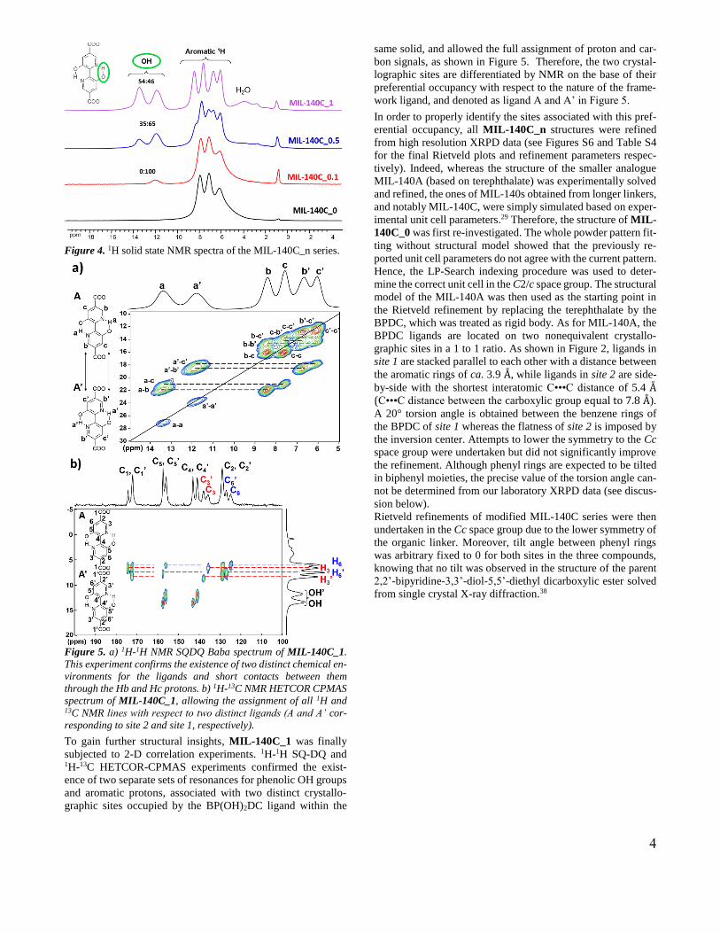
In depth structural analysis
Structural insights and quantitative information about the ar-
rangement of the ligands within the solids were obtained from
solid-state NMR analysis. 1H MAS NMR was first used to fol-
low specifically the proton involved in the ESIPT process
(Figure 4). The spectrum of MIL-140C_0 exhibits non-re-
solved resonances in the range 5-8 ppm assigned to the aromatic
protons of the linker. The 1H NMR of MIL-140C_1 shows a
more complex spectrum where beside resonances of water (4
ppm) and of OH defects in the ZrO7 chain (1-3 ppm),35-36 four
signals are now visible for aromatic protons at 6.1, 6.7, 7.6, and
8.5 ppm in addition to two new low-field resonances at 11.8 and
13.4 ppm. Detailed decomposition of the spectra can be found
in Figure S2 in Supporting Information. These particular low-
field signals, between 11 and 15 ppm, are signature of
deshielded protons involved in strong H-bonding and corre-
spond to the phenol groups of the ligands engaged in the ESIPT
process. The splitting of the signals of these protons and those
of the aromatics with the expected 1:1 ratio is an indication of
the existence of two crystallographic nonequivalent
BP(OH)2DC ligands, in line with the reported structure of MIL-
140s (see Figure 2). The partially substituted solids MIL-
140C_0.1 and MIL-140C_0.5 were then studied. First, upon
deconvolution of the 1H spectra (Figure S3), the amount of
BP(OH)DC was quantified. In line with the chemical analyses,
a preferential incorporation of the fluorescent ligand was found
both in MIL-140C_0.1 (16%) and MIL-140C_0.5 (65%) (see
Table S3). Interestingly, the distribution of the signals charac-
teristics of the phenol groups is no longer statistical in these sol-
ids. Indeed, at a low substitution rate (10%), a single signal is
observed at 11.8 ppm. This indicates that for MIL-140C_0.1,
the BP(OH)2DC are positioned in a preferential site. This pref-
erential occupancy is also observed with MIL-140C_0.5,
showing a ca 65:35 ratio between the signals at 11.8 and 13.4
ppm. The solids were further analyzed by 13C MAS NMR (Fig-
ures S3 and S4). For MIL-140C_0, the spectrum presents two
sets of resonances in the ranges 120-150 and 170-180 ppm, cor-
responding to aromatic and carboxylate carbons respectively.37
Signals in the same ranges are observed for MIL-140C_1, but
two additional resonances are also visible at 156.0 and 157.4
ppm, and attributed to the carbons bearing OH groups. As in the 1H NMR spectra, their relative proportion evolves depending on
the level of substitution, with a preferential occupancy of the
site associated with the signal at 156.0 ppm.
4
Figure 4. 1H solid state NMR spectra of the MIL-140C_n series.
Figure 5. a) 1H-1H NMR SQDQ Baba spectrum of MIL-140C_1.
This experiment confirms the existence of two distinct chemical en-
vironments for the ligands and short contacts between them
through the Hb and Hc protons. b) 1H-13C NMR HETCOR CPMAS
spectrum of MIL-140C_1, allowing the assignment of all 1H and 13C NMR lines with respect to two distinct ligands (A and A’ cor-
responding to site 2 and site 1, respectively).
To gain further structural insights, MIL-140C_1 was finally
subjected to 2-D correlation experiments. 1H-1H SQ-DQ and 1H-13C HETCOR-CPMAS experiments confirmed the exist-
ence of two separate sets of resonances for phenolic OH groups
and aromatic protons, associated with two distinct crystallo-
graphic sites occupied by the BP(OH)2DC ligand within the
same solid, and allowed the full assignment of proton and car-
bon signals, as shown in Figure 5. Therefore, the two crystal-
lographic sites are differentiated by NMR on the base of their
preferential occupancy with respect to the nature of the frame-
work ligand, and denoted as ligand A and A’ in Figure 5.
In order to properly identify the sites associated with this pref-
erential occupancy, all MIL-140C_n structures were refined
from high resolution XRPD data (see Figures S6 and Table S4
for the final Rietveld plots and refinement parameters respec-
tively). Indeed, whereas the structure of the smaller analogue
MIL-140A (based on terephthalate) was experimentally solved
and refined, the ones of MIL-140s obtained from longer linkers,
and notably MIL-140C, were simply simulated based on exper-
imental unit cell parameters.29 Therefore, the structure of MIL-
140C_0 was first re-investigated. The whole powder pattern fit-
ting without structural model showed that the previously re-
ported unit cell parameters do not agree with the current pattern.
Hence, the LP-Search indexing procedure was used to deter-
mine the correct unit cell in the C2/c space group. The structural
model of the MIL-140A was then used as the starting point in
the Rietveld refinement by replacing the terephthalate by the
BPDC, which was treated as rigid body. As for MIL-140A, the
BPDC ligands are located on two nonequivalent crystallo-
graphic sites in a 1 to 1 ratio. As shown in Figure 2, ligands in
site 1 are stacked parallel to each other with a distance between
the aromatic rings of ca. 3.9 Å, while ligands in site 2 are side-
by-side with the shortest interatomic C•••C distance of 5.4 Å (C•••C distance between the carboxylic group equal to 7.8 Å).
A 20° torsion angle is obtained between the benzene rings of
the BPDC of site 1 whereas the flatness of site 2 is imposed by
the inversion center. Attempts to lower the symmetry to the Cc
space group were undertaken but did not significantly improve
the refinement. Although phenyl rings are expected to be tilted
in biphenyl moieties, the precise value of the torsion angle can-
not be determined from our laboratory XRPD data (see discus-
sion below).
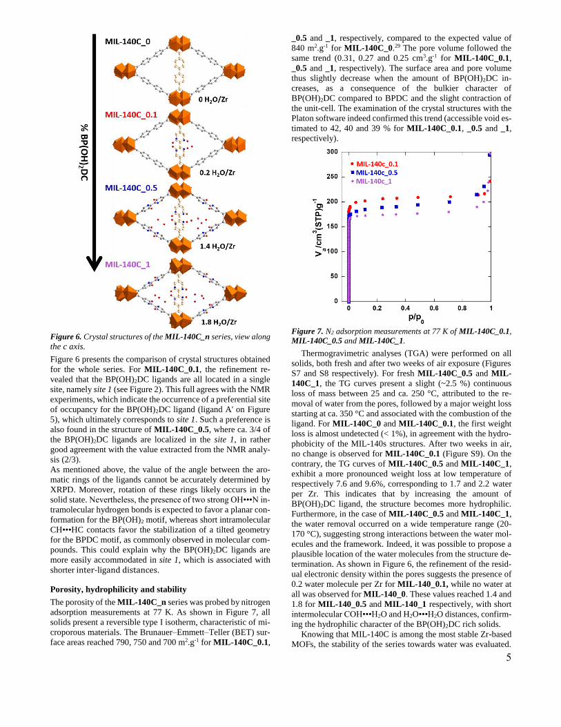
Rietveld refinements of modified MIL-140C series were then
undertaken in the Cc space group due to the lower symmetry of
the organic linker. Moreover, tilt angle between phenyl rings
was arbitrary fixed to 0 for both sites in the three compounds,
knowing that no tilt was observed in the structure of the parent
2,2’-bipyridine-3,3’-diol-5,5’-diethyl dicarboxylic ester solved
from single crystal X-ray diffraction.38
5
Figure 6. Crystal structures of the MIL-140C_n series, view along
the c axis.
Figure 6 presents the comparison of crystal structures obtained
for the whole series. For MIL-140C_0.1, the refinement re-
vealed that the BP(OH)2DC ligands are all located in a single
site, namely site 1 (see Figure 2). This full agrees with the NMR
experiments, which indicate the occurrence of a preferential site
of occupancy for the BP(OH)2DC ligand (ligand A' on Figure
5), which ultimately corresponds to site 1. Such a preference is
also found in the structure of MIL-140C_0.5, where ca. 3/4 of
the BP(OH)2DC ligands are localized in the site 1, in rather
good agreement with the value extracted from the NMR analy-
sis (2/3).
As mentioned above, the value of the angle between the aro-
matic rings of the ligands cannot be accurately determined by
XRPD. Moreover, rotation of these rings likely occurs in the
solid state. Nevertheless, the presence of two strong OH•••N in-
tramolecular hydrogen bonds is expected to favor a planar con-
formation for the BP(OH)2 motif, whereas short intramolecular
CH•••HC contacts favor the stabilization of a tilted geometry
for the BPDC motif, as commonly observed in molecular com-
pounds. This could explain why the BP(OH)2DC ligands are
more easily accommodated in site 1, which is associated with
shorter inter-ligand distances.
Porosity, hydrophilicity and stability
The porosity of the MIL-140C_n series was probed by nitrogen
adsorption measurements at 77 K. As shown in Figure 7, all
solids present a reversible type I isotherm, characteristic of mi-
croporous materials. The Brunauer–Emmett–Teller (BET) sur-
face areas reached 790, 750 and 700 m2.g-1 for MIL-140C_0.1,
_0.5 and _1, respectively, compared to the expected value of
840 m2.g-1 for MIL-140C_0.29 The pore volume followed the
same trend (0.31, 0.27 and 0.25 cm3.g-1 for MIL-140C_0.1,
_0.5 and _1, respectively). The surface area and pore volume
thus slightly decrease when the amount of BP(OH)2DC in-
creases, as a consequence of the bulkier character of
BP(OH)2DC compared to BPDC and the slight contraction of
the unit-cell. The examination of the crystal structures with the
Platon software indeed confirmed this trend (accessible void es-
timated to 42, 40 and 39 % for MIL-140C_0.1, _0.5 and _1,
respectively).
Figure 7. N2 adsorption measurements at 77 K of MIL-140C_0.1,
MIL-140C_0.5 and MIL-140C_1.
Thermogravimetric analyses (TGA) were performed on all
solids, both fresh and after two weeks of air exposure (Figures
S7 and S8 respectively). For fresh MIL-140C_0.5 and MIL-
140C_1, the TG curves present a slight (~2.5 %) continuous
loss of mass between 25 and ca. 250 °C, attributed to the re-
moval of water from the pores, followed by a major weight loss
starting at ca. 350 °C and associated with the combustion of the
ligand. For MIL-140C_0 and MIL-140C_0.1, the first weight
loss is almost undetected (< 1%), in agreement with the hydro-
phobicity of the MIL-140s structures. After two weeks in air,
no change is observed for MIL-140C_0.1 (Figure S9). On the
contrary, the TG curves of MIL-140C_0.5 and MIL-140C_1,
exhibit a more pronounced weight loss at low temperature of
respectively 7.6 and 9.6%, corresponding to 1.7 and 2.2 water
per Zr. This indicates that by increasing the amount of
BP(OH)2DC ligand, the structure becomes more hydrophilic.
Furthermore, in the case of MIL-140C_0.5 and MIL-140C_1,
the water removal occurred on a wide temperature range (20-
170 °C), suggesting strong interactions between the water mol-
ecules and the framework. Indeed, it was possible to propose a
plausible location of the water molecules from the structure de-
termination. As shown in Figure 6, the refinement of the resid-
ual electronic density within the pores suggests the presence of
0.2 water molecule per Zr for MIL-140_0.1, while no water at
all was observed for MIL-140_0. These values reached 1.4 and
1.8 for MIL-140_0.5 and MIL-140_1 respectively, with short
intermolecular COH•••H2O and H2O•••H2O distances, confirm-
ing the hydrophilic character of the BP(OH)2DC rich solids.
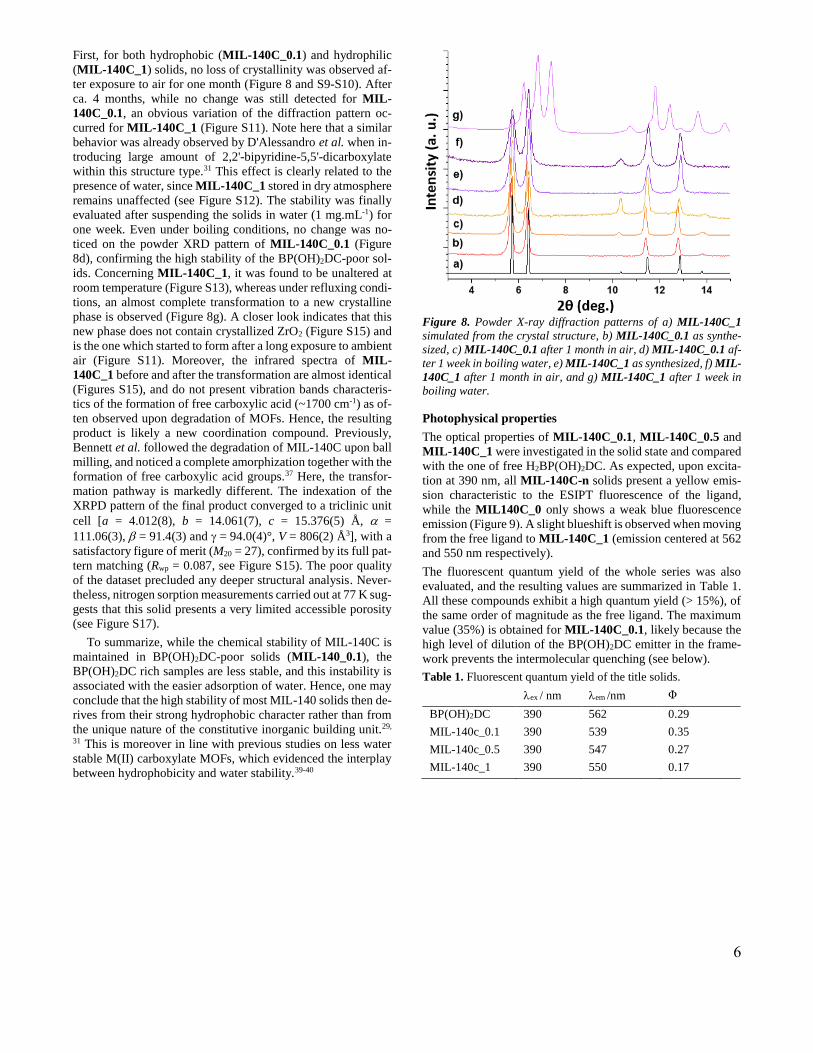
Knowing that MIL-140C is among the most stable Zr-based
MOFs, the stability of the series towards water was evaluated.
6
First, for both hydrophobic (MIL-140C_0.1) and hydrophilic
(MIL-140C_1) solids, no loss of crystallinity was observed af-
ter exposure to air for one month (Figure 8 and S9-S10). After
ca. 4 months, while no change was still detected for MIL-
140C_0.1, an obvious variation of the diffraction pattern oc-
curred for MIL-140C_1 (Figure S11). Note here that a similar
behavior was already observed by D'Alessandro et al. when in-
troducing large amount of 2,2'-bipyridine-5,5'-dicarboxylate
within this structure type.31 This effect is clearly related to the
presence of water, since MIL-140C_1 stored in dry atmosphere
remains unaffected (see Figure S12). The stability was finally
evaluated after suspending the solids in water (1 mg.mL-1) for
one week. Even under boiling conditions, no change was no-
ticed on the powder XRD pattern of MIL-140C_0.1 (Figure
8d), confirming the high stability of the BP(OH)2DC-poor sol-
ids. Concerning MIL-140C_1, it was found to be unaltered at
room temperature (Figure S13), whereas under refluxing condi-
tions, an almost complete transformation to a new crystalline
phase is observed (Figure 8g). A closer look indicates that this
new phase does not contain crystallized ZrO2 (Figure S15) and
is the one which started to form after a long exposure to ambient
air (Figure S11). Moreover, the infrared spectra of MIL-
140C_1 before and after the transformation are almost identical
(Figures S15), and do not present vibration bands characteris-
tics of the formation of free carboxylic acid (~1700 cm-1) as of-
ten observed upon degradation of MOFs. Hence, the resulting
product is likely a new coordination compound. Previously,
Bennett et al. followed the degradation of MIL-140C upon ball
milling, and noticed a complete amorphization together with the
formation of free carboxylic acid groups.37 Here, the transfor-
mation pathway is markedly different. The indexation of the
XRPD pattern of the final product converged to a triclinic unit
cell [a = 4.012(8), b = 14.061(7), c = 15.376(5) Å, =
111.06(3), = 91.4(3) and = 94.0(4)°, V = 806(2) Å3], with a
satisfactory figure of merit (M20 = 27), confirmed by its full pat-
tern matching (Rwp = 0.087, see Figure S15). The poor quality
of the dataset precluded any deeper structural analysis. Never-
theless, nitrogen sorption measurements carried out at 77 K sug-
gests that this solid presents a very limited accessible porosity
(see Figure S17).
To summarize, while the chemical stability of MIL-140C is
maintained in BP(OH)2DC-poor solids (MIL-140_0.1), the
BP(OH)2DC rich samples are less stable, and this instability is
associated with the easier adsorption of water. Hence, one may
conclude that the high stability of most MIL-140 solids then de-
rives from their strong hydrophobic character rather than from
the unique nature of the constitutive inorganic building unit.29,
31 This is moreover in line with previous studies on less water
stable M(II) carboxylate MOFs, which evidenced the interplay
between hydrophobicity and water stability.39-40
Figure 8. Powder X-ray diffraction patterns of a) MIL-140C_1
simulated from the crystal structure, b) MIL-140C_0.1 as synthe-
sized, c) MIL-140C_0.1 after 1 month in air, d) MIL-140C_0.1 af-
ter 1 week in boiling water, e) MIL-140C_1 as synthesized, f) MIL-
140C_1 after 1 month in air, and g) MIL-140C_1 after 1 week in
boiling water.
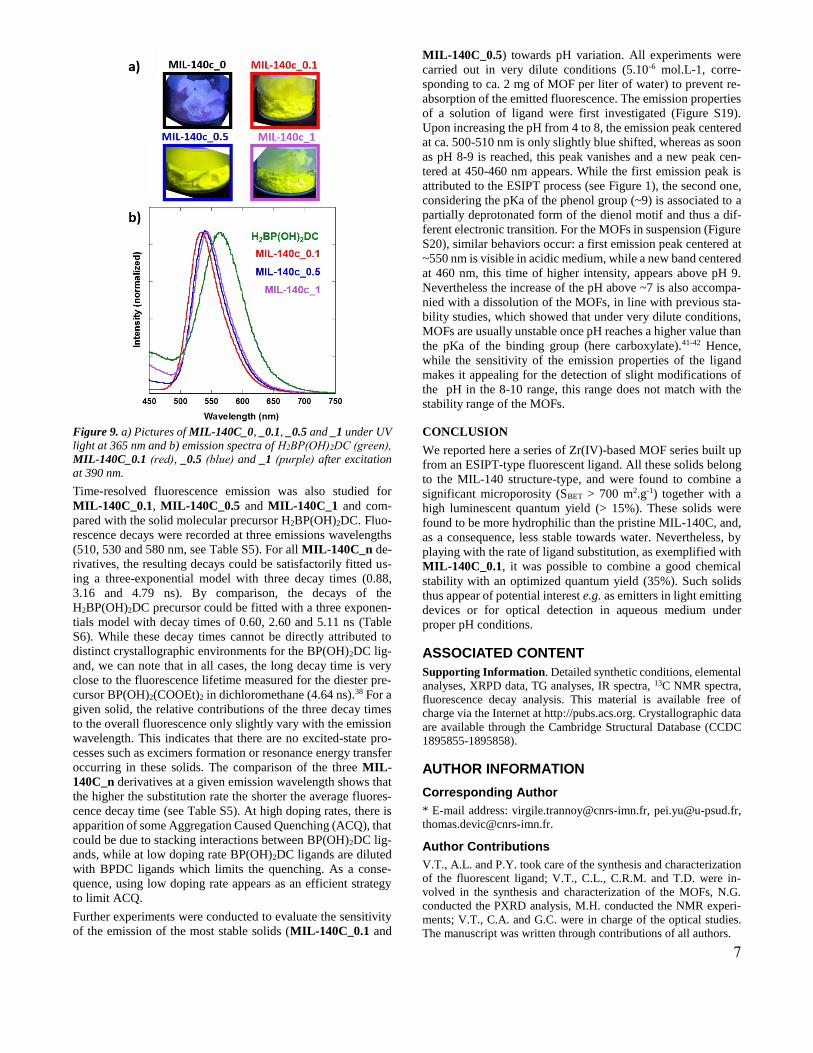
Photophysical properties
The optical properties of MIL-140C_0.1, MIL-140C_0.5 and
MIL-140C_1 were investigated in the solid state and compared
with the one of free H2BP(OH)2DC. As expected, upon excita-
tion at 390 nm, all MIL-140C-n solids present a yellow emis-
sion characteristic to the ESIPT fluorescence of the ligand,
while the MIL140C_0 only shows a weak blue fluorescence
emission (Figure 9). A slight blueshift is observed when moving
from the free ligand to MIL-140C_1 (emission centered at 562
and 550 nm respectively).
The fluorescent quantum yield of the whole series was also
evaluated, and the resulting values are summarized in Table 1.
All these compounds exhibit a high quantum yield (> 15%), of
the same order of magnitude as the free ligand. The maximum
value (35%) is obtained for MIL-140C_0.1, likely because the
high level of dilution of the BP(OH)2DC emitter in the frame-
work prevents the intermolecular quenching (see below).
Table 1. Fluorescent quantum yield of the title solids.
ex / nm em /nm Φ
BP(OH)2DC 390 562 0.29
MIL-140c_0.1 390 539 0.35
MIL-140c_0.5 390 547 0.27
MIL-140c_1 390 550 0.17
7
Figure 9. a) Pictures of MIL-140C_0, _0.1, _0.5 and _1 under UV
light at 365 nm and b) emission spectra of H2BP(OH)2DC (green),
MIL-140C_0.1 (red), _0.5 (blue) and _1 (purple) after excitation
at 390 nm.
Time-resolved fluorescence emission was also studied for
MIL-140C_0.1, MIL-140C_0.5 and MIL-140C_1 and com-
pared with the solid molecular precursor H2BP(OH)2DC. Fluo-
rescence decays were recorded at three emissions wavelengths
(510, 530 and 580 nm, see Table S5). For all MIL-140C_n de-
rivatives, the resulting decays could be satisfactorily fitted us-
ing a three-exponential model with three decay times (0.88,
3.16 and 4.79 ns). By comparison, the decays of the
H2BP(OH)2DC precursor could be fitted with a three exponen-
tials model with decay times of 0.60, 2.60 and 5.11 ns (Table
S6). While these decay times cannot be directly attributed to
distinct crystallographic environments for the BP(OH)2DC lig-
and, we can note that in all cases, the long decay time is very
close to the fluorescence lifetime measured for the diester pre-
cursor BP(OH)2(COOEt)2 in dichloromethane (4.64 ns).38 For a
given solid, the relative contributions of the three decay times
to the overall fluorescence only slightly vary with the emission
wavelength. This indicates that there are no excited-state pro-
cesses such as excimers formation or resonance energy transfer
occurring in these solids. The comparison of the three MIL-
140C_n derivatives at a given emission wavelength shows that
the higher the substitution rate the shorter the average fluores-
cence decay time (see Table S5). At high doping rates, there is
apparition of some Aggregation Caused Quenching (ACQ), that
could be due to stacking interactions between BP(OH)2DC lig-
ands, while at low doping rate BP(OH)2DC ligands are diluted
with BPDC ligands which limits the quenching. As a conse-
quence, using low doping rate appears as an efficient strategy
to limit ACQ.
Further experiments were conducted to evaluate the sensitivity
of the emission of the most stable solids (MIL-140C_0.1 and
MIL-140C_0.5) towards pH variation. All experiments were
carried out in very dilute conditions (5.10-6 mol.L-1, corre-
sponding to ca. 2 mg of MOF per liter of water) to prevent re-
absorption of the emitted fluorescence. The emission properties
of a solution of ligand were first investigated (Figure S19).
Upon increasing the pH from 4 to 8, the emission peak centered
at ca. 500-510 nm is only slightly blue shifted, whereas as soon
as pH 8-9 is reached, this peak vanishes and a new peak cen-
tered at 450-460 nm appears. While the first emission peak is
attributed to the ESIPT process (see Figure 1), the second one,
considering the pKa of the phenol group (~9) is associated to a
partially deprotonated form of the dienol motif and thus a dif-
ferent electronic transition. For the MOFs in suspension (Figure
S20), similar behaviors occur: a first emission peak centered at
~550 nm is visible in acidic medium, while a new band centered
at 460 nm, this time of higher intensity, appears above pH 9.
Nevertheless the increase of the pH above ~7 is also accompa-
nied with a dissolution of the MOFs, in line with previous sta-
bility studies, which showed that under very dilute conditions,
MOFs are usually unstable once pH reaches a higher value than
the pKa of the binding group (here carboxylate).41-42 Hence,
while the sensitivity of the emission properties of the ligand
makes it appealing for the detection of slight modifications of
the pH in the 8-10 range, this range does not match with the
stability range of the MOFs.
CONCLUSION
We reported here a series of Zr(IV)-based MOF series built up
from an ESIPT-type fluorescent ligand. All these solids belong
to the MIL-140 structure-type, and were found to combine a
significant microporosity (SBET > 700 m2.g-1) together with a
high luminescent quantum yield (> 15%). These solids were
found to be more hydrophilic than the pristine MIL-140C, and,
as a consequence, less stable towards water. Nevertheless, by
playing with the rate of ligand substitution, as exemplified with
MIL-140C_0.1, it was possible to combine a good chemical
stability with an optimized quantum yield (35%). Such solids
thus appear of potential interest e.g. as emitters in light emitting
devices or for optical detection in aqueous medium under
proper pH conditions.
ASSOCIATED CONTENT
Supporting Information. Detailed synthetic conditions, elemental
analyses, XRPD data, TG analyses, IR spectra, 13C NMR spectra,
fluorescence decay analysis. This material is available free of
charge via the Internet at http://pubs.acs.org. Crystallographic data
are available through the Cambridge Structural Database (CCDC
1895855-1895858).
AUTHOR INFORMATION
Corresponding Author
* E-mail address: [email protected], [email protected],
Author Contributions
V.T., A.L. and P.Y. took care of the synthesis and characterization
of the fluorescent ligand; V.T., C.L., C.R.M. and T.D. were in-
volved in the synthesis and characterization of the MOFs, N.G.
conducted the PXRD analysis, M.H. conducted the NMR experi-
ments; V.T., C.A. and G.C. were in charge of the optical studies.
The manuscript was written through contributions of all authors.
8
Funding Sources
This work was supported by a public grant overseen by the French
National Research Agency (ANR) as part of the “Inves-tissements
d’Avenir” Program no. CHARMMMAT ANR-11-LABX-0039.
ACKNOWLEDGMENT
We acknowledge the labex CHARMMMAT (ANR-11-LABEX-
0039) for the postdoctoral fellowships of Virgile Trannoy. Arnaud
Brosseau (PPSM) is gratefully acknowledged for his help with the
time-resolved fluorescence measurements.
REFERENCES
1. Allendorf, M. D.; Bauer, C. A.; Bhakta, R. K.; Houk, R. J.
T., Luminescent metal-organic frameworks. Chem. Soc. Rev. 2009, 38,
1330-1352.
2. Lustig, W. P.; Mukherjee, S.; Rudd, N. D.; Desai, A. V.; Li,
J.; Ghosh, S. K., Metal-organic frameworks: functional luminescent
and photonic materials for sensing applications. Chem. Soc. Rev. 2017,
46, 3242-3285.
3. Zhang, Y.; Yuan, S.; Day, G.; Wang, X.; Yang, X.; Zhou,
H.-C., Luminescent sensors based on metal-organic frameworks.
Coord. Chem. Rev. 2018, 354, 28-45.
4. Cui, Y.; Yue, Y.; Qian, G.; Chen, B., Luminescent
Functional Metal–Organic Frameworks. Chem. Rev. 2012, 112, 1126-
1162.
5. Liu, D.; Lu, K.; Poon, C.; Lin, W., Metal–Organic
Frameworks as Sensory Materials and Imaging Agents. Inorg. Chem.
2014, 53, 1916-1924.
6. Mahata, P.; Mondal, S. K.; Singha, D. K.; Majee, P.,
Luminescent rare-earth-based MOFs as optical sensors. Dalton Trans.
2017, 46, 301-328.
7. Müller-Buschbaum, K.; Beuerle, F.; Feldmann, C., MOF
based luminescence tuning and chemical/physical sensing.
Microporous Mesoporous Mater. 2015, 216, 171-199.
8. Monguzzi, A.; Ballabio, M.; Yanai, N.; Kimizuka, N.; Fazzi,
D.; Campione, M.; Meinardi, F., Highly Fluorescent Metal–Organic-
Framework Nanocomposites for Photonic Applications. Nano Lett.
2018, 18, 528-534.
9. Liu, X.-G.; Tao, C.-L.; Yu, H.-Q.; Chen, B.; Liu, Z.; Zhu,
G.-P.; Zhao, Z.; Shen, L.; Tang, B. Z., A new luminescent metal-
organic framework based on dicarboxyl-substituted tetraphenylethene
for efficient detection of nitro-containing explosives and antibiotics in
aqueous media. J. Mater. Chem. C 2018, 6, 2983-2988.
10. Li, Q.-Y.; Ma, Z.; Zhang, W.-Q.; Xu, J.-L.; Wei, W.; Lu, H.;
Zhao, X.; Wang, X.-J., AIE-active tetraphenylethene functionalized
metal-organic framework for selective detection of nitroaromatic
explosives and organic photocatalysis. Chem. Commun. 2016, 52,
11284-11287.
11. Deng, Y.; Chen, N.; Li, Q.; Wu, X.; Huang, X.; Lin, Z.;
Zhao, Y., Highly Fluorescent Metal–Organic Frameworks Based on a
Benzene-Cored Tetraphenylethene Derivative with the Ability To
Detect 2,4,6-Trinitrophenol in Water. Crystal Growth Des. 2017, 17,
3170-3177.
12. Eon, K. J.; Young, P. S., Advanced Organic Optoelectronic
Materials: Harnessing Excited-State Intramolecular Proton Transfer
(ESIPT) Process. Adv. Mater. 2011, 23, 3615-3642.
13. Padalkar, V. S.; Seki, S., Excited-state intramolecular
proton-transfer (ESIPT)-inspired solid state emitters. Chem. Soc. Rev.
2016, 45, 169-202.
14. Jayaramulu, K.; Kanoo, P.; George, S. J.; Maji, T. K.,
Tunable emission from a porous metal-organic framework by
employing an excited-state intramolecular proton transfer responsive
ligand. Chem. Commun. 2010, 46, 7906-7908.
15. Jayaramulu, K.; Narayanan, R. P.; George, S. J.; Maji, T. K.,
Luminescent Microporous Metal–Organic Framework with Functional
Lewis Basic Sites on the Pore Surface: Specific Sensing and Removal
of Metal Ions. Inorg. Chem. 2012, 51, 10089-10091.
16. Shustova, N. B.; Cozzolino, A. F.; Reineke, S.; Baldo, M.;
Dincă, M., Selective Turn-On Ammonia Sensing Enabled by High-
Temperature Fluorescence in Metal–Organic Frameworks with Open
Metal Sites. J. Am. Chem. Soc. 2013, 135, 13326-13329.
17. Douvali, A.; Tsipis, A. C.; Eliseeva, S. V.; Petoud, S.;
Papaefstathiou, G. S.; Malliakas, C. D.; Papadas, I.; Armatas, G. S.;
Margiolaki, I.; Kanatzidis, M. G.; Lazarides, T.; Manos, M. J., Turn‐
On Luminescence Sensing and Real‐Time Detection of Traces of
Water in Organic Solvents by a Flexible Metal–Organic Framework.
Angew. Chem. Int. Ed. 2015, 54, 1651-1656.
18. Douvali, A.; Papaefstathiou, G. S.; Gullo, M. P.; Barbieri,
A.; Tsipis, A. C.; Malliakas, C. D.; Kanatzidis, M. G.; Papadas, I.;
Armatas, G. S.; Hatzidimitriou, A. G.; Lazarides, T.; Manos, M. J.,
Alkaline Earth Metal Ion/Dihydroxy–Terephthalate MOFs: Structural
Diversity and Unusual Luminescent Properties. Inorg. Chem. 2015, 54,
5813-5826.
19. Chen, L.; Ye, J.-W.; Wang, H.-P.; Pan, M.; Yin, S.-Y.; Wei,
Z.-W.; Zhang, L.-Y.; Wu, K.; Fan, Y.-N.; Su, C.-Y., Ultrafast water
sensing and thermal imaging by a metal-organic framework with
switchable luminescence. Nature Commun. 2017, 8, 15985.
20. Pournara, A. D.; Douvali, A.; Diamantis, S.; Papaefstathiou,
G. S.; Hatzidimitriou, A. G.; Kaziannis, S.; Kosmidis, C.; Lazarides,
T.; Manos, M. J., A new Cd2+-dihydroxyterephthalate MOF: Synthesis,
crystal structure and detailed photophysical studies. Polyhedron 2018,
151, 401-406.
21. Bhattacharya, B.; Halder, A.; Paul, L.; Chakrabarti, S.;
Ghoshal, D., Eye-Catching Dual-Fluorescent Dynamic Metal–Organic
Framework Senses Traces of Water: Experimental Findings and
Theoretical Correlation. Chem. Eur. J. 2016, 22, 14998-15005.
22. Chen, L.; Zhang, H.; Pan, M.; Wei, Z.-W.; Wang, H.-P.; Fan,
Y.-N.; Su, C.-Y., An Efficient Visible and Near-Infrared (NIR)
Emitting SmIII Metal–Organic Framework (Sm-MOF) Sensitized by
Excited-State Intramolecular Proton Transfer (ESIPT) Ligand. Chem.
Asian J. 2016, 11, 1765-1769.
23. Li, Y.-P.; Li, S.-N.; Jiang, Y.-C.; Hu, M.-C.; Zhai, Q.-G., A
semiconductor and fluorescence dual-mode room-temperature
ammonia sensor achieved by decorating hydroquinone into a metal–
organic framework. Chem. Commun. 2018, 54, 9789-9792.
24. Low, J. J.; Benin, A. I.; Jakubczak, P.; Abrahamian, J. F.;
Faheem, S. A.; Willis, R. R., Virtual High Throughput Screening
Confirmed Experimentally: Porous Coordination Polymer Hydration.
J. Am. Chem. Soc. 2009, 131, 15834-15842.
25. Kim, M.; Cohen, S. M., Discovery, development, and
functionalization of Zr(iv)-based metal-organic frameworks.
CrystEngComm 2012, 14, 4096-4104.
26. Devic, T.; Serre, C., High valence 3p and transition metal
based MOFs. Chem. Soc. Rev. 2014, 43, 6097-6115.
27. Bai, Y.; Dou, Y.; Xie, L.-H.; Rutledge, W.; Li, J.-R.; Zhou,
H.-C., Zr-based metal–organic frameworks: design, synthesis,
structure, and applications. Chem. Soc. Rev. 2016, 45, 2327-2367.
28. Cavka, J. H.; Jakobsen, S.; Olsbye, U.; Guillou, N.;
Lamberti, C.; Bordiga, S.; Lillerud, K. P., A New Zirconium Inorganic
Building Brick Forming Metal Organic Frameworks with Exceptional
Stability. J. Am. Chem. Soc. 2008, 130, 13850-13851.
29. Guillerm, V.; Ragon, F.; Dan‐Hardi, M.; Devic, T.;
Vishnuvarthan, M.; Campo, B.; Vimont, A.; Clet, G.; Yang, Q.;
Maurin, G.; Férey, G.; Vittadini, A.; Gross, S.; Serre, C., A Series of
Isoreticular, Highly Stable, Porous Zirconium Oxide Based Metal–
Organic Frameworks. Angew. Chem. Int. Ed. 2012, 51, 9267-9271.
30. Liang, W.; D'Alessandro, D. M., Microwave-assisted
solvothermal synthesis of zirconium oxide based metal–organic
frameworks. Chem. Commun. 2013, 49, 3706-3708.
31. Liang, W.; Babarao, R.; Church, T. L.; D'Alessandro, D. M.,
Tuning the cavities of zirconium-based MIL-140 frameworks to
modulate CO2 adsorption. Chem. Commun. 2015, 51, 11286-11289.
9
32. Liang, W.; Babarao, R.; D’Alessandro, D. M., Microwave-
Assisted Solvothermal Synthesis and Optical Properties of Tagged
MIL-140A Metal–Organic Frameworks. Inorg. Chem. 2013, 52,
12878-12880.
33. Van de Voorde, B.; Damasceno Borges, D.; Vermoortele,
F.; Wouters, R.; Bozbiyik, B.; Denayer, J.; Taulelle, F.; Martineau, C.;
Serre, C.; Maurin, G.; De Vos, D., Isolation of Renewable Phenolics
by Adsorption on Ultrastable Hydrophobic MIL‐140 Metal–Organic
Frameworks. ChemSusChem 2015, 8, 3159-3166.
34. Topas V5: General Profile and Structure Analysis Software
for Powder Diffraction Data, Bruker AXS Ltd: 2014.
35. Salomon, W.; Roch-Marchal, C.; Mialane, P.;
Rouschmeyer, P.; Serre, C.; Haouas, M.; Taulelle, F.; Yang, S.;
Ruhlmann, L.; Dolbecq, A., Immobilization of polyoxometalates in the
Zr-based metal organic framework UiO-67. Chem. Commun. 2015, 51,
2972-2975.
36. Lawrence, M. C.; Schneider, C.; Katz, M. J., Determining
the structural stability of UiO-67 with respect to time: a solid-state
NMR investigation. Chem. Commun. 2016, 52, 4971-4974.
37. Bennett, T. D.; Todorova, T. K.; Baxter, E. F.; Reid, D. G.;
Gervais, C.; Bueken, B.; Van de Voorde, B.; De Vos, D.; Keen, D. A.;
Mellot-Draznieks, C., Connecting defects and amorphization in UiO-
66 and MIL-140 metal–organic frameworks: a combined experimental
and computational study. Phys. Chem. Chem. Phys. 2016, 18, 2192-
2201.
38. Yu, P.; Trannoy, V.; Leaustic, A.; Guillot, R.; Geffroy, B.;
Allain, C.; Clavier, G., manuscript in preparation.
39. Jasuja, H.; Huang, Y.-g.; Walton, K. S., Adjusting the
Stability of Metal–Organic Frameworks under Humid Conditions by
Ligand Functionalization. Langmuir 2012, 28, 16874-16880.
40. Jasuja, H.; Burtch, N. C.; Huang, Y.-g.; Cai, Y.; Walton, K.
S., Kinetic Water Stability of an Isostructural Family of Zinc-Based
Pillared Metal–Organic Frameworks. Langmuir 2013, 29, 633-642.
41. Cunha, D.; Ben Yahia, M.; Hall, S.; Miller, S. R.; Chevreau,
H.; Elkaïm, E.; Maurin, G.; Horcajada, P.; Serre, C., Rationale of Drug
Encapsulation and Release from Biocompatible Porous Metal–Organic
Frameworks. Chem.Mater. 2013, 25, 2767-2776.
42. Bellido, E.; Guillevic, M.; Hidalgo, T.; Santander-Ortega,
M. J.; Serre, C.; Horcajada, P., Understanding the Colloidal Stability of
the Mesoporous MIL-100(Fe) Nanoparticles in Physiological Media.
Langmuir 2014, 30, 5911-5920.