Embed Size (px)
Citation preview
Fluorescence Microscopy
SAKULRAT JITJARUEK
Product specialist
Scientific Instruments DivisionHollywood International Ltd
1
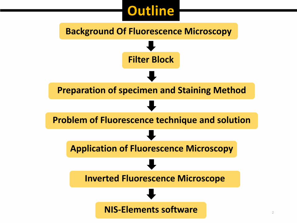
Inverted Fluorescence Microscope
Application of Fluorescence Microscopy
Problem of Fluorescence technique and solution
Preparation of specimen and Staining Method
Filter Block
2
Outline
Background Of Fluorescence Microscopy
NIS-Elements software
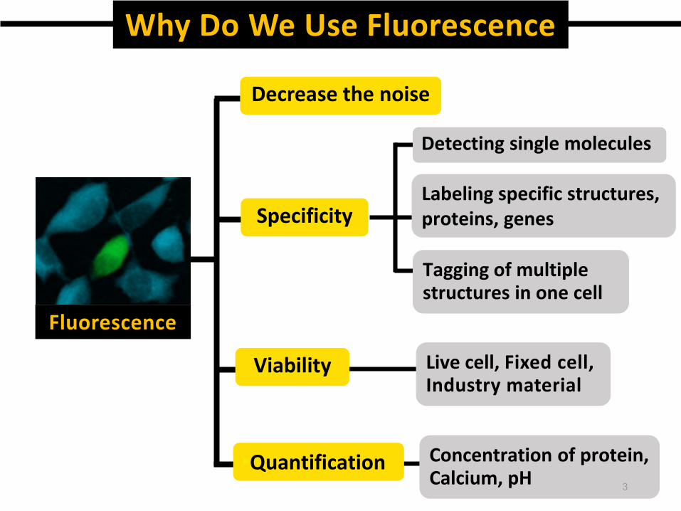
Why Do We Use Fluorescence
Fluorescence
Decrease the noise
Specificity
Detecting single molecules
Labeling specific structures, proteins, genes
Tagging of multiple structures in one cell
Viability Live cell, Fixed cell, Industry material
•Quantification Concentration of protein,Calcium, pH 3
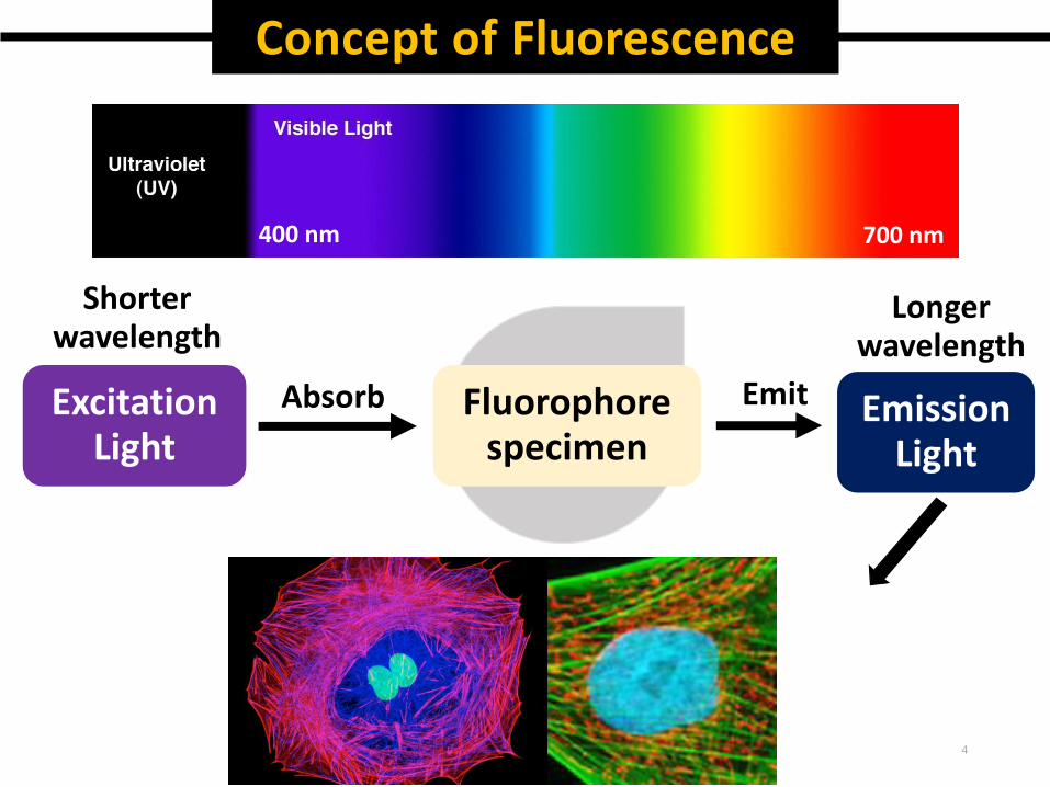
Concept of Fluorescence
Excitation Light
Fluorophore specimen
Absorb Emit Emission Light
Shorter wavelength
Longerwavelength
400 nm 700 nm
4
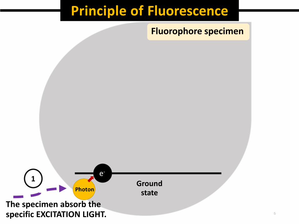
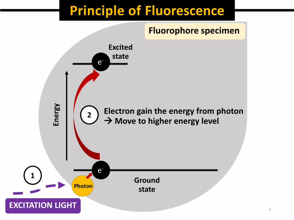
Principle of Fluorescence
Fluorophore specimen
e-
Ground state
The specimen absorb the specific EXCITATION LIGHT.
1
5
Principle of FluorescenceFluorophore specimen
e-
e-En
erg
y
Fluorophore specimen
Excitedstate
2
EXCITATION LIGHT
1
Electron gain the energy from photonMove to higher energy level
Ground state
6
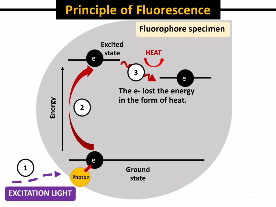
Principle of Fluorescence
e-
e-
Excitedstate
Ene
rgy
2
1
3
The e- lost the energy in the form of heat.
Fluorophore specimen
e-
Ground state
HEAT
EXCITATION LIGHT 7
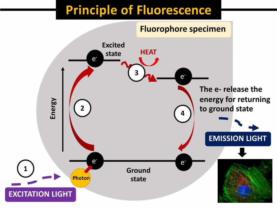
Principle of Fluorescence
e-
e-
Excitedstate
Ene
rgy
e-
e-1
2
3
4
The e- release the energy for returning to ground state
Fluorophore specimen
EMISSION LIGHT
Ground state
EXCITATION LIGHT
HEAT
8
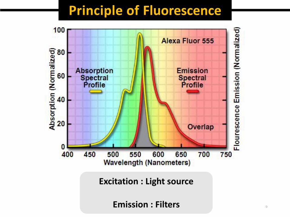
Principle of Fluorescence
Excitation : Light source
Emission : Filters 9
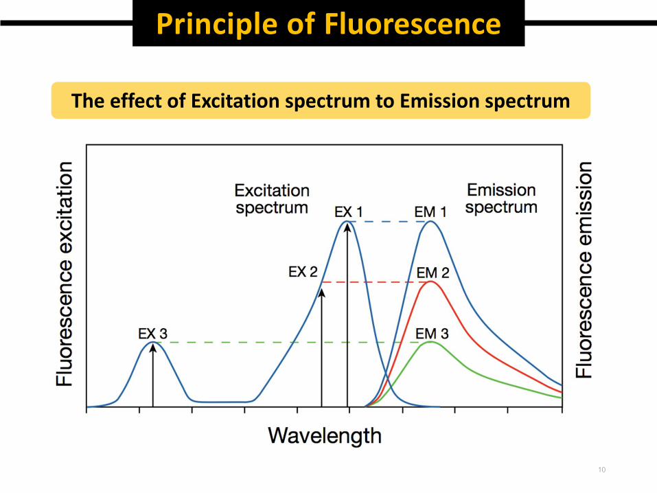
Principle of Fluorescence
The effect of Excitation spectrum to Emission spectrum
10
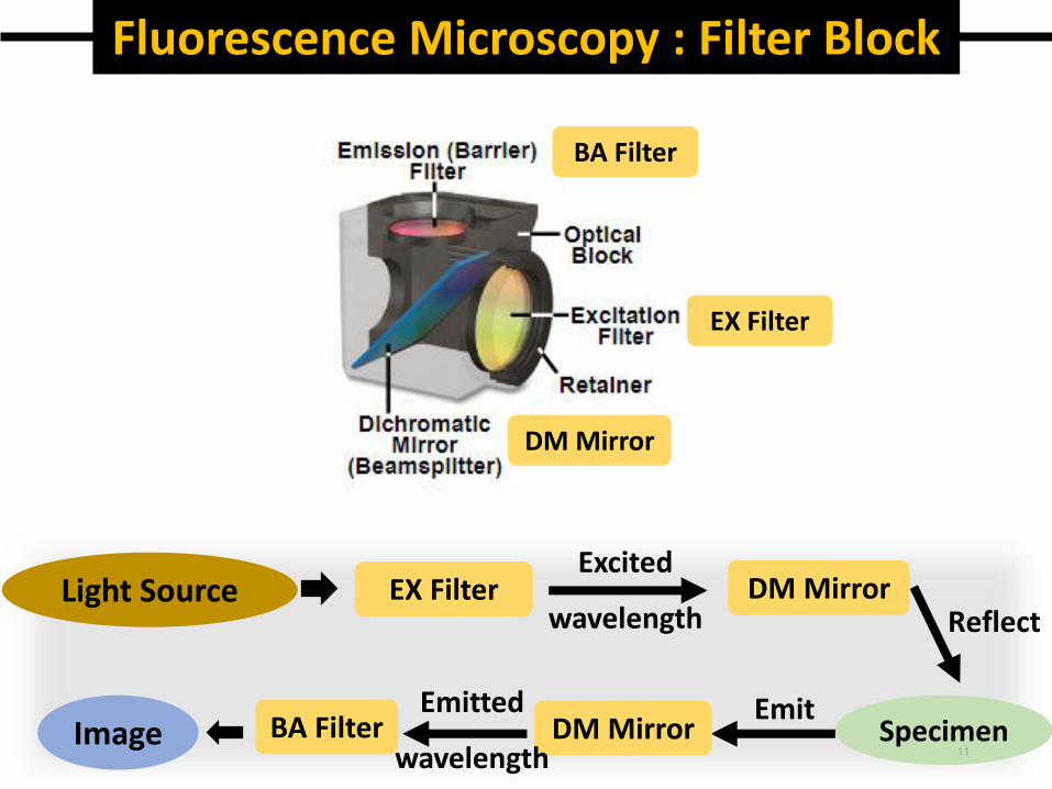
Fluorescence Microscopy : Filter Block
BA Filter
DM Mirror
EX Filter
Light Source EX FilterExcited
wavelength
Specimen
DM MirrorReflect
EmitDM Mirror
Emitted
wavelengthBA FilterImage
11
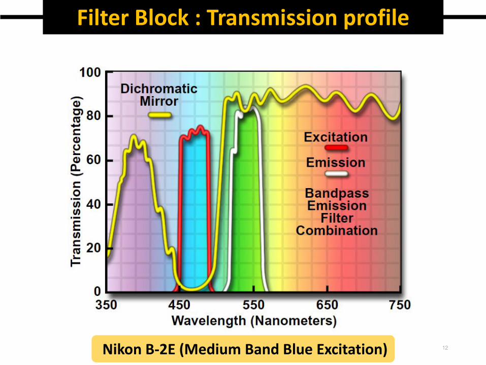
Filter Block : Transmission profile
Nikon B-2E (Medium Band Blue Excitation) 12
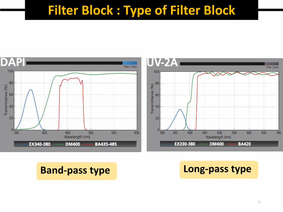
Filter Block : Type of Filter Block
Band-pass type Long-pass type
13
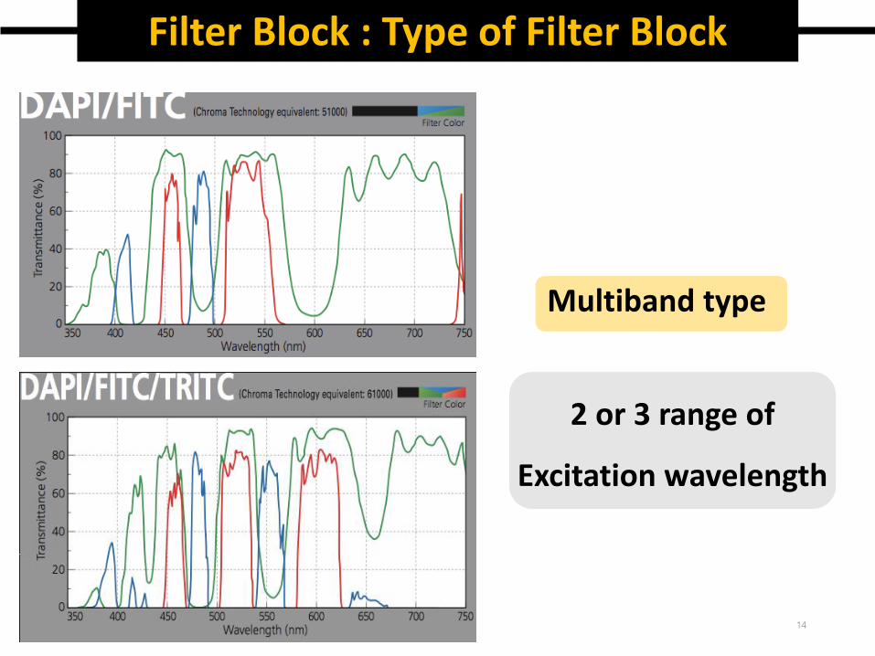
Filter Block : Type of Filter Block
Multiband type
2 or 3 range of
Excitation wavelength
14
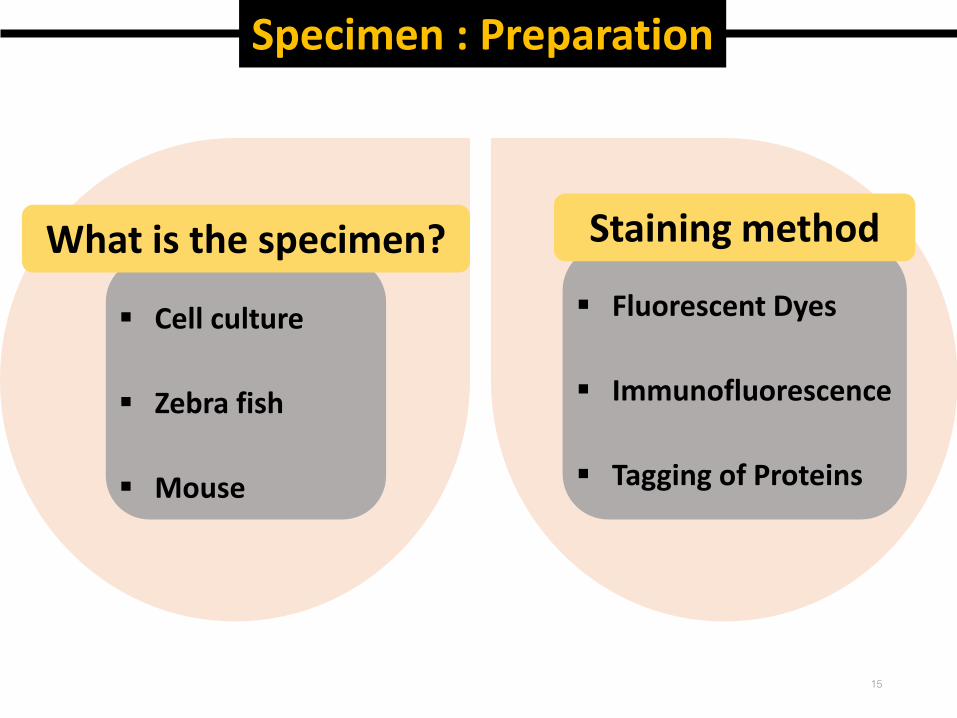
Fluorescent Dyes
Immunofluorescence
Tagging of Proteins
Cell culture
Zebra fish
Mouse
Specimen : Preparation
What is the specimen? Staining method
15
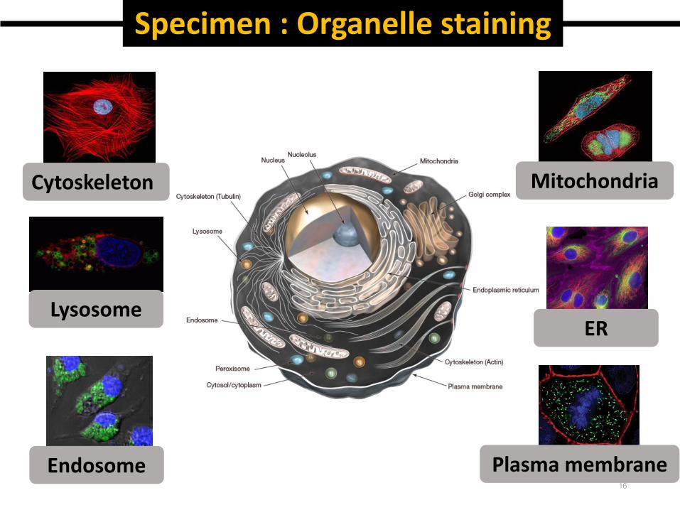
Specimen : Organelle staining
Cytoskeleton
Plasma membrane
ER
Mitochondria
Lysosome
Endosome16

Staining method : Fluorescent Dyes
▪ Taken up by the cells
▪ Incorporated and concentrated in specific subcellular
compartments
▪ Living cells are the mounted on a microscope slide and
examined in a fluorescence microscope.
▪ For Example : DAPI, Hoechst 33342, ER-Tracker, Mito-Tracker17
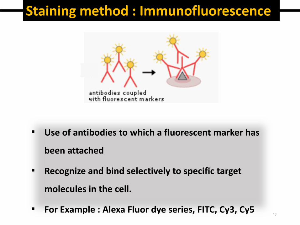
Staining method : Immunofluorescence
▪ Use of antibodies to which a fluorescent marker has
been attached
▪ Recognize and bind selectively to specific target
molecules in the cell.
▪ For Example : Alexa Fluor dye series, FITC, Cy3, Cy518
Staining method : Tagging of Proteins
▪ Modify cells so that they create their own fluorescing
molecules
▪ The location of that protein can be studied.
▪ It is also possible to watch the movements of the
proteins and its interactions with other cellular
components inside the cell.
▪ For Example : eGFP, eCPF, eYFP, DsRed, mCherry
19
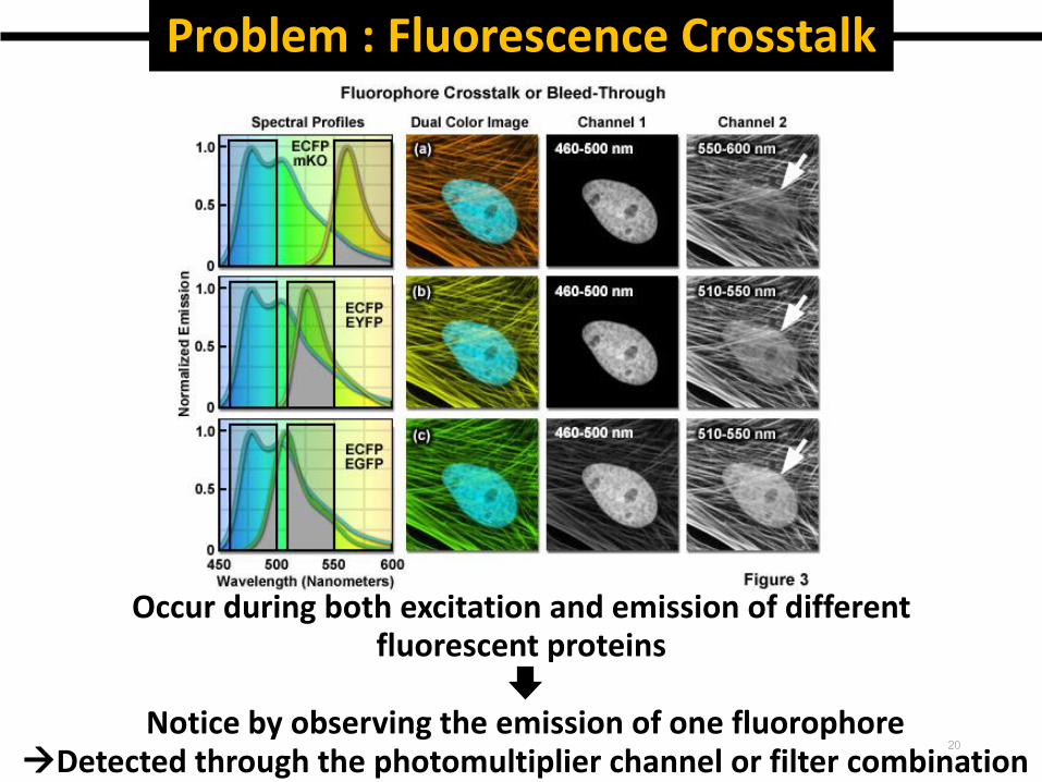
Problem : Fluorescence Crosstalk
Occur during both excitation and emission of different fluorescent proteins
Notice by observing the emission of one fluorophore Detected through the photomultiplier channel or filter combination
20
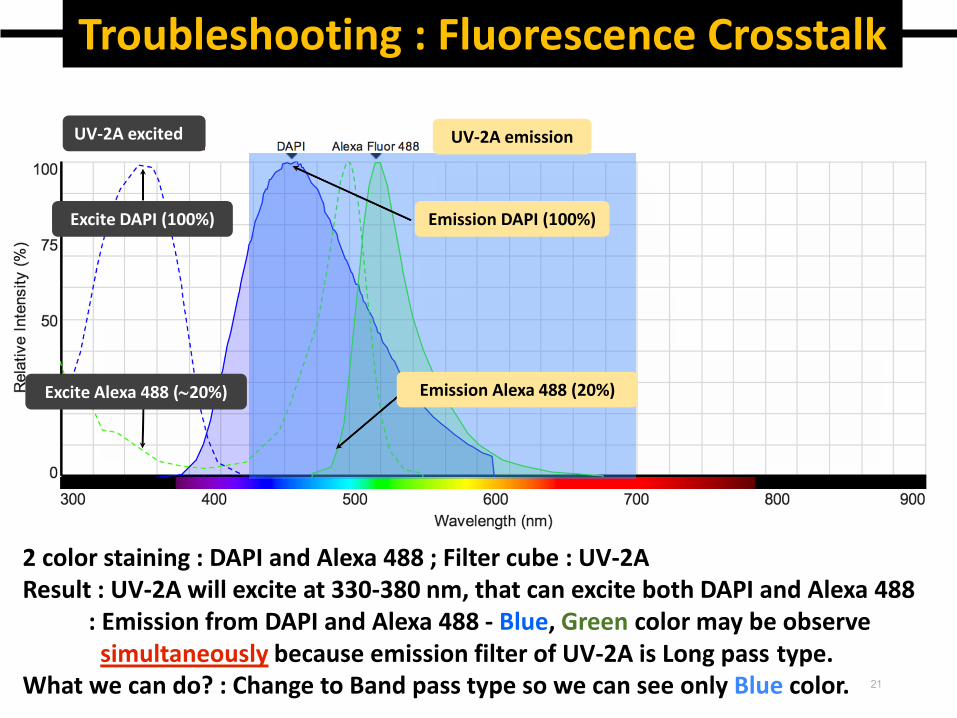
Troubleshooting : Fluorescence Crosstalk
2 color staining : DAPI and Alexa 488 ; Filter cube : UV-2AResult : UV-2A will excite at 330-380 nm, that can excite both DAPI and Alexa 488
: Emission from DAPI and Alexa 488 - Blue, Green color may be observesimultaneously because emission filter of UV-2A is Long pass type.
What we can do? : Change to Band pass type so we can see only Blue color. 21
UV-2A excited
Excite DAPI (100%)
Excite Alexa 488 (20%)
UV-2A emission
Emission DAPI (100%)
Emission Alexa 488 (20%)
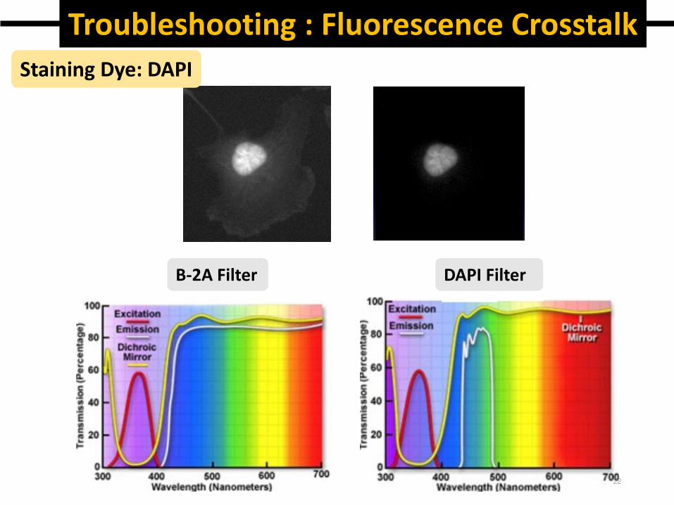
Troubleshooting : Fluorescence CrosstalkStaining Dye: DAPI
B-2A Filter DAPI Filter
22
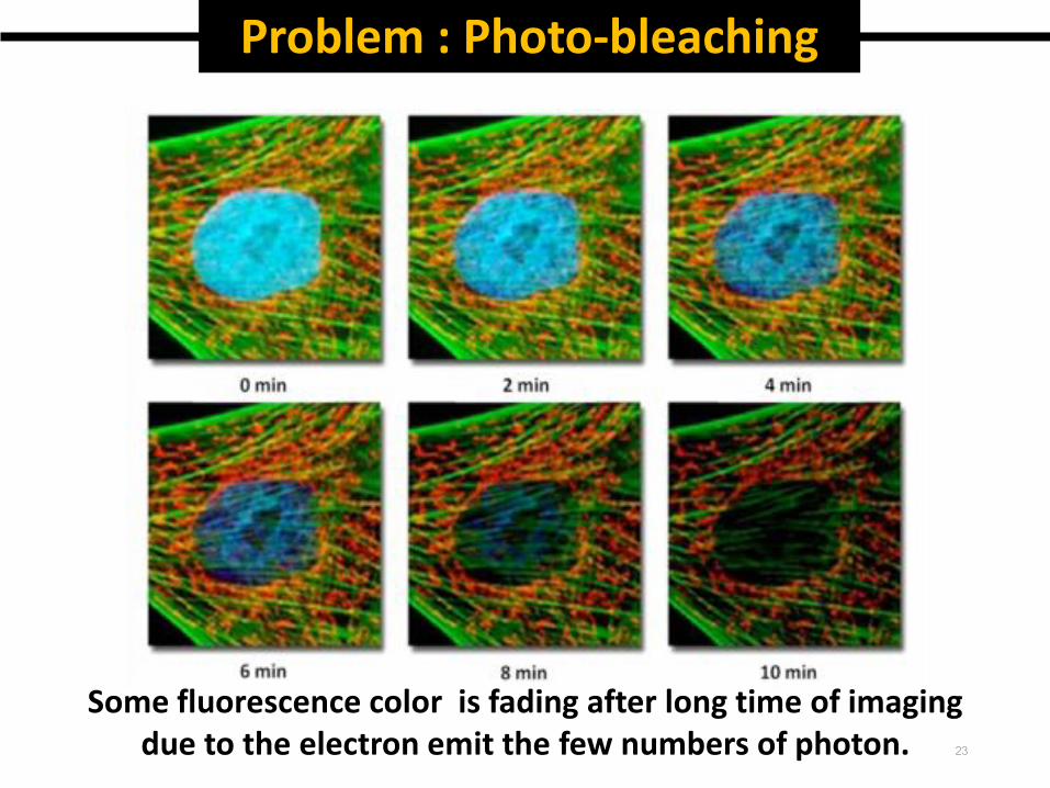
Problem : Photo-bleaching
Some fluorescence color is fading after long time of imagingdue to the electron emit the few numbers of photon. 23
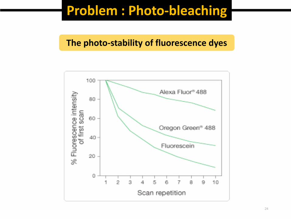
Problem : Photo-bleaching
The photo-stability of fluorescence dyes
24
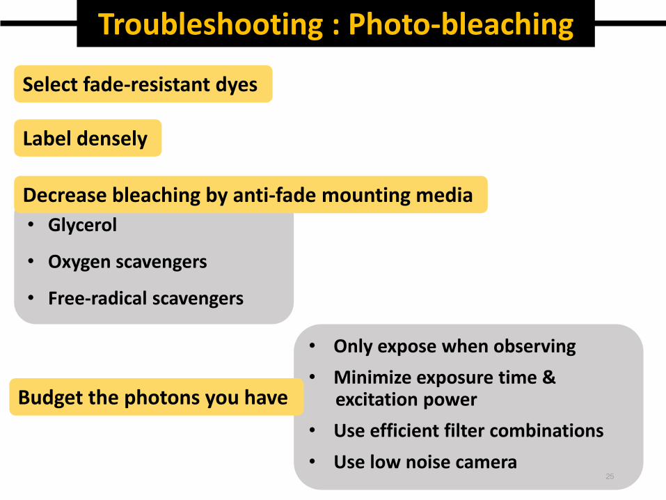
• Glycerol
• Oxygen scavengers
• Free-radical scavengers
Troubleshooting : Photo-bleaching
• Only expose when observing
• Minimize exposure time & excitation power
• Use efficient filter combinations
• Use low noise camera
Select fade-resistant dyes
Label densely
Decrease bleaching by anti-fade mounting media
Budget the photons you have
25
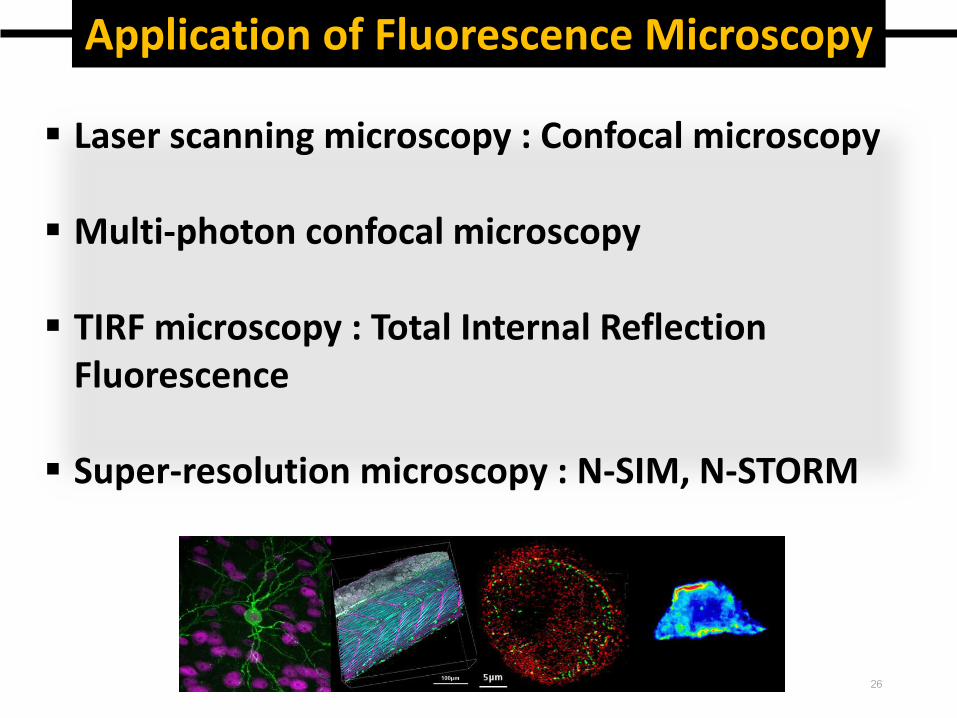
Application of Fluorescence Microscopy
Laser scanning microscopy : Confocal microscopy
Multi-photon confocal microscopy
TIRF microscopy : Total Internal Reflection Fluorescence
Super-resolution microscopy : N-SIM, N-STORM
26
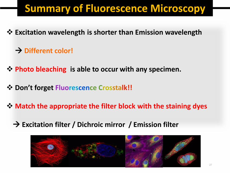
Summary of Fluorescence Microscopy
Excitation wavelength is shorter than Emission wavelength
Different color!
Photo bleaching is able to occur with any specimen.
Don’t forget Fluorescence Crosstalk!!
Match the appropriate the filter block with the staining dyes
Excitation filter / Dichroic mirror / Emission filter
27
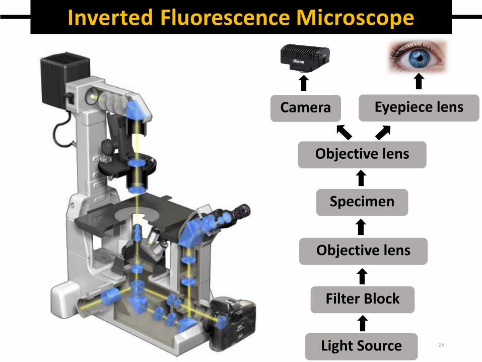
Inverted Fluorescence Microscope
Light Source
Filter Block
Specimen
Objective lens
Objective lens
Eyepiece lensCamera
28
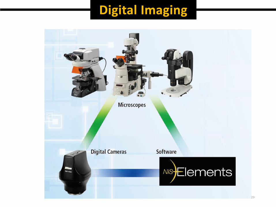
Digital Imaging
29
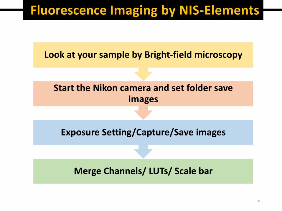
Fluorescence Imaging by NIS-Elements
Merge Channels/ LUTs/ Scale bar
Exposure Setting/Capture/Save images
Start the Nikon camera and set folder save images
Look at your sample by Bright-field microscopy
30
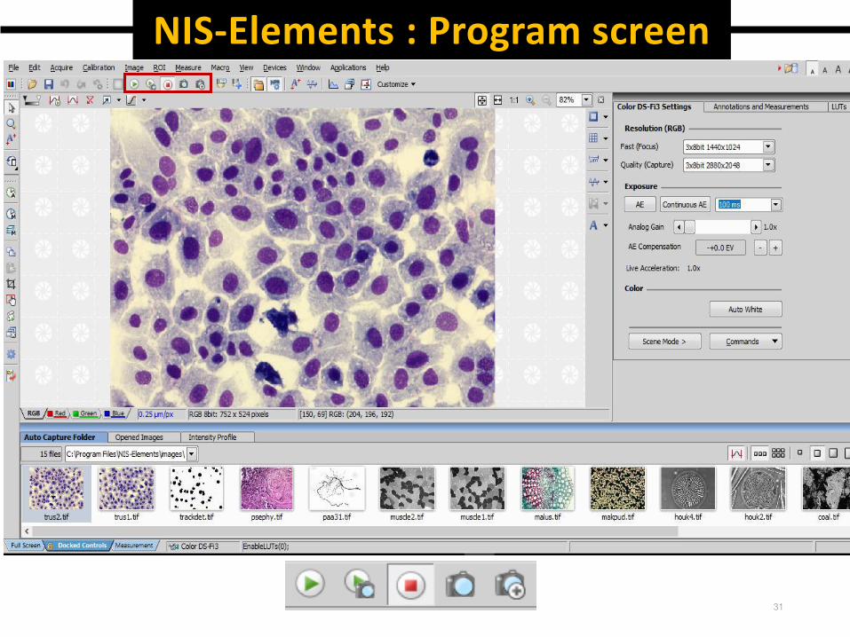
NIS-Elements : Program screen
31
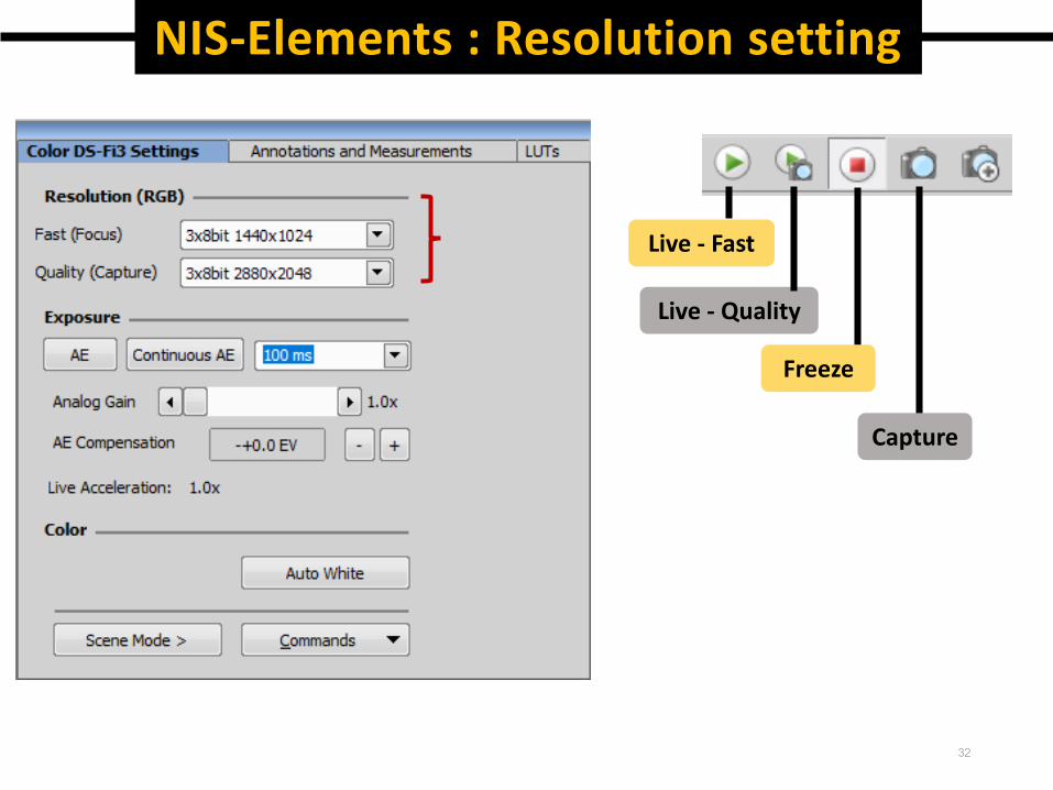
NIS-Elements : Resolution setting
Live - Fast
Live - Quality
Freeze
Capture
32
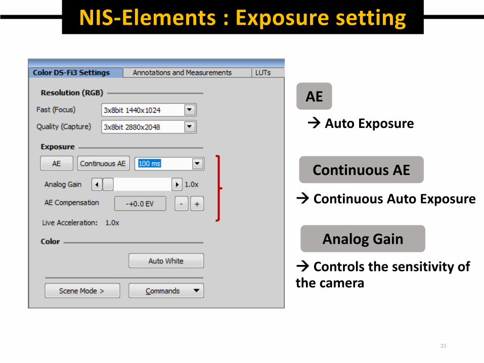
NIS-Elements : Exposure setting
AE
Auto Exposure
Continuous AE
Continuous Auto Exposure
Analog Gain
Controls the sensitivity of the camera
33
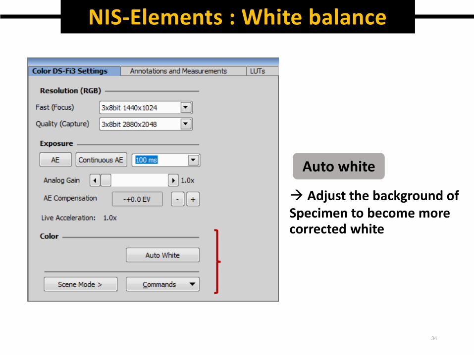
NIS-Elements : White balance
Auto white
Adjust the background ofSpecimen to become more corrected white
34
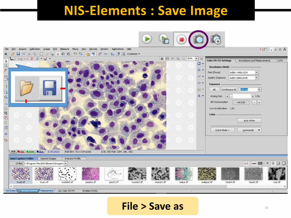
File > Save as
NIS-Elements : Save Image
35
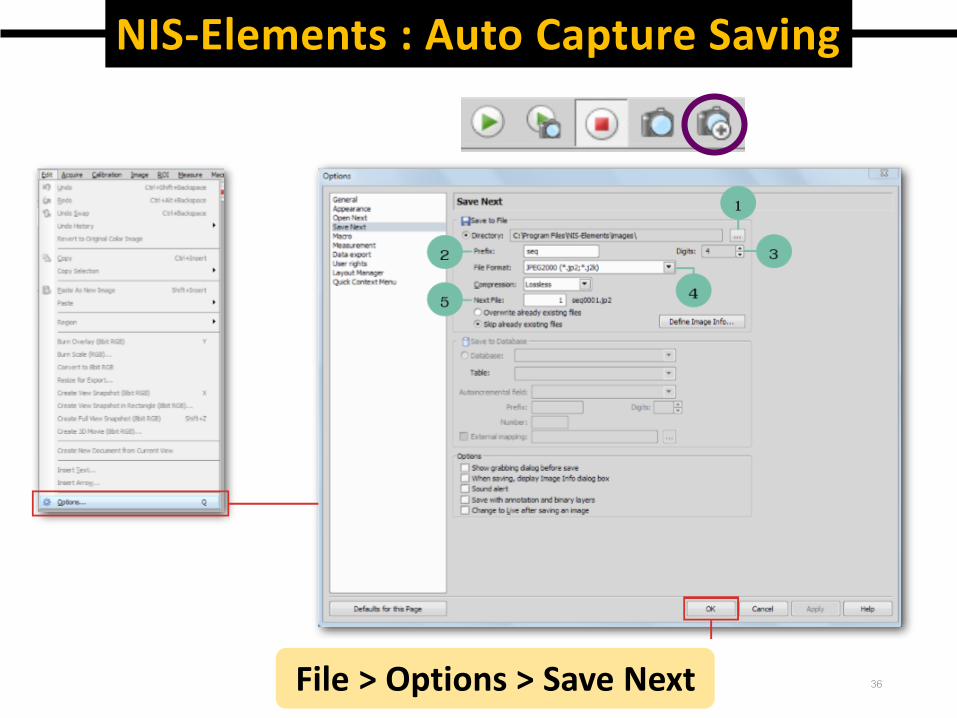
File > Options > Save Next
NIS-Elements : Auto Capture Saving
36
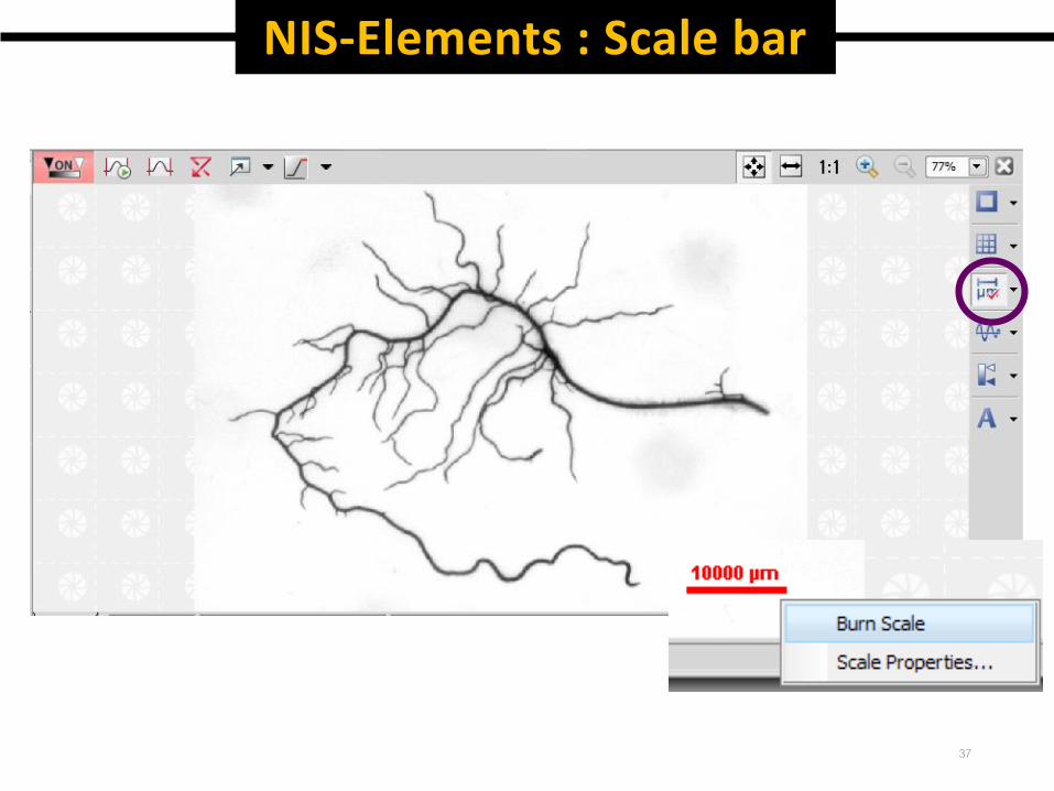
NIS-Elements : Scale bar
37
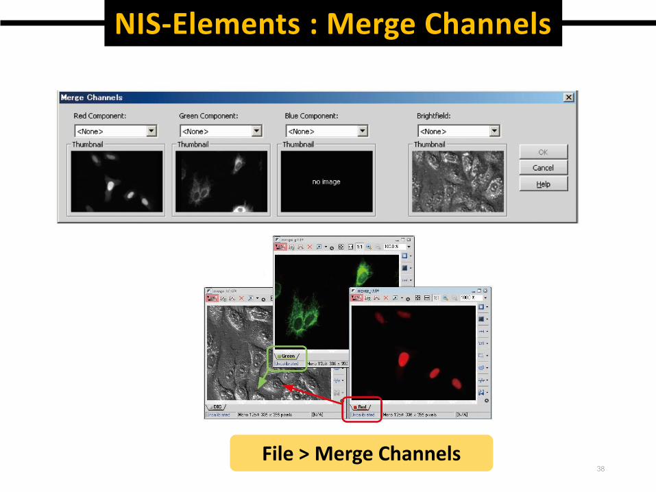
38File > Merge Channels
NIS-Elements : Merge Channels
39
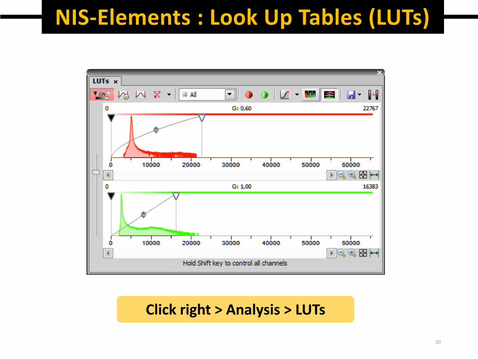
Click right > Analysis > LUTs
NIS-Elements : Look Up Tables (LUTs)