Embed Size (px)
Citation preview
Flexible, symmetry-directed approach to assemblingprotein cagesAaron Sciorea, Min Sub, Philipp Koldeweyc, Joseph D. Eschweilera, Kelsey A. Diffleya, Brian M. Linharesb,Brandon T. Ruotoloa, James C. A. Bardwellc,d,e, Georgios Skiniotisb,d, and E. Neil G. Marsha,d,1
aDepartment of Chemistry, University of Michigan, Ann Arbor, MI 48109; bLife Sciences Institute, University of Michigan, Ann Arbor, MI 48109; cDepartmentof Molecular, Cellular and Developmental Biology, University of Michigan, Ann Arbor, MI 48109; dDepartment of Biological Chemistry, University ofMichigan, Ann Arbor, MI 48109; and eHoward Hughes Medical Institute, University of Michigan, Ann Arbor, MI 48109
Edited by David Baker, University of Washington, Seattle, WA, and approved June 10, 2016 (received for review April 15, 2016)
The assembly of individual protein subunits into large-scale symmet-rical structures is widespread in nature and confers new biologicalproperties. Engineered protein assemblies have potential applica-tions in nanotechnology and medicine; however, a major challengein engineering assemblies de novo has been to design interactionsbetween the protein subunits so that they specifically assemble intothe desired structure. Here we demonstrate a simple, generalizableapproach to assemble proteins into cage-like structures that usesshort de novo designed coiled-coil domains to mediate assembly. Weassembled eight copies of a C3-symmetric trimeric esterase into awell-defined octahedral protein cage by appending a C4-symmetriccoiled-coil domain to the protein through a short, flexible linker se-quence, with the approximate length of the linker sequence deter-mined by computational modeling. The structure of the cage wasverified using a combination of analytical ultracentrifugation, nativeelectrospray mass spectrometry, and negative stain and cryoelectronmicroscopy. For the protein cage to assemble correctly, it was neces-sary to optimize the length of the linker sequence. This observationsuggests that flexibility between the two protein domains is impor-tant to allow the protein subunits sufficient freedom to assembleinto the geometry specified by the combination of C4 and C3 sym-metry elements. Because this approach is inherently modular andplaces minimal requirements on the structural features of the proteinbuilding blocks, it could be extended to assemble a wide variety ofproteins into structures with different symmetries.
coiled coils | protein design | native mass spectrometry | analyticalultracentrifugation | cryoelectron microscopy
The assembly of individual protein subunits into large-scalestructures, often from only one or a few types of protein
monomer, is widespread in nature; examples include viral capsids,multienzyme complexes, and intracellular storage compartments(1–4). These protein assemblies are generally characterized by ahigh degree of symmetry. An important consequence of the as-sembly process is the emergence of more complex biologicalproperties; well-studied examples include the dynamic polymeri-zation of actin (5) and tubulin fibrils (6) and the GroEL/GroES,which is a protein chaperone complex (7). In their assembledform, the basic ATPase activity inherent to each of these proteinsis harnessed toward the more complex tasks of motility and proteinrefolding, respectively. Consequently, there is significant interest inthe fields of synthetic biology and nanotechnology in designingnovel self-assembling proteins and adapting natural protein assem-blies for a range of applications broadly encompassing nanomedicineand materials science (4, 8–13).Early work by Yeates and coworkers (14, 15) recognized that
the principles of symmetry, often used in the design of inorganicmaterials, could be exploited to design either discrete, cage-likeprotein assemblies or extensive networks in one, two, and threedimensions. An important realization was that a large number ofcomplex symmetries could be generated from only two distinctsymmetry elements (for a protein, these must be rotationalsymmetries specified by its quaternary structure), provided the
orientation of the symmetry axes with respect to each other couldbe carefully controlled. These principles have now been quite widelyapplied to design both protein cages and protein networks (16–22).The principal challenge to researchers has been to design new in-teractions between the protein subunits that promote assembly inthe desired geometry, and, in particular, to align the angle betweensymmetry axes correctly. A variety of strategies have been used tofacilitate assembly; these include genetically linking two proteininteraction domains (14, 23, 24), the use of bifunctional ligands andmetal ions to coordinate proteins (19, 21, 22, 25), and the compu-tational design of new protein–protein interfaces (16, 17).Despite significant progress, the design of protein systems that
assemble into well-defined architectures remains a challenginggoal. Whereas genetically linking two protein interaction domainstogether is easy to accomplish, it has proven hard to achieve thenecessary degree of control over the orientation of the proteins. Inonly a few cases has this approach yielded assemblies that are suf-ficiently homogenous to characterize crystallographically (26, 27).More often genetically linking protein interaction domains result inpolydisperse protein assemblies (21, 22, 28–31); these are hard tocharacterize and are limited in their potential utility.More recently, the computation redesign of protein–protein in-
terfaces has met with some impressive successes, leading to theconstruction of rigid protein cages that could be characterizedcrystallographically (16, 17). However, this protein redesign iscomputationally intensive and requires very precise control of theprotein–protein interfaces to successfully direct assembly. Theprecision needed to successfully redesign protein–protein inter-faces limits the number of proteins amenable to this approach andrequires that many designed variants be experimentally screened to
Significance
The ability to organize biological molecules into new hierarchicalforms represents an important goal in synthetic biology. How-ever, designing new quaternary interactions between proteinsubunits has proved technically challenging and has generallyrequired extensive redesign of protein−protein interfaces. Here,we demonstrate a conceptually simple way to assemble a pro-tein into a well-defined geometric structure that uses coiled-coilsequences as “off-the-shelf” components. This approach is in-herently modular and adaptable to a wide range of proteins andsymmetries, opening up avenues for the construction of bio-logical structures with diverse geometries and wide-rangingfunctionalities.
Author contributions: A.S., M.S., P.K., B.T.R., and E.N.G.M. designed research; A.S., M.S.,P.K., J.D.E., K.A.D., and B.M.L. performed research; A.S., M.S., P.K., B.T.R., J.C.A.B., G.S.,and E.N.G.M. analyzed data; and A.S., M.S., P.K., B.T.R., J.C.A.B., G.S., and E.N.G.M. wrotethe paper.
The authors declare no conflict of interest.
This article is a PNAS Direct Submission.1To whom correspondence should be addressed. Email: [email protected].
This article contains supporting information online at www.pnas.org/lookup/suppl/doi:10.1073/pnas.1606013113/-/DCSupplemental.
www.pnas.org/cgi/doi/10.1073/pnas.1606013113 PNAS | August 2, 2016 | vol. 113 | no. 31 | 8681–8686
BIOCH
EMISTR
YCH
EMISTR
Y

identify well-folded assemblies. Moreover, the extensive reen-gineering of the protein surface that is often needed to con-struct the interface may negatively impact the biological activityof the designed protein.We aimed to develop a general approach to designing protein
assemblies that is largely independent of the structural details ofthe engineered protein and that does not require the orientationof the symmetry axes to be explicitly specified. Here, we describea strategy for assembling a trimeric protein into an octahedralcage using a small de novo designed, parallel four-helix bundlecoiled-coil domain that is genetically fused to the C terminus ofthe protein through a short, flexible linker. The structure of as-sembly is primarily specified by the symmetry of the coiled-coildomain. We show that, despite the flexibility of the linker, theresulting protein cage adopts a well-defined structure and ishighly homogeneous.
ResultsDesign Approach. In our design approach, we sought to develop aflexible, modular strategy in which the protein building block andthe coiled-coil domain function independently but, when geneti-cally linked together, assemble into a single structure of the de-sired symmetry. In general, attempts to design protein assemblieshave focused almost exclusively on combining trimeric (C3-sym-metric) proteins with dimeric (C2-symmetric) proteins, as theseare common quaternary structures (10). The combination of C3and C2 symmetry elements occurs in multiple point groups, somany geometries are compatible with assemblies made up of thesesymmetry elements. In contrast, the combination of C3 and C4symmetry elements is unique to the octahedral point group.Therefore, we attempted to construct an octahedral protein cagebased solely on combining proteins with these rotation symmetriesand without explicit orientation constraints.We surveyed several trimeric proteins in the Protein Data Bank
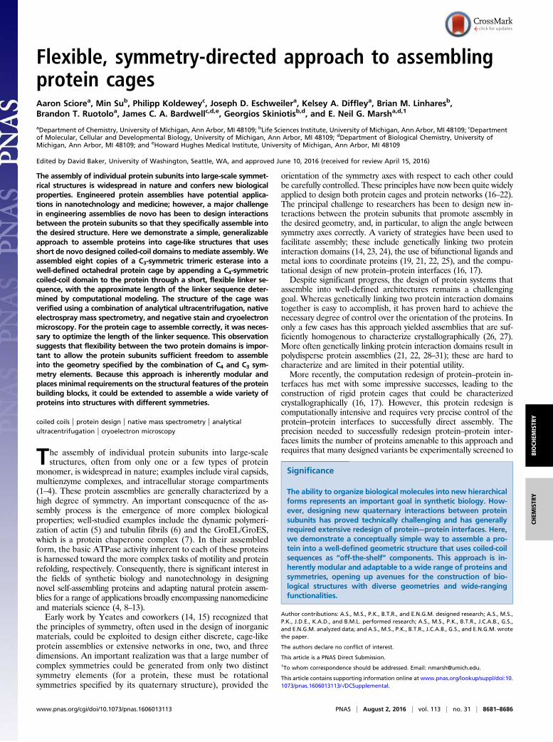
(PDB) and selected, as a test case, a trimeric esterase, PDB ID1ZOI (32). In this esterase, the C terminus is oriented toward theapex of the triangle formed by the C3-symmetric protein, posi-tioning it in approximately the right place to facilitate addition of
the C4-symmetric domain. (Fig. 1A). Natural, C4-symmetric pro-teins are rare, as most tetrameric proteins adopt a pseudo-D2“dimer-of-dimers” symmetry. Therefore, we used a de novodesigned coiled-coil protein as the C4 component. Coiled coils areamong the simplest and best-understood protein–protein interac-tions (33). As such, there are a large number of well-characterizeddesigns available as “off-the-shelf” components for use in proteinengineering applications, including dimeric, trimeric, tetrameric,pentameric, and hexameric designs in both parallel and antiparallelforms (34–36). A further advantage is that the strength of thecoiled-coil interaction can easily be manipulated by varying thenumber of heptad repeats. For our purposes, we selected a parallel,four-helix coiled coil in which the tetrameric arrangement isspecified by four repeating heptads in which Leu and Ile are pre-sent at the “a” and “d” positions of the canonical heptad (37); thecrystal structure of this protein, PDB ID 3R4A (37), shows that itpossesses close to perfect C4 symmetry.To determine the approximate minimum length of flexible linker
needed to connect the C terminus of the C3 protein with theN terminus of the C4 coiled coil, we aligned the C3 axis of theesterase and the C4 axis of the coiled coil along the C3 and C4 axes,respectively, of the octahedral point group. Using a search algo-rithm implemented in the program Rosetta (38), the angle of ro-tation of each protein about its symmetry axis and its distance fromthe origin were allowed to vary in a symmetrically constrainedmanner. The distance between the two termini was minimized,discarding any configurations with steric clashes (defined as anyintersubunit backbone atom distances shorter than 4 Å) (Fig. 1B).The modeling indicated that the coiled coils could either pointinward or outward. (The inward-pointing orientations were exam-ined by negatively translating the coiled-coil coordinates along thesymmetry axes indicated in Fig. 1B. This orientation is feasiblebecause the vertices of the trimeric esterase don’t pack togetherperfectly, leaving sufficient space for the coiled-coil domain to pointinward while still maintaining a compact structure.) Either orien-tation yielded a similar minimum distance between the termini ofthe esterase and coiled coil of ∼9.1 Å that could, in principle, be
Fig. 1. Design of a self-assembling octahedral protein cage. (A) Structures of the trimeric esterase (PDB 1ZOI) (C termini of the esterase are indicated by redspheres) and the tetrameric coiled coil (PDB 3R4A) used in the design. (B) Minimization of linker distance compatible with octahedral geometry. The proteinswere arrayed along the C3 (blue line) and C4 (green line) symmetry axes, and the distance between the N terminus of the coiled coil and the C terminus of theesterase (dashed red line) was minimized by symmetrically varying the rotation of the proteins about the symmetry axes and their radial distance whileavoiding steric clashes. (C) Distance-minimized structures were found to be compatible with the coiled-coil domains either facing inward (top structure) oroutward (bottom structure) with a minimum interterminus distance of ∼9.1 Å.
8682 | www.pnas.org/cgi/doi/10.1073/pnas.1606013113 Sciore et al.
bridged by a minimum of three amino acid residues (Fig. 1C). PDBfiles of the models are provided as Datasets S1 and S2.Based on the modeling, we constructed three synthetic genes
(Table S1) in which the C terminus of the trimeric esterase was ge-netically fused to the N terminus of the tetrameric coiled-coil domainthrough a flexible linker sequence comprising two, three, or fourglycine residues that potentially could span between 6 Å and 12 Å.We refer to these designs as Oct-2, Oct-3, and Oct-4, respectively.
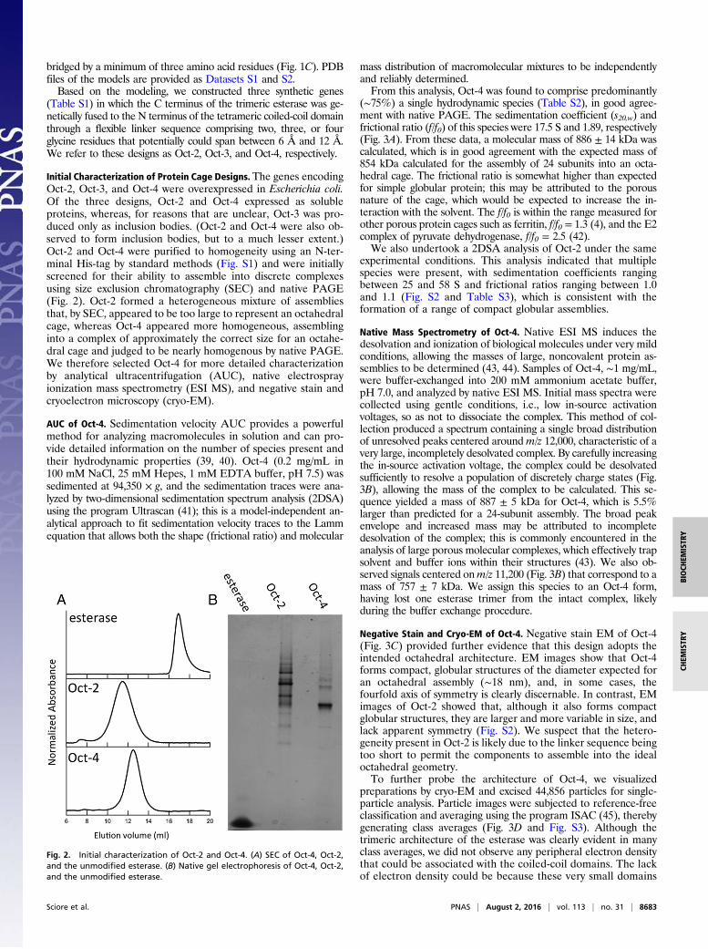
Initial Characterization of Protein Cage Designs. The genes encodingOct-2, Oct-3, and Oct-4 were overexpressed in Escherichia coli.Of the three designs, Oct-2 and Oct-4 expressed as solubleproteins, whereas, for reasons that are unclear, Oct-3 was pro-duced only as inclusion bodies. (Oct-2 and Oct-4 were also ob-served to form inclusion bodies, but to a much lesser extent.)Oct-2 and Oct-4 were purified to homogeneity using an N-ter-minal His-tag by standard methods (Fig. S1) and were initiallyscreened for their ability to assemble into discrete complexesusing size exclusion chromatography (SEC) and native PAGE(Fig. 2). Oct-2 formed a heterogeneous mixture of assembliesthat, by SEC, appeared to be too large to represent an octahedralcage, whereas Oct-4 appeared more homogeneous, assemblinginto a complex of approximately the correct size for an octahe-dral cage and judged to be nearly homogenous by native PAGE.We therefore selected Oct-4 for more detailed characterizationby analytical ultracentrifugation (AUC), native electrosprayionization mass spectrometry (ESI MS), and negative stain andcryoelectron microscopy (cryo-EM).
AUC of Oct-4. Sedimentation velocity AUC provides a powerfulmethod for analyzing macromolecules in solution and can pro-vide detailed information on the number of species present andtheir hydrodynamic properties (39, 40). Oct-4 (0.2 mg/mL in100 mM NaCl, 25 mM Hepes, 1 mM EDTA buffer, pH 7.5) wassedimented at 94,350 × g, and the sedimentation traces were ana-lyzed by two-dimensional sedimentation spectrum analysis (2DSA)using the program Ultrascan (41); this is a model-independent an-alytical approach to fit sedimentation velocity traces to the Lammequation that allows both the shape (frictional ratio) and molecular
mass distribution of macromolecular mixtures to be independentlyand reliably determined.From this analysis, Oct-4 was found to comprise predominantly
(∼75%) a single hydrodynamic species (Table S2), in good agree-ment with native PAGE. The sedimentation coefficient (s20,w) andfrictional ratio (f/f0) of this species were 17.5 S and 1.89, respectively(Fig. 3A). From these data, a molecular mass of 886 ± 14 kDa wascalculated, which is in good agreement with the expected mass of854 kDa calculated for the assembly of 24 subunits into an octa-hedral cage. The frictional ratio is somewhat higher than expectedfor simple globular protein; this may be attributed to the porousnature of the cage, which would be expected to increase the in-teraction with the solvent. The f/f0 is within the range measured forother porous protein cages such as ferritin, f/f0 = 1.3 (4), and the E2complex of pyruvate dehydrogenase, f/f0 = 2.5 (42).We also undertook a 2DSA analysis of Oct-2 under the same
experimental conditions. This analysis indicated that multiplespecies were present, with sedimentation coefficients rangingbetween 25 and 58 S and frictional ratios ranging between 1.0and 1.1 (Fig. S2 and Table S3), which is consistent with theformation of a range of compact globular assemblies.
Native Mass Spectrometry of Oct-4. Native ESI MS induces thedesolvation and ionization of biological molecules under very mildconditions, allowing the masses of large, noncovalent protein as-semblies to be determined (43, 44). Samples of Oct-4, ∼1 mg/mL,were buffer-exchanged into 200 mM ammonium acetate buffer,pH 7.0, and analyzed by native ESI MS. Initial mass spectra werecollected using gentle conditions, i.e., low in-source activationvoltages, so as not to dissociate the complex. This method of col-lection produced a spectrum containing a single broad distributionof unresolved peaks centered around m/z 12,000, characteristic of avery large, incompletely desolvated complex. By carefully increasingthe in-source activation voltage, the complex could be desolvatedsufficiently to resolve a population of discretely charge states (Fig.3B), allowing the mass of the complex to be calculated. This se-quence yielded a mass of 887 ± 5 kDa for Oct-4, which is 5.5%larger than predicted for a 24-subunit assembly. The broad peakenvelope and increased mass may be attributed to incompletedesolvation of the complex; this is commonly encountered in theanalysis of large porous molecular complexes, which effectively trapsolvent and buffer ions within their structures (43). We also ob-served signals centered onm/z 11,200 (Fig. 3B) that correspond to amass of 757 ± 7 kDa. We assign this species to an Oct-4 form,having lost one esterase trimer from the intact complex, likelyduring the buffer exchange procedure.
Negative Stain and Cryo-EM of Oct-4. Negative stain EM of Oct-4(Fig. 3C) provided further evidence that this design adopts theintended octahedral architecture. EM images show that Oct-4forms compact, globular structures of the diameter expected foran octahedral assembly (∼18 nm), and, in some cases, thefourfold axis of symmetry is clearly discernable. In contrast, EMimages of Oct-2 showed that, although it also forms compactglobular structures, they are larger and more variable in size, andlack apparent symmetry (Fig. S2). We suspect that the hetero-geneity present in Oct-2 is likely due to the linker sequence beingtoo short to permit the components to assemble into the idealoctahedral geometry.To further probe the architecture of Oct-4, we visualized
preparations by cryo-EM and excised 44,856 particles for single-particle analysis. Particle images were subjected to reference-freeclassification and averaging using the program ISAC (45), therebygenerating class averages (Fig. 3D and Fig. S3). Although thetrimeric architecture of the esterase was clearly evident in manyclass averages, we did not observe any peripheral electron densitythat could be associated with the coiled-coil domains. The lackof electron density could be because these very small domains
Fig. 2. Initial characterization of Oct-2 and Oct-4. (A) SEC of Oct-4, Oct-2,and the unmodified esterase. (B) Native gel electrophoresis of Oct-4, Oct-2,and the unmodified esterase.
Sciore et al. PNAS | August 2, 2016 | vol. 113 | no. 31 | 8683
BIOCH
EMISTR
YCH
EMISTR
Y
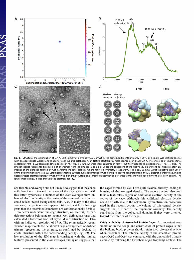
are flexible and average out, but it may also suggest that the coiledcoils face inward, toward the center of the cage. Consistent withthis latter hypothesis, a number of the class averages show en-hanced electron density at the center of the averaged particles thatcould reflect inward-facing coiled coils. Also, in many of the classaverages, the protein cages appear distorted, which further sug-gests that the assembled complexes are conformationally flexible.To better understand the cage structure, we used 34,980 par-
ticle projections belonging to the most well defined averages andcalculated a low-resolution 3D cryo-EM reconstruction of Oct-4with an indicated resolution of 17 Å. The symmetrically recon-structed map reveals the octahedral cage arrangement of distincttrimers representing the esterase, as confirmed by docking itscrystal structure within the corresponding density (Fig. 3D). Thelow resolution of the EM map is consistent with the limitedfeatures presented in the class averages and again suggests that
the cages formed by Oct-4 are quite flexible, thereby leading toblurring of the averaged density. The reconstruction also con-tains a featureless region of additional electron density at thecenter of the cage. Although this additional electron densitycould be partly due to the octahedral symmetrization procedureused in the reconstruction, the volume of this central densitysuggests that it is part of the oligomeric assembly. The densitycould arise from the coiled-coil domains if they were orientedtoward the interior of the cage.
Catalytic Activity of Assembled Protein Cages. An important con-sideration in the design and construction of protein cages is thatthe building block proteins should retain their biological activitywhen assembled. The esterase activity of the assembled proteincages Oct-2 and Oct-4 was compared with the unmodified trimericesterase by following the hydrolysis of p-nitrophenyl acetate. The
Fig. 3. Structural characterization of Oct-4. (A) Sedimentation velocity AUC of Oct-4. The protein sediments primarily (>75%) as a single, well-defined specieswith an appropriate weight and shape for a 24-subunit octahedron. (B) Native electrospray mass spectrum of intact Oct-4. The envelope of charge statescentered atm/z 12,600 corresponds to a species ofMr = 887 ± 5 kDa, whereas those centered atm/z = 11200 corresponds to a species ofMr = 757 ± 7 kDa. Thesmaller species represents dissociation of one trimer from the octahedral complex under the conditions of the Native MS experiment. (C) Negative stain EMimages of the particles formed by Oct-4. Arrows indicate particles where fourfold symmetry is apparent. (Scale bar, 20 nm.) (Inset) Negative stain EM ofunmodified trimeric esterase. (D, Left) Representative 2D class-averaged images of Oct-4 and projections generated from the 3D electron density map. (Right)Reconstructed electron density for Oct-4 viewed along the fourfold and threefold axes with one esterase trimer shown modeled into the electron density. Thelower images show a slice through the electron density.
8684 | www.pnas.org/cgi/doi/10.1073/pnas.1606013113 Sciore et al.

specific activity of the unmodified esterase determined in 25 mMHepes, pH 7.5, 100 mMNaCl, at 25 °C was 54 ± 4 μM·min−1·mg−1,whereas the specific activities of Oct-2 and Oct-4 were 19.5 ± 0.5and 20 ± 0.4 μM·min−1·mg−1, respectively.The reason for the lower specific activities of the assembled
proteins is currently unclear. It might be that assembly impedessubstrate access to the active site, or that it imposes small dis-tortions on the active site geometry or dynamics, both of whichcould lower activity. However, the retention of activity impliesthat the tertiary structure of the protein was not significantlyaltered by the assembly process.
DiscussionVarious studies have used symmetry-based methods for assemblyof threefold symmetric proteins into octahedral and tetrahedralcages using other protein domains, bifunctional cross-linkers,metal ions, or designed protein interfaces to direct assembly (14,16, 17, 19, 21–23, 25, 28–30). Common to these approaches hasbeen the combination of C3 and C2 symmetry elements, whichhas required that the orientation of the two symmetry elementsbe carefully controlled to prevent the formation of heteroge-neous assemblies. Here, we have shown that, by switching to acombination of C3 and C4 symmetry elements, it is possible toorganize a protein into a geometrically well-defined, large-scaleassembly without the need to explicitly specify the relative ori-entation of the two protein domains. To our knowledge, this isthe first example of a designed protein cage that incorporates aC4-symmetric element to mediate assembly.It is worth noting that the flexible connection between the C3
and C4 symmetry elements, in principle, also permits largerstructures of lower symmetry to be formed without violating the“4 × 3” valency rules. It is also possible that incompletely orincorrectly assembled structures could form that become kinet-ically trapped; this may explain the ensemble of larger assembliesthat are formed by Oct-2, which possesses a shorter linker se-quence. Indeed, some evidence for off-pathway assemblies wasalso evident in preparations of Oct-4, as evidenced by nativePAGE (Fig. 2B), although SEC largely removed these duringpurification (Fig. S1).We envisage that the coiled-coil domains act like “twist ties”
to hold the esterase trimers in a flexible octahedral configura-tion. As such, the assembly process is, in principle, independentof the structural details of the protein, requiring only optimiza-tion of the linker length connecting the two domains. This designstrategy provides a complementary approach to that of designingnew protein–protein interfaces, which produce rigid proteincages (16, 17). Also, because conformational dynamics are im-portant for the biological function of many proteins, by main-taining a looser association between subunits, the potential forinterfering with the protein’s biological activity is minimized. Weconsider that the simplicity and generality of this approach mayconfer advantages for many applications in synthetic biology,such as construction of enzyme nanoreactors, encapsulation ofprotein cargos, targeted drug delivery, and polyvalent display ofepitopes, where atomic-level precision is not necessary.The design strategy is inherently modular, and one can imagine
that, by combining proteins and coiled-coil domains with differentsymmetries, a variety of cages with different geometries could beconstructed. Coiled-coil designs have been described in whicholigomerization has been coupled to events such as metal binding(46), a redox environment (47), and pH changes (48). Such pro-grammability could be introduced into the design to make cageassembly and disassembly responsive to environmental conditionsor specific ligands. In addition, further optimization of the designmay be achieved by fine-tuning the coiled-coil interactions toimprove the kinetics of assembly to reduce misfolding and theformation of inclusion bodies.
Materials and MethodsConstruction of Genes Encoding Fusion Proteins. Codon-optimized genes li-gated into the expression vector pET28b were either commercially synthe-sized or derived from the other constructs using standard techniques. Thesequences of the proteins are included in Table S1.
Protein Expression and Purification. Expression constructs were transformedinto E. coli BL21(DE3) cells. Cells were grown in 2xYT medium with 50 mg/Lkanamycin at 37 °C. At an OD600 of 0.8, the temperature was reducedto 18 °C, and, at an OD600 of 1.0, protein expression was induced by additionof 0.1 mM IPTG; cells were grown for a further 18 h and harvested bycentrifugation.
All purification steps were performed on ice or at 4 °C. Cell pellets wereresuspended in 50 mMHepes buffer, pH 7.5, containing 1M urea, 300 mMNaCl,50mM imidazole, 5% (vol/vol) glycerol, SigmaFAST protease inhibitor, and 1mg/mLlysozyme, and then lysed by sonication. The lysate was clarified by centrifugationat 48,000 × g for 30 min and injected onto a HisTrap nickel–nitrilotriacetic acid(Ni-NTA) column, washedwith several volumes of the same buffer, and eluted with50 mM Hepes buffer, pH 7.5, containing 300 mM NaCl, 500 mM imidazole, and5% (vol/vol) glycerol. Fractions containing proteins of interest were pooled, di-alyzed against 25 mM Hepes buffer, pH 7.5, containing 100 mM NaCl and 2 mMEDTA, concentrated by ultrafiltration, and further purified by SEC on a Superose 6300/10 column equilibrated in the same buffer. Fractions containing proteins of thedesired oligomerization state were pooled and further concentrated for analysis.
AUC. Sedimentation velocity analysis was performed using a Beckman Pro-teome Lab XL-I analytical ultracentrifuge (Beckman Coulter) equipped withan AN60TI rotor. Samples were dialyzed against 25 mM Hepes buffer, pH 7.5,containing 100 mM sodium chloride and 1 mM EDTA. The hydrodynamicbehavior of the various proteins was analyzed at a protein concentrationwithinitial absorptions of 0.2 at 280 nm. Samples were loaded into precooledstandard sector-shaped, two-channel Epon centerpieces with 1.2-cm pathlength, and allowed to equilibrate at 6 °C for 2 h in the nonspinning rotorbefore sedimentation. Proteins were sedimented at 94,350 × g. Absorbancedata were collected at a wavelength of 280 nm. Sedimentation velocity datawere analyzed by 2DSA using the finite element modeling module providedwith the Ultrascan III software (www.ultrascan.uthscsa.edu). Confidencelevels for statistics were derived from 2DSA data refinement using a geneticalgorithm followed by 50 Monte Carlo simulations. Calculations were per-formed on the UltraScan LIMS cluster at the Bioinformatics Core Facility,University of Texas Health Science Center at San Antonio.
Native MS. After SEC, samples were concentrated to ∼5 mg/mL and thenbuffer-exchanged into 200 mM ammonium acetate, pH 7.0, using a Bio-spinP30 column (Bio-Rad, Inc.); 2–3 μL of the sample was loaded into glass cap-illary (approximate o.d. of 1.5–1.8 mm and wall thickness of 0.2 mm) beforemounting to the source of an Exactive Plus EMR mass spectrometer (ThermoFisher Scientific). An electrospray voltage of 1.2 kV was applied to thesample using a platinum wire inserted into the capillary, the source tem-perature was set to 175 °C, in-source CID was minimized to 1 V or 2 V, HCDwas 20 V, the resolution was set to 17,500, and other instrument parameterswere set as described previously (43).
EM Imaging. Protein complex samples were first screened by negative stain EM.The concentrated samples were diluted to ∼0.02 mg/mL and fixed on a gridusing conventional negative staining procedures (49). Imaging was performedat room temperature with a Morgagni 268(D) transmission electron micro-scope (FEI Co.) equipped with a tungsten filament operated at an accelerationvoltage of 100 kV and a mounted Orius SC200W CCD camera (Gatan).
For cryo-EM, 3 μL of concentrated sample solution was adsorbed on aglow-discharged Quantifoil grid (R2/2 200 mesh) and vitrified using aVitrobot (FEI Mark IV). The sample was imaged on a Tecnai TF20 trans-mission electron microscope (FEI Co.) equipped with a field emission electrongun operated at 200 kV. Images were recorded at a magnification of41,667× on a Gatan K2 Summit camera, and binned (2 × 2 pixels), resulting ina pixel size of 4.4 Å on the specimen level. All of the images were acquiredusing a low-dose procedure to minimize radiation damage to the samples,with a defocus value of 2–4 μm.
The 2D Classifications. A total of 44,856 particle images representing proteincages were manually excised using RELION (50). The contrast transfer functionparameters were determined and corrected through e2workflow.py (51). Parti-cles were then subjected to reference-free alignment, classification, and aver-aging using ISAC. The full set of candidate class averages is shown in Fig. S3. Fully
Sciore et al. PNAS | August 2, 2016 | vol. 113 | no. 31 | 8685
BIOCH
EMISTR
YCH
EMISTR
Y

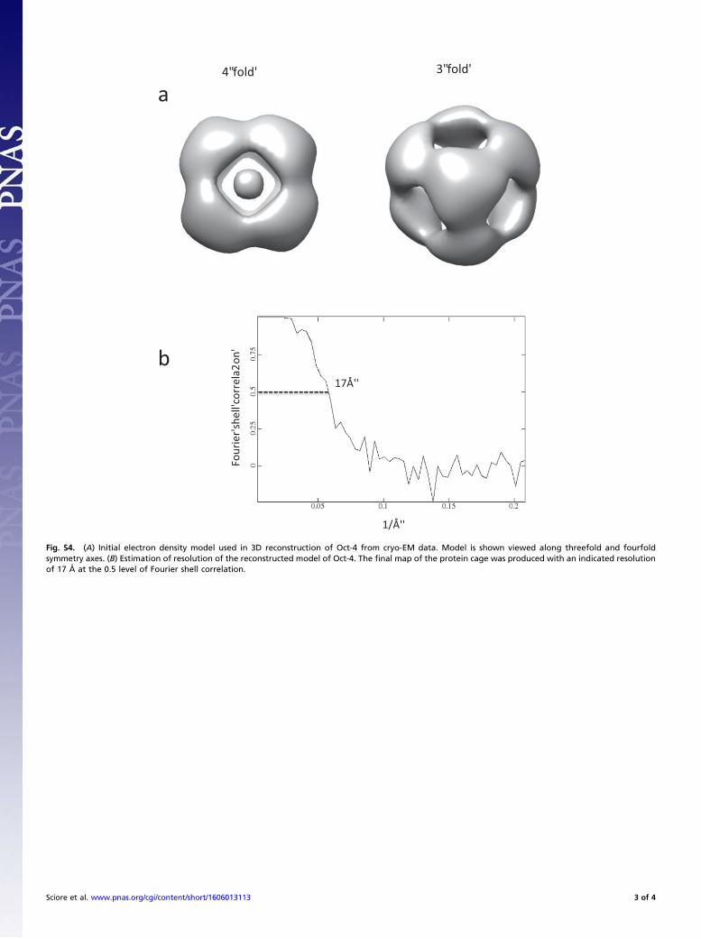
assembled and well-defined class average images were selected to generatethe initial mode using program e2initialmodel.py (Fig. S4A). Then, 34,980 particleswere extracted from those selected classes for 3D reconstruction using RELION. Initialmode was filtered to 60-Å resolution, and then subjected to 3D auto refinementwith initial angular sampling at 7.5°. Octahedral (O) symmetry was enforced duringreconstruction, and the final map of the protein cage was produced with an indi-cated resolution of 17 Å at the 0.5 level of Fourier shell correlation (Fig. S4B). Thecrystal structure of the esterase (PDB 1ZOI) was first manually docked in the mapwith the C terminus in close proximity to the fourfold axis. The fitting was thenrefined using the “fit in map” routine in CHIMERA (52). Map visualization, ren-dering, and figure generation were performed using CHIMERA.
ACKNOWLEDGMENTS. We thank Dr. N. P. King and Dr. W. Sheffler for valu-able assistance in performing the distance minimizations implemented withRosetta. We also thank Thermo Fisher Scientific and Vicki Wysocki [The OhioState University (OSU)] for access to the Orbitrap EMR instrument used in thesestudies, as part of the OSU Campus Chemical Instrumentation Center. Thiswork was supported in part by Department of Defense Multidisciplinary Uni-versity Research Initiative Grant DoD 59743-CH-MUR (to E.N.G.M.) and ArmyResearch Office Grant W911NF-11-1-0251 (to E.N.G.M). AUC calculations wereperformed on the UltraScan Laboratory Information Management Systemcluster at the Bioinformatics Core Facility, University of Texas Health ScienceCenter at San Antonio. These resources are supported in part by NSF XSEDEGrant MCB070038 (to Borries Demeler) and the Extended Collaborative Sup-port Service Program funded by NSF Award OCI-1053575.
1. Kuhn RJ, Rossmann MG (2005) Structure and assembly of icosahedral enveloped RNAviruses. Adv Virus Res 64:263–284.
2. Zhou ZH, McCarthy DB, O’Connor CM, Reed LJ, Stoops JK (2001) The remarkablestructural and functional organization of the eukaryotic pyruvate dehydrogenasecomplexes. Proc Natl Acad Sci USA 98(26):14802–14807.
3. Tanaka S, Sawaya MR, Yeates TO (2010) Structure and mechanisms of a protein-basedorganelle in Escherichia coli. Science 327(5961):81–84.
4. Jutz G, van Rijn P, Santos Miranda B, Böker A (2015) Ferritin: A versatile building blockfor bionanotechnology. Chem Rev 115(4):1653–1701.
5. Reisler E, Egelman EH (2007) Actin structure and function: What we still do not un-derstand. J Biol Chem 282(50):36133–36137.
6. Janke C (2014) The tubulin code: Molecular components, readout mechanisms, andfunctions. J Cell Biol 206(4):461–472.
7. Krishna KA, Rao GV, Rao KR (2007) Chaperonin GroEL: Structure and reaction cycle.Curr Protein Pept Sci 8(5):418–425.
8. Papapostolou D, Howorka S (2009) Engineering and exploiting protein assemblies insynthetic biology. Mol Biosyst 5(7):723–732.
9. King NP, Lai Y-T (2013) Practical approaches to designing novel protein assemblies.Curr Opin Struct Biol 23(4):632–638.
10. Lai Y-T, King NP, Yeates TO (2012) Principles for designing ordered protein assem-blies. Trends Cell Biol 22(12):653–661.
11. Channon K, Bromley EHC, Woolfson DN (2008) Synthetic biology through bio-molecular design and engineering. Curr Opin Struct Biol 18(4):491–498.
12. Uchida M, Qazi S, Edwards E, Douglas T (2015) Use of protein cages as a template forconfined synthesis of inorganic and organic nanoparticles. Methods Mol Biol 1252:17–25.
13. Patterson DP, Rynda-Apple A, Harmsen AL, Harmsen AG, Douglas T (2013) Biomimeticantigenic nanoparticles elicit controlled protective immune response to influenza.ACS Nano 7(4):3036–3044.
14. Padilla JE, Colovos C, Yeates TO (2001) Nanohedra: Using symmetry to design selfassembling protein cages, layers, crystals, and filaments. Proc Natl Acad Sci USA 98(5):2217–2221.
15. Yeates TO, Padilla JE (2002) Designing supramolecular protein assemblies. Curr OpinStruct Biol 12(4):464–470.
16. King NP, et al. (2014) Accurate design of co-assembling multi-component proteinnanomaterials. Nature 510(7503):103–108.
17. King NP, et al. (2012) Computational design of self-assembling protein nanomaterialswith atomic level accuracy. Science 336(6085):1171–1174.
18. Fletcher JM, et al. (2013) Self-assembling cages from coiled-coil peptide modules.Science 340(6132):595–599.
19. Brodin JD, et al. (2012) Metal-directed, chemically tunable assembly of one-, two- andthree-dimensional crystalline protein arrays. Nat Chem 4(5):375–382.
20. Lanci CJ, et al. (2012) Computational design of a protein crystal. Proc Natl Acad SciUSA 109(19):7304–7309.
21. Carlson JCT, et al. (2006) Chemically controlled self-assembly of protein nanorings.J Am Chem Soc 128(23):7630–7638.
22. Ringler P, Schulz GE (2003) Self-assembly of proteins into designed networks. Science302(5642):106–109.
23. Lai Y-T, Tsai K-L, Sawaya MR, Asturias FJ, Yeates TO (2013) Structure and flexibility ofnanoscale protein cages designed by symmetric self-assembly. J Am Chem Soc 135(20):7738–7743.
24. Kobayashi N, et al. (2015) Self-assembling nano-architectures created from a proteinnano-building block using an intermolecularly folded dimeric de novo protein. J AmChem Soc 137(35):11285–11293.
25. Huard DJE, Kane KM, Tezcan FA (2013) Re-engineering protein interfaces yieldscopper-inducible ferritin cage assembly. Nat Chem Biol 9(3):169–176.
26. Lai Y-T, Cascio D, Yeates TO (2012) Structure of a 16-nm cage designed by usingprotein oligomers. Science 336(6085):1129.
27. Lai Y-T, et al. (2014) Structure of a designed protein cage that self-assembles into ahighly porous cube. Nat Chem 6(12):1065–1071.
28. Raman S, Machaidze G, Lustig A, Aebi U, Burkhard P (2006) Structure-based design ofpeptides that self-assemble into regular polyhedral nanoparticles. Nanomedicine(Lond) 2(2):95–102.
29. Usui K, et al. (2009) Nanoscale elongating control of the self-assembled protein fil-ament with the cysteine-introduced building blocks. Protein Sci 18(5):960–969.
30. Patterson DP, et al. (2014) Characterization of a highly flexible self-assembling pro-tein system designed to form nanocages. Protein Sci 23(2):190–199.
31. Patterson DP, Desai AM, Holl MMB, Marsh ENG (2011) Evaluation of a symmetry-based strategy for assembling protein complexes. RSC Advances 1(6):1004–1012.
32. Elmi F, et al. (2005) Stereoselective esterase from Pseudomonas putida IFO12996reveals alpha/beta hydrolase folds for D-beta-acetylthioisobutyric acid synthesis.J Bacteriol 187(24):8470–8476.
33. Lupas AN, Gruber M (2005) The structure of alpha-helical coiled coils. Adv ProteinChem 70:37–78.
34. Fletcher JM, et al. (2012) A basis set of de novo coiled-coil peptide oligomers forrational protein design and synthetic biology. ACS Synth Biol 1(6):240–250.
35. Thomas F, Boyle AL, Burton AJ, Woolfson DN (2013) A set of de novo designed par-allel heterodimeric coiled coils with quantified dissociation constants in the micro-molar to sub-nanomolar regime. J Am Chem Soc 135(13):5161–5166.
36. Negron C, Keating AE (2014) A set of computationally designed orthogonal anti-parallel homodimers that expands the synthetic coiled-coil toolkit. J Am Chem Soc136(47):16544–16556.
37. Zaccai NR, et al. (2011) A de novo peptide hexamer with a mutable channel. NatChem Biol 7(12):935–941.
38. Das R, Baker D (2008) Macromolecular modeling with Rosetta. Annu Rev Biochem 77:363–382.
39. Demeler B, Saber H, Hansen JC (1997) Identification and interpretation of complexityin sedimentation velocity boundaries. Biophys J 72(1):397–407.
40. Demeler B, van Holde KE (2004) Sedimentation velocity analysis of highly heteroge-neous systems. Anal Biochem 335(2):279–288.
41. Demeler B (2005) UltraScan - A comprehensive data analysis software package foranalytical ultracentrifugation experiments. Analytical Ultracentrifugation: Techniquesand Methods, eds Scott DJ, Harding SE, Rowe AJ (R Soc Chem, London), pp 210–230.
42. Bosma HJ, De Kok A, Van Markwijk BW, Veeger C (1984) The size of the pyruvatedehydrogenase complex of Azotobacter vinelandii. Association phenomena. Eur JBiochem 140(2):273–280.
43. McKay AR, Ruotolo BT, Ilag LL, Robinson CV (2006) Mass measurements of increasedaccuracy resolve heterogeneous populations of intact ribosomes. J Am Chem Soc128(35):11433–11442.
44. Rose RJ, Damoc E, Denisov E, Makarov A, Heck AJR (2012) High-sensitivity Orbitrapmass analysis of intact macromolecular assemblies. Nat Methods 9(11):1084–1086.
45. Yang Z, Fang J, Chittuluru J, Asturias FJ, Penczek PA (2012) Iterative stable alignmentand clustering of 2D transmission electron microscope images. Structure 20(2):237–247.
46. Marsh ENG, DeGrado WF (2002) Noncovalent self-assembly of a heterotetramericdiiron protein. Proc Natl Acad Sci USA 99(8):5150–5154.
47. Zhou NE, Kay CM, Hodges RS (1993) Disulfide bond contribution to protein stability:Positional effects of substitution in the hydrophobic core of the two-stranded alpha-helical coiled-coil. Biochemistry 32(12):3178–3187.
48. Zimenkov Y, et al. (2006) Rational design of a reversible pH-responsive switch forpeptide self-assembly. J Am Chem Soc 128(21):6770–6771.
49. Ohi M, Li Y, Cheng Y, Walz T (2004) Negative staining and image classificationPowerful tools in modern electron microscopy. Biol Proced Online 6:23–34.
50. Scheres SHW (2012) RELION: Implementation of a Bayesian approach to cryo-EMstructure determination. J Struct Biol 180(3):519–530.
51. Tang G, et al. (2007) EMAN2: An extensible image processing suite for electron mi-croscopy. J Struct Biol 157(1):38–46.
52. Pettersen EF, et al. (2004) UCSF Chimera—A visualization system for exploratory re-search and analysis. J Comput Chem 25(13):1605–1612.
8686 | www.pnas.org/cgi/doi/10.1073/pnas.1606013113 Sciore et al.

Supporting InformationSciore et al. 10.1073/pnas.1606013113
Fig. S1. (A) SDS PAGE of proteins. Lane 1, protein standards; lane 2, unmodified esterase; lane 3, Oct-4; and lane 4, Oct-2. (B, Left) SEC of Oct-4 after pu-rification on Ni-NTA resin (solid trace). Fractions 1–5 were analyzed by native PAGE, pooled, and rechromatographed (dashed trace). (Right) Analysis of SECfractions by native PAGE. Lanes on the gel are: Ni, Oct-4 after purification on Ni-NTA resin; lanes 1–5, fractions 1–5; and pool, pooled material after SEC.
Fig. S2. Further characterization of Oct-2. (A) A 2DSA of Oct-2. The protein forms multiple species characterized by sedimentation coefficients that are largerthan expected for an octahedral cage. The low frictional ratios are consistent with the formation of globular complexes. (B) Negative stain EM of Oct-2. Theimages indicate that the protein assembles into a range of particle sizes, but no symmetry is apparent in the images, in contrast to the particles formed by Oct-4(Fig. 3D). (Scale bar, 20 nm.)
Sciore et al. www.pnas.org/cgi/content/short/1606013113 1 of 4

Fig. S3. The 2D class averages for Oct-4 from cryo-EM. A total of 44,856 particle images representing protein cages were excised using RELION. The selected particles were further subjected to reference-free alignment andclassified into 405 classes. For details, see The 2D Classifications.
Sciore et al. www.pnas.org/cgi/content/short/1606013113 2 of 4
4"fold' 3"fold'
Four
ier's
hell '
corr
ela2
on'
17Å''
1/Å''
a
b
Fig. S4. (A) Initial electron density model used in 3D reconstruction of Oct-4 from cryo-EM data. Model is shown viewed along threefold and fourfoldsymmetry axes. (B) Estimation of resolution of the reconstructed model of Oct-4. The final map of the protein cage was produced with an indicated resolutionof 17 Å at the 0.5 level of Fourier shell correlation.
Sciore et al. www.pnas.org/cgi/content/short/1606013113 3 of 4

Table S1. Amino acid sequences of proteins used in this study
The flexible linker region is shown in blue, and the coiled-coil sequence is in red.
Table S2. Hydrodynamic parameters for protein assemblies formed by Oct-4determined by sedimentation velocity AUC
SpeciesSedimentationcoefficient, S
Molecularweight, kDa
FrictionalRatio (f/f0)
Partialconcentration,
%
Solute 1 17.6 ± 0.1 886 ± 14 1.89 ± 0.02 73.3Solute 2 22.1 ± 0.07 489 ± 26 1.01 ± 0.04 18.5Solute 3 27.7 ± 0.3 728 ± 114 1.05 ± 0.1 4.5Solute 4 37.3 ± 0.2 1,145 ± 194 1.06 ± 0.09 2.3
For details, see AUC of Oct-4.
Table S3. Hydrodynamic parameters for protein assemblies formed by Oct-2determined by sedimentation velocity AUC
SpeciesSedimentationcoefficient, S
Molecularweight, kDa
Frictionalratio (f/f0)
Partialconcentration,
%
Solute 1 24.8 ± 0.4 649 ± 96 1.09 ± 0.11 2.2Solute 2 31.5 ± 0.1 905 ± 67 1.07 ± 0.05 21.5Solute 3 38.0 ± 0.2 1,128 ± 63 1.03 ± 0.04 27.4Solute 4 43.5 ± 0.3 1,357 ± 93 1.01 ± 0.05 20.3Solute 5 49.8 ± 0.8 1,681 ± 174 1.02 ± 0.06 13.9Solute 6 57.3 ± 0.7 2,113 ± 199 1.03 ± 0.07 7.4
For details, see AUC of Oct-4.
Other Supporting Information Files
Dataset S1 (TXT)Dataset S2 (TXT)
Sciore et al. www.pnas.org/cgi/content/short/1606013113 4 of 4