Embed Size (px)
Citation preview
170 a. vastardis, g. hadjipavlou, a. marjan, g. stojanovic, b. dial, k. kazakos
Acta Orthopædica Belgica, Vol. 83 - 1 - 2017 Acta Orthopædica Belgica, Vol. 83 - 1 - 2017
Georgios A. Vastardis, MD*†; Alexander G. Hadjipavlou, MD‡; Anna E. Marjan, BS*; Michael A. Stojanovic, BS*; Brian Dial, BS*; Konstantinos I. Kazakos MD ¥. * Department of Orthopaedic Surgery and Rehabilitation,Loyola University Medical Center, Maywood, Illinois, USA.
fractures. In the lower thoracic and particularly in the lumbar spine, the transpedicular approach is used to guide the trocar along the trajectory of the pedicle that offers a safe passage. However, accessing this safe route in the upper and middle thoracic spine is technically difficult and challenging for the surgeons, due to decreased pedicular diameter and the angulation of the pedicle, in the sagittal and axial plane (9,12,16,21). Additionally, lateral visualization
The company played no role in the design, conduct or reporting of the study or in the decision to submit the manuscript for publication.
Acta Orthop. Belg., 2017, 83, 170-179
Flexible memory-alloy instrumentation for unilateral transpedicular kyphoplasty and guided cement augmentation of the thoracic spine
Georgios A. vastardis, Alexander G. hadjipavlou, Anna E. marjan, Michael A. stojanovic,Brian dial, Konstantinos I. kazakos
From the Department of Orthopaedic Surgery and Rehabilitation, Loyola University Chicago, Maywood, USA
ORIGINAL STUDY
n Georgios A. Vastardis, MD† Musculoskeletal Biomechanics Laboratory, Department of Veterans Affairs, Edward Hines Jr. VA Hospital, Hines, Illinois, USA and Department of Orthopaedic Surgery and Department of Orthopaedic Surgery and Rehabilitation, Loyola, Maywood, Illinois, USA.
n Alexander G. Hadjipavlou, MDClinical Professor of Orthopaedic Surgery and Rehabi-litation. Department of Orthopaedic surgery and Rehabi-litation, University of Texas, Medical Branch, Galveston, Texas, USA.
n Anna E. Marjan, BSn Michael A. Stojanovic, BSn Brian Dial, BS
Department of Orthopaedic Surgery and Rehabilitation, Loyola University Medical Center, Maywood, Illinois, USA.
n Konstantinos I. Kazakos MD Professor of Orthopaedic surgery. Department of Ortho-paedic Surgery, Democritus University of Thrace, Greece.Correspondence : Georgios A. Vastardis MD, Department
of Orthopaedic Surgery and Rehabilitation,Loyola University Chicago, 2160 S. First Avenue, Maywood, IL 60153, USA.
Phone : +306936922952E-Mail : [email protected]© 2017, Acta Orthopædica Belgica.
The purpose of this novel study was to investigate the feasibility of unilateral transpedicular balloon kyphoplasty particularly of the upper thoracic vertebrae using an 11- gauge balloon and cement inserter, and to study the morphological parameters of the thoracic spine pedicles.We used four fresh frozen cadaveric thoracic spines with intact rib cages and skin for kyphoplasty from T1 to T12 vertebrae under C-arm fluoroscopy. The most limiting width of the pedicles 2.46+/-0.32mm was in the middle levels (T5-T8). The absolute minimum height of the pedicles was at T1 (3.80-3.87mm). All regions of the vertebral body were effectively targeted for cement augmentation. The average cement load of all the vertebral bodies was 43,22%.Using the kyphoplasty technique in combination with the pre-bent 11mm memory-alloy cement inserter allowed targeting of the desired position of the vertebral body for effective vertebral body cement augmentation.
Keywords : kyphoplasty ; upper thoracic spine ; unilateral transpedicular kyphoplasty.
INTRODUCTION
Vertebral kyphoplasty is a surgical intervention for restoring vertebral body height and osseous strength in both osteoporotic and metastatic compression

Acta Orthopædica Belgica, Vol. 83 - 1 - 2017
flexible memory-alloy instrumentation for unilateral transpedicular kyphoplasty 171
of the pedicle in the upper four thoracic vertebrae is almost technically impossible under fluoroscopy in the operating room.
For the above reasons, many surgeons are reluctant to attempt transpedicular kyphoplasty at the higher thoracic levels, especially with large-gauge trocars (e.g., an 8-gauge has an outer diameter of 4.2 mm). The extrapedicular approach has become the preferred choice. However, this approach is not without potential risks to the surrounding structures, such as the pulmonary cavity and segmental arteries (6,16,17).
The favored approach for kyphoplasty is a bilateral transpedicular instrumentation in order to ensure adequate cement augmentation of the entire vertebral body. Augmenting only a single half of the vertebral body may potentially lead to medial/lateral angular deformation of the vertebral body (4). A new vertebroplasty delivery cement system was designed to address this concern. A friendlier and more satisfactory vertebroplasty can be performed in the thoracic spine through a unilateral transpedicular approach using a thin cement delivery instrumentation. This consists of an 11gauge straight conventional trocar (Avamax) and a curved needle (AVAflex), for delivering the cement, made of memory alloy that bounces back into its pre-bent shape as soon as it exits the trocar within the vertebral body. This concept was supplied for the design of kyphoplasty instrumentation.
In the majority of vertebroplasty cases using the unilateral approach, the injected cement can cross the midline. Kim et al. showed that the clinical outcomes of the unilateral extrapedicular approach are no different than those treated with a bilateral approach in their study, that they performed a unilateral kyphoplasty but with a straight needle, inserted obliquely. (15). A cement quantity of 3-4 ml and 6-8 ml is found sufficient to restore the pre-fracture strength to the thoracolumbar and lumbar spine, respectively (2). A study using intact vertebral bodies demonstrated that the maximal stiffness and strength of the vertebral body are obtained when at least 20% of the vertebral body is augmented by volume with cement (14).
The development of a smaller gauge trocar and a thin pre-curved cement inserter instrument incited
us to investigate the feasibility of the unilateral transpedicular approach in the upper and middle thoracic spine.
The purpose of this study is threefold : A. To assess the feasibility of unilateral transpedicular kyphoplasty in upper and middle thoracic spine using an 11-gauge straight trocar (AVAmax®, CareFusion) with a memory alloy pre-curved vertebral needle that bounces back into shape after it exit the trocar within the vertebral body, introducing the cement for kyphoplasty (AVAflex®, CareFusion). B. To evaluate the cement distribution in the various compartments of the vertebral bodies after performing unilateral transpedicular kyphoplasty in the thoracic spine (T1-T12) with this new kyphoplasty instrumentation, and C. To investigate the morphology of the pedicle in these specimens.
MATERIALS AND METHODS
Four fresh frozen osteoporotic cadaveric thoracic specimens (age : 74 +/- 1.8 years) (T1-T12) with intact skin, muscles, and rib cage were used. The specimens were radiographically screened to exclude vertebral metastases, prior vertebral compression fractures and previous surgeries. The bone density of the specimens was determined by peripheral quantitative computed tomography (PQCT) scans.
Computed Tomography (CT) scans (0.6 mm slice thickness) were taken before and after the vertebral body cavitation and cement distribution using the commercial software package Mimics (Materialize Inc., Ann Arbor, MI). The inner trabecular bone and outer cortical bone in the sagittal and axial views at three distinct points along the pedicle were measured, for a total of nine measurements in each view. At each of these points, measurements were taken of the inner cancellous bone and the surrounding cortical bone on two sides, either inferior and superior in sagittal view or medial and lateral in axial view in the isthmus, the beginning of the pedicle, and the end of the pedicle. In addition, the angle of the pedicle axis was defined in axial and sagittal planes.
172 a. vastardis, g. hadjipavlou, a. marjan, g. stojanovic, b. dial, k. kazakos
Acta Orthopædica Belgica, Vol. 83 - 1 - 2017
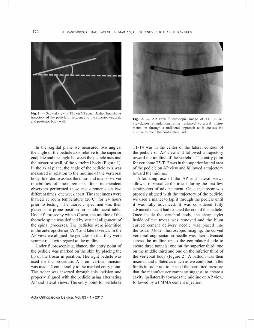
In the sagittal plane we measured two angles: the angle of the pedicle axis relative to the superior endplate and the angle between the pedicle axis and the posterior wall of the vertebral body (Figure 1). In the axial plane, the angle of the pedicle axis was measured in relation to the midline of the vertebral body. In order to assess the intra- and inter-observer reliabilities of measurements, four independent observers performed these measurements on two different times, one week apart. The specimens were thawed at room temperature (20˚C) for 24 hours prior to testing. The thoracic specimen was then placed in a prone position on a radiolucent table. Under fluoroscopy with a C-arm, the midline of the thoracic spine was defined by vertical alignment of the spinal processes. The pedicles were identified in the anteroposterior (AP) and lateral views. In the AP view we aligned the pedicles so that they were symmetrical with regard to the midline.
Under fluoroscopic guidance, the entry point of the pedicle was marked on the skin by placing the tip of the trocar in position. The right pedicle was used for the procedure. A 1 cm vertical incision was made, 2 cm laterally to the marked entry point. The trocar was inserted through this incision and properly aligned with the pedicle using alternating AP and lateral views. The entry point for vertebrae
T1-T4 was in the center of the lateral contour of the pedicle on AP view and followed a trajectory toward the midline of the vertebra. The entry point for vertebrae T5-T12 was in the superior-lateral area of the pedicle on AP view and followed a trajectory toward the midline.
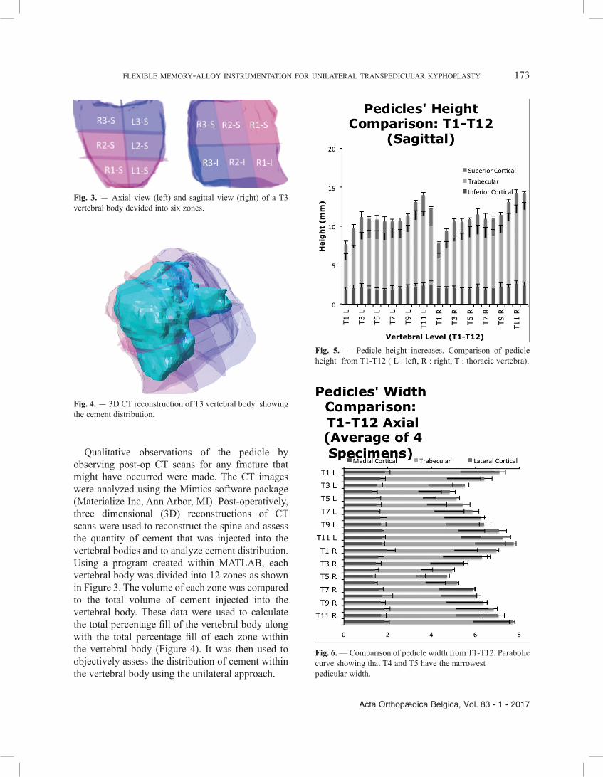
Alternating use of the AP and lateral views allowed to visualize the trocar during the first few centimeters of advancement. Once the trocar was properly aligned with the trajectory of the pedicle, we used a mallet to tap it through the pedicle until it was fully advanced. It was considered fully advanced once it had reached the end of the pedicle. Once inside the vertebral body, the sharp stylet inside of the trocar was removed and the blunt curved cement delivery needle was placed into the trocar. Under fluoroscopic imaging, the curved vertebral augmentation needle was then advanced across the midline up to the contralateral side to create three tunnels, one on the superior third, one on the middle third and one on the inferior third of the vertebral body (Figure 2). A balloon was then inserted and inflated as much as we could but in the limits in order not to exceed the permitted pressure that the manufacturer company suggest, to create a cavity ipsilaterally towards the midline on AP view, followed by a PMMA cement injection.
Fig. 1. — Sagittal view of T10 on CT scan. Dashed line shows trajectory of the pedicle in reference to the superior endplate and posterior body wall. Fig. 2. — AP view fluoroscopic image of T10 in AP
viewdemostratingdemonstrating reshaped vertebral instru-mentation through a unilateral approach as it crosses the midline to reach the contralateral side.
Acta Orthopædica Belgica, Vol. 83 - 1 - 2017
flexible memory-alloy instrumentation for unilateral transpedicular kyphoplasty 173
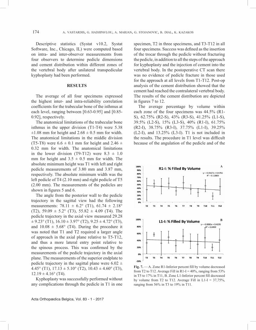
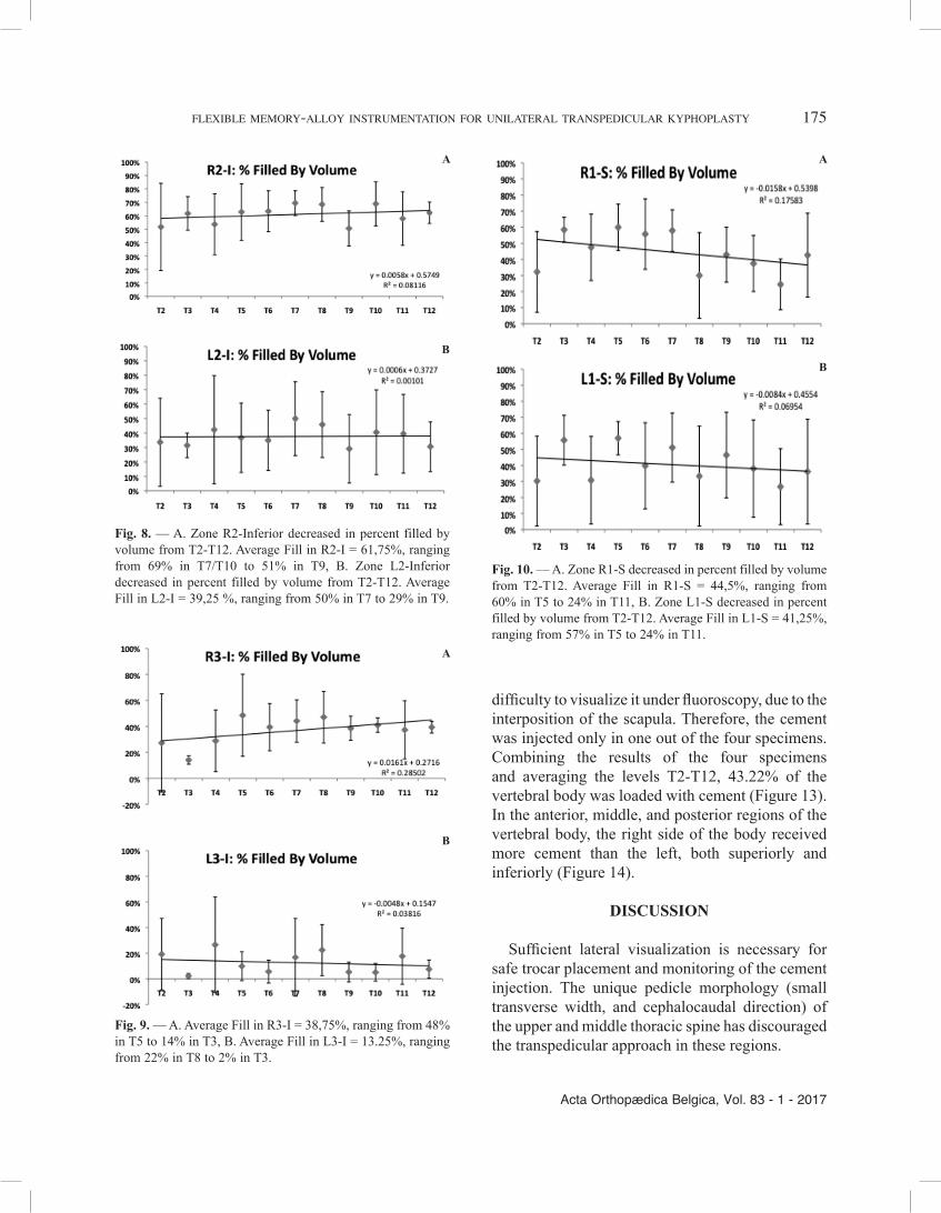
Qualitative observations of the pedicle by observing post-op CT scans for any fracture that might have occurred were made. The CT images were analyzed using the Mimics software package (Materialize Inc, Ann Arbor, MI). Post-operatively, three dimensional (3D) reconstructions of CT scans were used to reconstruct the spine and assess the quantity of cement that was injected into the vertebral bodies and to analyze cement distribution. Using a program created within MATLAB, each vertebral body was divided into 12 zones as shown in Figure 3. The volume of each zone was compared to the total volume of cement injected into the vertebral body. These data were used to calculate the total percentage fill of the vertebral body along with the total percentage fill of each zone within the vertebral body (Figure 4). It was then used to objectively assess the distribution of cement within the vertebral body using the unilateral approach.
Fig. 3. — Axial view (left) and sagittal view (right) of a T3 vertebral body devided into six zones.
Fig. 4. — 3D CT reconstruction of T3 vertebral body showing the cement distribution.
Fig. 5. — Pedicle height increases. Comparison of pedicle height from T1-T12 ( L : left, R : right, T : thoracic vertebra).
Fig. 6. — Comparison of pedicle width from T1-T12. Parabolic curve showing that T4 and T5 have the narrowest pedicular width.
174 a. vastardis, g. hadjipavlou, a. marjan, g. stojanovic, b. dial, k. kazakos
Acta Orthopædica Belgica, Vol. 83 - 1 - 2017
specimen, T2 in three specimens, and T3-T12 in all four specimens. Success was defined as the insertion of the trocar through the pedicle without fracturing the pedicle, in addition to all the steps of the approach for kyphoplasty and the injection of cement into the vertebral body. In the postoperative CT scan there was no evidence of pedicle fracture in those used for the approach at all levels from T1-T12. Post-op analysis of the cement distribution showed that the cement had reached the contralateral vertebral body.The results of the cement distribution are depicted in figures 7 to 12.
The average percentage by volume within each zone of the four specimens was 44.5% (R1-S), 62.75% (R2-S), 43% (R3-S), 41.25% (L1-S), 39.5% (L2-S), 15% (L3-S), 40% (R1-I), 61.75% (R2-I), 38.75% (R3-I), 37.75% (L1-I), 39.25% (L2-I), and 13.25% (L3-I). T1 is not included in the results. The procedure in T1 level was difficult because of the angulation of the pedicle and of the
Descriptive statistics (Systat v10.2, Systat Software, Inc., Chicago, IL) were computed based on intra- and inter-observer measurements from four observers to determine pedicle dimensions and cement distribution within different zones of the vertebral body after unilateral transpedicular kyphoplasty had been performed.
RESULTS
The average of all four specimens expressed the highest inter- and intra-reliability correlation coefficients for the trabecular bone of the isthmus at each level, ranging between [0.63-0.95] and [0.85-0.92], respectively.
The anatomical limitations of the trabecular bone isthmus in the upper division (T1-T4) were 5.38 ±1.08 mm for height and 2.68 ± 0.5 mm for width. The anatomical limitations in the middle division (T5-T8) were 6.6 ± 0.1 mm for height and 2.46 ± 0.32 mm for width. The anatomical limitations in the lower division (T9-T12) were 8.3 ± 1.0 mm for height and 3.5 ± 0.5 mm for width. The absolute minimum height was T1 with left and right pedicle measurements of 3.80 mm and 3.87 mm, respectively. The absolute minimum width was the left pedicle of T4 (2.10 mm) and right pedicle of T5 (2.00 mm). The measurements of the pedicles are shown in figures 5 and 6.
The angle from the posterior wall to the pedicle trajectory in the sagittal view had the following measurements: 78.11 ± 6.2° (T1), 61.74 ± 2.18° (T2), 59.09 ± 5.2° (T3), 55.82 ± 4.09 (T4). The pedicle trajectory in the axial view measured 29.28 ± 9.23° (T1), 16.10 ± 3.97° (T2), 9.25 ± 4.72° (T3), and 10.08 ± 5.68° (T4). During the procedure it was noted that T1 and T2 required a larger angle of approach in the axial plane relative to T5-T12, and thus a more lateral entry point relative to the spinous process. This was confirmed by the measurements of the pedicle trajectory in the axial plane. The measurements of the superior endplate to pedicle trajectory in the sagittal plane were 6.02 ± 4.45° (T1), 17.13 ± 5.10° (T2), 10.43 ± 4.60° (T3), 12.19 ± 4.16° (T4).
Kyphoplasty was successfully performed without any complications through the pedicle in T1 in one
Fig. 7. — A. Zone R1-Inferior percent fill by volume decreased from T2 to T12. Average Fill in R1-I = 40%, ranging from 53% in T5 to 17% in T11, B. Zone L1-Inferior percent fill decreased by volume from T2 to T12. Average Fill in L1-I = 37,75%, ranging from 56% in T3 to 19% in T11.
B
A
Acta Orthopædica Belgica, Vol. 83 - 1 - 2017
flexible memory-alloy instrumentation for unilateral transpedicular kyphoplasty 175
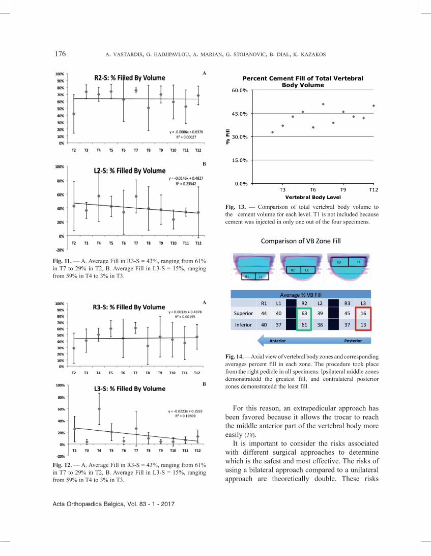
difficulty to visualize it under fluoroscopy, due to the interposition of the scapula. Therefore, the cement was injected only in one out of the four specimens. Combining the results of the four specimens and averaging the levels T2-T12, 43.22% of the vertebral body was loaded with cement (Figure 13). In the anterior, middle, and posterior regions of the vertebral body, the right side of the body received more cement than the left, both superiorly and inferiorly (Figure 14).
DISCUSSION
Sufficient lateral visualization is necessary for safe trocar placement and monitoring of the cement injection. The unique pedicle morphology (small transverse width, and cephalocaudal direction) of the upper and middle thoracic spine has discouraged the transpedicular approach in these regions.
Fig. 8. — A. Zone R2-Inferior decreased in percent filled by volume from T2-T12. Average Fill in R2-I = 61,75%, ranging from 69% in T7/T10 to 51% in T9, B. Zone L2-Inferior decreased in percent filled by volume from T2-T12. Average Fill in L2-I = 39,25 %, ranging from 50% in T7 to 29% in T9.
A
B
A
B
Fig. 9. — A. Average Fill in R3-I = 38,75%, ranging from 48% in T5 to 14% in T3, B. Average Fill in L3-I = 13.25%, ranging from 22% in T8 to 2% in T3.
A
B
Fig. 10. — A. Zone R1-S decreased in percent filled by volume from T2-T12. Average Fill in R1-S = 44,5%, ranging from 60% in T5 to 24% in T11, B. Zone L1-S decreased in percent filled by volume from T2-T12. Average Fill in L1-S = 41,25%, ranging from 57% in T5 to 24% in T11.
176 a. vastardis, g. hadjipavlou, a. marjan, g. stojanovic, b. dial, k. kazakos
Acta Orthopædica Belgica, Vol. 83 - 1 - 2017
For this reason, an extrapedicular approach has been favored because it allows the trocar to reach the middle anterior part of the vertebral body more easily (18).
It is important to consider the risks associated with different surgical approaches to determine which is the safest and most effective. The risks of using a bilateral approach compared to a unilateral approach are theoretically double. These risks
A
B
Fig. 11. — A. Average Fill in R3-S = 43%, ranging from 61% in T7 to 29% in T2, B. Average Fill in L3-S = 15%, ranging from 59% in T4 to 3% in T3.
A
B
Fig. 12. — A. Average Fill in R3-S = 43%, ranging from 61% in T7 to 29% in T2, B. Average Fill in L3-S = 15%, ranging from 59% in T4 to 3% in T3.
Fig. 13. — Comparison of total vertebral body volume to the cement volume for each level. T1 is not included because cement was injected in only one out of the four specimens.
Fig. 14. —Axial view of vertebral body zones and corresponding averages percent fill in each zone. The procedure took place from the right pedicle in all specimens. Ipsilateral middle zones demonstratedd the greatest fill, and contralateral posterior zones demonstratedd the least fill.

Acta Orthopædica Belgica, Vol. 83 - 1 - 2017
flexible memory-alloy instrumentation for unilateral transpedicular kyphoplasty 177
from T1-T12. This unexpected finding may suggest potential elastic properties inherent to cortical bone. The advantage given to the procedure by the thicker medial wall of the cortical bone in the pedicle or the angulation whith which the trocar can enter into the pedicle in the axial plane, allows for a trocar penetration without fracture (7). It can be argued that the failure to successfully perform transpedicular kyphoplasty in the upper thoracic spine in three out of four specimens at T1 and one out of four specimens at T2 was most likely due to the lack of proper visualization or improper technique, rather than an inadequate pedicle diameter. If the pedicle width was the limiting factor, then the results would have shown kyphoplasty to be unsuccessful at T3-T5, which was not the case. Therefore, it may be possible to perform transpedicular kyphoplasty up to T1 with a proper imaging visualization technique.
In the upper thoracic spine (levels above T5), lateral visualization is a major limiting factor to performing transpedicular kyphoplasty due to interference from the shoulder and scapula. Adequate visualization using fluoroscopy at higher thoracic levels is feasible with a proper technique that requires adducting or flexing and abducting the patient’s arms, which directs the scapula laterally (19). These maneuvers were not possible in this study since the specimens did not have arms and the scapula was fixed in the torso and could not be moved cephalad. It may be possible to achieve a better positioning and adequate visualization, and therefore successfully perform kyphoplasty at higher thoracic levels (T1 and T2) in patients who are able to assume the proper position.
According to one study, the cement distribution in vertebroplasty through a unilateral approach, has shown that the clinical outcomes are no different than those treated with a bilateral approach (15).
However, there is limited research describing the distribution of cement within the vertebral body after unilateral approach for kyphoplasty.
Since the majority of the compression fractures occur in the anterior region, the superior zones should be targeted for cement injection in order to restore the superior end plate height. Our study showed an increased fill in the superior zones relative to inferior zones, which is advantageous for the
include pedicle fracture, cement extravasation, and damage to surrounding structures. There are also other risks depending on whether one uses an extrapedicular approach or a transpedicular approach. The extrapedicular approach involves advancing the trocar through a “window” created by the transverse process and the rib, thus running a greater risk of damaging structures found in the soft tissues. In transpedicular approach, the pedicle serves also as a natural conduit and its surrounding cortical bone guiding the trocar into the vertebral body. However, this approach can give rise to disruption of the pedicle and spinal canal intrusion, leading to neurological deficits (16,18).
Some advocate the superiority of CT guided control to achieve adequate lateral imaging in the upper thoracic spine T1-T4 (23). However, this approach exposes both the surgeon and the patient to higher doses of radiation. Furthermore, as opposed to the operating theatre, the x-ray suite is not always readily available.
The effective use of a flexible curved vertebral needle made of memory alloy for augmentation vertebroplasty through a unilateral approach has shown to decrease the danger of penetrating the contralateral cortical wall of the vertebral body (20).
Previous reports studying thoracic pedicle anatomy demonstrated a great variability of pedicle morphology across gender, age and ethnicity (1,5,10,13).
The morphological data from this study reveal that the most physically confined level is at T4 and T5. These data also confirms previous reports (1,3,11,22). The inner diameter of the pedicle at its narrowest point, the isthmus, shows the highest correlation among the observers, making it the defining criterion for any anatomically limiting parameters.
The 11-gauge trocar has an outer diameter of 3.00 mm, which exceeds the isthmus width in the upper and middle levels (average widths are are 2.68 ± 0.50 and 2.46 ± 0.32 mm, respectively). Since pedicle height at all levels was greater than 3.0 mm, it was not a limitation to accessing the pedicle. Despite the width of the trocar exceeding that of the isthmus at multiple levels, post-operative CT scans showed no evidence of pedicle fracture
178 a. vastardis, g. hadjipavlou, a. marjan, g. stojanovic, b. dial, k. kazakos
Acta Orthopædica Belgica, Vol. 83 - 1 - 2017
5. Christodoulou AG, Apostolou T, Ploumis A, Terzidis I, Hantzokos I, Pournaras J. Pedicle dimensions of the thoracic and lumbar vertebrae in the Greek population. Clin Anat 2005 ; 18 : 404-8.
6. Chung HJ, Chung KJ, Yoon HS, et al. Comparative study of balloon kyphoplasty with unilateral versus bilateral approach in osteoporotic vertebral compression fractures. Internat Orthopaedics 2008; 32 : 817-820.
7. Dalbayrak S, Onen MR, Yilmaz M, Naderi S. Clinical and radiographic results of balloon kyphoplasty for treatment of vertebral body metastases and multiple myelomas. J Clin Neurosci 2010 ; 17 : 219-24.
8. Dufresne AC, Brunet E, Sola-Martinez MT, Rose M, Chiras J. Percutaneous vertebroplasty of the cervico-thoracic junction using an anterior route. Technique and results. Report of nine cases. J Neuroradiol 1998 ; 25 : 123-8.
9. Eleraky M, Papanastassiou I, Setzer M, Baaj AA, Tran ND, Vrionis FD Balloon kyphoplasty in the treatment of metastatic tumors of the upper thoracic spine. J Neurosurg Spine 2011 ; 14 :372-6.
10. Erkan S, Wu C, Mehbod AA, Cho W, Transfeldt EE. Biomechanical comparison of transpedicular versus extrapedicular vertebroplasty using polymethyl-methacrylate. J Spinal Disord Tech 2010 ; 23 : 180-5.
11. Garfin SR, Yuan HA, Reiley MA. New technologies in spine: kyphoplasty and vertebroplasty for the treatment of painful osteoporotic compression fractures. Spine (Phila Pa 1976) 2001, 15 ; 26 :1511-5.
12. Gerszten PC, Monaco EA 3rd. Complete percutaneous treatment of vertebral body tumors causing spinal canal compromise using a transpedicular cavitation, cement augmentation, and radiosurgical technique. Neurosurg Focus 2009 ; 27 : E9.
13. Hassan E, Liau KM, Ariffin I, Halim Yusof A. Internal morphometry of thoracic pedicles in the immature spine. Spine (Phila Pa 1976) 2010 1 ; 35 :1253-6.
14. Higgins K, Harten R, Langrana N, et al. Biomechanical effects of unipedicular vertebroplasty on intact vertebrae. Spine (Phila Pa 1976) 2003 ; 28 : 1540-1547.
15. Kim A, Jensen M, Dion J, et al. Unilateral transpedicular percutaneous vertebroplasty: Initial experience. Radiology 2002 ; 222 : 737-741.
16. Kim HS, Kim SW, Ju CI. Balloon kyphoplasty through extrapedicular approach in the treatment of middle thoracic osteoporotic compression fracture : T5-T8 level. J of Korean Neurosurg Soc 2007 ; 42 : 363-366.
17. Liebschner M, Rosenberg W, Keaveny T. Effects of bone cement volume and distribution on vertebral stiffness after vertebroplasty. Spine (Phila Pa 1976) 2001 ; 26 : 1547-1554.
18. Lien SB, Liou NH, Wu SS. Analysis of anatomic morphometry of the pedicles and the safe zone for through-pedicle procedures in the thoracic and lumbar spine. Eur Spine J 2007 ; 16 : 1215-22.
restoration of vertebral body height after a fracture. The posterior region is considered to be a “danger area” due to its proximity to the spinal canal, thus the reduced fill in zone 3 provides a greater degree of safety. It is critical to target cement placement at the desired location. The 11-gauge curved vertebral augmentation needle allows cement augmentation in all twelve zones of the vertebral body. By knowing the location and the extent of a vertebral compression fracture, the surgeon can use these data to discern which patients will benefit the most from this procedure.
In this study, pedicle disruption and spinal canal intrusion were not noted on the post-op CT scans. The blunt tip of the curved augmentation needle also decreased the danger of penetrating the cortical wall.
The major limitation of this study was its small sample size. We used four specimens because of cost constraints.
In conclusion, this novel instrumentation is safe and effective for unilateral percutaneous trans-pedicular balloon kyphoplasty in the mid and upper thoracic spine.
Acknowledgments
The authors would like to thank Prof. Avinash Patwardhan PhD director of Musculoskeletal Biomechanics Laboratory, Department of Veterans Affairs,Edward Hines Jr. VA Hospital, Hines, Illinois, USA, for provision of laboratory space and his technical support , Leonard I. Voronov MD,Phd and Michael Zindrick MD for advise and assistance during this study. The authors also deeply thank CareFusion, Vernon Hills, IL, USA, for financially supporting this study.
REFERENCES
1. Bayley E, Clamp J, Boszczyk BM. Percutaneous approach to the upper thoracic spine: optimal patient positioning. Eur Spine J. 2009 ; 18 : 1986-8.
2. Belkoff S, Mathis J, Jasper L, et al. The biomechanics of vertebroplasty. the effect of cement volume on mechanical behavior. Spine (Phila Pa 1976) 2001 ; 26 : 1537-1541.
3. Boszczyk BM, Bierschneider M, Hauck S, Beisse R, Potulski M, Jaksche H. Transcostovertebral kyphoplasty of the mid and high thoracic spine. Eur Spine J 2005 ; 14 : 992-9.
4. Chen B, Li Y, Xie D, et al. Comparison of unipedicular and bipedicular kyphoplasty on the stiffness and biomechanical balance of compression fractured vertebrae. Eur Spine J 2011 ; 20 : 1272-1280.
Acta Orthopædica Belgica, Vol. 83 - 1 - 2017
flexible memory-alloy instrumentation for unilateral transpedicular kyphoplasty 179
19. McLain RF, Ferrara L, Kabins M. Pedicle morphometry in the upper thoracic spine: limits to safe screw placement in older patients. Spine (Phila Pa 1976) 2002 ; 15 ; 27 : 2467-71.
20. Ortiz AO, Zoarski GH, Beckerman M. Kyphoplasty Tech Vasc Interv Radiol 2002 ; 5 : 239-49.
21. Ortiz O, Mathis JM Vertebral body reconstruction: techniques and tools. Neuroimaging Clin N Am 2010 ; 20 : 145-58.
22. Zhuang Z, Chen Y, Han H, Cai S, Wang X, Qi W, Kong K. Thoracic pedicle morphometry in different body height population : a three-dimensional study using reformatted computed tomography. Spine (Phila Pa 1976) 2011 ; 15 ; 36 : E1547-54.
23. Zindrick MR, Knight GW, Sartori MJ, Carnevale TJ, Patwardhan AG, Lorenz MA. Pedicle morphology of the immature thoracolumbar spine. Spine (Phila Pa 1976) 2000 ; 1 ; 25 : 2726-35.