Embed Size (px)
Citation preview
INTRODUCTION
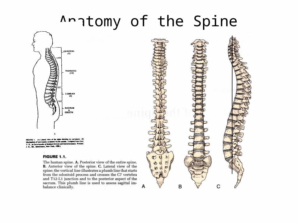
Spine• Backbone consisting of 7 cervical vertebrae, 12 thoracic vertebrae, 5 lumbar vertebrae• Mechanically, a long, slender, flexible, curved beam consisting of essentially similar segments
that can be considered in isolation
Mechanical Functions of the Spine• Structural support for the musculoskeletal torso• Flexibility of motion for activities• Protection of the spinal cord
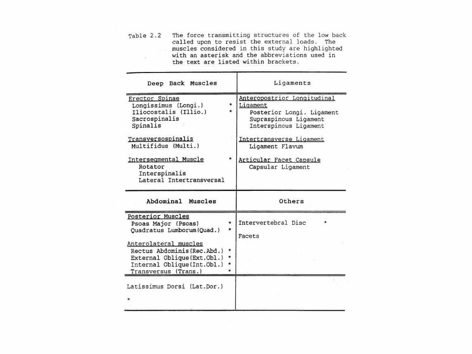
Elements for the Mechanical Stability of the Spine• Passive elements: Vertebrae, disc, facets, ligaments, ligaments• Active elements: Muscles• The stability is obtained through the highly developed dynamic neuromuscular control system
Disturbances• Biological factors• Acute or cumulative fatigue injuries• Surgical procedures
INTRODUCTION (CONT.)
Spinal Disorders• Tumors and TBs• Fractures• Deformities• Low back and neck pain
Back painRadicular pain (leg and arm pain)
Treatment Modalities• Conservative: Bed rest, Medication, Physical therapy, Exercise, Orthoses, Acupuncture, etc.• Non-conservative: Surgery
Why is the Biomechanics important for the spinal problems?• Mechanical stress due to excessive loading or vibration is a cause of various spinal disorders
1. Disc and facet degeneration2. Injuries3. Progression of deformity
• Spinal stability1. Prevention of the deformity progress2. Prevention of recurring pain
• Biomechanical investigations are capable of providing an objective assessment of the effects of injury, fatigue, and surgery on the human spine.
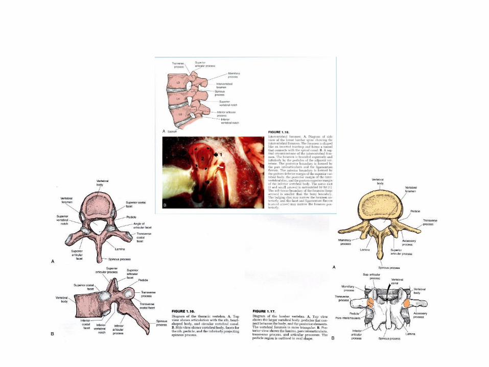
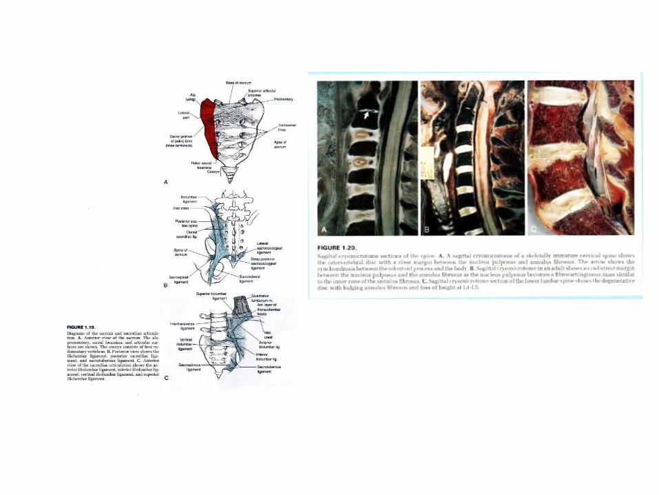
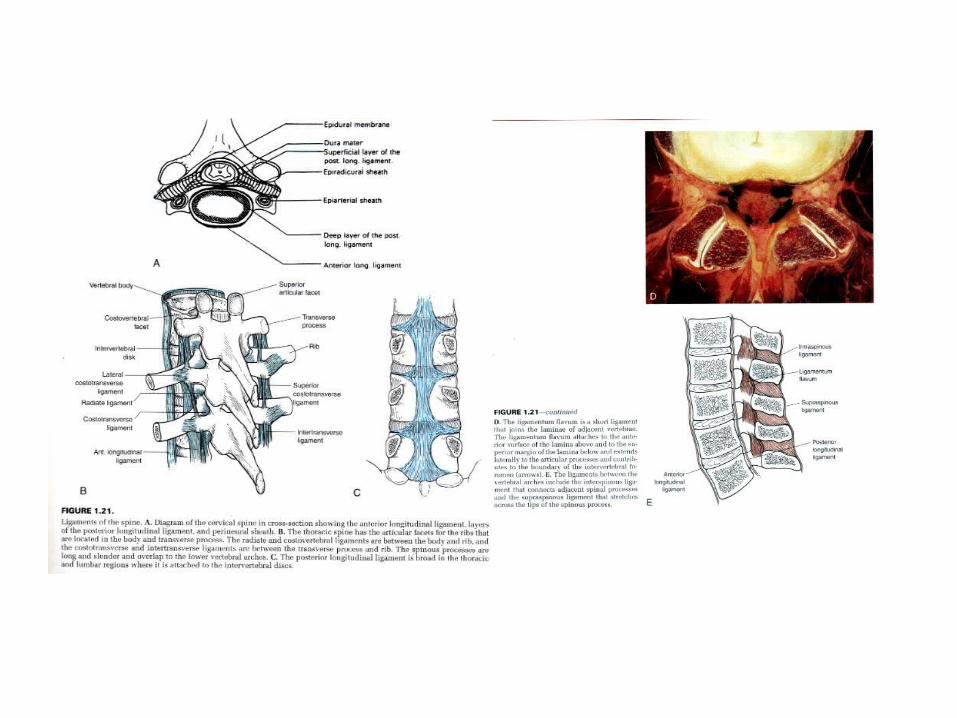
Anatomy of the Spine
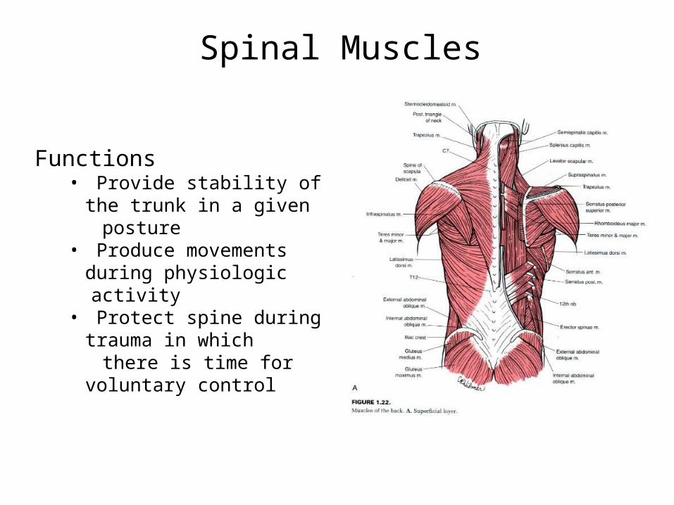
Spinal Muscles
Functions• Provide stability of the trunk in a
given posture• Produce movements during
physiologic activity• Protect spine during trauma in
which there is time for voluntary control
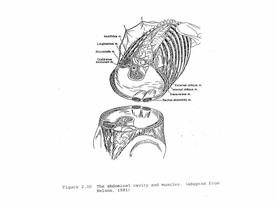

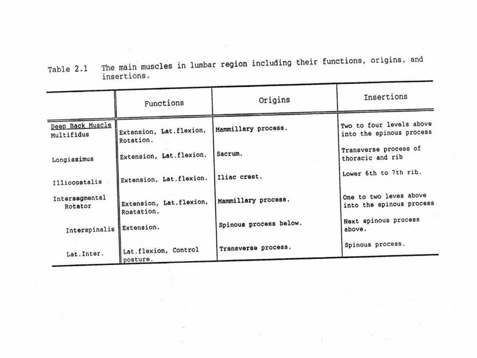
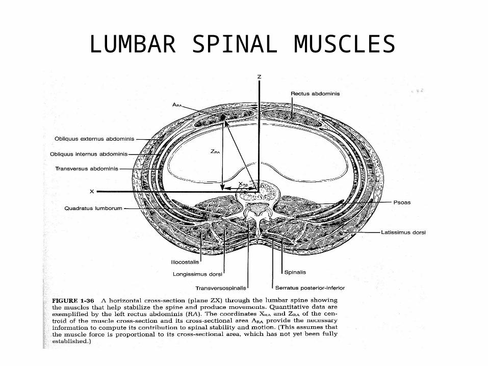
LUMBAR SPINAL MUSCLES
• Anatomy– Postvertebral
• Deep muscles: connects adjacent spinous processes or transverse processes
– Interspinales, Intertransversarii, rotatores, levatores costarum• Intermediate: arise from transverse process and attached to the spinous process of the the vertebra
above
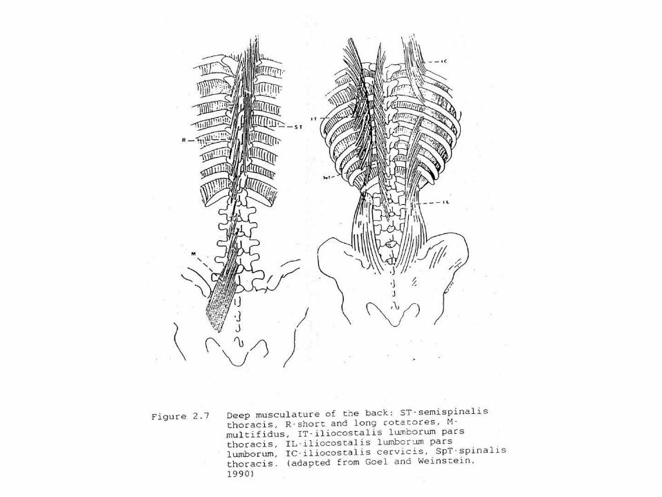
– multifidus (lumbosacral), semispinalis thoracis, semispinalis cervicis, semispinalis capitis
• Superficial: Erectorspinae
– iliocostalis (most laterally placed), longissimus, spinalis (most medially placed)
– Prevertebral: 4 abdominal muscles• external oblique, internal oblique, transversus abdominis (encircling the abdominal region), rectus
abdominis (located anteriorly at the mid-line)
LUMBAR SPINAL MUSCLES
• Erector Spinae:• a large muscle mass bordered anteriorly by the middle layer of the thoracolumbar fascia, transverse process, and laminae,
medially by the spinous processes, posteriorly and laterally by the posterior layer of the thoracolumbar fascia
• This muscle group originates from the sacrum on the medial and lateral sacral crest, the posterior surface of the iliac crest, the spinous processes, and the supraspinous ligament of the spine
– Spinalis Muscle (Medial)• begins at L2 vertebra and passes cephalad from spinous process to spinous process
• 3 portions: thoracic, cervical, and capitis portions although poorly developed in the lumbar spine
• acted in extension and lateral bending, but exerts minimal influence on the lumbar spinal mechanics and may be ignored for all practical purposes
– Longissimus Thoracis (Intermediate)• the longest of the ES group.
• Arises from the sacrum and transverse processes
• 2 portions: longissimus thoracis pars thoracis and longissimus thoracis pars lumborum (LT)
• acted in extension and lateral bending
– Iliocostalis Lumborum (Lateral)• begins from the iliac crest and attaches to the lower 6 to 7 ribs
• 2 portions: iliocostalis lumborum pars lumborum (IL) and iliocostalis lumborum pars thoracis (IT)
• acted in extension and lateral bending
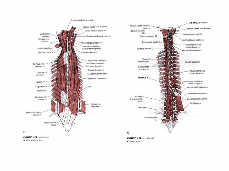
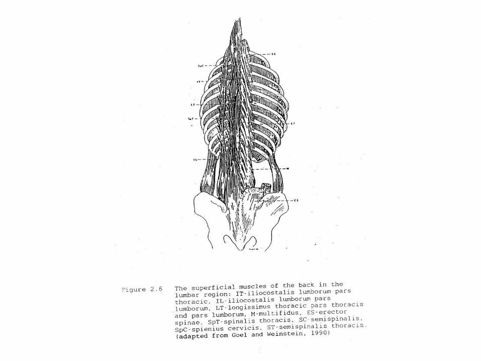
Muscles of the back in the Lumbar Region
• Transversospinalis Muscle Group:• This group lies deep to ES muscles• origins on the transverse processes and insertions on the spinous processes
– Multifidus (M) Muscle • best developed in the lumbar spine and extend the length of the vertebral colum• arises from the back of the sacrum in the sacral region;in the lumbar spine, from the mammillary processes to 2-4 level
above into the spinous processes starting with L5 spinous processes• The action is mostly posture control, but they also aid in extension, lateral bending and rotation
– Rotatores (R) Muscle• similar origins and insertions as the M muscle but only span 1 to 2 levels• 2 portions: long and short rotatores (LR and SR) depending on the length• This muscle can not be distinguished readily from the M muscles.• Same action as M
– Semispinalis Muscle (SC, SpC, SpT, ST)• does not extend to the lumbar spine• gets no or little biomechanical attention
Muscles of the back in the Lumbar Region
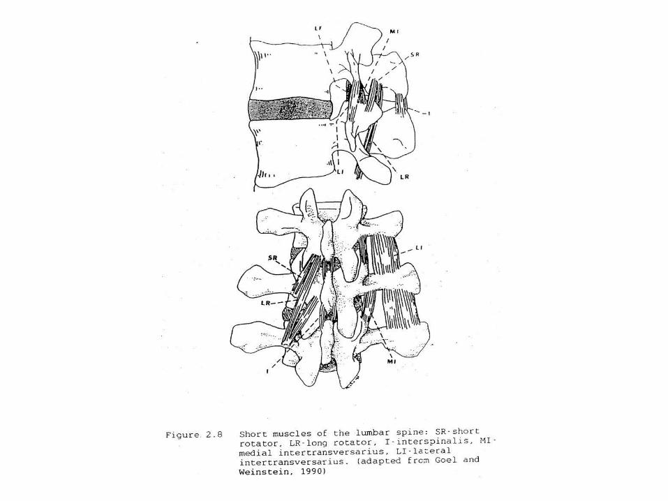
• Interspinalis (I):• originates from the spinous process below and insert on the next spinous process above; one either side of
the interspinous ligament
• There is occasionally one pair of muscles between the L5 and the sacrum like wise between the T12 and L1
• acted in extension
• Intertransversalis• 2 portions: medial intertransversalis (MI) and lateral intertransveralis (LI)
• MI originates on the mammillary process and inserts on the next cephalad mammillary process
• LI originates on the transverse process and inserts on the next cephalad transverse process
• acted for posture control and lateral bending
• Muscle Innervation• The paraspinal muscles are all segmentally innervated by the posterior primary rami of the spinal nerves.
• One exception is LI muscle, which is innervated by the ventral primary rami.
Muscles of the back in the Lumbar Region
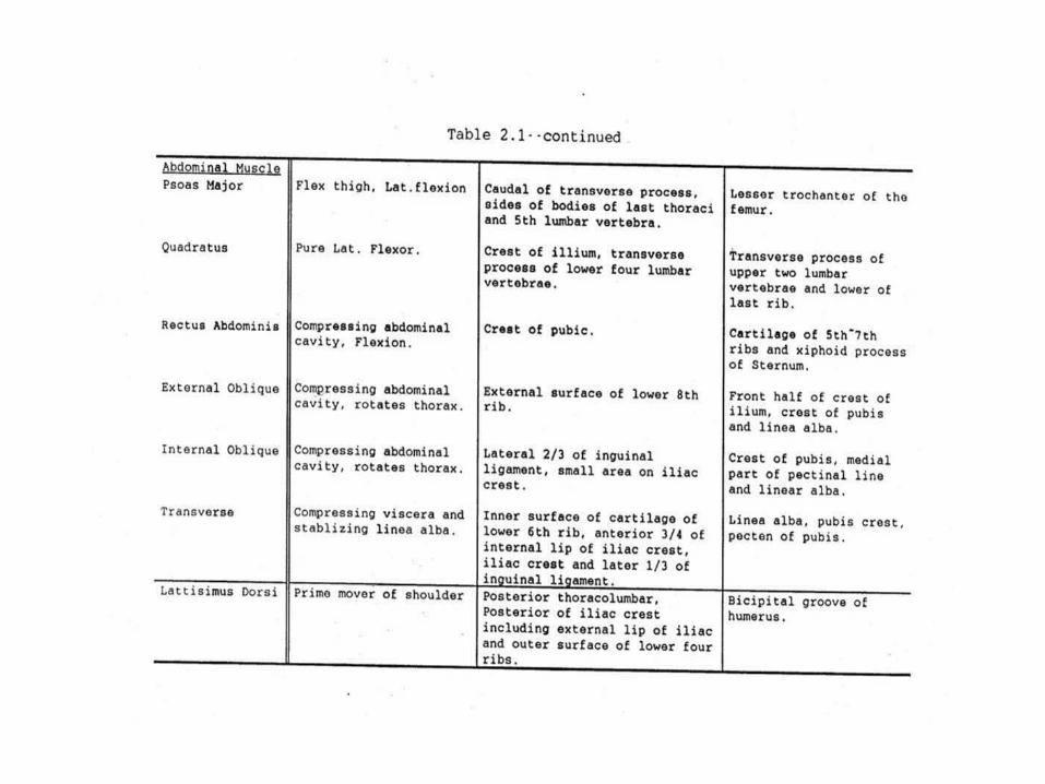
Abdominal Muscles
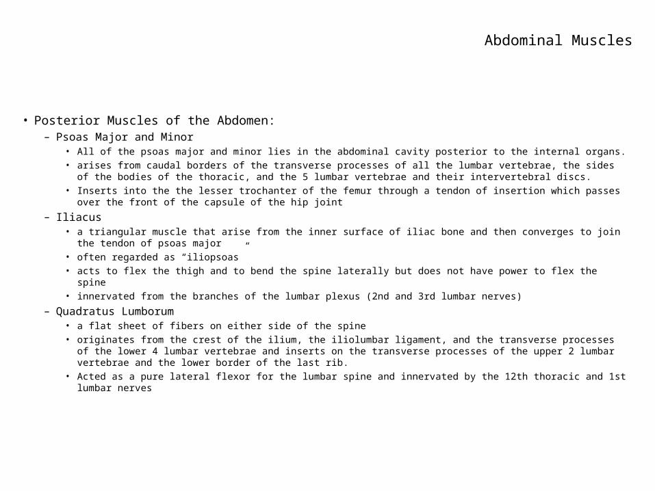
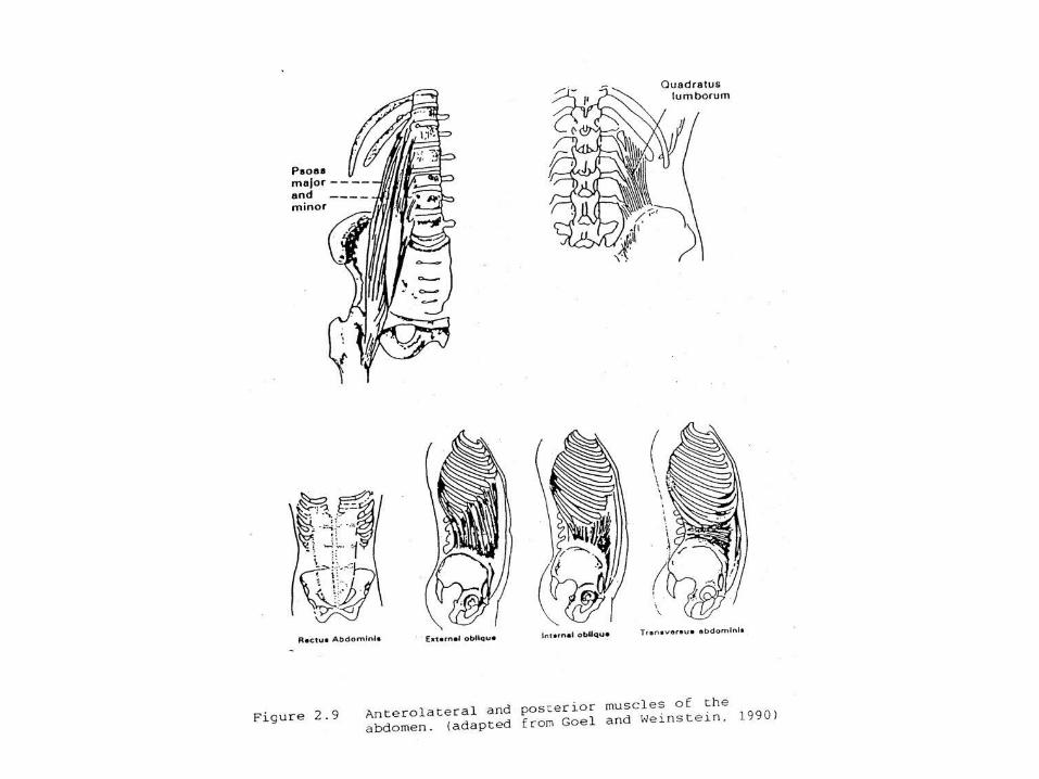
• Posterior Muscles of the Abdomen:– Psoas Major and Minor
• All of the psoas major and minor lies in the abdominal cavity posterior to the internal organs.• arises from caudal borders of the transverse processes of all the lumbar vertebrae, the sides of the bodies of the
thoracic, and the 5 lumbar vertebrae and their intervertebral discs.• Inserts into the the lesser trochanter of the femur through a tendon of insertion which passes over the front of the
capsule of the hip joint
– Iliacus• a triangular muscle that arise from the inner surface of iliac bone and then converges to join the tendon of psoas
major• often regarded as “iliopsoas”• acts to flex the thigh and to bend the spine laterally but does not have power to flex the spine• innervated from the branches of the lumbar plexus (2nd and 3rd lumbar nerves)
– Quadratus Lumborum• a flat sheet of fibers on either side of the spine• originates from the crest of the ilium, the iliolumbar ligament, and the transverse processes of the lower 4 lumbar
vertebrae and inserts on the transverse processes of the upper 2 lumbar vertebrae and the lower border of the last rib.• Acted as a pure lateral flexor for the lumbar spine and innervated by the 12th thoracic and 1st lumbar nerves
Abdominal Muscles
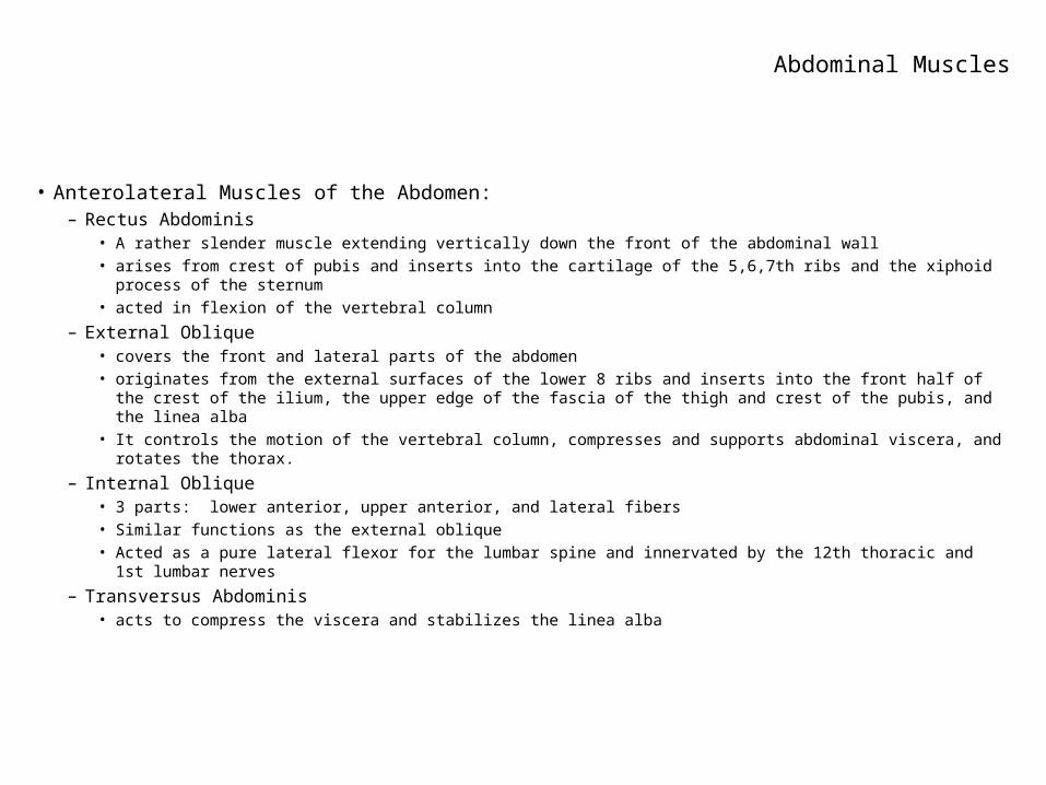
• Anterolateral Muscles of the Abdomen:– Rectus Abdominis
• A rather slender muscle extending vertically down the front of the abdominal wall• arises from crest of pubis and inserts into the cartilage of the 5,6,7th ribs and the xiphoid process of the sternum• acted in flexion of the vertebral column
– External Oblique• covers the front and lateral parts of the abdomen• originates from the external surfaces of the lower 8 ribs and inserts into the front half of the crest of the ilium, the
upper edge of the fascia of the thigh and crest of the pubis, and the linea alba• It controls the motion of the vertebral column, compresses and supports abdominal viscera, and rotates the thorax.
– Internal Oblique• 3 parts: lower anterior, upper anterior, and lateral fibers• Similar functions as the external oblique• Acted as a pure lateral flexor for the lumbar spine and innervated by the 12th thoracic and 1st lumbar nerves
– Transversus Abdominis• acts to compress the viscera and stabilizes the linea alba
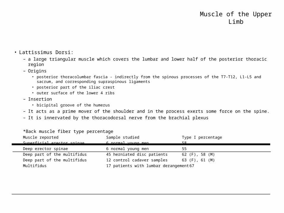
Muscle of the Upper Limb
• Lattissimus Dorsi:– a large triangular muscle which covers the lumbar and lower half of the posterior thoracic region
– Origins• posterior thoracolumbar fascia - indirectly from the spinous processes of the T7-T12, L1-L5 and sacrum, and corresponding
supraspinous ligaments• posterior part of the iliac crest• outer surface of the lower 4 ribs
– Insertion• bicipital groove of the humerus
– It acts as a prime mover of the shoulder and in the process exerts some force on the spine.
– It is innervated by the thoracodorsal nerve from the brachial plexus
*Back muscle fiber type percentageMuscle reported Sample studied Type I percentage
Superficial erector spinae 6 normal young men 58
Deep erector spinae 6 normal young men 55
Deep part of the multifidus 45 herniated disc patients 62 (F), 58 (M)
Deep part of the multifidus 12 control cadaver samples 63 (F), 61 (M)
Multifidus 17 patients with lumbar derangement 67