Embed Size (px)
Citation preview
FLEX THERAPIST CEUs 1422 Monterey Street, Suite C-102
San Luis Obispo, Ca 93401
Ph (805) 543-5100 Fax (805) 543-5106
www.flextherapistceus.com
Tennis Elbow – Treatment Approaches
Table of Contents
1. Relationships Between Biomechanics, Tendon Pathology, Pages 368 - 378
And Function in Individuals with Lateral Epicondylosis
2. Middle and Lower Trapezius Strengthening for the Pages 841 - 847
Management of Lateral Epicondylalgia: A Case Report
368 | june 2013 | volume 43 | number 6 | journal of orthopaedic & sports physical therapy
[ research report ]
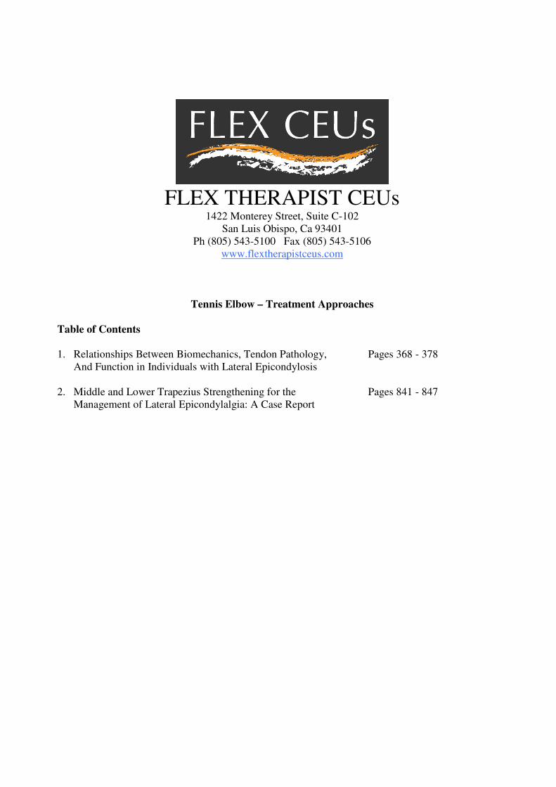
Lateral epicondylosis (LE) is a prevalent and costly musculoskeletal disorder of the common extensor tendon, characterized by degeneration of the tendon and frequently reported pain at the lateral aspect of the elbow.23,26 In addition, biomechanical
and sensorimotor deficits can occur and adversely impact upper extremity func-tion.4,8 These functional deficits may interfere with occupational tasks and activities of daily living, resulting in significant individual and occupational costs.16,48 Because the pathophysiology of LE is not well understood, treatment remains challenging, and LE is prone to recurrence.11,13
Tendon changes due to LE include dense populations of fibroblasts, vascu-lar hyperplasia, and disorganized colla-gen.28,39,43 The common extensor tendon origin in individuals with LE is usually thickened and shows increased signal intensity on magnetic resonance imag-ing (MRI). The region of greatest signal abnormality is usually at the origin of the extensor carpi radialis brevis tendon from the lateral epicondyle of the humerus. The areas of increased signal intensity within the diseased tendon usually cor-respond to areas of mucoid degeneration and neovascularization on histopatholog-ic analysis.27,43,46 Ultrasound also has been used to study LE, and findings include the presence of intratendinous calcifica-tion, tendon thickening, adjacent bone irregularity, focal hypoechoic regions, and diffuse heterogeneity of the common extensor tendon.9,10,33
TT STUDY DESIGN: Single-cohort descriptive and correlational study.
TT OBJECTIVES: To investigate the relation-ships between tendon pathology, biomechanical measures, and self-reported pain and function in individuals with chronic lateral epicondylosis.
TT BACKGROUND: Lateral epicondylosis has a multifactorial etiology and its pathophysiology is not well understood. Consequently, treatment remains challenging, and lateral epicondylosis is prone to recurrence. While tendon pathology, pain system changes, and motor impairments due to lateral epicondylosis are considered related, their relationships have not been thoroughly investi-gated.
TT METHODS: Twenty-six participants with either unilateral (n = 11) or bilateral (n = 15) chronic lateral epicondylosis participated in this study. Biomechanical measures (grip strength, rate of force development, and electromechanical delay) and measures of tendon pathology (magnetic reso-nance imaging and ultrasound) and self-reported pain and function (Patient-Rated Tennis Elbow Evaluation) were performed. Partial Spearman correlations, adjusting for covariates (age, gender, weight, and height), were used to evaluate the rela-tionship between self-reported pain, function, and biomechanical and tendon pathology measures.
TT RESULTS: Statistically significant correla-tions between biomechanical measures and the Patient-Rated Tennis Elbow Evaluation ranged in magnitude from 0.44 to 0.68 (P<.05); however, no significant correlation was observed between tendon pathology (magnetic resonance imaging and ultrasound) measures and the Patient-Rated Tennis Elbow Evaluation (r = –0.02 to 0.31, P>.05). Rate of force development had a stronger cor-relation (r = 0.54-0.68, P<.05) with self-reported function score than with grip strength (r = 0.35-0.47, P<.05) or electromechanical delay (r = 0.5, P<.05).
TT CONCLUSION: Biomechanical measures (pain-free grip strength, rate of force development, electromechanical delay) have the potential to be used as outcome measures to monitor progress in lateral epicondylosis. In comparison, the imaging measures (magnetic resonance imaging and ultra-sound) were useful for visualizing the pathophysi-ology of lateral epicondylosis. However, the severity of the pathophysiology was not related to pain and function, indicating that imaging measures may not provide the best clinical assessment. J Orthop Sports Phys Ther 2013;43(6):368-378. Epub 18 March 2013. doi:10.2519/jospt.2013.4411
TT KEY WORDS: grip strength, hand function, rate of force development, tennis elbow
1Trace Research and Development Center, Biomedical Engineering, University of Wisconsin-Madison, Madison, WI. 2Department of Biostatistics and Medical Informatics, University of Wisconsin-Madison, Madison, WI. 3Department of Family Medicine, School of Medicine and Public Health, University of Wisconsin-Madison, Madison, WI. 4Department of Radiology, School of Medicine and Public Health, University of Wisconsin-Madison, Madison, WI. 5Center for Musculoskeletal Research and School of Physiotherapy and Exercise Science, Griffith Health Institute, Griffith University, Gold Coast, Queensland, Australia. 6Department of Orthopedics and Rehabilitation, School of Medicine and Public Health, University of Wisconsin-Madison, Madison, WI. The Health Sciences Institutional Review Board of the University of Wisconsin-Madison approved the study protocol. Drs Sesto, Chourasia, and Buhr received support from the University of Wisconsin Clinical and Translational Science Award (NIH/NCRR 1 UL1RR025011). Dr Rabago was partially supported by the American Academy Family Practice Foundation’s Research Committee Joint Grant Awards Program (G0810). The authors certify that they have no affiliations with or financial involvement in any organization or entity with a direct financial interest in the subject matter or materials discussed in the article. Address correspondence to Dr Mary E. Sesto, 5110 Medical Sciences Center, 1300 University Avenue, Madison, WI 53706. E-mail: [email protected] T Copyright ©2013 Journal of Orthopaedic & Sports Physical Therapy®
AMRISH O. CHOURASIA, PhD1 • KEVIN A. BUHR, PhD2 • DAVID P. RABAGO, MD3 • RICHARD KIJOWSKI, MD4 • KENNETH S. LEE, MD4
MICHAEL P. RYAN, PhD5 • JESSICA M. GRETTIE-BELLING, BS3 • MARY E. SESTO, PT, PhD1,6
Relationships Between Biomechanics, Tendon Pathology, and Function in
Individuals With Lateral Epicondylosis
43-06 Chourasia.indd 368 5/21/2013 3:18:46 PM
journal of orthopaedic & sports physical therapy | volume 43 | number 6 | june 2013 | 369
Pain is the primary symptom of LE.23 The pain that patients with LE experi-ence may be due to changes in the ner-vous system, as a result of neuronal tissue changes as well as nociceptive and non-nociceptive processes.13,35 In patients with LE, pain can be assessed using pressure pain threshold and self-reported mea-sures such as the visual analog scale and the Patient-Rated Tennis Elbow Evalu-ation (PRTEE).12,45,50,53 The PRTEE is a commonly used, valid, reliable, and sen-sitive clinical instrument for assessment of pain and disability in individuals with chronic LE.32,40,45
Pain-free grip strength is the most commonly assessed motor impair-ment,5,49,51 and recently the effects of LE on reaction time,4,5,42 rate of force devel-opment, and electromechanical delay have been investigated.7 Rate of force de-velopment is considered to be a measure of the ability to rapidly generate strength and is associated with higher functional performance.1,47 Electromechanical delay represents the duration of the excita-tion contraction coupling in the muscle and the time to take up the slack in the elastic structures of the muscle-tendon unit.30 Chourasia et al7 found a lower rate of force development and longer elec-tromechanical delay in individuals with LE compared to controls. In addition, Bisset et al4,5 reported longer reaction times in individuals with LE compared to controls.
While tendon pathology, pain system changes, and motor impairments due to LE are considered related, their relation-ship has not been investigated thorough-ly. For example, Clarke et al9 observed a positive association between ultrasound findings and improvements in self-re-ported pain and function, but changes in motor performance were not evaluated. Similarly, Bisset et al4,5 investigated the effects of various interventions for LE on global improvement, grip strength, and sensorimotor measures but did not assess morphological changes in the common extensor tendon.
It is noteworthy that the majority of
studies on LE only include patients with LE of the dominant arm and exclude those with bilateral LE or unilateral LE of the nondominant arm.5,42 It is important that the relationships between the vari-ous components of LE be investigated in a heterogeneous sample. This may help to gain a better understanding of LE to improve assessment and treatment outcomes.
The overall objective of the study was to evaluate the relationships between self-reported pain and function (PRTEE) and measures of tendon pathology and biomechanics in individuals with LE. Secondary analyses evaluated the rela-tionships between biomechanical and tendon pathology measures. This infor-mation may help provide a better under-standing of the effect of LE on function and its association with biomechanical measures.
METHODS
Participants
Twenty-nine patients with LE were recruited and enrolled from various outpatient clinics in Madi-
son, WI from June 2009 to February 2010. These patients were participating in a therapeutic trial investigating the efficacy of prolotherapy for LE. Diagnos-tic criteria for LE included the presence of lateral elbow pain for more than 3 months, tenderness to palpation over the lateral epicondyle and/or extensor mech-anism, and pain present on at least 2 of the following provocation tests: resisted extension of the wrist or fingers, resisted supination, and passive stretch of the wrist extensors or supinator muscle. Ex-clusion criteria consisted of coexisting or previous medical history of rheumatoid or inflammatory arthritis, chronic pain diagnoses, diabetes mellitus, pregnancy, systemic nervous disease, neuropathy, or acute trauma to the fingers or hands. Additional exclusion criteria were prior upper extremity injury, concurrent upper extremity injury, unresolved litigation, and comorbidities that could interfere
with the ability to participate in the study. Two participants were excluded because they reported concurrent upper extrem-ity injury. Data from 1 participant were excluded because of instrumentation malfunction. Of the 26 eligible partici-pants, 11 had unilateral symptoms and 15 had bilateral symptoms (TABLE 1).
Informed consent was obtained for all participants in this study, in accordance with the study protocol, as approved by the University of Wisconsin-Madison Health Sciences Institutional Review Board.
Self-Reported MeasuresPRTEE Questionnaire Participants com-pleted the PRTEE, a condition-specific questionnaire that assesses both elbow pain and function. The PRTEE has good test-retest reliability, although reliability is less for patients with work-related LE (intraclass correlation coefficient [ICC] = 0.80) versus non–work-related LE (ICC = 0.94).40 It has also been used to deter-mine the effects of different interventions for LE.18,19
The PRTEE consists of a 5-item pain subscale and a 10-item function subscale. Each subscale is scored from 0 to 50, with 0 being the best score and 50 the worst score. The PRTEE composite score is the sum of the pain and function subscales and ranges from 0 to 100, with 0 being the best score and 100 the worst score.Visual Analog Scale All participants were asked to rate the average lateral elbow pain intensity for the previous week us-ing a 10-cm line ranging from zero (no pain) to 10 (most pain).
Biomechanical MeasuresBiomechanical measures included pain-free grip strength, rate of force devel-opment, and electromechanical delay. These measures were collected bilater-ally, and the results have been reported elsewhere.7 Only the results from the affected arm were used for the correla-tional analyses reported in this paper. In cases of bilaterally affected participants, the results from the more affected arm,
43-06 Chourasia.indd 369 5/21/2013 3:18:47 PM
370 | june 2013 | volume 43 | number 6 | journal of orthopaedic & sports physical therapy
[ research report ]
as determined by the visual analog scale scores, were used. When both arms were equally affected, the results from the dominant arm were used.Pain-Free Grip Strength Pain-free grip strength is a valid and reliable (ICC = 0.97) measure and is commonly used in research and clinical assessment of LE.4,5,49,52 For testing, as recommended for evaluation of grip strength in patients with LE,14,15 the participants were seated in a chair with the elbow in an extended posi-tion. The participants were then instruct-ed to squeeze the Baseline (Fabrication Enterprises, Inc, White Plains, NY) dyna-mometer for 5 seconds, avoiding discom-fort. Three replications with a 60-second interval between trials were performed. The average of the 3 replications was used as the pain-free grip strength.Rate of Force Development The mul-
tiaxis profile (MAP) dynamometer was used for measurement of rate of force development.24 The MAP dynamometer has demonstrated excellent test-retest re-liability (ICC = 0.99) and excellent con-current validity when compared to the Baseline dynamometer (r = 0.88-0.90).24
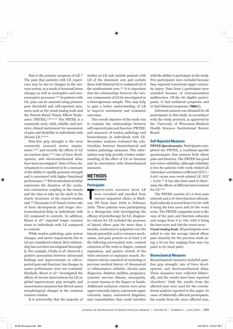
The participants were seated in a chair with the elbow in an extended position. Upon receipt of a randomly timed visual stimulus, participants were instructed to squeeze the handle as quickly and as hard as possible without pain for 5 sec-onds.7 The rate of force development was calculated by taking the time deriva-tive of the force signal (FIGURE 1).1 Rate of force development was measured at 30, 50, and 100 milliseconds from the onset of contraction. The onset of contraction was defined as a rise of 1 N from baseline level.29 Peak rate of force development
was also measured. This method is con-sistent with that of others for evaluating force development.1,2 Three repetitions, with a 60-second interval between them, were performed, and the average of the 3 repetitions was used for data analysis. The signals from the MAP dynamometer were sampled at a rate of 1000 samples per sec-ond, using a USB-6009 card (National Instruments Corporation, Austin, TX).
The MAP dynamometer also provides a measure of pain-free grip strength, but, due to a different handle geometry com-pared to the Baseline dynamometer, the grip strength values are different.24 We reported both grip strength values us-ing the variable names “pain-free grip strength-Baseline” and “pain-free grip strength-MAP.”Electromechanical Delay Electrome-chanical delay was measured using the MAP dynamometer force output and the raw electromyographic (EMG) signal from the extensor carpi radialis muscle. EMG signal was measured using a Tele-Myo 2400 EMG system (Noraxon USA Inc, Scottsdale, AZ). Electrodes were placed on the extensor carpi radialis at one third of the distance from the proxi-mal end of a line from the lateral epi-condyle to the distal end of the radius.38 Electrodes were placed on the extensor carpi radialis with the forearm in neutral position. The reference EMG electrode
TABLE 1 Participant Demographics (n = 26)
Abbreviations: LE, lateral epicondylosis; NA, not available.*Values are mean SD.†2 participants listed dual causes.
Value
Demographics
Sex (male, female), n 16, 10
Age, y* 48.2 8.7
Symptom duration, y* 3.4 3.5
Hand dominance (right, left), n 24, 2
Unilateral and bilateral symptoms (unilateral, bilateral), n 11, 15
Unilateral symptoms in dominant arm, n 9
Unilateral symptoms in nondominant arm, n 2
Prior treatment information, n
Physical therapy (yes) 23
Corticosteroid injections (yes) 13
Elbow brace (yes) 17
Occupational information
Work status (full time, part time, not working), n 23, 3, 0
Work restrictions (yes, no) 12, 14
Main reason for LE (self-reported), n†
Work 11
Sports 10
Home activity 4
Acute injury 2
Do not know 0
NA 1
Forc
e
Time
Stimulus
EMG signal onset
Force onset
0 ms
30 ms
50 ms
100 ms
EMD
RFD = d(Force)
dt
FIGURE 1. RFD and EMD measurements (figure is not to scale). Abbreviations: EMD, electromechanical delay; EMG, electromyogram; RFD, rate of force development.
43-06 Chourasia.indd 370 5/21/2013 3:18:48 PM
journal of orthopaedic & sports physical therapy | volume 43 | number 6 | june 2013 | 371
was attached to the lateral epicondyle of the right elbow. Prior to electrode placement, skin preparation was per-formed according to SENIAM (Surface ElectroMyoGraphy for the Non-Invasive Assessment of Muscles)22 guidelines. Disposable, self-adhesive Ag/AgCl dual snap electrodes (Noraxon USA Inc) with an individual electrode diameter of 1 cm and interelectrode distance of 2 cm were used. Preamplified EMG leads, with a differential gain of 500, connected the electrodes to a 16-channel TeleMyo 2400 wireless transmitter, with a 16-bit A/D converter and a bandwidth of 10 to 500 Hz. The EMG amplifier had a gain of 1000, an input impedance much greater than 100 MΩ, and a common-mode re-jection ratio greater than 100 dB. The signals were sampled at the rate of 1500 samples per second. The onset of muscle activation was defined as a deviation of 15 μV in the EMG signal from resting baseline level.29 The time between the onset of muscle activation based on the change in EMG signal intensity and the onset of force as measured with the MAP dynamometer is considered the electro-mechanical delay (FIGURE 1). Sufficient practice was provided to all participants to become comfortable with the testing procedures prior to completing 3 rep-etitions. The average of the 3 repetitions was used for data analysis.
Tendon Pathology MeasuresTendon pathology was assessed using
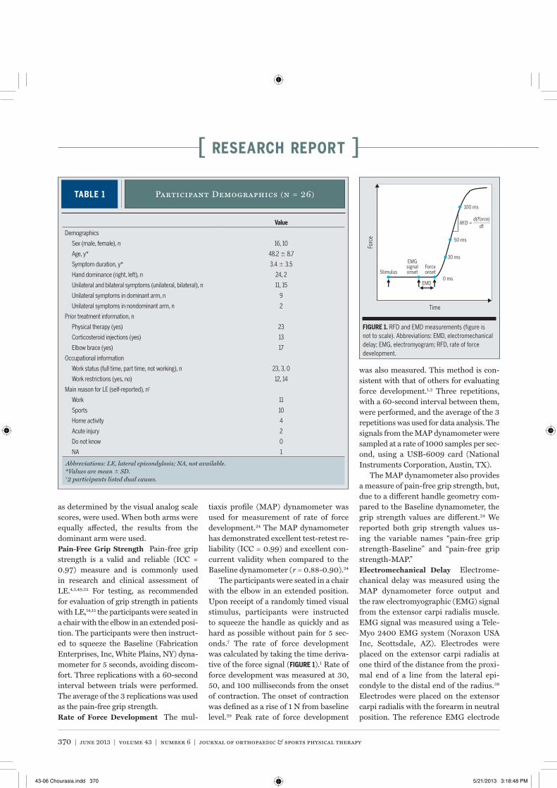
MRI and ultrasound. For those with uni-lateral LE, imaging was conducted on the affected arm. For individuals with bilat-eral LE, ultrasound was conducted on the most affected arm (as determined by the visual analog scale scores) and MRI was conducted bilaterally. For the corre-lational analyses used in this paper, we used the results from the more affected arm. When both arms were equally af-fected, the results from the dominant arm were used. Three participants did not complete the ultrasound assessment and 1 participant declined the MRI scan. Therefore, both MRI and ultrasound pa-rameters were available for 22 individu-als (elbows).Magnetic Resonance Imaging The MRI examination was performed using an ARTOSCAN (Esaote SpA, Genoa, Italy) 0.17-T extremity scanner. Axial and coro-nal intermediate-weighted fast spin-echo (FSE) and short tau inversion recovery (STIR) sequences of the elbow were used for semiquantitative assessment of disease severity (FIGURE 2). Intermediate-weighted FSE scan parameters were as follows: repetition time, 2050 millisec-onds; echo time, 18 milliseconds; slices, 7; gap, 1.0 mm; thickness, 3.5 mm; read-out field of view, 180; encoding field of view, 180; samples, 192; encoding num-ber, 192. STIR scan parameters were as follows: repetition time, 2050 millisec-onds; echo time, 34 milliseconds; inver-sion time, 75 milliseconds; slices, 7; gap, 1.0 mm; thickness, 3.5 mm; readout field
of view, 180; encoding field of view, 180; samples, 192; encoding number, 192. A semiquantitative grading scale was used to estimate the severity of chronic degen-eration and pathologic changes in the common extensor tendon origin.44 The grading scale is as follows:• Grade 0: normal common extensor
tendon that is of uniform low signal intensity on intermediate-weighted FSE and fat-suppressed, T2-weighted STIR images.
• Grade 1: common extensor tendon with mild tendinopathy that is thickened and has intermediate signal intensity on intermediate-weighted FSE and STIR images.
• Grade 2: common extensor tendon with moderate tendinopathy that is thinned and shows focal areas of intense fluid-like signal intensity on STIR images, which comprise less than 50% of the total cross-sectional diameter of the tendon.
• Grade 3: common extensor tendon with severe tendinopathy that is thinned and shows focal areas of intense fluid-like signal intensity on STIR images, which comprise more than 50% of the total cross-sectional diameter of the tendon.
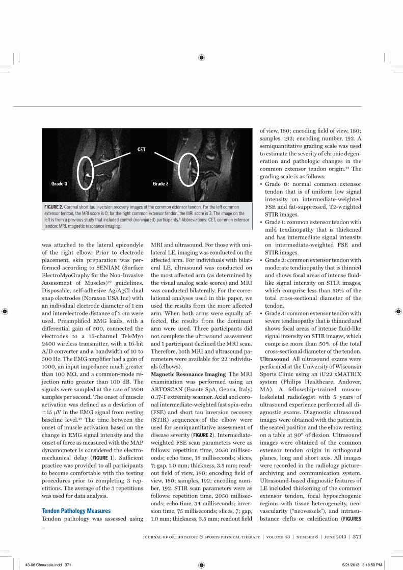
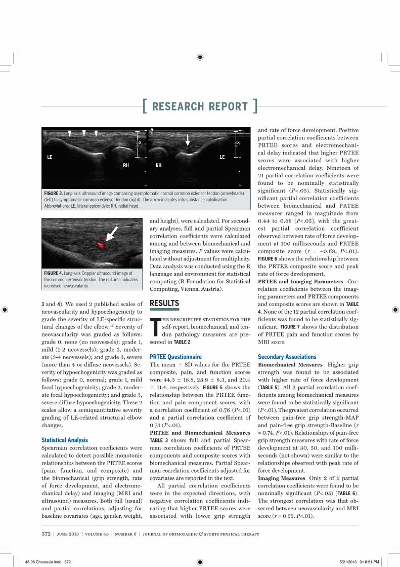
Ultrasound All ultrasound exams were performed at the University of Wisconsin Sports Clinic using an iU22 xMATRIX system (Philips Healthcare, Andover, MA). A fellowship-trained muscu-loskeletal radiologist with 5 years of ultrasound experience performed all di-agnostic exams. Diagnostic ultrasound images were obtained with the patient in the seated position and the elbow resting on a table at 90° of flexion. Ultrasound images were obtained of the common extensor tendon origin in orthogonal planes, long and short axis. All images were recorded in the radiology picture-archiving and communication system. Ultrasound-based diagnostic features of LE included thickening of the common extensor tendon, focal hypoechogenic regions with tissue heterogeneity, neo-vascularity (“neovessels”), and intrasu-bstance clefts or calcification (FIGURES
FIGURE 2. Coronal short tau inversion recovery images of the common extensor tendon. For the left common extensor tendon, the MRI score is 0; for the right common extensor tendon, the MRI score is 3. The image on the left is from a previous study that included control (noninjured) participants.8 Abbreviations: CET, common extensor tendon; MRI, magnetic resonance imaging.
43-06 Chourasia.indd 371 5/21/2013 3:18:50 PM
372 | june 2013 | volume 43 | number 6 | journal of orthopaedic & sports physical therapy
[ research report ]
3 and 4). We used 2 published scales of neovascularity and hypoechogenicity to grade the severity of LE-specific struc-tural changes of the elbow.36 Severity of neovascularity was graded as follows: grade 0, none (no neovessels); grade 1, mild (1-2 neovessels); grade 2, moder-ate (3-4 neovessels); and grade 3, severe (more than 4 or diffuse neovessels). Se-verity of hypoechogenicity was graded as follows: grade 0, normal; grade 1, mild focal hypoechogenicity; grade 2, moder-ate focal hypoechogenicity; and grade 3, severe diffuse hypoechogenicity. These 2 scales allow a semiquantitative severity grading of LE-related structural elbow changes.
Statistical AnalysisSpearman correlation coefficients were calculated to detect possible monotonic relationships between the PRTEE scores (pain, function, and composite) and the biomechanical (grip strength, rate of force development, and electrome-chanical delay) and imaging (MRI and ultrasound) measures. Both full (usual) and partial correlations, adjusting for baseline covariates (age, gender, weight,
and height), were calculated. For second-ary analyses, full and partial Spearman correlation coefficients were calculated among and between biomechanical and imaging measures. P values were calcu-lated without adjustment for multiplicity. Data analysis was conducted using the R language and environment for statistical computing (R Foundation for Statistical Computing, Vienna, Austria).
RESULTS
The descriptive statistics for the self-report, biomechanical, and ten-don pathology measures are pre-
sented in TABLE 2.
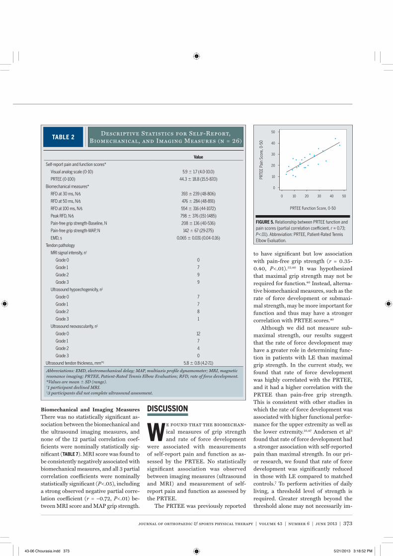
PRTEE QuestionnaireThe mean SD values for the PRTEE composite, pain, and function scores were 44.3 18.8, 23.9 8.3, and 20.4 11.4, respectively. FIGURE 5 shows the relationship between the PRTEE func-tion and pain component scores, with a correlation coefficient of 0.76 (P<.01) and a partial correlation coefficient of 0.73 (P<.01).PRTEE and Biomechanical Measures TABLE 3 shows full and partial Spear-man correlation coefficients of PRTEE components and composite scores with biomechanical measures. Partial Spear-man correlation coefficients adjusted for covariates are reported in the text.
All partial correlation coefficients were in the expected directions, with negative correlation coefficients indi-cating that higher PRTEE scores were associated with lower grip strength
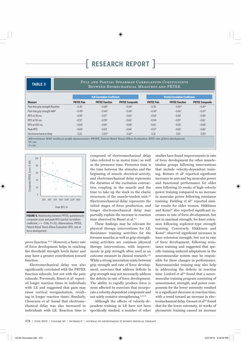
and rate of force development. Positive partial correlation coefficients between PRTEE scores and electromechani-cal delay indicated that higher PRTEE scores were associated with higher electromechanical delay. Nineteen of 21 partial correlation coefficients were found to be nominally statistically significant (P<.05). Statistically sig-nificant partial correlation coefficients between biomechanical and PRTEE measures ranged in magnitude from 0.44 to 0.68 (P<.05), with the great-est partial correlation coefficient observed between rate of force develop-ment at 100 milliseconds and PRTEE composite score (r = –0.68, P<.01). FIGURE 6 shows the relationship between the PRTEE composite score and peak rate of force development.PRTEE and Imaging Parameters Cor-relation coefficients between the imag-ing parameters and PRTEE components and composite scores are shown in TABLE
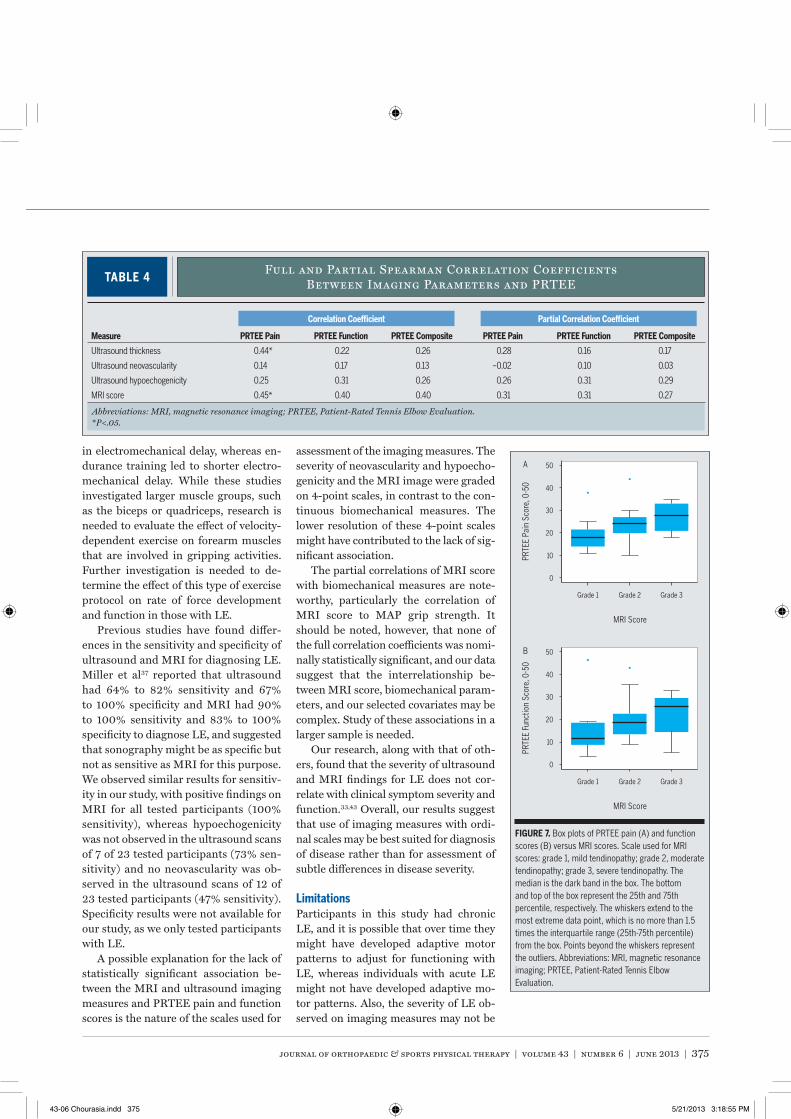
4. None of the 12 partial correlation coef-ficients was found to be statistically sig-nificant. FIGURE 7 shows the distribution of PRTEE pain and function scores by MRI score.
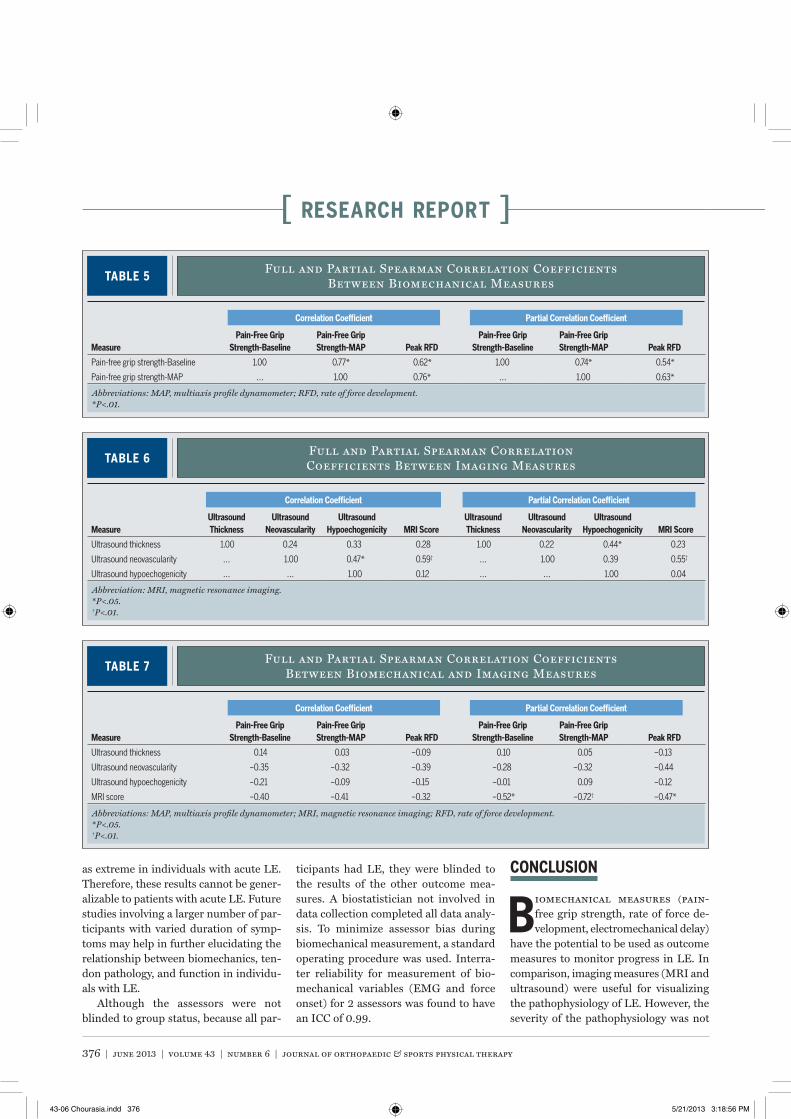
Secondary AssociationsBiomechanical Measures Higher grip strength was found to be associated with higher rate of force development (TABLE 5). All 3 partial correlation coef-ficients among biomechanical measures were found to be statistically significant (P<.01). The greatest correlation occurred between pain-free grip strength-MAP and pain-free grip strength-Baseline (r = 0.74, P<.01). Relationships of pain-free grip strength measures with rate of force development at 30, 50, and 100 milli-seconds (not shown) were similar to the relationships observed with peak rate of force development.Imaging Measures Only 2 of 6 partial correlation coefficients were found to be nominally significant (P<.05) (TABLE 6). The strongest correlation was that ob-served between neovascularity and MRI score (r = 0.55, P<.01).
FIGURE 3. Long-axis ultrasound image comparing asymptomatic normal common extensor tendon (arrowheads) (left) to symptomatic common extensor tendon (right). The arrow indicates intrasubstance calcification. Abbreviations: LE, lateral epicondyle; RH, radial head.
FIGURE 4. Long-axis Doppler ultrasound image of the common extensor tendon. The red area indicates increased neovascularity.
43-06 Chourasia.indd 372 5/21/2013 3:18:51 PM
journal of orthopaedic & sports physical therapy | volume 43 | number 6 | june 2013 | 373
Biomechanical and Imaging Measures There was no statistically significant as-sociation between the biomechanical and the ultrasound imaging measures, and none of the 12 partial correlation coef-ficients were nominally statistically sig-nificant (TABLE 7). MRI score was found to be consistently negatively associated with biomechanical measures, and all 3 partial correlation coefficients were nominally statistically significant (P<.05), including a strong observed negative partial corre-lation coefficient (r = –0.72, P<.01) be-tween MRI score and MAP grip strength.
DISCUSSION
We found that the biomechan-ical measures of grip strength and rate of force development
were associated with measurements of self-report pain and function as as-sessed by the PRTEE. No statistically significant association was observed between imaging measures (ultrasound and MRI) and measurement of self-report pain and function as assessed by the PRTEE.
The PRTEE was previously reported
to have significant but low association with pain-free grip strength (r = 0.35-0.40, P<.01).32,40 It was hypothesized that maximal grip strength may not be required for function.40 Instead, alterna-tive biomechanical measures, such as the rate of force development or submaxi-mal strength, may be more important for function and thus may have a stronger correlation with PRTEE scores.40
Although we did not measure sub-maximal strength, our results suggest that the rate of force development may have a greater role in determining func-tion in patients with LE than maximal grip strength. In the current study, we found that rate of force development was highly correlated with the PRTEE, and it had a higher correlation with the PRTEE than pain-free grip strength. This is consistent with other studies in which the rate of force development was associated with higher functional perfor-mance for the upper extremity as well as the lower extremity.25,47 Andersen et al3 found that rate of force development had a stronger association with self-reported pain than maximal strength. In our pri-or research, we found that rate of force development was significantly reduced in those with LE compared to matched controls.7 To perform activities of daily living, a threshold level of strength is required. Greater strength beyond the threshold alone may not necessarily im-
TABLE 2Descriptive Statistics for Self-Report,
Biomechanical, and Imaging Measures (n = 26)
Abbreviations: EMD, electromechanical delay; MAP, multiaxis profile dynamometer; MRI, magnetic resonance imaging; PRTEE, Patient-Rated Tennis Elbow Evaluation; RFD, rate of force development.*Values are mean SD (range).†1 participant declined MRI.‡3 participants did not complete ultrasound assessment.
Value
Self-report pain and function scores*
Visual analog scale (0-10) 5.9 1.7 (4.0-10.0)
PRTEE (0-100) 44.3 18.8 (15.5-87.0)
Biomechanical measures*
RFD at 30 ms, N/s 393 239 (48-806)
RFD at 50 ms, N/s 476 284 (48-891)
RFD at 100 ms, N/s 554 316 (44-1072)
Peak RFD, N/s 798 376 (151-1485)
Pain-free grip strength-Baseline, N 208 136 (40-536)
Pain-free grip strength-MAP, N 142 67 (29-275)
EMD, s 0.065 0.031 (0.04-0.16)
Tendon pathology
MRI signal intensity, n†
Grade 0 0
Grade 1 7
Grade 2 9
Grade 3 9
Ultrasound hypoechogenicity, n‡
Grade 0 7
Grade 1 7
Grade 2 8
Grade 3 1
Ultrasound neovascularity, n‡
Grade 0 12
Grade 1 7
Grade 2 4
Grade 3 0
Ultrasound tendon thickness, mm*‡ 5.8 0.8 (4.2-7.1)
PRTE
E Pa
in S
core
, 0-5
0
0
10
0 10 20 30 40 50
20
30
40
50
PRTEE Function Score, 0-50
FIGURE 5. Relationship between PRTEE function and pain scores (partial correlation coefficient, r = 0.73; P<.01). Abbreviation: PRTEE, Patient-Rated Tennis Elbow Evaluation.
43-06 Chourasia.indd 373 5/21/2013 3:18:52 PM
374 | june 2013 | volume 43 | number 6 | journal of orthopaedic & sports physical therapy
[ research report ]
prove function.40,45 However, a faster rate of force development helps in reaching the threshold strength levels faster and may have a greater contribution toward function.
Electromechanical delay was also significantly correlated with the PRTEE function subscale, but not with the pain subscale. Previously, Bisset et al5 report-ed longer reaction times in individuals with LE and suggested that pain may cause cortical reorganization, result-ing in longer reaction times. Similarly, Chourasia et al7 found that electrome-chanical delay was also increased in individuals with LE. Reaction time is
composed of electromechanical delay (also referred to as motor time) as well as the premotor time. Premotor time is the time between the stimulus and the beginning of muscle electrical activity, and electromechanical delay represents the duration of the excitation contrac-tion coupling in the muscle and the time to take up the slack in the elastic structures of the muscle-tendon unit.30 Electromechanical delay represents the initial stages of force production, and longer electromechanical delay may partially explain the increase in reaction time observed by Bisset et al.4,5
These findings may be relevant for physical therapy interventions for LE. Resistance training activities for the forearm muscles as well as grip-strength-ening activities are common physical therapy interventions, with improve-ments in grip strength often used as an outcome measure in clinical research.5,41 While a strong association exists between grip strength and rate of force develop-ment, exercises that address deficits in grip strength may not necessarily address the deficits in rate of force development. The ability to rapidly produce force is most affected by exercises that incorpo-rate a velocity-dependent component and not solely resistive strengthening.6,17,31
Although the effects of velocity-de-pendent training in LE have not been specifically studied, a number of other
studies have found improvements in rate of force development for other muscle-tendon groups following interventions that include velocity-dependent train-ing. Bottaro et al6 reported significant increases in arm and leg muscular power and functional performance for older men following 10 weeks of high-velocity power training compared to no increase in muscular power following resistance training. Fielding et al17 reported simi-lar results for older women. Häkkinen and Komi21 also reported significant in-creases in rate of force development, but not in maximal strength, for knee exten-sion following explosive-type strength training. Conversely, Häkkinen and Komi21 observed significant increases in knee extension strength, but not in rate of force development, following resis-tance training and suggested that spe-cific training-induced adaptations in the neuromuscular system may be respon-sible for these changes in performance. Neuromuscular training may also help in addressing the deficits in reaction time. Linford et al34 found that a neuro-muscular training program consisting of sensorimotor, strength, and power com-ponents for the lower extremity resulted in a significant decrease in reaction time, with a trend toward an increase in elec-tromechanical delay. Grosset et al20 found that for the lower extremity, 10 weeks of plyometric training caused an increase
TABLE 3Full and Partial Spearman Correlation Coefficients
Between Biomechanical Measures and PRTEE
Abbreviations: MAP, multiaxis profile dynamometer; PRTEE, Patient-Rated Tennis Elbow Evaluation; RFD, rate of force development.*P<.05.†P<.01.
Measure PRTEE Pain PRTEE Function PRTEE Composite PRTEE Pain PRTEE Function PRTEE Composite
Pain-free grip strength-Baseline –0.36 –0.48* –0.44* –0.35 –0.50* –0.47*
Pain-free grip strength-MAP –0.49* –0.44* –0.46* –0.44* –0.46* –0.47*
RFD at 30 ms –0.55† –0.57† –0.60† –0.53† –0.56† –0.59†
RFD at 50 ms –0.57† –0.59† –0.62† –0.54† –0.57† –0.61†
RFD at 100 ms –0.64† –0.65† –0.68† –0.61† –0.65† –0.68†
Peak RFD –0.60† –0.63† –0.64† –0.57† –0.65† –0.66†
Electromechanical delay 0.22 0.43* 0.42* 0.31 0.51† 0.50†
Full Correlation Coefficient Partial Correlation Coefficient
Com
posi
te P
RTEE
Sco
re, 0
-100
0
20
200 400 600 800 1000 1200 1400
40
60
80
100
Peak RFD, N
FIGURE 6. Relationship between PRTEE questionnaire composite score and peak RFD (partial correlation coefficient, r = –0.66, P<.01). Abbreviations: PRTEE, Patient-Rated Tennis Elbow Evaluation; RFD, rate of force development.
43-06 Chourasia.indd 374 5/21/2013 3:18:54 PM
journal of orthopaedic & sports physical therapy | volume 43 | number 6 | june 2013 | 375
in electromechanical delay, whereas en-durance training led to shorter electro-mechanical delay. While these studies investigated larger muscle groups, such as the biceps or quadriceps, research is needed to evaluate the effect of velocity-dependent exercise on forearm muscles that are involved in gripping activities. Further investigation is needed to de-termine the effect of this type of exercise protocol on rate of force development and function in those with LE.
Previous studies have found differ-ences in the sensitivity and specificity of ultrasound and MRI for diagnosing LE. Miller et al37 reported that ultrasound had 64% to 82% sensitivity and 67% to 100% specificity and MRI had 90% to 100% sensitivity and 83% to 100% specificity to diagnose LE, and suggested that sonography might be as specific but not as sensitive as MRI for this purpose. We observed similar results for sensitiv-ity in our study, with positive findings on MRI for all tested participants (100% sensitivity), whereas hypoechogenicity was not observed in the ultrasound scans of 7 of 23 tested participants (73% sen-sitivity) and no neovascularity was ob-served in the ultrasound scans of 12 of 23 tested participants (47% sensitivity). Specificity results were not available for our study, as we only tested participants with LE.
A possible explanation for the lack of statistically significant association be-tween the MRI and ultrasound imaging measures and PRTEE pain and function scores is the nature of the scales used for
assessment of the imaging measures. The severity of neovascularity and hypoecho-genicity and the MRI image were graded on 4-point scales, in contrast to the con-tinuous biomechanical measures. The lower resolution of these 4-point scales might have contributed to the lack of sig-nificant association.
The partial correlations of MRI score with biomechanical measures are note-worthy, particularly the correlation of MRI score to MAP grip strength. It should be noted, however, that none of the full correlation coefficients was nomi-nally statistically significant, and our data suggest that the interrelationship be-tween MRI score, biomechanical param-eters, and our selected covariates may be complex. Study of these associations in a larger sample is needed.
Our research, along with that of oth-ers, found that the severity of ultrasound and MRI findings for LE does not cor-relate with clinical symptom severity and function.33,43 Overall, our results suggest that use of imaging measures with ordi-nal scales may be best suited for diagnosis of disease rather than for assessment of subtle differences in disease severity.
LimitationsParticipants in this study had chronic LE, and it is possible that over time they might have developed adaptive motor patterns to adjust for functioning with LE, whereas individuals with acute LE might not have developed adaptive mo-tor patterns. Also, the severity of LE ob-served on imaging measures may not be
TABLE 4Full and Partial Spearman Correlation Coefficients
Between Imaging Parameters and PRTEE
Abbreviations: MRI, magnetic resonance imaging; PRTEE, Patient-Rated Tennis Elbow Evaluation.*P<.05.
Correlation Coefficient Partial Correlation Coefficient
Measure PRTEE Pain PRTEE Function PRTEE Composite PRTEE Pain PRTEE Function PRTEE Composite
Ultrasound thickness 0.44* 0.22 0.26 0.28 0.16 0.17
Ultrasound neovascularity 0.14 0.17 0.13 –0.02 0.10 0.03
Ultrasound hypoechogenicity 0.25 0.31 0.26 0.26 0.31 0.29
MRI score 0.45* 0.40 0.40 0.31 0.31 0.27
Correlation Coefficient Partial Correlation Coefficient
0
Grade 1 Grade 2 Grade 3
10
20
30
40
50
PRTE
E Pa
in S
core
, 0-5
0
MRI Score
A
0
Grade 1 Grade 2 Grade 3
10
20
30
40
50
PRTE
E Fu
nctio
n Sc
ore,
0-5
0
MRI Score
B
FIGURE 7. Box plots of PRTEE pain (A) and function scores (B) versus MRI scores. Scale used for MRI scores: grade 1, mild tendinopathy; grade 2, moderate tendinopathy; grade 3, severe tendinopathy. The median is the dark band in the box. The bottom and top of the box represent the 25th and 75th percentile, respectively. The whiskers extend to the most extreme data point, which is no more than 1.5 times the interquartile range (25th-75th percentile) from the box. Points beyond the whiskers represent the outliers. Abbreviations: MRI, magnetic resonance imaging; PRTEE, Patient-Rated Tennis Elbow Evaluation.
43-06 Chourasia.indd 375 5/21/2013 3:18:55 PM
376 | june 2013 | volume 43 | number 6 | journal of orthopaedic & sports physical therapy
[ research report ]
as extreme in individuals with acute LE. Therefore, these results cannot be gener-alizable to patients with acute LE. Future studies involving a larger number of par-ticipants with varied duration of symp-toms may help in further elucidating the relationship between biomechanics, ten-don pathology, and function in individu-als with LE.
Although the assessors were not blinded to group status, because all par-
ticipants had LE, they were blinded to the results of the other outcome mea-sures. A biostatistician not involved in data collection completed all data analy-sis. To minimize assessor bias during biomechanical measurement, a standard operating procedure was used. Interra-ter reliability for measurement of bio-mechanical variables (EMG and force onset) for 2 assessors was found to have an ICC of 0.99.
CONCLUSION
Biomechanical measures (pain-free grip strength, rate of force de-velopment, electromechanical delay)
have the potential to be used as outcome measures to monitor progress in LE. In comparison, imaging measures (MRI and ultrasound) were useful for visualizing the pathophysiology of LE. However, the severity of the pathophysiology was not
TABLE 5Full and Partial Spearman Correlation Coefficients
Between Biomechanical Measures
Abbreviations: MAP, multiaxis profile dynamometer; RFD, rate of force development.*P<.01.
MeasurePain-Free Grip
Strength-BaselinePain-Free Grip Strength-MAP Peak RFD
Pain-Free Grip Strength-Baseline
Pain-Free Grip Strength-MAP Peak RFD
Pain-free grip strength-Baseline 1.00 0.77* 0.62* 1.00 0.74* 0.54*
Pain-free grip strength-MAP … 1.00 0.76* … 1.00 0.63*
Correlation Coefficient Partial Correlation Coefficient
TABLE 6Full and Partial Spearman Correlation Coefficients Between Imaging Measures
Abbreviation: MRI, magnetic resonance imaging.*P<.05.†P<.01.
Correlation Coefficient Partial Correlation Coefficient
MeasureUltrasound Thickness
Ultrasound Neovascularity
Ultrasound Hypoechogenicity MRI Score
Ultrasound Thickness
Ultrasound Neovascularity
Ultrasound Hypoechogenicity MRI Score
Ultrasound thickness 1.00 0.24 0.33 0.28 1.00 0.22 0.44* 0.23
Ultrasound neovascularity … 1.00 0.47* 0.59† … 1.00 0.39 0.55†
Ultrasound hypoechogenicity … … 1.00 0.12 … … 1.00 0.04
Correlation Coefficient Partial Correlation Coefficient
TABLE 7Full and Partial Spearman Correlation Coefficients
Between Biomechanical and Imaging Measures
Abbreviations: MAP, multiaxis profile dynamometer; MRI, magnetic resonance imaging; RFD, rate of force development.*P<.05.†P<.01.
Correlation Coefficient Partial Correlation Coefficient
MeasurePain-Free Grip
Strength-BaselinePain-Free Grip Strength-MAP Peak RFD
Pain-Free Grip Strength-Baseline
Pain-Free Grip Strength-MAP Peak RFD
Ultrasound thickness 0.14 0.03 –0.09 0.10 0.05 –0.13
Ultrasound neovascularity –0.35 –0.32 –0.39 –0.28 –0.32 –0.44
Ultrasound hypoechogenicity –0.21 –0.09 –0.15 –0.01 0.09 –0.12
MRI score –0.40 –0.41 –0.32 –0.52* –0.72† –0.47*
Correlation Coefficient Partial Correlation Coefficient
43-06 Chourasia.indd 376 5/21/2013 3:18:56 PM
journal of orthopaedic & sports physical therapy | volume 43 | number 6 | june 2013 | 377
related to pain and function, indicating that imaging measures may not provide the best clinical assessment. t
KEY POINTSFINDINGS: Rate of force development has greater association with function than maximal strength. Imaging measures of tendon pathology were not significantly associated with self-report symptoms.IMPLICATIONS: Physical therapy interven-tions that include velocity-dependent training may result in improvements in rate of force development and function.CAUTION: Study participants had chronic LE. Results of the study may not be gen-eralizable for individuals with acute LE.
REFERENCES
1. Aagaard P, Simonsen EB, Andersen JL, Mag-nusson P, Dyhre-Poulsen P. Increased rate of force development and neural drive of human skeletal muscle following resistance training. J Appl Physiol. 2002;93:1318-1326. http://dx.doi.org/10.1152/japplphysiol.00283.2002
2. Andersen LL, Aagaard P. Influence of maximal muscle strength and intrinsic muscle contractile properties on contractile rate of force develop-ment. Eur J Appl Physiol. 2006;96:46-52. http://dx.doi.org/10.1007/s00421-005-0070-z
3. Andersen LL, Holtermann A, Jørgensen MB, Sjøgaard G. Rapid muscle activation and force capacity in conditions of chronic musculo-skeletal pain. Clin Biomech (Bristol, Avon). 2008;23:1237-1242. http://dx.doi.org/10.1016/j.clinbiomech.2008.08.002
4. Bisset LM, Coppieters MW, Vicenzino B. Sen-sorimotor deficits remain despite resolution of symptoms using conservative treatment in patients with tennis elbow: a randomized con-trolled trial. Arch Phys Med Rehabil. 2009;90:1-8. http://dx.doi.org/10.1016/j.apmr.2008.06.031
5. Bisset LM, Russell T, Bradley S, Ha B, Vicenzino BT. Bilateral sensorimotor abnormalities in unilateral lateral epicondylalgia. Arch Phys Med Rehabil. 2006;87:490-495. http://dx.doi.org/10.1016/j.apmr.2005.11.029
6. Bottaro M, Machado SN, Nogueira W, Scales R, Veloso J. Effect of high versus low-velocity resistance training on muscular fitness and functional performance in older men. Eur J Appl Physiol. 2007;99:257-264. http://dx.doi.org/10.1007/s00421-006-0343-1
7. Chourasia AO, Buhr KA, Rabago DP, Kijowski R, Irwin CB, Sesto ME. Effect of lateral epicondy-losis on grip force development. J Hand Ther. 2012;25:27-37. http://dx.doi.org/10.1016/j.jht.2011.09.003
8. Chourasia AO, Buhr KA, Rabago DP, Kijowski R, Sesto ME. The effect of lateral epicondylosis on upper limb mechanical parameters. Clin Biomech (Bristol, Avon). 2012;27:124-130. http://dx.doi.org/10.1016/j.clinbiomech.2011.08.014
9. Clarke AW, Ahmad M, Curtis M, Connell DA. Lateral elbow tendinopathy: correlation of ultra-sound findings with pain and functional disabil-ity. Am J Sports Med. 2010;38:1209-1214. http://dx.doi.org/10.1177/0363546509359066
10. Connell D, Burke F, Coombes P, et al. Sonograph-ic examination of lateral epicondylitis. AJR Am J Roentgenol. 2001;176:777-782. http://dx.doi.org/10.2214/ajr.176.3.1760777
11. Cook J. In search of the tendon holy grail: predictable clinical outcomes. Br J Sports Med. 2009;43:235. http://dx.doi.org/10.1136/bjsm.2009.058578
12. Coombes BK, Bisset L, Connelly LB, Brooks P, Vicenzino B. Optimising corticosteroid injec-tion for lateral epicondylalgia with the addition of physiotherapy: a protocol for a randomised control trial with placebo comparison. BMC Musculoskelet Disord. 2009;10:76. http://dx.doi.org/10.1186/1471-2474-10-76
13. Coombes BK, Bisset L, Vicenzino B. A new integrative model of lateral epicondylalgia. Br J Sports Med. 2009;43:252-258. http://dx.doi.org/10.1136/bjsm.2008.052738
14. De Smet L, Fabry G. Grip strength in patients with tennis elbow. Influence of elbow position. Acta Orthop Belg. 1996;62:26-29.
15. Dorf ER, Chhabra AB, Golish SR, McGinty JL, Pannunzio ME. Effect of elbow position on grip strength in the evaluation of lateral epicondylitis. J Hand Surg Am. 2007;32:882-886. http://dx.doi.org/10.1016/j.jhsa.2007.04.010
16. Fan ZJ, Silverstein BA, Bao S, et al. Quantitative exposure-response relations between physical workload and prevalence of lateral epicondy-litis in a working population. Am J Ind Med. 2009;52:479-490. http://dx.doi.org/10.1002/ajim.20700
17. Fielding RA, LeBrasseur NK, Cuoco A, Bean J, Mizer K, Fiatarone Singh MA. High-velocity resistance training increases skeletal muscle peak power in older women. J Am Geriatr Soc. 2002;50:655-662.
18. Garg R, Adamson GJ, Dawson PA, Shankwiler JA, Pink MM. A prospective randomized study comparing a forearm strap brace versus a wrist splint for the treatment of lateral epicondylitis. J Shoulder Elbow Surg. 2010;19:508-512. http://dx.doi.org/10.1016/j.jse.2009.12.015
19. Grewal R, MacDermid JC, Shah P, King GJ. Functional outcome of arthroscopic exten-sor carpi radialis brevis tendon release in chronic lateral epicondylitis. J Hand Surg Am. 2009;34:849-857. http://dx.doi.org/10.1016/j.jhsa.2009.02.006
20. Grosset JF, Piscione J, Lambertz D, Perot C. Paired changes in electromechanical delay and musculo-tendinous stiffness after endur-ance or plyometric training. Eur J Appl Physiol.
2009;105:131-139. http://dx.doi.org/10.1007/s00421-008-0882-8
21. Häkkinen K, Komi PV. Training-induced changes in neuromuscular performance under voluntary and reflex conditions. Eur J Appl Physiol Occup Physiol. 1986;55:147-155.
22. Hermens HJ, Freriks B, Disselhorst-Klug C, Rau G. Development of recommendations for SEMG sensors and sensor placement procedures. J Electromyogr Kinesiol. 2000;10:361-374.
23. Hong QN, Durand MJ, Loisel P. Treatment of lateral epicondylitis: where is the evidence? Joint Bone Spine. 2004;71:369-373. http://dx.doi.org/10.1016/j.jbspin.2003.05.002
24. Irwin CB, Sesto ME. Reliability and validity of the multiaxis profile dynamometer with younger and older participants. J Hand Ther. 2010;23:281-289. http://dx.doi.org/10.1016/j.jht.2010.02.002
25. Izquierdo M, Aguado X, Gonzalez R, López JL, Häkkinen K. Maximal and explosive force pro-duction capacity and balance performance in men of different ages. Eur J Appl Physiol Occup Physiol. 1999;79:260-267.
26. Johnson GW, Cadwallader K, Scheffel SB, Ep-perly TD. Treatment of lateral epicondylitis. Am Fam Physician. 2007;76:843-848.
27. Kijowski R, De Smet AA. Magnetic resonance imaging findings in patients with medial epicon-dylitis. Skeletal Radiol. 2005;34:196-202. http://dx.doi.org/10.1007/s00256-005-0896-9
28. Kraushaar BS, Nirschl RP. Tendinosis of the elbow (tennis elbow). Clinical features and find-ings of histological, immunohistochemical, and electron microscopy studies. J Bone Joint Surg Am. 1999;81:259-278.
29. Kubo K, Kanehisa H, Ito M, Fukunaga T. Effects of isometric training on the elasticity of hu-man tendon structures in vivo. J Appl Physiol. 2001;91:26-32.
30. LaRoche DP, Knight CA, Dickie JL, Lussier M, Roy SJ. Explosive force and fractionated reaction time in elderly low- and high-active women. Med Sci Sports Exerc. 2007;39:1659-1665. http://dx.doi.org/10.1249/mss.0b013e318074ccd9
31. LaRoche DP, Roy SJ, Knight CA, Dickie JL. Elderly women have blunted response to resistance training despite reduced an-tagonist coactivation. Med Sci Sports Exerc. 2008;40:1660-1668. http://dx.doi.org/10.1249/MSS.0b013e3181761561
32. Leung HB, Yen CH, Tse PY. Reliability of Hong Kong Chinese version of the Patient-rated Fore-arm Evaluation Questionnaire for lateral epicon-dylitis. Hong Kong Med J. 2004;10:172-177.
33. Levin D, Nazarian LN, Miller TT, et al. Lateral epi-condylitis of the elbow: US findings. Radiology. 2005;237:230-234. http://dx.doi.org/10.1148/radiol.2371040784
34. Linford CW, Hopkins JT, Schulthies SS, Freland B, Draper DO, Hunter I. Effects of neuromuscular training on the reaction time and electrome-chanical delay of the peroneus longus muscle. Arch Phys Med Rehabil. 2006;87:395-401. http://dx.doi.org/10.1016/j.apmr.2005.10.027
43-06 Chourasia.indd 377 5/21/2013 3:18:57 PM
378 | june 2013 | volume 43 | number 6 | journal of orthopaedic & sports physical therapy
[ research report ]
MORE INFORMATIONWWW.JOSPT.ORG@
35. Ljung BO, Alfredson H, Forsgren S. Neurokinin 1-receptors and sensory neuropeptides in tendon insertions at the medial and lateral epicondyles of the humerus. Studies on tennis elbow and medial epicondylalgia. J Orthop Res. 2004;22:321-327. http://dx.doi.org/10.1016/S0736-0266(03)00183-9
36. Maxwell NJ, Ryan MB, Taunton JE, Gillies JH, Wong AD. Sonographically guided intratendinous injection of hyperosmolar dextrose to treat chronic tendinosis of the Achilles tendon: a pilot study. AJR Am J Roentgenol. 2007;189:W215-W220. http://dx.doi.org/10.2214/AJR.06.1158
37. Miller TT, Shapiro MA, Schultz E, Kalish PE. Com-parison of sonography and MRI for diagnosing epicondylitis. J Clin Ultrasound. 2002;30:193-202. http://dx.doi.org/10.1002/jcu.10063
38. Mogk JP, Keir PJ. Crosstalk in surface electromy-ography of the proximal forearm during gripping tasks. J Electromyogr Kinesiol. 2003;13:63-71.
39. Nirschl RP. Elbow tendinosis/tennis elbow. Clin Sports Med. 1992;11:851-870.
40. Overend TJ, Wuori-Fearn JL, Kramer JF, Mac-Dermid JC. Reliability of a patient-rated forearm evaluation questionnaire for patients with lateral epicondylitis. J Hand Ther. 1999;12:31-37.
41. Peterson M, Butler S, Eriksson M, Svärdsudd K. A randomized controlled trial of exercise versus wait-list in chronic tennis elbow (lateral epicondylosis). Ups J Med Sci. 2011;116:269-279. http://dx.doi.org/10.3109/03009734.2011.60047
6 42. Pienimäki TT, Kauranen K, Vanharanta H. Bilater-
ally decreased motor performance of arms in patients with chronic tennis elbow. Arch Phys Med Rehabil. 1997;78:1092-1095.
43. Potter HG, Hannafin JA, Morwessel RM, DiCarlo EF, O’Brien SJ, Altchek DW. Lateral epicondylitis: correlation of MR imaging, surgical, and histo-pathologic findings. Radiology. 1995;196:43-46.
44. Rabago D, Kijowski R, Zgierska A, Yelland M, Scarpone M. Magnetic resonance imaging outcomes in a randomised, controlled trial of prolotherapy for lateral epicondylosis. Int Mus-culoskelet Med. 2010;32:117-123. http://dx.doi.org/10.1179/175361410X12652805808395
45. Rompe JD, Overend TJ, MacDermid JC. Valida-tion of the Patient-rated Tennis Elbow Evaluation Questionnaire. J Hand Ther. 2007;20:3-11. http://dx.doi.org/10.1197/j.jht.2006.10.003
46. Savnik A, Jensen B, Nørregaard J, Egund N, Danneskiold-Samsøe B, Bliddal H. Magnetic resonance imaging in the evaluation of treat-ment response of lateral epicondylitis of the el-bow. Eur Radiol. 2004;14:964-969. http://dx.doi.org/10.1007/s00330-003-2165-4
47. Shemmell J, Forner M, Tresilian JR, Riek S, Barry BK, Carson RG. Neuromuscular adaptation dur-ing skill acquisition on a two degree-of-freedom target-acquisition task: isometric torque produc-tion. J Neurophysiol. 2005;94:3046-3057. http://dx.doi.org/10.1152/jn.00670.2004
48. Silverstein B, Welp E, Nelson N, Kalat J. Claims incidence of work-related disorders of the upper extremities: Washington State, 1987 through 1995. Am J Public Health. 1998;88:1827-1833.
49. Smidt N, van der Windt DA, Assendelft WJ, et al. Interobserver reproducibility of the assess-ment of severity of complaints, grip strength, and pressure pain threshold in patients with lateral epicondylitis. Arch Phys Med Rehabil. 2002;83:1145-1150.
50. Sran M, Souvlis T, Vicenzino B, Wright A. Char-acterisation of chronic lateral epicondylalgia using the McGill pain questionnaire, visual analog scales, and quantitative sensory tests. Pain Clinic. 2001;13:251-259. http://dx.doi.org/10.1163/156856901753702429
51. Stratford PW, Levy DR. Assessing valid change over time in patients with lateral epicondylitis at the elbow. Clin J Sport Med. 1994;4:88-91.
52. Stratford PW, Levy DR, Gauldie S, Levy K, Miseferi D. Extensor carpi radialis tendonitis: a validation of selected outcome measures. Phys-iother Can. 1987;39:250-255.
53. Vicenzino B. Lateral epicondylalgia: a musculo-skeletal physiotherapy perspective. Man Ther. 2003;8:66-79.
BROWSE Collections of Articles on JOSPT’s Website
The Journal’s website (www.jospt.org) sorts published articles into more than 50 distinct clinical collections, which can be used as convenient entry points to clinical content by region of the body, sport, and other categories such as di�erential diagnosis and exercise or muscle physiology. In each collection, articles are cited in reverse chronological order, with the most recent first.
In addition, JOSPT o�ers easy online access to special issues and features, including a series on clinical practice guidelines that are linked to the International Classification of Functioning, Disability and Health. Please see “Special Issues & Features” in the right-hand column of the Journal website’s home page.
43-06 Chourasia.indd 378 5/21/2013 3:18:58 PM
journal of orthopaedic & sports physical therapy | volume 43 | number 11 | november 2013 | 841
[ case report ]
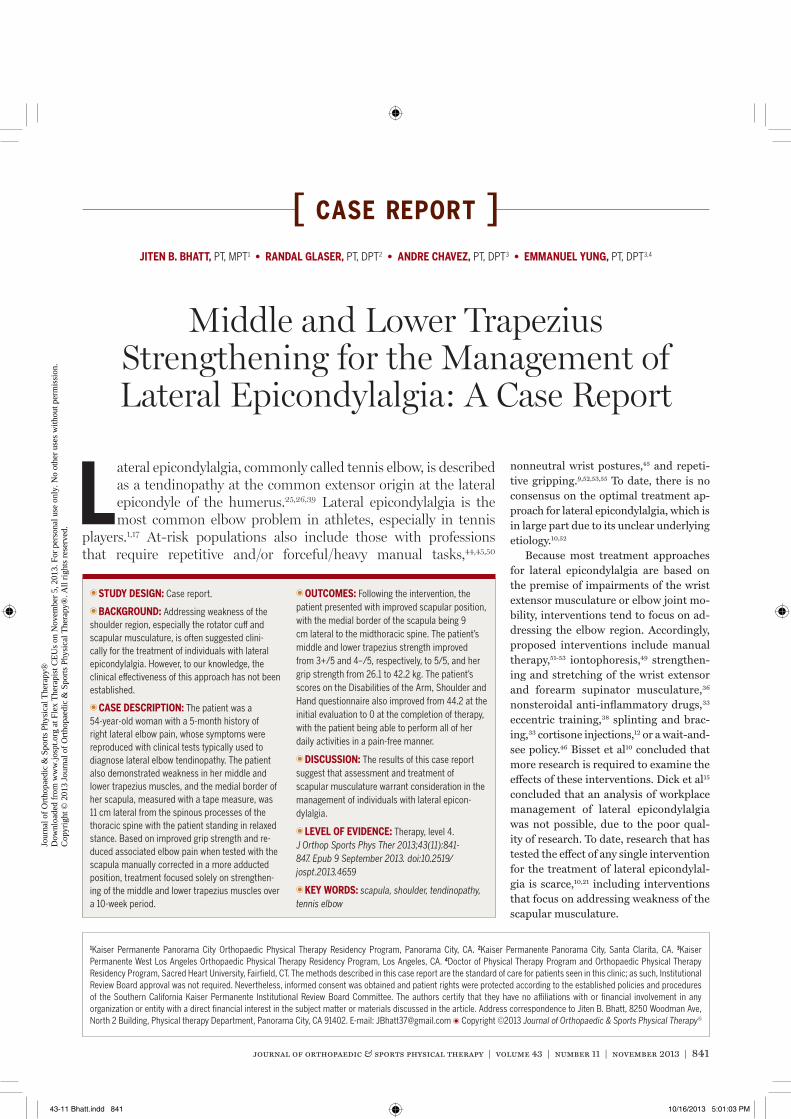
Lateral epicondylalgia, commonly called tennis elbow, is described as a tendinopathy at the common extensor origin at the lateral epicondyle of the humerus.25,26,39 Lateral epicondylalgia is the most common elbow problem in athletes, especially in tennis
players.1,17 At-risk populations also include those with professions that require repetitive and/or forceful/heavy manual tasks,44,45,50
nonneutral wrist postures,45 and repeti-tive gripping.9,52,53,55 To date, there is no consensus on the optimal treatment ap-proach for lateral epicondylalgia, which is in large part due to its unclear underlying etiology.10,52
Because most treatment approaches for lateral epicondylalgia are based on the premise of impairments of the wrist extensor musculature or elbow joint mo-bility, interventions tend to focus on ad-dressing the elbow region. Accordingly, proposed interventions include manual therapy,51-53 iontophoresis,49 strengthen-ing and stretching of the wrist extensor and forearm supinator musculature,36 nonsteroidal anti-inflammatory drugs,33 eccentric training,38 splinting and brac-ing,33 cortisone injections,12 or a wait-and-see policy.46 Bisset et al10 concluded that more research is required to examine the effects of these interventions. Dick et al15 concluded that an analysis of workplace management of lateral epicondylalgia was not possible, due to the poor qual-ity of research. To date, research that has tested the effect of any single intervention for the treatment of lateral epicondylal-gia is scarce,10,21 including interventions that focus on addressing weakness of the scapular musculature.
TT STUDY DESIGN: Case report.
TT BACKGROUND: Addressing weakness of the shoulder region, especially the rotator cuff and scapular musculature, is often suggested clini-cally for the treatment of individuals with lateral epicondylalgia. However, to our knowledge, the clinical effectiveness of this approach has not been established.
TT CASE DESCRIPTION: The patient was a 54-year-old woman with a 5-month history of right lateral elbow pain, whose symptoms were reproduced with clinical tests typically used to diagnose lateral elbow tendinopathy. The patient also demonstrated weakness in her middle and lower trapezius muscles, and the medial border of her scapula, measured with a tape measure, was 11 cm lateral from the spinous processes of the thoracic spine with the patient standing in relaxed stance. Based on improved grip strength and re-duced associated elbow pain when tested with the scapula manually corrected in a more adducted position, treatment focused solely on strengthen-ing of the middle and lower trapezius muscles over a 10-week period.
TT OUTCOMES: Following the intervention, the patient presented with improved scapular position, with the medial border of the scapula being 9 cm lateral to the midthoracic spine. The patient’s middle and lower trapezius strength improved from 3+/5 and 4–/5, respectively, to 5/5, and her grip strength from 26.1 to 42.2 kg. The patient’s scores on the Disabilities of the Arm, Shoulder and Hand questionnaire also improved from 44.2 at the initial evaluation to 0 at the completion of therapy, with the patient being able to perform all of her daily activities in a pain-free manner.
TT DISCUSSION: The results of this case report suggest that assessment and treatment of scapular musculature warrant consideration in the management of individuals with lateral epicon-dylalgia.
TT LEVEL OF EVIDENCE: Therapy, level 4. J Orthop Sports Phys Ther 2013;43(11):841-847. Epub 9 September 2013. doi:10.2519/jospt.2013.4659
TT KEY WORDS: scapula, shoulder, tendinopathy, tennis elbow
1Kaiser Permanente Panorama City Orthopaedic Physical Therapy Residency Program, Panorama City, CA. 2Kaiser Permanente Panorama City, Santa Clarita, CA. 3Kaiser Permanente West Los Angeles Orthopaedic Physical Therapy Residency Program, Los Angeles, CA. 4Doctor of Physical Therapy Program and Orthopaedic Physical Therapy Residency Program, Sacred Heart University, Fairfield, CT. The methods described in this case report are the standard of care for patients seen in this clinic; as such, Institutional Review Board approval was not required. Nevertheless, informed consent was obtained and patient rights were protected according to the established policies and procedures of the Southern California Kaiser Permanente Institutional Review Board Committee. The authors certify that they have no affiliations with or financial involvement in any organization or entity with a direct financial interest in the subject matter or materials discussed in the article. Address correspondence to Jiten B. Bhatt, 8250 Woodman Ave, North 2 Building, Physical therapy Department, Panorama City, CA 91402. E-mail: [email protected] T Copyright ©2013 Journal of Orthopaedic & Sports Physical Therapy®
JITEN B. BHATT, PT, MPT1 • RANDAL GLASER, PT, DPT2 • ANDRE CHAVEZ, PT, DPT3 • EMMANUEL YUNG, PT, DPT3,4
Middle and Lower Trapezius Strengthening for the Management of Lateral Epicondylalgia: A Case Report
43-11 Bhatt.indd 841 10/16/2013 5:01:03 PM
Jour
nal o
f O
rtho
paed
ic &
Spo
rts
Phys
ical
The
rapy
®
Dow
nloa
ded
from
ww
w.jo
spt.o
rg a
t Fle
x T
hera
pist
CE
Us
on N
ovem
ber
5, 2
013.
For
per
sona
l use
onl
y. N
o ot
her
uses
with
out p
erm
issi
on.
Cop
yrig
ht ©
201
3 Jo
urna
l of
Ort
hopa
edic
& S
port
s Ph
ysic
al T
hera
py®
. All
righ
ts r
eser
ved.
842 | november 2013 | volume 43 | number 11 | journal of orthopaedic & sports physical therapy
[ case report ]Clinically, based on expert opinion,
rotator cuff and scapular musculature weakness in individuals with lateral epi-condylalgia is commonly addressed by re-ducing the stresses placed on the wrist/forearm musculature.4,16 Alizadehkhaiyat et al4 identified weaker rotator cuff mus-culature in patients with lateral epicon-dylalgia compared to that of a control group, but did not address scapular mus-culature strength. More recently, Lucado et al29 reported the presence of lower tra-pezius muscle weakness in tennis players with lateral epicondylalgia. Clinically, the authors of this case report noted that modifying scapular position into adduc-tion in individuals with weak scapular musculature and an abducted scapula also modifies the position of the humerus and reduces lateral epicondylalgia symp-toms. More importantly, we have found that this scapular correction often results in improvement in grip strength.
The purpose of this case report was to document the effects of an exercise program focused solely on changing the strength of the scapular musculature and position of the scapula to address the pain and functional limitations of an individual with lateral epicondylalgia.
CASE DESCRIPTION
Patient Characteristics
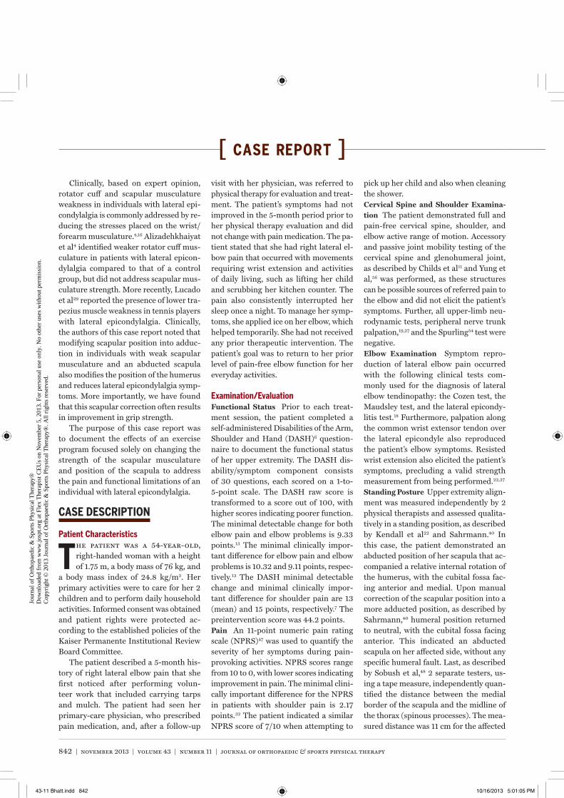
The patient was a 54-year-old, right-handed woman with a height of 1.75 m, a body mass of 76 kg, and
a body mass index of 24.8 kg/m2. Her primary activities were to care for her 2 children and to perform daily household activities. Informed consent was obtained and patient rights were protected ac-cording to the established policies of the Kaiser Permanente Institutional Review Board Committee.
The patient described a 5-month his-tory of right lateral elbow pain that she first noticed after performing volun-teer work that included carrying tarps and mulch. The patient had seen her primary-care physician, who prescribed pain medication, and, after a follow-up
visit with her physician, was referred to physical therapy for evaluation and treat-ment. The patient’s symptoms had not improved in the 5-month period prior to her physical therapy evaluation and did not change with pain medication. The pa-tient stated that she had right lateral el-bow pain that occurred with movements requiring wrist extension and activities of daily living, such as lifting her child and scrubbing her kitchen counter. The pain also consistently interrupted her sleep once a night. To manage her symp-toms, she applied ice on her elbow, which helped temporarily. She had not received any prior therapeutic intervention. The patient’s goal was to return to her prior level of pain-free elbow function for her everyday activities.
Examination/EvaluationFunctional Status Prior to each treat-ment session, the patient completed a self-administered Disabilities of the Arm, Shoulder and Hand (DASH)6 question-naire to document the functional status of her upper extremity. The DASH dis-ability/symptom component consists of 30 questions, each scored on a 1-to-5-point scale. The DASH raw score is transformed to a score out of 100, with higher scores indicating poorer function. The minimal detectable change for both elbow pain and elbow problems is 9.33 points.13 The minimal clinically impor-tant difference for elbow pain and elbow problems is 10.32 and 9.11 points, respec-tively.13 The DASH minimal detectable change and minimal clinically impor-tant difference for shoulder pain are 13 (mean) and 15 points, respectively.7 The preintervention score was 44.2 points.Pain An 11-point numeric pain rating scale (NPRS)47 was used to quantify the severity of her symptoms during pain-provoking activities. NPRS scores range from 10 to 0, with lower scores indicating improvement in pain. The minimal clini-cally important difference for the NPRS in patients with shoulder pain is 2.17 points.32 The patient indicated a similar NPRS score of 7/10 when attempting to
pick up her child and also when cleaning the shower.Cervical Spine and Shoulder Examina-tion The patient demonstrated full and pain-free cervical spine, shoulder, and elbow active range of motion. Accessory and passive joint mobility testing of the cervical spine and glenohumeral joint, as described by Childs et al11 and Yung et al,56 was performed, as these structures can be possible sources of referred pain to the elbow and did not elicit the patient’s symptoms. Further, all upper-limb neu-rodynamic tests, peripheral nerve trunk palpation,19,27 and the Spurling54 test were negative.Elbow Examination Symptom repro-duction of lateral elbow pain occurred with the following clinical tests com-monly used for the diagnosis of lateral elbow tendinopathy: the Cozen test, the Maudsley test, and the lateral epicondy-litis test.18 Furthermore, palpation along the common wrist extensor tendon over the lateral epicondyle also reproduced the patient’s elbow symptoms. Resisted wrist extension also elicited the patient’s symptoms, precluding a valid strength measurement from being performed.22,37
Standing Posture Upper extremity align-ment was measured independently by 2 physical therapists and assessed qualita-tively in a standing position, as described by Kendall et al22 and Sahrmann.40 In this case, the patient demonstrated an abducted position of her scapula that ac-companied a relative internal rotation of the humerus, with the cubital fossa fac-ing anterior and medial. Upon manual correction of the scapular position into a more adducted position, as described by Sahrmann,40 humeral position returned to neutral, with the cubital fossa facing anterior. This indicated an abducted scapula on her affected side, without any specific humeral fault. Last, as described by Sobush et al,48 2 separate testers, us-ing a tape measure, independently quan-tified the distance between the medial border of the scapula and the midline of the thorax (spinous processes). The mea-sured distance was 11 cm for the affected
43-11 Bhatt.indd 842 10/16/2013 5:01:05 PM
Jour
nal o
f O
rtho
paed
ic &
Spo
rts
Phys
ical
The
rapy
®
Dow
nloa
ded
from
ww
w.jo
spt.o
rg a
t Fle
x T
hera
pist
CE
Us
on N
ovem
ber
5, 2
013.
For
per
sona
l use
onl
y. N
o ot
her
uses
with
out p
erm
issi
on.
Cop
yrig
ht ©
201
3 Jo
urna
l of
Ort
hopa
edic
& S
port
s Ph
ysic
al T
hera
py®
. All
righ
ts r
eser
ved.
journal of orthopaedic & sports physical therapy | volume 43 | number 11 | november 2013 | 843
side and 9 cm for the nona� ected side, whereas previously reported normal val-ues were (mean � SD) 8.3 � 1.25 cm.48
Grip Strength Grip strength was as-sessed with and without manual scapular correction. The purpose of this test was to assess the e� ect scapular position had on the patient’s grip strength and elbow pain during a gripping task. For testing, a Jamar dynamometer (Sammons Preston Rolyan, Bolingbrook, IL) was used, and the patient’s elbow was fl exed to 90°, with the wrist and forearm held in neutral and the glenohumeral joint in a neutral rest-ing position. Only scapular position was modifi ed between the tests, and for each scapular position the patient performed 3 trials of 5 seconds in duration. With the scapula in its usual resting position, the patient had a grip strength of 26.1 kg prior to lateral elbow pain rated at 7/10. In comparison, with the scapula passive-ly manually corrected into a more ad-ducted and posteriorly tilted position, as described by Sahrmann,40 grip strength, without any elbow pain, was 33.7 kg.Scapular Musculature Strength testing of the middle and lower trapezius was performed in a prone testing position,
as described by Kendall et al,22 with the scapula in its usual resting position. Test-ing was performed independently by 2 licensed physical therapists, and scores from 0 to 5 were used as described by Kendall et al.22 Middle trapezius strength was considered to be 3+/5, whereas lower trapezius strength was 4–/5.Electromyography and Grip Strength To potentially gain additional insight on the infl uence of scapular repositioning on the activation level of the wrist musculature, electromyographic (EMG) signals for the extensor carpi radialis brevis (ECRB) and biceps brachii were collected when performing a standardized grip task with and without correction of scapular posi-tion. The EMG signals were recorded at 1500 Hz and subsequently band-pass fi l-tered (80-250 Hz). Data were full-wave rectifi ed, and a moving-average smooth-ing algorithm (50-millisecond window) was used to generate a linear envelope. EMG processing and smoothing were performed using MyoResearch XP Mas-ter Edition 1.08.17 (Noraxon USA Inc, Scottsdale, AZ). Disposable, self-adhesive Ag/AgCl dual electrodes (Noraxon USA Inc) were used, spaced 2 cm apart over the ECRB and biceps brachii muscles, according to the criterion described by Delagi and Perotto.14
For testing, the same dynamometer and shoulder and elbow position de-
scribed earlier were used. The patient was instructed on maintaining a standard grip force of 31.75 kg for 5 seconds. Once able to perform this task, after a 3-min-ute rest period, the patient was asked to perform 6 trials of 5 seconds in duration, while EMG and force data were recorded. The fi rst 3 trials were performed with the patient in her usual scapular resting posi-tion. Following another 3-minute rest pe-riod, the last 3 trials were performed with an examiner giving tactile cues for the patient to hold her scapula in a more ad-ducted position.40 The results indicated a reduction of EMG signal amplitude for both the ECRB (decreased by 44%) and the biceps brachii (23%) when perform-ing the grip task with active correction of scapular position.
DiagnosisBased on the clinical examination, we determined that the patient matched the presentation for lateral epicondylal-gia. We also postulated that signifi cant weakness of her scapular musculature and poor scapular positioning might have contributed to her clinical condi-tion. Therefore, it was our hypothesis that an exercise program focused on improv-ing the strength of the scapular adductor muscles would result in pain resolution with gripping tasks and improved scapu-lar position.
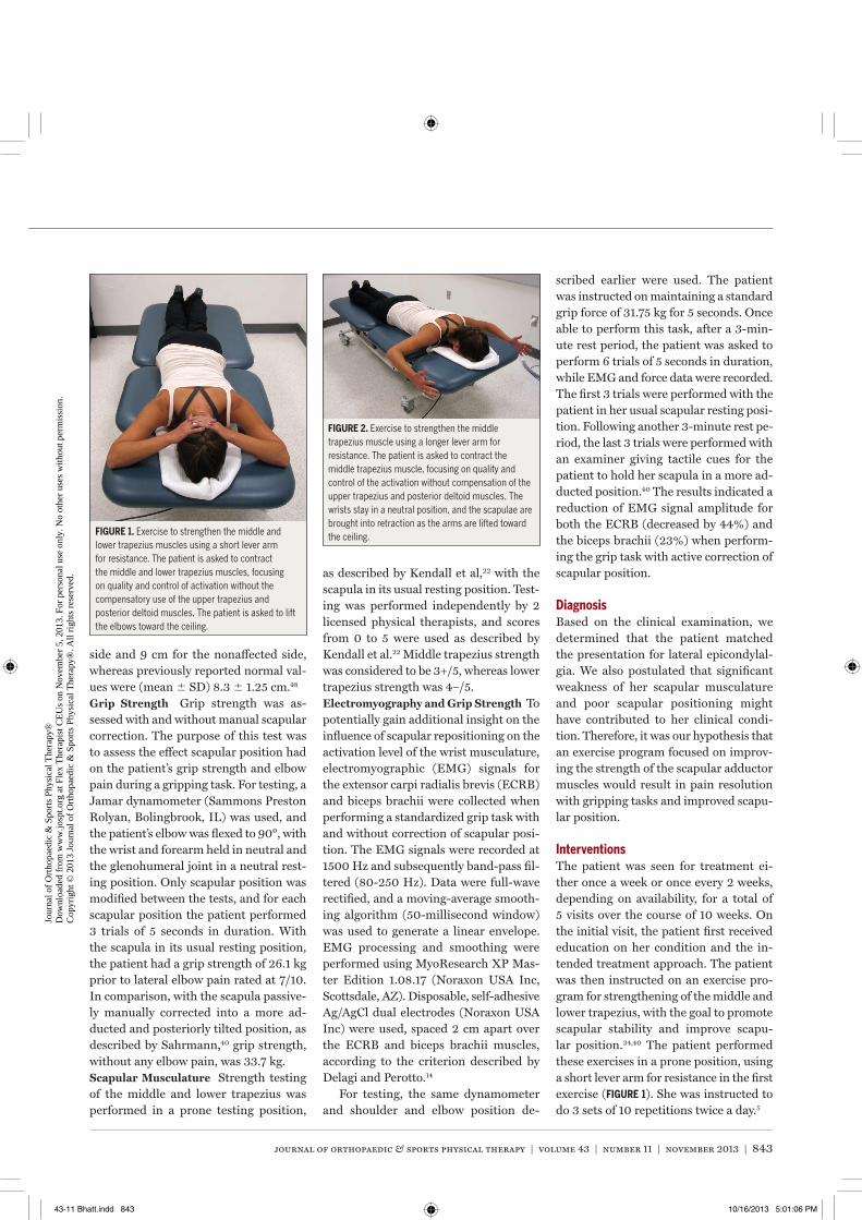
InterventionsThe patient was seen for treatment ei-ther once a week or once every 2 weeks, depending on availability, for a total of 5 visits over the course of 10 weeks. On the initial visit, the patient fi rst received education on her condition and the in-tended treatment approach. The patient was then instructed on an exercise pro-gram for strengthening of the middle and lower trapezius, with the goal to promote scapular stability and improve scapu-lar position.34,40 The patient performed these exercises in a prone position, using a short lever arm for resistance in the fi rst exercise (FIGURE 1). She was instructed to do 3 sets of 10 repetitions twice a day.5
FIGURE 1. Exercise to strengthen the middle and lower trapezius muscles using a short lever arm for resistance. The patient is asked to contract the middle and lower trapezius muscles, focusing on quality and control of activation without the compensatory use of the upper trapezius and posterior deltoid muscles. The patient is asked to lift the elbows toward the ceiling.
FIGURE 2. Exercise to strengthen the middle trapezius muscle using a longer lever arm for resistance. The patient is asked to contract the middle trapezius muscle, focusing on quality and control of the activation without compensation of the upper trapezius and posterior deltoid muscles. The wrists stay in a neutral position, and the scapulae are brought into retraction as the arms are lifted toward the ceiling.
43-11 Bhatt.indd 843 10/16/2013 5:01:06 PM
Jour
nal o
f O
rtho
paed
ic &
Spo
rts
Phys
ical
The
rapy
®
Dow
nloa
ded
from
ww
w.jo
spt.o
rg a
t Fle
x T
hera
pist
CE
Us
on N
ovem
ber
5, 2
013.
For
per
sona
l use
onl
y. N
o ot
her
uses
with
out p
erm
issi
on.
Cop
yrig
ht ©
201
3 Jo
urna
l of
Ort
hopa
edic
& S
port
s Ph
ysic
al T
hera
py®
. All
righ
ts r
eser
ved.
844 | november 2013 | volume 43 | number 11 | journal of orthopaedic & sports physical therapy
[ CASE REPORT ]
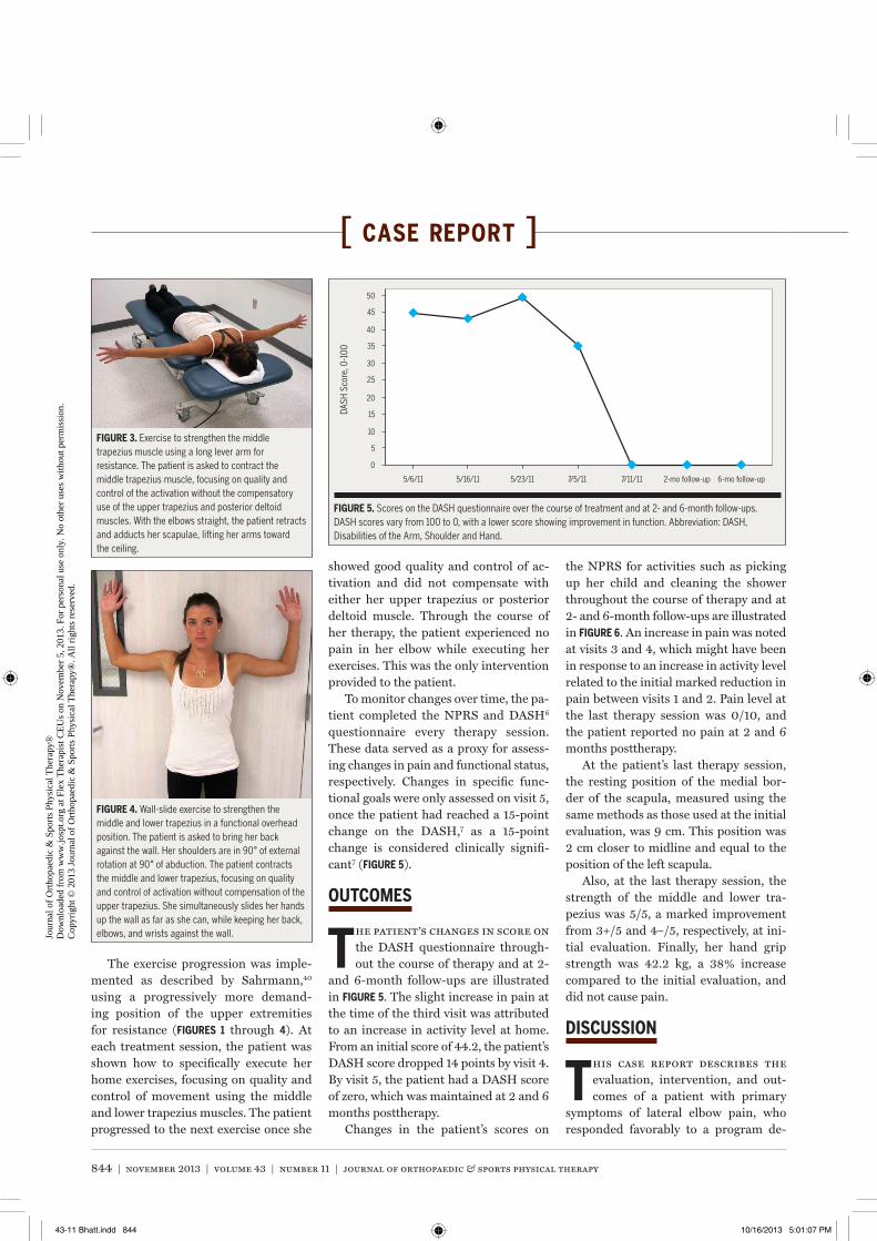
The exercise progression was imple-mented as described by Sahrmann,40
using a progressively more demand-ing position of the upper extremities for resistance (FIGURES 1 through 4). At each treatment session, the patient was shown how to specifi cally execute her home exercises, focusing on quality and control of movement using the middle and lower trapezius muscles. The patient progressed to the next exercise once she
showed good quality and control of ac-tivation and did not compensate with either her upper trapezius or posterior deltoid muscle. Through the course of her therapy, the patient experienced no pain in her elbow while executing her exercises. This was the only intervention provided to the patient.
To monitor changes over time, the pa-tient completed the NPRS and DASH6
questionnaire every therapy session. These data served as a proxy for assess-ing changes in pain and functional status, respectively. Changes in specifi c func-tional goals were only assessed on visit 5, once the patient had reached a 15-point change on the DASH,7 as a 15-point change is considered clinically signifi -cant7 (FIGURE 5).
OUTCOMES
The patient’s changes in score on the DASH questionnaire through-out the course of therapy and at 2-
and 6-month follow-ups are illustrated in FIGURE 5. The slight increase in pain at the time of the third visit was attributed to an increase in activity level at home. From an initial score of 44.2, the patient’s DASH score dropped 14 points by visit 4. By visit 5, the patient had a DASH score of zero, which was maintained at 2 and 6 months posttherapy.
Changes in the patient’s scores on
the NPRS for activities such as picking up her child and cleaning the shower throughout the course of therapy and at 2- and 6-month follow-ups are illustrated in FIGURE 6. An increase in pain was noted at visits 3 and 4, which might have been in response to an increase in activity level related to the initial marked reduction in pain between visits 1 and 2. Pain level at the last therapy session was 0/10, and the patient reported no pain at 2 and 6 months posttherapy.
At the patient’s last therapy session, the resting position of the medial bor-der of the scapula, measured using the same methods as those used at the initial evaluation, was 9 cm. This position was 2 cm closer to midline and equal to the position of the left scapula.
Also, at the last therapy session, the strength of the middle and lower tra-pezius was 5/5, a marked improvement from 3+/5 and 4–/5, respectively, at ini-tial evaluation. Finally, her hand grip strength was 42.2 kg, a 38% increase compared to the initial evaluation, and did not cause pain.
DISCUSS ION
This case report describes the evaluation, intervention, and out-comes of a patient with primary
symptoms of lateral elbow pain, who responded favorably to a program de-
0
5
10
15
20
5/6/11 5/16/11 5/23/11 7/5/11 7/11/11 2-mo follow-up 6-mo follow-up
25
30
35
40
45
50
DASH
Sco
re, 0
-100
FIGURE 5. Scores on the DASH questionnaire over the course of treatment and at 2- and 6-month follow-ups. DASH scores vary from 100 to 0, with a lower score showing improvement in function. Abbreviation: DASH, Disabilities of the Arm, Shoulder and Hand.
FIGURE 3. Exercise to strengthen the middle trapezius muscle using a long lever arm for resistance. The patient is asked to contract the middle trapezius muscle, focusing on quality and control of the activation without the compensatory use of the upper trapezius and posterior deltoid muscles. With the elbows straight, the patient retracts and adducts her scapulae, lifting her arms toward the ceiling.
FIGURE 4. Wall-slide exercise to strengthen the middle and lower trapezius in a functional overhead position. The patient is asked to bring her back against the wall. Her shoulders are in 90° of external rotation at 90° of abduction. The patient contracts the middle and lower trapezius, focusing on quality and control of activation without compensation of the upper trapezius. She simultaneously slides her hands up the wall as far as she can, while keeping her back, elbows, and wrists against the wall.
43-11 Bhatt.indd 844 10/16/2013 5:01:07 PM
Jour
nal o
f O
rtho
paed
ic &
Spo
rts
Phys
ical
The
rapy
®
Dow
nloa
ded
from
ww
w.jo
spt.o
rg a
t Fle
x T
hera
pist
CE
Us
on N
ovem
ber
5, 2
013.
For
per
sona
l use
onl
y. N
o ot
her
uses
with
out p
erm
issi
on.
Cop
yrig
ht ©
201
3 Jo
urna
l of
Ort
hopa
edic
& S
port
s Ph
ysic
al T
hera
py®
. All
righ
ts r
eser
ved.
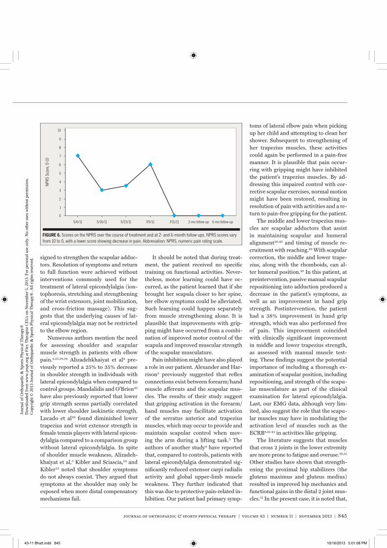
journal of orthopaedic & sports physical therapy | volume 43 | number 11 | november 2013 | 845
signed to strengthen the scapular adduc-tors. Resolution of symptoms and return to full function were achieved without interventions commonly used for the treatment of lateral epicondylalgia (ion-tophoresis, stretching and strengthening of the wrist extensors, joint mobilization, and cross-friction massage). This sug-gests that the underlying causes of lat-eral epicondylalgia may not be restricted to the elbow region.
Numerous authors mention the need for assessing shoulder and scapular muscle strength in patients with elbow pain.4,23,24,34 Alizadehkhaiyat et al4 pre-viously reported a 25% to 35% decrease in shoulder strength in individuals with lateral epicondylalgia when compared to control groups. Mandalidis and O’Brien30 have also previously reported that lower grip strength seems partially correlated with lower shoulder isokinetic strength. Lucado et al29 found diminished lower trapezius and wrist extensor strength in female tennis players with lateral epicon-dylalgia compared to a comparison group without lateral epicondylalgia. In spite of shoulder muscle weakness, Alizadeh-khaiyat et al,3 Kibler and Sciascia,24 and Kibler23 noted that shoulder symptoms do not always coexist. They argued that symptoms at the shoulder may only be exposed when more distal compensatory mechanisms fail.
It should be noted that during treat-ment, the patient received no specific training on functional activities. Never-theless, motor learning could have oc-curred, as the patient learned that if she brought her scapula closer to her spine, her elbow symptoms could be alleviated. Such learning could happen separately from muscle strengthening alone. It is plausible that improvements with grip-ping might have occurred from a combi-nation of improved motor control of the scapula and improved muscular strength of the scapular musculature.
Pain inhibition might have also played a role in our patient. Alexander and Har-rison2 previously suggested that reflex connections exist between forearm/hand muscle afferents and the scapular mus-cles. The results of their study suggest that gripping activation in the forearm/hand muscles may facilitate activation of the serratus anterior and trapezius muscles, which may occur to provide and maintain scapular control when mov-ing the arm during a lifting task.2 The authors of another study4 have reported that, compared to controls, patients with lateral epicondylalgia demonstrated sig-nificantly reduced extensor carpi radialis activity and global upper-limb muscle weakness. They further indicated that this was due to protective pain-related in-hibition. Our patient had primary symp-
toms of lateral elbow pain when picking up her child and attempting to clean her shower. Subsequent to strengthening of her trapezius muscles, these activities could again be performed in a pain-free manner. It is plausible that pain occur-ring with gripping might have inhibited the patient’s trapezius muscles. By ad-dressing this impaired control with cor-rective scapular exercises, normal motion might have been restored, resulting in resolution of pain with activities and a re-turn to pain-free gripping for the patient.
The middle and lower trapezius mus-cles are scapular adductors that assist in maintaining scapular and humeral alignment20,40 and timing of muscle re-cruitment with reaching.40 With scapular correction, the middle and lower trape-zius, along with the rhomboids, can al-ter humeral position.40 In this patient, at preintervention, passive manual scapular repositioning into adduction produced a decrease in the patient’s symptoms, as well as an improvement in hand grip strength. Postintervention, the patient had a 38% improvement in hand grip strength, which was also performed free of pain. This improvement coincided with clinically significant improvement in middle and lower trapezius strength, as assessed with manual muscle test-ing. These findings suggest the potential importance of including a thorough ex-amination of scapular position, including repositioning, and strength of the scapu-lar musculature as part of the clinical examination for lateral epicondylalgia. Last, our EMG data, although very lim-ited, also suggest the role that the scapu-lar muscles may have in modulating the activation level of muscles such as the ECRB8,41-43 in activities like gripping.
The literature suggests that muscles that cross 2 joints in the lower extremity are more prone to fatigue and overuse.28,35 Other studies have shown that strength-ening the proximal hip stabilizers (the gluteus maximus and gluteus medius) resulted in improved hip mechanics and functional gains in the distal 2 joint mus-cles.31 In the present case, it is noted that,
0
1
2
3
4
5/6/11 5/16/11 5/23/11 7/5/11 7/11/11 2-mo follow-up 6-mo follow-up
5
6
7
8
9
10
NPR
S Sc
ore,
0-1
0
FIGURE 6. Scores on the NPRS over the course of treatment and at 2- and 6-month follow-ups. NPRS scores vary from 10 to 0, with a lower score showing decrease in pain. Abbreviation: NPRS, numeric pain rating scale.
43-11 Bhatt.indd 845 10/16/2013 5:01:08 PM
Jour
nal o
f O
rtho
paed
ic &
Spo
rts
Phys
ical
The
rapy
®
Dow
nloa
ded
from
ww
w.jo
spt.o
rg a
t Fle
x T
hera
pist
CE
Us
on N
ovem
ber
5, 2
013.
For
per
sona
l use
onl
y. N
o ot
her
uses
with
out p
erm
issi
on.
Cop
yrig
ht ©
201
3 Jo
urna
l of
Ort
hopa
edic
& S
port
s Ph
ysic
al T
hera
py®
. All
righ
ts r
eser
ved.
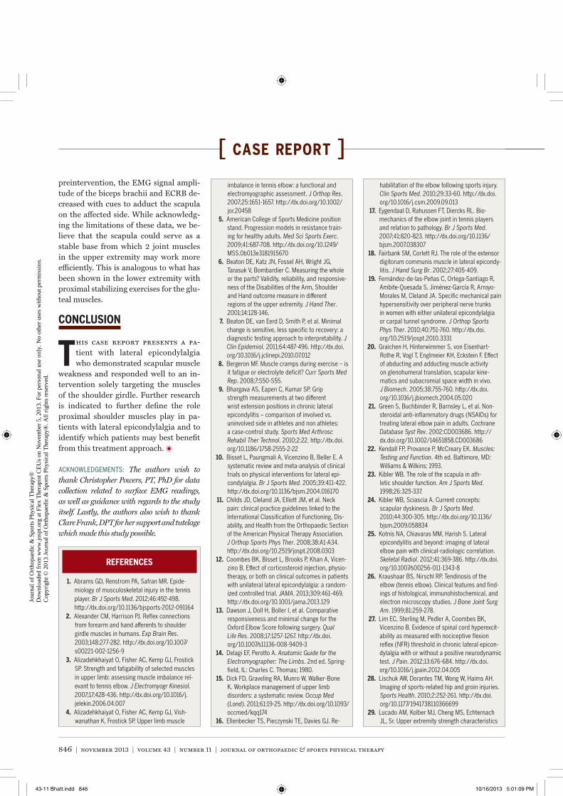
846 | november 2013 | volume 43 | number 11 | journal of orthopaedic & sports physical therapy
[ case report ]preintervention, the EMG signal ampli-tude of the biceps brachii and ECRB de-creased with cues to adduct the scapula on the affected side. While acknowledg-ing the limitations of these data, we be-lieve that the scapula could serve as a stable base from which 2 joint muscles in the upper extremity may work more efficiently. This is analogous to what has been shown in the lower extremity with proximal stabilizing exercises for the glu-teal muscles.
CONCLUSION
This case report presents a pa-tient with lateral epicondylalgia who demonstrated scapular muscle
weakness and responded well to an in-tervention solely targeting the muscles of the shoulder girdle. Further research is indicated to further define the role proximal shoulder muscles play in pa-tients with lateral epicondylalgia and to identify which patients may best benefit from this treatment approach. t
ACKNOWLEDGEMENTS: The authors wish to thank Christopher Powers, PT, PhD for data collection related to surface EMG readings, as well as guidance with regards to the study itself. Lastly, the authors also wish to thank Clare Frank, DPT for her support and tutelage which made this study possible.
REFERENCES
1. Abrams GD, Renstrom PA, Safran MR. Epide-miology of musculoskeletal injury in the tennis player. Br J Sports Med. 2012;46:492-498. http://dx.doi.org/10.1136/bjsports-2012-091164
2. Alexander CM, Harrison PJ. Reflex connections from forearm and hand afferents to shoulder girdle muscles in humans. Exp Brain Res. 2003;148:277-282. http://dx.doi.org/10.1007/s00221-002-1256-9
3. Alizadehkhaiyat O, Fisher AC, Kemp GJ, Frostick SP. Strength and fatigability of selected muscles in upper limb: assessing muscle imbalance rel-evant to tennis elbow. J Electromyogr Kinesiol. 2007;17:428-436. http://dx.doi.org/10.1016/j.jelekin.2006.04.007
4. Alizadehkhaiyat O, Fisher AC, Kemp GJ, Vish-wanathan K, Frostick SP. Upper limb muscle
imbalance in tennis elbow: a functional and electromyographic assessment. J Orthop Res. 2007;25:1651-1657. http://dx.doi.org/10.1002/jor.20458
5. American College of Sports Medicine position stand. Progression models in resistance train-ing for healthy adults. Med Sci Sports Exerc. 2009;41:687-708. http://dx.doi.org/10.1249/MSS.0b013e3181915670
6. Beaton DE, Katz JN, Fossel AH, Wright JG, Tarasuk V, Bombardier C. Measuring the whole or the parts? Validity, reliability, and responsive-ness of the Disabilities of the Arm, Shoulder and Hand outcome measure in different regions of the upper extremity. J Hand Ther. 2001;14:128-146.
7. Beaton DE, van Eerd D, Smith P, et al. Minimal change is sensitive, less specific to recovery: a diagnostic testing approach to interpretability. J Clin Epidemiol. 2011;64:487-496. http://dx.doi.org/10.1016/j.jclinepi.2010.07.012
8. Bergeron MF. Muscle cramps during exercise – is it fatigue or electrolyte deficit? Curr Sports Med Rep. 2008;7:S50-S55.
9. Bhargava AS, Eapen C, Kumar SP. Grip strength measurements at two different wrist extension positions in chronic lateral epicondylitis – comparison of involved vs. uninvolved side in athletes and non athletes: a case-control study. Sports Med Arthrosc Rehabil Ther Technol. 2010;2:22. http://dx.doi.org/10.1186/1758-2555-2-22
10. Bisset L, Paungmali A, Vicenzino B, Beller E. A systematic review and meta-analysis of clinical trials on physical interventions for lateral epi-condylalgia. Br J Sports Med. 2005;39:411-422. http://dx.doi.org/10.1136/bjsm.2004.016170
11. Childs JD, Cleland JA, Elliott JM, et al. Neck pain: clinical practice guidelines linked to the International Classification of Functioning, Dis-ability, and Health from the Orthopaedic Section of the American Physical Therapy Association. J Orthop Sports Phys Ther. 2008;38:A1-A34. http://dx.doi.org/10.2519/jospt.2008.0303
12. Coombes BK, Bisset L, Brooks P, Khan A, Vicen-zino B. Effect of corticosteroid injection, physio-therapy, or both on clinical outcomes in patients with unilateral lateral epicondylalgia: a random-ized controlled trial. JAMA. 2013;309:461-469. http://dx.doi.org/10.1001/jama.2013.129
13. Dawson J, Doll H, Boller I, et al. Comparative responsiveness and minimal change for the Oxford Elbow Score following surgery. Qual Life Res. 2008;17:1257-1267. http://dx.doi.org/10.1007/s11136-008-9409-3
14. Delagi EF, Perotto A. Anatomic Guide for the Electromyographer: The Limbs. 2nd ed. Spring-field, IL: Charles C. Thomas; 1980.
15. Dick FD, Graveling RA, Munro W, Walker-Bone K. Workplace management of upper limb disorders: a systematic review. Occup Med (Lond). 2011;61:19-25. http://dx.doi.org/10.1093/occmed/kqq174
16. Ellenbecker TS, Pieczynski TE, Davies GJ. Re-
habilitation of the elbow following sports injury. Clin Sports Med. 2010;29:33-60. http://dx.doi.org/10.1016/j.csm.2009.09.013
17. Eygendaal D, Rahussen FT, Diercks RL. Bio-mechanics of the elbow joint in tennis players and relation to pathology. Br J Sports Med. 2007;41:820-823. http://dx.doi.org/10.1136/bjsm.2007.038307
18. Fairbank SM, Corlett RJ. The role of the extensor digitorum communis muscle in lateral epicondy-litis. J Hand Surg Br. 2002;27:405-409.
19. Fernández-de-las-Peñas C, Ortega-Santiago R, Ambite-Quesada S, Jiménez-García R, Arroyo-Morales M, Cleland JA. Specific mechanical pain hypersensitivity over peripheral nerve trunks in women with either unilateral epicondylalgia or carpal tunnel syndrome. J Orthop Sports Phys Ther. 2010;40:751-760. http://dx.doi.org/10.2519/jospt.2010.3331
20. Graichen H, Hinterwimmer S, von Eisenhart-Rothe R, Vogl T, Englmeier KH, Eckstein F. Effect of abducting and adducting muscle activity on glenohumeral translation, scapular kine-matics and subacromial space width in vivo. J Biomech. 2005;38:755-760. http://dx.doi.org/10.1016/j.jbiomech.2004.05.020
21. Green S, Buchbinder R, Barnsley L, et al. Non-steroidal anti-inflammatory drugs (NSAIDs) for treating lateral elbow pain in adults. Cochrane Database Syst Rev. 2002:CD003686. http://dx.doi.org/10.1002/14651858.CD003686
22. Kendall FP, Provance P, McCreary EK. Muscles: Testing and Function. 4th ed. Baltimore, MD: Williams & Wilkins; 1993.
23. Kibler WB. The role of the scapula in ath-letic shoulder function. Am J Sports Med. 1998;26:325-337.
24. Kibler WB, Sciascia A. Current concepts: scapular dyskinesis. Br J Sports Med. 2010;44:300-305. http://dx.doi.org/10.1136/bjsm.2009.058834
25. Kotnis NA, Chiavaras MM, Harish S. Lateral epicondylitis and beyond: imaging of lateral elbow pain with clinical-radiologic correlation. Skeletal Radiol. 2012;41:369-386. http://dx.doi.org/10.1007/s00256-011-1343-8
26. Kraushaar BS, Nirschl RP. Tendinosis of the elbow (tennis elbow). Clinical features and find-ings of histological, immunohistochemical, and electron microscopy studies. J Bone Joint Surg Am. 1999;81:259-278.
27. Lim EC, Sterling M, Pedler A, Coombes BK, Vicenzino B. Evidence of spinal cord hyperexcit-ability as measured with nociceptive flexion reflex (NFR) threshold in chronic lateral epicon-dylalgia with or without a positive neurodynamic test. J Pain. 2012;13:676-684. http://dx.doi.org/10.1016/j.jpain.2012.04.005
28. Lischuk AW, Dorantes TM, Wong W, Haims AH. Imaging of sports-related hip and groin injuries. Sports Health. 2010;2:252-261. http://dx.doi.org/10.1177/1941738110366699
29. Lucado AM, Kolber MJ, Cheng MS, Echternach JL, Sr. Upper extremity strength characteristics
43-11 Bhatt.indd 846 10/16/2013 5:01:09 PM
Jour
nal o
f O
rtho
paed
ic &
Spo
rts
Phys
ical
The
rapy
®
Dow
nloa
ded
from
ww
w.jo
spt.o
rg a
t Fle
x T
hera
pist
CE
Us
on N
ovem
ber
5, 2
013.
For
per
sona
l use
onl
y. N
o ot
her
uses
with
out p
erm
issi
on.
Cop
yrig
ht ©
201
3 Jo
urna
l of
Ort
hopa
edic
& S
port
s Ph
ysic
al T
hera
py®
. All
righ
ts r
eser
ved.
journal of orthopaedic & sports physical therapy | volume 43 | number 11 | november 2013 | 847
@ MORE INFORMATIONWWW.JOSPT.ORG
in female recreational tennis players with and without lateral epicondylalgia. J Orthop Sports Phys Ther. 2012;42:1025-1031. http://dx.doi.org/10.2519/jospt.2012.4095
30. Mandalidis D, O’Brien M. Relationship between hand-grip isometric strength and isokinetic moment data of the shoulder stabilisers. J Bodyw Mov Ther. 2010;14:19-26. http://dx.doi.org/10.1016/j.jbmt.2008.05.001
31. Mascal CL, Landel R, Powers C. Management of patellofemoral pain targeting hip, pelvis, and trunk muscle function: 2 case reports. J Orthop Sports Phys Ther. 2003;33:647-660. http://dx.doi.org/10.2519/jospt.2003.33.11.647
32. Michener LA, Snyder AR, Leggin BG. Respon-siveness of the numeric pain rating scale in patients with shoulder pain and the effect of sur-gical status. J Sport Rehabil. 2011;20:115-128.
33. Nilsson P, Baigi A, Sward L, Möller M, Månsson J. Lateral epicondylalgia: a structured pro-gramme better than corticosteroids and NSAID. Scand J Occup Ther. 2012;19:404-410. http://dx.doi.org/10.3109/11038128.2011.620983
34. Page P, Frank CC, Lardner R. Assessment and Treatment of Muscle Imbalance: The Janda Ap-proach. Champaign, IL: Human Kinetics; 2010.
35. Palmer WE, Kuong SJ, Elmadbouh HM. MR imaging of myotendinous strain. AJR Am J Roentgenol. 1999;173:703-709. http://dx.doi.org/10.2214/ajr.173.3.10470908
36. Pienimäki TT, Tarvainen TK, Siira PT, Van-haranta H. Progressive strengthening and stretching exercises and ultrasound for chronic lateral epicondylitis. Physiotherapy. 1996;82:522-530. http://dx.doi.org/10.1016/S0031-9406(05)66275-X
37. Rainville J, Noto DJ, Jouve C, Jenis L. Assess-ment of forearm pronation strength in C6 and C7 radiculopathies. Spine (Phila Pa 1976). 2007;32:72-75. http://dx.doi.org/10.1097/01.brs.0000251002.39417.2c
38. Raman J, MacDermid JC, Grewal R. Effective-ness of different methods of resistance exercises in lateral epicondylosis—a systematic review. J Hand Ther. 2012;25:5-25; quiz 26. http://dx.doi.org/10.1016/j.jht.2011.09.001
39. Ruiz-Ruiz B, Fernández-de-las-Peñas C, Ortega-
Santiago R, Arendt-Nielsen L, Madeleine P. Topo-graphical pressure and thermal pain sensitivity mapping in patients with unilateral lateral epi-condylalgia. J Pain. 2011;12:1040-1048. http://dx.doi.org/10.1016/j.jpain.2011.04.001
40. Sahrmann SA. Diagnosis and Treatment of Movement Impairment Syndromes. St Louis, MO: Mosby; 2001.
41. Schwellnus MP. Cause of exercise associated muscle cramps (EAMC) – altered neuromuscu-lar control, dehydration or electrolyte depletion? Br J Sports Med. 2009;43:401-408. http://dx.doi.org/10.1136/bjsm.2008.050401
42. Schwellnus MP. Muscle cramping in the mara-thon: aetiology and risk factors. Sports Med. 2007;37:364-367.
43. Schwellnus MP, Derman EW, Noakes TD. Aetiolo-gy of skeletal muscle ‘cramps’ during exercise: a novel hypothesis. J Sports Sci. 1997;15:277-285. http://dx.doi.org/10.1080/026404197367281
44. Shiri R, Viikari-Juntura E. Lateral and medial epicondylitis: role of occupational factors. Best Pract Res Clin Rheumatol. 2011;25:43-57. http://dx.doi.org/10.1016/j.berh.2011.01.013
45. Shiri R, Viikari-Juntura E, Varonen H, Heliövaara M. Prevalence and determinants of lateral and medial epicondylitis: a population study. Am J Epidemiol. 2006;164:1065-1074. http://dx.doi.org/10.1093/aje/kwj325
46. Smidt N, van der Windt DA, Assendelft WJ, Deville WL, Korthals-de Bos IB, Bouter LM. Corticosteroid injections, physiotherapy, or a wait-and-see policy for lateral epicondy-litis: a randomised controlled trial. Lancet. 2002;359:657-662. http://dx.doi.org/10.1016/S0140-6736(02)07811-X
47. Smidt N, van der Windt DA, Assendelft WJ, et al. Interobserver reproducibility of the assess-ment of severity of complaints, grip strength, and pressure pain threshold in patients with lateral epicondylitis. Arch Phys Med Rehabil. 2002;83:1145-1150.
48. Sobush DC, Simoneau GG, Dietz KE, Levene JA, Grossman RE, Smith WB. The Lennie test for measuring scapular position in healthy young adult females: a reliability and validity study. J Orthop Sports Phys Ther. 1996;23:39-50. http://
dx.doi.org/10.2519/jospt.1996.23.1.39 49. Stefanou A, Marshall N, Holdan W, Siddiqui A. A
randomized study comparing corticosteroid in-jection to corticosteroid iontophoresis for lateral epicondylitis. J Hand Surg Am. 2012;37:104-109. http://dx.doi.org/10.1016/j.jhsa.2011.10.005
50. van Rijn RM, Huisstede BM, Koes BW, Burdorf A. Associations between work-related factors and specific disorders at the elbow: a system-atic literature review. Rheumatology (Oxford). 2009;48:528-536. http://dx.doi.org/10.1093/rheumatology/kep013
51. Vicenzino B. Lateral epicondylalgia: a musculo-skeletal physiotherapy perspective. Man Ther. 2003;8:66-79.
52. Vicenzino B, Cleland JA, Bisset L. Joint manipulation in the management of lateral epicondylalgia: a clinical commentary. J Man Manip Ther. 2007;15:50-56. http://dx.doi.org/10.1179/106698107791090132
53. Vicenzino B, Smith D, Cleland J, Bisset L. Development of a clinical prediction rule to identify initial responders to mobilisation with movement and exercise for lateral epicondylal-gia. Man Ther. 2009;14:550-554. http://dx.doi.org/10.1016/j.math.2008.08.004
54. Wainner RS, Fritz JM, Irrgang JJ, Boninger ML, Delitto A, Allison S. Reliability and diagnostic accuracy of the clinical examination and patient self-report measures for cervical radiculopathy. Spine (Phila Pa 1976). 2003;28:52-62.
55. Walker-Bone K, Palmer KT, Reading I, Coggon D, Cooper C. Occupation and epicondylitis: a pop-ulation-based study. Rheumatology (Oxford). 2012;51:305-310. http://dx.doi.org/10.1093/rheumatology/ker228
56. Yung E, Asavasopon S, Godges JJ. Screening for head, neck, and shoulder pathology in patients with upper extremity signs and symptoms. J Hand Ther. 2010;23:173-185; quiz 186. http://dx.doi.org/10.1016/j.jht.2009.11.004
SEND Letters to the Editor-in-Chief
JOSPT welcomes letters related to professional issues or articles published in the Journal. The Editor-in-Chief reviews and selects letters for publica-tion based on the topic’s relevance, importance, appropriateness, and timeliness. Letters should include a summary statement of any conflict of interest, including financial support related to the issue addressed. In addition, letters are copy edited, and the correspondent is not typically sent a version to approve. Letters to the Editor-in-Chief should be sent electronically to [email protected]. Authors of the relevant manuscript are given the opportunity to respond to the content of the letter.
43-11 Bhatt.indd 847 10/16/2013 5:01:10 PM
Jour
nal o
f O
rtho
paed
ic &
Spo
rts
Phys
ical
The
rapy
®
Dow
nloa
ded
from
ww
w.jo
spt.o
rg a
t Fle
x T
hera
pist
CE
Us
on N
ovem
ber
5, 2
013.
For
per
sona
l use
onl
y. N
o ot
her
uses
with
out p
erm
issi
on.
Cop
yrig
ht ©
201
3 Jo
urna
l of
Ort
hopa
edic
& S
port
s Ph
ysic
al T
hera
py®
. All
righ
ts r
eser
ved.