Embed Size (px)
Citation preview
FLAVIN AMINE OXIDASES FROM THE MONOAMINE OXIDASE
STRUCTURAL FAMILY UTILIZE A HYDRIDE TRANSFER MECHANISM
A Dissertation
by
MICHELLE HENDERSON POZZI
Submitted to the Office of Graduate Studies of Texas A&M University
in partial fulfillment of the requirements for the degree of
DOCTOR OF PHILOSOPHY
May 2010
Major Subject: Biochemistry
FLAVIN AMINE OXIDASES FROM THE MONOAMINE OXIDASE
STRUCTURAL FAMILY UTILIZE A HYDRIDE TRANSFER MECHANISM
A Dissertation
by
MICHELLE HENDERSON POZZI
Submitted to the Office of Graduate Studies of Texas A&M University
in partial fulfillment of the requirements for the degree of
DOCTOR OF PHILOSOPHY
Approved by:
Co-Chairs of Committee, Paul F. Fitzpatrick
Gregory D. Reinhart Committee Members, Mary Bryk Frank M. Raushel Head of Department, Gregory D. Reinhart
May 2010
Major Subject: Biochemistry
iii
ABSTRACT
Flavin Amine Oxidases from the Monoamine Oxidase Structural Family Utilize a
Hydride Transfer Mechanism. (May 2010)
Michelle Henderson Pozzi, B.S., Sam Houston State University
Co-Chairs of Advisory Committee: Dr. Paul F. Fitzpatrick Dr. Gregory D. Reinhart
The amine oxidase family of enzymes has been the center of numerous
mechanistic studies because of the medical relevance of the reactions they catalyze. This
study describes transient and steady-state kinetic analyses of two flavin amine oxidases,
mouse polyamine oxidase (PAO) and human lysine specific demethylase (LSD1), to
determine the mechanisms of amine oxidation.
PAO is a flavin adenine dinucleotide (FAD)-dependent enzyme that catalyzes the
oxidation of N1-acetylated polyamines. The pH-dependence of the kcat/Kamine indicates
that the monoprotonated form of the substrate is required for catalysis, with the N4
nitrogen next to the site of CH bond cleavage being unprotonated. Stopped-flow
spectroscopy shows that the pH-dependence of the rate constant for flavin reduction,
kred, displays a pKa of 7.3 with a decrease in activity at acidic pH. This is consistent with
an uncharged nitrogen being required for catalysis. Mutating Lys315 to methionine has
no effect on the kcat/Kamine-pH profile with the substrate spermine, and the kred value only
shows a 1.5-fold decrease with respect to wild-type PAO. The mutation results in a 30-
fold decrease in kcat/KO2. Solvent isotope effects and proton inventories are consistent
iv
with Lys315 accepting a proton from a water molecule hydrogen-bonded to the flavin
N5 during flavin oxidation.
Steady-state and transient kinetic studies of para-substituted N,N’-dibenzyl-1,4-
diaminobutanes as substrates for PAO show that the kred values for each correlate with
the van der Waals volume (VW) and the σ value. The coefficient for VW is the same at
pH 8.6 and 6.6, whereas the ρ value increases from -0.59 at pH 8.6 to -0.09 at pH 6.6.
These results are most consistent with a hydride transfer mechanism.
The kinetics of oxidation of a peptide substrate by human lysine specific
demethylase (LSD1) were also studied. The kcat/KM pH-profile is bell-shaped, indicating
the need for one unprotonated nitrogen next to the site of CH bond cleavage and another
protonated nitrogen. The kcat and kred values are equal, and identical isotope effects are
observed on kred, kcat, and kcat/KM, indicating that CH bond cleavage is rate-limiting with
this substrate.
v
DEDICATION
This dissertation is dedicated to my family. For my parents and grandmother,
who have taught me through their words and actions how to live this life as stated in
Philippians 3:14, “I press on toward the goal for the prize of the upward call of God in
Christ Jesus.” To my husband for all the support, love, and friendship we share,
strengthened by our faith and values to recognize true achievements in life, “Let not a
wise man boast of his wisdom, and let not the mighty man boast of his might, let not a
rich man boast of his riches; but let him who boasts boast of this, that he understands and
knows Me” (Jeremiah 9:23-24). To my son, the highlight of my life, the greatest blessing
and responsibility I have ever been given, and the one I want to give all opportunities.
And finally to my precious little dogs who faithfully wait for my return from work every
day and sit with me during long nights of study.

vi
ACKNOWLEDGEMENTS
I would like to thank my advisor Dr. Paul F. Fitzpatrick for his expertise,
guidance and support, and my committee members for their involvement in overseeing
my progress throughout my graduate career. I would like to thank all of my lab mates
who have been great co-workers and great friends. I would like to thank the many
friends I have made in graduate school, who have made the bad times better and the
good times great. And finally, I would like to thank my family for their unwavering
support, encouragement, love, and most importantly, their prayers.
vii
ABBREVIATIONS
DAAO D-Amino acid oxidase
FAD Flavin adenine dinucleotide
FMN Flavin mononucleotide
Fms1 S. cerevisiae spermine oxidase
LAAO L-Amino acid oxidase
LSD1 Lysine-specific demethylase 1
MAO Monoamine oxidase
MTOX N-Methyltryptophan oxidase
PAO Polyamine oxidase
SSAT Spermidine/spermine N1-acetyltransferase
TMO Tryptophan 2-monooxygenase
viii
TABLE OF CONTENTS
Page
ABSTRACT .............................................................................................................. iii
DEDICATION .......................................................................................................... v
ACKNOWLEDGEMENTS ...................................................................................... vi
ABBREVIATIONS................................................................................................... vii
TABLE OF CONTENTS .......................................................................................... viii
LIST OF FIGURES................................................................................................... x
LIST OF TABLES .................................................................................................... xii
CHAPTER
I INTRODUCTION................................................................................ 1
Polyamine Metabolism and Regulation ......................................... 1 Polyamine Oxidase......................................................................... 6 Flavoprotein Amine Oxidases ........................................................ 9 II pH DEPENDENCE OF A MAMMALIAN POLYAMINE OXIDASE: INSIGHTS INTO SUBSTRATE SPECIFICITY AND THE ROLE OF LYSINE 315 .............................................................. 19 Experimental Procedures................................................................ 23 Results ............................................................................................ 26 Discussion ...................................................................................... 39
III LYS315 PLAYS A ROLE IN THE OXIDATIVE-HALF REACTION IN MAMMALIAN POLYAMINE OXIDASE .............. 44 Experimental Procedures................................................................ 47 Results ............................................................................................ 49
Discussion ...................................................................................... 54
ix
CHAPTER Page
IV MECHANISTIC STUDIES OF PARA-SUBSTITUTED N,N’-
DIBENZYL-1,4-DIAMINOBUTANES AS SUBSTRATES FOR A MAMMALIAN POLYAMINE OXIDASE......................................... 59
Experimental Procedures................................................................ 62 Results ............................................................................................ 66 Discussion ...................................................................................... 80
V USE OF pH AND KINETIC ISOTOPE EFFECTS TO ESTABLISH
CHEMISTRY AS RATE-LIMITING IN OXIDATION OF A PEPTIDE SUBSTRATE BY LSD1..................................................... 86
Experimental Procedures................................................................ 90 Results ............................................................................................ 93 Discussion ...................................................................................... 99
VI SUMMARY ......................................................................................... 103
REFERENCES.......................................................................................................... 107
VITA ......................................................................................................................... 125
x
LIST OF FIGURES
FIGURE Page
1.1 Polyamine structures .................................................................................. 3 1.2 Polyamine metabolism ............................................................................... 5 1.3 PAO oxidation of N1-acetylspermine ........................................................ 8 1.4 General reaction mechanism for flavoprotein amine oxidases .................. 10 1.5 Conversion of riboflavin to FMN and FAD............................................... 10 1.6 Electronic conversion of oxidized flavin to semiquinone to reduced flavin ............................................................................................. 10
1.7 UV-visible spectra of oxidized flavin (solid line), semiquinone (dotted line), and reduced flavin (dashed line) ....................................................... 11
1.8 The SET mechanism .................................................................................. 14
1.9 The polar nucleophilic mechanism ............................................................ 16
1.10 The hydride transfer mechanism ................................................................ 17
2.1 A, kcat/Kamine-pH profile of wild type PAO with N1-acetylspermine, N1-acetylspermidine, and spermine, and K315M PAO with spermine ..... 27
2.2 Structures of N1-acetylspermidine and N1-acetylspermidine analogues... 31
2.3 pKi-pH profile of wild type PAO with (A) N1-acetyl-N3-pentyl-1,3-diaminopropane, 1,8-diaminooctane, and N1-acetyl-1,8-diaminooctane
and (B) N1-acetyl-1,12-diaminododecane and 1,12-diaminododecane..... 32
2.4 The reduction of PAO by 1 mM N1-acetylspermine at pH 7.5, 20 ºC ...... 35
2.5 pH dependence of kred for wild type and K315M PAO with N1- acetylspermine at 20 ºC.............................................................................. 36
2.6 Relative positions of the conserved active site lysine and the FAD in human MAO A, human MAO B, maize PAO, S. cerevisiae spermine
xi
FIGURE Page
oxidase Fms1, Calloselasma rhodostoma L-amino acid oxidase, and human LSD1. ............................................................................................. 38
3.1 ClustalW multiple sequence alignments of the substrate binding domain for mouse PAO, human spermine oxidase, human MAO-B, human MAO-A, maize PAO, and Fms1 ................................................................ 46
3.2 The kcat/KO2-pH profile for wt and K315M PAO with N1- acetylspermine............................................................................................ 50
3.3 Solvent isotope effects on kcat/KO2: (A) wild type and (B) K315M PAO in H2O and D2O at pH 10 or pD 10.4, 20ºC with 1 mM N1- acetylspermine............................................................................................ 51
3.4 Proton inventories of wild type and K315M PAO..................................... 52
3.5 Hydrogen bond network of “Lys-H2O-N5” motif in mammalian PAO..... 58
4.1 (A) kcat/Km-pH profiles of PAO with N,N’-(benzyl)2-1,4-diaminobutane, N,N’-(4-CH3O-benzyl)2-1,4-diaminobutane, and N,N’-(4-CF3-benzyl)2- 1,4-diaminobutane as substrates................................................................. 68
4.2 Spectral changes during reduction of PAO by N,N’-(4-X-benzyl)2-1,4-diaminobutanes at pH 6.6, 30 °C ............................................................... 73
4.3 pH dependence of kred for PAO oxidation of N,N’-(benzyl)2- 1,4-diaminobutane at 30 °C........................................................................ 74
4.4 Correlation of the kred values for para-substituted N,N’-(4-X -benzyl)2- 1,4-diaminobutanes as substrates for PAO with the van der Waals volume (VW) of the substituents at pH 6.6 and pH 8.6 .............................. 78
5.1 LSD1 oxidation of lysine in histone3......................................................... 89
5.2 kcat/KM pH profiles for LSD1 with protiated and deuterated H3K4 21-mer dimethylated peptide .................................................................................. 96
5.3 Spectral changes during reduction of LSD1 by the H3K4 21-mer dimethylated peptide .................................................................................. 98
xii
LIST OF TABLES
TABLE Page 2.1 pKa values for PAO substrates and inhibitors ............................................ 28 3.1 Steady-state kinetic parameters for wild type and K315M PAO with 1 mM N1-acetylspermine and varied concentrations of oxygen................... 50 4.1 Kinetic parameters of PAO with N,N’-(4-X-benzyl)2-1,4- diaminobutanes as substrates at pH 8.6, 30 °C .......................................... 67 4.2 pKa values for kcat/KDBDB-pH profiles for N,N’-(4-X-benzyl)2-1,4-
diaminobutanes as substrates for PAO....................................................... 69 4.3 Effect of pH on the D(kcat/Km) and Dkcat values for N,N’-(4-X-benzyl)2- 1,4-diaminobutanes as substrates for PAO ................................................ 72 4.4 Rate constants and deuterium kinetic isotope effects for reduction of PAO by substituted N,N’-(4-X-benzyl)2-1,4-diaminobutanes ................... 75 4.5 Correlation analysis of log kred for N,N’-(4-X-benzyl)2-1,4- diaminobutanes with hydrophobic, steric and electronic parameters......... 77 4.6 Correlation analysis of log kcat/KM (µM-1 s-1) and log kcat (s
-1) with N,N’-(4-X-benzyl)2-1,4-diaminobutanes at 30 °C with hydrophobic, steric and electronic parameters at pH 8.6 ................................................. 79
5.1 Kinetic parameters of LSD1 with the H3K4 21-mer dimethylated peptide as a substrate.................................................................................. 95
5.2 Effect of pH on the deuterium kinetic isotope effects on the oxidation of the H3K4 21-mer dimethylated peptide by LSD1 ..................................... 96
1
CHAPTER I
INTRODUCTION
POLYAMINE METABOLISM AND REGULATION
Polyamines were originally isolated as “three-sided” crystals from human semen
in 1678 by Antonie van Leeuwenhook (1), though the empirical formula of the crystals
was not deduced until 1924 (2). Polyamines include N,N’-bis(3-amino-propyl)butane-
1,4-diamine and N-(3-aminopropyl)butane-1,4-diamine, commonly referred to as
spermine and spermidine for the source of their discovery, and 1,4-diaminobutane,
which derives its common name of putrescine from its offensive smell that is associated
with putrefying flesh. Oftentimes polyamines are referred to as supercations due to the
multiple positive charges carried on nitrogens within the aliphatic chain. Polyamines,
specifically spermine and spermidine, have been shown to interact with DNA, bridging
the major and minor grooves (3). Structural studies indicate that these “bridging
interactions” occur within an individual DNA molecule (4), and that polyamines
selectively bind pyrimidine residues (5). Polyamine interactions have been suggested to
not only alter DNA structure, but also influence its function (6). For example, in the
nucleosome, the concentration of polyamines has been correlated with partial unwinding
of DNA resulting in exposure of potential transcriptional regulators binding sites (7).
This dissertation follows the style of Biochemistry.

2
Though the positive charge associated with polyamines is a defining
characteristic, polyamines in a cell vary in length, number of potential positive charges,
flexibility and acetylation, indicating that cells requires multiple polyamines for differing
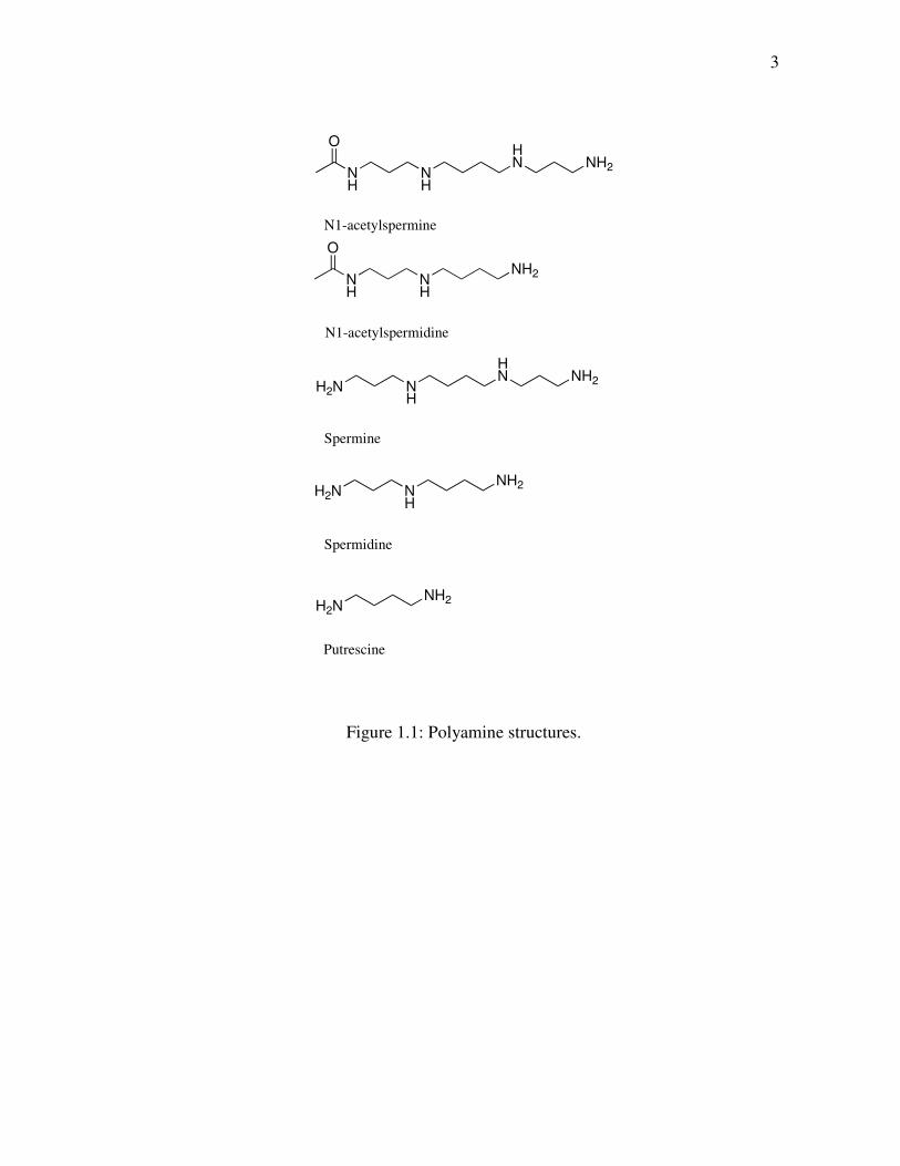
functions. Polyamines include spermine, spermidine, their acetylated derivatives, and
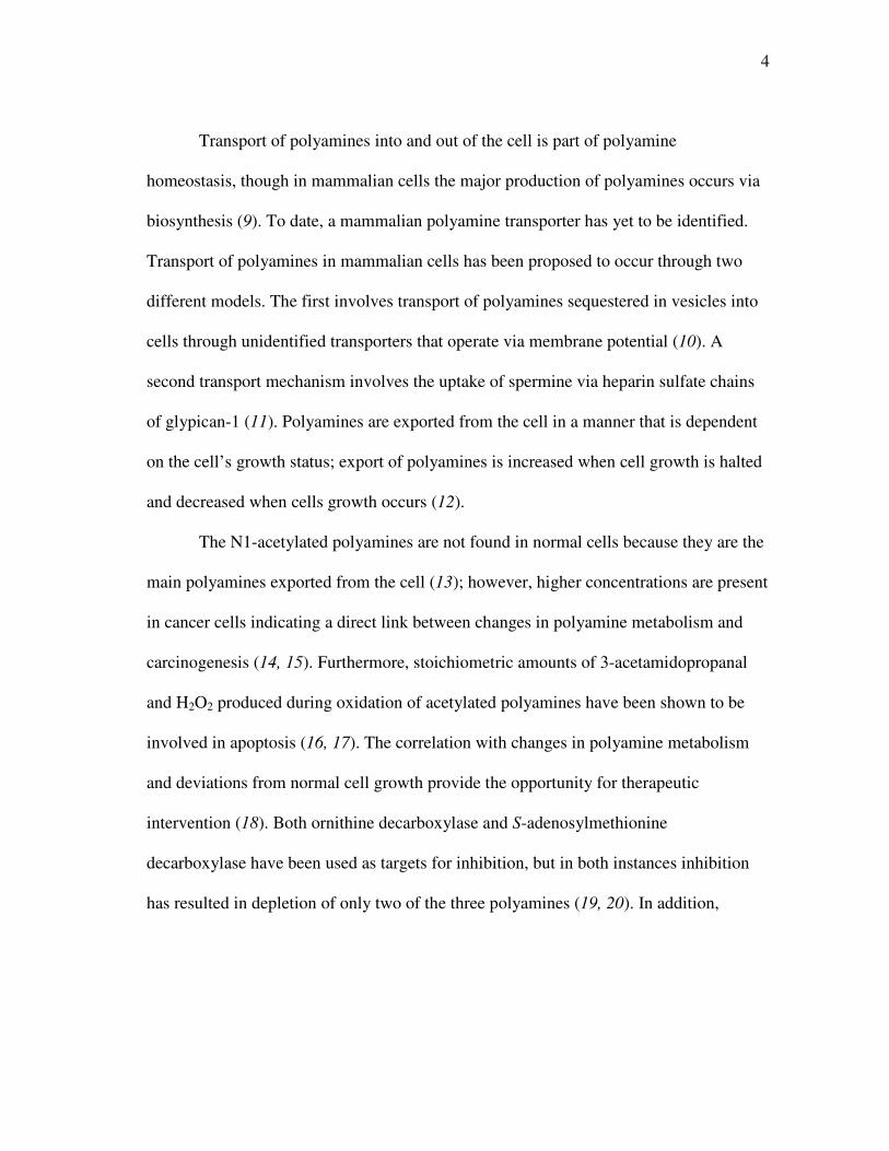
putrescine (Figure 1.1). Polyamine metabolism is a complex system that orchestrates
biosynthesis, degradation and transport (Figure 1.2, modified from Ref (8)). In
eukaryotic cells, the three polyamines spermine, spermidine and putrescine are
synthesized from L-arginine and L-methionine. Putrescine is synthesized from L-
arginine via arginase and ornithine decarboxylase. Putrescine can be converted into
spermidine and spermine by spermidine synthase and spermine synthase, respectively,
using an aminopropyl group from decarboxylated S-adenosylmethionine. The
aminopropyl group is generated from L-methionine in two consecutive reactions
involving methionine adenosyltransferase and S-adenosylmethionine decarboxylase.
Polyamine degradation begins with spermidine/spermine N1-acetyltransferase
(SSAT) using acetyl-CoA to acetylate spermine and spermidine, producing N1-
acetylspermine and N1-acetylspermidine. Polyamine oxidase (PAO) oxidizes N1-
acetylspermine and N1-acetylspermidine to spermidine and putrescine, producing 3-
acetamidopropanal and H2O2 as byproducts. More recently, it was determined that an
enzyme catalyzes the back-conversion of spermine to produce spermidine, 3-
aminopropanal, and H2O2. This enzyme has been named spermine oxidase to distinguish
it from the aforementioned PAO.
3
NH
NH
O
NH2
N1-acetylspermidine
NH
NH
OHN NH2
N1-acetylspermine
H2N NH
HN NH2
Spermine
H2N NH
NH2
Spermidine
H2NNH2
Putrescine
Figure 1.1: Polyamine structures.
4
Transport of polyamines into and out of the cell is part of polyamine
homeostasis, though in mammalian cells the major production of polyamines occurs via
biosynthesis (9). To date, a mammalian polyamine transporter has yet to be identified.
Transport of polyamines in mammalian cells has been proposed to occur through two
different models. The first involves transport of polyamines sequestered in vesicles into
cells through unidentified transporters that operate via membrane potential (10). A
second transport mechanism involves the uptake of spermine via heparin sulfate chains
of glypican-1 (11). Polyamines are exported from the cell in a manner that is dependent
on the cell’s growth status; export of polyamines is increased when cell growth is halted
and decreased when cells growth occurs (12).
The N1-acetylated polyamines are not found in normal cells because they are the
main polyamines exported from the cell (13); however, higher concentrations are present
in cancer cells indicating a direct link between changes in polyamine metabolism and
carcinogenesis (14, 15). Furthermore, stoichiometric amounts of 3-acetamidopropanal
and H2O2 produced during oxidation of acetylated polyamines have been shown to be
involved in apoptosis (16, 17). The correlation with changes in polyamine metabolism
and deviations from normal cell growth provide the opportunity for therapeutic
intervention (18). Both ornithine decarboxylase and S-adenosylmethionine
decarboxylase have been used as targets for inhibition, but in both instances inhibition
has resulted in depletion of only two of the three polyamines (19, 20). In addition,
5
Methionine
S-adenosylmethionine
DecarboxylatedS-adenosylmethionine
Methylthioadenosine
Methylthioadenosine
Arginine
L-Ornithine
Putrescine
Spermidine
Spermine
Intracellular Extracellular
CO2
N1-acetylspermine
N1-acetylspermidine
MethionineAdenosyltransferase
S-adenosylmethionineDecarboxylase
SpermidineSynthase
SpermineSynthase Spermine
Oxidase
Spermidine/Spermine
N1-acetyltransferase
Polyamine Oxidase
Arginase
OrnithineDecarboxylase
Spermidine/Spermine
N1-acetyltransferase
Polyamine Oxidase
Uptake
ExportCO2
Figure 1.2: Polyamine metabolism.
6
both of these enzymes have rapid turnover in mammalian cells (21), making long term
inhibition difficult. Since therapeutically the goal is to deplete polyamine levels within a
cell, SSAT and PAO, which play a direct role in the catabolism of polyamines, are
receiving greater attention in drug design. To date, an inhibitor specific for SSAT has
not been developed, though it has been shown that inhibitors of other enzymes in
polyamine metabolism induce SSAT activity also (22). Inhibitors of PAO have shown to
be promising, with a number of clinical trials underway (23, 24). Studies using a lung
carcinoma cell line suggest that induction of PAO results in increased production of
H2O2 linked with cytotoxicity and eventual apoptosis (25). These results taken
cumulatively show the potential for the use of polyamine analogues as
chemopreventative and anticancer drugs, necessitating the need to better understand the
mechanisms of each of the enzymes involved in polyamine metabolism.
POLYAMINE OXIDASE
PAO contains a non-covalently bound flavin adenine dinucleotide (FAD) and is
one of two enzymes that are involved in polyamine catabolism as previously described.
Despite PAO having been implicated in cancer, ischemic tissue damage and apoptosis,
little attention has been paid to the mechanism of this enzyme, necessitating the need to
do so. The cloning, sequencing and expression of mouse PAO (26) presents the
opportunity to study a mammalian PAO in an effort to gain important mechanistic
information that can be used for better and more efficient drug design.
7
Mammalian PAOs are differentiated from plant PAOs based on the site of CH
bond cleavage. In a mammalian PAO, N1-acetylated polyamines are oxidized on the
exo-side of their N4-amino groups (Figure 1.3) producing an imine-intermediate that is
non-enzymatically hydrolyzed. 3-Acetamidopropanal is formed, which can be
enzymatically deacetylated to produce cytotoxic 3-aminopropanal (27). Mouse PAO
exhibits a preference for polyamine substrates in the order N1-acetyl spermine ≈ N1-
acetyl spermidine >> N1,N12-diacetyl spermine >> spermine (28). With plant PAOs, the
carbon on the endo-side of the N4-nitrogens of spermidine and spermine is oxidized,
producing 4-aminobutyraldehyde and 3-(aminopropyl)-4-aminobutyraldehyde,
respectively.
The difference in substrate specificity of enzymes involved in polyamine
metabolism raises questions about nomenclature to differentiate between such enzymes.
Specifically, PAO’s preference for N1-acetylated polyamines distinguishes it from an
amine oxidase that catalyzes the oxidation of spermine, more accurately defining the
latter as a spermine oxidase. From this perspective, it could be argued that the preference
for plant PAO`s to oxidize spermine and spermidine over their acetylated derivatives
would more accurately classify these enzymes as being spermine oxidases.
8
HN
O
H2N
NH2
NH3
O2
H2O2
HN
O
HN
NH2
NH3
+ H3NNH2
NH3
N1-acetylspermine
spermidine
HN
O
O
Flox
Flred
Figure 1.3: PAO oxidation of N1-acetylspermine.
9
FLAVOPROTEIN AMINE OXIDASES
PAO is a member of a family of enzymes known as flavin amine oxidases. The
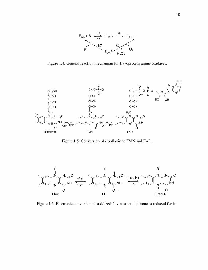
overall reaction of these enzymes can be divided into two half-reactions (Figure 1.4).
The reductive half-reaction is characterized by the reduction of the flavin cofactor upon
the oxidation of substrate. The oxidative half-reaction involves the oxidation of enzyme
with reduced FAD by molecular oxygen, producing H2O2 and an imine intermediate that
is non-enzymatically hydrolyzed.
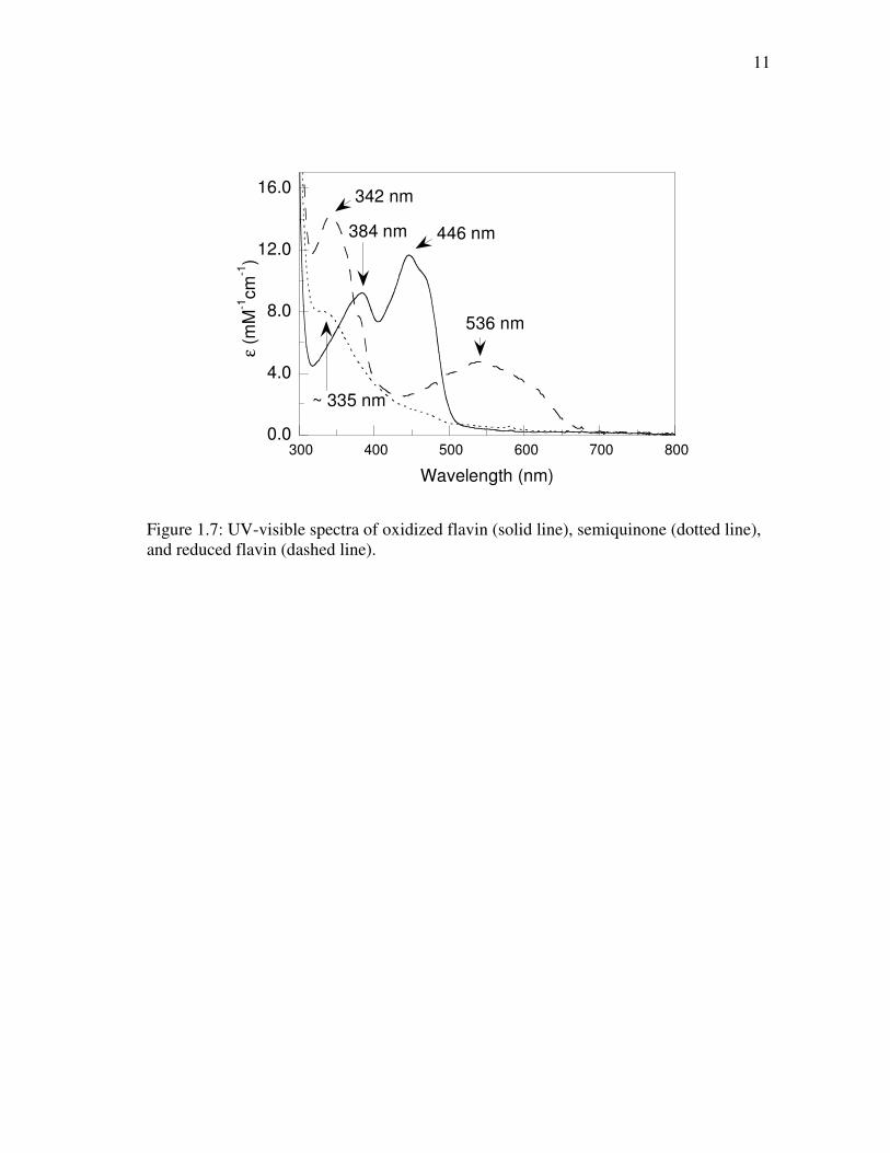
The flavin cofactor exists in three forms. The first form is riboflavin (vitamin
B2), which can be converted to the other two forms flavin mononucleotide (FMN) and
flavin adenine dinucleotide (FAD) as shown in Figure 1.5 (modified from ref (29)).
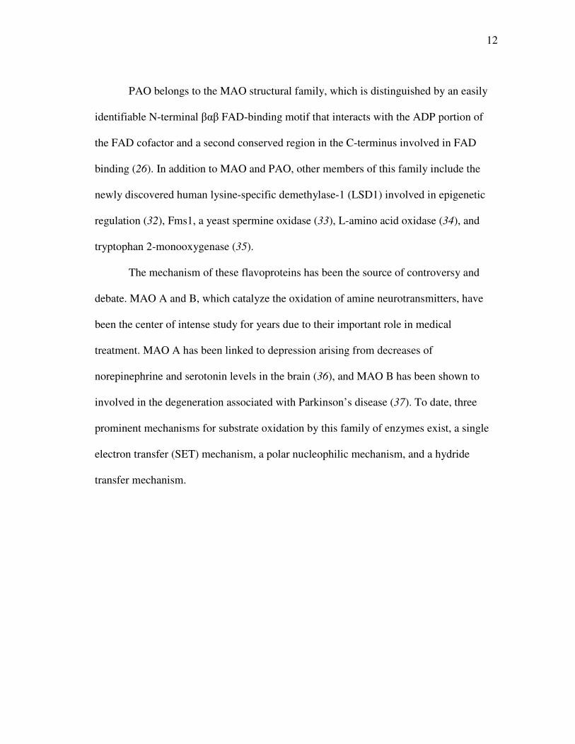
Flavins possess a highly conjugated ring system that gives them a chemical reactivity
that allows them to accept one electron, forming flavin semiquinone, or two electrons to
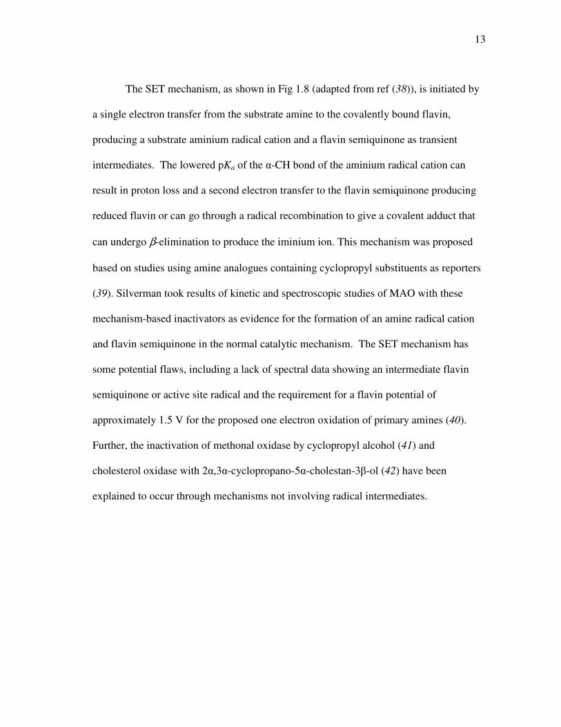
form reduced flavin (Figure 1.6, modified from ref (29)). Figure 1.7 shows
representative UV-visible absorbance spectra for the oxidized, semiquinone, and reduced
states of flavin, with indicated wavelengths of maximum absorbance in each state (30).
Flavin oxidation potentials are in the range of 0 to -200 mV, though -500 mV has been
observed in flavodoxins (29). Flavins can be non-covalently bound in flavoenzymes, as
is the case with PAO (26), or covalently bound as with MAO, which occurs via a
thioether linkages between a cysteinyl residue and the 8α-methylene of the isoalloxazine
ring (31).
10
EOX + S EOXSk1
k2
k3EREDP
EOXPO2
H2O2
k5k7
P
Figure 1.4: General reaction mechanism for flavoprotein amine oxidases.
N
N
NH
N8a
4a
O
O
CH2
ATP ADP N
N
NH
N O
O
CH2
ATP PPi N
N
NH
N O
O
H2C
P
O
O
O P
O
O
O O
OHHO
N
N
N
N
NH2
Riboflavin FMN FAD
CHOH
CHOH
CHOH
CH2OH
CHOH
CHOH
CHOH
CHOH
CHOH
CHOH
CH2OP
O
O
OCH2O
Figure 1.5: Conversion of riboflavin to FMN and FAD.
N
N
NH
N O
O
R
Flox
N
N
NH
HN O
O
R
Fl
+1e-
-1e- NH
N
NH
N O
O
R
FlredH-
+1e-, H+
-1e-
Figure 1.6: Electronic conversion of oxidized flavin to semiquinone to reduced flavin.
11
0.0
4.0
8.0
12.0
16.0
300 400 500 600 700 800
ε (m
M-1
cm
-1)
Wavelength (nm)
536 nm
446 nm384 nm
342 nm
~ 335 nm
Figure 1.7: UV-visible spectra of oxidized flavin (solid line), semiquinone (dotted line), and reduced flavin (dashed line).
12
PAO belongs to the MAO structural family, which is distinguished by an easily
identifiable N-terminal βαβ FAD-binding motif that interacts with the ADP portion of
the FAD cofactor and a second conserved region in the C-terminus involved in FAD
binding (26). In addition to MAO and PAO, other members of this family include the
newly discovered human lysine-specific demethylase-1 (LSD1) involved in epigenetic
regulation (32), Fms1, a yeast spermine oxidase (33), L-amino acid oxidase (34), and
tryptophan 2-monooxygenase (35).
The mechanism of these flavoproteins has been the source of controversy and
debate. MAO A and B, which catalyze the oxidation of amine neurotransmitters, have
been the center of intense study for years due to their important role in medical
treatment. MAO A has been linked to depression arising from decreases of
norepinephrine and serotonin levels in the brain (36), and MAO B has been shown to
involved in the degeneration associated with Parkinson’s disease (37). To date, three
prominent mechanisms for substrate oxidation by this family of enzymes exist, a single
electron transfer (SET) mechanism, a polar nucleophilic mechanism, and a hydride
transfer mechanism.
13
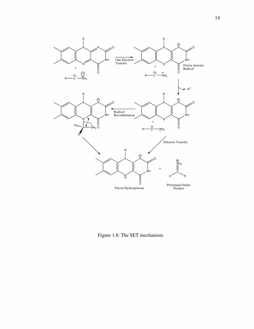
The SET mechanism, as shown in Fig 1.8 (adapted from ref (38)), is initiated by
a single electron transfer from the substrate amine to the covalently bound flavin,
producing a substrate aminium radical cation and a flavin semiquinone as transient
intermediates. The lowered pKa of the α-CH bond of the aminium radical cation can
result in proton loss and a second electron transfer to the flavin semiquinone producing
reduced flavin or can go through a radical recombination to give a covalent adduct that
can undergo β-elimination to produce the iminium ion. This mechanism was proposed
based on studies using amine analogues containing cyclopropyl substituents as reporters
(39). Silverman took results of kinetic and spectroscopic studies of MAO with these
mechanism-based inactivators as evidence for the formation of an amine radical cation
and flavin semiquinone in the normal catalytic mechanism. The SET mechanism has
some potential flaws, including a lack of spectral data showing an intermediate flavin
semiquinone or active site radical and the requirement for a flavin potential of
approximately 1.5 V for the proposed one electron oxidation of primary amines (40).
Further, the inactivation of methonal oxidase by cyclopropyl alcohol (41) and
cholesterol oxidase with 2α,3α-cyclopropano-5α-cholestan-3β-ol (42) have been
explained to occur through mechanisms not involving radical intermediates.
14
Figure 1.8: The SET mechanism.
N
N
NH
N O
O
R
RH2C NH2
N
N
NH
N O
O
R
One ElectronTransfer
RH2C NH2
Flavin AnionicRadical
Base Abstraction
H
N
N
NH
N O
O
R
RHC NH2
RadicalRecombination
N
N
NH
N O
O
R
NH2
R
H
NH
N
NH
N O
O
R
Electron Transfer
NH2
C
H R
Flavin HydroquinoneProtonated Imine
Product
+ +
+
+
15
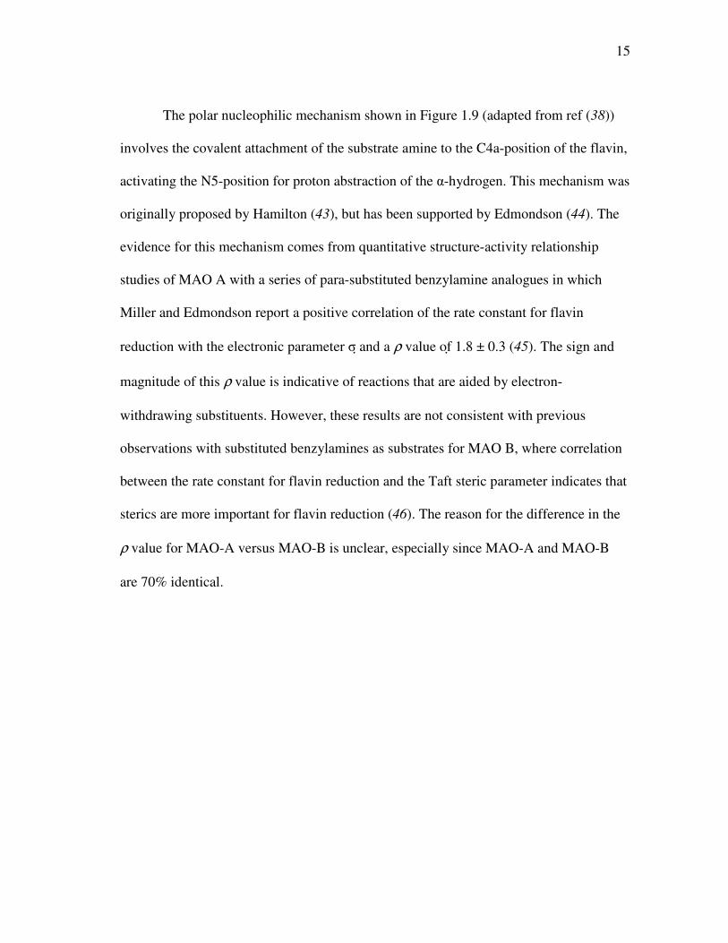
The polar nucleophilic mechanism shown in Figure 1.9 (adapted from ref (38))
involves the covalent attachment of the substrate amine to the C4a-position of the flavin,
activating the N5-position for proton abstraction of the α-hydrogen. This mechanism was
originally proposed by Hamilton (43), but has been supported by Edmondson (44). The
evidence for this mechanism comes from quantitative structure-activity relationship
studies of MAO A with a series of para-substituted benzylamine analogues in which
Miller and Edmondson report a positive correlation of the rate constant for flavin
reduction with the electronic parameter σ and a ρ value of 1.8 ± 0.3 (45). The sign and
magnitude of this ρ value is indicative of reactions that are aided by electron-
withdrawing substituents. However, these results are not consistent with previous
observations with substituted benzylamines as substrates for MAO B, where correlation
between the rate constant for flavin reduction and the Taft steric parameter indicates that
sterics are more important for flavin reduction (46). The reason for the difference in the
ρ value for MAO-A versus MAO-B is unclear, especially since MAO-A and MAO-B
are 70% identical.
16
Figure 1.9: The polar nucleophilic mechanism.
N
N
N H
N O
O
R
H 3 C
H 2 C
S
E n z
N H 2 C
R
H H
N H
N
N H
N O
O
R
H 3 C
H 2 C
S
E n z
N H 2 C H
R
N H
N
N H
N O
O
R
H 3 C
H 2 C
S
E n z
+ N H 2 C
H R
17
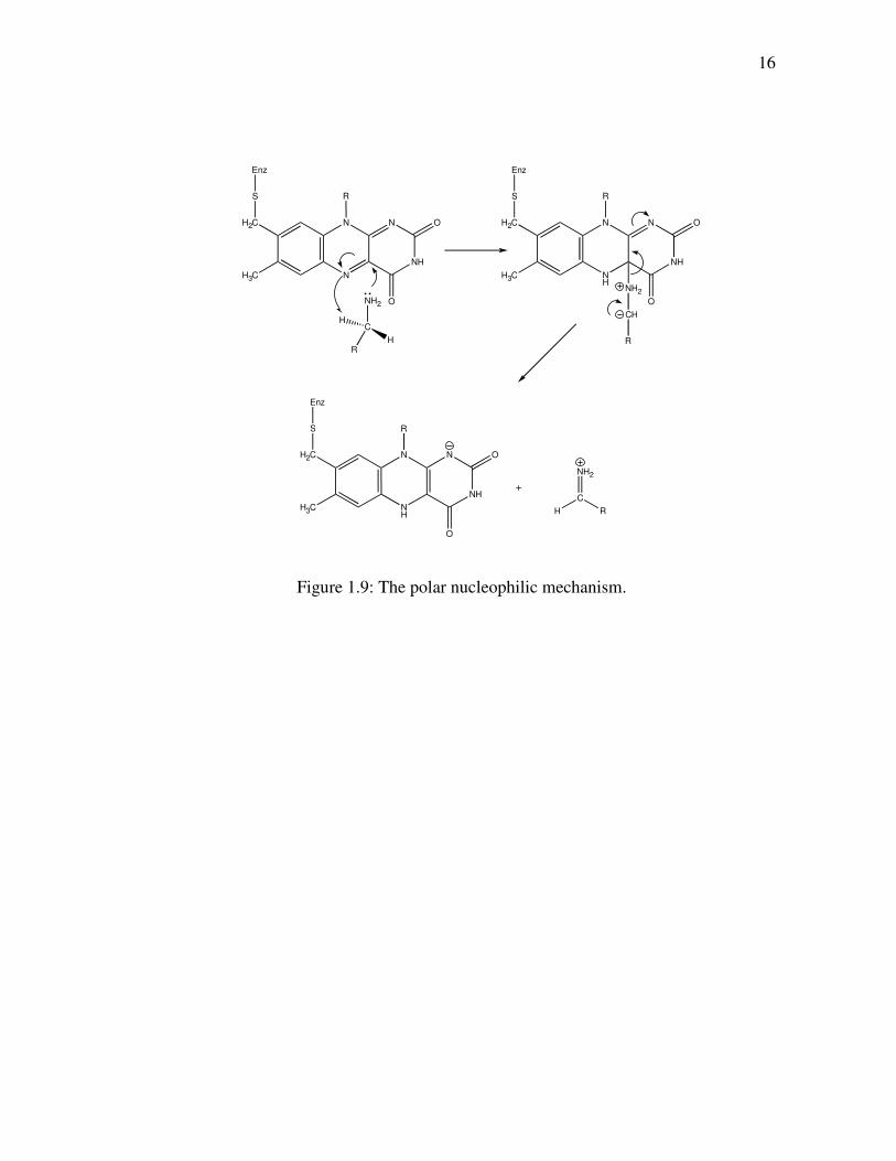
The hydride transfer mechanism is the generally accepted mechanism of the
flavoprotein amine oxidase D-amino acid oxidase (DAAO), which has a different
structure than the MAO structural family. This mechanism, as shown in Figure 1.10,
involves the direct transfer of a hydride equivalent from the substrate to the flavin
cofactor with no intermediate steps. This mechanism has received greater attention in
recent years as the mechanism of flavoprotein amine oxidases in light of new kinetic
data. Deuterium and 15N kinetic isotope effect studies of flavin amine oxidases,
including DAAO (47, 48), tryptophan-2-monooxygenase (TMO) (49), and N-
methyltryptophan oxidase (MTOX) (50, 51), are most consistent with a hydride transfer
mechanism. Furthermore, these results indicate that a common kinetic mechanism is
shared between the DAAO structural family, which includes DAAO (52) and MTOX
(50), and the MAO structural family that includes TMO (53).
Figure 1.10: The hydride transfer mechanism.
R CH
NH2
+
H
NH
N
NH
N O
O
R
NH2
C
H R
+
N
N
NH
N O
O
R
18
PAO presents the opportunity to study a flavin amine oxidase in an effort to
determine the chemical mechanism, assess the potential importance of residues within
the active site, and to gain insight into substrate specificity. Results of this study with a
mammalian PAO can serve to offer structural and mechanistic information that can be
utilized in drug design for anti-cancer agents targeted for PAO.
19
CHAPTER II
pH DEPENDENCE OF A MAMMALIAN POLYAMINE OXIDASE: INSIGHTS
INTO SUBSTRATE SPECIFICITY AND THE ROLE OF LYSINE 315∗∗∗∗
The polyamines spermine and spermidine are essential for cell proliferation, with
higher levels being found in rapidly growing cells (4, 54). This observation suggests that
compounds which decrease the levels of polyamines in cells have potential as
antineoplastic agents. Indeed, the polyamine biosynthetic pathway has been heavily
studied with the goal of developing enzyme inhibitors (24, 54, 55). The pathway begins
with the formation of putrescine from ornithine catalyzed by ornithine decarboxylase.
Putrescine is then converted to spermine by two sequential reactions catalyzed by
spermidine synthase, forming first spermidine and then spermine, using decarboxylated
S-adenosylmethionine as the propylamine donor in both steps. In the opposite direction,
catabolism of spermine requires the sequential action of two enzymes (4). First,
acetylation of spermine by spermidine/spermine N1-acetylspermine acetyltransferase
forms N1-acetylspermine. This is then converted to spermidine by the flavoenzyme
polyamine oxidase (PAO). The same two enzymes also catalyze the acetylation of
spermidine to N1-acetylspermidine and the subsequent oxidation to putrescine. Very
recently, several mammalian tissues have been found to contain a flavoenzyme capable
of oxidizing spermine directly to spermidine (56-58); while referred to occasionally as
PAO, it is more accurately a spermine oxidase. ∗ Reproduced with permission from Henderson Pozzi, M., Gawandi, V., and Fitzpatrick, P.F. (2009) Biochemistry 48, 1508-1516. Copyright 2009 American Chemical Society.
20
The reaction of mammalian PAO is shown in Figure 1.3. The enzyme cleaves the
exo carbon-hydrogen bond of its substrate, forming spermidine and N-acetyl-3-
aminopropanaldehyde from N1-acetylspermine or putrescine and N-acetyl-3-
aminopropanaldehyde from N1-acetylspermidine. There are also plant PAOs, of which
the maize enzyme is the best-characterized (59-62). While the mammalian enzymes
oxidize spermine to 3-aminopropanaldehyde and spermidine, the plant enzymes oxidize
the endo bond of spermine to form propane-1,3-diamine and N-(3-aminopropyl)-4-
aminobutyraldehyde (63). The structural bases for the difference in substrate specificity
between polyamine and spermine oxidases and in the site of substrate oxidation between
the plant and animal enzymes are not known.
The general reaction of flavin amine oxidases such as PAO can be divided into
two half reactions. In the reductive half reaction a hydride equivalent is transferred from
the substrate to the flavin, while the oxidative half reaction involves the oxidation of the
reduced flavin by molecular oxygen, producing H2O2. The steady-state kinetic
mechanism has previously been determined for mouse PAO (26). Consistent with the
results for most flavoprotein oxidases (64), the kinetic pattern is ping-pong due to the
reductive half reaction being effectively irreversible. Consequently, the kcat/Km value for
the amine substrate includes the steps in the reductive half-reaction from amine binding
through flavin reduction, while the kcat/Km value for oxygen is the second order rate
constant for reoxidation of the reduced flavin. This simplifies analysis of the individual
kinetic parameters, since the kcat/Km value for the amine substrate is independent of the
21
oxygen concentration, while the rate constant for flavin reduction can readily be
determined using rapid-reaction methods in the absence of oxygen.
The chemical mechanism of the reductive half-reaction of flavoprotein amine
oxidases has been quite controversial (65). Oxidation of an amine substrate by an amine
oxidase necessarily involves the removal of two protons and two electrons as the carbon-
nitrogen single bond is converted to a double bond. The various mechanistic proposals
for the flavin amine oxidases have included most of the possible combinations by which
this can occur (65, 66). Cleavage of the carbon-hydrogen bond could occur by removal
of the hydrogen as a proton, a hydrogen atom, or a hydride (44, 67, 68), with some
mechanisms involving formation of an amine-flavin adduct as an intermediate (44). In
contrast, the hydrogen is generally proposed to be removed from the nitrogen as a
proton, with the disagreement over when this occurs in the reaction. Thus, the proton
could be lost to solvent before the amine binds to the protein (48) or to an active site
base either before (69-71) or concurrent with cleavage of the carbon-hydrogen bond
(69). Thus, establishing the catalytic mechanism of an amine oxidase necessarily
requires knowledge of the timing of removal of hydrogens from both the carbon and the
nitrogen. In addition, in the case of the proton on the nitrogen, loss of the proton from
the bound substrate would require an active site base.
The flavin amine oxidases can be divided into two structural classes, the
MAO/PAO family (34, 60) and the D-amino acid oxidase (DAAO)/sarcosine oxidase
family (72). No structure of a mammalian PAO has been described to date. However,
structures are available for maize PAO and for S. cerevisiae spermine oxidase (Fms1)
22
(73, 74). These enzymes both belong to the monoamine oxidase (MAO) family of
flavoprotein amine oxidases (73). The sequences of mammalian PAOs align well with
these and other members of this family (26, 73, 75). The available structures of members
of the MAO family show that all contain a conserved lysyl residue in the active site (32,
34, 73, 74, 76). This residue is part of a “Lys-H2O-N5” motif in which the lysyl side
chain forms hydrogen bonds to the N5 of the isoalloxazine ring via an intervening water
molecule. This lysyl residue has been proposed to be an active site base which accepts a
proton from either the protonated amine of the substrate prior to its oxidation or from a
water molecule to form hydroxide for hydrolysis of an imine intermediate (34, 59).
This chapter describes the use of the effects of pH on the steady-state and
reductive half-reaction kinetics of mouse PAO to probe substrate specificity and
establish the protonation states of polyamines required for catalysis. In addition, the role
the conserved lysyl residue plays in amine oxidation by this enzyme has been analyzed.
23
EXPERIMENTAL PROCEDURES
Materials. Spermine was purchased from Acros Organics (Geel, Belgium), and
1,8-diaminooctane and 1,12-diaminododecane were purchased from Aldrich
(Milwaukee, WI). N1-Acetylspermine and N1-acetylspermidine were synthesized as
previously described (77); N1-acetylspermidine was also purchased from Fluka
(Switzerland). Substrates were synthesized by Dr. Vijay Gawandi of Texas A&M
University (78).
Expression and Purification of Recombinant Proteins. Mouse PAO was purified
as previously described (79) with a few minor changes. The pellet resulting from the
final 65% ammonium sulfate precipitation was resuspended in 50 mM potassium
phosphate and 10% glycerol (pH 7.5) and dialyzed overnight with two buffer changes.
The resulting protein sample was then centrifuged at 22,400xg for 30 min at 4 ºC to
remove precipitated protein. The purified protein was stored at -80 ºC. The concentration
of active enzyme was determined from the flavin visible absorbance spectrum, using an
ε458 value of 10,400 M-1 cm-1.
The K315M mutation was introduced using the Stratagene QuikChange site-
directed mutagenesis method and the mutagenic primer 5’-
GGCTTCGGTACCAACAACATGATCTTCCTCGAGTTC -3’, which contains the
K315M mutation (shown in bold) and a silent mutation at Leu318 that results in the
addition of an AvaI site (underlined) used in screening colonies. The DNA sequence of
the entire gene was verified to ensure that no unwanted mutations occurred. Purification
of the mutant enzyme was done using the same procedure as for wild type PAO.
24
Assays. Steady state kinetic assays were performed in air-saturated buffers on a
computer-interfaced Hansatech (Hansatech Instruments) or YSI oxygen (Yellow Springs
Instrument, Inc.) electrode. Assays were initiated by the addition of enzyme. All buffers
contained 10% glycerol; 50 mM Tris-HCl, 50 mM CHES, and 50 mM CAPS were used
for the pH ranges 7-8.5, 9.0-9.5, or 10-11 respectively.
Rapid-reaction kinetic experiments were conducted at 20 ºC on an Applied
Photophysics SX-18MV stopped-flow spectrophotometer. The night before the
experiment, the instrument was flushed with anaerobic buffer followed by a solution of
36 nM glucose oxidase in 5 mM glucose, 50 mM Tris-HCl, pH 7.5. For enzyme
solutions, anaerobic conditions were established by applying cycles of vacuum and
argon, while substrate solutions were bubbled with argon. All buffers contained 10%
glycerol and 5 mM glucose; 200 mM PIPES, 200 mM Tris-HCl, and 200 mM CHES
were used for the pH ranges 6.5-6.9, 7-8.9, or 9.0-9.5, respectively. Glucose oxidase was
added to all anaerobic solutions at a final concentration of 36 nM before loading them
onto the stopped-flow spectrophotometer.
Data Analysis. Kinetic data were analyzed using the programs KaleidaGraph
(Adelbeck Software, Reading, PA) and Igor (WaveMetrics, Lake Oswego, OR). Initial
rate data obtained by varying the concentration of a single substrate were fit to the
Michaelis-Menten equation. The effects of pH on kinetic parameters were fit to
equations 2.1-2.3. Equation 2.1 applies for a kinetic parameter which decreases below
pK1 due to the protonation of a single moiety. Equation 2.2 applies for a kinetic
parameter which decreases above pK2 due to the protonation of a single moiety.
25
Equation 2.3 applies for a kinetic parameter which decreases below pK1 due to
protonation of a single moiety and decreases above pK2 due to deprotonation of a single
moiety. In all three equations, c is the pH-independent value. In each, y is the kinetic
parameter being measured, c is the pH-independent value, K1 is the ionization constant
for a residue which must be unprotonated, and K2 is the ionization constant for a residue
which must be protonated.
log y = log (c/(1 + H/K1)) (2.1)
log y = log (c/(1 + K2/H)) (2.2)
log y = log (c/(1 + H/K1 + K2/H)) (2.3)
Analysis of stopped-flow data was done using both KaleidaGraph and SPECFIT
(Spectrum Software Associates, Marlborough, MA). To determine the kinetic
parameters for the reduction of wild type PAO by N1-acetylspermine, stopped-flow
traces were fit to equation 2.4, which describes a triphasic exponential decay, where k1,
k2, and k3 are first order rate constants, A1, A2, A3 correspond to the absorbance changes
in each phase, and A∞ is the final absorbance. Equation 2.5 was used to fit the biphasic
traces obtained for K315M PAO.
At = A1e-k1t + A2e
-k2t + A3e-k3t + A∞ (2.4)
At = A1e-k1t + A2e
-k2t+ A∞ (2.5)
26
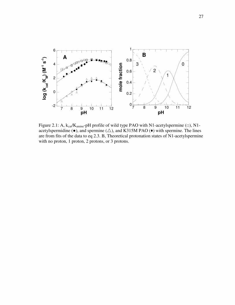
RESULTS
kcat/Kamine- pH Profile. Previous steady-state kinetic studies at pH 7.6 indicated
that the substrate preference for mouse PAO is N1-acetylspermine, N1-acetylspermidine,
and then spermine, with spermine being a significantly slower substrate than the
acetylated compounds (26). The effect of pH on the kinetic parameter kcat/Kamine was
determined for each of these three substrates. The results are shown in Figure 2.1A, and
the resulting pKa values are summarized in Table 2.1. The pH profiles for all three
substrates exhibit decreases in activity at both low and high pH, consistent with a
requirement for one moiety in the enzyme or substrate that must be protonated for
substrate recognition and/or oxidation and one which must be unprotonated. Both
acetylated substrates have bell-shaped curves with two distinguishable pKa values. In
contrast, with spermine the pH profile exhibits a sharp optimum so that the two pKa
values are too close together to resolve; consequently, only the average of the two pKa
values could be determined with this substrate.
27
-2
0
2
4
6
7 8 9 10 11 12
A
log
(k
ca
t/KM)
(M-1
s-1
)
pH
0
0.2
0.4
0.6
0.8
1
7 8 9 10 11 12
B
mo
le f
rac
tio
n
pH
1
0
2
3
Figure 2.1: A, kcat/Kamine-pH profile of wild type PAO with N1-acetylspermine (□), N1-acetylspermidine (●), and spermine (�), and K315M PAO (♦) with spermine. The lines are from fits of the data to eq 2.3. B, Theoretical protonation states of N1-acetylspermine with no proton, 1 proton, 2 protons, or 3 protons.
28
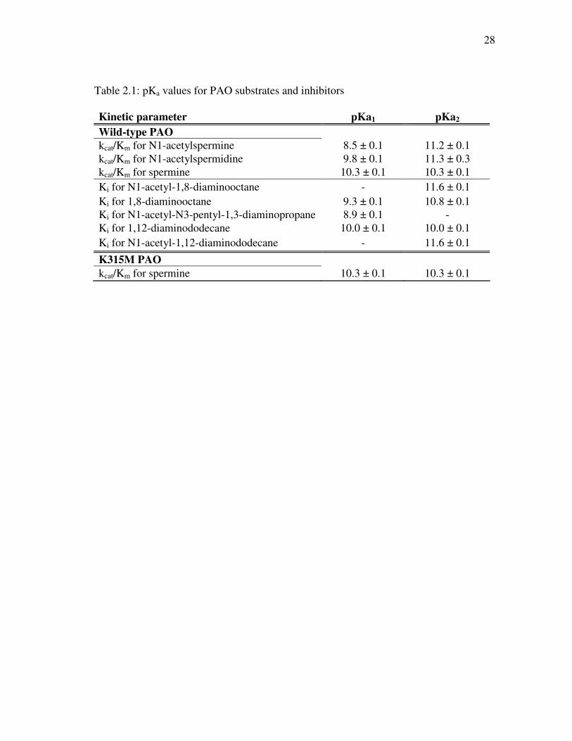
Table 2.1: pKa values for PAO substrates and inhibitors
Kinetic parameter pKa1 pKa2
Wild-type PAO
kcat/Km for N1-acetylspermine 8.5 ± 0.1 11.2 ± 0.1 kcat/Km for N1-acetylspermidine 9.8 ± 0.1 11.3 ± 0.3 kcat/Km for spermine 10.3 ± 0.1 10.3 ± 0.1 Ki for N1-acetyl-1,8-diaminooctane - 11.6 ± 0.1 Ki for 1,8-diaminooctane 9.3 ± 0.1 10.8 ± 0.1 Ki for N1-acetyl-N3-pentyl-1,3-diaminopropane 8.9 ± 0.1 - Ki for 1,12-diaminododecane 10.0 ± 0.1 10.0 ± 0.1 Ki for N1-acetyl-1,12-diaminododecane - 11.6 ± 0.1
K315M PAO kcat/Km for spermine 10.3 ± 0.1 10.3 ± 0.1
29
Protonation States of Polyamines as a Function of pH. A likely basis for one or
both of the pKa values in the kcat/Kamine-pH profiles is the protonation state of the
substrates. Figure 2.1B shows the effect of pH on the mole fractions of N1-
acetylspermine with zero, one, two, or three protons (78). The monoprotonated form is
maximal at pH 10.1, in good agreement with the pH optimum in the kcat/Kamine-pH
profile of 9.9, while the diprotonated form is maximal at pH 9.0. For spermine the pH
maximum for the monoprotonated form is 10.4-10.5 (78, 80, 81), similarly closer to the
pH optima of 10.3 than is the maximum for the diprotonated form of 9.4-9.5. The
agreement with N1-acetylspermidine is not as good, in that the optimum in the
kcat/Kamine-pH profile is 10.5 while the calculated maximum for the monoprotonated
form is 9.9 (78). However, a requirement for the diprotonated form of N1-
acetylspermidine would contribute a single pKa of 9.1 for a group which must be
protonated to the kcat/Kamine-pH profile, while a requirement for the uncharged form
would contribute a single pKa value of 10.6 (78), in contrast to the observed bell-shaped
profile. Thus, the kcat/Kamine-pH profiles are most consistent with the active form of the
substrate having a single charge.
PAO pH Dependence of Inhibition. While the kcat/Kamine-pH profiles are
consistent with the monoprotonated forms of the substrate being preferred, they do not
establish which nitrogen in each substrate must be charged. In addition, the kcat/Kamine-
pH profiles reflect both binding and catalysis. To determine the preferred protonation
states of individual nitrogens in substrates for productive binding, analogues lacking one
or more nitrogens (Figure 2.2) were characterized as inhibitors. 1,8-Diaminooctane, N1-
30
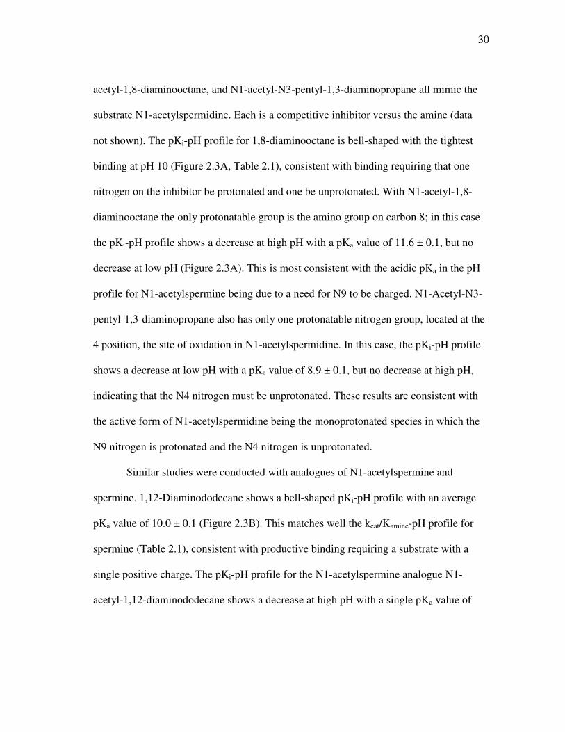
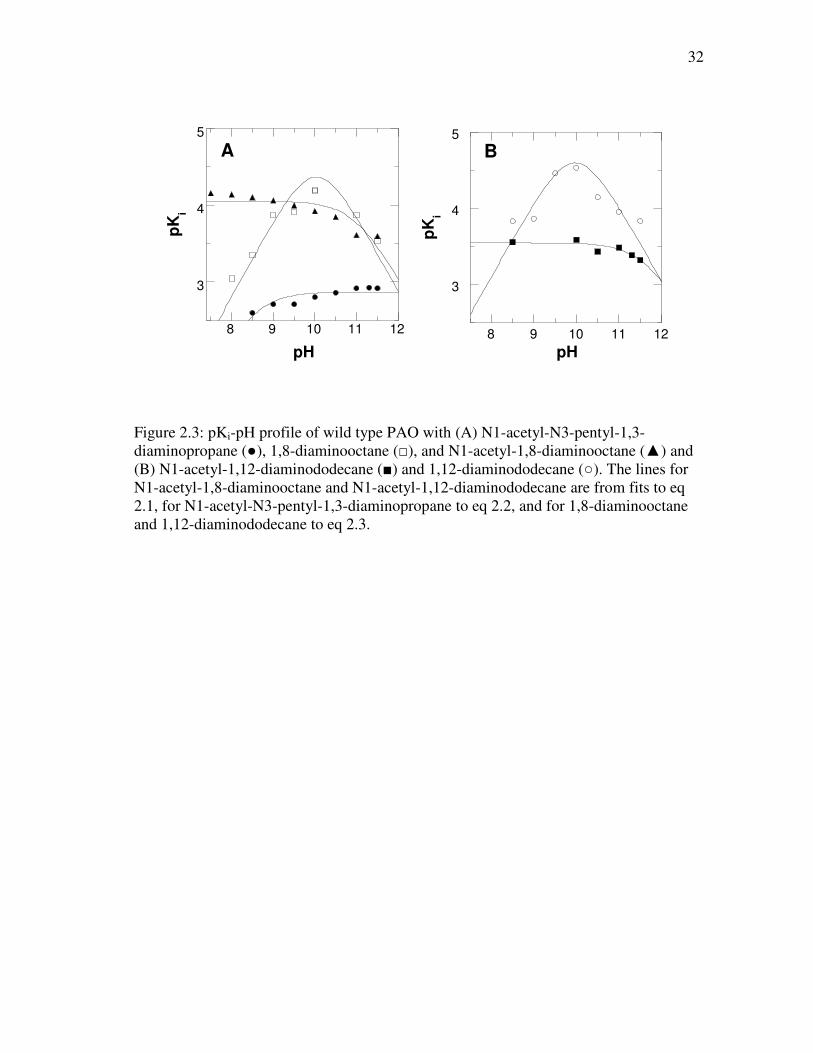
acetyl-1,8-diaminooctane, and N1-acetyl-N3-pentyl-1,3-diaminopropane all mimic the
substrate N1-acetylspermidine. Each is a competitive inhibitor versus the amine (data
not shown). The pKi-pH profile for 1,8-diaminooctane is bell-shaped with the tightest
binding at pH 10 (Figure 2.3A, Table 2.1), consistent with binding requiring that one
nitrogen on the inhibitor be protonated and one be unprotonated. With N1-acetyl-1,8-
diaminooctane the only protonatable group is the amino group on carbon 8; in this case
the pKi-pH profile shows a decrease at high pH with a pKa value of 11.6 ± 0.1, but no
decrease at low pH (Figure 2.3A). This is most consistent with the acidic pKa in the pH
profile for N1-acetylspermine being due to a need for N9 to be charged. N1-Acetyl-N3-
pentyl-1,3-diaminopropane also has only one protonatable nitrogen group, located at the
4 position, the site of oxidation in N1-acetylspermidine. In this case, the pKi-pH profile
shows a decrease at low pH with a pKa value of 8.9 ± 0.1, but no decrease at high pH,
indicating that the N4 nitrogen must be unprotonated. These results are consistent with
the active form of N1-acetylspermidine being the monoprotonated species in which the
N9 nitrogen is protonated and the N4 nitrogen is unprotonated.
Similar studies were conducted with analogues of N1-acetylspermine and
spermine. 1,12-Diaminododecane shows a bell-shaped pKi-pH profile with an average
pKa value of 10.0 ± 0.1 (Figure 2.3B). This matches well the kcat/Kamine-pH profile for
spermine (Table 2.1), consistent with productive binding requiring a substrate with a
single positive charge. The pKi-pH profile for the N1-acetylspermine analogue N1-
acetyl-1,12-diaminododecane shows a decrease at high pH with a single pKa value of
31
11.6 ± 0.1 (Figure 2.3B), confirming that a nitrogen not located next to the site of CH
bond cleavage must be protonated for catalysis.
NH
NH2
O
NH
NH
O
NH
O
NH2
H2NNH2
1,8-diaminooctane
N1-acetyl-1,8-diaminooctane N1-acetyl-1,12-diaminododecane
H2NNH2
1,12-diaminododecane
N1-acetyl-N3-pentyl-1,3-diaminopropane
NH
NH
O
NH2
N1-acetylspermidine
NH
NH
OHN NH2
N1-acetylspermine
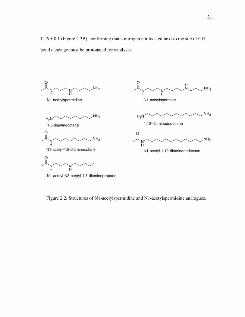
Figure 2.2: Structures of N1-acetylspermidine and N1-acetylspermidine analogues.
32
3
4
5
8 9 10 11 12
pK
i
pH
A
3
4
5
8 9 10 11 12
pK
i
pH
B
Figure 2.3: pKi-pH profile of wild type PAO with (A) N1-acetyl-N3-pentyl-1,3-diaminopropane (●), 1,8-diaminooctane (□), and N1-acetyl-1,8-diaminooctane (▲) and (B) N1-acetyl-1,12-diaminododecane (■) and 1,12-diaminododecane (○). The lines for N1-acetyl-1,8-diaminooctane and N1-acetyl-1,12-diaminododecane are from fits to eq 2.1, for N1-acetyl-N3-pentyl-1,3-diaminopropane to eq 2.2, and for 1,8-diaminooctane and 1,12-diaminododecane to eq 2.3.
33
pH Dependence of Flavin Reduction. To address the effect of pH on catalysis
rather than binding, stopped-flow spectroscopy was used to determine the rate constant
for flavin reduction by N1-acetylspermine as a function of pH. Reactions were carried
out at 20 ºC instead of 30 ºC because much of the reaction occurred in the dead time of
the instrument at the higher temperature. Over the pH range 6.5-9.5, the flavin
absorbance at 458 nm showed the same behavior: an initial decrease in absorbance, a
slower, slight increase in absorbance, and finally a slow decrease in absorbance (Figure
2.4A). Data could not be obtained at pH 10 and above due to enzyme instability. The
same kinetic behavior was seen when the reaction was monitored from 320-600 nm by
photodiode array spectroscopy; this approach also allowed the spectra of the
intermediates to be obtained (Figure 2.4B). The initial fast phase of the reaction accounts
for the majority of the change in amplitude and has a rate constant that is dependent on
substrate concentration (Figure 2.4C). This phase can be attributed to the rapid and
reversible binding of N1-acetylspermine with no detectable change in the flavin
spectrum, followed by flavin reduction. The slowest two rate constants are independent
of substrate concentration and slower than kcat (Figure 2.4C); therefore, they are not
relevant to catalysis.
The effect of pH on the rate constant for reduction of PAO at saturating
concentrations of N1-acetylspermine (kred) is shown in Figure 2.5. The value of this
kinetic parameter shows a decrease at acidic pH with a pKa of 7.3 ± 0.1, indicating that a
group in the ES complex must be unprotonated for reduction. Flavin reduction is
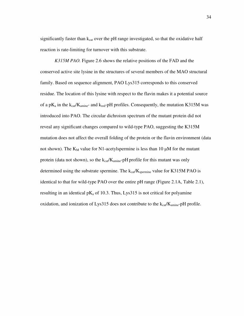
34
significantly faster than kcat over the pH range investigated, so that the oxidative half
reaction is rate-limiting for turnover with this substrate.
K315M PAO. Figure 2.6 shows the relative positions of the FAD and the
conserved active site lysine in the structures of several members of the MAO structural
family. Based on sequence alignment, PAO Lys315 corresponds to this conserved
residue. The location of this lysine with respect to the flavin makes it a potential source
of a pKa in the kcat/Kamine- and kred-pH profiles. Consequently, the mutation K315M was
introduced into PAO. The circular dichroism spectrum of the mutant protein did not
reveal any significant changes compared to wild-type PAO, suggesting the K315M
mutation does not affect the overall folding of the protein or the flavin environment (data
not shown). The KM value for N1-acetylspermine is less than 10 µM for the mutant
protein (data not shown), so the kcat/Kamine-pH profile for this mutant was only
determined using the substrate spermine. The kcat/Kspermine value for K315M PAO is
identical to that for wild-type PAO over the entire pH range (Figure 2.1A, Table 2.1),
resulting in an identical pKa of 10.3. Thus, Lys315 is not critical for polyamine
oxidation, and ionization of Lys315 does not contribute to the kcat/Kamine-pH profile.
35
0
0.05
0.1
0.15
0.01 0.1 1 10
AA
458
t (s)
0
0.05
0.1
0.15
0.2
350 400 450 500 550
B
Ab
so
rba
nc
e
wavelength (nm)
1
2
3
4
0
2
4
40
120
200
0 500 1000 1500 2000
C
ko
bs (
s-1
)
[N1-Acetylspermine] (µM)
0
0.05
0.1
0.15
0.2
0.25
350 400 450 500 550
Ab
so
rba
nc
e
wavelength (nm)
1
2
3
Figure 2.4: The reduction of PAO by 1 mM N1-acetylspermine at pH 7.5, 20 ºC. (A) Absorbance changes at 458 nm upon reduction of 20 µM wild-type PAO by 1 mM N1-acetylspermine. The line is from a fit to eq 2.4. (B) Absorbance spectra of flavin intermediates observed in the reductive half reaction of wild-type PAO. (C) The dependence of the individual rate constants on the N1-acetylspermine concentration for wild-type (first phase(●), second phase (■) and third phase (♦)) and K315M (first phase (○) and second phase (□)) PAO. The lines are from fits of the concentration dependence on the rate constant for the first phase to the Michaelis-Menten equation. (D) Absorbance spectra of the flavin intermediates observed in the reductive half reaction of K315M PAO.
D
36
1
1.5
2
2.5
3
6 7 8 9 10
log
kre
d (
s-1
)
pH
Figure 2.5: pH dependence of kred for wild type (●) and K315M (□) PAO with N1-acetylspermine at 20 ºC. The lines are from fits of the data to eq 2.2.
37
To further investigate the role of Lys315 in catalysis, the effect of pH on the
value of kred with N1-acetylspermine was determined for the mutant protein. The
changes in the flavin spectrum upon reduction of K315M PAO are biphasic, with a fast
phase exhibiting a large change in absorbance and a slower phase exhibiting a smaller
amplitude (Figure 2.4C). As with the wild-type enzyme, the rate constant for the fast
phase shows a dependence on substrate concentration, while the rate constant for the
slow phase is independent of substrate concentration and is slower than the kcat value for
the mutant protein (Figure 2.4C). At pH 9.5, the value of kred is not decreased
substantially from the value for the wild-type enzyme (240 s-1 versus 440 s-1), indicating
that this mutation does not have a significant effect on the reductive half reaction. The
kred-pH profile for K315M PAO is similar to that observed for the wild-type enzyme,
with a basic shift in the pKa to 7.8 ± 0.1 (Figure 2.5).
38
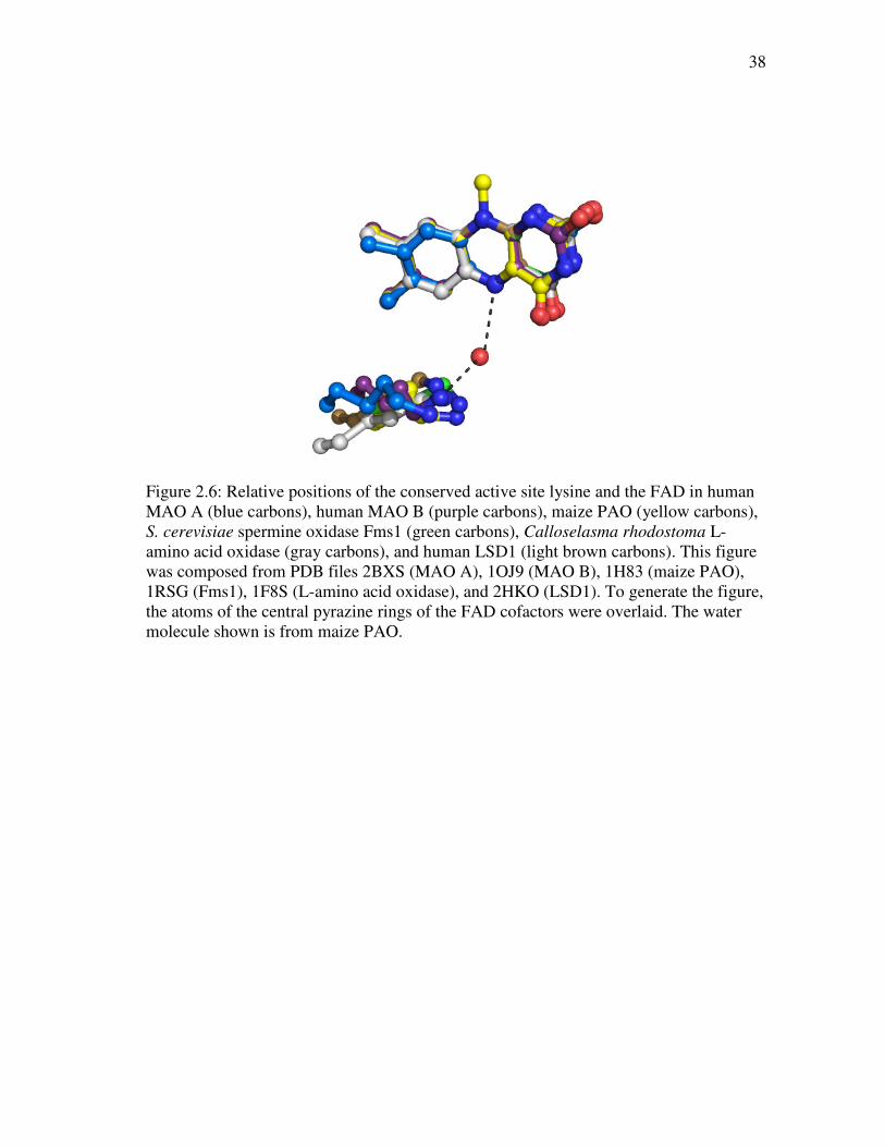
Figure 2.6: Relative positions of the conserved active site lysine and the FAD in human MAO A (blue carbons), human MAO B (purple carbons), maize PAO (yellow carbons), S. cerevisiae spermine oxidase Fms1 (green carbons), Calloselasma rhodostoma L-amino acid oxidase (gray carbons), and human LSD1 (light brown carbons). This figure was composed from PDB files 2BXS (MAO A), 1OJ9 (MAO B), 1H83 (maize PAO), 1RSG (Fms1), 1F8S (L-amino acid oxidase), and 2HKO (LSD1). To generate the figure, the atoms of the central pyrazine rings of the FAD cofactors were overlaid. The water molecule shown is from maize PAO.
39
DISCUSSION
The protonation state of the amine substrate required for productive binding has
been a subject of controversy among those studying flavin amine oxidases. For example,
Harris et al. (69) proposed for DAAO that a coupled deprotonation/dehydrogenation of
the protonated substrate occurs in which a proton is transferred to the solvent. However,
Denu and Fitzpatrick (47) reported that DAAO does not show a solvent isotope effect,
leading to the conclusion that the amino group of the substrate must be uncharged.
Further evidence for the amine being in the neutral form was provided by measurement
of 15N isotope effects for DAAO, which indicated that the amino group must be
unprotonated for catalysis (48). Zhao and Jorns (82) subsequently concluded from
studies of monomeric sarcosine oxidase that the amine substrate within the enzyme-
substrate complex must be unprotonated for flavin reduction. The situation with the
MAO/PAO family has been less clear. Jones et al. (71) concluded that uncharged
inhibitors bind MAO A better, but the predominant species of the amine in the pH range
of 7-9 is protonated and therefore must be the substrate. In contrast, Dunn et al. (83)
concluded from kinetic isotope and pH studies with MAO A that deprotonation of the
amine is required for catalysis. The studies reported here clearly show that mammalian
PAO requires that the N4 nitrogen next to the site of CH bond cleavage be unprotonated
for CH bond cleavage to occur. This is consistent with the results of Dunn et al. (83)
with MAO, establishing that the enzymes of the MAO/PAO family all require an
unprotonated nitrogen for amine oxidation, as do the members of the DAAO/sarcosine
oxidase family. This requirement for a substrate with a neutral nitrogen extends the
40
mechanistic similarities of these two structural classes of flavin amine oxidases, a clear
example of convergent evolution of enzyme mechanisms.
Comparison of the effect of pH on the protonation state of each substrate with its
kcat/Kamine-pH profiles establishes that the monoprotonated forms are required for
catalysis. More specifically, the pKi-pH profiles for the inhibitors establish that these
pKas can be attributed to specific nitrogens in the substrates. In N1-acetylspermidine, the
nitrogen next to the carbon being oxidized must be unprotonated and the N10-nitrogen
must be protonated. N1-Acetylspermine and spermine are more complicated due to the
increased number of nitrogens. For N1-acetylspermine the N4-nitrogen must be
unprotonated, but the data for the inhibitors do not establish whether it is the N10- or
N14-nitrogen that must be protonated. However, the kcat/Kamine values for N1-
acetylspermine and N1-acetylspermidine are identical at the pH optimum, suggesting
that it is the N10-nitrogen that is protonated, as is the case for N1-acetylspermidine. A
similar case can be made for spermine. These results suggest that both protonated and
unprotonated forms of the substrate can bind, but only the protonated form can react.
Thus, the pKas in the kcat/Kamine-pH profiles are due to the substrate and not an ionizable
residue within the active site of the enzyme.
The requirement for the monoprotonated substrate provides a potential
mechanism of discrimination against spermine by PAO, since an acetyl moiety would
prevent the terminal nitrogen from ionizing and thereby result in a very large increase in
the fraction of substrate in the correctly protonated form. Although the kcat/Kamine values
are essentially identical at pH 10 for the two natural substrates N1-acetylspermine and
41
N1-acetylspermidine, at the physiological pH of approximately 8.2 (84), N1-
acetylspermine is the far better substrate at physiological pH. Compared with N1-
acetylspermidine, N1-acetylspermine has a broader pH profile (Figure 2.1A). This is
most readily explained by a difference in the forward commitments of the two
substrates, with N1-acetylspermine being a more sticky substrate.
The kred-pH profile for wild type PAO shows a pKa of 7.3 with N1-
acetylspermine as substrate. The pH profile for kred reports on the protonation states of
ionizable groups in the enzyme-substrate complex required for reduction. The decrease
in activity at acidic pH can be attributed to the substrate bound to the enzyme. The
incorrectly protonated form of the substrate must be able to bind but not to react. If one
assumes that substrate binding is at equilibrium, a likely oversimplification, the
difference between the pKa of the reactive nitrogen when bound to the enzyme and free
in solution of 2.1 establishes that the correctly protonated form binds about 100-fold
more tightly than the form with N4 protonated. Monomeric sarcosine oxidase and MAO
A show similar perturbations of the amine pKa upon binding (82, 83), suggesting that the
active sites of these enzymes also preferentially bind the form of the substrate with the
critical nitrogen in its neutral form.
Although numerous mechanisms have been put forth for flavin amine oxidases,
most recent data support the mechanism as direct hydride transfer. Kinetic studies using
15N isotope effects have ruled out the possibility of a polar nucleophilic addition
mechanism (49, 51). The 15N isotope effects are consistent with a single electron transfer
mechanism, but the failure to detect any intermediate with a natural substrate for any
42
flavin amine oxidase and the very unfavorable redox change for single electron transfer
from an amine to an oxidized flavin provide arguments against such a mechanism.
Reduction of wild-type PAO by N1-acetylspermine shows multiple phases, with the rate
constant for the fastest phase reflecting amine oxidation, while the slower phases are
likely due to product release from reduced enzyme, a step that is not significant during
turnover in the presence of oxygen. More critically the flavin spectrum showed no
intermediate between fully oxidized and fully reduced flavin during reduction of wild
type PAO by N1-acetylspermine over the entire pH range studied, indicating that
oxidation of the amine substrate to the imine occurs in a single step. This result is
consistent with what has been observed with other flavin amine oxidases (45, 69, 85,
86).
The conserved active site lysyl residue in flavin amine oxidases provides a
potential source of a pKa in the kcat/Kamine profile. The role of this residue has previously
been examined in several members of this family. In maize PAO, when Lys300 is
similarly mutated to a methionine, a 1400-fold decrease in kred is observed, suggesting
an important but undefined role for this residue in substrate oxidation (59). The
corresponding K661A mutation in human LSD1 completely abolished demethylase
activity (87). In contrast, the substitution of methionine for this lysine in PAO results in
no change in the kcat/Kspm value or the pH profile with spermine, and the rate constant for
flavin reduction by N1-acetylspermine shows only a 1.8-fold decrease at pH 9.5. This
rules out Lys315 acting as an active site base in mouse PAO or playing any other critical
role in the reductive half-reaction. The kred-pH profile for K315M PAO shows a slight
43
basic shift in the pKa as compared to that for wild type PAO; this can be attributed to a
change in the active site environment due to the loss of the charged lysine. The reasons
for the differences in the effects of mutating this residue among the different flavin
amine oxidases is not apparent. It may be that this residue plays a critical role in
positioning the flavin or otherwise stabilizing the active site structure, and that different
flavin amine oxidases simply tolerate the loss of this interaction more than others.
In conclusion, the present study establishes the protonation state of the amine
required for productive binding to PAO, and presumably for the other members of the
MAO/PAO family. The results will be of use in further studies of the mechanism of
amine oxidation, for interpretation of the effects of site-directed mutagenesis, for design
of inhibitors, and for understanding the different substrate specificities and reactivities of
polyamine and spermine oxidases. The results rule out a critical role for Lys315 in
polyamine oxidation and further support hydride transfer from the neutral amine as the
mechanism of flavin amine oxidases.
44
CHAPTER III
LYS315 PLAYS A ROLE IN THE OXIDATIVE-HALF REACTION IN
MAMMALIAN POLYAMINE OXIDASE
Polyamine levels have been correlated to cell growth and differentiation and
tumor growth (8, 15, 54) necessitating a better understanding of polyamine levels and
the enzymes that regulate their corresponding levels. Polyamine oxidase (PAO) plays an
important role in polyamine homeostasis, specifically by catalyzing the oxidation of N1-
acetylspermine (Figure 1.3) and N1-acetylspermine to produce spermine and putrescine,
respectively. PAO is of particular interest because its N1-acetylated polyamine
substrates are not found in normal cells because they are the main polyamines exported
from the cell (13). However, cancer cells display higher concentrations of N1-acetylated
polyamines indicating a direct link between changes in polyamine metabolism and
carcinogenesis (14, 15), and leading to interest in exploring the role of PAO as an anti-
cancer drug target.
PAO is a flavoprotein amine oxidase that contains a non-covalently bound FAD.
The general mechanism for flavoprotein amine oxidases including PAO, can be divided
into two half-reactions (Figure 1.4). The reductive half-reaction consists of the transfer
of a hydride equivalent from the substrate to the flavin, producing reduced flavin and
oxidized amine. This step is essentially irreversible; therefore, steady-state kinetic
analysis is simplified since the kcat/Km value for the amine substrate is independent of the
oxygen concentration. The oxidative half-reaction involves the oxidation of reduced
45
flavin by molecular oxygen forming H2O2; thus the kcat/KO2 value is the second order
rate constant for the bimolecular reaction with reduced enzyme and oxygen.
To date, no structures for mammalian PAOs have been described. As a result,
site-directed mutagenesis provides the only approach to identify residues important for
catalysis. Structures of other members of the MAO flavoprotein amine oxidase structural
family, including MAO-A (76), MAO-B (75), maize PAO (73), lysine-specific
demethylase 1 (LSD1) (32), and Fms1, a yeast spermine oxidase (74), serve as a basis to
design PAO mutants. Alignment of sequences of members of the MAO structural family
reveal a conserved lysine in the substrate binding domain of PAO that is highly
conserved throughout this family (Figure 3.1), and structural alignments show that this
residue is spatially conserved as part of a “Lys-H2O-N5” structural motif (Figure 2.6).
Earlier studies of the role of Lys315 in mouse PAO indicated that the residue does not
play a critical role in the reductive half-reaction (Chapter II), contrary to observations
with maize PAO (59) and LSD1 (87).
Though the members of the MAO structural family contain the conserved active
site lysyl residue, flavoprotein amine oxidases that belong to the D-amino acid oxidase
structural family do not. Though it is unclear why the loss of this lysyl residue is better
tolerated in PAO (Chapter II) than maize PAO (59) or LSD1 (87), it could be postulated
that residue plays a structural role rather than a catalytic one. The studies presented here
are aimed at establishing the role of Lys315 in the mechanism of PAO.
46
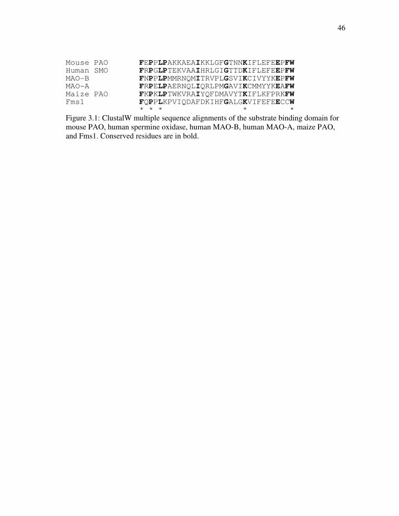
Mouse PAO FEPPLPAKKAEAIKKLGFGTNNKIFLEFEEPFW
Human SMO FRPGLPTEKVAAIHRLGIGTTDKIFLEFEEPFW
MAO-B FNPPLPMMRNQMITRVPLGSVIKCIVYYKEPFW
MAO-A FRPELPAERNQLIQRLPMGAVIKCMMYYKEAFW
Maize PAO FKPKLPTWKVRAIYQFDMAVYTKIFLKFPRKFW
Fms1 FQPPLKPVIQDAFDKIHFGALGKVIFEFEECCW
* * * * * Figure 3.1: ClustalW multiple sequence alignments of the substrate binding domain for mouse PAO, human spermine oxidase, human MAO-B, human MAO-A, maize PAO, and Fms1. Conserved residues are in bold.
47
EXPERIMENTAL PROCEDURES
Materials. Spermine was purchased from Acros Organics (Geel, Belgium) and
N1-acetylspermine was purchased from Fluka (Switzerland). Deuterium oxide was
purchased from Cambridge Isotope Laboratories, Inc (Andover, MA).
Expression and Purification of K315M. Wild type and K315M PAO were
expressed and purified as previously described (Chapter II).
Assays. Steady-state kinetic assays were performed in air-saturated buffers
conducted on a computer-interfaced Hansatech (Hansatech Instruments) electrode.
Assays that required varying the concentration of oxygen was done by bubbling nitrogen
in the Hansatech electrode chamber containing buffer and polyamine substrate. All
assays were initiated by the addition of enzyme. All buffers contained 10% glycerol; 50
mM PIPES, 50 mM Tris-HCl, 50 mM CHES and 50 mM CAPS were used for the pH
ranges of 6.6, 7.1-8.6, 9.1-9.6, and 10, respectively. Solvent isotope effects were
performed in buffers containing 50 mM CHES (pH 9 or pD 9.4) or 50 mM CAPS (pH
10, or pD 10.4) with a viscosity of 1.3 prepared in either H2O or D2O. Glycerol buffer
with a viscosity of 1.3 was prepared as established by Segur and Oberstar (88). A
concentration of 1 mM N1-acetylspermine was used in all assays. Due to the
hygroscopic nature of N1-acetylspermine, the concentration of substrate was determined
enzymatically.
Data Analysis. Steady-state kinetic parameters were determined based on fits to
the Michaelis-Menten equation using the program KaleidaGraph (Adelbeck Software,
Reading, PA). Data for the kcat/KO2-pH profile of wild type PAO was fit to equation 3.1,

48
which applies for a kinetic parameter that decreases below pK1 due to the protonation of
a single moiety, y is the kinetic parameter being measured, c is the pH-independent
value, and K1 is the ionization constant for a residue which must be unprotonated. Eqs
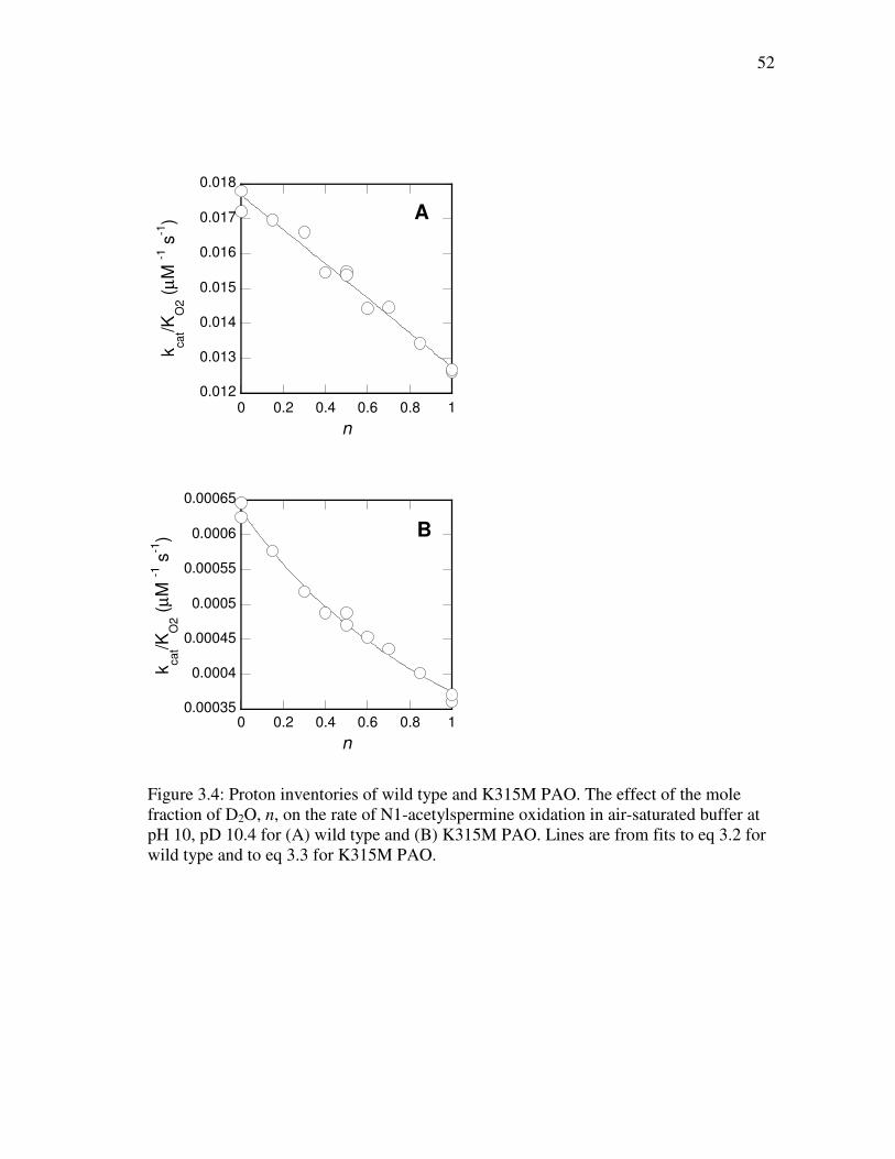
3.2 and 3.3 were used to fit the proton inventories for wild type and K315M PAO,
respectively. Eq 3.2 describes a linear proton inventory arising from a single proton, in
which (v/e)n is the rate in air saturated buffer with a mole fraction of D2O n, (v/e)0 is the
rate in H2O, and KIE denotes the calculated isotope effect. Eq 3.3 describes a proton
inventory of a solvent isotope effect that is due to the reactant.
log y = log (c/(1 + H/K1)) (3.1)
(v/e)n = (v/e)0*(1-n+(n/KIE)) (3.2)
(v/e)n = (v/e)0/(1-n+(n/KIE)) (3.3)
49
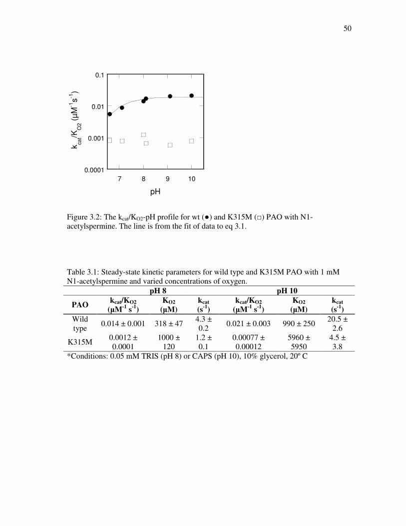
RESULTS
kcat/KO2-pH profile. Table 1 shows the kinetic parameters for wild type PAO at
pH 8 and 10. Steady-state kinetic assays were performed using 1 mM of N1-
acetylspermine while varying the concentration of oxygen over the pH range of 6.6-10.
The resulting kcat/KO2-pH profile for wild type PAO is shown in Figure 3.2 and shows a
pKa of 7.0 ± 0.1 with a decrease in activity at acidic pH.
Solvent Isotope Effects and Proton Inventory. Experiments to determine the
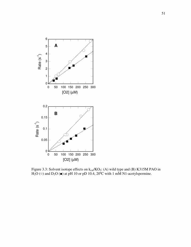
solvent isotope effect on the kinetic parameter kcat/KO2 for wild type PAO were
performed at pH 10 (Figure 3.3A) and result in a value of 1.43 ± 0.05. To test whether
this is a true solvent isotope effect and not due to the viscosity of the D2O buffer, the
effect of 10% (w/w) glycerol on the kcat/KO2 value was determined. This concentration of
glycerol results in η equal to 1.3, the viscosity of D2O. The activity increased in the
presence of glycerol, for an inverse viscosity effect of 0.85 ± 0.03. This establishes that
the decrease in kcat/KO2 in D2O is not due to the viscosity of the D2O solution. Next, a
proton inventory was conducted at pH 10 and pD 10.4 (Figure 3.4A) in an effort to
determine the number of protons responsible for the observed isotope effect. Data from
this experiment was best fit to a linear dependence of the rate on the mole fraction of
D2O, indicating this solvent isotope arises from a single exchangeable proton.
50
0.0001
0.001
0.01
0.1
7 8 9 10
kcat/K
O2 (
µM
-1s
-1)
pH
Figure 3.2: The kcat/KO2-pH profile for wt (●) and K315M (□) PAO with N1-acetylspermine. The line is from the fit of data to eq 3.1.
Table 3.1: Steady-state kinetic parameters for wild type and K315M PAO with 1 mM N1-acetylspermine and varied concentrations of oxygen.
pH 8 pH 10
PAO kcat/KO2
(µM-1
s-1
)
KO2
(µM)
kcat
(s-1
)
kcat/KO2
(µM-1
s-1
)
KO2
(µM)
kcat
(s-1
)
Wild type
0.014 ± 0.001 318 ± 47 4.3 ± 0.2
0.021 ± 0.003 990 ± 250 20.5 ±
2.6
K315M 0.0012 ± 0.0001
1000 ± 120
1.2 ± 0.1
0.00077 ± 0.00012
5960 ± 5950
4.5 ± 3.8
*Conditions: 0.05 mM TRIS (pH 8) or CAPS (pH 10), 10% glycerol, 20º C
51
0
1
2
3
4
5
6
0 50 100 150 200 250 300
A
Rate
(s
-1)
[O2] (µM)
0
0.05
0.1
0.15
0.2
0 50 100 150 200 250 300
B
Rate
(s
-1)
[O2] (µM)
Figure 3.3: Solvent isotope effects on kcat/KO2: (A) wild type and (B) K315M PAO in H2O (○) and D2O (■) at pH 10 or pD 10.4, 20ºC with 1 mM N1-acetylspermine.
52
0.012
0.013
0.014
0.015
0.016
0.017
0.018
0 0.2 0.4 0.6 0.8 1
A
kcat/K
O2 (
µM
-1 s
-1)
n
0.00035
0.0004
0.00045
0.0005
0.00055
0.0006
0.00065
0 0.2 0.4 0.6 0.8 1
B
kcat/K
O2 (
µM
-1 s
-1)
n
Figure 3.4: Proton inventories of wild type and K315M PAO. The effect of the mole fraction of D2O, n, on the rate of N1-acetylspermine oxidation in air-saturated buffer at pH 10, pD 10.4 for (A) wild type and (B) K315M PAO. Lines are from fits to eq 3.2 for wild type and to eq 3.3 for K315M PAO.
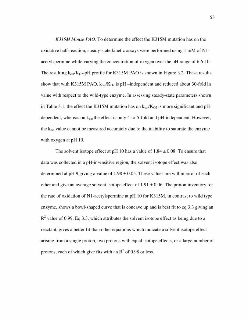
53
K315M Mouse PAO. To determine the effect the K315M mutation has on the
oxidative half-reaction, steady-state kinetic assays were performed using 1 mM of N1-
acetylspermine while varying the concentration of oxygen over the pH range of 6.6-10.
The resulting kcat/KO2-pH profile for K315M PAO is shown in Figure 3.2. These results
show that with K315M PAO, kcat/KO2 is pH –independent and reduced about 30-fold in
value with respect to the wild-type enzyme. In assessing steady-state parameters shown
in Table 3.1, the effect the K315M mutation has on kcat/KO2 is more significant and pH-
dependent, whereas on kcat the effect is only 4-to-5-fold and pH-independent. However,
the kcat value cannot be measured accurately due to the inability to saturate the enzyme
with oxygen at pH 10.
The solvent isotope effect at pH 10 has a value of 1.84 ± 0.08. To ensure that
data was collected in a pH-insensitive region, the solvent isotope effect was also
determined at pH 9 giving a value of 1.98 ± 0.05. These values are within error of each
other and give an average solvent isotope effect of 1.91 ± 0.06. The proton inventory for
the rate of oxidation of N1-acetylspermine at pH 10 for K315M, in contrast to wild type
enzyme, shows a bowl-shaped curve that is concave up and is best fit to eq 3.3 giving an
R2 value of 0.99. Eq 3.3, which attributes the solvent isotope effect as being due to a
reactant, gives a better fit than other equations which indicate a solvent isotope effect
arising from a single proton, two protons with equal isotope effects, or a large number of
protons, each of which give fits with an R2 of 0.98 or less.
54
DISCUSSION
Structural information compiled from crystal structures of various flavin amine
oxidases presents the opportunity to investigate the functional importance of structural
motifs in PAO. One such motif is the hydrogen bond network that exists between a
conserved lysyl residue in the active site to the N5 atom of the FAD cofactor via a water
molecule. When the lysine of this “Lys-H2O-N5” structural motif was mutated to a
methionine in maize PAO, the rate constant for flavin reduction was reduced 1400-fold
(59). Similarly, the Lys661Ala mutation in LSD1 abolished demethylase activity (32).
The effect of the K315M mutation in PAO on the reductive half-reaction has been the
source of earlier study, showing modest effects in the rate of flavin reduction (Chapter
II).
In an effort to better understand the oxidative half reaction, results of wild type
PAO studies show that kcat/KO2 is pH-dependent with an increase in activity observed at
higher pH. This result indicates that deprotonation of a moiety within the enzyme
enhances oxidation. To better understand the role of the conserved Lysine residue found
in the active site of members of the MAO structural family, the mutant K315M was
characterized. K315M PAO shows kcat/KO2 values that are depressed in comparison to
wild type and are pH-independent (Figure 3.2, Table 3.1). From these results, two
conclusions can be drawn. First, Lys315 plays a role in the oxidative half-reaction,
though not a critical one. Second, the deprotonation in wild type PAO with a pKa of 7.0
± 0.1 is due to the protonation of Lys315. The mutant still maintains the ability to
oxidize substrate, a decrease that is greater than an order of magnitude is observed in the
55
kcat/KO2 rate constant, with a smaller 4-to-5-fold effect observed for kcat (Table 3.1). The
results seen in the oxidative half-reaction are in contrast to the modest 1.5-fold decrease
observed for the rate of flavin reduction with N1-acetylspermine (Chapter II). The
importance positively charged residues have in establishing electrophilicity and polarity
of the active site of flavoproteins is documented in the literature. In addition to the
aforementioned conserved lysyl residue in members of the MAO structural family,
glucose oxidase possesses His516 which plays an important role in the oxidation of
reduced flavin by contributing a positive charge that facilitates the formation of O2- (89).
Results with the flavoprotein choline oxidase, which catalyzes the oxidation of choline
to glycine betaine, indicates involvement of two histidines, His351 and His466, both of
which must be protonated for optimal catalysis and aid in transition state stabilization of
the alkoxide species (90, 91). What is different with PAO is that it is the loss of that
positive charge that enhances activity. It appears that this lysyl residue in PAO must be
uncharged to hydrogen bond to water as depicted in Figure 3.5. Thus, it is postulated that
the role of the hydrogen bond network between the uncharged Lys315, a water molecule,
and the N5 of the flavin within PAO is to enhance the rate of oxidation of the reduced
flavin. The proton bonded to the N5 atom of the flavin must be removed during
oxidation of reduced flavin. It could therefore be postulated that Lys315 in PAO
removes a proton from the water molecule, activating it to remove the proton bound at
the N5 position, thus facilitating reformation of oxidized flavin for the next round of
catalysis.
56
Solvent isotope effects with wild type and K315M PAO reflect a significant
change occurring within the active site by removal of this lysyl residue. The increase in
the solvent isotope effect seen with K315M PAO suggests that the lysyl residue is
replaced with water molecules. Further evidence of this comes from the change in proton
inventory of wild type PAO, which shows that a single proton is exchangeable to
solvent, compared to that of K315M PAO where a significant change is observed with
the simplest explanation being the presence of an additional water molecule in the active
site. Though another water molecule can bind within the active site in the absence of
Lys315, the inefficiency of such a replacement is reflected in a decrease by an order of
magnitude in kcat/KO2. With PAO, previous studies indicated that under single turnover
conditions a slow step is observed that is slower than turnover (Chapter II). It is probable
that this is product release in the absence of oxygen; thus reduced enzyme reacts with
molecular oxygen before product is released, and product release is rate-limiting. The
lesser effect of 4-5 fold observed for kcat with mutant enzyme reflects that the full effect
of the mutation is partially masked due to another partially-rate limiting step such as
product release.
The results of this study with mammalian PAO establish the role of Lys315 as
being involved in a hydrogen bond network (Figure 3.5), in which the deprotonated form
of Lys315 serves to enhance the rate of oxidation of reduced flavin via a water molecule.
The removal of this lysyl residue results in the presence of an additional water molecule
in the active site as reflected in solvent isotope effects and proton inventory. It remains
unclear as to why differences exist with K300M maize PAO (59) as compared to
57
K315M mouse PAO (Chapter II) with respect to the rate of flavin reduction. Perhaps, the
simplest explanation is that in maize PAO, Lys300 does not play a direct role in
substrate oxidation, but rather causes enzyme instability resulting from structural effects,
whereas in mouse PAO, the loss of this residue is structurally less dramatic. What can be
concluded is that in mammalian PAO, this conserved lysyl residue plays a direct, though
not vital, role in the oxidative half-reaction.
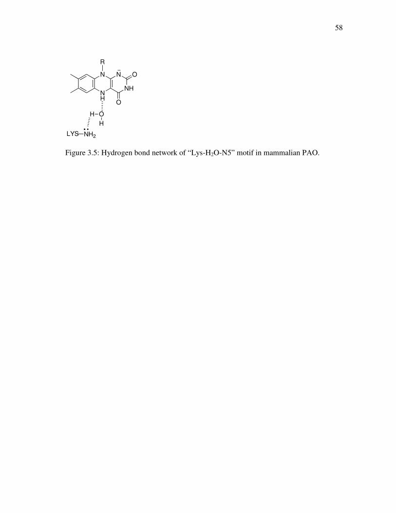
58
NH
N
NH
N O
O
R
OH
H
LYS NH2
Figure 3.5: Hydrogen bond network of “Lys-H2O-N5” motif in mammalian PAO.

59
CHAPTER IV
MECHANISTIC STUDIES OF PARA-SUBSTITUTED N,N’-DIBENZYL-1,4-
DIAMINOBUTANES AS SUBSTRATES FOR A MAMMALIAN POLYAMINE
OXIDASE
Polyamine oxidase (PAO) is a flavoprotein that plays a pivotal role in polyamine
metabolism by combining biosynthesis and degradation pathways with the regulated
import and export of polyamines (92). The activity of PAO in maintaining polyamine
homeostasis indicates it has an important role in facilitating apoptosis in cancer cells
(93). Specifically, PAO catalyzes the oxidation of N1-acetylspermine and N1-
acetylspermidine to the products spermidine and putrescine, respectively, producing
H2O2 as a by-product. Recent studies show the potential for targeting tumor cells by
inducing PAO with specific polyamine analogues that leads to the cytotoxic production
of H2O2 in a tumor specific manner, including solid tumors found in lung, breast, colon,
and prostate (94, 95). PAO, not to be confused with a spermine oxidase, preferentially
reacts with acetylated polyamines, though it can also oxidize spermine (26). The study
presented here uses PAO from mouse, and as a distinction from plant PAOs where CH
bond cleavage occurs on the endo-side of spermine (63), mammalian PAOs catalyze CH
bond cleavage on the exo-side of the N4-nitrogen in N1-acetylspermine and N1-
acetylspermidine.
PAO is a member of the flavin amine oxidase superfamily and belongs to the
structural family that includes monoamine oxidase A and B (MAO-A/B) (60), lysine-
60
specific demethylase (LSD1) (32), L-amino acid oxidase (34), and tryptophan 2-
monooxygenase (TMO) (53) amongst others. Sequence alignments reveal low sequence
identity of PAO with other flavoprotein amine oxidases (26), though no evidence has
been found to suggest that these enzymes utilize different mechanisms. The reaction of
PAO, like other flavoprotein amine oxidases, can be divided into two half-reactions. The
reductive half-reaction consists of substrate binding and flavin reduction upon substrate
oxidation, and the oxidative-half reaction involves the oxidation of reduced flavin via
molecular oxygen. The steady-state kinetic mechanism for mouse PAO displays the
ping-pong kinetic pattern (26), as is observed with most flavoprotein oxidases (64). Due
to the irreversibility of the reductive half-reaction, analysis of the kcat/Km parameter for
the amine substrate can be conducted in air saturated buffers since its value is
independent of the oxygen concentration. To determine the rate constant for flavin
reduction, anaerobic rapid-reaction methods are utilized to monitor changes in the
observed flavin spectrum going from oxidized to reduced.
There are multiple mechanisms that have been presented for the mode by which
CH bond cleavage is achieved. Three mechanisms that have become more prominent in
the discussion of flavoprotein amine oxidases include the single electron transfer
mechanism (Figure 1.8) (39), the polar nucleophilic mechanism (Figure 1.9) (45), and
the hydride transfer mechanism (Figure 1.10) (49, 51, 96, 97). In order to determine the
mechanism by which PAO functions, the study described here investigates the charge
61
development during the transition state of oxidation by a mammalian PAO using N,N’-
(4-X-benzyl)2-1,4-diaminobutanes as substrates. The influence of the different
substituents on the rate of oxidation of polyamine analogues varies and can be assessed
in terms of steric (VW and ES), hydrophobic (π), and electronic properties (σ, σ+ and σ-).
Analysis of these effects through correlations of steady-state and transient kinetic rate
constants allows the opportunity to investigate the effects such groups have on the
development of the charge in the chemical step during the transition state. Kinetic
isotope effects as a function of pH were determined with N,N’-(4-X-benzyl)2-1,4-
diaminobutanes in order to gain information about the timing of CH bond cleavage
during oxidation of the substrate, allowing further insight into the nature of the transition
state. These results observed for PAO are compared with results of structure-activity
relationships observed with MAO and are discussed in terms of the various mechanisms
proposed for flavoprotein amine oxidases.
62
EXPERIMENTAL PROCEDURES
Syntheses. All substrates used in this study were synthesized by Dr. Vijay
Gawandi of Texas A&M University.
Expression and Purification of Recombinant PAO Proteins. Cells were grown to
an O.D. of 0.6 before being induced with 0.15 mM IPTG, and grown overnight at 20 °C.
Cells were harvested by centrifugation at 22400 g for 30 min. Cell paste was
resuspended in 50 mM potassium phosphate (pH 7.5), 10% glycerol, 2 µM leupeptin and
pepstatin, and 100 µg/mL phenylmethanesulfonyl fluoride and lysozyme and lysed by
sonication. After centrifugation at 22400 g for 30 min, the resulting supernatant was
loaded onto a HisTrap FF precharged Ni Sepharose™ 6 Fast Flow (GE Lifesciences)
column washed with ten column volumes of 50 mM potassium phosphate (pH 7.5), 10%
glycerol, 0.4 M sodium chloride, 10 mM imidazole. The protein was eluted using a 20-
column volume gradient of 10 mM – 200 mM imidazole. The resulting protein was
pooled and concentrated by a 65% ammonium sulfate precipitation, centrifuged at 22400
g for 30 min, and resuspended in minimal volume of 50 mM potassium phosphate (pH
7.5), 10% glycerol, and dialyzed overnight in the same buffer. After a final
centrifugation at 22400 g for 30 min, the purified enzyme was stored at -80 °C in 50 mM
potassium phosphate (pH 7.5), 10% glycerol. The concentration of active enzyme was
determined from the flavin visible absorbance spectrum using an ε458 value of 10,400 M-
1 cm-1 (79).
Steady-State Assays. Steady-state kinetic parameters for PAO were determined
using a computer-interfaced Hansatech Clark oxygen electrode (Hansatech Instruments).
63
All assays were performed at 30 oC and were initiated by addition of enzyme. The
buffers used for pH range 7.1-8.6 was 50 mM Tris-HCl, 50 mM CHES at pH 9.1-9.6,
and 50 mM CAPS at pH 10.1. All buffers contained 10 % glycerol. Enzyme was used to
check the concentration of substrates; the concentration of substrate based on the oxygen
consumption assay was twice that based on molecular weight, indicating an ability of
PAO to recognize and oxidize both sides of N,N’-(4-X-benzyl)2-1,4-diaminobutanes.
Rapid Reaction Kinetics. Rapid reaction kinetic measurements were performed
on an Applied Photophysics SX-18MV stopped-flow spectrophotometer using both
single wavelength and photodiode array detection. To establish anaerobic conditions,
the instrument was loaded with anaerobic buffer containing 5 mM glucose and 36 nM of
glucose oxidase and left overnight. Enzyme solutions containing 5 mM glucose were
made anaerobic by applying cycles of argon and vacuum; substrate solutions containing
5 mM glucose were made anaerobic by bubbling with argon. Glucose oxidase was added
to all anaerobic solutions at a final concentration of 36 nM.
64
Data Analysis. Data were analyzed using the programs Kaleidagraph (Adelbeck
Software, Reading, PA) and Igor (WaveMetrics, Lake Oswego, OR). Initial rate data
were fit to the Michaelis-Menten equation to determine steady-state kinetic parameters.
The kcat/KDBDB-pH profile was fit to eq 4.1, where y is the value of the kinetic parameter,
c is its pH-independent value, K1 is the ionization constant for a moiety that must be
unprotonated for maximum activity and K2 is the ionization constant for a moiety that
must be protonated for maximum activity.
log y = log[c/(1+H/K1 + K2/H)] (4.1)
Kinetic isotope effects on steady-state parameters were determined from fits to
eq 4.2 or 4.3, where E is the isotope effect on the parameters kcat/KDBDB and kcat, s is the
concentration of substrate, Fi is the fraction of heavy isotope in the substrate, and, in eq
4.3, Kai is the substrate inhibition constant. Some of the substrates exhibited substrate
inhibition and were consequently fit with eq 4.3.
v = (V*s)/((KM+s)*(1+(E-1)*Fi) (4.2)
v = (V*s)/((KM *(1+Fi*(E-1))+s*(1+Fi*(E-1))+s2/Kai) (4.3)
Rapid reaction kinetic traces were fit to single and double exponential decays
(eqs 4.4 and 4.5); here, k1 and k2 are the first order rate constants, A1 and A2 are the

65
absorbance changes associated with the two phases, At is the total absorbance change,
and A∞ is the final absorbance.
At = A1e-k1t + A∞
(4.4)
At = A1e-k1t + A2e
-k2t + A∞ (4.5)
The kred-pH profile for PAO oxidation of N,N’-(4-H-benzyl)2-1,4-
diaminobutanes was fit to eq 4.6, where y is the value of the kinetic parameter, yL is the
value of the low plateau in the sigmoidal curve, ∆yL is the change in rate between the
high and low plateaus, and K1 is the ionization constant.
log y = log (yL + (∆yL / (1 + H/K1)) (4.6)
66
RESULTS
N, N’-dibenzyl-diamines As Substrates. Several N, N’-dibenzyl-diamines were
previously shown to be substrates for partially purified polyamine oxidase from pig
liver, producing benzaldehyde as product; the highest activity was seen with the 1,3-
diaminopropane and 1,4-diaminobutane derivatives (13). With purified recombinant
mouse enzyme in hand, we determined the kinetics of oxidation of N, N’-dibenzyl-1,2-
diaminoethane, N,N’-dibenzyl-1,3- diaminopropane, and N, N’-1,4-dibenzyl-
diaminobutane in air-saturated buffer at pH 8.6. No activity was detected with the
diaminoethane derivative. The apparent kcat, Km, and kcat/Km values for N, N’-dibenzyl-
1,3- diaminopropane were 6.1 ± 0.2 s-1, 40 ± 6 µM, and 0.15 ± 0.02 µM-1s-1,
respectively. For N, N’-dibenzyl-1,4-diaminobutane, the values of these kinetic
parameters were 0.80 ± 0.02 s-1, 15 ± 2 µM, and 0.06 ± 0.01 µM-1s-1. The latter Km value
is within a factor of two of the value reported for the pig enzyme at 37 oC; the kinetic
parameters for N, N’-dibenzyl-1,3-diaminopropane were not reported previously. The
lower activity of N, N’-dibenzyl-1,4-diaminobutane suggested that chemistry is more
likely to be rate-limiting with this compound. Consequently, analogs of this compound
were selected for further study.
Steady-State Kinetics. The steady-state kinetic parameters apparent kcat, kcat/KM,
and KM were determined for PAO at pH 8.6 and 30 °C for seven N,N’-(4-X-benzyl)2-
1,4-diaminobutanes as substrates (Table 4.1). The enzyme displays ping pong kinetics so
assays were conducted in air-saturated buffers; therefore, the kcat parameter calculated is
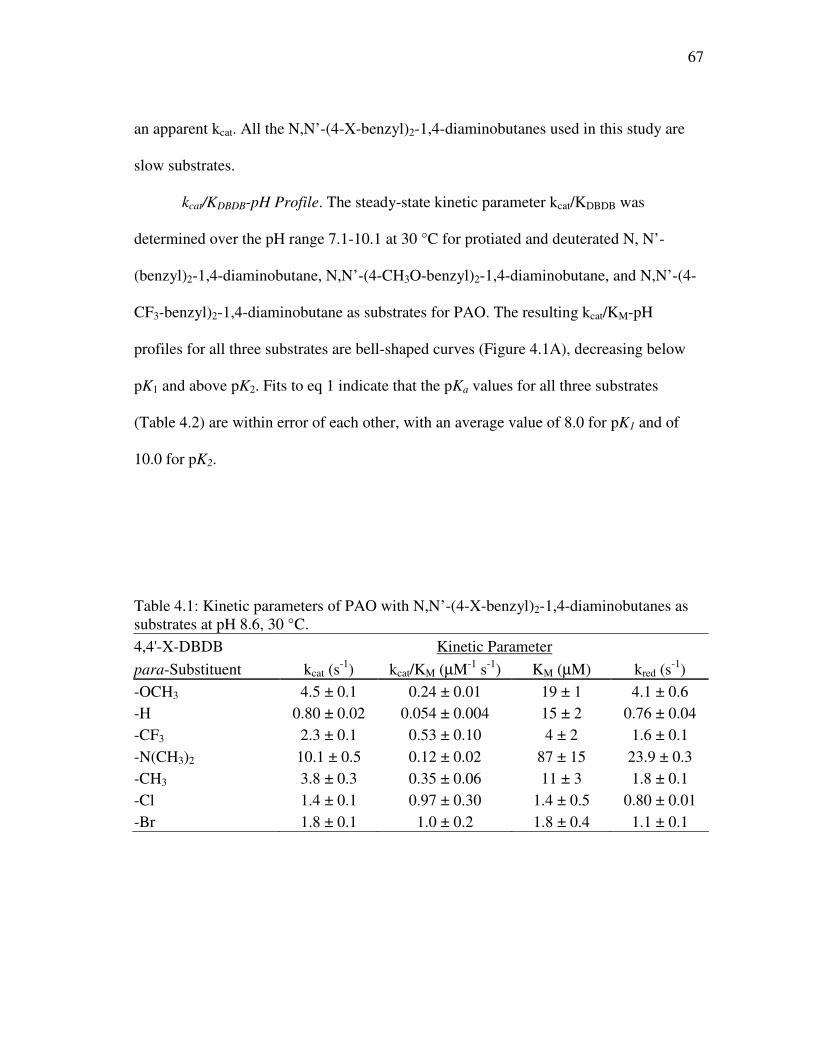
67
an apparent kcat. All the N,N’-(4-X-benzyl)2-1,4-diaminobutanes used in this study are
slow substrates.
kcat/KDBDB-pH Profile. The steady-state kinetic parameter kcat/KDBDB was
determined over the pH range 7.1-10.1 at 30 °C for protiated and deuterated N, N’-
(benzyl)2-1,4-diaminobutane, N,N’-(4-CH3O-benzyl)2-1,4-diaminobutane, and N,N’-(4-
CF3-benzyl)2-1,4-diaminobutane as substrates for PAO. The resulting kcat/KM-pH
profiles for all three substrates are bell-shaped curves (Figure 4.1A), decreasing below
pK1 and above pK2. Fits to eq 1 indicate that the pKa values for all three substrates
(Table 4.2) are within error of each other, with an average value of 8.0 for pK1 and of
10.0 for pK2.
Table 4.1: Kinetic parameters of PAO with N,N’-(4-X-benzyl)2-1,4-diaminobutanes as substrates at pH 8.6, 30 °C. 4,4'-X-DBDB Kinetic Parameter
para-Substituent kcat (s-1) kcat/KM (µM-1 s-1) KM (µM) kred (s
-1) -OCH3 4.5 ± 0.1 0.24 ± 0.01 19 ± 1 4.1 ± 0.6 -H 0.80 ± 0.02 0.054 ± 0.004 15 ± 2 0.76 ± 0.04 -CF3 2.3 ± 0.1 0.53 ± 0.10 4 ± 2 1.6 ± 0.1 -N(CH3)2 10.1 ± 0.5 0.12 ± 0.02 87 ± 15 23.9 ± 0.3 -CH3 3.8 ± 0.3 0.35 ± 0.06 11 ± 3 1.8 ± 0.1 -Cl 1.4 ± 0.1 0.97 ± 0.30 1.4 ± 0.5 0.80 ± 0.01 -Br 1.8 ± 0.1 1.0 ± 0.2 1.8 ± 0.4 1.1 ± 0.1
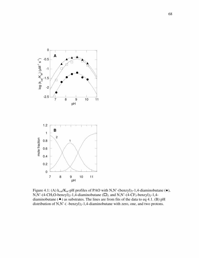
68
-2.5
-2
-1.5
-1
-0.5
0
7 8 9 10 11
log (
kcat/K
M)
(µM
-1 s
-1)
pH
A
0
0.2
0.4
0.6
0.8
1
1.2
7 8 9 10 11
B
mole
fra
ctio
n
pH
2
1
Figure 4.1: (A) kcat/Km-pH profiles of PAO with N,N’-(benzyl)2-1,4-diaminobutane (●), N,N’-(4-CH3O-benzyl)2-1,4-diaminobutane (�), and N,N’-(4-CF3-benzyl)2-1,4-diaminobutane (�) as substrates. The lines are from fits of the data to eq 4.1. (B) pH distribution of N,N’-( -benzyl)2-1,4-diaminobutane with zero, one, and two protons.
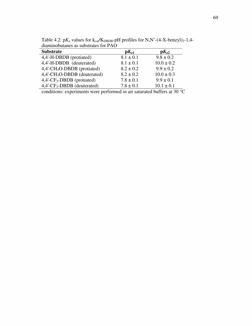
69
Table 4.2: pKa values for kcat/KDBDB-pH profiles for N,N’-(4-X-benzyl)2-1,4-diaminobutanes as substrates for PAO Substrate pKa1 pKa2
4,4'-H-DBDB (protiated) 8.1 ± 0.1 9.8 ± 0.2 4,4'-H-DBDB (deuterated) 8.1 ± 0.1 10.0 ± 0.2 4,4'-CH3O-DBDB (protiated) 8.2 ± 0.2 9.9 ± 0.2 4,4'-CH3O-DBDB (deuterated) 8.2 ± 0.2 10.0 ± 0.3 4,4'-CF3-DBDB (protiated) 7.8 ± 0.1 9.9 ± 0.1 4,4'-CF3-DBDB (deuterated) 7.8 ± 0.1 10.1 ± 0.1 conditions: experiments were performed in air saturated buffers at 30 °C
70
Steady-State Kinetic Isotope Effects. Deuterium kinetic isotope effects were
determined using di-deuterated N,N’-(4-X-benzyl)2-1,4-diaminobutanes, which results in
isotope effects that reflect both the primary and secondary effects. Data obtained from
varying the concentration of N,N’-(4-X-benzyl)2-1,4-diaminobutanes in air-saturated
buffer were best fit using eq 4.2, which assigns equal isotope effects on Dkcat/KDBDB and
Dkcat. In instances where substrate inhibition was observed, eq 4.3 was used instead. For
each of the three substrates used, the isotope effect on kcat/KDBDB and kcat is less than two
and are pH independent (Table 4.3). These three substrates are representative of the
different substituents from the series of the N,N’-(4-X-benzyl)2-1,4-diaminobutanes used
for study here, and lack of any notable changes observed in the kinetic isotope effect did
not necessitate further studies with other analogues.
Flavin Reduction Kinetics. Stopped-flow experiments were conducted to
determine directly the effects that para-substituents have on the rate of flavin reduction,
kred. To obtain rate constants as a function of substrate concentration, reactions were
monitored at 458 nm (Figure 4.2A, closed symbols); with the majority of the substrates,
the spectral changes upon mixing enzyme and substrate could be fit to a single
exponential decay. The exception was N,N’-(4-N(CH3)2-benzyl)2-1,4-diaminobutane; in
this case the traces were fit better to a double exponential decay (Figure 4.2A, closed
symbols), where the second rate constant is slower than turnover and therefore not
catalytically relevant. These experiments were conducted at both pH 8.6, near the pH
optimum, and at pH 6.6. Experiments at pH 9.6 were attempted, but enzyme instability
at such a high pH prevented the acquisition of usable data. For each substrate, a
71
photodiode array detector was used to monitor the spectral changes during the course of
the reaction (Figure 4.2B). Global analyses of the data showed that flavin reduction
occurs in a single step, with no observable intermediates between fully oxidized and
fully reduced flavin (Figure 4.2C). These results are summarized in Table 4. Similar
analyses were carried out with the deuterated substrates; the resulting isotope effects on
the kred values range from 1.3 to 2.9 for N,N’-(4-X-benzyl)2-1,4-diaminobutane
substrates and are pH independent (Table 4.4). The isotope effects on kred are
comparable to those on kcat/Km and kcat.
The pH-profile of the rate of flavin reduction with the substrate N’-(benzyl)2-1,4-
diaminobutane is a sigmoid curve with an increase in activity at higher pH (Figure 4.3).
Deprotonation of a group within the ES complex having a pKa of 8.2 ± 0.3 results in an
increase in activity of approximately 4-fold. The rate constant at low pH is 0.20 ± 0.01 s-
1 and at high pH is 0.82 ± 0.13 s-1.
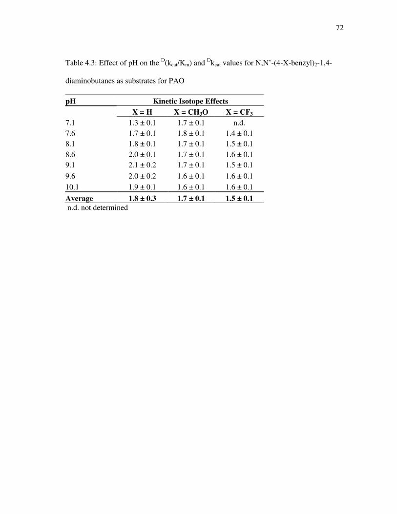
72
Table 4.3: Effect of pH on the D(kcat/Km) and Dkcat values for N,N’-(4-X-benzyl)2-1,4-
diaminobutanes as substrates for PAO
pH Kinetic Isotope Effects
X = H X = CH3O X = CF3 7.1 1.3 ± 0.1 1.7 ± 0.1 n.d. 7.6 1.7 ± 0.1 1.8 ± 0.1 1.4 ± 0.1 8.1 1.8 ± 0.1 1.7 ± 0.1 1.5 ± 0.1 8.6 2.0 ± 0.1 1.7 ± 0.1 1.6 ± 0.1 9.1 2.1 ± 0.2 1.7 ± 0.1 1.5 ± 0.1
9.6 2.0 ± 0.2 1.6 ± 0.1 1.6 ± 0.1 10.1 1.9 ± 0.1 1.6 ± 0.1 1.6 ± 0.1
Average 1.8 ± 0.3 1.7 ± 0.1 1.5 ± 0.1
n.d. not determined
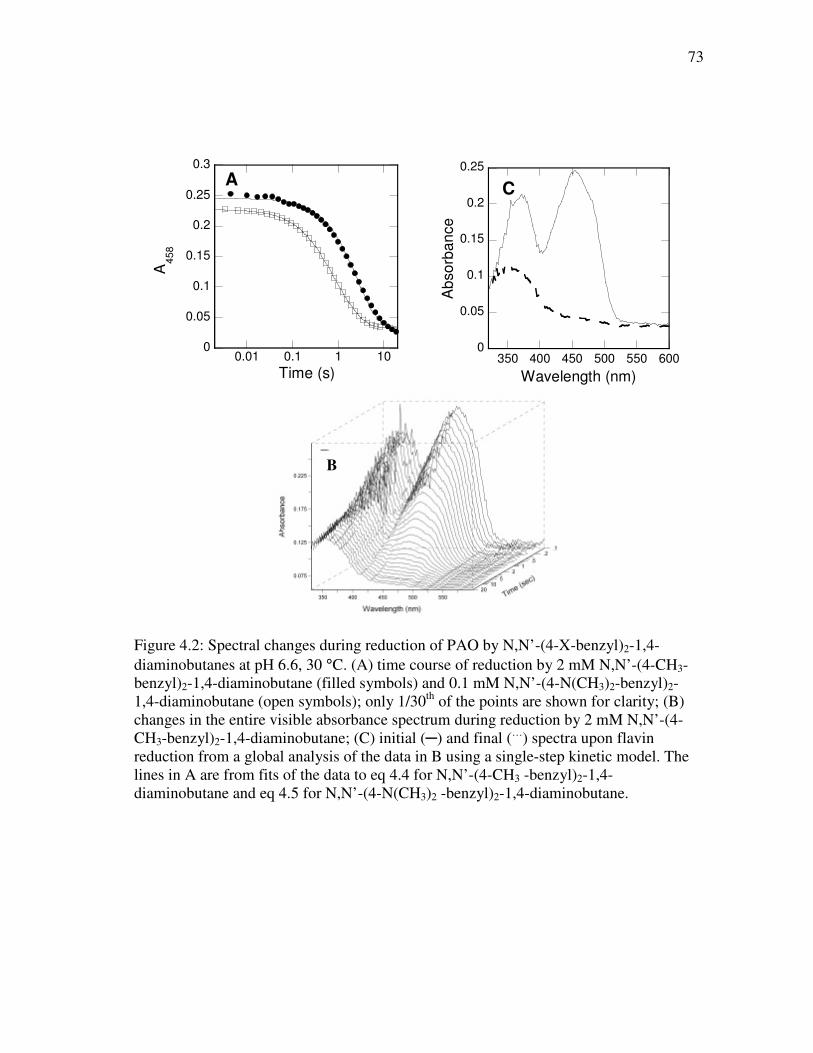
73
0
0.05
0.1
0.15
0.2
0.25
0.3
0.01 0.1 1 10
AA
45
8
Time (s)
0
0.05
0.1
0.15
0.2
0.25
350 400 450 500 550 600
C
Ab
so
rba
nce
Wavelength (nm)
Figure 4.2: Spectral changes during reduction of PAO by N,N’-(4-X-benzyl)2-1,4-diaminobutanes at pH 6.6, 30 °C. (A) time course of reduction by 2 mM N,N’-(4-CH3-benzyl)2-1,4-diaminobutane (filled symbols) and 0.1 mM N,N’-(4-N(CH3)2-benzyl)2-1,4-diaminobutane (open symbols); only 1/30th of the points are shown for clarity; (B) changes in the entire visible absorbance spectrum during reduction by 2 mM N,N’-(4-CH3-benzyl)2-1,4-diaminobutane; (C) initial (─) and final (…) spectra upon flavin reduction from a global analysis of the data in B using a single-step kinetic model. The lines in A are from fits of the data to eq 4.4 for N,N’-(4-CH3 -benzyl)2-1,4-diaminobutane and eq 4.5 for N,N’-(4-N(CH3)2 -benzyl)2-1,4-diaminobutane.
B
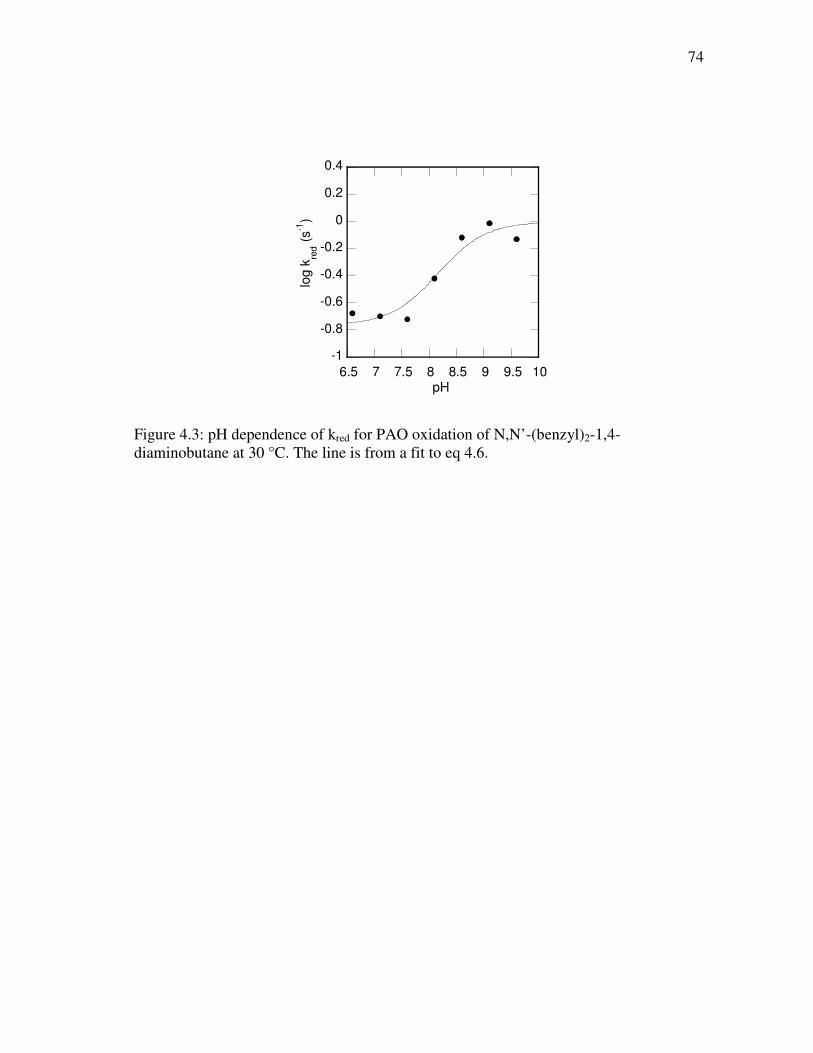
74
-1
-0.8
-0.6
-0.4
-0.2
0
0.2
0.4
6.5 7 7.5 8 8.5 9 9.5 10
log k
red (
s-1
)
pH
Figure 4.3: pH dependence of kred for PAO oxidation of N,N’-(benzyl)2-1,4-diaminobutane at 30 °C. The line is from a fit to eq 4.6.
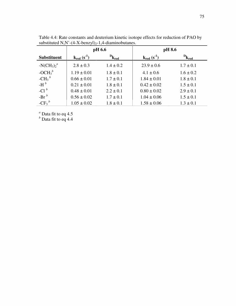
75
Table 4.4: Rate constants and deuterium kinetic isotope effects for reduction of PAO by substituted N,N’-(4-X-benzyl)2-1,4-diaminobutanes.
pH 6.6 pH 8.6
Substituent kred (s-1
) Dkred kred (s
-1)
Dkred
-N(CH3)2a 2.8 ± 0.3 1.4 ± 0.2 23.9 ± 0.6 1.7 ± 0.1
-OCH3b 1.19 ± 0.01 1.8 ± 0.1 4.1 ± 0.6 1.6 ± 0.2
-CH3 b 0.66 ± 0.01 1.7 ± 0.1 1.84 ± 0.01 1.8 ± 0.1
-H b 0.21 ± 0.01 1.8 ± 0.1 0.42 ± 0.02 1.5 ± 0.1 -Cl b 0.48 ± 0.01 2.2 ± 0.1 0.80 ± 0.02 2.9 ± 0.1 -Br b 0.56 ± 0.02 1.7 ± 0.1 1.04 ± 0.06 1.5 ± 0.1 -CF3
b 1.05 ± 0.02 1.8 ± 0.1 1.58 ± 0.06 1.3 ± 0.1
a Data fit to eq 4.5 b Data fit to eq 4.4
76
Linear Correlation Analysis. The effect of the substituent at the para-position of
the aromatic rings on the rate constant for flavin reduction can be analyzed using single-
or multiple-parameter linear correlations with electronic (σ, σ+, σ-), hydrophobic (π), and
steric parameters (VW and ES, where VW is the van der Waals volume and ES is the Taft
steric parameter). Eq 4.7 describes a single-parameter linear correlation where ρ is the
parameter coefficient for the σ parameter and C is a constant. Eq 4.8 describes a two-
parameter linear correlation where ρ and A are the coefficients for the two-parameters
being analyzed.
log kred = ρσ + C (4.7)
log kred = ρσ + Aπ (or VW or ES) + C (4.8)
The results of the analyses are summarized in Table 4.5. The best single-
parameter correlation is with the van der Waals volume at both pH 8.6 and 6.6 (Figure
4.4, Table 4.5). However, a two-parameter linear correlation using the electronic
parameter σ and steric parameter VW shows a lower χ2 value than the single-parameter
correlation with just VW (Table 4.5). The ρ value is more negative at pH 8.6 by 0.5 as
compared with pH 6.6, whereas the coefficient for VW is essentially unaffected by pH
(Table 4.5).
Analyses of the effects the substituents of N,N’-(4-X-benzyl)2-1,4-
diaminobutanes have on the steady-state kinetic parameters kcat/KM and kcat are
summarized in Table 4.6. The best linear correlation for kcat/KM is obtained with the
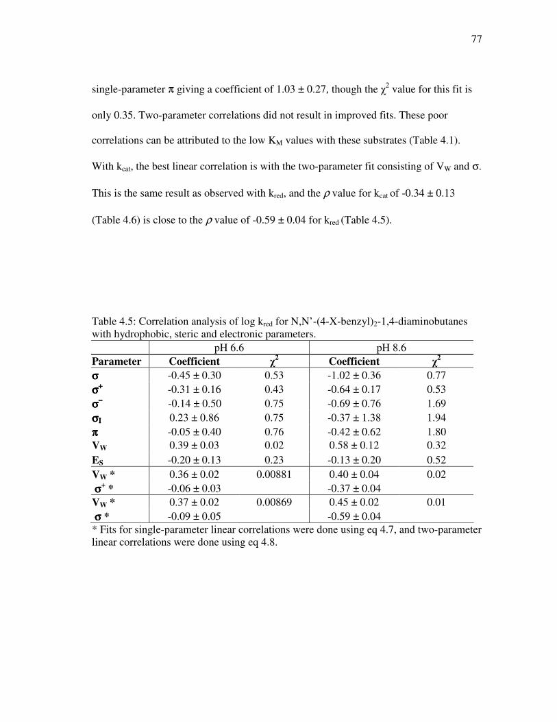
77
single-parameter π giving a coefficient of 1.03 ± 0.27, though the χ2 value for this fit is
only 0.35. Two-parameter correlations did not result in improved fits. These poor
correlations can be attributed to the low KM values with these substrates (Table 4.1).
With kcat, the best linear correlation is with the two-parameter fit consisting of VW and σ.
This is the same result as observed with kred, and the ρ value for kcat of -0.34 ± 0.13
(Table 4.6) is close to the ρ value of -0.59 ± 0.04 for kred (Table 4.5).
Table 4.5: Correlation analysis of log kred for N,N’-(4-X-benzyl)2-1,4-diaminobutanes with hydrophobic, steric and electronic parameters. pH 6.6 pH 8.6 Parameter Coefficient χ
2 Coefficient χ
2
σσσσ -0.45 ± 0.30 0.53 -1.02 ± 0.36 0.77 σσσσ++++ -0.31 ± 0.16 0.43 -0.64 ± 0.17 0.53 σσσσ−−−− -0.14 ± 0.50 0.75 -0.69 ± 0.76 1.69 σσσσΙΙΙΙ 0.23 ± 0.86 0.75 -0.37 ± 1.38 1.94 ππππ -0.05 ± 0.40 0.76 -0.42 ± 0.62 1.80 VW 0.39 ± 0.03 0.02 0.58 ± 0.12 0.32 ES -0.20 ± 0.13 0.23 -0.13 ± 0.20 0.52 VW * 0.36 ± 0.02 0.00881 0.40 ± 0.04 0.02 σσσσ+
* -0.06 ± 0.03 -0.37 ± 0.04 VW * 0.37 ± 0.02 0.00869 0.45 ± 0.02 0.01 σσσσ * -0.09 ± 0.05 -0.59 ± 0.04 * Fits for single-parameter linear correlations were done using eq 4.7, and two-parameter linear correlations were done using eq 4.8.
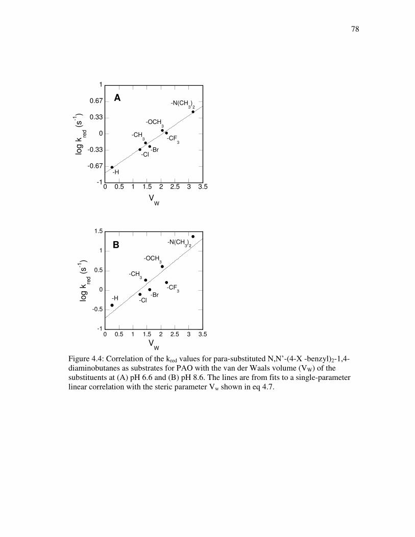
78
-1
-0.67
-0.33
0
0.33
0.67
1
0 0.5 1 1.5 2 2.5 3 3.5
A
log
kre
d (
s-1
)
VW
-H
-CH3
-OCH3
-CF3
-Br-Cl
-N(CH3)2
-1
-0.5
0
0.5
1
1.5
0 0.5 1 1.5 2 2.5 3 3.5
B
log
kre
d (
s-1
)
VW
-N(CH3)2
-OCH3
-CH3
-H -Cl-Br
-CF3
Figure 4.4: Correlation of the kred values for para-substituted N,N’-(4-X -benzyl)2-1,4-diaminobutanes as substrates for PAO with the van der Waals volume (VW) of the substituents at (A) pH 6.6 and (B) pH 8.6. The lines are from fits to a single-parameter linear correlation with the steric parameter Vw shown in eq 4.7.
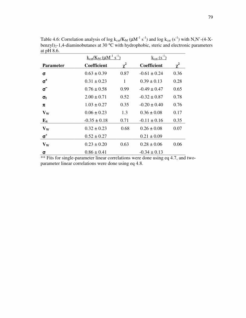
79
Table 4.6: Correlation analysis of log kcat/KM (µM-1 s-1) and log kcat (s-1) with N,N’-(4-X-
benzyl)2-1,4-diaminobutanes at 30 °C with hydrophobic, steric and electronic parameters at pH 8.6.
kcat/KM (µM-1 s-1) kcat (s-1)
Parameter Coefficient χ2 Coefficient χ
2
σσσσ 0.63 ± 0.39 0.87 -0.61 ± 0.24 0.36
σσσσ++++ 0.31 ± 0.23 1 0.39 ± 0.13 0.28
σσσσ−−−− 0.76 ± 0.58 0.99 -0.49 ± 0.47 0.65
σσσσI 2.00 ± 0.71 0.52 -0.32 ± 0.87 0.78
ππππ 1.03 ± 0.27 0.35 -0.20 ± 0.40 0.76
VW 0.06 ± 0.23 1.3 0.36 ± 0.08 0.17
ES -0.35 ± 0.18 0.71 -0.11 ± 0.16 0.35
VW 0.32 ± 0.23 0.68 0.26 ± 0.08 0.07
σσσσ+ 0.52 ± 0.27 0.21 ± 0.09
VW 0.23 ± 0.20 0.63 0.28 ± 0.06 0.06
σσσσ 0.86 ± 0.41 -0.34 ± 0.13 ** Fits for single-parameter linear correlations were done using eq 4.7, and two-parameter linear correlations were done using eq 4.8.
80
DISCUSSION
These studies with N,N’-(4-X-benzyl)2-1,4-diaminobutane present the
opportunity to address the minimal substrate required for PAO recognition. Specifically,
the ability of PAO to oxidize N,N’-(4-X-benzyl)2-1,4-diaminobutane indicates that PAO
requires two nitrogens separated by four carbons, and hence the diaminobutane core with
the appropriate protonation state is what is important for binding and catalysis. Previous
studies of PAO with the natural substrates N1-acetylspermine and N1-acetylspermidine
are consistent with the nitrogen at the site of CH bond cleavage being neutral and one
other nitrogen being positively charged in the reactive form of the substrate (Chapter II).
The kcat/KM-pH profiles described here are consistent with earlier observations using the
natural substrates, that the monoprotonated form of N,N’-(4-X-benzyl)2-1,4-
diaminobutane is the active form of the substrate. Studies of PAO with N1,N12-
bisethylspermine (BESPM) and N1,N11-bis(ethyl)-nor-spermine (BENSPM) reveal that
PAO also recognizes them as substrates and catalyzes their oxidation (26, 79). BESPM
and BENSPM, like the N,N’-(4-X-benzyl)2-1,4-diaminobutanes, contain the
diaminobutane core that exists in the appropriate protonation state for catalysis.
However, spermidine and N8-acetylspermidine, are competitive inhibitors (26) despite
containing the diaminobutane core. In comparing the structures of these polyamines, it
appears as though having the diaminobutane core is the minimal substrate requirement
for substrate recognition, but that length and volume play an important role in orienting
the substrate in a manner compatible for catalysis.
81
The transient kinetic studies indicate that the rate constant kred is slightly lower
than kcat for the N,N’-(4-X-benzyl)2-1,4-diaminobutanes. One explanation for the lower
kred is that the enzyme is slightly less active at higher enzyme concentrations. The
similar kred and kcat values establish that reduction is the rate-limiting step in catalysis
(Table 4.1). Furthermore, the spectral changes observed for all seven N,N’-(4-X-
benzyl)2-1,4-diaminobutanes establish that flavin reduction occurs in a single step, with
no observation of any intermediates. N,N’-(4-X-Benzyl)2-1,4-diaminobutanes are slow
substrates in which chemistry is the rate-limiting step, thus allowing for the build-up of
intermediates if such intermediates exist. The rate constant associated with flavin
reduction for N1-acetylspermine is over 1000-fold greater (Chapter II) than those
observed with N,N’-(4-X-benzyl)2-1,4-diaminobutanes. The kred-pH profiles observed
for N1-acetylspermine displays a decrease in activity at acidic pH with a pKa of 7.3 ±
0.1, consistent with the ability of substrate in the incorrectly protonated form to bind to
the enzyme, but not react. The kred-pH profile for N,N’-(4-H-benzyl)2-1,4-diaminobutane
is a sigmoid curve, indicating that both the protonated and deprotonated forms of the
enzyme-substrate complex are active, but deprotonation enhances activity or that the
proton can be lost from the ES complex at a rate of ~0.2 s-1. The change in activity
between the protonated and deprotonated forms is only ~4-fold. Coupled with the fact
that the isotope effect on kred is pH-independent, this suggests that the deprotonation that
enhances the rate of reduction is a local perturbation that does not affect the chemistry
step. It is feasible that the proton is lost to solvent since it is unlikely a base exists in the
active site to accept the proton. Previous studies of K315M PAO show that this Lys315
82
does not behave as an active site base, and furthermore does not play a role in flavin
reduction (Chapter II). Thus, the loss of a proton to solvent seems the most likely
explanation. With natural substrates that display a rate constant for flavin reduction that
is significantly faster, the rate constant of 0.20 s-1 associated with the loss of a proton
during the transition-state is not observed, indicating that this deprotonation is not
significant for catalysis with physiological substrates. A source of this deprotonation
could be explained by the presence of water molecules that can pack inside the active
site with the N,N’-(4-X-benzyl)2-1,4-diaminobutane substrates that are not present in the
active site with N1-acetylspermine bound.
The isotope effects on kcat/KDBDB and kcat for N,N’-(4-X -benzyl)2-1,4-
diaminobutanes in Table 3 are less than two for the three substrates analyzed and are pH-
independent, indicating that this may be the intrinsic effect. Similar pH-dependent
isotope effects on kred (Table 4.4) are observed in the oxidation of N,N’-(4-X-benzyl)2-
1,4-diaminobutanes by PAO, indicating that there are no external commitments. These
results cumulatively indicate that CH bond cleavage is the rate-limiting step in reduction.
The small magnitude of the isotope effects can be indicative of either an internal
commitment or a very early transition state. Similar small isotope effects have been seen
with other flavoprotein amine oxidases including LSD1 (Chapter VI), DAAO (98), and
TMO (99).
Analysis of single- and two-parameter linear correlations of kred with electronic
(σ, σ+, and σ-), hydrophobic (π), and steric (VW and ES) parameters at the pH optimum
of 8.6 and at pH 6.6 allows for assessment of the charge development during the
83
transition state. Two-parameter linear correlations with VW and σ show the best fits. At
both pH 6.6 and 8.6, the coefficient associated with VW is pH-independent, whereas the
electronic parameter coefficient approaches zero away from the pH optimum. Going
from the pH optimum of 8.6 to the pH value of 6.6 results in an increase in the ρ value
from -0.59 to -0.09; thus, the effect of the substrate amine pKa could be to make the ρ
value more positive at low pH by ~0.5. Therefore, it is likely that the pH-dependence of
the correlation observed with kred and the σ factor at high pH but not a low pH is due to
the pKa of N,N’-(4-X-benzyl)2-1,4-diaminobutanes in the ES complex and not charge
development in the transition state. Another explanation could be the loss of a proton at
a rate of 0.2 s-1 that results from additional water molecules hydrogen-bonded within the
active site of PAO with N,N’-(4-X-benzyl)2-1,4-diaminobutane substrates.
These results with PAO are consistent with studies of oxidation of para-
substituted benzylamine analogues by MAO-B, where a correlation between kred the Taft
steric parameter indicating that steric orientation of the substrate is important for flavin
reduction (46). However, with monoamine oxidase A (45), Miller and Edmondson report
a positive correlation for kred and the electronic parameter σ with a ρ value of 1.8 ± 0.3.
The reason for the differences in the ρ value observed for MAO-A versus MAO-B is not
clear, especially in light of the fact that MAO-A and MAO-B are 70% identical.
Furthermore, results from structure-activity relationships with MAO-A in the oxidation
of phenethylamine analogues show that the kred value decreases with an increase in the
VW parameter of the substituent in a linear manner and that binding affinities of a series
of arylalkylamine analogues have the best linear correlation with the Taft steric
84
parameter of the alkyl side chain (100). No electronic contribution was observed. A
difference between the linear correlations with electronic parameters in the oxidation of
benzylamine and phenethylamine analogues is expected. The presence of the extra CH2
group in phenethylamine would cause a 2-3 fold reduction in the ρ value (101).
However, a significant ρ value is still expected. A possible explanation for the linear
correlations with the electronic parameter σ observed for MAO-A with para-substituted
benzylamine analogues is that the source of the linear correlation is due to the pKa of the
benzylamine substrates, rather than the charge development of ES complex in the
transition state, as is observed with PAO. This is a likely explanation since amine
substrates are able to ionize at the pH value studied, and thus can contribute to
correlations with electronic parameters. As with PAO, the ρ value increases going above
the pKa, -0.59 to -0.09, so the argument that the ρ value may be pH sensitive and the
experiment should be done in the pH-insensitive region is valid.
The mechanism of flavin amine oxidases has been a source of debate for years
with three prominant mechanisms at the center of the controversy including single
electron transfer, polar nucleophilic addition, and hydride transfer mechanisms. The
single electron transfer mechanism proposed by Silverman involves the formation of a
radical intermediate of the flavin or a residue within the enzyme’s active site (Figure 1.8)
(39). The polar nucleophilic mechanism as put forth by Edmondson is a concerted
reaction involving the formation of a 4a-alkylated isoalloxazine ring that activates the
N5 of the flavin for proton abstraction of the substrate α-hydrogen (Figure 1.9) (45).
The hydride transfer mechanism is the simplest mechanism, involving the direct transfer
85
of a hydride from the α-C of the substrate to the N5 position of the flavin (Figure 1.10).
This last mechanism has received greater attention for the MAO/PAO family only in
recent years (47, 49-51, 97) despite the fact that it is and has been the accepted
mechanism for D-amino acid oxidase (48, 66).
A large amount of data has been generated in recent years concerning
flavoprotein amine oxidases. To date, no flavin intermediates have been observed, which
supports the hydride transfer mechanism. 15N kinetic isotope effects for N-
methyltryptophan oxidase (51), TMO (49), and DAAO (48) rule out the polar
nucleophilic mechanism. The small ρ value presented here for PAO with N’-(4-X-
benzyl)2-1,4-diaminobutanes indicates a lack of charge development in the transition
state, consistent with hydride transfer. Interpretation of all this data in terms of the three
potential mechanisms proposed for flavoprotein amine oxidases provides an
overwhelming amount of evidence in favor of the hydride transfer mechanism.
86
CHAPTER V
USE OF pH AND KINETIC ISOTOPE EFFECTS TO ESTABLISH CHEMISTRY
AS RATE-LIMITING IN OXIDATION OF A PEPTIDE SUBSTRATE BY LSD1∗∗∗∗
Nucleosomes, the basic subunits of chromatin, are composed of 146 bp of DNA
wrapped around an octamer of four core histones (H3, H4, H2A and H2B) (102). Each
octamer consists of an H3-H4 histone tetramer plus an H2A-H2B dimer and is connected
to the adjacent octamers via an H1 linker histone (102). Post-translational modifications
of the N-terminal tails of histones, which extend freely beyond the nucleosome core,
have been shown to affect chromatin structure and the accessibility to DNA of proteins
essential for transcription, repair, and replication (103). Lysine-specific demethylase
(LSD1) is an FAD-containing amine oxidase that catalyzes the demethylation of mono-
and dimethylated lysyl residues in the N-terminal tails of histones, thereby playing a role
in the epigenetic regulation of gene transcription in cells (76). LSD1 demethylates lysine
residues 4 and 9 of histone H3 (H3K4 and H3K9) (104, 105); in addition, the enzyme
can demethylate the tumor suppressor p53 (106). The discoveries of LSD1 and of the
JmjC family of iron(II)-alpha-ketoglutarate-dependent histone demethylases (107) have
demonstrated that methylation of histones is not permanent; instead, like other post-
translational modifications, it is part of a reversible process. The effects of histone
methylation vary depending on the site of modification, the degree of methylation, the
∗ Reproduced with permission from Gaweska, H., Henderson Pozzi, M., Schmidt, D. M. Z., McCafferty, D. G., and Fitzpatrick, P.F. (2009) Biochemistry 48, 5440-5445. Copyright 2009 American Chemical Society.
87
sites and nature of adjacent modifications, and the protein complexes in proximity to the
modification (108). LSD1 functions as both a repressor and activator of gene
transcription; for example, demethylation of Lys4 of histone H3 results in gene
repression (109), while androgen-dependent demethylation of mono- or dimethylated
Lys9 results in an activated transcriptional state (105). LSD1 has been implicated in the
maintenance of disease, such as neuroblastoma (110), its expression correlates with
high-risk prostate tumors (111), and it plays regulatory roles in cell differentiation (112,
113).
Flavoprotein oxidases utilize a flavin cofactor to catalyze oxidation of a substrate
CX bond, transferring a hydride equivalent to the flavin; the reduced flavin is then
oxidized by molecular oxygen, producing H2O2 (114). LSD1 oxidizes the carbon-
nitrogen bond between the methyl group and the epsilon amine of lysyl residues,
forming an imine intermediate that is nonenzymatically hydrolyzed to produce
formaldehyde and the demethylated lysine (Figure 5.1). There are two structural families
of flavoprotein amine oxidases; LSD1 belongs to the monoamine oxidase family (32). A
number of mechanisms have been proposed for amine oxidation by flavoenzymes,
including direct transfer of a hydride transfer from the substrate to the flavin, formation
of a substrate carbanion that then transfers electrons to the flavin, and two single electron
transfers to form an intermediate radical pair with a subsequent proton transfer (65).
However, kinetic isotope effects on the reactions of a number of flavoprotein amine and
alcohol oxidases support direct hydride transfer as the mechanism for members of both
families of amine oxidases and for flavoprotein oxidases in general (48, 49, 51, 66, 115).
88
While there have been many studies focusing on the role of LSD1 in gene
transcription and protein expression and its interactions with other proteins, few have
focused on the kinetics of the enzyme. Several mechanism-based inhibitors of
monoamine oxidase and their derivatives have been shown to inhibit LSD1 (109, 116-
120), providing insights into the interactions with substrates and demonstrating the
mechanistic similarity of the enzyme to monoamine oxidase. Forneris et al. have
described a pH profile of LSD1 (121) and analyzed the reaction of the photo-reduced
enzyme with oxygen, demonstrating that the oxidation is typical of a flavoprotein
oxidase (122). Still, there has been no detailed analysis of the reaction with a normal
peptide substrate that allows for conclusions regarding mechanism. LSD1 is highly
specific for the N-terminus of histone H3. Indeed, the minimal substrate is a peptide
composed of the N-terminal 21 amino acid residues of H3 (121). We describe here
mechanistic studies of oxidation of such a peptide substrate by LSD1.
89
CH3
(CH2)4
N
(CH2)4
N
(CH2)4
NH3C CH2
(CH2)4
NH
FAD FADH2
O2H2O2
H2O
OH
HCHO
H3C H3CCH2H3C
Figure 5.1: LSD1 oxidation of lysine in histone3.
90
EXPERIMENTAL PROCEDURES
Materials. Horseradish peroxidase and Ampliflu Red were purchased from
Sigma. Fmoc amino acids were from Novabiochem, Applied Biosystems, or Advanced
ChemTech. 2H-Formaldehyde (20% solution in D2O) was from Cambridge Isotopes.
Sodium cyanoborodeuteride was from CDN Isotopes.
H3K4 21-mer Dimethylated Peptide Synthesis. A peptide corresponding to the
first 21 amino acids of histone H3 with a dimethylated lysine at the fourth residue
(ARTK(diMe)QTARKSTGGKAPRKQLA) was synthesized by Helena Gaweska of
Duke University (123). Similarly, the deuterated dimethylated H3K4 peptide was also
synthesized by Helena Gaweska.
LSD1 Expression and Purification. A truncated form of human LSD1, lacking
the first 150 amino acids (76), was expressed and purified by Helena Gaweska of Duke
University (123). The concentration of LSD1 was determined using the absorbance at
458 nm and an extinction coefficient based on that for free FAD (11,600 cm-1M-1).
Assays. Steady state kinetic assays monitoring oxygen consumption were
performed on a computer-interfaced Hansatech (Hansatech Instruments) or YSI (Yellow
Springs Instrument, Inc.) oxygen electrode. All assays were conducted at 25 °C and were
initiated by the addition of enzyme. A fluorescence assay, coupling hydrogen peroxide
formation to oxidation of Ampliflu Red by horseradish peroxidase, was used to
determine kcat/Km values due to the low concentrations of substrate required. The
product resorufin was detected using a fluorescence plate reader (Molecular Devices
SpectraMax Gemini EM) with an excitation wavelength of 560 nm and emission at 590
91
nm. Experiments done using the fluorescence assay were conducted by Helena Gaweska
of Duke University. The coupled fluorescence assay resulted in initial rates that were
twofold slower than those detected using the oxygen electrode. Consequently, the rates
obtained with the coupled assay were corrected by this factor. The constant ionic
strength buffer 0.1 M Aces, 52 mM Tris, and 52 mM ethanolamine (124) was used for
assays at all pH values. Preincubation of enzyme in buffers of pH higher than 9.5 for 60
minutes eliminated LSD1 activity.
Rapid-reaction kinetic experiments were conducted at 25 ºC on an Applied
Photophysics SX-18MV stopped-flow spectrophotometer. Anaerobic conditions were
established by applying cycles of vacuum and argon to enzyme solutions, while substrate
solutions were bubbled with argon. The buffer was again ACES/Tris/ethanolamine, plus
5 mM glucose, at pH 7.5. Glucose oxidase was added to all anaerobic solutions at a final
concentration of 36 nM before loading them onto the stopped-flow spectrophotometer.
Data Analysis. Steady-state kinetic data were analyzed using the program
KaleidaGraph (Adelbeck Software, Reading, PA). Initial rate data obtained by varying
the concentration of a single substrate were fit to the Michaelis-Menten equation. The
effects of pH on kinetic parameters were fit to eq 5.1, which applies for a kinetic
parameter which decreases below pK1 due to protonation of a single moiety and
decreases above pK2 due to deprotonation of a single moiety. Here, y is the kinetic
parameter being measured, c is its pH-independent value, and K1 and K2 are the
ionization constants for moieties which must be unprotonated or protonated,
respectively. Eq 5.2 was used to determine kinetic isotope effects using the program Igor
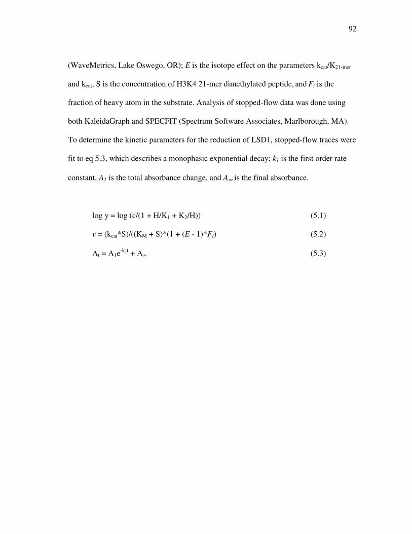
92
(WaveMetrics, Lake Oswego, OR); E is the isotope effect on the parameters kcat/K21-mer
and kcat, S is the concentration of H3K4 21-mer dimethylated peptide, and Fi is the
fraction of heavy atom in the substrate. Analysis of stopped-flow data was done using
both KaleidaGraph and SPECFIT (Spectrum Software Associates, Marlborough, MA).
To determine the kinetic parameters for the reduction of LSD1, stopped-flow traces were
fit to eq 5.3, which describes a monophasic exponential decay; k1 is the first order rate
constant, A1 is the total absorbance change, and A∞ is the final absorbance.
log y = log (c/(1 + H/K1 + K2/H)) (5.1)
v = (kcat*S)/((KM + S)*(1 + (E - 1)*Fi) (5.2)
At = A1e-k1t + A∞ (5.3)
93
RESULTS
Steady-State Kinetics. Steady-state kinetic parameters for LSD1 were determined
at pH 7.5 and 25 °C with the dimethylated H3K4 21-mer as substrate. This peptide has
the sequence of the N-terminal 21 residues of histone H3, the physiological substrate of
LSD1, and contains N, N-dimethyllysine at residue 4. It represents the minimum length
required for detectable activity (121). While the kcat/K21-mer values could be determined
in air-saturated buffer because LSD1 exhibits ping pong kinetics (122), kcat values were
determined with 1.1 mM oxygen and 50 µM peptide to ensure saturation with both
substrates. In addition, the kcat/KO2 values were determined by varying the concentration
of oxygen in the presence of 50 µM peptide. The results are summarized in Table 1. The
kcat and kcat/K21-mer values are in reasonable agreement with the values previously
reported by Forneris et al. (121).
pH Profiles. The effect of pH on the kcat/K21-mer values was determined over the
pH range 7.0–9.5 using a buffer system that maintained constant ionic strength over the
entire pH range. At pH values above 9.5, the enzyme lost activity, while the activity was
too low below pH 7 to be measured reliably. Because these assays are not carried out
with saturating oxygen concentrations, the resulting kcat values were only apparent, and
the kcat-pH profile need not reflect the actual pKa values. The kcat/K21-mer data yield a
bell-shaped pH profile (Figure 5.2), decreasing above and below pH 8.5-9. This behavior
is consistent with the involvement of two groups on the free enzyme or substrate, one of
which must be protonated and one deprotonated for activity. When the data were fit
assuming different pKa values for the two groups, the resulting pKa values were within
94
0.3 of one another (results not shown), too close to reliably discriminate. Consequently,
only the average of the pKa values of the two groups can be determined. The data were
therefore fit using eq 5.1, which assigns the same pKa value to the two groups, to obtain
the average pKa value of 8.7 ± 0.1.
Kinetic Isotope Effects Deuterium kinetic isotope effects were determined with
the 21-mer in which both methyl groups on the dimethylated lysyl residue contained
three deuteriums. The results at pH 7.5 are summarized in Table 5.1. Data obtained by
varying the concentration of the peptide in air-saturated buffer were best fit by eq 5.2,
which assigns equal values to Dkcat/K21-mer and Dkcat, for an isotope effect of 3.2 ± 0.1.
Alternatively, the Dkcat value was determined at fixed saturating concentrations of
peptide and oxygen of 50 µM and 1.1 mM, respectively. The resulting value was 2.9 ±
0.2. The kinetic isotope effect on kcat/K21-mer and kcat was then determined over the pH
range 7-9.5, fitting the data in each case to eq 5.2. The isotope effect was pH-
independent, with an average value of 3.5 ± 0.3 (Table 5.2). The effect of pH on the
kcat/K21-mer value for the deuterated peptide is shown in Figure 5.2; these data were fit to
eq 5.1 to yield an average pKa value of 8.8 ± 0.1, within error of the value for the
nondeuterated peptide.
95
Table 5.1: Kinetic parameters of LSD1 with the H3K4 21-mer dimethylated peptide as a substrate∗
Kinetic parameter Value kcat/K21-mer (mM-1 s-1) 38 ± 2a kcat/KO2 (mM-1 s-1) 1.0 ± 0.2b K21-mer (µM) 2.6 ± 0.2a
KO2 (µM) 195 ± 40b
kcat (s-1) 0.199 ± 0.013c
kred (s-1) 0.231 ± 0.004e
D(kcat/K21-mer) 3.2 ± 0.1a,d
Dkcat 2.9 ± 0.2c
3.2 ± 0.1a,d Dkred
3.3 ± 0.1e
∗Conditions: pH 7.5, 25 °C. aDetermined in air-saturated buffer. bDetermined using 50 µM H3K4 21-mer dimethylated peptide. cDetermined using 50 µM H3K4 21-mer dimethylated peptide and 1.1 mM oxygen. dCalculated using eq 5.2. eDetermined using 200 µM 21-mer dimethylated peptide.
96
-4
-3
-2
-1
7 8 9 10
log
(k
cat/K
21m
er)
(µM
-1 s
-1)
pH
Figure 5.2: kcat/KM pH profiles for LSD1 with protiated (closed circles) and deuterated (open squares) H3K4 21-mer dimethylated peptide. The lines are from fits of the data to eq 5.1.
Table 5.2: Effect of pH on the deuterium kinetic isotope effects on the oxidation of the H3K4 21-mer dimethylated peptide by LSD1*
pH Kinetic isotope effect 7.0 4.0 ± 0.2 7.5 3.2 ± 0.1 8.0 3.3 ± 0.1 8.5 3.4 ± 0.1 9.0 3.7 ± 0.2 9.5 3.6 ± 0.1
Average 3.5 ± 0.3 *Determined in air saturated buffers and calculated using eq 5.2.
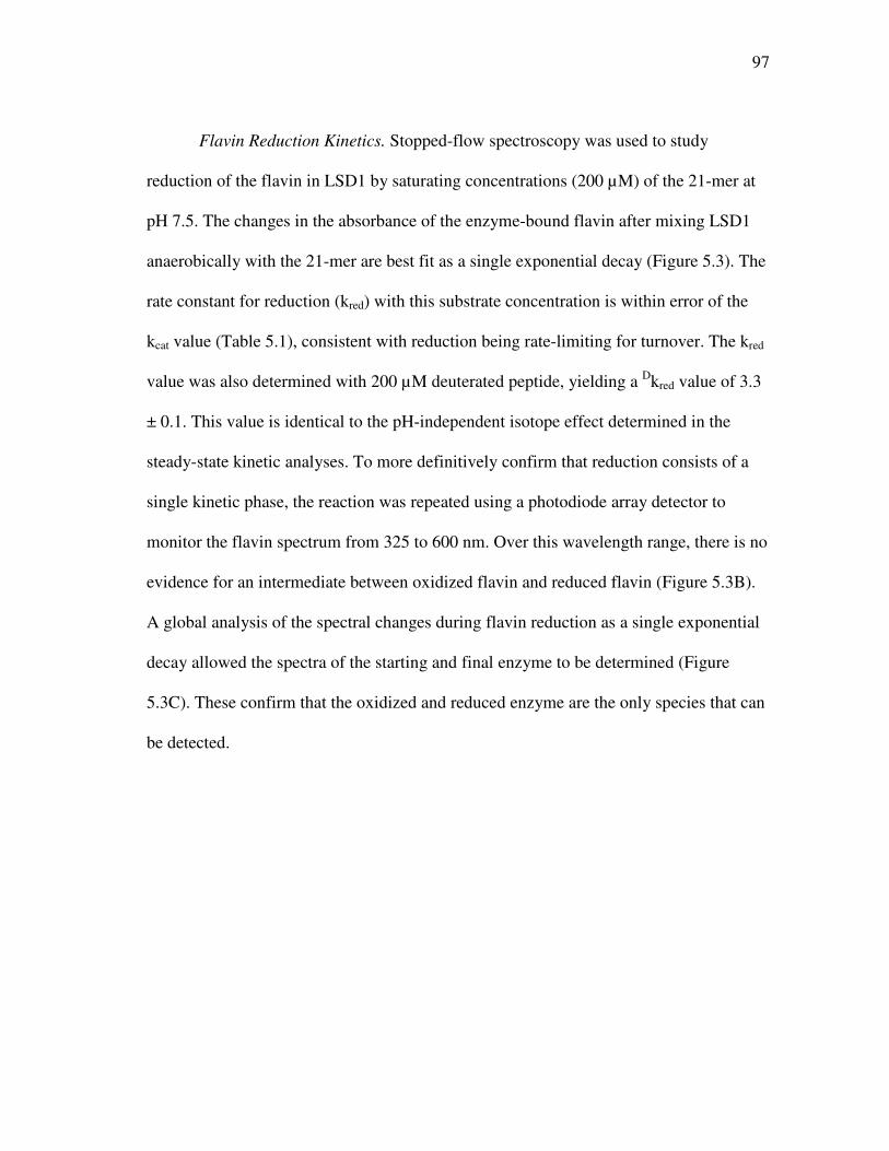
97
Flavin Reduction Kinetics. Stopped-flow spectroscopy was used to study
reduction of the flavin in LSD1 by saturating concentrations (200 µM) of the 21-mer at
pH 7.5. The changes in the absorbance of the enzyme-bound flavin after mixing LSD1
anaerobically with the 21-mer are best fit as a single exponential decay (Figure 5.3). The
rate constant for reduction (kred) with this substrate concentration is within error of the
kcat value (Table 5.1), consistent with reduction being rate-limiting for turnover. The kred
value was also determined with 200 µM deuterated peptide, yielding a Dkred value of 3.3
± 0.1. This value is identical to the pH-independent isotope effect determined in the
steady-state kinetic analyses. To more definitively confirm that reduction consists of a
single kinetic phase, the reaction was repeated using a photodiode array detector to
monitor the flavin spectrum from 325 to 600 nm. Over this wavelength range, there is no
evidence for an intermediate between oxidized flavin and reduced flavin (Figure 5.3B).
A global analysis of the spectral changes during flavin reduction as a single exponential
decay allowed the spectra of the starting and final enzyme to be determined (Figure
5.3C). These confirm that the oxidized and reduced enzyme are the only species that can
be detected.
98
0.06
0.07
0.08
0.09
0.1
0.01 0.1 1 10 100
AA
45
8
Time (s)
0
0.02
0.04
0.06
0.08
0.1
350 400 450 500 550 600
C
Ab
so
rba
nc
e
Wavelength (nm)
Figure 5.3: Spectral changes during reduction of LSD1 by the H3K4 21-mer dimethylated peptide. A, time course at 458 nm with protiated (closed circles) or deuterated (open circles) substrate (only 1/30th of the points are shown for clarity); B, Changes in the entire visible absorbance spectrum during reduction by the deuterated substrate; C, Initial (─) and final (…) spectra upon LSD1 flavin reduction by deuterated H3K4 21-mer dimethyl peptide obtained from a global analysis of the data in B using a single-step kinetic model. The lines in A are from fits of the data to eq 5.3.
99
DISCUSSION
While the physiological substrate for LSD1 is the histone H3, mechanistic
studies with the entire protein substrate present significant problems, including the
difficulty of preparing the dimethylated substrate. Consequently, mechanistic insight
requires the use of a minimal substrate which still retains the specificity determinants of
the larger protein. LSD1 has no detectable activity with peptides containing fewer than
16 of the N-terminal residues of histone H3, and a peptide containing the first 21
residues, as was utilized here, is as active a substrate as a peptide containing the first 30
residues (121). Epigenetic modifications of histone H3 that alter the reactivity of the
intact protein with LSD1 cause similar decreases in activity when incorporated into a 21-
mer (122), establishing the latter as a valid model for the larger protein substrate. Indeed,
incorporation of a propargyl moiety onto Lys4 of the 21-mer yields an efficient
mechanism-based inhibitor (116). The three-dimensional structure of the inactivated
enzyme shows extensive contacts with the first 7 residues, while the remainder of the
peptide is not visible (118). This combination of solution and structural studies
establishes the validity of the 21-mer as a model for mechanistic studies.
The steady-state and transient kinetic studies presented here establish that the
rate-limiting step in oxidation of the dimethylated H3K4 21-mer by LSD1 is CH bond
cleavage. The kred value, the rate constant for flavin reduction, is identical to the kcat
value for LSD1. Therefore, the reductive half reaction is rate-limiting for turnover with
this peptide substrate. The identity of the primary deuterium isotope effects on kcat/K21-
mer, kcat, and kred further identifies CH bond cleavage as the rate limiting step. The pH-
100
independence of the isotope effect on kcat/K21-mer is also consistent with fully rate-
limiting CH bond cleavage and suggests that this isotope effect is the intrinsic deuterium
isotope effect (125). An isotope effect of 3.2 ± 0.1 is substantially smaller than the
limiting semi-classical value of about 7 for a primary deuterium isotope effect (126); in
the present case, the value also includes the secondary isotope effects from the two other
deuterium atoms on each methyl group. The small magnitude of the isotope effect
suggests that the oxidation of this substrate by LSD1 has an early transition state. A late
transition state would also yield a small isotope effect, but a late transition state would
be expected for an unfavorable reaction. In contrast, at all concentrations of substrate
used here, the reductive half-reaction goes to completion rather than reaching an
equilibrium suggesting that the oxidation of the 21-mer is favorable. Similarly small
kinetic isotope effects have been reported for other flavin amine oxidases, including D-
amino acid oxidase with glycine as substrate (98) and tryptophan 2-monooxygenase with
tryptophan as substrate (99).
No intermediates between oxidized and reduced enzyme were observed in the
oxidation of the 21-mer by LSD1. The lack of an intermediate with a substrate whose
rate-limiting step is CH bond cleavage is not consistent with several mechanisms
proposed for flavin amine oxidases, especially mechanisms involving easily detectable
flavin radicals or covalent substrate-flavin adducts. The present results are fully
consistent with direct hydride transfer as the mechanism of LSD1, in line with
observations from other flavoprotein amine oxidases. LSD1 is inactivated by
cyclopropyl substrates (120), and inactivation by such compounds is frequently taken as
101
evidence for radical intermediates (39). However, a number of flavoprotein oxidases for
which hydride transfer is the accepted mechanism can be inactivated by cyclopropyl
substrates (42, 127).
The kinetic parameter kcat/K21-mer exhibits a pH optimum of 8.7 in the studies
described here. While the kcat/Km value for a substrate can reflect the protonation states
of both the enzyme and substrate required for productive binding and catalysis, recent
studies with the related flavoprotein polyamine oxidase suggest that the kcat/Km-pH
profile reflects the required protonation state of the substrate. With N1-acetylspermine,
N1-acetylspermidine, and spermine as the substrate for mammalian polyamine oxidase,
the kcat/Kamine-pH optimum matches the pH at which the substrate has only one
positively charged nitrogen (Chapter II). The pH dependence of the inhibition by
spermidine analogues further established that the nitrogen in the CN bond of the
polyamine being oxidized by polyamine oxidase must be uncharged for oxidation,
consistent with observations of other amine oxidases (48-50) and with the expectations
for a hydride transfer mechanism. With both polyamine oxidase (Chapter II) and
monoamine oxidase (83), the need for the nitrogen at the reaction site to be neutral is
clearly seen in the pH dependence of the reductive half reaction of the enzyme,
demonstrating the requirement for a neutral nitrogen for catalysis. The substrate used in
the present study contains the first 21 residues of the N-terminus of histone H3. Based
on the precedents of other amine oxidases, the dimethylated Lys4 must be uncharged for
catalysis and therefore is responsible for the acidic limb of the pH profile. Protonation of
Lys4 does not prevent binding since the trimethylated analog of this peptide binds tightly
102
to LSD1, although it is not a substrate (121). The basic limb of the kcat/K21-mer-pH profile
can be attributed to one of the three other lysyl residues in the substrate.
The members of the monoamine oxidase family, including LSD1, contain a
conserved lysyl residue in the active site that is part of a “Lys-H2O-N5” structural motif
(32, 34, 73-76). This residue clearly could be a source of a pKa in the kcat/K21-mer-pH
profile. Mutation of this lysine in LSD1 to an alanine results in a complete loss of
activity (87), and in maize polyamine oxidase the corresponding lysine to methionine
mutation results in a 1400-fold decrease in kred (59). However, the same lysine to
methionine mutation in mouse polyamine oxidase results in no change in the kcat/Kamine-
pH profile and only a slight decrease in the value of kred (Chapter II), suggesting that this
residue plays a subtle structural rather than a catalytic role in these enzymes and
therefore does not contribute to the pH dependence.
The rate constant for the chemical step in LSD1 is 2-5 orders of magnitude
slower than values reported for other flavoprotein amine oxidases (46, 59, 86, 128-130).
This suggests that evolution has not optimized catalysis for this enzyme. Instead, it is
likely that the requirements for rapid catalysis are secondary to high specificity in this
case. Given the physiological role of LSD1 in regulation of gene expression, high
catalytic activity would be much less critical than very high specificity. Moreover, while
high catalytic activity is necessary for metabolic enzymes to maintain balance among
metabolic pathways, epigenetic regulation such as DNA histone methylation operates on
a very long time scale and would not require rate constants on the order of 102-104 s-1.
103
CHAPTER VI
SUMMARY
The purpose of this study is to determine the chemical mechanism by which
mammalian polyamine oxidase (PAO) catalyzes the oxidation of polyamines. PAO
catalyzes the oxidation of N1-acetylspermine and N1-acetylspermidine to produce N-
acetyl-3-aminopropanaldehyde and spermidine or putrescine. Structurally, PAO is a
member of the monoamine oxidase family of flavoproteins. The three mechanisms
proposed for flavin amine oxidases are a single electron transfer mechanism producing a
radical intermediate, a polar nucleophilic mechanism forming a covalent adduct
intermediate, and a direct hydride transfer.
The effects of pH on kinetic parameters for mouse PAO were determined to gain
insight into the protonation state of the polyamine required for catalysis and the roles of
ionizable residues in the active site in amine oxidation. The kcat/Kamine-pH profiles are
bell-shaped for N1-acetylspermine, N1-acetylspermidine, and spermine, with each of the
profiles being consistent with the productive form of the substrate possessing a single
positively charged nitrogen. The pKi-pH profiles for a series of polyamine analogs are
most consistent with the reactive form of the substrate having a neutral nitrogen at the
site of oxidation and one positively charged nitrogen. With N1-acetylspermine as the
substrate, the value of the limiting rate constant for flavin reduction, kred, is pH
dependent, decreasing below a pKa value of 7.3, offering further validation for the
requirement of an uncharged nitrogen for substrate oxidation. In PAO, Lys315
104
corresponds to a conserved active site residue found throughout the monoamine oxidase
family. Mutating of Lys315 to methionine does not affect the kcat/Kamine profile for
spermine, the kred value with N1-acetylspermine is only decreased by 1.8-fold compared
to wild-type enzyme, and the pKa of the kred-pH profile with N1-acetylspermine shows a
basic shift to 7.8. These results rule out Lys315 as a source of a pKa in the kcat/Kamine or
kcat/kred profiles and establish that this residue does not play a critical role in amine
oxidation by PAO.
Steady-state kinetic parameters and the pH-dependence of kcat/KO2 for wild-type
and K315M PAO establish that Lys315 plays a role in the oxidative half-reaction. A 30-
fold decrease in kcat/KO2 is observed for the mutant enzyme. The kcat/KO2-pH profiles for
wild-type and K315M PAO indicate that the unprotonated form of Lys315 is required
for optimal catalysis. Solvent isotope effects and proton inventories of wild-type and
K315M PAO suggest that the absence of this lysyl residue can be overcome by
replacement with a water molecule. The “Lys-H2O-N5” structural motif identified in
members of the MAO structural family, in PAO must involve a hydrogen-bond network
between unprotonated Lys315 and the active site water molecule. Thus it is conceivable
that Lys315 enhances the rate of oxidation of reduced flavin by facilitating the removal
of the proton from the N5 position of reduced FAD.
The influence that substituents in the aromatic ring of N,N’-(4-X-benzyl)2-1,4-
diaminobutanes have on steady-state kinetic parameters and the kred value for PAO is the
focus of Chapter IV. The kcat/KM-pH profiles for N, N’-dibenzyl-1,4-diaminobutane and
the CH3O- and CF3-substituted compounds all show a bell-shaped curve, consistent with
105
the monoprotonated form of the substrate being required for binding and catalysis. The
rate constants of flavin reduction are >1000-fold slower than those observed for natural
substrates, and kinetics with all analogues reveal that the rate-limiting step in turnover is
flavin reduction. The kred-pH profile displays a sigmoidal shape, with a pKa of 8.2 ± 0.3.
Isotope effects on kcat, kcat/KM, and kred, are pH-independent, with an average value of 2,
indicating an internal commitment or an early transition state. The kred values for N,N’-
(4-X-benzyl)2-1,4-diaminobutanes exhibit a correlation with the van der Waals volume
(VW) of the substituent that is pH independent, and with the σ parameter. The ρ value is
-0.059 at pH 8.6, and -0.09 at pH 6.6. The change in the ρ value as a function of pH can
be attributed to changes in pKa of the N,N’-(4-X-benzyl)2-1,4-diaminobutane substrates
in the ES complex. The data presented here are best interpreted as further support for the
hydride transfer mechanism.
The mechanism of oxidation of a peptide substrate by the flavoprotein lysine-
specific demethylase (LSD1) was examined using the effects of pH and isotopic
substitution on steady-state and rapid-reaction kinetic parameters as presented in Chapter
V. The peptide substrate is composed of the first 21 residues of the N-terminal tail of
histone H3, with a dimethylated lysyl residue at position 4. At pH 7.5, kred equals kcat,
indicating that the reductive half reaction is rate-limiting at physiological pH.
Deuteration of the lysyl methyls results in identical kinetic isotope effects of 3.1 ± 0.2 on
the kred, kcat and kcat/Km values for the peptide, establishing CH bond cleavage as rate-
limiting with this substrate. No intermediates between oxidized and reduced flavin are
detectable by stopped-flow spectroscopy, consistent with the expectation for a direct
106
hydride transfer mechanism. The kcat/Km pH-profile for the peptide is bell-shaped,
consistent with a requirement that the nitrogen at the site of oxidation be uncharged and
that at least one of the other lysyl residues be charged for catalysis. The D(kcat/Km) value
for the peptide is pH-independent, suggesting that the observed value is the intrinsic
deuterium kinetic isotope effect for oxidation of this substrate.
107
REFERENCES
(1) van Leeuwenhoek, A. (1678) Observations D. Anthonii Leeuwenhoek, de Natis e
semine genitali Animalculis. Philos. Trans. R. Soc. London 12, 1040-1043.
(2) Dudley, H. W., Rosenheim, M.C., and Rosenheim, O. (1924) The chemical
constitution of spermine. 1. The isolation of spermine from animal tissues and
the preparation of its salts. Biochem. J. 18, 1263-1272.
(3) Matthews, H. R. (1993) Polyamine, chromatin structure and transcription.
BioEssays 15, 561-567.
(4) Tabor, C. W., and Tabor, H. (1984) Polyamines. Annu. Rev. Biochem. 53, 749-
790.
(5) Xaio, L., Swank, R.A., and Matthews, H.R. (1991) Photoaffinity polyamines:
Sequence specific interactions with DNA. Nucleic Acid Res. 19, 3701-3708.
(6) Basu, H. S., Wright, W. D., Deen, D. F., Roti-Roti, J., and Marton, L. J. (1993)
Treatment with a polyamine analogue alters DNA matrix association in HeLa
cell nuclei: A nucleoid halo assay. Biochemistry 32, 4073-4076.
(7) Morgan, J. E., Blankenship, J. W., and Matthews, H. R. (1987) Polyamines and
acetylpolyamines increase the stability and alter the conformation of nucleosome
core particles. Biochemistry 26, 3643-3649.
(8) Wallace, H. M., Fraser, A. V., and Hughes, A. (2003) A perspective of
polyamine metabolism. Biochem. J. 376, 1-14.
108
(9) Wallace, H. M. (1996) Polyamines in human health. Proc. Nutr. Soc. 55, 419-
431.
(10) Soulet, D., Gagnon, B., Rivest, S., Audette, M., and Poulin, R. (2004) A
fluorescent probe of polyamine transport accumulates into intracellular acidic
vesicles via a two-step mechanism. J. Biol. Chem. 279, 49355-49366.
(11) Belting, M., Mani, K., Jonsson, M., Cheng, F., Sandgren, S., Jonsson, S., Ding,
K., Delcros, J.G., and Fransson, L.A. (2003) Glypican-1 is a vehicle for
polyamine uptake in mammalian cells. J. Biol. Chem. 278, 47181-47189.
(12) Seiler, N., Delcros, J.G., and Moulinoux, J.P. (1996) Polyamine transport in
mammalian cells. An update. Int. J. Biochem. Cell. Biol. 28, 843-861.
(13) Bolkenius, F. N., and Seiler, N. (1981) Acetyl derivatives as intermediates in
polyamine catabolism. Int. J. Biochem. 13, 287-292.
(14) Kingsnorth, A. N., and Wallace, H. M. (1985) Elevation of monoacetylated
polyamines in human breast cancers. J. Cancer Clin. Oncol. 21, 1057-1062.
(15) Wallace, H. M., Duthie, J., Evans, D.M., Lamond, S., Nicoll, K.M., and Heys,
S.D. (2000) Alterations in polyamine catabolic enzymes in human breast cancer
tissue. Clin. Cancer Res. 6, 3657-3661.
(16) Parchment, R. E., and Pierce, G. B. (1989) Polyamine oxidation, programmed
cell death, and regulation of melanoma in the murine embryonic limb. Cancer
Res. 49, 6680-6686.
(17) Averill-Bates, D. A., Agostinelli, E., Przybytkowski, E. and Mondovi, B. (1994)
Aldehyde dehydrogenase and cytotoxicity of purified bovine serum amine
109
oxidase and spermine in Chinese hamster ovary cells. Biochem. Cell Biol. 72, 36-
42.
(18) Wallace, H. M., and Fraser, A.V. (2004) Inhibitors of polyamine metabolism:
Review article. Amino Acids 26, 353-365.
(19) Gerner, E. W. a. M., P.S. (1986) Restoration of polyamine contents in rat
hepatoma (HTC) cells after inhibition of polyamine biosynthesis: Relationship
with cell proliferation. Eur. J. Biochem. 156, 31-35.
(20) Byers, T. L., Ganem, B., and Pegg, A.E. (1992) Cytostasis induced in L1210
murine leukaemia cells by the S-adenosyl-L-methionine decarboxylase inhibitor
5'([(Z)-4-amino-2-butenyl]methylamino)-5'-deoxyadenosine may be due to
hypusine depletion. Biochem. J. 287, 717-724.
(21) Pegg, A. E. (1986) Recent advances in the biochemistry of polyamines in
eukaryotes. Biochem. J. 234, 249-262.
(22) Karvonen, E., Kauppinen, L., Partanen, T., and Poso, H. (1985) Irreversible
inhibition of putrescine-stimulated S-adenosyl-L-methionine decarboxylase by
berenil and pentamidine. Biochem. J. 231, 165-169.
(23) Wolff, A. C., Armstrong, D.K., Fetting, J.H., Carducci, M.K., Riley, C.D.,
Bender, J.F., Casero, Jr, R.A., and Davidson, N.E. (2003) A Phase II study of the
polyamine analog N1,N11-diethylnorspermine (DENSpm) daily for five days
every 21 days in patients with previously treated metastatic breast cancer. Clin.
Cancer Res. 9, 5922-5928.
110
(24) Lin, P. K. T., Dance, A.M., Bestwick, C., and Milne, L. (2003) The biological
activities of new polyamine derivatives as potential therapeutic agents. Biochem.
Soc. Trans. 31, 407-410.
(25) Ha, H. C., Woster, P.M., Yager, J.D, and Casero, Jr., R.A. (1997) The role of
polyamine catabolism in polyamine analogue-induced programmed cell death.
Proc. Natl. Acad. Sci. U.S.A. 94, 11557-11562.
(26) Wu, T., Yankovskaya, V., and McIntire, W. S. (2003) Cloning, sequencing, and
heterologous expression of the murine peroxisomal flavoprotein, N1-acetylated
polyamine oxidase. J. Biol. Chem. 278, 20514-20525.
(27) Houen, G., Bock, K., and Jensen, A.L. (1994) HPLC and NMR investigation of
the serum amine oxidase catalyzed oxidation of polyamines. Acta. Chem. Scand.
48, 52-60.
(28) Vujcic, S., Liang, P., Diegelman, P., Kramer, D. L., and Porter, C. W. (2003)
Genomic identification and biochemical characterization of the mammalian
polyamine oxidase involved in polyamine back-conversion. Biochem. J. 370, 19-
28.
(29) Silverman, R. B. (2002) The organic chemistry of enzyme-catalyzed reactions,
Elsevier Sciences, San Diego, California.
(30) Gadda, G., and Fitzpatrick, P.F. (1998) Biochemical and physical
characterization of the active FAD-containing form of nitroalkane oxidase from
Fusarium oxysprum. Biochemistry 37, 6154-6164.
111
(31) Kearney, E. B., Salach, J.I., Walker, W.H., Seng, R.L., Kenney, W., Zeszotek,
E., and Singer, T.P. (1971) The covalently-bound flavin of hepatic monoamine
oxidase. 1. Isolation and sequence of a flavin peptide and evidence for binding at
the 8alpha position. Eur. J. Biochem. 24, 321-327.
(32) Chen, Y., Yang, Y., Wang, F., Wan, K., Yamane, K., Zhang, Y., and Lei, M.
(2006) Crystal structure of human histone lysine-specific demethylase 1 (LSD1).
Proc. Natl. Acad. Sci. U.S.A. 103, 13956-13961.
(33) Landry, J., and Sternglanz, R. (2003) Yeast Fms1 is a FAD-utilizing polyamine
oxidase. Biochem. Biophys. Res. Commun. 303, 771-776.
(34) Pawelek, P. D., Cheah, J., Coulombe, R., Macheroux, P., Ghisla, S., and
Vrielink, A. (2000) The structure of L-amino acid oxidase reveals the substrate
trajectory into a enantiomerically conserved active site. EMBO J. 19, 429404215.
(35) Sobrado, P., and Fitzpatrick, P.F. (2003) Analysis of the role of the active site
residue Arg98 in the flavoprotein tryptophan 2-monooxygenase, a member of the
L-amino oxidase family. Biochemistry 42, 13826-13832.
(36) Knoll, J. (1992) Pharmacological basis of the therapeutic effect of (-)deprenyl in
age-related neurological diseases. Med. Res. Rev. 12, 505-524.
(37) Rinne, U. (1987) R-(-)-deprenyl as an adjuvant to levodopa in the treatment of
Parkinson's disease. J. Neural. Transm. 25 Suppl, 149-155.
(38) Edmondson, D. E., Mattevi, A., Binda, C., Li, M., and Hubalek, F. (2004)
Structure and mechanism of monoamine oxidase. Curr. Med. Chem. 11, 1983-
1993.
112
(39) Silverman, R. B. (1995) Radical ideas about monoamine oxidase. Acc. Chem.
Res. 28, 335-342.
(40) Newton-Vinson, P., and Edmondson, D. E. (1999) High-level expression,
structural, kinetic and redox characterization of recombinant human liver
monoamine oxidase B., in Flavins and Flavoproteins (Ghisla, S., Kroneck, P.,
Macheroux, P., and Sund, H., Ed.) pp 431, Agency for Scientific Publications,
Berlin.
(41) Sherry, B., and Abeles, R.H. (1985) Mechanism of action of methanol oxidase,
reconstitution of methanol oxidase with 5-deazaflavin, and inactivation of
methanol oxidase by cyclopropanol. Biochemistry 24, 2594-2605.
(42) McCann, A. E., and Sampson N.S. (2002) A C6-flavin adduct Is the major
product of irreversible inactivation of cholesterol oxidase by 2α,3α-
cyclopropano-5α-cholestan-3β-ol. J. Am. Chem. Soc. 122, 35-39.
(43) Hamilton, G. A. (1971) Proton in biological redox reactions. Prog. Bioorg.
Chem. 1, 83-157.
(44) Edmondson, D. E., Binda, C., Mattevi, A. (2007) Structural insights into the
mechanism of amine oxidation by monoamine oxidases A and B. Arch. Biochem.
Biophys. 264, 269-276.
(45) Miller, J. R., and Edmondson, D. E. (1999) Structure-activity relationships in the
oxidation of para-substituted benzylamine analogues by recombinant human
liver monoamine oxidase A. Biochemistry 38, 13670-13683.
113
(46) Walker, M. C., and Edmondson, D.E. (1994) Structure-activity relationships in
the oxidation of benzylamine analogues by bovine liver mitochondrial
monoamine oxidase B. Biochemistry 33, 7088-7098.
(47) Denu, J. M., and Fitzpatrick, P. F. (1994) Intrinsic primary, secondary, and
solvent kinetic isotope effects on the reductive half-reaction of D-amino acid
oxidase: evidence against a concerted mechanism. Biochemistry 33, 4001-4007.
(48) Kurtz, K. A., and Fitzpatrick, P.F. (2000) Nitrogen isotope effects as probes of
the mechanism of D-amino acid oxidase. J. Am. Chem. Soc 122, 12896-12897.
(49) Ralph, E. C., Anderson, M. A., Cleland, W. W., and Fitzpatrick, P. F. (2006)
Mechanistic studies of the flavoenzyme tryptophan 2-monooxygenase:
Deuterium and 15N kinetic isotope effects on alanine oxidation by an L-amino
acid oxidase. Biochemistry 45, 15844-15852.
(50) Ralph, E. C., and Fitzpatrick, P. F. (2005) pH and kinetic isotope effects on
sarcosine oxidation by N-methyltryptophan oxidase. Biochemistry 44, 3074-
3081.
(51) Ralph, E. C., Hirschi, J. S., Anderson, M. A., Cleland, W. W., Singleton, D. A.,
and Fitzpatrick, P. F. (2007) Insights into the mechanism of the flavoprotein-
catalyzed amine oxidation from nitrogen isotope effects of the reaction of N-
methyltryptophan oxidase. Biochemistry 46, 7655-7664.
(52) Mattevi, A., Vanoni, M.A., Todone, F., Rizzi, M., Teplyakov, A., Coda, A.,
Bolognesi, M., and Curti, B. (1996) Crystal Structure of D-amino acid oxidase: A
114
case of active site mirror-image convergent evolution with flavocytochrome b2.
Proc. Natl. Acad. Sci. U.S.A. 93, 7496-7501.
(53) Sobrado, P., and Fitzpatrick, P.F. (2002) Analysis of the roles of amino acid
residues in the flavoprotein tryptophan 2-monooxygenase modified by 2-oxo-3-
pentynoate: Characterization of His338, Cys339, and Cys511 mutant enzymes.
Arch. Biochem. Biophys. 402, 24-30.
(54) Marton, L. J., and Pegg, A.E. (1995) Polyamines as targets for therapeutic
intervention. Annu. Rev. Pharmacol. Toxicol. 35, 55-91.
(55) Wang, Y., and Casero, R.A., Jr. (2006) Mammalian polyamine catabolism: a
therapeutic target, a pathological problem, or both? J. Biochem. (Tokyo) 139, 17-
25.
(56) Vujcic, S., Diegelman, P., Bacchi, C.J., Kramer, D.L., and Porter, C.W. (2002)
Identification and characterization of a novel flavin-containing spermine oxidase
of mammalian cell origin. Biochem. J. 367, 665-667.
(57) Wang, Y., Murray-Stewert, T., Devereux, W., Hacker, A., Frydman, B., Woster,
P.M. and Casero, R.A., Jr. (2003) Properties of purified recombinant human
polyamine oxidase, PAOh1/SMO. Biochem. Biophys. Res. Commun. 304.
(58) Cervelli, M., Polticelli, F., Federico, R., and Mariottini, P. (2003) Heterologous
expression and characterization of mouse spermine oxidase. J. Biol. Chem. 278,
5271-5276.
(59) Polticelli, F., Basran, J., Faso, C., Cona, A., Minervini, G., Angeline, R.,
Federico, R., Scrutton, N. S., and Tavladoraki, P. (2005) Lys300 plays a major
115
role in the catalytic mechanism of polyamine oxidase. Biochemistry 44, 16108-
16120.
(60) Binda, C., Mattevi, A., and Edmondson, D. E. (2002) Structure-function
relationships in flavoenzyme-dependent amine oxidases. J. Biol. Chem. 277,
23973-23976.
(61) Binda, C., Angelini, R., Federico, R., Ascenzi, P., and Mattevi, A. (2001)
Structural bases for inhibitor binding and catalysis in polyamine oxidase.
Biochemistry 40, 2766-2776.
(62) Bellelli, A., Angelini, R., Laurenzi, M., and Federico, R. (1997) Transient
kinetics of polyamine oxidase from Zea mays L. Arch. Biochem. Biophys. 343,
146-148.
(63) Sebela, M., Radova, A., Angelini, R., Tavladoraki, P., Frebort, I.I., and Pec, P.
(2001) FAD-containing polyamine oxidases: A timely challenge for researchers
in biochemistry and physiology of plants. Plant Sci. 160, 197-207.
(64) Bright, H. J., and Porter, D.J.T. (1975) Flavoprotein oxidases, in The Enzymes
(Boyer, P., Ed.) pp 421-505, Academic Press, New York.
(65) Fitzpatrick, P. F. (2001) Substrate dehydrogenation by flavoproteins. Acc. Chem.
Res. 42, 299-307.
(66) Fitzpatrick, P. F. (2007) Insights into the mechanisms of flavoprotein oxidases
from kinetic isotope efects. J. Label Compd. Radiopharm. 50, 1016-1025.
(67) Scrutton, N. S. (2004) Chemical aspects of amine oxidation by flavoprotein
enzymes. Nat. Prod. Rep. 21, 722-730.
116
(68) Zhao, G., and Jorns, M.S. (2006) Spectral and kinetic characterization of the
Michaelis charge transfer complex in monomeric sarcosine oxidase.
Biochemistry 45, 5985-5992.
(69) Harris, C. M., Pollegioni, L., and Ghisla, S. (2001) pH and kinetic isotope effects
in D-amino acid oxidase catalysis. Eur. J. Biochem. 268, 5504-5520.
(70) Zhao, G., Song, H., Chen, Z., Mathews, S., and Jorns, M.S. (2002) Monomeric
sarcosine oxidase: Role of histidine 269 in catalysis. Biochemistry 41, 9751-
9764.
(71) Jones, T. Z., Balsa, D., Unzeta, M., and Ramsay, R.R. (2007) Variations in
activity and inhibition with pH: The protonated amine is the substrate for
monoamine oxidase, but uncharged inhibitors bind better. J. Neural Transm. 113,
707-712.
(72) Trickey, P., Wagner, M.A., Jorns, M.S., and Mathews, F.S. (1999) Monomeric
sarcosine oxidase: Role of histidine 269 in catalysis. Structure 7, 331-345.
(73) Binda, C., Coda, C., Angelini, R., Ascenzi, P., and Mattevi, A. (1999) A 30-Å
long U-shaped catalytic tunnel in the crystal structure of polyamine oxidase.
Structure 7, 265-276.
(74) Huang, Q., Liu, Q., and Hao, Q. (2005) Crystal structures of Fms1 and its
complex with spermine reveal substrate specficity. J. Mol. Biol. 348, 951-959.
(75) Binda, C., Newton-Vinson, P., Hubalek, F., Edmondson, D.E., and Mattevi, A.
(2002) Structure of human monoamine oxidase B, a drug target for the treatment
of neurological disorders. Nat. Struct. Biol. 9, 22-26.
117
(76) Ma, J., Yoshimura, M., Yamashita, E., Nakagawa, A., Ito, A., and Tsukihara, T.
(2004) Structure of rat monoamine oxidase A and its specific recognitions for
substrates and inhibitors. J. Mol. Biol. 338, 103-114.
(77) Gawandi, V., and Fitzpatrick, P.F. (2007) The synthesis of deuterium-labeled
spermine, N1-acetylspermine and N1-acetylspermidine. J. Label Compd.
Radiopharm. 50, 666-670.
(78) Henderson Pozzi, M., Gawandi, V., and Fitzpatrick, P.F. (2009) pH Dependence
of a mammalian polyamine oxidase: Insights into substrate specificity and the
role of lysine 315. Biochemistry 48, 1508-1516.
(79) Royo, M., and Fitzpatrick, P.F. (2005) Mechanistic studies of mouse polyamine
oxidase with N1,N12-bisethylspermine as a substrate. Biochemistry 44, 7079-
7084.
(80) Frassineti, C., Ghelli, S., Gans, P., Sabatini, A., Moruzzi, M.S., and Vacca, A.
(1995) Nuclear magnetic resonance as a tool for determining protonation
constants of natural polyprotic bases in solution. Anal. Biochem. 231, 374-382.
(81) Bencini, A., Bianchi, A., Garcia-Espana, E., Micheloni, M., and Remirez, J.A.
(1999) Proton coordination by polyamine compounds in aqueous solution.
Coord. Chem. Rev. 188, 97-156.
(82) Zhao, G., and Jorns, M.S. (2005) Ionization of zwitterionic amine substrates
bound to monomeric sarcosine oxidase. Biochemistry 44, 16866-16874.
(83) Dunn, R. V., Marshall, K.R., Munro, A.W., and Scrutton, N.S. (2008) The pH
dependence of kinetic isotope effects in monoamine oxidase A indicates

118
stabilization of the neutral amine in the enzyme-substrate complex. FEBS
Journal 275, 3850-3858.
(84) Dansen, T. B., Wirtz, K.W.A., Wanders, R.J.A., and Pap, E.H.W. (1999)
Peroxisomes in human fibroblasts have a basic pH. Nat. Cell Biol. 2, 51-53.
(85) Ghisla, S., and Massey, V. (1991) L-Lactate Oxidase, in Chemistry and
Biochemistry of Flavoenzymes, Vol. II (Muller, F., Ed.) pp 243-289, CRC Press,
Boca Raton.
(86) Emanuele, J. J., and Fitzpatrick, P.F. (1995) Mechanistic studies of the
flavoprotein tryptophan 2-monooxygenase. 1. Kinetic mechanism. Biochemistry
34, 3710-3715.
(87) Lee, M. G., Wynder, C., Cooch, N., and Shiekhattar, R. (2005) An essential role
for CoREST in nucleosomal histone 3 lysine 4 demethylation. Nature 437, 432-
435.
(88) Segur, J. B., and Oberstar, H.E. (1951) Viscosity of glycerol and Its aqueous
solutions. Ind. Eng. Chem. 43, 2117-2120.
(89) Roth, J. P., and Klinman, J.P. (2002) Catalysis of electron transfer during
activation of O2 by the flavoprotein glucose oxidase. Proc. Natl. Acad. Sci.
U.S.A. 100, 62-67.
(90) Rungsrisuriyachai, K., and Gadda, G. (2008) On the role of histidine 351 in the
reaction of alcohol oxidation by choline oxidase. Biochemistry 47, 6762-6769.
(91) Ghanem, M., and Gadda, G. (2005) On the catalytic role of the conserved active
site residue His466 of choline oxidase. Biochemistry 44, 893-904.
119
(92) Seiler, N. (1987) Functions of polyamine acetylation. Can. J. Physiol.
Phatmacol. 65, 2024-2035.
(93) Thomas, T., and Thomas, T. J. (2003) Polyamine metabolism and cancer. J. Cell
Mol. Med. 7, 113-126.
(94) Wang, Y., Devereux, W., Woster, P.M., Stewart, T.M., Hacker, A., and Casero,
R.A. Jr. (2001) Cloning and characterization of a human polyamine oxidase that
is inducible by polyamine analogue exposure. Cancer Res. 61, 5370-5373.
(95) Casero, R. A., Jr., Wang, Y., Stewart, T.M., Devereux, W., Hacker, A., Wang,
Y., Smith, R., and Woster, P.M. (2003) The role of polyamine catabolism in anti-
tumour drug response. Biochem. Soc. Trans. 31, 361-365.
(96) Pollegioni, L., Blodig, W., and Ghisla, S. (1997) On the mechanism of D-amino
acid oxidase. J. Biol. Chem. 272, 4924-4934.
(97) Fitzpatrick, P. F. (2004) Carbanion versus hydride transfer mechanisms in
flavoprotein-catalyzed dehydrogenations. Bioorg. Chem. 32, 125-139.
(98) Denu, J. M., and Fitzpatrick, P. F. (1992) pH and kinetic isotope effects on the
reductive half-reaction of D-amino acid oxidase. Biochemistry 31, 8207-8215.
(99) Emanuele, J. J., and Fitzpatrick, P.F. (1995) Mechansitic studies of the
flavoprotein tryptophan 2-monooxygenase. 2. pH and kinetic isotope effects.
Biochemistry 34, 3716-3723.
(100) Nandigama, R. K., and Edmondson, D.E. (2000) Structure-activity relations in
the oxidation of phenethylamine analogues by recombinant human liver
monoamine oxidase A. Biochemistry 39, 15258-15265.
120
(101) Taft, J., R.W., and Lewis, I.C. (1959) Evaluation of resonance effects on
reactivity by application of the linear inductive energy relationship. V.
Concerning a σR scale of resonance effects. J. Am. Chem. Soc. 81, 5343-5352.
(102) Luger, K., Mader, A.W., Richmond, R.K., Sargent, D.F., and Richmond, T.J.
(1997) Crystal structure of the nucleosome core particle at 2.8 Å resolution.
Nature 389, 251-260.
(103) Jenuwein, T., and Allis, C.D. (2001) Translating the histone code. Science 293,
1074-1080.
(104) Shi, Y., Lan, F., Matson, C., Mulligan, P., Whetsine, J.R., Cole, P.A., and
Casero, R.A. (2004) Histone demethylation mediated by the nuclear amine
oxidase homolog LSD1. Cell. Mol. Life Sci. 119, 941-953.
(105) Metzger, E., Wissmann, M., Yin, N., Muller, J.M., Schneider, R., Peters,
A.H.F.M., Gunther, T., Buettner, R., and Schule, R. (2005) LSD1 demethylates
repressive histone marks to promote androgen-receptor-dependent transcription.
Nature 437, 436-439.
(106) Huang, J., Sengupta, R., Espejo, A.B., Lee, M.G., Dorsey, J.A., Richter, M.,
Opravil, S., Shiekhattar, R., Bedford, M.T., Jenuwein, T., and Berger, S.L.
(2007) p53 is regulated by the lysine demethylase LSD1. Nature 449, 105-108.
(107) Tsukada, Y., Fang, J., Erdjument-Bromage, H., Warren, M.E., Borchers, C.H.,
Tempst, P., and Zhang, Y. (2006) Histone demethylation by a family of JmjC
domain-containing proteins. Nature 439, 811-816.
121
(108) Shilatifard, A. (2008) Molecular implementation and physiological roles for
histone H3 lysine 4 (H3K4) methylation. Curr. Opin. Cell Biol. 20, 341-348.
(109) Lee, M. G., Wynder, C., Schmidt, D.M., McCafferty, D.G., and Shiekhattar, R.
(2006) Histone H3 Lysine 4 demethylation is a target of nonselective
antidepressive medications. Chem. Biol. 13, 563-567.
(110) Schulte, J. H., Lim, S., Schramm, A., Friedrichs, N., Koster, J., Versteeg, R., Ora,
I., Pajtler, K., Klein-Hitpass, L., Kuhfittig-Kulle, S., Metzger, E., Schule, R.,
Eggert, A., Buettner, R., and Kirfel, J. (2009) Lysine-specific demethylase 1 is
strongly expressed in poorly differentiated neuroblastoma: Implications for
therapy. Cancer Res. 69, 2065-2071.
(111) Kahl, P., Gullotti, L., Heukamp, L.C., Wolf, S., Friedrichs, N., Vorreuther, R.,
Solleder, G., Bastian, P.J., Ellinger, J., Metzger, E., Schule, R., and Buettner, R.
(2006) Androgen receptor coactivators lysine-specific histone demethylase 1 and
four and a half LIM domain protein 2 predict risk of prostate cancer recurrence.
Cancer Res. 66, 11341-11347.
(112) Zhu, X., Wang, J., Ju, B.G., and Rosenfeld, M.G. (2007) Signaling and
epigenetic regulation of pituitary development. Curr. Opin. Cell Biol. 19, 605-
611.
(113) Su, S. T., Ying, H.Y., Chiu, Y.K., Lin, F.R., Chen, M.Y., and Lin, K.I. (2009)
Involvement of histone demethylase LSD1 in Blimp1-mediated gene repression
during plasma cell differentiation. Mol. Cell. Biol. 29, 1421-1431.
122
(114) Fraaije, M. W., and Mattevi, A. (2000) Flavoenzymes: Diverse catalysts with
recurrent features. Trends Biochem. Sci. 25, 126-132.
(115) Sobrado, P., and Fitzpatrick, P.F. (2003) Solvent and primary deuterium isotope
effects show that lactate CH and OH bond cleavages are concerted in Y254F
flavocytochrome b2, consistent with a hydride transfer mechanism. Biochemistry
42, 15208-15214.
(116) Culhane, J. C., Szewczuk, L.M., Liu, X., Da, G., Marmorstein, R., and Cole, P.A.
(2006) A mechanism-based inactivator for histone demethylase LSD1. J. Am.
Chem. Soc. 128, 4536-4537.
(117) Szewczuk, L. M., Culhane, J.C., Yang, M., Majumdar, A., Yu, H., and Cole, P.A.
(2007) Mechanistic analysis of a suicide inactivator of histone demethylase
LSD1. Biochemistry 46, 6892-6902.
(118) Yang, M., Culhane, J.C., Szewczuk, L.M., Jalili, P., Ball, H.L., Machius, M.,
Cole, P.A., and Yu, H. (2007) Structural basis for the inhibition of the LSD1 by
the antidepressant trans-2-phenylcyclopropylamine. Biochemistry 46, 8058-8065.
(119) Gooden, D. M., Schmidt, D.M.Z., Pollock, J.A., Kabadi, A.M., and McCafferty,
D.G. (2008) Facile synthesis of substituted trans-2-arylcyclopropylamine
inhibitors of the human histone demethylase LSD1 and monoamine oxidases A
and B. Bioorg. Med. Chem. Lett. 18, 3047-3051.
(120) Schmidt, D. M., and McCafferty, D. G. (2007) trans-2-Phenylcyclopropylamine
in a mechanism-based inactivator of the histone demethylase LSD1. Biochem.
46, 4408-4416.
123
(121) Forneris, F., Binda, C., Vanoni, M.A., Battaglioli, E., and Mattvie, A. (2005)
Human histone demethylase LSD1 reads the histone code. J. Biol. Chem. 280,
41360-41365.
(122) Forneris, F., Binda, C., Dall'Aglio, A., Fraaije, M.W., Battaglioli, E., and
Mattevi, A. (2006) A highly specific mechanism of histone H3-K4 recognition
by histone demethylase LSD1. J. Biol. Chem. 281, 35289-35295.
(123) Gaweska, H., Henderson Pozzi, M., Schmidt, D. M. Z., McCafferty, D. G., and
Fitzpatrick, P.F. (2009) Use of pH and kinetic isotope effects to establish
chemistry as rate-limiting in oxidation of a peptide substrate by LSD1.
Biochemistry 48, 5440-5445.
(124) Ellis, K. J., and Morrison, J.F. (1982) Buffers of constant ionic strength for
studying pH-dependent processes. Methods Enzymol. 87, 405-426.
(125) Cook, P. F., and Cleland, W.W. (1981) pH variation of isotope effects in
enzyme-catalyzed reactions. 1. Isotope- and pH-dependent steps the same.
Biochemistry 20, 1797-1805.
(126) Jencks, W. P. (1969) Catalysis in chemistry and enzymology, McGraw-Hill
Books, Inc, New York.
(127) Chen, Z. W., Zhao, G., Martinovic, S., Jorns, M.S., and Mathews, F.S. (2005)
Structure of the sodium borohydride-reduced N-(cyclopropyl)glycine adduct of
the flavoenzyme monomeric sarcosine oxidase. Biochemistry 44, 15444-15450.
(128) Massey, V., and Gibson, Q.H. (1964) Role of semiquinones in flavoprotein
catalysis. Fed. Proc. 23, 18-29.
124
(129) Massey, V., and Curti, R. (1967) On the reaction mechanism of Crotalus
adamanteus L-amino acid oxidase. J. Biol. Chem. 242, 1259-1264.
(130) Basran, J., Bhanji, N., Basran, A., Nietlispach, D., Mistry, S., Meskys, R., and
Scrutton, N.S. (2002) Mechanistic aspects of the covalent flavoprotein
dimethylglycine oxidase of Arthrobacter globiformis studied by stopped-flow
spectrophotometry. Biochemistry 41, 4733-4743.
125
VITA
Michelle Henderson Pozzi TAMU Dept of Biochemistry 103 Biochemistry Bldg 2128 TAMU College Station, TX 77843-2128 [email protected] Education B.S., Chemistry, Sam Houston State University, 2004 Ph. D., Texas A&M University, 2010 Honors and Awards 2000-01 Alpha Lambda Delta-National Academic Freshman Honor Society, Texas Lutheran University 2000-02 Pace-Setters Scholarship, Texas Lutheran University Provost List, Texas Lutheran University 2002-03 Undergraduate Academic Achievement Scholarship, Sam Houston State University Deans List, Sam Houston State University National Deans List 2002-04 Earl H. Burrough Endowed Scholarship, Sam Houston State University Deans List, Sam Houston State University Alpha Chi Academic Honor Society, Sam Houston State University Beta Beta Beta National Biological Honor Society, Sam Houston State University 2004 Graduated summa cum laude, Sam Houston State University 2004-06 NIH Molecular Biophysics Trainee, Texas A&M University Publications Gaweska, H., Henderson Pozzi, M., Schmidt, D.M.Z., McCafferty, and Fitzpatrick, P.F. (2009) Use of pH and Kinetic Isotope Effects to Establish Chemistry as Rate-Limiting in Oxidation of Peptide Substrate. Biochemistry 48, 5440-5445.
Henderson Pozzi, M., Gawandi, V., and Fitzpatrick, P.F. (2009) pH-Dependence of a Mammalian Polyamine Oxidase: Insights into Substrate Specificity and the Role of Lysine 315. Biochemistry 48, 1508-1516. Livingston, A.L., Kundu, S., Henderson Pozzi, M., Anderson, D.W., and David, S.S. (2005) Insights into the Roles of Tyrosine 82 and Glycine 253 in the Escherichia coli Adenine Glycosylase MutY. Biochemistry 44, 14179-14190.